Abstract
This systematic review was aimed to evaluate occurrence of secondary caries and marginal adaptation in ion-releasing materials versus resin composite. Electronic search of PubMed, Scopus, and Open Grey databases with no date or language restrictions until May 21st, 2021, was conducted. Randomized clinical trials that compared ion-releasing restorations versus resin composite were included. For quantitative analysis, a random-effects meta-analysis with risk difference as an effect measure and a 95% confidence interval was used. Quality of evidence was assessed using The Grading of Recommendations, Assessment, Development, and Evaluation criteria. The risk of bias was evaluated using the Cochran Collaboration Risk of Bias tool. The inclusion criteria were met by 22 studies, and 10 studies were included in the meta-analysis. Three follow-up periods (1 year, 18 months–2 years, and 3 years) were evaluated. The overall quality of evidence for secondary caries and marginal adaptation outcomes was low. The results of the meta-analysis showed no significant difference (p > 0.05) in both outcomes between ion-releasing materials and resin composite. The occurrence of secondary caries was not dependent on the nature of the restorative material. It is more likely a complex process that involves the same risk factors as primary carious lesions.
Subject terms: Health care, Medical research
Introduction
Over the last decade, remarkable advances in resin composite formulations have been made to address clinical challenges. Bulk-placement techniques, new filler formulations, and simplified adhesion protocols have resulted in a more user-friendly application1,2. However, the clinical problems of technique sensitivity, polymerization shrinkage, and lack of antibacterial properties remained unchanged3–5 and similarly, the main reasons for its failure remain to be secondary caries and bulk fractures1,6.
Secondary caries can be defined as caries lesions at the margins of existing restorations7 or caries associated with restorations or sealants (CARS) (secondary caries and caries around restorations are used synonymously in this review)8,9. The complexity of caries around restorations is related to its multifactorial origin, combining the pathological pathway of primary carious lesions with the influence of the formulations of different restorative materials9. It has been reported that thicker biofilms accumulate around resin composite than glass ionomer restorations10. In vivo plaque studies have also shown that the levels of lactic acid-producing bacteria are significantly higher around resin composite restorations than on either amalgam or glass ionomer restorations11,12. Therefore, fluoride-releasing materials that possess remineralization and/or antibacterial properties have gained popularity in recent years13 with the hope of preventing secondary caries formation.
Conventional glass ionomer cement (GICs) and its evolutions such as: high-viscosity glass ionomer (HV-GIC), resin-modified glass ionomer (RMGIC), and compomers are the most frequently used fluoride-releasing restorative materials. An inherent disadvantage of GIC is its low fracture toughness, which limits its clinical applications to low load-bearing areas such as the buccal and lingual surfaces. Nevertheless, increasing the powder-liquid ratio, and modifications in its chemical composition have shown to lead to improved physical properties and prolonged clinical survival14,15.
Modified versions of the conventionally set GIC such as HV-GIC were introduced with the hope of extending the indications of GIC to include load-bearing areas on posterior teeth to provide an alternative for patients with limited resources16–18. Promising 10-years clinical results have recently emerged for HV-GIC used in class I and II restorations, where no restoration had to be replaced due to unacceptable clinical wear19. In addition to HV-GIC, glass hybrid materials such as Equia Forte were introduced in 2015. According to the manufacturer, these materials are modified with highly reactive glass particles of different sizes to significantly increase their mechanical properties20,21.
Nonetheless, the clinical indications of GIC and its evolutions in multiple-surface restorations in the stress-bearing posterior regions of the mouth are still limited due to their poor fracture toughness, tensile strength, wear resistance, and hardness. A recent systematic review reported that the annual failure rates of approximal or multi-surface GIC restorations were greater than those of single-surface occlusal restorations22. A solution to counteract this limitation of GIC is to incorporate resin composite restorations (which have superior mechanical properties than GIC) with reactive fillers that can protect the tooth against secondary caries23. Up to press date, there are several new commercially available ion-releasing composites with claimed bioactivity such as ACTIVA™ BioACTIVE-RESTORATIVE™ (Pulpdent Corporation, Watertown, MA, USA), Cention N (Ivoclar Vivadent, Schaan, Liechtenstein), and Surefil one (Dentsply Sirona). These materials are relatively recent additions to the realm of ion-releasing materials, that are claimed by their respective manufacturer, to release sufficient amounts of ions other than fluoride to promote remineralization24–26 around restorations. Tiskaya et al. 27, reported significant release of Al3+ and Ca2+ ions from Cention N and Activa Bioactive in acidic media of pH 4, which in turn indicate an ability to protect against secondary caries.
Clinical investigations regarding their ability to inhibit caries around restorations are scarce in the current literature. While in vitro studies have shown that fluoride-releasing restorative materials such as GICs can inhibit tooth demineralization adjacent to restoration margins28–30, the caries inhibitory effect of these new ion-releasing materials remains unclear. Therefore, this systematic review and meta-analysis were aimed to answer the following question: Is there a difference in the occurrence of secondary caries and marginal adaptation in ion-releasing restorations compared to resin composite?
Materials and methods
The recommendation of the preferred reporting items for systematic reviews and meta-analysis (PRISMA) were followed in this review31,32.
Eligibility criteria and PICO question
The research question was as follows: Is there a difference in the incidence secondary caries and marginal adaptation in ion-releasing restorations compared to resin composite?
The following PICO questions were established:
Population: patients with permanent dentition in need of restorations.
Intervention: ion-releasing restorations. From here forth, the term ‘ion-releasing’ will be used in this article to encompass fluoride and all other ion-releasing materials. All GIC derivatives including (RMGIC, HV-GIC, conventional GIC, and glass hybrid), polyacid-modified composite (compomer), giomer, and any material stated by the manufacturer to be capable of ion-release will be in the intervention group.
Comparison: the intervention should be compared with a resin composite restoration applied in conjunction with any adhesive system.
Outcomes: caries around restorations and marginal adaptation.
Inclusion criteria
Randomized clinical trials in patients with permanent dentition comparing an ion-releasing material to resin composite in any form of cavities (Black’s Class I, II, V) and non-carious cervical lesions (NCCLs).
Parallel or split-mouth studies.
A minimum follow-up period of 1 year.
Evaluation criteria: FDI criteria and/or USPHS.
The investigated materials must be commercially available. Any study investigating discontinued products was excluded.
Exclusion criteria
Editorial letters, pilot studies, historical reviews, literature reviews, systematic reviews, in vitro studies, cohort, observational and descriptive studies, such as case reports and case series.
- Randomized clinical trials were excluded if.
- Ion-releasing materials were compared to each other with no resin composite restoration as a reference for comparison.
- Restorations were done on primary teeth,
- The follow-up period was less than 1 year.
Information source and search strategy
An electronic search within the following databases (Medline via PubMed and Scopus) was conducted until May 21st, 2021. Grey literature was searched through the Open Grey database http://www.opengrey.eu/.
The following keywords were used in the electronic search: “FDI criteria AND randomized clinical trial”, “modified USPHS criteria AND randomized clinical trials”, “Secondary caries OR caries adjacent to restorations and randomized clinical trials”, “marginal adaptation and randomized clinical trial”, “ion releasing restorations OR bioactive resin composite OR bio interactive restorations AND clinical trials”. To identify ongoing clinical trials, we also searched the ClinicalTrials.gov website. The outcome of the search among the abovementioned databases was comprehensively checked and duplicated results was excluded.
To minimize publication bias, no language or publication date restrictions were applied. Two reviewers (E.H. and H.H.) independently extracted data and assessed their eligibility and risk of bias. Any disagreements were resolved by consulting a third reviewer (H.C.).
Study selection and assessment of eligibility
According to the search strategy, assessment of the eligibility of trials was performed by the two reviewers according to the relevance of the title. Abstracts of studies that could not be excluded based on the title were retrieved and evaluated. At the final stage of evaluation, full texts were assessed to determine if they met the predetermined inclusion criteria. The included studies received an identification code composed of the first author’s last name and the year of publication.
Two reviewers extracted data from included studies such as the number of patients and restorations per group, intervention, and comparator, follow-up period, study design, evaluation criteria, adhesive strategy, cavity design, isolation technique, patient’s age, settings, and location of data collection. In studies that reported multiple follow-up periods, data from the longest follow-up were extracted. If more than one type of resin composite was used, the data were combined into a single entry. For ion-releasing restorations, GIC-based restorations (HVGIC, glass hybrid, and RMGIC) were combined into a single entry and compomer restorations were pooled together.
Assessment of risk of bias
The Risk of Bias (RoB) of the included studies was assessed using the Cochrane Collaboration Risk of Bias Tool (version 2.0) for RCTs33. The six domains of the RoB Tool are assessment of random sequence generation, allocation concealment, blinding of participants and personnel, blinding of the outcome assessors, incomplete outcome data (attrition bias), selective outcome reporting, and other sources of bias. In this study, the other sources of bias domain was not included. Each entry received a judgment of low, unclear, or high risk of bias. At the study level, a study was considered at low risk of bias if all 5 domains of the RoB tool for each outcome were at low risk of bias. If one or more domains were judged to have unclear risk, the study was judged to have unclear risk. If at least one item was considered at high risk of bias, the study was considered to have a high risk of bias.
Assessment of quality of evidence
The confidence in evidence was evaluated using the Grading of Recommendations Assessment, Development, and Evaluation (GRADE)34. According to GRADE, the body of evidence can be rated as high, moderate, low, or very low. The GRADE pro-Guideline Development Tool (www.gradepro.org) was used to create a summary-of-findings table.
The strength of cumulative evidence was assessed based on, the risk of bias, inconsistencies, indirectness, imprecision, and publication bias. The data were summarized in the summary of findings (Table 2). The quality of evidence for the first 4 domains may be downgraded by 1, 2, or 3 levels based on “serious or very serious risks. Publication bias may either be suspected or undetected. In the case of suspected bias, downgrading by 2 levels was made35,36.
Table 2.
Quality assessment of the included studies according to the GRADE tool.
| Certainty assessment | Summary of findings | ||||||||||
|---|---|---|---|---|---|---|---|---|---|---|---|
| Participants (studies) Follow up | Risk of bias | Inconsistency | Indirectness | Imprecision | Publication bias | Overall certainty of evidence | Study event rates (%) | Relative effect (95% CI) | Anticipated absolute effects | ||
| With resin composite restorations | With Ion releasing material (GIC) | Risk with resin composite restorations | Risk difference with Ion releasing material (GIC) | ||||||||
| Secondary caries—1-year follow-up | |||||||||||
| 1677 (12 RCTs) | SeriousA | Not serious | Not serious | SeriousB | none | ⨁⨁◯◯ LOW | 8/880 (0.9%) | 6/797 (0.8%) | Not estimable | 9 per 1000 |
0 fewer per 1000 (from 10 fewer to 10 more) |
| Secondary caries—18 months to 2 years follow-up | |||||||||||
|
1087 (8 RCTs) |
SeriousA | Not serious | Not serious | SeriousB | none |
⨁⨁◯◯ LOW |
8/594 (1.3%) | 9/493 (1.8%) | Not estimable | 13 per 1000 |
0 fewer per 1000 (from 10 fewer to 10 more) |
| Secondary caries—Three-year follow-up | |||||||||||
|
321 (3 RCTs) |
SeriousA | Not serious | Not serious | SeriousB | none |
⨁⨁◯◯ LOW |
0/163 (0.0%) | 0/158 (0.0%) | Not estimable | 0 per 1000 |
0 fewer per 1000 (from 20 fewer to 20 more) |
| Marginal adaptation—One-Year follow-up | |||||||||||
|
1386 (9 RCTs) |
SeriousA | Not serious | Not serious | SeriousB | none |
⨁⨁◯◯ LOW |
6/748 (0.8%) | 30/638 (4.7%) | Not estimable | 8 per 1,000 |
10 fewer per 1000 (from 30 fewer to 20 more) |
| Marginal adaptation—18 months to 2 years follow-up | |||||||||||
|
1018 (7 RCTs) |
SeriousA | Not serious | Not serious | SeriousB | none |
⨁⨁◯◯ LOW |
10/559 (1.8%) | 48/459 (10.5%) | Not estimable | 18 per 1,000 |
30 fewer per 1000 (from 80 fewer to 20 more) |
| Marginal adaptation – Three-year follow-up | |||||||||||
|
321 (3 RCTs) |
SeriousA | Not serious | Not serious | SeriousB | none |
⨁⨁◯◯ LOW |
0/163 (0.0%) | 0/158 (0.0%) | Not estimable | 0 per 1,000 |
0 fewer per 1000 (from 20 fewer to 20 more) |
CI: Confidence interval.
A: most of the information is from studies with an unclear or high risk of bias.
B: Control and intervention arms had no events.
High quality: We are very confident that the true effect lies close to that of the estimate of the effect.
Moderate quality: We are moderately confident in the effect estimate: The true effect is likely to be close to the estimate of the effect, but there is a possibility that it is substantially different.
Low quality: Our confidence in the effect estimate is limited: The true effect may be substantially different from the estimate of the effect.
Very low quality: We have very little confidence in the effect estimate: The true effect is likely to be substantially different from the estimate of effect.
Synthesis of data
Data were analysed using Revman 5.4 (Review Manager Version 5.4, The Cochrane Collaboration, Copenhagen, Denmark). Data from included studies were either dichotomous for the “Secondary Caries” outcome measure or ordinal for the “Marginal Adaptation” outcome measure. Marginal adaptation data were dichotomized to NO representing Alpha and Bravo scores of the modified USPHS criteria, and scores 1 and 2 of the FDI criteria, or YES corresponding to Charlie and Delta scores of the modified USPHS criteria, and 3, 4, and 5 scores of the FDI criteria. Risk differences as an effect measure with 95% confidence intervals and random effects model were employed. Heterogeneity was evaluated using the Q test and I2 statistics, where 25%, 50%, and 75% represent low, moderate heterogeneity, and high heterogeneity respectively. For both the outcomes (secondary caries and marginal adaptation), data from 3 follow-up periods were included, i.e., 1 year, 18 months—2 years, and 3 years. For secondary caries outcome, two analyses were performed, one with all types of cavities, and one for load-bearing cavities.
Results
Search details
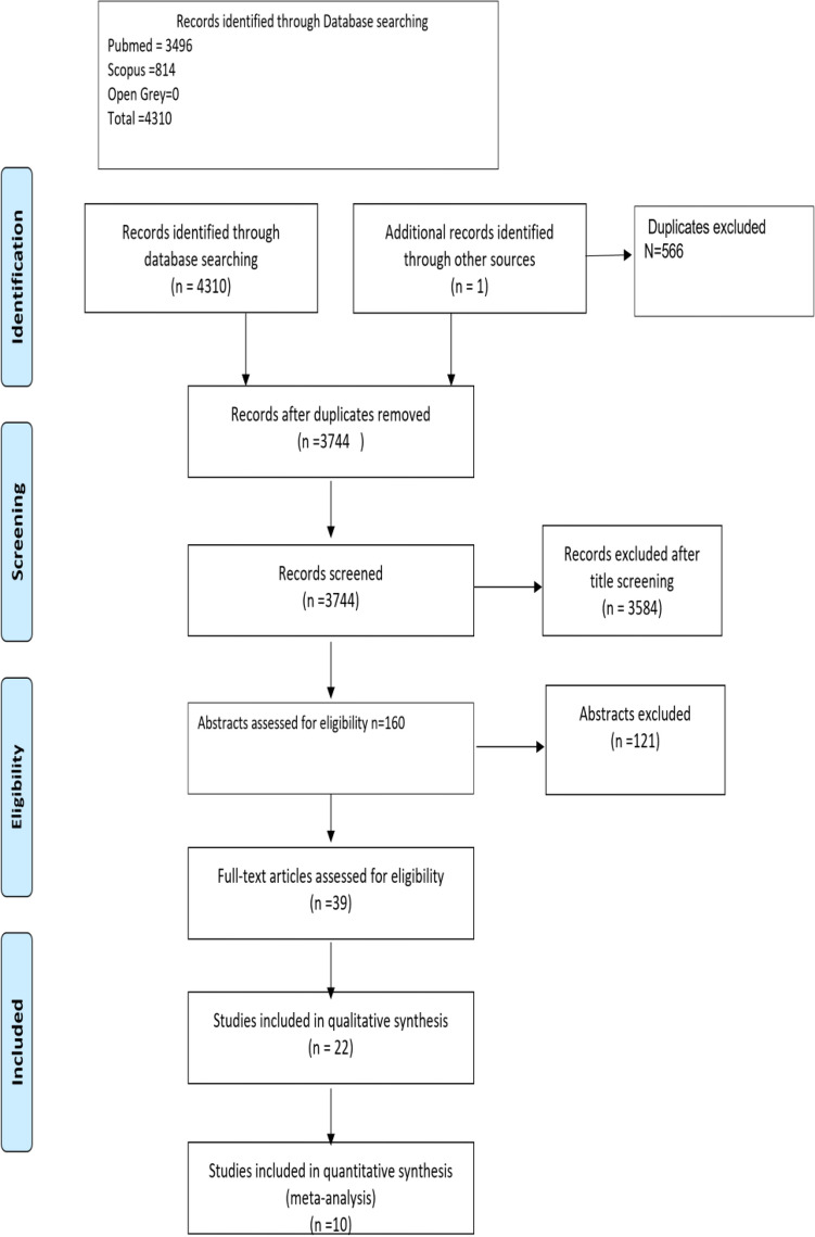
The initial search in the databases resulted in 3744 studies being identified after duplicates exclusion. After title screening, 3584 articles were excluded, and the remaining 160 abstracts were further assessed for eligibility. Articles that had multiple reports corresponding to different follow-up periods were combined into a single entry and the data of the longest follow-up were included in this study. This process culminated in 39 studies that were to be progressed to full-text analysis. Subsequent full-text analysis of these studies resulted in 22 studies that met the inclusion criteria (Fig. 1).
Figure 1.
Prisma flow chart of the study selection process.
Risk of bias evaluation
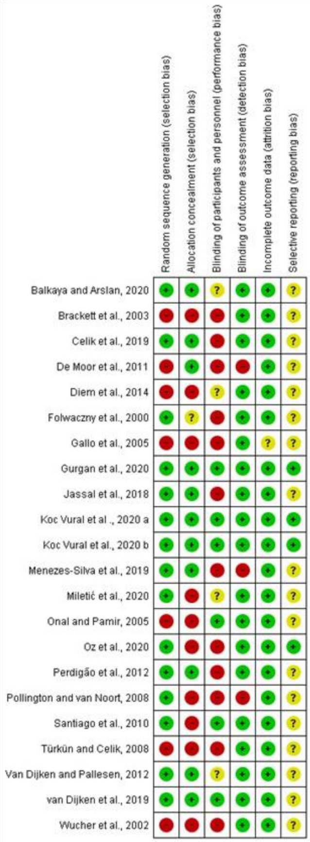
Overall, 3 studies were deemed to have a low risk of bias19,37,38, 3 studies showed39–41 unclear risk of bias while the remaining 16 studies had a high risk of bias. Seven studies17,42–47 did not report random sequence generation, while 50% of the included studies reported allocation concealment. Performance bias was unclear in the majority of studies (16 out of 22), while outcome assessment was blinded in all studies except for 343,48,49. No attrition bias was noticed in any of the included studies except for one44, which did not adequately report the number of dropouts (Fig. 2).
Figure 2.

Risk of bias summary: authors' judgments about each risk of bias item for each included study. Filled Green circle Low ROB Filled Red circle High ROB Filled Yellow Circle Unclear ROB.
Included studies characteristics
The characteristics and methodological assessment of the 22 included studies are summarized in Table 1. In 15 of the included studies16,19,37,38,41–44,46–48,50–53, split-mouth design was employed while 7 studies reported a parallel study design17,39,40,45,49,54,55. Most of the studies employed the modified USPHS criteria for restorations evaluation except for 4 studies16,17,50,51 that used FDI criteria. One study43 used the McComb et al., criteria56. Five studies used HV-GIC16,17,19,39,49. Two studies used glass hybrid38,51. Resin-modified glass ionomer was used in 9 studies37,41–43,45,50,52,54,57, while 2 studies used conventional GIC43,53. Compomer (poly-acid modified composite) was used in 7 studies40,44–48,54. Most of the studies used nano- or micro-hybrid composite. Bulk-fill composite was used in one study39. Nano-filled composite was used in 2 studies46,57 while one study used micro-filled composite44. Most follow-up periods ranged between 2 and 3 years. Long-term follow-up was reported in 2 studies19,40 which had a follow-up period of 10 and 7 years respectively. One study41 was terminated after 1 year due to an unacceptable failure rate. Class II cavities were reported in 7 studies19,39,41,47,49,51. Class I cavities were evaluated in 3 studies 17,19,41. Non-carious cervical lesions were evaluated in 11 studies16,38,42,44–46,48,50,52,53,57. Class V carious lesions were evaluated in 4 studies37,40,43,54. For HV-GIC, glass hybrid, and conventional GIC, Cavity conditioner of poly-acrylic acid was used in all studies except 2 which did not report any type of pre-treatment38,53. For RMGIC, 2 studies used 37% phosphoric acid etching for 5 s37,41. Two studies used Vitremer primer45,52 while another study used GC cavity conditioner for RMGIC, and Ketac nano primer for nano-filled RMGIC42,57. For Compomer, 5 studies used self-etch adhesive (SE)40,45,46,48,54, while 2 studies used etch-and-rinse adhesive (ER)44,47.
Table 1.
Characteristics of the included studies.
| Study ID | 1. Ion-releasing material | 2. Type of composite | 3. Evaluation criteria | 4. Number of restorations/per group | 5. Total number of restorations and/patients | 6. Follow-up period | 7. Location/settings of data collection | 8. Trial design | 9. Recall rate | 10. Secondary caries detection |
|---|---|---|---|---|---|---|---|---|---|---|
| Balkaya et al.39 | Glass hybrid: Equia Forte Fil a |
1.Bulk-fill resin composite: Filtek Bulk Fill Posterior a 2. Micro hybrid composite: Charisma Smart c |
Modified USPHS |
1. Equia Forte/34 2. Filtek Bulkfill /38 3. Charisma smart /37 |
109/54 | 2 Years | Turkey/University | Parallel | 100% | Visual-tactile with mirror, intraoral photographs, prob and bitewing radiographs |
| Gurgan et al.19 | 1. HVGIC: Equia Fil a | 1. Microhybrid resin composite: Gradia Direct Posterior a | Modified USPHS |
1. Equia Fil/40 class I, 30 class II 2. Gradia Direct Posteior/40 class I, 30 class II |
140/59 | 10 Years | Turkey/University | Split-mouth | 88.1% | Visual-tactile with mirror, coloured photographs and prob |
| Koc Vural et al.37 | 1. RMGIC: Riva LC J | 1. Microhybrid composite: Spectrum TPH3 e | Modified USPHS |
1. Riva LC/55 2. Spectrum TPH3/55 |
110/33 | 3 Years | Turkey/University | Split-mouth | 90.91% | Visual-tactile method with mouth mirror and explorer under the dental light unit |
| Koc Vural et al.38 | Glass hybrid: Equia Forte Fil a | 1. Nanofilled composite: Ceram X One Universal e | Modified USPHS |
1. Equia Forte Fil/74 2. Ceram X One/74 |
148/52 | 2 Years | Turkey/University | Split-mouth | 88% | Visual with the aid of coloured photographs |
| Miletić et al.51 | Glass hybrid: Equia Forte Fil a | 1. Nanohybrid composite/Tetric Evoceram c | FDI |
1. Equia Forte/179 2. Tetric Evoceram/178 |
358/184 | 2 Years | Multicenter: Croatia, Italy, Turkey, and Serbia/University | Split-mouth | 90.6% | Visual-tactile with (magnification 2.5X), mirrors, and very thin (250-μm-thick) dental probes |
| Oz et al.53 | Conventional GIC: Fuji Bulk a | 1.MFR Hybrid Composite/Gaenial Posterior a | Modifies USPHS |
1. Fuji Bulk/67 2. Gaenial Posterior/67 |
134/30 | 1 Year | Turkey/University | Split-mouth | 93% | Visual-tactile with mirrors, probes, and air streams |
| Celik et al.16 | 1. HVGIC: Equia Fil a | 1.MFR Hybrid Composite G-aenial Posterior a | FDI |
1. Equia Fil /67 2. G-aenial/67 |
134/22 | 3 Years | Turkey/University | Split-mouth | 82% | Visual-tactile using a mirror and an explorer |
| Menezes-Silva et al.49 | 1. HVGIC: Equia Fil a | 2. Filtek Z350 XT Universal b | Modified USPHS |
1. Equia Fil/77 2. Filtek Z350/77 |
154/154 | 1 year | Brazil/17 public primary schools | Parallel | 94.8% | Visual-tactile with photographs, mirror, and ballpoint periodontal prob |
| Van Dijken et al.41 | 1. RMGIC: Activa Bioactive f | 1. Nanofilled composite: Ceram X e | Modified USPHS |
1. Activa Bioactive/82 2. Ceram X/82 |
164/67 | 1 Year | Sweden/University | Split-mouth | 96.3% | Visual-tactile using mirror and explorer and radiographs one-year recall |
| Jassal et al.50 | 1. RMGIC: GC II LC a | 1. Microfine hybrid compiste/Solar X a | FDI |
1. GC II LC/98 2. Solar X , passive adhesive application/98 3. Solar X, rigouros adhesive application/98 |
294/56 | 18 Months | India/n.r | Split-mouth | 90.81% | Visual using dental-operating microscope at 1 × magnification |
| Diem t al.17 | 1. HVGIC: Fuji IX GP Extra a | 1. Microfine hybrid Composite: Solar a | FDI |
1. Fuji IX GP Extra/87 2. Fuji IX GP Extra with G-coat plus/84 3. Solar /83 |
254/91 | 3 Years | Vietnam/Primary school in semi-rual area | Parallel | 77.9% | Visual using headlight, natural light, and digital photographs |
| Van Dijken et al.40 | 1. Compomer: Dyract AP e | 1. Hybrid compiste/Tetric Ceram c | Modified USPHS |
1. Dyract AP/69 2. Tetric Ceram/70 |
139/60 | 7 Years | University | Parallel | 97.1% | Visual-tactile using a mirror, and an explorer |
| Perdigão et al.57 |
1. RMGIC: Fuji II LC a 2. Nanofilled RMGIC: Ketac Nano b |
1. Nanofilled composite: Filtek Suprem Plus b | Modified USPHS |
1. Fuji II LC/31 2. Ketac Nano/30 3. Filtek Suprem/31 |
92/33 | 1 Year | Brazil/University | Parallel | 84.8% | Visual using a mirror and intra-oral-coloured photographs at 1.5 × magnification |
| De Moor et al.43 |
1. Conventional GIC: Ketac Fil b 2. RMGIC: Photac Fil b |
1. Microhybrid compiste: Herculite XRV d | McComb et al., criteria |
1. Ketac Fil/35 2. Photac Fil/35 3. Herculite/35 |
105/35 | 2 Years | Belgium/Privat practice | Split-mouth | 77.1% | Tactile using an explorer |
| Santiago et al.52 | 1. RMGIC: Vitremer b | 2. Nanohybrid composite: Tetric Ceram c | Modified USPHS |
1. Vitremer/35 2. Tetric Ceram/35 |
70/35 | 2 Years | Brazil/University | Split-mouth | 93.3% | Visual-tactile using a mirror, and an explorer |
| Pollington et al.48 | 1. Compomer: Hytac b | 1. Universal Hybrid composite: Pertac II b | Modified USPHS |
1. Hytac/30 2. Pertac II/30 |
60/30 | 3 Years | United Kingdom/University | Split-mouth | 100% | Visual-tactile (no details are mentioned) |
| Türkün et al.46 | 1. Compomer: Dyract e | 1. Nanofilled composite: Filtek Supreme b | USPHS |
1. Dyract/50 2. Filtek Supreme/50 |
100/24 | 2 Years | Turkey/University | Split-mouth | 100% | Visual-tactile using a mirror, an explorer and radiographs |
| Gallo et al.44 | 1. Compomer: F 2000 b | 1. Microfilled composite: Silux Plus b | Modified USPHS |
1. F 2000 + Single bond (ER)/30 2. F 2000 + SE primer/30 3. Silux Plus + Single bond/30 |
90/30 | 3 Years | USA/University | Split-mouth | 100% |
Visual-tactile (No details are mentioned) |
| Onal et al.45 |
1. RMGIC: Vitremer b 2. Compomer: F 2000 b 3. Compomer: Dyract e |
1. Universal composite: Valus Plus b | Modified USPHS |
1. Vitremer /24 2. F 2000/38 3. Dyract 64 4. Valus Plus/22 |
130/30 | 2 Years | Turkey/University | Parallel ara> | 93.8% | Visual-tactile (no details are mentioned) |
| Brackett et al.42 | 1. RMGIC: Fuji II LC a | 1. Microhybrid composite/Z250 b | Modified USPHS |
1. Fuji II LC/37 2. Z250/37 |
74/24 | 2 Years | Mexico/University | Split-mouth | 73% | Visual-tactile (no details are mentioned) |
| Wucher et al.47 | 1. Compomer: Dyract e | 1. Microhybrid compiste: Spectrum TPH e | USPHS |
1. Dyract/23 2 Dyract covered with Spectrum /23 3. Spectrum TPH/23 |
69/23 | 3 Years | South Africa/Private practice | Split-mouth | 86.9% | Visual-tactile using mirror, periodontal rob, and periapical radiographs at 1-year recalls |
| Folwaczny., et al.54 |
1. Compomer: Dyract e 2. RMGIC: Fuji II LC a 3. RMGIC: Photac Fil b |
1. Hybrid Compoiste: Tetric Ceram c | Modified USPHS |
1. Dyract/79 2. Fuji II LC/51 3. Tetric Ceram/36 |
197/37 | 2 Years | Germany/University setting | Parallel | N.r | Visual-tactile using mirrors and a prob |
| Study ID | 10. Black’s classification | 11. Cavity design and size | 12. Gingival margin location/enamel bevel | 13. Moisture control | 14. Adhesive technique/Composite | 15. Adhesive technique/Ion-releasing material | 16. Patient’s age Mean ± SD [Range], in years | |||
|---|---|---|---|---|---|---|---|---|---|---|
| Balkaya et al.39 | Class II | Conservative slot design | Enamel/no bevel | Cotton pellets and suction | Single Bond Universal b/SE | Polyacrylic acid conditioner a | 2220–32 | |||
| Gurgan et al.19 | Class I and II | Conservative | Enamel/no bevel | Cotton rolls | G-bond a/One step SE | Polyacrylic acid conditioner a | 2415–37 | |||
| Koc Vural et al.37 | Class V (carious) | Conservative | Dentine/no bevel | Cotton rolls and saliva ejector | Prime & Bond NT e/2-step ER | 37% phosphoric acid for 5 s | 52.69 ± 9.737–88 | |||
| Koc Vural et al.38 | Class V (NCCL) | Wedge shaped, and saucer-shaped | N.R | Cotton rolls and saliva ejector | Prime & Bond Elect One e/Univeral adhseive with selective enamel etching | No preconditioning | 55 ± 8.340,42–71 | |||
| Miletić et al.51ara> | Class II | Conservative, moderate to large | Enamel/no bevel |
Rubber dam for composite High suction and cotton roll for GIC |
Adhese c/2-step SE | Polyacrlic acid condition a | > 18 | |||
| Oz et al.53 | Class V (NCCL) | Non-retentive | Enamel + dentine/n.r | Cotton rolls | G-premio bond a/Universal adhesive in ER mode | No pre-treatment |
61.8 ± n.r Patient had at least one systemic disease |
|||
| Celik et al.16 | Class V (NCCL) | Wedge or saucer-shaped | Dentine/no bevel | Cotton rolls, retraction cord, and a saliva aspirator | Optibond FLd/a 3-step ER | Polyacrylic acid a | 47.8 ± nr34–62 | |||
| Menezes-Silva et al.49 | Class II |
GIC/ATR Composite/conservative |
Dentine/retention grooves for GIC | Cotton rolls | Single Bond Universal b | Polyacrylic acid a | N.r8–19 | |||
| Van Dijken et al.41 | Class I and II | Retentive cavity | N.r/no bevel | Cotton rolls and suction | Xeno select e/1- step SE | Etching for 5 s with phosphoric acid | 58.3 ± n.r37–85 | |||
| Jassal et al.50 | Class V (NCCL) | Non-retentive | Enamel + dentine/no bevel | Cotton rolls and retraction cord | G-bond a/1- step SE | Polyacrylic acid | > 18 | |||
| Diem et al.17 | Class I | Adhesive cavity preparation | No bevel | Cotton rolls | G-bond a/1- step SE | Polyacrylic acid | N.r11,12 with occlusal caries in permanent first molars | |||
| Van Dijken et al.40 | Class V (carious) | Non-retentive | Dentine/no bevel | Cotton rolls and saliva suction device | Xeno III e/1- step SE | Xeno III e/1- step SE | 61.5 ± n.r40,43–83 | |||
| Perdigão et al.57 | Class V (NCCL) | Non-retentive | n.r/no bevel | Cotton rolls | FGM k/2-step ER |
1. Ketac Nano primer b . Polyacrylic acid conditioner with Fuji LC |
48.7 ± n.r30–78 | |||
| De Moor et al.43 | Class V (carious) | Conventional cavity preparation | Enamel/bevel | N.r | Optibond FL d/3-step ER | Polyacrylic acid a |
45 [n.r] Head and neck cancer patients |
|||
| Santiago et al.52 | Class V (NCCL) | Non-retentive | Enamel/no bevel | Rubber dam | Excite c/2-step ER | Vitremer Primer b | N.r 18–50 | |||
| Pollington et al.48 | Class V (NCCL) | Non-retentive | Enamel + dentine/no bevel | Cotton rolls and high suction | Prompt L-Pop b/1-step | Prompt L-Pop b/1-step | 54 [N.r] | |||
| Türkün et al.46 | Class V (NCCL) | Non-retentive | No bevel | Cotton rolls and retraction cord | Clearfil protect y/2-step SE | Clearfil protect y/2-step SE | 44 25–54 | |||
| Gallo et al.44 | Class V (NCCL) | Non-retetnive | Enamel + dentine/bevel | Rubber dam | Single Bond b/2-step ER |
1. Single Bond b/2-step ER 2. F 2000 b/SE |
N.r | |||
| Onal et al.45 | Class V (NCCL) | Non-retentive | Enamel + dentine/no bevel | Cotton rolls and suction | Scotchbond b/3-step ER |
1. Vitremer Primer b 2 |
N.r 27–63 | |||
| Brackett et al.42 | Class V (NCCL) | Non-retentive | Enamel + dentine/n.r | Cotton rolls and retraction cord | Single Bond b/3-step ER | Polyacrylic acid a | 47 ± n.r 28–72 | |||
| Wucher et al.47 | Class II | Conventional design | N.r/no bevels | Cotton rolls and saliva ejector | Prime and Bond 2.1 e/2-step ER | Prime and Bond 2.1 e/2-step ER | N.r 25–61 | |||
| Folwaczny et al.54 | Class V (carious and NCCL) | Non-retentive | Enamel + dentine/bevel | Cotton rolls | Syntac C/3-step ER | PSA Dyract e/2-step SE | N.r 26–66 |
a: GC,Tokyo, Japan,
b: 3 M.
c: Heraeus Kulzer, Ha-nau, Germany.
d: Kerr—Sybron Gmbh, Karlsruhe, Germany.
e: Dentsply,Konstanz, Germany.
C: Vivadent, Schaan, Liechtenstein),
f: Pulpdent, Watertown, MA, USA).
j: SDI, Bayswater, Australia).
y: Kuraray; Osaka, Japan).
k: Joinville, Brazil.
n.r: not reported.
ER: Etch-and-rinse.
SE: Self-etch.
ART: atraumatic restorative technique.
RC: resin composite.
For resin composite,8 studies used ER adhesive system16,37,42–45,47,52,57 while 9 studies used SE adhesives17,19,40,41,46,48,50,51,54. Two studies used a universal adhesive in selective etch mode38,49, 1 in SE mode39, and 1 in ER mode53. For moisture control, cotton rolls and saliva ejectors were reported in the majority of studies except for 3 studies that used rubber dam isolation44,51,52.
Patients in all studies had no systemic diseases except two43,53. In one study53, patients were required to have at least one systemic disease and the other one43 included subjects who were xerostomic, head and neck, cancer patients who received radiation therapy. Ten studies16,19,37–39,41,49–51,53 were published in the years (2018–2020) with 6 in 2020, 3 in 2019, and 1 in 2018. No studies were identified from January to May of 2021. Five studies17,40,43,52,57 were published between 2010 and 2014. Seven studies42,44–48,54 were published before 2010.
Descriptive analysis
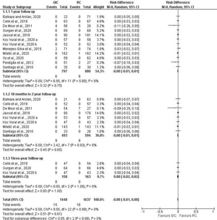
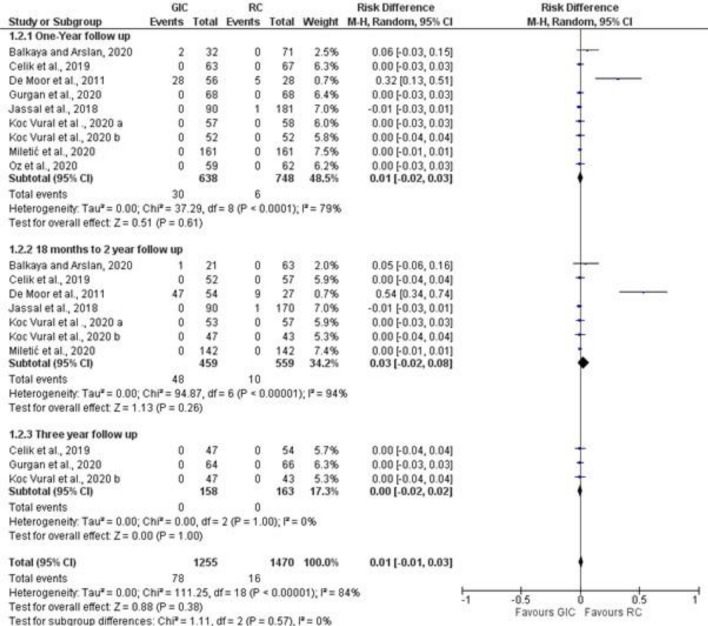
Studies that reported secondary caries and marginal adaptation in different follow-up periods were included in the meta-analysis (Figs. 3, 4, 5). For secondary caries outcome for all types of cavities, the meta-analysis was grouped as follows: ion releasing materials (GIC) vs resin composite (RC) with the following 3 follow-up periods, i. e. 1 year, 18–24 months, and 3 years. For secondary caries in load-bearing cavities, ion-releasing material (GIC and compomer) vs resin composite, and data were extracted from the last follow-up.
Figure 3.
Forest plot of comparison: Ion releasing restoration (GIC) versus resin composite, outcome: 1.1 Secondary caries for all types of cavities.
Figure 4.
Forest plot of comparison: Ion releasing restoration versus resin composite, outcome: 1.2 Secondary caries for load-bearing cavities.
Figure 5.
Forest plot of comparison: Ion releasing restoration (GIC) versus resin composite, outcome: 1.3 marginal adaptation.
The difference in the number of studies in each follow-up is attributed to whether the outcome was reported by the authors. For marginal adaptation outcome, GIC vs resin composite comparison was evaluated at the same 3 follow-up periods. secondary caries was not reported in all studies that compared compomer and resin composite at different follow-up periods. Therefore, no meta-analysis was performed for compomer vs resin composite comparison. Out of a total of 1448 GIC restorations, only 15 showed secondary caries with a percentage of 0.8%. Similarly, 16 composite restorations failed due to caries out of 1637 with a percentage of 0.9%. In all studies that compared compomer and resin composite, no occurrence of secondary caries was observed over the follow-up periods which ranged between 2 and 3 years.
Meta-analysis
Secondary caries
The risk difference for the comparison between GIC and RC for the 1-year and18 month–2 years follow-up periods was -0.00 with 95% CI between [− 0.1–0.01]. The 3-year follow-up risk difference was 0.00 with 95% CI between [− 0.2–0.02] with no occurrence of secondary caries in both arms. There was no statistically significant difference (P = 0.61) between GIC and RC in secondary caries development at any of the follow-up periods. For a total of 1448 GIC restorations, 15 failed due to secondary caries, in comparison with 16 out of 1637 composite restorations. Overall heterogeneity was low with I2 = 0%. (Fig. 3). For secondary caries in load-bearing cavities, the risk difference was 0.0 with 95% CI between [− 0.01–0.02]. No statistically significant difference (P = 0.77) was found between ion-releasing material and secondary caries.
Marginal adaptation
The risk difference for the 1-year follow-up was 0.0.1 with 95% CI between [− 0.02–0.03]. Heterogeneity was high with an I2 = 75%. No statistically significant difference (p ˃ 0.5) was found between the 2 materials. At 18 months–2 years follow-up, the risk difference was 0.03 with 95% CI between [− 0.02–0.08]. Heterogeneity was high with an I2 = 94%. At the 3-year follow-up, the risk difference was 0.00 with 95%CI between [− 0.02–0.02]. Heterogeneity was low with an I2 = 0%.
The overall risk difference was 0.01 with 95%CI between [− 0.01–0.03]. Out of a total of 1255 GIC restorations, 78 showed unacceptable marginal adaptation compared to 16 out of 1470 RC restorations. No statistically significant difference was found between the 2 materials. Overall heterogeneity was high with an I2 = 84%. (Fig. 4).
GRADE quality of evidence
Assessment of the quality of evidence for secondary caries and marginal adaptation outcomes for the 3 follow-up periods (1 year, 18–24 months, and 3 years) was low. This finding suggests that the confidence in the effect estimate is limited, and that further research is likely to have an impact on the confidence of the estimate of effect (Table 2).
Discussion
This systematic review discussed the occurrence of secondary caries in ion-releasing materials versus resin composite. Glass ionomer and its derivatives are the most clinically reported ion-releasing materials. Compomer was less frequently used. The results of the meta-analysis showed no significant difference between the secondary caries in resin composite and all derivatives of GIC.
Secondary caries is influenced by several factors with the most frequent ones being: the location of the lesion (cervical, proximal, or occlusal), patient’s caries risk, age, and socioeconomic status, operator’s skills variation, and detection methods and criteria58. The majority of studies included in this review were conducted in university settings with trained operators and under standardized conditions with patients who demonstrated moderate oral hygiene. This could explain the low number of events. Secondary caries was found to be more frequent in practice-based settings7. This could be attributed to the technique sensitivity of composite placement that requires highly skilled and calibrated operators which is often the case in university settings59. Regarding operative procedures, the majority of studies in this review used cotton rolls and saliva ejectors for moisture control while only 3 studies reported rubber dam isolation. Previous literature reported no significant difference between the survival of composite restorations performed under either of the isolation protocols60.
The location of the lesion is an important factor that could explain the generally low incidence of events. Around 45% of the included studies involved NCCL which are less affected by secondary caries than posterior occlusal and proximal cavities59. Secondary caries is reported to be more frequent with deep proximal restorations with gingival margins extending beyond the cementoenamel junction with dentine and cementum as the substrate61,62. Furthermore, the placement of such restorations is highly technique sensitive and isolation in every restorative step cannot be strictly followed9.
The Patient’s caries susceptibility is crucial in secondary caries development, as primary caries and secondary caries are inherently the same diseases and consequently patients with high caries risk are more suspectable to secondary caries63. The findings of this review were based on the results of studies performed on a population of healthy individuals with good to moderate oral hygiene and with no debilitating conditions. One exception is the study by De Moor et al.43, in which the population was head and neck xerostomic cancer patients who received radiation therapy. De Moor et al. 43, reported a significantly higher failure rate due to secondary caries in resin composite restorations in comparison with conventional GIC. Nevertheless, the findings of this study cannot be generalized as this population is highly specific. However, the difference in the performance of different materials in populations with compromised oral health indicates that patient factors could be more influential than the choice of material.
Adhesive strategy and interfacial gap formation were speculated to play a role in secondary caries development. Gaps at the margins of restorations can permit bacterial invasion and biofilm accumulation along the tooth/restoration interface64. However, until now there is no Conesus in the literature regarding the role of gaps in secondary caries development. In a study by Kidd et al.,65, it was suggested that microleakage cannot solely induce active demineralization beneath a restoration, only when bacterial invasion takes place at the composite-restoration interface, the size of the gap becomes pertinent.
The durability of the adhesive interface is critical for the survival of resin composite restorations, especially with dentin margins. Several attempts have been made to increase the durability of adhesives to dentine including using MMPs inhibitors, biomimetic remineralization, and increasing the hydrophobicity of the adhesive66–68. The adhesion protocols in this systematic review varied between etch-and-rinse (9 studies) and self-etch adhesives (8 studies), while 2 studies used universal adhesives in selective etch mode38,49, one in SE mode39 and one in ER mode53. The findings of this systematic review suggest that regarding secondary caries development, all adhesive strategies performed similarly considering the low number of events. In a previous study that utilized a short-term in vitro biofilm model69, the adhesive type affected carious lesion development and progression in gaps. However, a recent systematic review and Network meta-analysis showed similar performance of all adhesive strategies in preventing secondary caries70. It is worth mentioning that the impact of adhesive strategy/type on secondary caries development was not assessed quantitatively in this review, considering the overall scarcity of secondary caries occurrence in the included follow-up periods.
The short follow-up period (2–3 years) in the majority of studies might have contributed to an overall low incidence of events. Longer-term follow-up clinical trials showed an increased reporting of secondary carious lesions71,72. According to the findings of a recent review59, the highest mean incidence of secondary caries development was recorded after five years. Interestingly, the only long-term 10-year follow-up study for posterior restorations (class I and II) in this review19, did not report failure due to secondary caries for composites and glass ionomer restorations over the 10-year observational period. Furthermore, the detection methods and criteria of evaluation might have played a role in reporting secondary caries. According to a systematic review by Brouwer et al.73, only visual assessment would mean that 40% of secondary carious lesions will be missed, while 20% of sound surfaces will be misdiagnosed as carious. Until now, there is no clear consensus on what constitutes a secondary carious lesion that requires intervention8,9.
While the findings of in vitro studies74,75 reported a reduced risk of secondary caries in ion-releasing restorations such as GICs and their derivatives, the relation between the restorative material and secondary caries development is not clear in clinical settings. It is worth mentioning that clinical reporting in the form of randomized clinical trials on the recently developed ion-releasing materials is still scarce. Developments such as RMGIC with ionic resin matrix (Activa Bioactive) which is claimed to release ions in sufficient quantities to induce remineralization and inhibit secondary caries have not been thoroughly evaluated. The short-term performance was disappointing with an unacceptable failure rate due to the absence of an adhesive41. (a protocol no longer recommended by the manufacturer). Recent in vitro data regarding the ion-releasing Cention n showed its ability to neutralize the acidic environment76. However, no clinical evidence in the literature is available to validate the laboratory data.
The quality of the interface between the tooth structure and the restoration can play a significant role in the occurrence of secondary caries. While not the only route for secondary caries, the presence of a defective restoration margin can allow acidic fluids or biofilm to enter the interface via gaps. However, there is currently no agreement on the role of microleakage in the development of caries near composites. Nonetheless, some in vivo and in vitro studies suggest that the presence of a gap next to a composite restoration can result in the formation of a "wall lesion.". The literature also suggests the presence of a correlation between the size of the gap and the size of the dentinal wall lesions59,64,77–79.
The results of marginal adaptation between GIC derivatives showed comparable performance with resin composite restorations with no significant difference between them. Marginal adaptation of restorations is highly dependent on the quality of the adhesive interface80,81. Traditionally, attachment of resin composite restorations was achieved through micromechanical adhesion that involved the etching of the dental substrates82. Due to their user-friendly application, simplified universal adhesives have grown in popularity. According to the literature, these adhesives are a single-bottle, no-mix adhesive system that works well with any adhesion strategy and bonds adequately to tooth structure as well as various direct and indirect restorative materials83,84. However, the simplification came at the expense of hydrophilicity which can lead to water seepage through the hybrid layer causing nano leakage 85. Therefore, different protocols have been suggested to improve the performance of simplified adhesives including increasing the application time86, the addition of a hydrophobic resin layer over the adhesive87, and application of several layers of the simplified adhesive88. There is no clear consensus in the literature on the optimal way to improve the long-term performance of simplified adhesives.
The results of this systematic review showed a wide variation in the adhesion protocol for the ion-releasing materials, ranging from no pre-treatments to polyacrylic acid conditioners, ER, and SE adhesives. Nevertheless, the overall incidence of marginal deterioration was low. It is important to highlight that the adherent substrate which is a determining factor in the quality of the adhesion, is not consistent in all studies, with margins being in enamel, dentine, or cementum. GICs were applied in the majority of studies after pre-treatment with a cavity conditioner of poly-acrylic acid. It has been proposed that a tooth-GIC interaction interphase layer is seen after GIC comes in contact with pre-treated dentin, as the pre-treatment facilitates diffusion of ions into the demineralized substrate89,90.
A recent systematic review has shown that this interphase layer is notably resistant to acidic dissolution and hence improving the quality of the adhesive interface91. It is important to note that in this review, scores 1 and 2 of the FDI criteria in the marginal adaptation outcome were considered to be a sign of no significant marginal deterioration. This was done to distinguish early stages of marginal deterioration between ion-releasing materials and resin composite restorations. Since, the presence of small marginal gaps, ditches could potentially be a culprit in secondary caries development.
The risk of bias in more than 60% of the included studies was high, with only 3 studies reporting a low risk of bias19,37,38. Performance bias was high or unclear in most studies as the nature and presentation of the used materials are different and easily identified by dentists. It should be noted that the overall risk of bias of the study was not considered as a ground for meta-analysis exclusion. Therefore, the results of this analysis should be cautiously interpreted. The GRADE assessment of the quality of evidence was low for both outcomes (secondary caries and marginal adaptation) which weakens confidence in the effect estimate. Consequently, the true effect might be substantially different from the estimate of the effect. Imprecision and risk of bias for both outcomes had to be downgraded by one level each. The risk of bias for 2 of the primary domains (performance bias and selection bias) was high for studies that contributed to the weight of the analysis. The absence of events in control and intervention arms led to a downgrading for impression by one level92.
There are some limitations to this review. Firstly, no restriction was placed on the date of publication. Studies that were published in the early 2000s presented a higher risk of bias and inadequate reporting which affected their quality assessment. Although the Consolidated Standards of Reporting Trials (CONSORT) statement was developed in 199693 and undergone a couple of revisions94,95, many clinical trial reports remained inadequate. Furthermore, short follow-up periods resulted in an overall low number of events. Also, several new ion-releasing materials have emerged in the last 5 years. The results of this analysis were based on two broad categories of materials (GICs and compomers). The findings of this review cannot be applied to all commercially available ion-releasing materials.
Conclusions
Within the limitation of this work, this systematic review and meta-analysis revealed that secondary caries occurrence is not dependent on the ion-releasing capability of restorative material.
Short-term follow-ups are a common denominator among the available body of evidence. Longer follow-ups are recommended to accurately detect the performance of different restorative materials after prolonged clinical service.
Several new ion-releasing materials lack high-quality clinical reporting and need further investigations.
Supplementary Information
Author contributions
Conception and design of study: H.H. H.A.M.A.M.A., S.H.M., E.A.; Acquisition of data: E.A., H.H. H.A.M.A.M.A., H.C., M.M., S.H.M.; Analysis and/or interpretation of data: E.A., H.H. H.A.M.A.M.A.; Drafting the manuscript: E.A., H.C., H.H. H.A.M.A.M.A., S.H.M.; Revising the manuscript critically for important intellectual content: H.C., H.H. H.A.M.A.M.A., S.H.M.
Funding
This study was funded by 3 M/Key Opinion Leader Scholarship.
Data availability
The data used in this article are available upon request from the corresponding author.
Competing interests
The authors declare no competing interests.
Footnotes
Publisher's note
Springer Nature remains neutral with regard to jurisdictional claims in published maps and institutional affiliations.
Supplementary Information
The online version contains supplementary material available at 10.1038/s41598-022-19622-6.
References
- 1.van Dijken JW, Pallesen U. A randomized 10-year prospective follow-up of Class II nanohybrid and conventional hybrid resin composite restorations. J. Adhes. Dent. 2014;16:585–592. doi: 10.3290/j.jad.a33202. [DOI] [PubMed] [Google Scholar]
- 2.van Dijken JW, Pallesen U. Randomized 3-year clinical evaluation of Class I and II posterior resin restorations placed with a bulk-fill resin composite and a one-step self-etching adhesive. J. Adhes. Dent. 2015;17:81. doi: 10.3290/j.jad.a33502. [DOI] [PubMed] [Google Scholar]
- 3.Barata JS, Casagrande L, Pitoni CM, De Araujo FB, Garcia-Godoy F, Groismann S. Influence of gaps in adhesive restorations in the development of secondary caries lesions: An in situ evaluation. Am. J. Dent. 2012;25:244–248. [PubMed] [Google Scholar]
- 4.Da Rosa Rodolpho PA, Donassollo TA, Cenci MS, Loguercio AD, Moraes RR, Bronkhorst EM, et al. 22-Year clinical evaluation of the performance of two posterior composites with different filler characteristics. Dent. Mater. 2011;27:955–963. doi: 10.1016/j.dental.2011.06.001. [DOI] [PubMed] [Google Scholar]
- 5.Kramer N, Kunzelmann KH, Garcia-Godoy F, Haberlein I, Meier B, Frankenberger R. Determination of caries risk at resin composite margins. Am. J. Dent. 2007;20:59–64. [PubMed] [Google Scholar]
- 6.Opdam NJ, van de Sande FH, Bronkhorst E, Cenci MS, Bottenberg P, Pallesen U, et al. Longevity of posterior composite restorations: A systematic review and meta-analysis. J. Dent. Res. 2014;93:943. doi: 10.1177/0022034514544217. [DOI] [PMC free article] [PubMed] [Google Scholar]
- 7.Mjor IA, Toffenetti F. Secondary caries: A literature review with case reports. Quintessence Int. 2000;31:165–179. [PubMed] [Google Scholar]
- 8.Machiulskiene V, Campus G, Carvalho JC, Dige I, Ekstrand KR, Jablonski-Momeni A, et al. Terminology of dental caries and dental caries management: Consensus report of a workshop organized by ORCA and cariology research group of IADR. Caries Res. 2020;54:7–14. doi: 10.1159/000503309. [DOI] [PubMed] [Google Scholar]
- 9.Askar H, Krois J, Gostemeyer G, Bottenberg P, Zero D, Banerjee A, et al. Secondary caries: What is it, and how it can be controlled, detected, and managed? Clin Oral Investig. 2020;24:1869–1876. doi: 10.1007/s00784-020-03268-7. [DOI] [PubMed] [Google Scholar]
- 10.de Fucio SB, Puppin-Rontani RM, de Carvalho FG, Mattos-Graner Rde O, Correr-Sobrinho L, Garcia-Godoy F. Analyses of biofilms accumulated on dental restorative materials. Am. J. Dent. 2009;22:131–136. [PubMed] [Google Scholar]
- 11.Hansel C, Leyhausen G, Mai UE, Geurtsen W. Effects of various resin composite (co)monomers and extracts on two caries-associated micro-organisms in vitro. J. Dent. Res. 1998;77:60–67. doi: 10.1177/00220345980770010601. [DOI] [PubMed] [Google Scholar]
- 12.Zalkind MM, Keisar O, Ever-Hadani P, Grinberg R, Sela MN. Accumulation of Streptococcus mutans on light-cured composites and amalgam: An in vitro study. J. Esthet. Dent. 1998;10:187–190. doi: 10.1111/j.1708-8240.1998.tb00356.x. [DOI] [PubMed] [Google Scholar]
- 13.Pires PM, Neves AA, Makeeva IM, Schwendicke F, Faus-Matoses V, Yoshihara K, et al. Contemporary restorative ion-releasing materials: Current status, interfacial properties and operative approaches. Br Dent J. 2020;229:450–458. doi: 10.1038/s41415-020-2169-3. [DOI] [PubMed] [Google Scholar]
- 14.Lohbauer U. Dental glass ionomer cements as permanent filling materials? – Properties. Limit. Future Trends. 2010;3:76–96. [Google Scholar]
- 15.Banerjee A. The role of glass-ionomer cements in minimum intervention (MI) caries management. In: Sidhu SK, editor. Glass-Ionomers in Dentistry. Cham: Springer International Publishing; 2016. pp. 81–96. [Google Scholar]
- 16.Celik EU, Tunac AT, Yilmaz F. Three-year clinical evaluation of high-viscosity glass ionomer restorations in non-carious cervical lesions: A randomised controlled split-mouth clinical trial. Clin. Oral. Investig. 2019;23:1473–1480. doi: 10.1007/s00784-018-2575-y. [DOI] [PubMed] [Google Scholar]
- 17.Diem VT, Tyas MJ, Ngo HC, Phuong LH, Khanh ND. The effect of a nano-filled resin coating on the 3-year clinical performance of a conventional high-viscosity glass-ionomer cement. Clin. Oral Investig. 2014;18:753–759. doi: 10.1007/s00784-013-1026-z. [DOI] [PubMed] [Google Scholar]
- 18.Friedl K, Hiller KA, Friedl KH. Clinical performance of a new glass ionomer based restoration system: A retrospective cohort study. Dent. Mater. 2011;27:1031–1037. doi: 10.1016/j.dental.2011.07.004. [DOI] [PubMed] [Google Scholar]
- 19.Gurgan S, Kutuk ZB, Yalcin Cakir F, Ergin E. A randomized controlled 10 years follow up of a glass ionomer restorative material in class I and class II cavities. J Dent. 2020;94:103175. doi: 10.1016/j.jdent.2019.07.013. [DOI] [PubMed] [Google Scholar]
- 20.Najeeb S, Khurshid Z, Zafar MS, Khan AS, Zohaib S, Marti JM, et al. Modifications in glass ionomer cements: Nano-sized fillers and bioactive nanoceramics. Int. J. Mol. Sci. 2016 doi: 10.3390/ijms17071134. [DOI] [PMC free article] [PubMed] [Google Scholar]
- 21.Salinovic I, Stunja M, Schauperl Z, Verzak Z, Ivanisevic Malcic A, Brzovic Rajic V. Mechanical properties of high viscosity glass ionomer and glass hybrid restorative materials. Acta Stomatol. Croat. 2019;53:125–131. doi: 10.15644/asc53/2/4. [DOI] [PMC free article] [PubMed] [Google Scholar]
- 22.Ruengrungsom C, Palamara JEA, Burrow MF. Comparison of ART and conventional techniques on clinical performance of glass-ionomer cement restorations in load bearing areas of permanent and primary dentitions: A systematic review. J. Dent. 2018;78:1–21. doi: 10.1016/j.jdent.2018.07.008. [DOI] [PubMed] [Google Scholar]
- 23.Vallittu PK, Boccaccini AR, Hupa L, Watts DC. Bioactive dental materials-Do they exist and what does bioactivity mean? Dent. Mater. 2018;34:693–694. doi: 10.1016/j.dental.2018.03.001. [DOI] [PubMed] [Google Scholar]
- 24.Garoushi S, Vallittu PK, Lassila L. Characterization of fluoride releasing restorative dental materials. Dent. Mater. J. 2018;37:293–300. doi: 10.4012/dmj.2017-161. [DOI] [PubMed] [Google Scholar]
- 25.Todd, J. C. Scientific Documentation: Cention N. Ivoclar Vivadent AG, research and development. Sci. Doc. 1–58. (2016).
- 26.Francois P, Fouquet V, Attal JP, Dursun E. Commercially available fluoride-releasing restorative materials: A review and a proposal for classification. Materials (Basel) 2020 doi: 10.3390/ma13102313. [DOI] [PMC free article] [PubMed] [Google Scholar]
- 27.Tiskaya M, Al-eesa NA, Wong FSL, Hill RG. Characterization of the bioactivity of two commercial composites. Dent. Mater. 2019;35:1757–1768. doi: 10.1016/j.dental.2019.10.004. [DOI] [PubMed] [Google Scholar]
- 28.Glasspoole EA, Erickson RL, Davidson CL. Demineralization of enamel in relation to the fluoride release of materials. Am. J. Dent. 2001;14:8–12. [PubMed] [Google Scholar]
- 29.Yaman SD, Er O, Yetmez M, Karabay GA. In vitro inhibition of caries-like lesions with fluoride-releasing materials. J. Oral Sci. 2004;46:45–50. doi: 10.2334/josnusd.46.45. [DOI] [PubMed] [Google Scholar]
- 30.Okida RC, Mandarino F, Sundfeld RH, de Alexandre RS, Sundefeld ML. In vitro-evaluation of secondary caries formation around restoration. Bull. Tokyo Dent. Coll. 2008;49:121–128. doi: 10.2209/tdcpublication.49.121. [DOI] [PubMed] [Google Scholar]
- 31.Hutton B, Salanti G, Caldwell DM, Chaimani A, Schmid CH, Cameron C, Ioannidis JP, Straus S, Thorlund K, Jansen JP, Mulrow C, Catalá-López F, Gøtzsche PC, Dickersin K, Boutron I, Altman DG, Moher D. The PRISMA extension statement for reporting of systematic reviews incorporating network meta-analyses of health care interventions: checklist and explanations. Ann. Intern. Med. 2015;162:777–784. doi: 10.7326/M14-2385. [DOI] [PubMed] [Google Scholar]
- 32.Moher D, Liberati A, Tetzlaff J, Altman DG, Group P. Preferred reporting items for systematic reviews and meta-analyses: The PRISMA statement. PLoS Med. 2009;6:e1000097. doi: 10.1371/journal.pmed.1000097. [DOI] [PMC free article] [PubMed] [Google Scholar]
- 33.Higgins JPT, Altman DG, Gøtzsche PC, Jüni P, Moher D, Oxman AD, et al. The Cochrane Collaboration’s tool for assessing risk of bias in randomised trials. J. BMJ. 2011;343:d5928. doi: 10.1136/bmj.d5928. [DOI] [PMC free article] [PubMed] [Google Scholar]
- 34.Alonso-Coello P, Schunemann HJ, Moberg J, Brignardello-Petersen R, Akl EA, Davoli M, et al. GRADE Evidence to Decision (EtD) frameworks: a systematic and transparent approach to making well informed healthcare choices. 1: Introduction. BMJ. 2016;353:i2016. doi: 10.1136/bmj.i2016. [DOI] [PubMed] [Google Scholar]
- 35.Guyatt GH, Oxman AD, Vist GE, Kunz R, Falck-Ytter Y, Alonso-Coello P, et al. GRADE: An emerging consensus on rating quality of evidence and strength of recommendations. BMJ. 2008;336:924–926. doi: 10.1136/bmj.39489.470347.AD. [DOI] [PMC free article] [PubMed] [Google Scholar]
- 36.Balshem H, Helfand M, Schunemann HJ, Oxman AD, Kunz R, Brozek J, et al. GRADE guidelines: 3. Rating the quality of evidence. J. Clin. Epidemiol. 2011;64:401–406. doi: 10.1016/j.jclinepi.2010.07.015. [DOI] [PubMed] [Google Scholar]
- 37.Koc Vural U, Kerimova L, Kiremitci A. Clinical comparison of a micro-hybride resin-based composite and resin modified glass ionomer in the treatment of cervical caries lesions: 36-month, split-mouth, randomized clinical trial. Odontology. 2020 doi: 10.1007/s10266-020-00550-8. [DOI] [PubMed] [Google Scholar]
- 38.Koc Vural U, Meral E, Ergin E, Gürgan S. Twenty-four-month clinical performance of a glass hybrid restorative in non-carious cervical lesions of patients with bruxism: A split-mouth, randomized clinical trial. Clin. Oral Investig. 2020;24:1229–1238. doi: 10.1007/s00784-019-02986-x. [DOI] [PubMed] [Google Scholar]
- 39.Balkaya H, Arslan S. A two-year clinical comparison of three different restorative materials in class II cavities. Oper Dent. 2020;45:E32–e42. doi: 10.2341/19-078-c. [DOI] [PubMed] [Google Scholar]
- 40.van Dijken JW, Pallesen U. A 7-year randomized prospective study of a one-step self-etching adhesive in non-carious cervical lesions. The effect of curing modes and restorative material. J. Dent. 2012;40:1060–1067. doi: 10.1016/j.jdent.2012.08.017. [DOI] [PubMed] [Google Scholar]
- 41.van Dijken JWV, Pallesen U, Benetti A. A randomized controlled evaluation of posterior resin restorations of an altered resin modified glass-ionomer cement with claimed bioactivity. Dent. Mater. 2019;35:335–343. doi: 10.1016/j.dental.2018.11.027. [DOI] [PubMed] [Google Scholar]
- 42.Brackett WW, Dib A, Brackett MG, Reyes AA, Estrada BE. Two-year clinical performance of Class V resin-modified glass-lonomer and resin composite restorations. Oper. Dent. 2003;28:477–481. [PubMed] [Google Scholar]
- 43.De Moor RJ, Stassen IG, van’t Veldt Y, Torbeyns D, Hommez GM. Two-year clinical performance of glass ionomer and resin composite restorations in xerostomic head- and neck-irradiated cancer patients. Clin. Oral Investig. 2011;15:31–38. doi: 10.1007/s00784-009-0355-4. [DOI] [PubMed] [Google Scholar]
- 44.Gallo JR, Burgess JO, Ripps AH, Walker RS, Ireland EJ, Mercante DE, et al. Three-year clinical evaluation of a compomer and a resin composite as Class V filling materials. Oper. Dent. 2005;30:275–281. [PubMed] [Google Scholar]
- 45.Onal B, Pamir T. The two-year clinical performance of esthetic restorative materials in noncarious cervical lesions. J. Am. Dent. Assoc. 2005;136:1547–1555. doi: 10.14219/jada.archive.2005.0085. [DOI] [PubMed] [Google Scholar]
- 46.Türkün LS, Celik EU. Noncarious class V lesions restored with a polyacid modified resin composite and a nanocomposite: A two-year clinical trial. J. Adhes. Dent. 2008;10:399–405. [PubMed] [Google Scholar]
- 47.Wucher M, Grobler SR, Senekal PJ. A 3-year clinical evaluation of a compomer, a composite and a compomer/composite (sandwich) in class II restorations. Am. J. Dent. 2002;15:274–278. [PubMed] [Google Scholar]
- 48.Pollington S, van Noort R. A clinical evaluation of a resin composite and a compomer in non-carious Class V lesions. A 3-year follow-up. Am. J. Dent. 2008;21:49–52. [PubMed] [Google Scholar]
- 49.Menezes-Silva R, Velasco SRM, Bastos RS, Molina G, Honório HM, Frencken JE, et al. Randomized clinical trial of class II restoration in permanent teeth comparing ART with composite resin after 12 months. Clin. Oral Investig. 2019;23:3623–3635. doi: 10.1007/s00784-018-2787-1. [DOI] [PubMed] [Google Scholar]
- 50.Jassal M, Mittal S, Tewari S. Clinical effectiveness of a resin-modified glass ionomer cement and a mild one-step self-etch adhesive applied actively and passively in noncarious cervical lesions: An 18-month clinical trial. Oper. Dent. 2018;43:581–592. doi: 10.2341/17-147-c. [DOI] [PubMed] [Google Scholar]
- 51.Miletić I, Baraba A, Basso M, Pulcini MG, Marković D, Perić T, et al. Clinical performance of a glass-hybrid system compared with a resin composite in the posterior region: Results of a 2-year multicenter study. J. Adhes. Dent. 2020;22:235–247. doi: 10.3290/j.jad.a44547. [DOI] [PubMed] [Google Scholar]
- 52.Santiago SL, Passos VF, Vieira AH, Navarro MF, Lauris JR, Franco EB. Two-year clinical evaluation of resinous restorative systems in non-carious cervical lesions. Braz. Dent. J. 2010;21:229–234. doi: 10.1590/s0103-64402010000300010. [DOI] [PubMed] [Google Scholar]
- 53.Oz FD, Meral E, Ergİn E, Gurgan S. One-year evaluation of a new restorative glass ionomer cement for the restoration of non-carious cervical lesions in patients with systemic diseases: A randomized, clinical trial. J. Appl. Oral Sci. 2020;28:e20200311. doi: 10.1590/1678-7757-2020-0311. [DOI] [PMC free article] [PubMed] [Google Scholar]
- 54.Folwaczny M, Loher C, Mehl A, Kunzelmann KH, Hinkel R. Tooth-colored filling materials for the restoration of cervical lesions: A 24-month follow-up study. Oper. Dent. 2000;25:251–258. [PubMed] [Google Scholar]
- 55.Perdigão J, Dutra-Corrêa M, Saraceni CH, Ciaramicoli MT, Kiyan VH, Queiroz CS. Randomized clinical trial of four adhesion strategies: 18-month results. Oper. Dent. 2012;37:3–11. doi: 10.2341/11-222-c. [DOI] [PubMed] [Google Scholar]
- 56.McComb D, Erickson RL, Maxymiw WG, Wood RE. A clinical comparison of glass ionomer, resin-modified glass ionomer and resin composite restorations in the treatment of cervical caries in xerostomic head and neck radiation patients. Oper. Dent. 2002;27:430–437. [PubMed] [Google Scholar]
- 57.Perdigão J, Dutra-Corrêa M, Saraceni SH, Ciaramicoli MT, Kiyan VH. Randomized clinical trial of two resin-modified glass ionomer materials: 1-year results. Oper. Dent. 2012;37:591–601. doi: 10.2341/11-415-c. [DOI] [PubMed] [Google Scholar]
- 58.Demarco FF, Collares K, Correa MB, Cenci MS, Moraes RR, Opdam NJ. Should my composite restorations last forever? Why are they failing? Braz. Oral Res. 2017;31:e56. doi: 10.1590/1807-3107BOR-2017.vol31.0056. [DOI] [PubMed] [Google Scholar]
- 59.Nedeljkovic I, Teughels W, De Munck J, Van Meerbeek B, Van Landuyt KL. Is secondary caries with composites a material-based problem? Dent. Mater. 2015;31:e247–e277. doi: 10.1016/j.dental.2015.09.001. [DOI] [PubMed] [Google Scholar]
- 60.Raskin A, Setcos JC, Vreven J, Wilson NH. Influence of the isolation method on the 10-year clinical behaviour of posterior resin composite restorations. Clin. Oral Investig. 2000;4:148–152. doi: 10.1007/s007840000069. [DOI] [PubMed] [Google Scholar]
- 61.Mjor IA. Clinical diagnosis of recurrent caries. J. Am. Dent. Assoc. 2005;136:1426–1433. doi: 10.14219/jada.archive.2005.0057. [DOI] [PubMed] [Google Scholar]
- 62.Kuper NK, Opdam NJ, Ruben JL, de Soet JJ, Cenci MS, Bronkhorst EM, et al. Gap size and wall lesion development next to composite. J. Dent. Res. 2014;93:108S–S113. doi: 10.1177/0022034514534262. [DOI] [PMC free article] [PubMed] [Google Scholar]
- 63.Nedeljkovic I, De Munck J, Vanloy A, Declerck D, Lambrechts P, Peumans M, et al. Secondary caries: Prevalence, characteristics, and approach. Clin. Oral Investig. 2020;24:683–691. doi: 10.1007/s00784-019-02894-0. [DOI] [PubMed] [Google Scholar]
- 64.Cenci MS, Pereira-Cenci T, Cury JA, Ten Cate JM. Relationship between gap size and dentine secondary caries formation assessed in a microcosm biofilm model. Caries Res. 2009;43:97–102. doi: 10.1159/000209341. [DOI] [PubMed] [Google Scholar]
- 65.Kidd EA, Fejerskov O. What constitutes dental caries? Histopathology of carious enamel and dentin related to the action of cariogenic biofilms. J. Dent. Res. 2004 doi: 10.1177/154405910408301s07. [DOI] [PubMed] [Google Scholar]
- 66.Tjaderhane L, Nascimento FD, Breschi L, Mazzoni A, Tersariol IL, Geraldeli S, et al. Strategies to prevent hydrolytic degradation of the hybrid layer-A review. Dent. Mater. 2013;29:999–1011. doi: 10.1016/j.dental.2013.07.016. [DOI] [PMC free article] [PubMed] [Google Scholar]
- 67.Toledano M, Yamauti M, Osorio E, Osorio R. Zinc-inhibited MMP-mediated collagen degradation after different dentine demineralization procedures. Caries Res. 2012;46:201–207. doi: 10.1159/000337315. [DOI] [PubMed] [Google Scholar]
- 68.Tezvergil-Mutluay A, Agee KA, Hoshika T, Carrilho M, Breschi L, Tjaderhane L, et al. The requirement of zinc and calcium ions for functional MMP activity in demineralized dentin matrices. Dent. Mater. 2010;26:1059–1067. doi: 10.1016/j.dental.2010.07.006. [DOI] [PMC free article] [PubMed] [Google Scholar]
- 69.Kuper NK, van de Sande FH, Opdam NJ, Bronkhorst EM, de Soet JJ, Cenci MS, et al. Restoration materials and secondary caries using an in vitro biofilm model. J. Dent. Res. 2015;94:62–68. doi: 10.1177/0022034514553245. [DOI] [PMC free article] [PubMed] [Google Scholar]
- 70.Askar H, Krois J, Gostemeyer G, Schwendicke F. Secondary caries risk of different adhesive strategies and restorative materials in permanent teeth: Systematic review and network meta-analysis. J. Dent. 2021;104:103541. doi: 10.1016/j.jdent.2020.103541. [DOI] [PubMed] [Google Scholar]
- 71.Laske M, Opdam NJM, Bronkhorst EM, Braspenning JCC, Huysmans M. Ten-year survival of class II restorations placed by general practitioners. JDR Clin. Trans. Res. 2016;1:292–299. doi: 10.1177/2380084416663192. [DOI] [PubMed] [Google Scholar]
- 72.Opdam NJ, Bronkhorst EM, Loomans BA, Huysmans MC. 12-year survival of composite vs. amalgam restorations. J. Dent. Res. 2010;89:1063–1067. doi: 10.1177/0022034510376071. [DOI] [PubMed] [Google Scholar]
- 73.Brouwer F, Askar H, Paris S, Schwendicke F. Detecting secondary caries lesions: A systematic review and meta-analysis. J. Dent. Res. 2016;95:143–151. doi: 10.1177/0022034515611041. [DOI] [PubMed] [Google Scholar]
- 74.Askar H, Brouwer F, Lehmensiek M, Paris S, Schwendicke F. The association between loading of restorations and secondary caries lesions is moderated by the restoration material elasticity. J. Dent. 2017;58:74–79. doi: 10.1016/j.jdent.2017.01.002. [DOI] [PubMed] [Google Scholar]
- 75.Hetrodt F, Lausch J, Meyer-Lueckel H, Conrads G, Apel C. Evaluation of restorative materials containing preventive additives in a secondary caries model in vitro. Caries Res. 2019;53:447–456. doi: 10.1159/000496401. [DOI] [PubMed] [Google Scholar]
- 76.Gupta N, Jaiswal S, Nikhil V, Gupta S, Jha P, Bansal P. Comparison of fluoride ion release and alkalizing potential of a new bulk-fill alkasite. J. Conserv. Dent. 2019;22:296–299. doi: 10.4103/JCD.JCD_74_19. [DOI] [PMC free article] [PubMed] [Google Scholar]
- 77.Nassar HM, Gonzalez-Cabezas C. Effect of gap geometry on secondary caries wall lesion development. Caries Res. 2011;45:346–352. doi: 10.1159/000329384. [DOI] [PubMed] [Google Scholar]
- 78.Thomas RZ, Ruben JL, ten Bosch JJ, Fidler V, Huysmans MC. Approximal secondary caries lesion progression, a 20-week in situ study. Caries Res. 2007;41:399–405. doi: 10.1159/000104799. [DOI] [PubMed] [Google Scholar]
- 79.Espejo LC, Simionato MR, Barroso LP, Netto NG, Luz MA. Evaluation of three different adhesive systems using a bacterial method to develop secondary caries in vitro. Am. J. Dent. 2010;23:93–97. [PubMed] [Google Scholar]
- 80.Kakaboura A, Rahiotis C, Watts D, Silikas N, Eliades G. 3D-marginal adaptation versus setting shrinkage in light-cured microhybrid resin composites. Dent. Mater. 2007;23:272–278. doi: 10.1016/j.dental.2006.01.020. [DOI] [PubMed] [Google Scholar]
- 81.He Z, Shimada Y, Sadr A, Ikeda M, Tagami J. The effects of cavity size and filling method on the bonding to Class I cavities. J. Adhes. Dent. 2008;10:447–453. [PubMed] [Google Scholar]
- 82.Foxton RM. Current perspectives on dental adhesion: (2) Concepts for operatively managing carious lesions extending into dentine using bioactive and adhesive direct restorative materials. Jpn. Dent. Sci. Rev. 2020;56:208–215. doi: 10.1016/j.jdsr.2020.08.003. [DOI] [PMC free article] [PubMed] [Google Scholar]
- 83.Matos AB, Trevelin LT, Silva B, Francisconi-Dos-Rios LF, Siriani LK, Cardoso MV. Bonding efficiency and durability: current possibilities. Braz. Oral Res. 2017;31:e57. doi: 10.1590/1807-3107BOR-2017.vol31.0057. [DOI] [PubMed] [Google Scholar]
- 84.Alex G. Universal adhesives: the next evolution in adhesive dentistry? Compend. Contin. Educ. Dent. 2015;36:15–26. [PubMed] [Google Scholar]
- 85.Van Landuyt KL, Snauwaert J, Peumans M, De Munck J, Lambrechts P, Van Meerbeek B. The role of HEMA in one-step self-etch adhesives. Dent. Mater. 2008;24:1412–1419. doi: 10.1016/j.dental.2008.02.018. [DOI] [PubMed] [Google Scholar]
- 86.Toledano M, Proenca JP, Erhardt MC, Osorio E, Aguilera FS, Osorio R, et al. Increases in dentin-bond strength if doubling application time of an acetone-containing one-step adhesive. Oper. Dent. 2007;32:133–137. doi: 10.2341/06-32. [DOI] [PubMed] [Google Scholar]
- 87.Sezinando A, Luque-Martinez I, Munoz MA, Reis A, Loguercio AD, Perdigao J. Influence of a hydrophobic resin coating on the immediate and 6-month dentin bonding of three universal adhesives. Dent. Mater. 2015;31:e236–e246. doi: 10.1016/j.dental.2015.07.002. [DOI] [PubMed] [Google Scholar]
- 88.Chasqueira AF, Arantes-Oliveira S, Portugal J. Effect of changes to the manufacturer application techniques on the shear bond strength of simplified dental adhesives. J. Appl. Biomater. Funct. Mater. 2013;11:e117–e121. doi: 10.5301/jabfm.5000156. [DOI] [PubMed] [Google Scholar]
- 89.Toledano M, Osorio R, Osorio E, Cabello I, Toledano-Osorio M, Aguilera FS. In vitro mechanical stimulation facilitates stress dissipation and sealing ability at the conventional glass ionomer cement-dentin interface. J. Dent. 2018;73:61–69. doi: 10.1016/j.jdent.2018.04.006. [DOI] [PubMed] [Google Scholar]
- 90.Yilmaz Y, Gurbuz T, Kocogullari ME. The influence of various conditioner agents on the interdiffusion zone and microleakage of a glass lonomer cement with a high viscosity in primary teeth. Oper. Dent. 2005;30:105–112. [PubMed] [Google Scholar]
- 91.Mustafa HA, Soares AP, Paris S, Elhennawy K, Zaslansky P. The forgotten merits of GIC restorations: A systematic review. Clin. Oral Investig. 2020;24:2189–2201. doi: 10.1007/s00784-020-03334-0. [DOI] [PubMed] [Google Scholar]
- 92.Castellini G, Bruschettini M, Gianola S, Gluud C, Moja L. Assessing imprecision in Cochrane systematic reviews: A comparison of GRADE and Trial Sequential Analysis. Syst. Rev. 2018;7:110. doi: 10.1186/s13643-018-0770-1. [DOI] [PMC free article] [PubMed] [Google Scholar]
- 93.Begg C, Cho M, Eastwood S, Horton R, Moher D, Olkin I, et al. Improving the quality of reporting of randomized controlled trials. The CONSORT statement. JAMA. 1996;276:637–639. doi: 10.1001/jama.276.8.637. [DOI] [PubMed] [Google Scholar]
- 94.Moher D, Schulz KF, Altman DG. The CONSORT statement: revised recommendations for improving the quality of reports of parallel-group randomised trials. Lancet. 2001;357:1191–1194. doi: 10.1016/S0140-6736(00)04337-3. [DOI] [PubMed] [Google Scholar]
- 95.Schulz KF, Altman DG, Moher D, Grop C. CONSORT 2010 Statement: updated guidelines for reporting parallel group randomised trials. Trials. 2010;11:32. doi: 10.1186/1745-6215-11-32. [DOI] [PMC free article] [PubMed] [Google Scholar]
Associated Data
This section collects any data citations, data availability statements, or supplementary materials included in this article.
Supplementary Materials
Data Availability Statement
The data used in this article are available upon request from the corresponding author.


