Abstract
Purpose
To explore the prognostic value and the correlates of NT-proBNP in patients with acute infective endocarditis, a life-threatening disease, with an often unpredictable outcome given by the lack of reliable prognostic parameters.
Methods
We retrospectively studied 337 patients admitted to our centre between January 1, 2006 and September 30, 2020 with available NT-proBNP level at admission. Our analyses were performed considering NT-proBNP as both a categorical variable, using the median value as the cut-off level, and numerical variable. Study end points were in-hospital mortality, cardiac surgery and 1 year survival.
Results
NT-proBNP was an independent predictor of in-hospital mortality (OR 14.9 [95%C.I. 2.46–90.9]; P = .003). Levels below 2926 pg/mL were highly predictive of a favorable in-hospital outcome (negative predictive value 96.6%). Patients with higher NT-proBNP levels showed a significantly lower survival rate at 1 year follow-up (log-rank P = .005). NT-proBNP was strongly associated with chronic kidney disease (P < .001) and significantly higher in patients with prior chronic heart failure (P = .001). NT-proBNP was tightly related to staphylococcal IE (P = .001) as well as with higher CRP and hs-troponin I (P = 0.023, P < .001, respectively).
Conclusion
Our results confirm the remarkable prognostic role of NT-proBNP in patients with IE and provide novel evidences of its multifaceted correlates in this unique clinical setting. Our data strongly support the incorporation of NT-proBNP into the current diagnostic work-up of IE.
Supplementary Information
The online version contains supplementary material available at 10.1007/s15010-022-01813-y.
Keywords: Endocarditis, NT-proBNP, Prognostic value, Heart failure
Introduction
Infective Endocarditis (IE) is a life-threatening disease often complicated by acute (de novo) or acute on chronic heart failure (HF) [1], significantly impacting mortality. Recently, efforts have been done to predict the disease prognosis as accurately and quickly as possible, aiming to tailor the therapeutic approach [2, 3].
Along this path, the role of several bio-markers of systemic inflammation and/or organ damage has been investigated, including B-type Natriuretic Peptides (BNP and NT-proBNP), procalcitonin (PCT), troponin, copeptin and pro-adrenomedullin [4–6].
B-type natriuretic peptide (BNP) is synthetized and secreted by cardiomyocytes in response to mechanical strain and inflammatory stimuli [7]. Several studies have demonstrated its important role in the management of patients with HF [8–12] and sepsis [13, 14]. Thus, serum levels of this marker are currently considered key for diagnosis and risk stratification in patients with HF [15]. Moreover, its serum levels have been correlated to sepsis and sepsis-associated cardiac dysfunction [16–22].
IE is a unique disease model in this regard, as often characterized at the same time by both HF and sepsis. Indeed, these conditions represent the most common and serious complications of IE and have a major impact on prognosis [1, 12, 14].
Despite of the emerging evidences suggesting a role for BNP in predicting prognosis of patients with IE [23–26], its clinical correlates and its value for patient risk stratification in this setting remain to be established [1].
Accordingly, in this study we analyzed in depth the correlates and the prognostic value of NT-proBNP in a large sample of patients with acute IE.
Patients and methods
Study design
This was a retrospective study conducted at the Unit of Infectious & Transplant Medicine, Monaldi Hospital, University of Campania “Luigi Vanvitelli”, involving patients who received a diagnosis of IE admitted between January 2006 and September 2020. IE diagnosis was done according to existing criteria over time (modified Duke criteria until 2015 [27] and ESC criteria from 2016 on [1]). In the study period (2006–2020), we observed in our Hospital, a regional referral center for IE, 568 IE cases. NT-proBNP serum levels, measured on Hospital admission or a few days later in the acute phase of the disease, and before cardiac surgery, were available in 337 IE patients, who were included in this study.
This study was approved by the Ethics Committee of the University of Campania “Luigi Vanvitelli” and AORN Ospedali dei Colli. All patients gave their informed consent to the anonymous use of their clinical data.
Patients included
Detailed data of patients were available as part of a standardized protocol of IE evaluation in use at our Unit, which includes a baseline clinical evaluation, clinical history, physical examination and demographics, chest X-ray, abdominal ultrasound scan and laboratory analyses (including C-reactive protein [CRP], creatinine, high-sensitivity cardiac troponin I [hs-troponin I] and d-dimers). According to the protocol, a trans-thoracic echocardiogram (TTE) was performed in all patients within 72 h of admission, followed by a trans-esophageal echocardiogram (TEE) where needed. Detailed information about IE characteristics (on native valve, prosthetic valve or cardiac implantable electronic device [CIED]), endocardial vegetation (size and position), and isolated causative pathogens were also collected. In terms of infection site, patients with vegetation located on more than one site were recorded as multisite. Moreover, patients with CIED endocarditis and any valve vegetation were considered as CIED-related IE. Patients with prosthetic and native valve involvement were considered as prosthetic IE.
Embolic events, defined as acute complications causing overt clinical manifestations [28], as well as valve perforation and abscesses (diagnosed by means of echocardiography and/or intra-operative findings) were also recorded. Chronic HF prior to IE onset, chronic obstructive pulmonary disease (COPD), ischemic heart disease (IHD), diabetes mellitus, liver disease, malignant neoplasia, chronic kidney disease (CKD) and renal replacement therapy (intermittent haemodialysis) were considered as the main co-morbidities. Data regarding cardiac surgery were also collected.
Study end points
Patients were followed-up after discharge from hospital until death or loss to follow up. The study end points were in-hospital mortality and surgical treatment. Mid-term survival was assessed up to one year after discharge.
Biochemical assays
NT-proBNP levels were measured on clinical samples obtained during the routine laboratory work up using the ElectroChemiLuminescence ImmunoAssay (ECLIA) system (Elecsys 2010 analyzer). NT-proBNP was preferred over BNP due to its more stable circulating levels and the absence of significant interaction with drugs. This assay has a lower detection limit of 5 pg/mL and a linearity range up to 35,000 pg/mL. Normal value threshold is ≤ 125 pg/mL.
Statistical analyse
Our analyses were performed considering NT-proBNP both as a categorical variable, separating cases with NT-proBNP below or above the median value of the sample distribution, and as numerical variable. Numerical variables are presented as median and interquartile range (IQR), while categorical/nominal data are presented as number and percentage.The Mann–Whitney U test or Kruskal–Wallis test (for more than two groups) were used to assess statistical significance of the differences between numerical groups of variables whilst the Chi-square test was used to compare differences between the categorical variables. Logistic regression analyses of independent predictors of NT-proBNP levels and hospital mortality was performed by block entering in the model all variables significantly associated with each of these outcomes on the univariate analysis. Correlation between numerical values was assessed by Spearman’s coefficient. To evaluate the predictive performance of biomarker levels on IE outcome, we calculated the area under Receiver Operating Characteristic (ROC) curve, entering in-hospital mortality as the state variable.Furthermore, an analysis of 1 year survival was performed drawing Kaplan–Meier curves, assessing differences by the log-rank test.The significance level was set at 5% and all tests were 2-tailed. All analyses were performed using the statistical software for Windows Statistical Package for Social Sciences v. 22 (SPSS, Inc., Chicago, Illinois, USA).
Results
The study cohort included 337 acute IE patients with a median age of 64 years and a high proportion of males (237 pts, 70.3%). The median NT-proBNP level was 1689.0 pg/mL. Baseline clinical characteristics of the study group are shown in Table 1. Cardiac surgery was indicated in 246 patients (73.4%) and actually performed in 208 (62.3%) subjects. Despite of an existing indication, surgery was not performed in 39 out of 337 patients (11.5%) because of high preoperative risk prediction, death before surgery or patient refusal. Embolic events occurred in 104 patients (30.9%). The in-hospital mortality rate was 9.6%.
Table 1.
Baseline clinical features of the 337 IE cases studied
| General characteristics | |
|---|---|
| Total Number | 337 |
| Age, years, median [IQR] | 64 [51.5–73] |
| Male gender, number (%) | 237 (70.3) |
| Female gender, number (%) | 100 (29.7) |
| Comorbidities, number (%) | |
| Chronic heart failure (prior to IE onset) | 101 (30) |
| Chronic obstructive pulmonary disease | 73 (21.7) |
| Ischemic heart disease | 66 (19.6) |
| Diabetes mellitus | 62 (18.4) |
| Liver disease | 61 (18.1) |
| Chronic kidney disease | 59 (17.5) |
| Malignant neoplasia | 49 (14.5) |
| Renal replacement therapy (intermittent haemodialysis) | 18 (5.3) |
| Biochemical data, median [IQR] | |
| NT-proBNP, pg/mL | 1689 [497–5703] |
| Creatinine, mg/dL | 1.0 [0.8–1.3] |
| HS-troponin I, ng/mL | 0.06 [0.02–0.65] |
| D-dimers, ng/mL | 605 [296.5–1101.0] |
| C-reactive protein, mg/dL | 5.3 [2.5–9.9] |
| Vegetation location, number (%) | |
| Aortic valve | 115 (34.3) |
| Mitral valve | 79 (23.6) |
| Tricuspid valve | 18 (5.4) |
| Pulmonary valve | 7 (2.1) |
| Cardiac implantable electronic device | 86 (25.7) |
| Multivalve involvement | 28 (8.4) |
| Other | 2 (0.6) |
| IE subtype, number (%) | |
| Native valve | 145 (43.3) |
| Prosthetic valve | 93 (27.8) |
| Cardiac implantable electronic device | 86 (25.7) |
| Other | 11 (3.3) |
| IE causative pathogen, number (%) | |
| S. aureus | 53 (15.7) |
| Coagulase negative staphylococci | 63 (18.7) |
| Streptococcus spp. | 97 (28.8) |
| Enterococcus spp. | 52 (15.4) |
| Gram negatives | 14 (4.2) |
| Candida spp. | 2 (0.6) |
| Other microorganisms | 7 (2.1) |
| Negative cultures | 49 (14.5) |
| Cardiac surgery, number (%) | |
| Surgical indication (2 missing) | 246 (73.4) |
| Surgical treatment (3 missing) | 208 (62.3) |
| Complications, number (%) | |
| Embolic events | 104 (30.9) |
| Valve perforation | 29 (8.6) |
| Annular abscess | 46 (13.6) |
| In hospital mortality (2 missing), number (%) | |
| Discharged alive | 303 (90.4) |
| Discharged dead | 32 (9.6) |
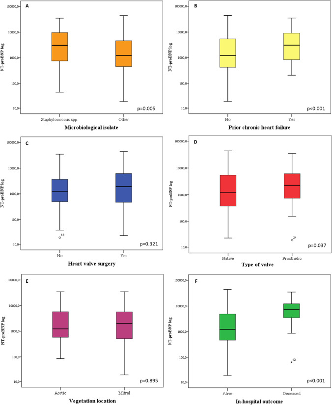
On univariate analysis (Table 2), performed considering NT-proBNP as a categorical variable, we observed a statistically significant association between NT-proBNP levels and older age (P < 0.001). Older patients were more likely to have CHF and CKD, both conditions related to a higher NT-proBNP level (data not shown). NT-proBNP levels were also related to IE etiology (Table 2). Significantly higher levels of this biomarker were observed in staphylococcal IE (including S. aureus and coagulase negative staphylococci) compared with cases due to other microorganisms (Enterococcus spp, Streptococcus spp, etc.) (P = 0.001). The median value of NT-proBNP in staphylococcal IE was 2633.0 pg/mL, significantly higher than in IE of other etiology (1245.0 pg/mL; P = 0.005) (Fig. 1A). In addition, we observed a strong association of NT-proBNP with other biomarkers, such as creatinine, hs-troponin I, d-dimers and CRP (P < 0.001, P < 0.001, P < 0.001 and P = 0.023, respectively) (Table 2). Spearman correlation confirmed these findings (Supplementary Fig. 1).NT-proBNP levels increased directly with raising serum creatinine concentrations (r = 0.462; P < 0.001) (Supplementary Fig. 1A) and were also significantly higher in patients with any of the major comorbidities considered. Specifically, a strong association (P < 0.001) was observed with IHD and CKD (Table 2). Moreover, prior chronic HF also translated into higher NT-proBNP levels (P = 0.001). The median value of NT-proBNP was 2701 pg/mL in prior HF patients and 1272.5 pg/mL in those without (P < 0.001) (Fig. 1B).
Table 2.
Univariate and multivariate analyses of clinical, biochemical and outcome variables associated to NT-proBNP levels in the study cohort (n = 337)
| Parameter | Univariate analysis | P | Multivariate analysis | P | |
|---|---|---|---|---|---|
| NT-proBNP | OR [CI 95%] | ||||
| ≤ 1689.0 (n = 169) | > 1689.0 (n = 168) | ||||
| General characteristics | |||||
| Age, median [IQR] | 56 [46–68.5] | 69 [59–75] | < 0.001 | 1.046 [1.019–1.074] | 0.001 |
| Sex, n (%) | 0.096 | ||||
| M | 126 (74.5) | 111 (65.7) | |||
| F | 43 (25.5) | 57 (34.5) | |||
| Vegetation location, n (%) | 0.054 | ||||
| Aortic valve | 64 (38) | 51 (30.5) | |||
| Mitral valve | 38 (22.6) | 41 (24.5) | |||
| Tricuspid valve | 14 (8.3) | 4 (2.4) | |||
| Pulmonary valve | 4 (2.5) | 3 (1.8) | |||
| CIED | 35 (20.8) | 51 (30.5) | |||
| Other | 0 (0) | 2 (1.3) | |||
| Multisite location | 13 (7.8) | 15 (9) | |||
| Heart side, n (%) | 0.731 | ||||
| Left-sided | 113 (67.3) | 105 (63.2) | |||
| Right-sided | 51 (30.3) | 56 (33.7) | |||
| Bilateral | 4 (2.4) | 5 (30.1) | |||
| Type of valve, n (%) | 0.145 | ||||
| Native | 83 (65.3) | 62 (55.8) | |||
| Prosthetic | 44 (34.7) | 49 (44.2) | |||
| Microorganisms, n (%) | < 0.001 | 1.246 [0.957–1.621] | 0.102 | ||
| S. aureus | 21 (12.4) | 32 (19) | |||
| Other Staphylococcus | 22 (13) | 41 (24.4) | |||
| Streptococcus spp. | 66 (39) | 31 (18.4) | |||
| Enterococcus spp. | 28 (16.6) | 24 (14.3) | |||
| Gram negative | 4 (2.4) | 10 (6) | |||
| Candida spp. | 0 (0) | 2 (1.2) | |||
| Other microorganisms | 2 (1.2) | 5 (3) | |||
| Negative culture | 26 (15.4) | 23 (13.7) | |||
| Microorganisms, n (%) | 0.001 | 0.249 [0.083–0.751] | 0.014 | ||
| Staphylococcus spp | 43 (25.4) | 73 (43.4) | |||
| Other microorganisms | 126 (74.6) | 95 (56.6) | |||
| Biochemical data, median [IQR] | |||||
| C-reactive protein, mg/dL | 4.7 [2.4–8.6] | 6.3 [2.9–11.7] | 0.023 | 1.018 [0.982–1.055] | 0.337 |
| Creatinine, mg/dL | 0.9 [0.7–1] | 1.2 [0.9–1.7] | < 0.001 | 1.629 [0.986–2.690] | 0.057 |
| HS-troponin I, ng/mL | 0.02 [0.01–0.19] | 0.11 [0.04–2.415] | < 0.001 | 1.001 [0.997–1.005] | 0.793 |
| D-dimers, ng/mL | 507 [251–922] | 724 [356.7–1412.2] | < 0.001 | 1.000 [1.000–1.000] | 0.893 |
| Comorbidities, n (%) | |||||
| Chronic heart failure (prior to IE onset) | 37 (22) | 64 (38) | < 0.001 | 0.676 [0.311–1.469] | 0.322 |
| Ischemic heart disease | 20 (11.8) | 46 (27.4) | < 0.001 | 2.360 [0.891–6.247] | 0.084 |
| Chronic obstructive pulmonary disease | 27 (15.9) | 46 (27.4) | 0.012 | 1.144 [0.487–2.686] | 0.757 |
| Diabetes | 23 (13.6) | 39 (23.2) | 0.025 | 0.393 [0.145–1.061] | 0.065 |
| Liver disease | 35 (20.7) | 26 (15.5) | 0.258 | ||
| Malignant neoplasia | 18 (10.6) | 31 (18.3) | 0.046 | 2.244 [0.879–5.729] | 0.091 |
| Chronic kidney disease | 4 (2.3) | 55 (32.7) | < 0.001 | 13.339 [2.530–70.340] | 0.002 |
| Haemodialysis | 0 (0) | 18 (10.7) | < 0.001 | N/A | N/A |
| Cardiac surgerya, n (%) | |||||
| Surgical indication | 114 (68.2) | 132 (78.5) | 0.036 | 2.237 [0.967–5.175] | 0.060 |
| Surgical treatment | 95 (56.8) | 113 (67.6) | 0.055 | ||
| Complications, n (%): | |||||
| Embolic events | 54 (32) | 50 (29.7) | 0.724 | ||
| Valve rupture | 18 (10.6) | 11 (6.5) | 0.244 | ||
| Annular abscess | 19 (11.2) | 27 (16) | 0.208 | ||
| In-hospital outcome, n (%) | < 0.001 | 7.671 [1.550–37.961] | 0.013 | ||
| Survivors | 164 (97.7) | 139 (83.3) | |||
| Non-survivors | 4 (2.3) | 28 (16.7) | |||
aIncludes both open-heart valve surgery and CIED extraction procedures
Fig. 1.
NT-proBNP levels in different IE patient subgroups (log-transformed NT-proBNP levels are shown). Each boxplot depicts the NT-proBNP median levels among each subgroup of IE patients. Panel A Microbiological isolates (Staphylococcus spp vs others). Panel B Prior heart failure. Panel C Heart valve surgery. Panel D type of valve (native vs prosthetic). Panel E Vegetation location (aortic vs mitral). Panel F in-hospital outcome (alive vs deceased)
Concerning cardiac surgery, an additional analysis was carried out excluding CIED-IE (86 pts, who underwent percutaneous lead extraction) or cases with an undefined location (4 pts). Therefore, 247 patients with valvular IE were assessed, showing a median NT-proBNP of 1500 pg/mL. The univariate analysis demonstrated no statistically significant relationship between NT-proBNP and open-heart valve surgery (P = 0.283 for indication; P = 0.371 for surgery performed). NT-proBNP median levels were 1941 pg/mL in those who underwent surgery and 1240.5 pg/mL in those who did not (P = 0.321) (Fig. 1C). There were no differences in NT-proBNP between cases with surgical indication but not operated, cases with indication and operated and cases without indication (data not shown).
At univariate analysis, the proportion of patients with higher NT-proBNP levels did not significantly differ between prosthetic and native valve IE (52.6% vs 42.7%; P = 0.145) (Table 2). However, the median value of this biomarker was 2242 [721.5–6512.5] pg/mL in the former vs 1226 [374.5–5410.5] pg/mL in the latter (P = 0.037) (Fig. 1D).
We then investigated whether NT-proBNP serum levels differed according with IE location (excluding cases with multivalve/multisite involvement). Patients with aortic valve IE did not show a higher level of NT-proBNP compared with mitral IE (1984 [495–5758] pg/mL vs 1245 [566–5793]; P = 0.895) (Fig. 1E). Similarly, there was no difference in NT-proBNP levels between right-sided and left-sided IE (Table 2).
We found no association between NT-proBNP and vegetation size (P = 0.974). In contrast, in valvular IE, those who had paravalvular abscesses showed significantly higher NT-proBNP levels (2954 [865–7564] vs 1245 [425–5410] pg/mL in those without abscesses; P = 0.036).
Patients with implantable cardioverter defibrillator IE had higher NT-proBNP levels than those with pacemaker IE (2550 [1599–6951] vs 1263 [309–3116] pg/mL; P = 0.003). The same was observed for hs-troponin I and creatinine (P = 0.032 and P = 0.002, respectively).
NT-proBNP was strongly associated with in-hospital mortality (P < 0.001) (Table 2). The median value of NT-proBNP in patients who died in hospital was significantly higher than in those discharged alive (5162 [3082–13546] vs 1488 [458–4984] pg/mL; P < 0.001) (Fig. 1F). In the logistic regression multivariate model, age, causative pathogen (staphylococci vs others), CKD and in-hospital mortality were independent correlates of a higher NT-proBNP level (Table 2).
In the univariate analysis considering in-hospital mortality as outcome, an association was found with type of valve (prosthetic vs native) (P = 0.004), surgical indication (P = 0.02) (but not surgical treatment [P = 0.450]), causative microorganism (P < 0.001) and levels of NT-proBNP (P < 0.001), d-dimers (P = 0.011), hs-troponin I (P = 0.043) and CRP (P = 0.022) (data not shown). In the logistic regression multivariate model, higher NT-proBNP levels were an independent predictor of in-hospital mortality (P = 0.003), together with type of valve involved (prosthetic vs native) and d-dimer levels (P = 0.005 and P = 0.002, respectively) (Table 3 and Supplementary Fig. 2).
Table 3.
Multivariate analysis for predicting the occurrence of in-hospital mortality,considering NT-proBNP as a categorical variable
| Parameter | Multivariate analysis | |
|---|---|---|
| OR [CI 95%] | P | |
| Clinical characteristics | ||
| Type of valve | 5.775 [1.722–19.363] | 0.005 |
| Native | ||
| Prosthetic | ||
| Biochemical data | ||
| NT-proBNP | 14.971 [2.465–90.923] | 0.003 |
| C-reactive protein | 1.019 [0.967–1.075] | 0.481 |
| D-dimers | 1.000 [1.000–1.001] | 0.002 |
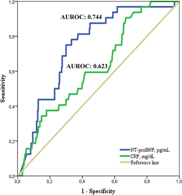
The sensitivity and specificity of NT-proBNP levels in predicting in-hospital mortality were evaluated by means of a ROC curve analysis. In this analysis, an NT-proBNP cut-off value of 2926.5 pg/mL showed a sensitivity of 78.1% and a specificity of 66.7% (AUROC 0.744; [CI 95%: 0.66–0.82], P < 0.001), with a positive predictive value of 19.8% and a negative predictive value of 96.6%. Interestingly, NT-proBNP performed better when compared to CRP. Indeed, with a best cut-off of 6.1 mg/dL, CRP showed a sensitivity of 59.4% and a specificity of 58.2% (AUROC 0.623; [CI 95%: 0.52–0.71], P = 0.022) (Fig. 2).
Fig. 2.

Receiver Operating Characteristic (ROC) curve analysis evaluating the predictive value of in-hospital mortality of the NT-proBNP and C-reative protein (CRP).For each of the analysed biomarker the area under the ROC curve is shown. The best cut-off for NT-proBNP (blue line) is 2926.5 pg/mL whether the cut-off for CRP (green line) is 6.1 mg/dL
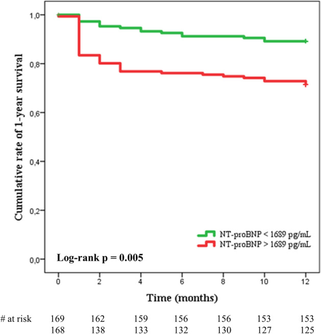
Finally, a Kaplan–Meier survival analysis was performed to assess the influence of admission NT-proBNP levels on the 1 year survival of IE. This analysis showed that patients with NT-proBNP serum levels above the median had a significantly greater mortality rate throughout the 1-year follow-up (log rank P = 0.005) (Fig. 3).
Fig. 3.

Kaplan–Meier survival curves depicting the cumulative rate of survival at 1 year according to NT-proBNP levels above or below the median total cohort value (1689 pg/mL). The number of cases at risk according to the displayed time-lime is shown at the bottom
Discussion
In this study, we provide novel insights into the role of a major cardiac biomarker in IE. NT-proBNP levels showed a strong and clinically relevant prognostic role in IE, both during hospitalization and the subsequent follow-up. Levels below a defined cut off were highly predictive for a favorable outcome. Interestingly, our data suggest NT-proBNP levels correlate with markers of the infectious process rather than with drivers of IE related HF. Indeed, patients with staphylococcal infection, higher CRP and d-dimers were those showing higher NT-proBNP levels. In contrast, concentration of this biomarker did not significantly correlate with aortic valve infection—a predictor of de novo HF in IE [29]—left-sided infection or need for heart valve surgery. Neither prior HF was an independent determinant of NT-proBNP levels.
The association between NT-proBNP and age, in line with Wei et al. results [26], may be partially related to the growing rate of comorbidities, such as CHF and CKD, among the elderly. However, age was an independent predictor of increased NT-proBNP levels (Table 2), suggesting further studies are needed to explain this finding.
Our results showed that serum levels of NT-proBNP differ significantly also according with IE etiology. Indeed, higher levels were observed in patients with IE due to staphylococci, 63% of whom presented with concentrations above the median value. By contrast, most cases with streptococcal IE, well-known for a less aggressive presentation and a less ominous outcome than staphylococcal IE [30], showed a lower NT-proBNP level.
The interesting correlation between NT-proBNP and other biomarkers (hs-troponin I and d-dimers) encourages further investigation on their clinical value in patients with IE. Indeed, previous studies have already suggested a link between sepsis-induced cardiac dysfunction and troponin [16–20], as well as its role in predicting septic patient prognosis [14]. Moreover, following the profound alteration of coagulation system typically ensuing in septic patients, d-dimers may be extremely elevated [31, 32].
The significant correlation of NT-proBNP with serum creatinine levels and prior CKD should be discussed. CKD was the only comorbidity independently associated to a higher NT-proBNP. These results, consistent with current knowledge, further suggest the need for clinicians to evaluate NT-proBNP levels in light of actual kidney function [33]. However, when we performed all described analyses using an NT-proBNP value normalized for serum creatinine, findings were essentially confirmed (data not shown).
The lack of association between NT-proBNP levels and heart side involved deserves further comment. In our cohort, most patients with right-heart involvement were CIED-related IE, and were therefore carriers of a cardiac structural disorder leading to CIED placement. However, in a sub-analysis in which we excluded patients with CIED, we still did not observe significant differences between left-sided and right-sided IE in terms of NT-proBNP levels, further suggesting the association of this biomarker with inflammation rather than cardiac dysfunction.
We expected a higher level of NT-proBNP in patients requiring or undergoing cardiac surgery. In contrast, and similar to Wei et al. findings [26], patients who underwent open heart valve surgery did not show increased levels of this biomarker. Thus, at present we cannot support a role for higher NT-proBNP levels in the decision to proceed with surgery in IE patients.
An important prognostic value of NT-proBNP in IE patients emerges from this study. Indeed, the relationship between NT-proBNP levels and in-hospital mortality was statistically significant and independent of other variables. Noteworthy, 28 of 32 (87.5%) patients who died during hospitalization had NT-proBNP levels above the median value. Our results corroborate those from Wei et al. who found a similar sensitivity and specificity of NT-proBNP (76.2% and 69.1%, respectively for a cutoff value of 2260 pg/mL) [26]. Likewise, NT-proBNP prognostic value extended in the mid-term as patients with increased NT-proBNP levels on admission for IE showed a significantly lower 1 year survival.
Our results are consistent with most but not all prior literature. Kahveci et al. [23] showed a relationship between NT-proBNP and in-hospital mortality, HF, creatinine and troponin on a smaller cohort. By contrast, these investigators did not observe any association between NT-proBNP and CRP or Staph. aureus IE. Interestingly, they describe an association between this biomarker and end-systolic left ventricular diameter, end-diastolic left ventricular diameter and other echocardiographic parameters that, unfortunately, were not available in our patient cohort.
Siciliano et al. [25] found that BNP correlated with HF, a larger vegetation (≥ 10 mm), Staph aureus IE, CRP, diabetes mellitus and creatinine. In contrast, Shiue et al. [24] did not show any relationship between BNP and outcome, HF, creatinine and diabetes, but only with right-sided IE, severe aortic and mitral regurgitation, paravalvular abscesses and troponin. Some of these differences might be related to the different biochemical properties of BNP and NT-proBNP. Indeed, the latter has a longer half-life (25–120 min) and is biologically inactive [34], possibly reflecting HF and/or sepsis more accurately than BNP.
Limitations of this study include its retrospective design, the absence of recorded echocardiography data and the relatively low in-hospital mortality of the study sample.
In conclusion, our results confirm the remarkable prognostic role of NT-proBNP in patients with IE and provide novel evidences of its multifaceted correlates in this unique clinical setting. Our data strongly support the incorporation of NT-proBNP into the current diagnostic work-up of IE.
Supplementary Information
Below is the link to the electronic supplementary material.
Acknowledgements
Members of the Monaldi Hospital Cardiovascular Infection Study Group:
Emanuele Durante-Mangoni MD, PhD, Domenico Iossa PhD, Lorenzo Bertolino MD, Maria Paola Ursi MD, Fabiana D’Amico BSc, Arta Karruli MD, Mohammad Ramadan MD, Roberto Andini MD, Rosa Zampino MD (Internal Medicine); Mariano Bernardo MSc, Giuseppe Ruocco MSc (Microbiology); Giovanni Dialetto MD, Franco Enrico Covino MD, Sabrina Manduca MD (Echocardiography); Alessandro Della Corte MD, PhD, Luca S. De Santo MD, PhD, Antonio Carozza MD, PhD, Marisa De Feo MD, PhD (Cardiac Surgery); Stefano De Vivo MD (Electrophysiology); Maria Luisa De Rimini MD (Nuclear Medicine); Nicola Galdieri MD (Intensive Care).
Funding
Open access funding provided by Università degli Studi della Campania Luigi Vanvitelli within the CRUI-CARE Agreement. The authors did not receive support from any organization for the submitted work.
Declarations
Conflict of interest
The authors have no competing interests to declare that are relevant to the content of this article.
Contributor Information
Emanuele Durante-Mangoni, Email: emanuele.durante@unicampania.it.
The Monaldi Hospital Cardiovascular Infection Study Group:
Emanuele Durante-Mangoni, Domenico Iossa, Lorenzo Bertolino, Maria Paola Ursi, Fabiana D’Amico, Arta Karruli, Mohammad Ramadan, Roberto Andini, Rosa Zampino, Mariano Bernardo, Giuseppe Ruocco, Giovanni Dialetto, Franco Enrico Covino, Sabrina Manduca, Alessandro Della Corte, Luca S. De Santo, Antonio Carozza, Marisa De Feo, Stefano De Vivo MD, Maria Luisa De Rimini, and Nicola Galdieri
References
- 1.Habib G, Lancellotti P, Antunes MJ, et al. ESC Guidelines for the management of infective endocarditis (version 2015) Eur Heart J. 2015;36:3075–3128. doi: 10.1093/eurheartj/ehv319. [DOI] [PubMed] [Google Scholar]
- 2.Scheggi V, Merilli I, Marcucci R, et al. Predictors of mortality and adverse events in patients with infective endocarditis: a retrospective real world study in a surgical centre. BMC Cardiovasc Disord. 2021;21:28. doi: 10.1186/s12872-021-01853-6. [DOI] [PMC free article] [PubMed] [Google Scholar]
- 3.Ternhag A, Cederstrom A, Torner A, et al. A nationwide cohort study of mortality risk and long-term prognosis in infective endocarditis in Sweden. PLoS ONE. 2013;8:e67519. doi: 10.1371/journal.pone.0067519. [DOI] [PMC free article] [PubMed] [Google Scholar]
- 4.Snipsøyr MG, Ludvigsenc M, Petersen E, et al. A systematic review of biomarkers in the diagnosis of infective endocarditis. Int J Cardiol. 2016;202:564–570. doi: 10.1016/j.ijcard.2015.09.028. [DOI] [PubMed] [Google Scholar]
- 5.Siciliano RF, Gualandro DM, Bittencourt MS, et al. Biomarkers for prediction of mortality in left-sided infective endocarditis. Int J Infect Dis. 2020;96:25–30. doi: 10.1016/j.ijid.2020.03.009. [DOI] [PubMed] [Google Scholar]
- 6.Zampino R, Iossa D, Ursi MP, et al. Prognostic value of pro-adrenomedullin and copeptin in acute infective endocarditis. BMC Infect Dis. 2021;21:23. doi: 10.1186/s12879-020-05655-7. [DOI] [PMC free article] [PubMed] [Google Scholar]
- 7.Hall C. Essential biochemistry and physiology of (NT-pro)BNP. Eur J Heart Fail. 2004;15:257–260. doi: 10.1016/j.ejheart.2003.12.015. [DOI] [PubMed] [Google Scholar]
- 8.Tang WH, Francis GS, Morrow DA, et al. National Academy of Clinical Biochemistry Laboratory Medicine National Academy of Clinical Biochemistry Laboratory Medicine practice guidelines Clinical utilization of cardiac biomarker testing in heart failure. Circulation. 2007;116:99 109. doi: 10.1016/j.clinbiochem.2007.07.002. [DOI] [PubMed] [Google Scholar]
- 9.Doust JA, Pietrzak E, Dobson A, et al. How well does B-type natriuretic peptide predict death and cardiac events in patients with heart failure: systematic review. BMJ. 2005;330:625. doi: 10.1136/bmj.330.7492.625. [DOI] [PMC free article] [PubMed] [Google Scholar]
- 10.Bettencourt P, Azevedo A, Pimenta J, et al. N-terminal-pro-brain natriuretic peptide predicts outcome after hospital discharge in heart failure patients. Circulation. 2004;110:2168–2174. doi: 10.1136/bmj.330.7492.625. [DOI] [PubMed] [Google Scholar]
- 11.Davis ME, Richards AM, Nicholls MG, et al. Introduction of metoprolol increases plasma B-type cardiac natriuretic peptides in mild, stable heart failure. Circulation. 2006;113:977–985. doi: 10.1161/CIRCULATIONAHA.105.567727. [DOI] [PubMed] [Google Scholar]
- 12.Pfisterer M, Buser P, Rickli H, et al. TIME-CHF Investigators. BNP-guided vs symptom-guided heart failure therapy: the trial of intensified vs standard medical therapy in elderly patients with congestive heart failure (TIME-CHF) randomized trial. JAMA. 2009;301:383–392. doi: 10.1001/jama.2009.2. [DOI] [PubMed] [Google Scholar]
- 13.Post F, Weilemann LS, Messow CM, et al. B-type natriuretic peptide as a marker for sepsis-induced myocardial depression in intensive care patients. Crit Care Med. 2008;36:3030–3037. doi: 10.1097/CCM.0b013e31818b9153. [DOI] [PubMed] [Google Scholar]
- 14.Kandil E, Burack J, Sawas A, et al. B-type natriuretic peptide: a biomarker for the diagnosis and risk stratification of patients with septic shock. Arch Surg. 2008;143:242–246. doi: 10.1001/archsurg.2007.69. [DOI] [PubMed] [Google Scholar]
- 15.Mueller C, McDonald K, de Boer RA, et al. Heart failure association of the European society of cardiology heart failure association of the European society of cardiology practical guidance on the use of natriuretic peptide concentrations. Eur J Heart Fail. 2019;216:715–731. doi: 10.1002/ejhf.1494. [DOI] [PubMed] [Google Scholar]
- 16.Fenton KE, Parker MM. Cardiac function and dysfunction in sepsis. Clin Chest Med. 2016;37:289–298. doi: 10.1016/j.ccm.2016.01.014. [DOI] [PubMed] [Google Scholar]
- 17.Romero-Bermejo FJ, Ruiz-Bailen M, Gil-Cebrian J, et al. Sepsis-induced cardiomyopathy. Curr. Cardiol Rev. 2011;7:163–183. doi: 10.2174/157340311798220494. [DOI] [PMC free article] [PubMed] [Google Scholar]
- 18.Antonucci E, Fiaccadori E, Donadello K, et al. Myocardial depression in sepsis: from pathogenesis to clinical manifestations and treatment. J Crit Care. 2014;29:500–511. doi: 10.1016/j.jcrc.2014.03.028. [DOI] [PubMed] [Google Scholar]
- 19.Wilhelm J, Hettwer S, Schuermann M, et al. Severity of cardiac impairment in the early stage of community-acquired sepsis determines worse prognosis. Clin Res Cardiol. 2013;102:735–744. doi: 10.1007/s00392-013-0584-z. [DOI] [PubMed] [Google Scholar]
- 20.Sanfilippo F, Corredor C, Fletcher N, et al. Diastolic dysfunction and mortality in septic patients: a systematic review and meta-analysis. Intensive Care Med. 2015;41:1004–1013. doi: 10.1007/s00134-015-3748-7. [DOI] [PubMed] [Google Scholar]
- 21.Landesberg G, Gilon D, Meroz Y, et al. Diastolic dysfunction and mortality in severe sepsis and septic shock. Eur Heart J. 2012;33:895–903. doi: 10.1093/eurheartj/ehr351. [DOI] [PMC free article] [PubMed] [Google Scholar]
- 22.Vieillard-Baron A, Caille V, Charron C, et al. Actual incidence of global left ventricular hypokinesia in adult septic shock. Crit Care Med. 2008;36:1701–1706. doi: 10.1097/CCM.0b013e318174db05. [DOI] [PubMed] [Google Scholar]
- 23.Kahveci G, Bayrak F, Mutlu B, et al. Prognostic value of N-terminal pro-B-type natriuretic peptide in patients with active infective endocarditis. Am J Cardiol. 2007;99:1429–1433. doi: 10.1016/j.amjcard.2006.12.071. [DOI] [PubMed] [Google Scholar]
- 24.Shiue AB, Stancoven AB, Purcell JB, et al. Relation of level of B-type natriuretic peptide with outcomes in patients with infective endocarditis. Am J Cardiol. 2010;106:1011–1015. doi: 10.1016/j.amjcard.2010.05.034. [DOI] [PubMed] [Google Scholar]
- 25.Siciliano RF, Gualandro DM, Mueller C, et al. Incremental value of B-type natriuretic peptide for early risk prediction of infective endocarditis. Int J Infect Dis. 2014;29:120–124. doi: 10.1016/j.ijid.2014.08.017. [DOI] [PubMed] [Google Scholar]
- 26.Wei XB, Liu YH, He PC, et al. Prognostic value of N-terminal prohormone brain natriuretic peptide for in-hospital and long-term outcomes in patients with infective endocarditis. Eur J Prev Cardiol. 2017;24:676–684. doi: 10.1177/2047487316686436. [DOI] [PubMed] [Google Scholar]
- 27.Li JS, Sexton DJ, Mick N, et al. Proposed modifications to the Duke criteria for the diagnosis of infective endocarditis. Clin Infect Dis. 2000;30:633–638. doi: 10.1086/313753. [DOI] [PubMed] [Google Scholar]
- 28.Durante Mangoni E, Adinolfi LE, Tripodi MF, et al. Risk factors for “major” embolic events in hospitalized patients with infective endocarditis. Am Heart J. 2003;146:311–316. doi: 10.1016/S0002-8703(02)94802-7. [DOI] [PubMed] [Google Scholar]
- 29.Hasbun R, Vikram HR, Barakat LA, et al. Complicated left-sided native valve endocarditis in adults: risk classification for mortality. JAMA. 2003;289:1933–1940. doi: 10.1001/jama.289.15.1933. [DOI] [PubMed] [Google Scholar]
- 30.Yombi JC, Yuma SN, Pasquet A, et al. Staphylococcal versus Streptococcal infective endocarditis in a tertiary hospital in Belgium: epidemiology, clinical characteristics and outcome. Acta Clin Belg. 2017;72:417–423. doi: 10.1080/17843286.2017.1309341. [DOI] [PubMed] [Google Scholar]
- 31.Amaral A, Opal SM, Vincent JL. Coagulation in sepsis. Intensive Care Med. 2004;30:1032–1040. doi: 10.1007/s00134-004-2291-8. [DOI] [PubMed] [Google Scholar]
- 32.Schutte T, Thijs A, Smulders YM. Never ignore extremely elevated D-dimer levels: they are specific for serious illness. Neth J Med. 2016;74:443–448. [PubMed] [Google Scholar]
- 33.Chenevier-Gobeaux C, Claessens YE, Voyer S, et al. Influence of renal function on N-terminal pro-brain natriuretic peptide (NT-proBNP) in patients admitted for dyspnoea in the Emergency Department: comparison with brain natriuretic peptide (BNP) Clin Chim Acta. 2005;361:167–175. doi: 10.1016/j.cccn.2005.05.021. [DOI] [PubMed] [Google Scholar]
- 34.Kroll MH, Twomey PJ, Srisawasdi P. Using the single-compartment ratio model to calculate half-life, NT-proBNP as an example. Clin Chim Acta. 2007;380:197–202. doi: 10.1016/j.cca.2007.02.031. [DOI] [PubMed] [Google Scholar]
Associated Data
This section collects any data citations, data availability statements, or supplementary materials included in this article.


