Abstract
Psoriasis is a common chronic inflammatory skin disease. The diversity and heterogeneity of immune cells in human skin have been studied in recent years, but the spatial distribution of immune cells at the single-cell level in the human psoriatic epidermis and dermis remains unclear. In this study, we mapped psoriatic skin immune cells from paired lesional, perilesional, and nonlesional skin samples using mass cytometry. Phenotypic dendritic cells (DCs) were found in the psoriatic epidermis and dermis. Psoriatic dermal CD1c+CD11b+ cDC2s migrated to the epidermis in the perilesional skin during the preinitiation stage. CD1c+CD11b+ cDC2s rapidly replaced EpCAM+CD11clow LC cells and initiated inflammation. Simultaneously, CD207+CD11chi LC and CD5+ T cells accumulated in the psoriatic epidermis and orchestrated epidermal inflammation in psoriasis. The immune cell pool in the psoriatic dermis primarily included APCs and T cells. However, unlike that in the dermis, the epidermal immune environment was more significant and coincided with the inflammation occurring during psoriasis.
The epidermal immune microenvironment plays a dominant role in psoriasis. Langerhans cells, epidermis-resident memory T cells and macrophages together contribute to healthy epidermal immune homeostasis. However, psoriatic CD1c+CD11b+ epidermal cDC2s are positioned in the perilesional area, replacing EpCAM+CD11clow LCs rapidly and initiating inflammation. Epidermal CD141+ cDC1s, CD1c+ cDC2s, CD14+ moDCs, and BDCA2+ pDCs orchestrate psoriatic inflammation. Meanwhile, CD11chi LCs and CD5+ T cells accumulate in the psoriatic epidermis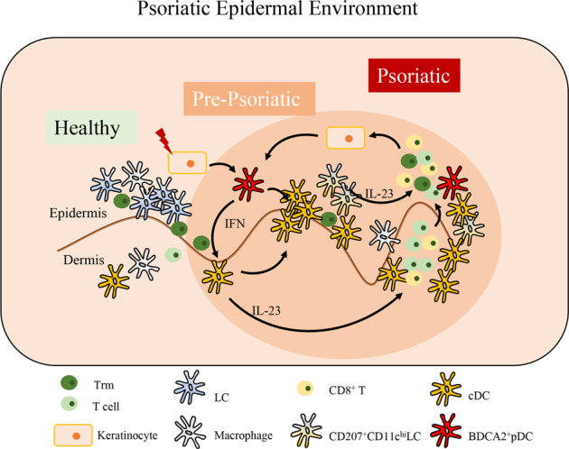
Keywords: Psoriasis, Cutaneous immune, Mass cytometry, Dendritic cell
Subject terms: Autoimmunity, Mechanisms of disease
Introduction
As the first-line battlefield of a host and both a physical and immunological barrier, the skin is involved in defending against exogenous pathogens and physical dangers and in maintaining tissue homeostasis [1, 2]. The epidermis, dermis, and subcutaneous tissue constitute the skin, while the epidermis and papillary dermis are the predominant parts of the cutaneous immune microenvironment [3]. Abundant skin-resident and skin-infiltrating immune cells of both the innate and adaptive immune systems induce diverse protective and pathogenic immune responses [4]. In addition to immune cells, nonimmune components in the skin, such as keratinocytes, play essential roles in interactions with immune cells via soluble mediators [3]. Cytokines released by immune cells affect the activation and proliferation of nonimmune cells and create an intricate inflammatory network during the initiation, propagation, and maintenance of skin inflammation.
Psoriasis is a common chronic inflammatory skin disease that affects ~125 million people worldwide [5], and it is mediated by a variety of immune responses. The pathogenesis of psoriasis is complex and has not been fully elucidated. Langerhans cells (LCs), plasmacytoid dendritic cells (pDCs), myeloid dendritic cells (mDCs), Th1 cells, Th17 cells, CD8+ T cells, macrophages (MACs), and innate lymphoid cells (ILCs) are involved in the initiation and development of psoriasis [6–8]. Dendritic cells are considered to be the primary drivers of psoriasis [9, 10]. In addition, macrophages, T cells, and LCs interact with keratinocytes and contribute to psoriatic inflammation. In contrast to that of circulating DC subsets, the heterogeneity of cutaneous DCs has not been fully elucidated [11]. Several functionally distinct epidermal and dermal DC subsets have been highlighted for their accumulation and contribution to the inflammatory environment through the production of TNF-α, IL-12, and IL-23 [12]. The IL-23/IL-17 axis is thought to be the central pathway among the immune pathways involved in the pathogenesis of psoriasis [8]. Furthermore, heterogeneous T cells, such as Th1 cells, Th2 cells, Th17 cells, Th9 cells, T follicular helper cells (Tfhs), regulatory T cells (Tregs), and CD8+ cells, have been found to infiltrate psoriatic skin, and all of them take part in orchestrating the inflammatory process [13]. Nevertheless, a more comprehensive phenotypic and functional depiction of these cells is required to further the understanding of the epidermal and dermal immune microenvironments, especially during the different stages of psoriatic inflammation.
In this study, we profiled the heterogeneity of immune cells, especially DCs and T cells in the epidermis and dermis of nonlesional (NL), perilesional (PL), lesional (L), and cured-lesional (CL) skin of psoriasis patients, by performing cytometry by time of flight (CyTOF) and multicolor flow cytometry. The dynamics of epidermal and dermal immune cells during the initiation, plaque progression and resolution phases of psoriasis were studied. Our data highlight that the epidermal immune microenvironment plays a dominant role in the development of psoriasis.
Materials and methods
Human samples
Human skin tissue specimens were obtained from the lesional, perilesional, nonlesional, or cured-lesional skin of psoriasis patients and the normal skin of healthy donors undergoing reconstructive surgery at the Department of Dermatology, Second Affiliated Hospital of Zhejiang University (Hangzhou, China). The study was approved by the Institutional Review Board of the Second Affiliated Hospital, Zhejiang University School of Medicine. Signed consent forms were collected from all patients and donors. The information for the donors involved in this study is summarized in Supplemental Table 1 and Table 2, including data generated by mass cytometry (PSO: n = 12, HC: n = 9), flow cytometry and immunofluorescence staining (PSO: n = 8, HC: n = 6). Skin tissues were processed into epidermal and dermal single-cell suspensions separately. After digestion with dispase (GibcoTM) at 37 °C for 90 min, the epidermis and dermis were separated and then cut into small pieces that were enzymatically digested (RPMI 1640 medium containing 1 mg/ml collagenase IV, 250 μg/mL hyaluronase, and 20 μg/mL DNase I) for 90 min at 37 °C. The epidermal and dermal cell suspensions were obtained by filtering through a 70-μm cell strainer. An average of 2 × 10⁶ cells were processed and analyzed per specimen.
Cytometry by time of flight (CyTOF/mass cytometry) staining, acquisition, and data analysis
Antibodies were either purchased as preconjugated products from Fluidigm (DVS Sciences) or purchased as purified products and conjugated in-house using MaxPar x8 Polymer Kits (Fluidigm) according to the manufacturer’s instructions. The mass cytometry antibodies used are shown in Supplementary Table 3. A total of 1–3 × 10⁶ cells for each sample were washed with protein-free PBS, stained with 1 μM cisplatin for 5 min at room temperature, and then stained for cell-surface markers in staining medium (PBS containing 0.5% bovine serum albumin and 0.02% sodium azide) for 30 min at 4 °C. The cells were fixed and stained with DNA Intercalator-Ir overnight. The Foxp3/Transcription Factor Staining Buffer Set (eBioscience) was used, followed by intracellular staining for 30 minutes at 4 °C, washing, and storage at 4 °C until acquisition. The cells were washed twice with deionized water prior to adding EQ normalization beads containing Ce140, Eu151, Eu153, Ho165 and Lu175 (Fluidigm) and were acquired on a Helios instrument. After normalization and randomization of values near zero using Helios software, FCS files were uploaded for analysis.
CyTOF/mass cytometry data analysis
Mass cytometry data were first debarcoded using a doublet filtering scheme with mass-tagged barcodes and then manually gated to retain live, singlet, valid immune cells. Data generated from different batches were normalized through the bead normalization method. To obtain accurate immune subset information, we ran the X-shift algorithm on all samples. All cell events in each individual sample were pooled and included in this analysis.
Flow cytometry staining and analysis
All antibodies used for flow cytometry are listed in Supplementary Table 4. Epidermal and dermal single-cell suspensions were prepared as described above. For surface marker staining, cells were stained with the Zombie UV Fixable Viability Kit (BioLegend) for 15 min, incubated with the Fc receptor blocker TruStain FcX (BioLegend) for 10 min and then incubated with a cocktail of antibodies specific for cell-surface markers for 30 min at 4 °C in the dark. For intracellular TNF-α and IL-17A staining, cells were first stimulated with Cell Activation Cocktail (with brefeldin A, BioLegend) for 6 h; for IL-23p19 detection, cells were stimulated with 2 μg/ml resiquimod (Sigma) for 6 h in the presence of brefeldin A (GolgiPlug, BD Biosciences). After stimulation, cells were stained for surface marker detection first (as described above), followed by fixation and permeabilization using BD Cytofix/Cytoperm (BD Biosciences) or the Foxp3/Transcription Factor Staining Buffer Set (eBioscience). The cells were then incubated with anti-cytokine antibodies or anti-Foxp3 for 30 min at 4 °C in the dark. Data were acquired on a CytoFLEX LX (Beckman Coulter) and analyzed by using Kaluza Analysis Software (Beckman Coulter).
Immunofluorescence staining
For section staining, human skin tissues were embedded in OCT compound, frozen, cryosectioned (10 μm), and fixed in 4% paraformaldehyde for 10 min. The sections were blocked with a solution of 5% normal goat serum, 1% bovine serum albumin and 0.1% Triton in PBS for 1 h at room temperature and then stained with anti-human CD3 (Abcam, cat: ab16669, clone: SP7, dilution: 1:500) and anti-human CD11c (Abcam, cat: ab11029, clone: 3.9, dilution: 1:500) overnight at 4 °C. After washing in PBS, the sections were stained with fluorochrome-conjugated secondary antibodies (Thermo Fisher Scientific, goat anti-mouse IgG (H + L), Alexa Fluor™ 555, cat: A-21422, dilution: 1:500; goat anti-rabbit IgG (H + L), Alexa Fluor™ 488, cat: A-11008, dilution: 1:500) for 1 h at room temperature in the dark. DAPI (Roche, cat: 10236276001, dilution: 1:5000) incubation for 5 min was used for nuclear staining. Then, the sections were visualized under a confocal microscope (Leica, TCS SP8) and analyzed with the accompanying software.
Statistical analysis
Statistical analysis was performed using unpaired (comparisons of two groups from different individuals) or paired (comparisons of two groups from the same individuals) two-tailed Student’s t tests or one-way ANOVA (comparisons among more than two groups) in GraphPad Prism software. A p value less than 0.05 was considered significant (*p < 0.05, **p < 0.01***p < 0.001, and ****p < 0.0001).
Results
High-dimensional analysis of the human immune cell atlas in the healthy and psoriatic epidermis and dermis
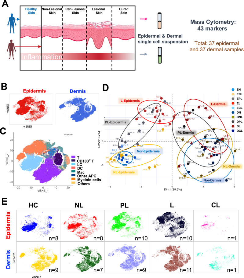
Early inflammatory changes can be observed in the perilesional skin of affected psoriatic patients. [14] In addition, the nonlesional skin displays a prepsoriasis phenotype [15]. To better investigate the dynamic immune microenvironment in psoriatic inflammation, our multilesion CyTOF/mass cytometry scheme was performed with 37 epidermal and 37 paired dermal tissue specimens from 12 psoriasis patients and 9 healthy donors (Fig. 1A, B and Supplementary Table 1). Thus, the epidermis and dermis of patients with nonlesional, perilesional, lesional, and cured-lesional psoriatic skin were paired and collected (Fig. 1E). All specimens were digested into single-cell suspensions and stained for immune- and function-related markers (Supplementary Table 3). As expected, the primary immune cells in human psoriatic and healthy skin were T cells, LCs, DCs, macrophages, and a few myeloid cells (Fig. 1C).
Fig. 1.
Human skin immune cell atlas of healthy and psoriatic skin based on CyTOF analysis. A The strategy for sample collection, tissue processing and analysis of 74 samples. All samples were separated into epidermal and dermal single-cell suspensions and stained with 43 markers for CyTOF (Supplementary Table 3). CyTOF single-cell data were clustered using PhenoGraph to identify common populations across patients. B viSNE plots of CD45+ cells from the epidermis and dermis for 74 samples (epidermal cells, red; dermal cells, blue). C viSNE plot dimensionality reduction and PhenoGraph clustering of CD45 + immune cells across all samples. The immune cell compartment comprised all major immune lineages in psoriasis, including T cells, CD103+ T cells, LCs, DCs, MACs, other APCs and myeloid cells. D Principal component analysis (PCA) of CD45+ immune cells from all samples. The colored circles represent 10 different subgroups (EN, epidermis of normal skin; ENL, epidermis of nonlesional psoriatic skin; EPL, epidermis of perilesional psoriatic skin; EL, epidermis of lesional skin; ECL, epidermis of cured-lesional psoriatic skin; DN, dermis of normal skin; DNL, dermis of nonlesional psoriatic skin; DPL, dermis of perilesional psoriatic skin; DL, dermis of lesional psoriatic skin; and DCL, dermis of cured-lesional psoriatic skin). E viSNE plots of CD45 + immune cells in each subgroup. The groups included normal skin from healthy donors (HCs) (epidermis, n = 8; dermis, n = 9) and psoriatic skin (NL in the epidermis, n = 8; NL in the dermis, n = 7; PL in the epidermis, n = 10; PL in the dermis, n = 9; L in the epidermis, n = 10; L in the dermis, n = 11; CL in the epidermis, n = 1; CL in the dermis, n = 1)
Notably, the composition of immune cell subsets varied between the epidermis and dermis as well as among the different stages of psoriatic lesions (Fig. 1D, E). Principal component analysis (PCA) revealed that the epidermis of perilesional (EPL) psoriatic skin was in an intermediate position between the lesional and nonlesional epidermis, while dermal groups appeared to have a more mixed stage (Fig. 1D). These results indicated that the alteration in the epidermal immune microenvironment was more consistent with the development of psoriasis than was the alteration in the dermal microenvironment.
The heterogeneous immune cell landscape in the healthy and psoriatic epidermis
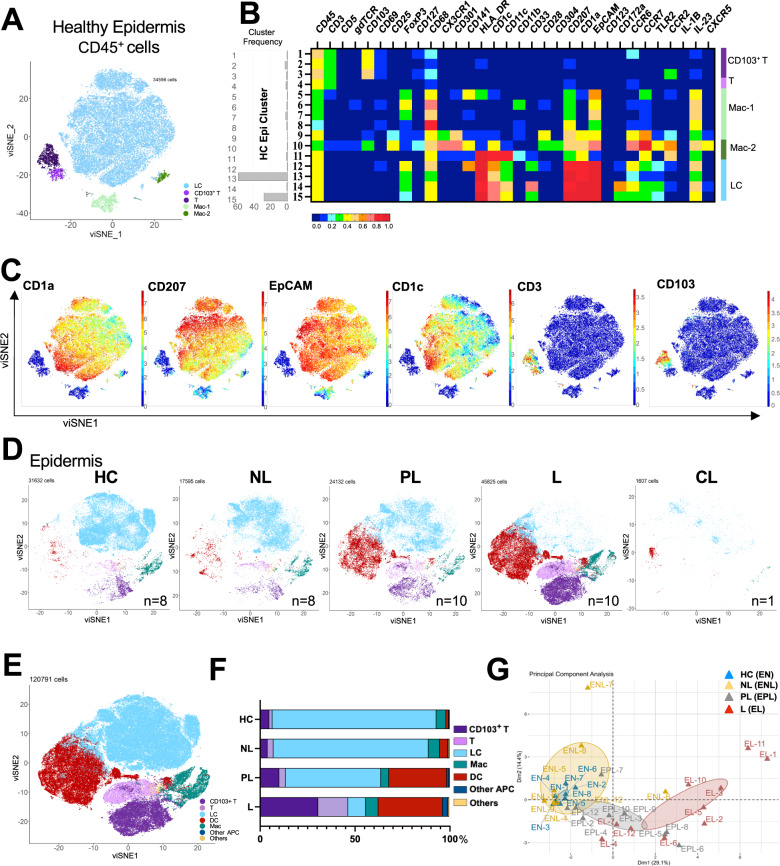
To better analyze psoriatic epidermal immune cells, we first analyzed the cells in the epidermis of healthy skin (n = 8) [2]. Langerhans cells (LCs) (HC EpiC-12, HC EpiC-13, HC EpiC-14, and HC EpiC-15) hallmarked by specific expression of CD1a, CD207 and EpCAM were found to be the dominant immune cells in normal human skin, along with a few T cells (HC EpiC-4), CD103+ resident T cells (HC EpiC-1, HC EpiC-2, and HC EpiC-3), and MACs (Fig. 2A–C).
Fig. 2.
Heterogeneous immune cell landscapes of the healthy and psoriatic epidermis. A viSNE plots of CD45+ immune cells in healthy epidermis from 8 samples. B Heatmap of PhenoGraph clusters of CD45+ immune cells. Rows represent 15 clusters of single cells within individual healthy epidermis samples grouped by metacluster. C viSNE plots of the normalized expression levels of representative phenotypic markers (top) for the eight healthy epidermis samples. D viSNE plots of CD45+ immune cells from the epidermal samples of 5 groups (HC, NL, PL, L, and CL) are shown. E viSNE plots of CD45+ immune cells from all epidermal samples colored by the relative expression of CyTOF markers, with six cell populations indicated (right). F Representative bar chart of the proportions of the epidermal immune cell subsets (CD103 + T cells, T cells, LCs, DCs and other APCs) in four subgroups (CL excluded). G PCA of CD45+ immune cells from the epidermal samples of 4 groups (EN, epidermis of normal skin; ENL, epidermis of nonlesional psoriatic skin; EPL, epidermis of perilesional psoriatic skin; and EL, epidermis of lesional skin). HC healthy control, NL nonlesional, PL perilesional, L lesional, CL cured-lesional
To visualize the distribution of cell populations in the psoriatic epidermis, we analyzed all epidermal samples from both healthy controls and psoriasis patients (n = 37). Group viSNE and PCA analyses showed that psoriatic epidermal immune cells were present in higher numbers and had notably more complex compositions, especially in the perilesional and lesional psoriatic skin (Fig. 2C–G). These results indicated the heterogeneity of immune cells in the peri-psoriatic epidermis during the preinitiation stage.
Inflammatory subpopulations of epidermal DCs in psoriasis
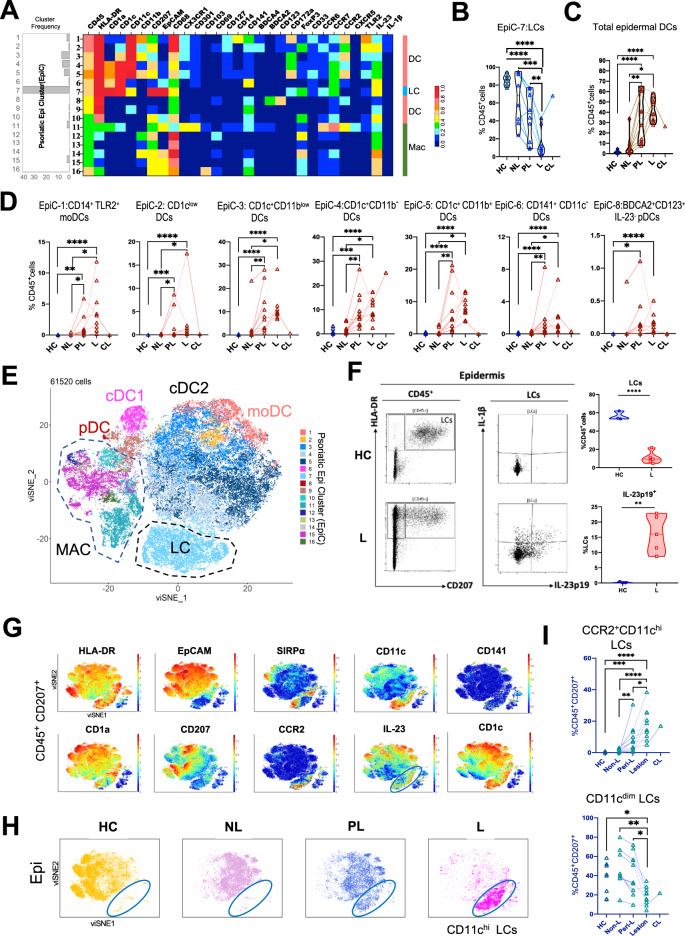
Skin antigen-presenting cells (APCs), including dendritic cells, monocytes, and macrophages, play key roles in initiating, modulating, and resolving cutaneous inflammation [16]. To further investigate the diversity and heterogeneity of these immune cells in the psoriatic epidermis, we clustered all epidermal APC clusters (Psoriatic Epidermis cluster-1 to Psoriatic Epidermis cluster-16, EpiC-1 to EpiC-16 in Fig. 3A). Regarding the percentage of each cell group, we found that over 85% of the epidermal CD45+ cells were CD207+ EpCAM+ CD1a+ LCs (EpiC-7) in the healthy human epidermis (Fig. 3B). In contrast, the frequency of LCs was dramatically decreased in the PL and L psoriatic epidermis and recovered in the CL epidermis (Fig. 3B). Instead of LCs, epidermal DCs were clearly present (Fig. 3C). Total epidermal DCs were significantly increased in perilesional and lesional psoriatic skin (Fig. 3C).
Fig. 3.
Diversity of human psoriatic epidermal antigen-presenting cells (APCs). A Heatmap of PhenoGraph clusters across the epidermis for all patients revealed distinct APC metaclusters in CD45+ immune cells. B Relative frequency of epidermal CD207+ EpCAM+ CD11c+ LCs (EpiC-7) in CD45+ cells in the five groups. C Relative frequency of total epidermal DCs in CD45+ immune cells in the five subgroups. D Relative frequency of epidermal DC subsets (EpiC-1, EpiC-2, EpiC-3, EpiC-4, EpiC-5, EpiC-6, and EpiC-8) in CD45+ immune cells in the five subgroups selected from A. E viSNE plots of psoriatic epidermal APCs. F Representative flow cytometry plots and statistical analysis of LCs (CD207+ HLA-DR+) in CD45+ cells and IL-23p19+ LCs in total LCs in the HC and L epidermis after stimulation with resiquimod for 6 hours (HC, n = 3; L, n = 5). G viSNE plots of the normalized expression levels of representative phenotypic markers (top) of CD45+ CD207+ cells from all 74 samples. H viSNE plots of CD45+ CD207+ immune cells from the epidermal samples of 4 groups (HC, NL, PL, and L) are shown. Blue circles represent EpCAM- CD11chi CCR2+ LCs. I Relative frequencies of CCR2+ CD11chi LCs and CD11cdim LCs in CD45+ CD207+ cells in the five subgroups. B, C, D, I Statistical analysis was performed by one-way ANOVA. F Statistical analysis was performed by unpaired t tests. *p < 0.05, **p < 0.01, ***p < 0.001, ****p < 0.0001. HC healthy control, EpiC epidermal cluster, NL nonlesional, PL perilesional, L lesional, CL cured-lesional
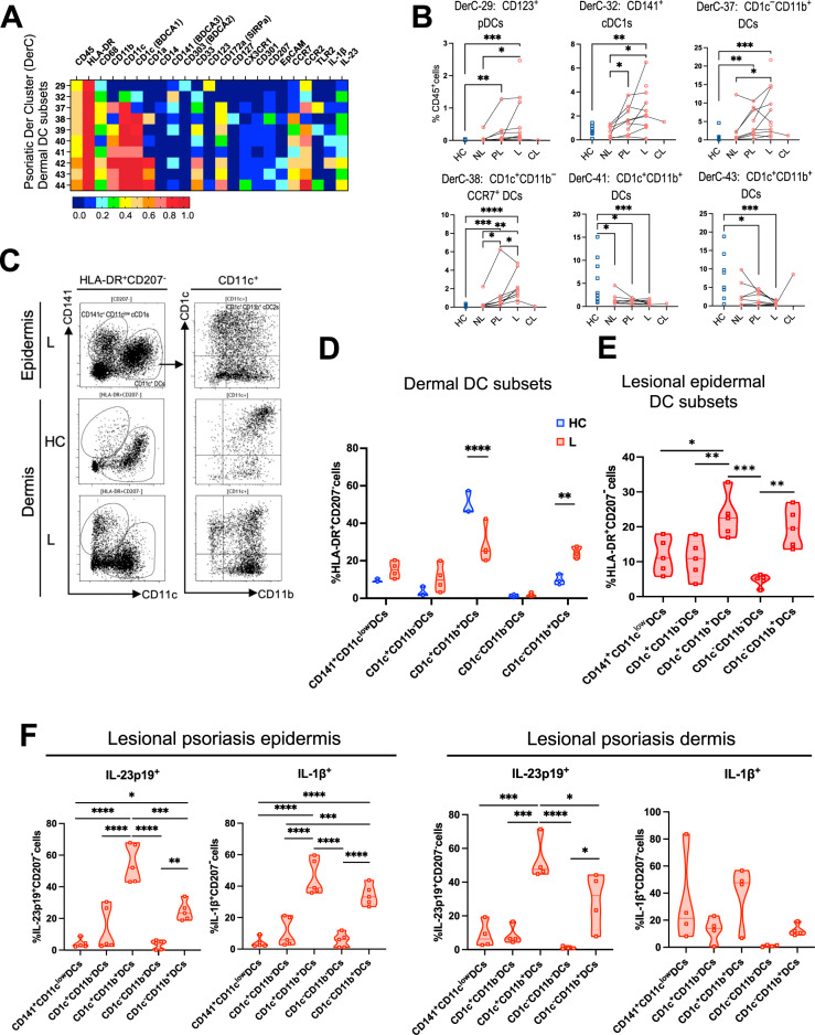
DCs comprise plasmacytoid DCs (pDCs), conventional DCs (cDCs) and monocyte-derived DCs. The current consensus describes two main subsets of conventional DCs (cDCs), known as type 1 cDCs (cDC1s, CD141+CLEC9A+XCR1+ cDC1s in humans) and type 2 cDCs (cDC2s, CD1c+ cDC2s in humans), which have been identified in all tissues [17, 18]. Tissue DCs are a highly dynamic and heterogeneous population, with functionally specialized subsets imprinted and modulated by the local microenvironment [16]; for example, CD11blow DC2s are found only in the skin [19, 20]. There are still many unknowns in regard to cutaneous DCs. Therefore, we were highly interested in DCs in psoriatic tissue, especially in the epidermis. In Fig. 3D, E, epidermal DCs were divided into 4 main subtypes: CD14+ TLR2+ moDCs (EpiC-1), BDCA2+CD123+IL-23- pDCs (EpiC-8), CD141+ CD11c- cDC1s (EpiC-2) and CD1c+ cDC2s (EpiC-3, EpiC-4, and EpiC-5). Epidermal cDC2s composed the majority of cutaneous DCs in psoriasis, with similar frequencies in the PL and L epidermis; these cells were composed of CD1c+ CD11blow DCs (EpiC-3), CD1c+ CD11b− DCs (EpiC-4) and CD1c+ CD11b+ DCs (EpiC-5) (Fig. 3D, E). In addition, flow cytometry detected these DC subsets in the psoriatic epidermis (Fig. 6).
Fig. 6.
Phenotypic profiling and enumeration of dermal DCs in psoriatic and healthy skin. A Heatmap of PhenoGraph clusters across the dermis for all patients revealed distinct APC metaclusters in CD45+ immune cells. B Relative frequencies of dermal DC subsets (DerC-29, DerC-32, DerC-37, DerC-38, DerC-41, and DerC-43) in CD45+ immune cells in the five subgroups selected from A. C Representative flow cytometry plots for CD141+ cDC1s, CD1c−CD11b+ DCs, CD1c+CD11b- DCs, CD1c+CD11b+ DCs, CD1c−CD11b- DCs and CD1c−CD11b+ DCs in the lesional epidermis and healthy and lesional dermis (HC, n = 3; L, n = 5). D Statistical analysis of the above five DC subsets in HLA-DR+ CD207- cells in the HC and L dermis identified through flow cytometry. E Statistical analysis of the above five DC subsets in HLA-DR+ CD207- cells in the L epidermis identified through flow cytometry. F Statistical analysis of IL-23p19- or IL-1β-expressing DCs in the lesional epidermis and dermis after stimulation with resiquimod for 6 h. B, E, F Statistical analysis was performed by one-way ANOVA. D Statistical analysis was performed by unpaired t tests. *p < 0.05, **p < 0.01, ***p < 0.001, ****p < 0.0001. HC healthy control, EpiC epidermal cluster, NL nonlesional, PL perilesional, L lesional, CL cured-lesional
Epidermal CD11chi Langerhans cells release IL-23 in psoriasis
The number of LCs in psoriasis has been debated in recent decades, as has the role of LCs in psoriatic inflammation [21–25]. LC heterogeneity was recently found by single-cell approaches [26]. Consistent with the data in Fig. 3B, flow cytometry detected HLA-DR+ CD207+ LCs in eight additional samples (Supplementary Table 2), showing that the frequency of LCs was dramatically decreased in psoriatic lesional skin compared with healthy control (HC) skin. However, these LCs had an increased capacity to secrete IL-23p19 in the psoriatic lesional epidermis (Fig. 3F). These results led us to hypothesize that LCs may have opposite functional subsets. We then analyzed all of the cutaneous CD45+CD207+ cells (Fig. 3G–I). Intriguingly, utilizing a CyTOF approach, our results revealed the heterogeneity of LCs. The data showed that the percentage of CD11dim LCs decreased from the healthy control epidermis to the nonlesional, perilesional, and lesional epidermis. However, CCR2+CD11chi LCs (blue circle) were detected only in the perilesional and lesional epidermis (Fig. 3H, I), and these subpopulations of LCs were more likely to produce IL-23 (Fig. 3G). In conclusion, LCs with higher expression of CCR2 and CD11c had the capacity to produce IL-23 in psoriatic lesions.
CD5+ T cells have an intensified ability to produce IL-17A and TNF-α in the psoriatic epidermis and dermis
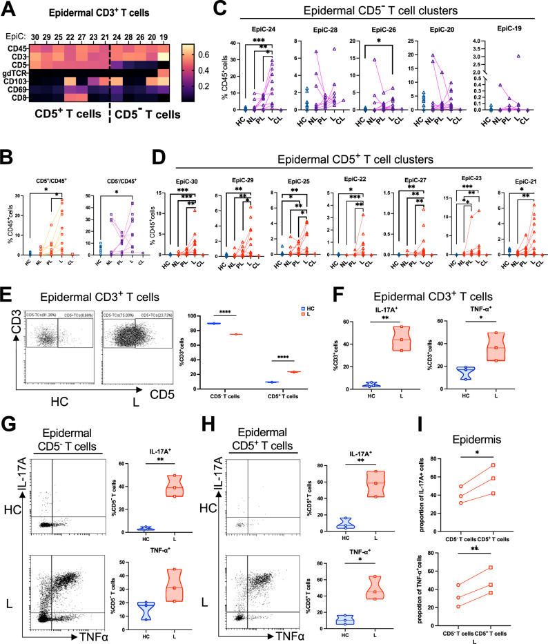
In addition to APCs, increased T cells and associated cytokines are hallmarks of psoriasis and are involved in the pathogenesis and treatment of psoriasis [27–30]. Next, we focused on epidermal CD3+ T cells. Not only the number but also the heterogeneity of epidermal T cells were increased (Figs. 2E, F, 4A). CD5 expression defined two types of T cells in the psoriatic epidermis (Fig. 4A). Interestingly, the percentage of total or heterogeneous CD5+ T-cell clusters (EpiC-30, EpiC-29, EpiC-25, EpiC-22, EpiC-27, EpiC-23, and EpiC-21) showed a gradually increasing tendency from the nonlesional epidermis to the perilesional and finally lesional psoriatic epidermis and was higher than that in the healthy epidermis (Fig. 4B, D). For CD5- T cells (EpiC-24, EpiC-28, EpiC-26, EpiC-20, and EpiC-19), a similar increasing tendency was observed in the psoriatic epidermis (Fig. 4C). As verification of the mass cytometry data, flow cytometric analysis showed that the proportion of CD5+ T cells was markedly higher in the psoriatic epidermis than in the healthy epidermis (Fig. 4E). It was obvious that psoriatic lesional epidermal CD3+ T cells had an enhanced capacity to produce IL-17A and TNF-α compared with healthy control T cells (Fig. 4F). Both CD5+ and CD5- T cells in the psoriatic epidermis could release more IL-17A and TNF-α than those in HC skin (Fig. 4G, H), but the CD5+ T cells seemed to have a stronger cytokine-releasing capacity than the CD5- T cells due to the significantly higher proportions of IL-17A- and TNF-α-positive cells (Fig. 4I). Additionally, both healthy and lesional dermal CD3+ T cells secreted IL-17A and TNF-α, while lesional dermal T cells exhibited significantly increased IL-17A production (Supplementary Fig. 1B). Although the percentage of CD5+ or CD5- T cells was nearly equal between the healthy and lesional dermis (Supplementary Fig. 1C, D), the capacity of these two dermal T-cell subsets to secrete IL-17A was enhanced in the inflammatory psoriatic environment (Supplementary Fig. 1E, F). Consistent with the results for the epidermis, lesional dermal CD5+ T cells also had a stronger potential to release IL-17A and TNF-α than did CD5- T cells due to the significantly higher proportions of IL-17A- and TNF-α-positive cells (Supplementary Fig. 1G). Overall, CD5+ T cells, especially epidermal CD5+ T cells, provide a more important contribution to the pathogenesis of psoriasis by intensively producing IL-17A and TNF-α.
Fig. 4.
CD5+ T cells have an enhanced ability to produce IL-17A and TNF-α in the psoriatic epidermis. A Heatmap of PhenoGraph clusters of T cells with or without CD5 expression across all epidermal samples. B Relative frequencies of epidermal CD5+ and CD5- T cells in CD45+ cells in the five subgroups. C Relative frequencies of five subclusters of epidermal CD5- T cells in CD45+ cells in the five subgroups. D Relative frequencies of seven subclusters of epidermal CD5+ T cells in CD45+ cells in the five subgroups. E Representative flow cytometry plots and statistical analysis of CD5+ and CD5- T cells in the HC and L epidermis (HC, n = 3; L, n = 3). F Statistical analysis of IL-17A+ T cells and TNF-α+ T cells in CD3+ T cells in the HC and L epidermis identified through flow cytometry. G, H Representative flow cytometry plots and statistical analysis of IL-17A+ T cells and TNF-α+ T cells in CD5- T cells and IL-17A+ T cells and TNF-α+ T cells in CD5+ T cells in the HC and L epidermis after stimulation with Cell Activation Cocktail for 6 h. I Comparisons of the proportions of IL-17A+ cells and TNF-α+ cells between lesional epidermal CD5+ and CD5- T cells. C, D Statistical analysis was performed by one-way ANOVA. E–H Statistical analysis was performed by unpaired t tests. I Statistical analysis was performed by a paired t test. *p < 0.05, **p < 0.01, ***p < 0.001, ****p < 0.0001. EpiC epidermal cluster, HC healthy control, NL nonlesional, PL perilesional, L lesional, CL cured-lesional
The psoriatic dermis as an inflammatory cell pool composed of T cells and APCs
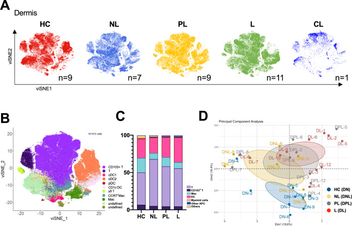
To further understand the dynamics of the immune system in the dermis, another important battlefield for psoriatic inflammation, we analyzed dissected single dermal cells from the five groups (Fig. 5A, B). All dermal CD45+ cells were evaluated for the expression of 43 protein markers by mass cytometry, and 97.5% of the cells were defined (Fig. 5B, C). First, we found that T cells and APCs (DCs and MACs) were two dominant types of dermal immune cells under both the homeostatic and psoriatic inflammatory conditions (Fig. 5B, C). Although there seemed to be no significant differences in the diversity or abundance of immune cell clusters among the groups (Fig. 5C), PCA clearly distinguished the psoriatic (NL, PL, and L) dermis from the healthy dermis (HC) and showed a gradual separation from the nonlesional skin to the perilesional and finally lesional skin, which coincided with the progression of psoriatic inflammation (Fig. 5D). To deeply investigate the pathological immune cell subtypes, we next focused on the diversity and heterogeneity of dermal DCs (Fig. 6A). The majority of dermal DC subsets were increased in the psoriatic environment, including CD123+ pDCs (Psoriatic Dermal Cluster-29, DerC-29), CD141+ cDC1s (DerC-32), CD1c−CD11b+ cDC2s (DerC-37), and CD1c+CD11b−CCR7+ cDC2s (DerC-38). However, the frequency of dermal CD1c+CD11b+ cDC2s (DerC-41 and DerC-43) was lower in the psoriatic dermis (NL, PL, and L) than in the healthy control dermis (Fig. 6A, B). The dynamic trend for dermal CD1c+CD11b+ cDC2s was opposite that for epidermal CD1c+CD11b+ cDC2s (EpiC-5, in Fig. 3D). Flow cytometry data also showed a consistent result, with the proportion of CD1c+CD11b+ cDC2s being decreased in the lesional dermis but increased in the lesional epidermis compared with the corresponding healthy control tissues (Fig. 6C–E). Consequently, these results raised the hypothesis that the original resting dermal CD1c+CD11b+ cDC2s migrated to the epidermis and were chemoattracted by the triggers of psoriasis. We mapped the cutaneous APC trajectory, and our pseudotime analysis showed similar results (Supplementary Fig. 2). Moreover, these CD1c+CD11b+ cDC2s had a much stronger ability to produce IL-23p19 and IL-1β than did other DC subsets in both the epidermis and the dermis (Fig. 6F and Supplementary Fig. 3A, B). In addition, the majority of lesional dermal DC subsets had an enhanced capacity for IL-23p19 and IL-1β production compared with the corresponding healthy control subsets, although some comparisons were not statistically significant (Supplementary Fig. 3C). Together, these results indicated that the influx of CD1c+CD11b+ cDC2s from the dermis to the epidermis might be a hallmark of psoriasis initiation.
Fig. 5.
The psoriatic dermis contributed an inflammatory cell pool of T cells and APCs. A viSNE plots of CD45+ immune cells from the dermal samples of five groups (HC, NL, PL, L, and CL) are shown. B viSNE plots of CD45+ immune cells from all dermal samples. C Representative bar chart of the proportions of the dermal immune cell subsets (T cells, CD103+ T cells, MACs, myeloid cells and other APCs) in four subgroups (CL excluded). D PCA of CD45+ immune cells from the dermal samples of four groups (DN, dermis of normal skin; DNL, dermis of nonlesional psoriatic skin; DPL, dermis of perilesional psoriatic skin; and DL, dermis of lesional psoriatic skin). HC healthy control, NL nonlesional, PL perilesional, L lesional, CL cured-lesional
Human cutaneous CD103+ cell and Treg levels were increased in psoriasis
CD103, also known as alpha E integrin, is expressed by skin-resident cells, including tissue-resident memory T cells (TRMs) [31]. With long-term survival (longevity) and low migration in peripheral tissues, skin TRMs can respond rapidly in defense against infections and play crucial roles in the recurrence of chronic skin autoimmune diseases, including psoriasis [32–35].
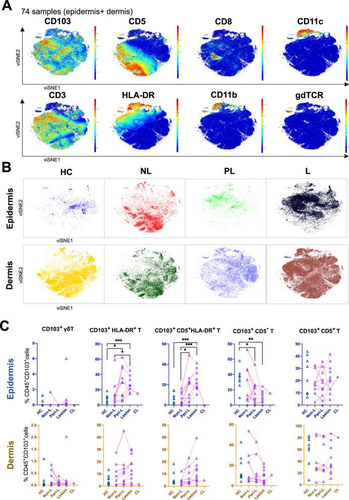
In our data, CD103+ cells were relatively rare cell clusters in the skin immune system. Therefore, to better understand resident cells in psoriatic tissue, we specifically selected all CD45+CD103+ cells in the epidermis and dermis from both the psoriasis patients and the HCs for an additional analysis. The majority of cutaneous CD103+ cells were CD3+ T cells, which were reportedly skin TRMs, and few were CD11b+ or CD11b- HLA-DR+ CD11c+ DCs and other HLA-DR+ cells (Fig. 7A). The cutaneous CD103+ cells exhibited more dramatic dynamics in the psoriatic epidermis than in the dermis (Fig. 7B, 7C).
Fig. 7.
The heterogeneity of human cutaneous CD103+ T cells in psoriatic and healthy skin. A viSNE plots of the normalized expression levels of representative phenotypic markers (top) of CD45+ CD103+ cells from all 74 samples. B viSNE plots of the epidermal and dermal CD45+ CD103+ cells of 5 groups (HC, NL, PL, L, and CL) are shown. C Relative frequencies of five epidermal and dermal CD103+ subclusters in CD45+ CD103+ cells in five subgroups. C Statistical analysis was performed by one-way ANOVA. *p < 0.05, **p < 0.01, ***p < 0.001, ****p < 0.0001. HC healthy control, Non-L nonlesional, Peri-L perilesional, CL cured-lesional
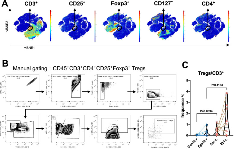
CD4+CD25+Foxp3+ Tregs are considered to play fundamental roles in maintaining immune homeostasis and regulating local tissue inflammation by suppressing immune responses [36]. In psoriasis, impaired suppressive function of Tregs leads to an altered T-helper 17/Treg balance [36]. However, the exact functions of Tregs in psoriasis pathogenesis remain mostly unclear, and the Treg frequency in lesional skin is controversial [37–40]. Therefore, we examined the frequency of Tregs in lesional and healthy skin. As expected, our viSNE plot showed a limited number of CD4+CD25+Foxp3+ cells (Fig. 8A). Subsequently, we manually gated CD45+CD3+CD4+CD25+Foxp3+ Tregs (Fig. 8B), which showed that the proportion of lesional Tregs was increased in both the epidermis and the dermis of psoriasis patients compared with the corresponding tissues of healthy controls (Fig. 8C).
Fig. 8.
Tregs were increased in the human psoriatic epidermis and dermis. A viSNE plots of the normalized expression levels of representative phenotypic markers (top) of CD45+ cells from all 74 samples. Black circles represent CD3+CD25+Foxp3+CD127- CD4+ Tregs. B Manual gating strategy for CD45+CD3+CD4+CD25+Foxp3+ Tregs in all samples. C Statistical analysis of the epidermal and dermal Tregs in CD3+ T cells in HC and L skin. C Statistical analysis was performed by one-way ANOVA, and exact p values are shown on the graph. Der dermis, Epi epidermis, L lesional
Abundant T cells and DCs were present in the epidermis and dermis of perilesional and lesional psoriatic skin
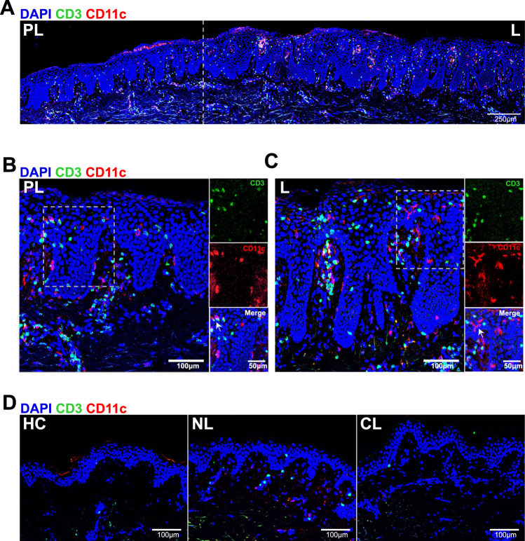
On the basis of the above results, heterogeneous subsets of T cells and DCs were identified to be endowed with distinct functional potential depending on their phenotype and local immune microenvironment. To visually present the dynamic infiltration of these immune cells during the initiation and progression of psoriatic inflammation, immunofluorescence staining for CD3+ T cells and CD11c+ DCs in psoriatic nonlesional, perilesional, lesional, and cured-lesional skin and in healthy control skin was performed (negative control staining with isotype controls for each primary antibody confirmed specificity, Supplementary Fig. 4). To eliminate interference by CD11c+ LCs, CD207 staining (LC specific marker) and CD11c staining were used to differentiate LCs and DCs (Supplementary Fig. 4C). Both CD207+ CD11c+ LCs and CD207- CD11c+ DCs were distributed in the psoriatic lesional epidermis, as shown in Supplementary Fig. 4C. We also analyzed the percentage of CD207+ CD11c+ LCs and CD207- CD11c+ DCs in the psoriatic lesional epidermis through flow cytometry. The results showed that epidermal CD207-CD11c+ DCs were much more abundant than CD207+ CD11c+ LCs (Supplementary Fig. 4D, E), which was consistent with the results shown in Fig. 3. A panoramic scan of typical psoriatic skin showed that abundant T cells and DCs were present in the epidermis and dermis of perilesional and lesional skin (Fig. 9A), and in line with our data, these immune cells increased progressively from the nonlesional skin to the peri-lesional and finally lesional skin but were nearly absent in healthy control and cured-lesional skin, especially in the epidermis (Fig. 9B, C and Supplementary Fig. 4A). These results suggested that the epidermal immune microenvironment might play a dominant role in psoriasis development due to the progressive infiltration of T cells and DCs from the perilesional stage to the lesional stage.
Fig. 9.
Immunofluorescence staining for CD3+ T cells and CD11c+ DCs in psoriatic and healthy skin. A Panoramic scan of typical psoriatic PL and L skin with immunofluorescence staining for CD3+ T cells and CD11c+ DCs. The dotted line indicates the boundary between the PL and L areas. DAPI (blue), CD3 (green), and CD11c (red). Scale bars, 250 μm. B, C Magnified images of the immunofluorescence staining for CD3+ T cells and CD11c+ DCs in the psoriatic PL and L skin. DAPI (blue), CD3 (green), and CD11c (red). Scale bars, 100 μm, 50 μm. D Immunofluorescence staining for CD3+ T cells and CD11c+ DCs in HC and psoriatic NL and CL skin. DAPI (blue), CD3 (green), and CD11c (red). Scale bars, 100 μm. HC healthy control, NL nonlesional, PL perilesional, L lesional, CL cured-lesional
Discussion
In both homeostasis and disease, reconstructing the dynamic landscape of identified cells is crucial to better understanding immune responses and to addressing therapeutic issues. Immune cells are characterized by diversity, specificity, plasticity, and adaptability, and these properties enable them to contribute to disease pathogenesis [41].
Our findings provide a comprehensive map of the unsupervised clustering and visualization of cutaneous immune cells in psoriatic skin at the single-cell protein level through a combination of mass cytometry by CyTOF, unbiased analysis and flow cytometry. Unbiased heterogeneity was surprisingly detected between the psoriatic epidermis and dermis, which showed a deep dissection of the immune infiltrate distributed in the epidermis and dermis of psoriatic lesions. The inflammatory and autoimmune process underlying psoriasis markedly reshapes the diseased perilesional and lesional areas in the epidermis more than those in the dermis.
Cellular beauty is skin deep [42]. Reynolds et al. recently used single-cell sequencing to generate an atlas of human skin, identifying heterogeneous cutaneous cell populations in two inflammatory skin diseases, atopic dermatitis and psoriasis [42]. Tc17/TH17 cells are dominant in the epidermis of lesional psoriatic skin [42], as validated in our data showing that T cells enriched in the inflamed psoriatic epidermis possibly contributed to sustaining the disease process. Furthermore, we found that psoriatic lesional epidermal CD5+ T cells had an enhanced ability to produce IL-17A and TNF-α, which might be the primary contribution to the maintenance of psoriasis. CD5+ T cells also had a similar potential to release IL-17A and TNF-α in the epidermis and dermis in the inflamed psoriatic environment. However, only a small number of T-cell markers were used in our mass cytometry experiment due to the limitations of CyTOF when compared with single-cell RNA sequencing. Therefore, it is difficult to say that dendritic cells play a more important role in the epidermal immune microenvironment of psoriasis than do T cells.
Yager et al. revealed key expanded intermediate monocytes and macrophages in psoriatic arthritis synovial fluid using mass cytometry, and these cells were found to spontaneously produce the proinflammatory modulators osteopontin and CCL2, which have potential pathogenic and therapeutic importance [43]. In addition, rheumatoid arthritis joint synovial tissues were defined using the inflammatory cell states through a combination of single-cell transcriptomics and mass cytometry [44]. Since psoriasis is a systemic, immune-mediated inflammatory disorder, peripheral blood mononuclear cells (PBMCs) have been analyzed by mass cytometry in previous studies [45, 46].
In particular, dendritic cells have an important function in skin inflammatory diseases [47, 48], including atopic dermatitis [49] and psoriasis [10, 50, 51]. Juliana et al. revealed the heterogeneity of human DC subsets in the blood, skin, spleen, and tonsils that roughly showed primarily human skin-resident cDC2s and LCs [52]. More specific and functional DC subsets were identified in the skin by single-cell analysis, such as CD14+ type 3 dendritic cells in psoriasis [10] and the LAMP3+CCR7+ and CD1A+FCER1A+ DC populations in atopic dermatitis [49]. Dermal CD11b− cDCs express CD207 (also known as the C-type lectin langerin) and include CD103+ and CD103− cells [53]. However, to date, the research on psoriatic epidermal and dermal DCs has not been clear. In this study, diversified epidermal and dermal DCs were increased in psoriasis. These DCs functionally orchestrate psoriatic inflammation. In addition, we found that a subpopulation of CD1c+ CD11b+ cDC2s migrated from the dermis to the epidermis driven by psoriatic cues and developed an increased capacity to produce IL-23p19 and IL-1β. The influx of CD1c+CD11b+ DCs from the dermis to the epidermis is a hallmark of psoriasis initiation. Our results provide comprehensive insights into the cutaneous DC subpopulations that could contribute to the development of DC-based therapeutics [10].
Additionally, resident Langerhans cells are indispensable in cutaneous inflammation [51]. Our data showed that CD207+CD11chi LCs were present in the psoriatic epidermis, while the total number of LCs was decreased significantly. CD207+CD11chi LCs have an increased capacity to produce IL-23. The source of the skin pathological cytokine IL-23 is important in the pathogenesis of psoriasis and its translation is relevant to treatment. Therefore, it seems that inflammation apparently recruits inflammatory DC populations to replace epidermal “modulatory” Langerhans cells, and any residual cells also become proinflammatory by producing more IL-23.
In conclusion, our data showed the dramatic dynamics of epidermal DCs and LCs during psoriasis development. Psoriatic dermal CD1c+CD11b+ cDC2s migrated to the epidermis in the perilesional skin, rapidly replacing EpCAM+CD11clow LCs to prepare the tissue and initiate inflammation. CD141+ cDC1s, CD1c+ cDC2s, CD14+ moDCs, and BDCA2+ pDCs orchestrated the psoriatic epidermal immune microenvironment. Simultaneously, CD207+CD11chi LCs and CD5+ T cells accumulated in the psoriatic epidermis. The immune cell pool in the psoriatic dermis primarily included DCs and T cells. Compared with that of the dermis, the epidermal immune environment was more significant and coincided with the inflammation occurring during psoriasis.
Supplementary information
Acknowledgements
This study was supported by grants from the National Natural Science Foundation of China (No. 81930089, 82103709, and 82230104). The authors wish to thank PLTTECH (ZJ, China) for providing technical support and kind assistance with the mass cytometry analysis. The authors wish to thank Prof. Qing-Sheng Mi for providing kind advice on this project.
Author contributions
MZ and XYM designed the research studies; XYM and MZ supervised the methods and administered and funded the project; YZ, FX and XYC conducted the experiments; YZ and FX acquired the data and analyzed the data; BXY, ZYW and SQC provided reagents; YZ wrote the manuscript; and FX and XYM edited the paper. YZ, FX and XYC conducted experiments including processing samples and performing CyTOF staining and flow cytometry, and YZ refined concepts and wrote the manuscript; these three authors contributed equally to this work.
Funding
This study was supported by grants from the National Natural Science Foundation of China (Nos. 81930089, 82103709, and 82230104).
Competing interests
The authors declare no competing interests.
Ethics approval and consent to participate
The study was approved by the Ethics Committee of the Second Affiliated Hospital, Zhejiang University School of Medicine.
Footnotes
Consent to participate Informed consent was obtained from all individual participants included in the study.
These authors contributed equally: Yuan Zhou, Fan Xu, Xue-Yan Chen.
Supplementary information
The online version contains supplementary material available at 10.1038/s41423-022-00940-8.
References
- 1.Chen YE, Fischbach MA, Belkaid Y. Skin microbiota-host interactions. Nature. 2018;553:427–36. doi: 10.1038/nature25177. [DOI] [PMC free article] [PubMed] [Google Scholar]
- 2.Kobayashi T, Naik S, Nagao K. Choreographing immunity in the skin epithelial barrier. Immunity. 2019;50:552–65. doi: 10.1016/j.immuni.2019.02.023. [DOI] [PMC free article] [PubMed] [Google Scholar]
- 3.Kabashima K, Honda T, Ginhoux F, Egawa G. The immunological anatomy of the skin. Nat Rev Immunol. 2019;19:19–30. doi: 10.1038/s41577-018-0084-5. [DOI] [PubMed] [Google Scholar]
- 4.Dainichi T, Hanakawa S, Kabashima K. Classification of inflammatory skin diseases: a proposal based on the disorders of the three-layered defense systems, barrier, innate immunity and acquired immunity. J Dermatol Sci. 2014;76:81–9. doi: 10.1016/j.jdermsci.2014.08.010. [DOI] [PubMed] [Google Scholar]
- 5.Michalek IM, Loring B, John SM. A systematic review of worldwide epidemiology of psoriasis. J Eur Acad Dermatol Venereol. 2017;31:205–12. doi: 10.1111/jdv.13854. [DOI] [PubMed] [Google Scholar]
- 6.Armstrong AW, Read C. Pathophysiology, clinical presentation, and treatment of psoriasis: a review. JAMA. 2020;323:1945–60. doi: 10.1001/jama.2020.4006. [DOI] [PubMed] [Google Scholar]
- 7.Polese B, Zhang H, Thurairajah B, King IL. Innate lymphocytes in psoriasis. Front Immunol. 2020;11:242. doi: 10.3389/fimmu.2020.00242. [DOI] [PMC free article] [PubMed] [Google Scholar]
- 8.Griffiths CEM, Armstrong AW, Gudjonsson JE, Barker J. Psoriasis. Lancet. 2021;397:1301–15. doi: 10.1016/S0140-6736(20)32549-6. [DOI] [PubMed] [Google Scholar]
- 9.Wang A, Bai Y. Dendritic cells: the driver of psoriasis. J Dermatol. 2020;47:104–13. doi: 10.1111/1346-8138.15184. [DOI] [PubMed] [Google Scholar]
- 10.Nakamizo S, Dutertre CA, Khalilnezhad A, Zhang XM, Lim S, Lum J, et al. Single-cell analysis of human skin identifies CD14+ type 3 dendritic cells co-producing IL1B and IL23A in psoriasis. J Exp Med. 2021;218:e20202345. [DOI] [PMC free article] [PubMed]
- 11.Solimani F, Meier K, Ghoreschi K. Emerging topical and systemic JAK inhibitors in dermatology. Front Immunol. 2019;10:2847. doi: 10.3389/fimmu.2019.02847. [DOI] [PMC free article] [PubMed] [Google Scholar]
- 12.Martini E, Wikén M, Cheuk S, Gallais Sérézal I, Baharom F, Ståhle M, et al. Dynamic changes in resident and infiltrating epidermal dendritic cells in active and resolved psoriasis. J Investig Dermatol. 2017;137:865–73. doi: 10.1016/j.jid.2016.11.033. [DOI] [PubMed] [Google Scholar]
- 13.Das D, Akhtar S, Kurra S, Gupta S, Sharma A. Emerging role of immune cell network in autoimmune skin disorders: An update on pemphigus, vitiligo and psoriasis. Cytokine Growth Factor Rev. 2019;45:35–44. doi: 10.1016/j.cytogfr.2019.01.001. [DOI] [PubMed] [Google Scholar]
- 14.Delic D, Wolk K, Schmid R, Gabrielyan O, Christou D, Rieber K, et al. Integrated microRNA/mRNA expression profiling of the skin of psoriasis patients. J Dermatol Sci. 2020;97:9–20. doi: 10.1016/j.jdermsci.2019.11.003. [DOI] [PubMed] [Google Scholar]
- 15.Nosbaum A, Dahel K, Goujon C, Nicolas JF, Mengeaud V, Vocanson M. Psoriasis is a disease of the entire skin: non-lesional skin displays a prepsoriasis phenotype. Eur J Dermatol. 2021;31:143–54. doi: 10.1684/ejd.2021.4015. [DOI] [PubMed] [Google Scholar]
- 16.Kashem SW, Haniffa M, Kaplan DH. Antigen-presenting cells in the skin. Annu Rev Immunol. 2017;35:469–99. doi: 10.1146/annurev-immunol-051116-052215. [DOI] [PubMed] [Google Scholar]
- 17.Guilliams M, Dutertre CA, Scott CL, McGovern N, Sichien D, Chakarov S, et al. Unsupervised high-dimensional analysis aligns dendritic cells across tissues and species. Immunity. 2016;45:669–84. doi: 10.1016/j.immuni.2016.08.015. [DOI] [PMC free article] [PubMed] [Google Scholar]
- 18.Eisenbarth SC. Dendritic cell subsets in T cell programming: location dictates function. Nat Rev Immunol. 2019;19:89–103. doi: 10.1038/s41577-018-0088-1. [DOI] [PMC free article] [PubMed] [Google Scholar]
- 19.Henri S, Poulin LF, Tamoutounour S, Ardouin L, Guilliams M, de Bovis B, et al. CD207+ CD103+ dermal dendritic cells cross-present keratinocyte-derived antigens irrespective of the presence of Langerhans cells. J Exp Med. 2010;207:189–206. doi: 10.1084/jem.20091964. [DOI] [PMC free article] [PubMed] [Google Scholar]
- 20.Mayer JU, Hilligan KL, Chandler JS, Eccles DA, Old SI, Domingues RG, et al. Homeostatic IL-13 in healthy skin directs dendritic cell differentiation to promote TH2 and inhibit TH17 cell polarization. Nat Immunol. 2021;22:1538–50. doi: 10.1038/s41590-021-01067-0. [DOI] [PubMed] [Google Scholar]
- 21.Yan B, Liu N, Li J, Li J, Zhu W, Kuang Y, et al. The role of Langerhans cells in epidermal homeostasis and pathogenesis of psoriasis. J Cell Mol Med. 2020;24:11646–55. doi: 10.1111/jcmm.15834. [DOI] [PMC free article] [PubMed] [Google Scholar]
- 22.Eaton LH, Mellody KT, Pilkington SM, Dearman RJ, Kimber I, Griffiths C. Impaired Langerhans cell migration in psoriasis is due to an altered keratinocyte phenotype induced by interleukin-17. Br J Dermatol. 2018;178:1364–72. doi: 10.1111/bjd.16172. [DOI] [PubMed] [Google Scholar]
- 23.Eidsmo L, Martini E. Human langerhans cells with pro-inflammatory features relocate within psoriasis lesions. Front Immunol. 2018;9:300. doi: 10.3389/fimmu.2018.00300. [DOI] [PMC free article] [PubMed] [Google Scholar]
- 24.Fujita H, Shemer A, Suárez-Fariñas M, Johnson-Huang LM, Tintle S, Cardinale I, et al. Lesional dendritic cells in patients with chronic atopic dermatitis and psoriasis exhibit parallel ability to activate T-cell subsets. J Allergy Clin Immunol. 2011;128:574–82. doi: 10.1016/j.jaci.2011.05.016. [DOI] [PubMed] [Google Scholar]
- 25.Komine M, Karakawa M, Takekoshi T, Sakurai N, Minatani Y, Mitsui H, et al. Early inflammatory changes in the “perilesional skin” of psoriatic plaques: is there interaction between dendritic cells and keratinocytes? J Investig Dermatol. 2007;127:1915–22. doi: 10.1038/sj.jid.5700799. [DOI] [PubMed] [Google Scholar]
- 26.Liu X, Zhu R, Luo Y, Wang S, Zhao Y, Qiu Z, et al. Distinct human Langerhans cell subsets orchestrate reciprocal functions and require different developmental regulation. Immunity. 2021;54:2305–20. doi: 10.1016/j.immuni.2021.08.012. [DOI] [PubMed] [Google Scholar]
- 27.Sabat R, Wolk K, Loyal L, Döcke WD, Ghoreschi K. T cell pathology in skin inflammation. Semin Immunopathol. 2019;41:359–77. doi: 10.1007/s00281-019-00742-7. [DOI] [PMC free article] [PubMed] [Google Scholar]
- 28.Hawkes JE, Chan TC, Krueger JG. Psoriasis pathogenesis and the development of novel targeted immune therapies. J Allergy Clin Immunol. 2017;140:645–53. doi: 10.1016/j.jaci.2017.07.004. [DOI] [PMC free article] [PubMed] [Google Scholar]
- 29.Liu J, Chang HW, Huang ZM, Nakamura M, Sekhon S, Ahn R, et al. Single-cell RNA sequencing of psoriatic skin identifies pathogenic Tc17 cell subsets and reveals distinctions between CD8(+) T cells in autoimmunity and cancer. J Allergy Clin Immunol. 2021;147:2370–80. doi: 10.1016/j.jaci.2020.11.028. [DOI] [PMC free article] [PubMed] [Google Scholar]
- 30.Girolomoni G, Strohal R, Puig L, Bachelez H, Barker J, Boehncke WH, et al. The role of IL-23 and the IL-23/TH 17 immune axis in the pathogenesis and treatment of psoriasis. J Eur Acad Dermatol Venereol. 2017;31:1616–26. doi: 10.1111/jdv.14433. [DOI] [PMC free article] [PubMed] [Google Scholar]
- 31.Khalil S, Bardawil T, Kurban M, Abbas O. Tissue-resident memory T cells in the skin. Inflamm Res. 2020;69:245–54. doi: 10.1007/s00011-020-01320-6. [DOI] [PubMed] [Google Scholar]
- 32.Chen L, Shen Z. Tissue-resident memory T cells and their biological characteristics in the recurrence of inflammatory skin disorders. Cell Mol Immunol. 2020;17:64–75. doi: 10.1038/s41423-019-0291-4. [DOI] [PMC free article] [PubMed] [Google Scholar]
- 33.Yang K, Kallies A. Tissue-specific differentiation of CD8(+) resident memory T cells. Trends Immunol. 2021;42:876–90. doi: 10.1016/j.it.2021.08.002. [DOI] [PubMed] [Google Scholar]
- 34.Leijten EF, van Kempen TS, Olde Nordkamp MA, Pouw JN, Kleinrensink NJ, Vincken NL, et al. Tissue-resident memory CD8+ T cells from skin differentiate psoriatic arthritis from psoriasis. Arthritis Rheumatol. 2021;73:1220–32. doi: 10.1002/art.41652. [DOI] [PMC free article] [PubMed] [Google Scholar]
- 35.Samat AAK, van der Geest J, Vastert SJ, van Loosdregt J, van Wijk F. Tissue-resident memory T cells in chronic inflammation-local cells with systemic effects? Cells. 2021;10:409. [DOI] [PMC free article] [PubMed]
- 36.Nussbaum L, Chen YL, Ogg GS. Role of regulatory T cells in psoriasis pathogenesis and treatment. Br J Dermatol. 2021;184:14–24. doi: 10.1111/bjd.19380. [DOI] [PubMed] [Google Scholar]
- 37.Zhang L, Yang XQ, Cheng J, Hui RS, Gao TW. Increased Th17 cells are accompanied by FoxP3(+) Treg cell accumulation and correlated with psoriasis disease severity. Clin Immunol. 2010;135:108–17. doi: 10.1016/j.clim.2009.11.008. [DOI] [PubMed] [Google Scholar]
- 38.Yun WJ, Lee DW, Chang SE, Yoon GS, Huh JR, Won CH, et al. Role of CD4CD25FOXP3 regulatory T cells in psoriasis. Ann Dermatol. 2010;22:397–403. doi: 10.5021/ad.2010.22.4.397. [DOI] [PMC free article] [PubMed] [Google Scholar]
- 39.Sanchez Rodriguez R, Pauli ML, Neuhaus IM, Yu SS, Arron ST, Harris HW, et al. Memory regulatory T cells reside in human skin. J Clin Investig. 2014;124:1027–36. doi: 10.1172/JCI72932. [DOI] [PMC free article] [PubMed] [Google Scholar]
- 40.Keijsers RR, van der Velden HM, van Erp PE, de Boer-van Huizen RT, Joosten I, Koenen HJ, et al. Balance of Treg vs. T-helper cells in the transition from symptomless to lesional psoriatic skin. Br J Dermatol. 2013;168:1294–302. doi: 10.1111/bjd.12236. [DOI] [PubMed] [Google Scholar]
- 41.Efremova M, Vento-Tormo R, Park JE, Teichmann SA, James KR. Immunology in the era of single-cell technologies. Annu Rev Immunol. 2020;38:727–57. doi: 10.1146/annurev-immunol-090419-020340. [DOI] [PubMed] [Google Scholar]
- 42.Reynolds G, Vegh P, Fletcher J, Poyner E, Stephenson E, Goh I, et al. Developmental cell programs are co-opted in inflammatory skin disease. Science. 2021;371:eaba6500. [DOI] [PMC free article] [PubMed]
- 43.Yager N, Cole S, Lledo Lara A, Maroof A, Penkava F, Knight JC, et al. Ex vivo mass cytometry analysis reveals a profound myeloid proinflammatory signature in psoriatic arthritis synovial fluid. Ann Rheum Dis. 2021;80:1559–67. doi: 10.1136/annrheumdis-2021-220280. [DOI] [PubMed] [Google Scholar]
- 44.Zhang F, Wei K, Slowikowski K, Fonseka CY, Rao DA, Kelly S, et al. Defining inflammatory cell states in rheumatoid arthritis joint synovial tissues by integrating single-cell transcriptomics and mass cytometry. Nat Immunol. 2019;20:928–42. doi: 10.1038/s41590-019-0378-1. [DOI] [PMC free article] [PubMed] [Google Scholar]
- 45.Guo R, Zhang T, Meng X, Lin Z, Lin J, Gong Y, et al. Lymphocyte mass cytometry identifies a CD3-CD4+ cell subset with a potential role in psoriasis. JCI Insight. 2019;4:e125306. [DOI] [PMC free article] [PubMed]
- 46.Solberg SM, Aarebrot AK, Sarkar I, Petrovic A, Sandvik LF, Bergum B, et al. Mass cytometry analysis of blood immune cells from psoriasis patients on biological therapy. Eur J Immunol. 2021;51:694–702. doi: 10.1002/eji.202048857. [DOI] [PubMed] [Google Scholar]
- 47.Xue D, Tabib T, Morse C, Lafyatis R. Transcriptome landscape of myeloid cells in human skin reveals diversity, rare populations and putative DC progenitors. J Dermatol Sci. 2020;97:41–9. doi: 10.1016/j.jdermsci.2019.11.012. [DOI] [PubMed] [Google Scholar]
- 48.Ginhoux F, Guilliams M, Merad M. Expanding dendritic cell nomenclature in the single-cell era. Nat Rev Immunol. 2022. [DOI] [PubMed]
- 49.He H, Suryawanshi H, Morozov P, Gay-Mimbrera J, Del Duca E, Kim HJ, et al. Single-cell transcriptome analysis of human skin identifies novel fibroblast subpopulation and enrichment of immune subsets in atopic dermatitis. J Allergy Clin Immunol. 2020;145:1615–28. doi: 10.1016/j.jaci.2020.01.042. [DOI] [PubMed] [Google Scholar]
- 50.Lou F, Sun Y, Xu Z, Niu L, Wang Z, Deng S, et al. Excessive polyamine generation in keratinocytes promotes self-RNA sensing by dendritic cells in psoriasis. Immunity. 2020;53:204–16. doi: 10.1016/j.immuni.2020.06.004. [DOI] [PubMed] [Google Scholar]
- 51.Singh TP, Zhang HH, Borek I, Wolf P, Hedrick MN, Singh SP, et al. Monocyte-derived inflammatory Langerhans cells and dermal dendritic cells mediate psoriasis-like inflammation. Nat Commun. 2016;7:13581. doi: 10.1038/ncomms13581. [DOI] [PMC free article] [PubMed] [Google Scholar]
- 52.Alcántara-Hernández M, Leylek R, Wagar LE, Engleman EG, Keler T, Marinkovich MP, et al. High-dimensional phenotypic mapping of human dendritic cells reveals interindividual variation and tissue specialization. Immunity. 2017;47:1037–50. doi: 10.1016/j.immuni.2017.11.001. [DOI] [PMC free article] [PubMed] [Google Scholar]
- 53.Malissen B, Tamoutounour S, Henri S. The origins and functions of dendritic cells and macrophages in the skin. Nat Rev Immunol. 2014;14:417–28. doi: 10.1038/nri3683. [DOI] [PubMed] [Google Scholar]
Associated Data
This section collects any data citations, data availability statements, or supplementary materials included in this article.


