Abstract
The fine-scale cell-free DNA fragmentation patterns in early-stage cancers are poorly understood. We developed a de novo approach to characterize the cell-free DNA fragmentation hotspots from plasma whole-genome sequencing. Hotspots are enriched in open chromatin regions, and, interestingly, 3′end of transposons. Hotspots showed global hypo-fragmentation in early-stage liver cancers and are associated with genes involved in the initiation of hepatocellular carcinoma and associated with cancer stem cells. The hotspots varied across multiple early-stage cancers and demonstrated high performance for the diagnosis and identification of tissue-of-origin in early-stage cancers. We further validated the performance with a small number of independent case–control-matched early-stage cancer samples.
Supplementary Information
The online version contains supplementary material available at 10.1186/s13073-022-01141-8.
Keywords: Cell-free DNA, Fragmentation hotspots, Open chromatin regions, Cancer early detection, Tissues-of-origin
Background
Circulating cell-free DNA (cfDNA) from patients’ plasma is a promising non-invasive biomarker for disease diagnosis [1]. The fragmentation patterns of cfDNA are not evenly distributed in the genome and altered in cancer, bringing enormous signals from both tumor and peripheral immune cells to detect early-stage cancers [2–4]. Recently, several patterns have been derived to capture the full spectrums of the cfDNA fragmentation in cancer, such as patterns near transcription start sites (TSS) and transcription factor binding sites (TFBS), orientation-aware cfDNA fragmentation (OCF), the preferred-ended position of cfDNA, motif diversity score (MDS), large-scale fragmentation patterns at mega-base level (DELFI), nucleosome positioning (window protection score, WPS), and multi-modality integrations [2, 5–14]. However, the studies of fragmentation patterns at selected known regulatory elements, such as TSS [7], TFBS [10], and known open chromatin regions in selected immune cells (OCF) [9], limited their opportunities to unbiasedly characterize the genome-wide fragmentation aberrations on other regulatory regions in early-stage cancers. The preferred-ended position of cfDNA has not been associated with known gene-regulatory elements yet [8]. The end motif and MDS [11] is a summary statistic score for each patient that does not allow further explorations of its association with specific gene-regulatory elements. The large-scale fragmentation patterns at mega-base level (DELFI) [2] are challenging to be associated with the fine-scale gene-regulatory elements, genes, pathways, and therefore further druggable targets for the interventions of early-stage cancers. These challenges limited their potential opportunity to characterize the underlying unknown gene-regulatory aberrations during the initiations of early-stage cancers.
To conquer these challenges, we need an unbiased genome-wide approach to narrow down the regions of interest from cfDNA fragments directly. A previous study on cfDNA from healthy and late-stage cancers de novo characterized the regions with high WPS signals that are associated with nucleosome occupancies [6]. Nucleosome occupancies inside the cells are usually measured by MNase-seq, which is not comprehensively performed at various primary cell types across different human pathological conditions, such as cancer. Thus, the characterization of nucleosome-occupied regions from cfDNA, such as WPS, will still limit our scope to dissect the potential regulatory aberrations in cancer. However, the reduced fragmentation process (“fragmentation coldspots”) at nucleosome-occupied regions, on the other side, indicates the potential existence of an increased fragmentation process (“fragmentation hotspots”) in the open chromatin regions. The open chromatin region is a hallmark of DNA regulatory elements and has recently been comprehensively profiled by ATAC-seq and DNase-seq at many primary cell types across different pathological and physiological conditions, including cancer and immune cells [15, 16]. Transcription factors, which are critical for disease progression, usually bind the open chromatin regions rather than the nucleosome-occupied regions [17]. Therefore, instead of identifying “fragmentation coldspots” at nucleosome-occupied regions, we hypothesize that the characterization of cfDNA “fragmentation hotspots,” potentially enriched in open chromatin regions and gene-regulatory elements, will not only boost the power for the identification of nuanced pathological conditions, such as early-stage cancer, but also elucidate the unknown in vivo gene-regulatory mechanisms indicated by the cfDNA fragmentation patterns from patients’ plasma.
Here, we developed a computational approach, named Cell fRee dnA fraGmentation (CRAG), to de novo identify the genome-wide cfDNA fragmentation hotspots by utilizing the weighted fragment coverages from cfDNA paired-end WGS data. We observed the high enrichment of these fragmentation hotspots at open chromatin regions and related gene-regulatory elements. We demonstrated the cfDNA fragmentation aberrations in early-stage cancers. Finally, as a proof-of-concept study, we showed the possibility to utilize these cancer-specific fragmentation hotspots for the detection and localization of multiple cancers, mostly early-stage.
Methods
Datasets and sample cohorts used in the study
Public datasets used in this study are listed in Additional file 1: Table S1. During the method development process for the hotspot detection, we utilized the deeply sequenced cfDNA WGS data (BH01) from Snyder et al. 2016 study [6]. For cfDNA WGS in liver cancer, we utilized 32 healthy, 90 HCC, 67 HBV, and 36 Cirrhosis samples from Jiang et al. 2015 study [41]. For the application of cfDNA WGS in multiple other cancers, we utilized 423 samples from Cristiano et al. 2019 study [2], including 215 healthy, 54 breast cancer, 26 bile duct cancer, 27 colorectal cancer, 27 gastric cancer, 12 lung cancer, 28 ovarian cancer, and 34 pancreatic cancer samples. For the internal validation samples, the tumor plasma samples (n=33, breast and liver cancer) were selected from the UC Biorepository (UCB). The UCB prospectively collects blood, tumor, and matching normal tissues under biospecimen collection and distribution protocols approved by the University of Cincinnati Institutional Review Board (2012-3923). Written informed consent was obtained from individuals with suspected or confirmed cancers prior to the collection and storage of biospecimens. Demographic, clinical, and pathological data were captured from electronic health records and stored in the UCB biospecimen management database. All biospecimens and corresponding data were de-identified by the UCB. Blood was collected preoperatively and processed for serum, plasma, and buffy coat. The normal plasma samples (n=33) were selected from the UC Fernald Community Cohort to match the age, gender, and lifestyle (smoking history and alcohol usage). The UC Fernald Community Cohort prospectively collects whole blood, plasma, serum, urine, and urine with buffer using a protocol designed to minimize environmental contamination, which was approved by the University of Cincinnati Institutional Review Board (2012-3745). All normal biospecimens and corresponding data were de-identified by the UC Fernald Community Cohort. All the plasma samples were stored at -80°C until distributed for research use. The quality of the specimens had been assessed several times during the storage (primarily for degradation of protein and DNA), and found no evidence of genomic DNA degradation. Clinical information about patients and sample details are provided in Additional file 1: Table S2.
Preprocess of whole-genome sequencing data
The adapter was trimmed by Trimmomatic (v0.36) [18] in paired-end mode with the following parameters: ILLUMINACLIP:TruSeq3-PE.fa:2:30:10:2:true MINLEN:36. After adapter trimming, reads were aligned to the human genome (GRCh37, human_g1k_v37.fa) using BWA-MEM 0.7.15 [19] with default parameters. PCR-duplicate fragments were removed by samblaster (v0.1.24) [20]. Only high-quality autosomal reads were used for all downstream analyses (both ends uniquely mapped, either end with a mapping quality score of 30 or greater, properly paired, not supplementary alignment, and not a PCR duplicate). In addition, fragments shorter than 50bp and longer than 1000bp are excluded from downstream analysis.
Preprocess of whole-genome bisulfite sequencing data
DNA methylation levels measured by whole-genome bisulfite sequencing (WGBS) in cfDNA were obtained from the previous publications (details in Additional file 1: Table S1) [21, 22]. Single-end WGBS from cfDNA was processed by the following internal pipeline. Based on FastQC results on the distribution of four nucleotides along the sequencing cycle, the adapter was trimmed by Trim Galore! (v0.6.0) [23] with cutadapt (v2.1.0) [24] and with parameters “--clip_R1 10” and “--clip_R1 10 --three_prime_clip_R1 13.” After the adapter trimming, reads were aligned to the human genome (GRCh37, human_g1k_v37.fa) by Biscuit (v0.3.10.20190402) with default parameters. PCR-duplicate reads were marked by samtools (v1.9) [25]. Only high-quality reads were used for all the downstream analyses (uniquely mapped, mapping quality score of 30 or greater, and not a PCR duplicate). The methylation level at each CpG was called by Bis-SNP (v0.90) with default parameters in bissnp_easy_usage.pl [26].
Identification of cfDNA fragmentation hotspots by CRAG
Fragment coverages and sizes are both essential parts of the cfDNA fragmentation patterns. However, popular peak calling tools, such as MACS2 [27], cannot utilize the signals from two dimensions. Thus, we created an integrated fragmentation score (IFS) by weighting the fragment coverage based on the ratio of average fragment size in the window versus that in the whole chromosome. We also found that hotspots called by IFS showed a better enrichment at regulatory regions than those called by fragment coverage alone. We utilized a 200-bp sliding window with a 20-bp step to scan each chromosome (autosome only). In the ith window:
| 1 |
| 2 |
where Ci is the IFS score round down to the nearest integer in the ith window, ni is the number of fragments whose mid-points are located within the ith window, li is the average fragment size in the ith window, and L is the average fragment size in the whole chromosome. Windows overlapped with dark regions or with average mappability scores smaller than 0.9 were removed. Dark regions were defined by the merged DAC blacklist and Duke Excluded from the UCSC Table Browser. Mappability score was generated by the GEM mappability program on the human reference genome (GRCh37, human_g1k_v37.fa, 51mer) [28].
The negative binomial (NB) model was previously utilized for the ChIP-seq peak calling [29]. Across the genome, the background IFS score is far from constant. The Poisson distribution, which requires a single fixed distribution mean (lambda), is not an ideal model to fit the overdispersed data. Instead, NB distribution can be viewed as a compound Poisson distribution, i.e., a continuous mixture of Poisson distributions of dispersed, gamma-distributed lambdas. Thus, we found that the negative binomial model is better than the Poisson model by allowing the background IFS scores to vary across the genome. Here, we assumed the background Ci following the NB distribution.
| 3 |
We denoted the sample mean and sample variance as μ and ν. Thus, we can estimate NB parameters as follows:
| 4 |
| 5 |
We utilized the NB model to test whether the Ci in the ith window was significantly smaller than the local background (50 kb) and global background (the whole chromosome). In R (v4.2.0), we can calculate p-values using the following function:
| 6 |
where q is the observed IFS score in the window. Based on median absolute deviation (MAD), we identified the outlier of the IFS score (MAD > 5) and excluded them from the p-value calculation. For each window, we took the larger value between the local background and the global background p-value and then performed multiple hypothesis correction (Benjamini and Hochberg method). Only windows with an adjusted p-value smaller than a cut-off (FDR ≤ 0.2) were kept for further analysis. Finally, significant windows with a distance of less than 200bp to each other were merged as the final hotspots.
To remove the possible sequence composition bias caused by G+C% content, similar to the previous study [2], locally weighted smoothing linear regression (loess, span = 0.75) was utilized to regress out the G+C% (GC) covariates from the raw IFS score in each window. In R, we used the loess function for the calculation. The mean IFS score in each chromosome was added back to the residual value after the correction. The hotspots were called based on the corrected IFS finally.
To check the possible fragmentation bias caused by k-mer, we first calculated the expected IFS by using the average IFS at each possible type of dimer (16 types) across the genome. Then, at each location, the adjusted IFS was calculated by dividing the original IFS with the expected IFS based on the dimer composition at that location. Finally, the adjusted IFS at each location was multiplied by the ratios between the average adjusted IFS and the average expected IFS in the same chromosome.
Utilizing cfDNA fragmentation level to predict the open chromatin regions
We utilized the cfDNA fragmentation from healthy (BH01) [6] as the benchmark dataset. Open chromatin regions from neutrophils, which release 20–60% cfDNA in healthy, are not available yet. We cannot determine whether the ones that are predicted as open regions but not in the ATAC-seq/DNase-seq in immune cell types are false positive or still true positive but just the open regions in neutrophil, which is missing. Therefore, we used the conserved active chromatin regions and closed chromatin regions to benchmark the performance. We generated a balanced positive and negative group randomly sampled from two types of regions: (1) constitutively open regions: we used the −150- to 50-bp regions around the transcription start sites which are overlapped with TssA chromHMM states shared across all cell types (15-state chromHMM segmentation from NIH Epigenome Roadmap Consortium); (2) constitutively closed regions: we used the Quies chromHMM states shared across all cell types. We further randomly sampled the intervals from these constitutively closed regions with matched GC content and mappability as the constitutively open regions. We utilized the IFS score and k-mer (k=2) composition within these two types of regions as the features and applied the random forest model with default parameters (tree number = 100) in the setting of ten-fold cross-validation.
Cancer early detection by cfDNA fragmentation hotspots
Here, we took the classification of liver cancer vs. healthy controls as an example. Ten-fold cross-validation was applied to evaluate the performance. In the training dataset, all the liver cancer samples and healthy samples were pooled to identify the hotspots, respectively. We used all the hotspots as the feature for the classification. It is well known that the sequencing depths will largely affect the number of peaks called in ChIP-seq and ATAC-seq [30]. In Cristiano et al. dataset [2], the sample size in the healthy group is ten times larger than that in any cancer type, which will lead to the uneven sequencing depths between healthy controls and cancers. Thus, by following the similar procedures in the previous publication [30], we downsampled the number of healthy controls to the same size as cancer before hotspot calling in each comparison (e.g., breast cancer vs. healthy). IFS before and after GC bias correction were both tested. IFS after GC bias correction was shown in the main figure for the classification. Only genomic regions at ±100bp of the hotspot center were used to retrieve the IFS in each sample (the same strategy was used in PCA and unsupervised clustering analysis). The IFS at each corresponding hotspot was z-score transformed based on the mean and standard deviation at each chromosome of each sample. Finally, a support vector machine (SVM) classifier with linear kernel and default parameters (fitcsvm function at Matlab 2019b) was applied. At the testing dataset, the z-score-transformed IFS in each sample was retrieved at the hotspot regions identified from the training set in that particular fold. The average area under the curve (AUC) and 95% confidence interval (95% CI) of the AUC were calculated based on the classification results of the testing dataset across the ten-folds. Specifically, we first compute the standard error of AUC (standard deviation divided by the square root of the iteration number). Then, we multiply the standard error value by the z-score to obtain the margin of error. Finally, we add or subtract the margin of error from the mean AUC to obtain the confidence interval. To avoid the randomness of the data split, we repeated the cross-validation randomly 10 times.
Tissues-of-origin predictions by cfDNA fragmentation hotspots
Only samples predicted as cancers were kept for the tissues-of-origin analysis. The saturation analysis of the fragment number needed for hotspot calling suggested that 200 million fragments are required to achieve the saturated performance (Additional file 2: Fig. S1, Details in Supplementary Methods). Thus, pathological conditions with less than 200 million fragments in total were not used for the tissues-of-origin analysis (e.g., lung cancer). Bile duct cancer was at the boundary condition. Therefore, we performed the analysis with or without bile duct cancer. By ten-fold cross-validation, similar to that in the cancer early detection part, hotspots for each cancer type in the training set were identified. The z-score-transformed IFS after GC bias correction in each sample was obtained as the feature. Since the total number of fragments in breast cancer is much larger than that in the other cancer types, we downsampled breast cancer to the median sample size across all the cancer types. The centroid in each cancer type was then calculated by the z-score-transformed IFS across all the hotspots in the training set. In the testing dataset, each sample was assigned to the top two candidate cancers based on their distance to the centroids in each cancer type identified at the training set. The distance was calculated by corr function with “Type” of “Spearman” at Matlab 2019b. To further narrow down the best candidate cancer type, decision tree models (fitctree function at Matlab 2019b) were learned to identify the better candidate by all the hotspots in each possible pair of cancer types at the training set. Finally, we applied the corresponding decision tree model on the top two candidates to further characterize the best candidate at the testing dataset.
Low-coverage cfDNA WGS on plasma samples for independent validation
CfDNA was isolated using the MagMAX Cell-Free DNA Isolation Kit (Applied Biosystems) from these plasma samples. The concentration and size distribution of cfDNA were measured by Qubit (Invitrogen) and BioAnalyzer (Agilent), respectively. Samples were randomly distributed into different batches for library preparation and sequencing. Cases and their matched controls were always generated in the same batch. Library construction was performed on 1ng of cfDNA using the KAPA HyperPrep Kit (Roche) and NEXTFLEX Unique Dual Index Barcodes (300nM final concentration, PerkinElmer). Libraries were sequenced on Illumina NovaSeq 6000 in PE150 mode. The cfDNA WGS data generated here was processed in the same way as we did for the public cfDNA WGS dataset. The z-score-transformed IFS signals were extracted at the hotspots, which were identified in the public dataset from the same cancer types and their controls. The normalized IFS signals were finally utilized for the performance validation of the machine learning models.
Results
CRAG: a probabilistic model to characterize the cell-free DNA fragmentation hotspots
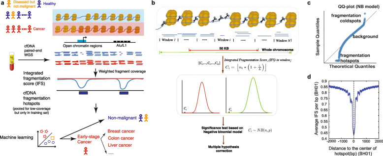
We proposed a computational approach to de novo characterize the fine-scale genomic regions with higher fragmentation rates than the local and global backgrounds, defined as cfDNA fragmentation hotspots (Fig. 1 a, b). Since both fragment coverages and sizes are essential parts of evaluating the fragmentation process, we weighed the fragment coverages in each region by the ratio of average fragment sizes in the region versus that in the whole chromosome, named integrated fragmentation score (IFS) (details in the “Methods” section). The negative binomial model we proposed correctly captured the variation of IFS in the background and indicated the existence of cfDNA fragmentation hotspots (Fig. 1 c, details in the “Methods” section). We utilized both local (50kb) and global (whole chromosome) backgrounds to identify the significant hotspots, which is especially useful when the focal copy number changes exist. Since sequencing coverages are usually affected by the G+C% content, we also normalized the IFS signals with the G+C% content within the regions (details in the “Methods” section). We used the cfDNA deep WGS data (BH01, ~100X) [6] from the healthy non-pregnant individuals as the primary dataset to evaluate our approach in healthy individuals. In the BH01 dataset, we identified 138,938 cfDNA fragmentation hotspots. The IFS distributions in both BH01 and another independent dataset from a healthy individual (IH01, ~100X) showed expected depletions at the center of BH01 hotspots (Fig. 1 d, Additional file 2: Fig. S2a), suggesting that we correctly capture the genome-wide fragmentation hotspots.
Fig. 1.
The schematic of the CRAG approach. a The schematic of using cfDNA hotspots for the cancer diagnosis. b The schematic of cfDNA hotspot identification. c The Q–Q plot for the negative binomial modeling of IFS score distribution. d The distribution of IFS around the hotspots (BH01, healthy)
It is well known that the fragment coverages and lengths from next-generation sequencing, including cfDNA WGS, are affected by the sequence compositions [6, 31, 32]. To check if the depletion of IFS signals is just due to the bias of sequence compositions, we normalized the IFS signals by k-mer composition (n=2) at BH01 hotspots (details in the “Methods” section). We did not observe any change in the overall distribution of fragmentation patterns before and after the correction (Additional file 2: Fig. S2b). These results suggested that our model robustly captured the fragmentation hotspots in cfDNA WGS.
Cell-free DNA fragmentation hotspots are highly enriched in open chromatin regions and active gene-regulatory elements
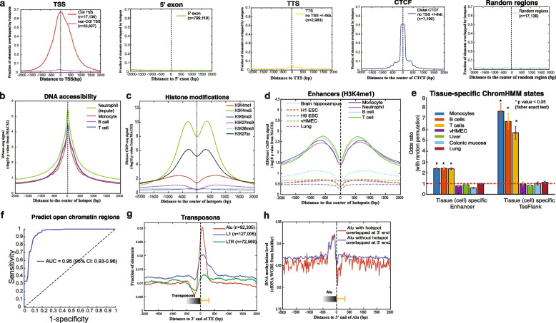
We next sought to characterize the genomic distributions of these fragmentation hotspots in healthy individuals (BH01). Similar to the previous studies on the open chromatin regions [33], the fragmentation hotspots from cfDNA are highly enriched at the CpG island (CGI) promoters and CTCF insulators, but not enriched at the non-CGI promoters, 5′ exon boundaries, transcription termination sites (TTSs), and random genomic regions (Fig. 2 a). We plotted the distributions of publicly available DNA accessibility and active/repressive histone modification marks from the major hematopoietic cell types around the hotspots. We found the high enrichment of epigenetic marks related to the active regulatory element as expected (Fig. 2 b, c, Additional file 2: Fig. S3-S4). Moreover, the enhancer mark H3K4me1, from hematopoietic cell types but not other cell types, showed a high enrichment around the hotspots, which is consistent with previous studies that hematopoietic cell types are the major contributors to cfDNA in healthy individuals [21, 34, 35] (Fig. 2 d, Additional file 2: Fig. S4). To further understand the enrichment of fragmentation hotspots at different chromatin states, we utilized the 15-state chromHMM segmentation results across different cell types from the NIH Roadmap Epigenomics Mapping Consortium [30]. The hotspots mainly showed the enrichment in the tissue/cell-type-specific chromHMM states from hematopoietic cell types but not other cell types (Fig. 2 e). The evolutionary conservation score (phastCons) in hotspots is significantly higher than in matched random regions (two-sided Mann–Whitney U test, p < 2.2e−16, Additional file 2: Fig. S5) [36], further suggesting the enrichment of the functional regulatory elements.
Fig. 2.
CfDNA fragmentation hotspots are enriched at active gene-regulatory regions in healthy. a The overlap of cfDNA fragmentation hotspots (BH01, healthy) and CGI transcription starting sites (TSSs), non-CGI TSSs, 5′exon boundary (no TSS or CTCF within ± 2 kb), transcription termination sites (TTSs) (no TSS or CTCF within ± 2 kb), CTCF transcription factor binding sites (no TSS within ± 4 kb), and random genomics regions. b The DNA accessibility levels from hematopoietic cells around the cfDNA fragmentation hotspots (BH01, healthy). c The histone modification levels from monocytes around the cfDNA fragmentation hotspots (BH01, healthy). d The H3K4me1 histone modification levels from hematopoietic (solid lines) and non-hematopoietic (dashed lines) cells around the cfDNA fragmentation hotspots (BH01, healthy). e The enrichment of hotspots at tissue-specific chromHMM states (Enhancer and TssFlank). Odds ratio is compared with matched random regions (matched chromosome and length, repeated 10 times). The error bar is based on the 95% confidence interval. p-value is calculated based on the Fisher exact test. f The receiver operating characteristic (ROC) curve for the prediction of open chromatin regions by using cfDNA fragmentation level at the hotspots from the constitutively open and closed regions. g The overlap of cfDNA fragmentation hotspots (BH01, healthy) and 3′end of transposons (Alu, L1, and LTR). h The cfDNA methylation level from healthy individuals (Sun et al. 2015 PNAS) [21] around the 3′end of Alu that overlapped or not overlapped with the cfDNA fragmentation hotspots (BH01, healthy)
We further asked if we could predict the open chromatin regions by using fragmentation alone. Neutrophils are one of the major contributors to the cfDNA in healthy individuals (20–60%) [21, 35]. However, the open chromatin regions in neutrophils are still missing. Thus, we utilized the matched constitutively open regions and closed regions across different cell types to benchmark the accuracy that we can detect the open chromatin regions by the fragmentation level. We achieved the 0.95 (95% CI: 0.93–0.96) area under the curve (AUC) to predict the known open chromatin regions (Fig. 2 f, details in the “Methods” section), further suggesting the strong link between fragmentation hotspots and open chromatin regions.
We next asked if we could detect other unknown regulatory potentials from cfDNA fragmentation hotspots. We collected 523 publicly available open chromatin region datasets measured by DNase-seq or ATAC-seq across different cell types (details in Additional file 1: Table S1). These cell types are the major known contributors to cfDNA in healthy non-pregnant individuals, including liver and rest or activated immune cells from the Roadmap Epigenomics Consortium, ENCODE, BLUEPRINT, and other publications [16, 30, 37–39]. Interestingly, after excluding the potential overlap with all these known open chromatin regions, we noticed a high enrichment of hotspots not within but right after the 3′ end of transposable elements (TEs). To exclude the possible artifact of reads mapping caused by the sequence composition bias, we examined the distribution of mappability and G+C% content right after the 3′ end of TEs and did not notice the significant bias there (Fig. 2 g, Additional file 2: Fig. S6a, b). The motif enrichment results at these hotspots right after the 3′end of TEs further suggested the high enrichment of pioneer transcription factors, such as OCT (POU, Homeobox), which usually bind the nucleosome-occupied regions (Additional file 2: Fig. S6c) [40]. Moreover, we observed the differences in DNA methylation at the same regions (right after the 3′end of Alu) with or without the overlap of hotspots, which indicates the potential functional association between hotspots and the local epigenetic status, besides nucleosome occupancy, after the 3′end of TEs (Fig. 2 h).
Taken together, in healthy individuals, these de novo characterized cfDNA fragmentation hotspots are highly enriched in open chromatin regions and active gene-regulatory elements and can potentially reveal other unknown regulatory elements from cfDNA WGS.
Cell-free DNA fragmentation hotspots reveal the potential gene-regulatory aberrations in early-stage cancer
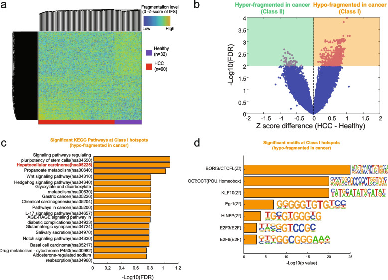
We next sought to explore whether or not the cfDNA fragmentation dynamics in the hotspots can reflect the aberrations of gene-regulatory elements in early-stage cancer. We collected the publicly available low-coverage cfDNA WGS (~1X/sample) from 90 patients with early-stage hepatocellular carcinoma (HCC, 85 of them are Barcelona Clinic Liver Cancer stage A, 5 of them are stage B) and 32 healthy individuals from the same study [41, 42]. Since these cfDNA WGS are all sequenced with low coverage, we estimated the minimum number of fragments required by CRAG (see Supplementary Methods and Additional file 2: Fig. S1) and pooled these low-coverage cfDNA WGS to obtain enough fragments for the hotspot calling in each condition. The unsupervised hierarchical clustering of the top 10,000 most variable hotspots showed a clear fragmentation dynamic between early-stage HCC and healthy (Fig. 3 a, Additional file 2: Fig. S7). The volcano plot of the false discovery rate (FDR, two-sample t-test) and z-score difference of IFS between HCC and healthy across all the fragmentation hotspots showed a large fraction of hypo-fragmented hotspots in early-stage HCC (Fig. 3 b).
Fig. 3.
The aberrations of cfDNA fragmentation patterns at hotspots in early-stage liver cancer. a Unsupervised clustering on the z-score of IFS at the top 10,000 most variable cfDNA fragmentation hotspots called from HCC and healthy samples. Hotspots are selected based on the variance of the IFS values across all the samples. b Volcano plot of z-score differences and −log10 FDR-value (two-way Student’s t-test) for the aberration of IFS in cfDNA fragmentation hotspots between early-stage HCC and healthy. c The significant KEGG pathways for the genes associated with class I hotspots. Cistrome-GO was utilized to associate hotspots with their targeted genes. d The motifs that are significantly enriched within class I hotspots
To further understand the molecular mechanism behind these fragmentation aberrations at the hotspots, we split the significantly differentiated fragmentation hotspots (FDR<0.01) into two groups: class I (hypo-fragmented in cancer) and class II (hyper-fragmented in cancer) (Fig. 3 b, Additional file 1: Table S3). We associated these hotspots with their targeted genes by CistromeGO and identified the enrichment of Gene Ontology Biological Processes (GO BPs) at these genes (Additional file 2: Fig. S8) [43]. Genes associated with class I hotspots are enriched in “cell adhesion” related GO BPs. For example, epithelial cell adhesion molecule (EpCAM) genes within the GO: 0098742 were considered the marker of HCC cancer stem cells for a long time [44, 45]. Genes associated with class I hotspots are relatively enriched in “cysteine endopeptidases,” “apoptosis,” and “purine biosynthesis” related GO BPs, which were all associated with cancer progression and invasion in the previous studies [46, 47]. We also characterize the KEGG pathway enrichment at the class I hotspots (hypo-fragmented in cancer), which are highly associated with HCC initiations, such as hepatocellular carcinoma (hsa05225) and signal pathway regulating pluripotency of stem cells (hsa04550) (Fig. 3 c). Interestingly, we found the motif enrichment of BORIS/CTCFL at the class I hotspots (hypo-fragmented in cancer) but not at the class II hotspots (hyper-fragmented in cancer) (Fig. 3 d), which suggested the potential associations with the changes in three-dimensional chromatin organizations.
To understand the cell type specificity of these cancer-specific hotspots, we performed the enrichment analysis at chromatin states from different cell types. In the Epigenome Roadmap studies, Enhancer and TssFlank are considered to be mostly cell-type-specific [30]. Compared to class I hotspots (hypo-fragmented in HCC, i.e., open in healthy), we found that the class II hotspots (hyper-fragmented in HCC, i.e., open in HCC) are significantly enriched in cell-type-specific chromHMM states from liver and liver cancer (HepG2) but not other cell types (Additional file 2: Fig. S9a).
In summary, in early-stage cancer, we found the global aberrations of fragmentation patterns at the cfDNA fragmentation hotspots, which bring together the signals mostly from peripheral immune cells and potentially small fractions from tumor tissues. These aberrations at the hotspots are highly associated with the alterations of regulatory elements and genes related to the initiation of cancer.
Cell-free DNA fragmentation hotspots for the detection and localization of multiple early-stage cancers
Next, we asked if we could utilize the cfDNA fragmentation hotspots for the diagnosis of early-stage cancer. The diagnosis of early-stage HCC is usually compared with not only healthy individuals but also patients with liver diseases. Thus, we collected additional cfDNA WGS datasets in 67 patients with chronic HBV infection and 36 patients with HBV-associated liver cirrhosis from the same study as above [41]. Unsupervised hierarchical clustering at the most variable hotspots showed the clear dynamics of the fragmentation patterns among early-stage HCC, HBV, cirrhosis, and healthy controls (Additional file 2: Fig. S10-11). Utilizing the ten-fold cross-validation, we identified hotspots only in the samples pooled in the training dataset to avoid the information leak to the test dataset. We utilized the z-score-transformed IFS from the cfDNA fragmentation hotspots as the features for the classification by a linear support vector machine (SVM) approach (details in the “Methods” section). Overall, for the comparison between HCC and healthy, we obtained 91% sensitivity at 100% specificity (96% sensitivity at 100% specificity after GC bias correction) (Additional file 1: Table S4-5, Additional file 2: Fig. S12). For the comparison between HCC and all other non-cancer controls, we obtained 83% sensitivity at 100% specificity (88% sensitivity at 100% specificity after GC bias correction). Both comparisons showed higher performance than other methods, especially fragmentation at known regulatory elements, from the same dataset with the same data split (Additional file 2: Fig. S13, Additional file 1: Table S6-7).
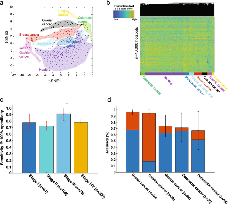
We further extended our study from early-stage HCC to multiple other cancer types. We collected publicly available low-coverage cfDNA WGS data (~1X/sample) from 208 patients across seven different kinds of cancer (88% in stages I–III, colon, breast, lung, gastric, bile duct, ovary, and pancreatic cancer) and 215 healthy controls in the same study [2, 42]. We applied a similar strategy as the HCC study above for the hotspot calling (pool the samples in the training dataset). Across seven different types of cancer and healthy conditions, the z-score-transformed IFS signals in the most variable fragmentation hotspots showed clear cancer-specific fragmentation patterns in both t-SNE visualization and unsupervised hierarchical clustering (Fig. 4 a, b, Additional file 2: Fig. S14, details in Supplementary Methods). We also performed the enrichment analysis at chromatin states similar to that in HCC above. Compared to class I hotspots (hypo-fragmented in lung cancer, i.e., open in healthy), we found that the class II hotspots (hyper-fragmented in lung cancer, i.e., open in cancer) are significantly enriched in cell-type-specific chromHMM states from lung cancer cells (A549) (Additional file 2: Fig. S9b).
Fig. 4.
The detection and localization of multiple early-stage cancers. a t-SNE visualization on the z-score of IFS (after GC bias correction) at the most variable cfDNA fragmentation hotspots (one-way ANOVA test with p-value < 0.01) across multiple different early-stage cancer types and healthy conditions. b Unsupervised clustering on z-score of IFS (after GC bias correction) at the top 40,000 most variable cfDNA fragmentation hotspots across multiple different early-stage cancer types and healthy conditions. c The sensitivity across different cancer stages at 100% specificity to distinguish cancer and healthy condition by using IFS (after GC bias correction) at cfDNA fragmentation hotspots. Error bars represent 95% confidence intervals. d Percentages of patients that were correctly classified by one of the two most likely types (sum of orange and blue bars) or the most likely type (blue bar). Error bars represent 95% confidence intervals
By ten-fold cross-validation, the linear SVM model showed a consistent high classification performance across different stages for its high sensitivity at high specificity (72% sensitivity to 91% sensitivity at 100% specificity, Fig. 4 c, Additional file 1: Table S8). Across different cancer types, we achieved 48 to 95% sensitivity at 100% specificity. Particularly, at 100% specificity, we achieved 95% sensitivity (95% CI: 85–100%) in colorectal cancer, 93% sensitivity (95% CI: 85–100%) in breast cancer, and 90% sensitivity (95% CI: 80–100%) in gastric cancer, which of these are poorly detected at high specificity level by other liquid biopsy approaches in the same dataset [2, 48–51] (Additional file 2: Fig. S15-16, Additional file 1: Table S8, Additional file 1: Table S9). In the other cancer types, the performance is largely comparable to the previous results [2]. Since the cfDNA cancer diagnosis is mostly affected by the tumor fractions, even in early-stage cancers, we estimated the tumor fractions in each sample by the copy number based approach (ichorCNA) [52]. A recent study suggested that the increases in signal breadth could increase the sensitivity even with a low tumor burden in cfDNA [53]. De novo hotspot characterization will expand the signals and thus increase the sensitivity. Our approach indeed showed a consistently high performance across samples with different tumor fractions (Additional file 2: Fig. S17).
To validate the model performance in an independent test dataset, we generated low-coverage cfDNA WGS at plasma from two types of cancers: early-stage HCC (n=8, stages I–II) and breast cancer (n=25, stages I–III), together with their matched healthy controls (1:1 matched age, gender, alcohol usage, and smoking history, meta-data details in Additional file 1: Table S2). We utilized the model trained in Cristiano 2019 data (breast vs. healthy) and Jiang 2015 data (HCC vs. healthy) and applied it directly on the test set. We obtained high performance in both independent datasets and showed superior performance to other approaches, especially known regulatory elements, in the same test sets (Additional file 2: Fig. S18, Additional file 1: Table S10). However, the ROC curve could show perfect separation between cancer and non-cancer controls in the test set but fail to validate either the sensitivity or the specificity based on the fixed cut-off when applying to the clinical cases. To further validate the clinical application of our approach, we performed the batch effect correction at the training and test sets (details in Supplementary Methods, Additional file 2: Fig. S19). We fixed the cut-off of our model in the training set of the training dataset and compared the performance of the model at the validation set in the training dataset and the independent test set. Our model still showed high performance in the test dataset (87.5% sensitivity at 87.5% specificity in early-stage liver cancer and 56% sensitivity at 80% specificity in early-stage breast cancer) (Additional file 1: Table S11).
Next, we asked whether or not we could identify the tissues-of-origin of cancer samples by using the fine-scale fragmentation levels alone. In the cancer-positive samples identified above by the machine learning algorithm, without any clinical information about the patients, we further localized the sources of cancer to one or two anatomic sites in a mean of 80% of these patients across five different cancer types and 76% accuracy across six different cancer types. Furthermore, we were able to localize the source of the positive test to a single organ in a median of 62% of these patients (Fig. 4 d, Additional file 1: Table S12, Additional file 2: Fig. S20) (details in the “Methods” section). The prediction accuracy varies among tumor types, from 67% (95% CI: 36–97%) in pancreatic cancer to 97% (95% CI: 92–100%) in breast cancer (Fig. 4 d and Additional file 1: Table S12), but significantly higher than random choices by the frequency of samples in each cancer type (Additional file 2: Fig. S21).
Overall, our proof-of-concept study on the publicly available cfDNA WGS dataset suggested that the de novo characterization of cfDNA fragmentation hotspots is a promising novel approach for the diagnosis and localization of multiple early-stage cancers.
Discussion
In summary, we developed a computational approach, named CRAG, to de novo identify the cfDNA fragmentation hotspots by weighting fragment coverages with the fragment size information. The cfDNA fragmentation hotspots are highly enriched at open chromatin regions and active gene-regulatory elements. While in early-stage cancers, a significant proportion of these hotspots are hypo-fragmented. These hypo-fragmented hotspots in early-stage cancer are mostly enriched in GO terms and pathways related to the initiation of cancer, which further suggests the functional importance of these cancer-specific hypo-fragmented hotspots. In addition, the BORIS/CTCFL motif is enriched at these hypo-fragmented hotspots, which suggests the potential three-dimensional chromatin organization changes during the initiation of early-stage cancer that has been reported before but not revealed by the non-invasive cfDNA approaches [54]. Overall, our results suggested that the de novo characterization of fine-scale cfDNA fragmentation hotspots is critical to revealing the unknown gene-regulatory aberrations in pathological conditions.
Compared to the fragmentation studies at the known regulatory elements, such as TSS and TFBS, our de novo approach shows several advantages. First, the de novo approach will expand the signal breadth. The tumor content in cfDNA is low in most early-stage cancers. Recent studies suggested that the increase of signal breadth could increase the sensitivity even with a low tumor burden in cfDNA [53]. The aberration of regulatory elements in cancer involves both genes and distal regulatory elements [15, 55]. De novo characterization will expand the signals from known genes’ promoters to many distal regulatory elements and thus increase the sensitivity. Second, the landscape of known regulatory elements has not been well characterized in tumor and immune cells from early-stage cancer patients yet. The initiation of early-stage cancer involves the interaction between the tumor and the immune environment [56, 57]. The chromatin accessibility landscape has recently been profiled in late-stage tumors in cancer patients and immune cells in healthy individuals [15, 16]. However, the landscape of regulatory elements from early-stage tumors and the immune environment of patients with early-stage cancers are still not known. Therefore, it is challenging to utilize comprehensive regulatory element maps in tumor and immune cells for the study of early-stage cancers. Finally, a lot of distal regulatory elements do not contain well-defined motifs or TFBS. Moreover, tissue-specific and cancer-specific TFBS are also not well-defined across many diseases, especially early-stage cancers. Characterization of motifs and TFBS from sequence directly cannot represent the whole aberration map of regulatory elements in early-stage cancers. Our benchmark results over the known regulatory elements also supported this conclusion (Additional file 2: Fig. S18, Additional file 1: Table S10).
The in vivo fragmentation process is complicated. There is a significant computational challenge to identify the fragmentation hotspots compared to the identification of fragmentation coldspots at nucleosome-occupied regions in cfDNA WGS [6]. For example, genomic regions with a higher fragmentation rate do not always indicate the open chromatin regions. Furthermore, besides nucleosomes, both biological (e.g., DNA methylation and histone modifications) [58, 59] and technical artifacts (e.g., G+C%, k-mer, and mappability) [31, 60] can affect the measurements of fragmentation level. After excluding the known effects of open chromatin regions and technical artifacts, our genome-wide analysis here revealed the enrichment of hotspots after the 3′end of transposable elements and potentially associated with local DNA methylation level, which suggested the unknown origin of the cfDNA fragmentation processes.
Previous efforts had been made to characterize the nucleosome-free regions by using the depletion of coverages from MNase-seq/ChIP-seq assay [61]. The measurement of cfDNA fragmentation here, however, involves information from both fragment coverages and sizes. CRAG can be further improved by better integrating the fragment coverages and sizes, or even with more dimensions, such as the fragment orientation, jagged ends, and endpoint, to fully capture the spectrum of fragmentation. Also, G+C% bias is known to affect the peak calling result in ChIP-seq/ATAC-seq [62]. A better statistical model with the incorporation of GC normalization on both the fragment coverages and sizes will improve our method’s performance. PCR-free library preparation for WGS will also mitigate the concerns of GC bias and other sequencing artifacts [63].
Our study here on the detection and localization of early-stage cancer is still in the proof-of-concept stage. There are still several limitations. First, due to the limited number of publicly available early-stage cancer cfDNA datasets, the classification performance here is mainly evaluated by multi-fold cross-validation on a relatively small sample size cohort in each cancer type without strictly matched healthy controls, similar to other cfDNA WGS studies [2]. We only generated small-scale datasets from two cancer types for independent validations. Multiple independent large-scale prospective cohorts with strictly matched controls will be a better way to assess the power of our approach for the diagnosis of early-stage cancer. Second, previous studies suggested that pre-analytic differences in the patient populations could bring the artifact for fragmentomic studies and finally affect the diagnosis performance [64–69]. Unified sample collection, experimental workflow, and better computational approaches to adjust these cofounders are still needed. Third, we pooled the low-coverage WGS samples from the same condition for the hotspot calling, which may cause a problem with a small number of samples. Due to the random dropout of the fragment coverages and many genomic windows in the genome, the number of falsely discovered hotspots without any biological interpretations will increase. Our current strategy by filtering low mappability regions and correcting GC bias is helpful to reduce the false-positive rate for the hotspot detection. However, the accuracy of IFS signals at individual hotspots from each sample is still severely affected by the low-coverage data. Recent efforts showed the possibility to integrate genome-wide mutational patterns at low-coverage WGS to enable the ultra-sensitive detection of cancer samples with low tumor burden [53], which is similar to our strategy for the IFS signals at low-coverage samples here. Since we narrow down the regions of interest, even with missing values at part of the loci, many other hotspots from the same sample will still provide informative signals rather than noises for the model to make the classifications. In the future, appropriate statistical models for the imputation of missing fragmentation patterns are still needed to mitigate the missing data problem. Lastly, in some cancer types, our fine-scale study here showed complementary classification performance compared with that in the previous large-scale fragmentation study in the same dataset [2]. For example, our results on gastric, breast, and colorectal cancer outperformed previous large-scale fragmentation studies, while for bile duct, pancreatic, and lung cancer, the performance is reversed. Future combinations of the fragmentation patterns at multi-scales and information from other modalities or clinical meta-data may further improve the performance.
Conclusions
Our study here provides a de novo approach to non-invasively detect multiple early-stage cancers simultaneously on an existing matured high-throughput platform in a cost-effective way. It also paves the road to further elucidate the unknown gene-regulatory mechanisms in pathological conditions through the cfDNA fragmentation hotspots.
Supplementary Information
Additional file 1: Supplementary Tables. All the supplementary tables in the study.
Additional file 2: Supplementary Methods and Figures. Supplementary Methods and all Supplementary Figures in the study.
Acknowledgements
The authors greatly acknowledge Dr. Yuk Ming Dennis Lo and his circulating nucleic acids research group in the Chinese University of Hong Kong, Dr. Jay Shendure and his research group in the University of Washington, and Drs. Robert B. Scharpf and Victor E. Velculescu and their research group in the Johns Hopkins University School of Medicine for their cfDNA data. The authors acknowledge Dr. Li Wang for her suggestions and comments on the manuscript. The authors also acknowledge Jeanette M. Buckholz for the significant effort she invested in locating the study subjects. This work is supported by the computational resources from Biomedical Informatics (BMI) high-performance computing cluster in CCHMC.
Authors’ contributions
Y.L. conceived the study. Y.L. and X.Z. designed the methodological framework. X.Z. implemented the methods in MATLAB. H.Z. implemented the methods in R. X.Z., H.Z., and Y.L. performed the data analysis. H.F. generated the cfDNA WGS data for validation. K.L. D. M, S.M.P., and H.F. coordinated the plasma samples and provided input for the study design of the validation. Y.L., X.Z., H.Z, and H.F. wrote the manuscript together. All the authors read and approved the final manuscript.
Funding
This work was supported by the CCHMC start-up grant, Trustee Award, CCTST mentored pilot translational award, CCHMC innovation grant, and R56HG012360 from NHGRI to Y.L. This work also used the Extreme Science and Engineering Discovery Environment (XSEDE), which is supported by the National Science Foundation grant number ACI-1548562. This work used the XSEDE at the Pittsburgh Supercomputing Center (PSC) through allocation MCB190124P and MCB190006P. The authors acknowledge the Fernald infrastructure grant (R24ES028527 and P30ES006096) from NIEHS for the maintenance of the data and biospecimens, which were used as our validation samples.
Availability of data and materials
All the public datasets used in this study are listed with detailed information and URLs in Additional file 1: Table S1. The raw sequencing data (validation dataset generated by us) are deposited at dbGap with controlled access (dbGap id: phs003062.v1.p1, https://www.ncbi.nlm.nih.gov/projects/gap/cgi-bin/study.cgi?study_id=phs003062.v1.p1) [70]. The de-identified fragment files mapped to hg19 are available at Zenodo.org (breast.tar and liver.tar, DOI: 10.5281/zenodo.6914806) [71]. All data needed to evaluate the conclusions in the paper are present in the paper and/or the Supplementary Materials. CRAG is implemented in MATLAB and R (v4.2.0). The source code is freely available on GitHub (https://github.com/epifluidlab/cragr.git) (R version) [72] and (https://github.com/epifluidlab/CRAG.git) (MATLAB version) [73] under the MIT license for academic researchers. The source code, readme files, hotspot location, intermediate analysis scripts, and intermediate results are available at Zenodo.org (10.5281/zenodo.6914806) [71].
Declarations
Ethics approval and consent to participate
This research study was approved by the Cincinnati Children’s Hospital Medical Center Institutional Review Board (#2019-0601) in accordance with the Declaration of Helsinki. De-identified plasma sample collection was approved by the University of Cincinnati Institutional Review Board (2012-3923 and 2012-3745). All participants provided written informed consent to participate.
Consent for publication
Not applicable.
Competing interests
A PCT patent (Y.L., X.Z., and H.Z.) was filed by Cincinnati Children’s Hospital Medical Center. Y.L. owns stocks from Freenome Inc. The remaining authors declare that they have no competing interests.
Footnotes
Publisher’s Note
Springer Nature remains neutral with regard to jurisdictional claims in published maps and institutional affiliations.
Xionghui Zhou, Haizi Zheng, and Hailu Fu contributed equally.
References
- 1.Heitzer E, Haque IS, Roberts CES, Speicher MR. Current and future perspectives of liquid biopsies in genomics-driven oncology. Nat Rev Genet. 2019;20:71–88. doi: 10.1038/s41576-018-0071-5. [DOI] [PubMed] [Google Scholar]
- 2.Cristiano S, Leal A, Phallen J, Fiksel J, Adleff V, Bruhm DC, et al. Genome-wide cell-free DNA fragmentation in patients with cancer. Nature. 2019;570:385–389. doi: 10.1038/s41586-019-1272-6. [DOI] [PMC free article] [PubMed] [Google Scholar]
- 3.Chabon JJ, Hamilton EG, Kurtz DM, Esfahani MS, Moding EJ, Stehr H, et al. Integrating genomic features for non-invasive early lung cancer detection. Nature. 2020;580:245–251. doi: 10.1038/s41586-020-2140-0. [DOI] [PMC free article] [PubMed] [Google Scholar]
- 4.Mouliere F, Robert B, Arnau Peyrotte E, Del Rio M, Ychou M, Molina F, et al. High fragmentation characterizes tumour-derived circulating DNA. PLoS One. 2011;6:e23418. doi: 10.1371/journal.pone.0023418. [DOI] [PMC free article] [PubMed] [Google Scholar]
- 5.Liu Y. At the dawn: cell-free DNA fragmentomics and gene regulation. Br J Cancer. 2022;126:379–390. doi: 10.1038/s41416-021-01635-z. [DOI] [PMC free article] [PubMed] [Google Scholar]
- 6.Snyder MW, Kircher M, Hill AJ, Daza RM, Shendure J. Cell-free DNA comprises an in vivo nucleosome footprint that informs its tissues-of-origin. Cell. 2016;164:57–68. doi: 10.1016/j.cell.2015.11.050. [DOI] [PMC free article] [PubMed] [Google Scholar]
- 7.Ulz P, Thallinger GG, Auer M, Graf R, Kashofer K, Jahn SW, et al. Inferring expressed genes by whole-genome sequencing of plasma DNA. Nat Genet. 2016;48:1273–1278. doi: 10.1038/ng.3648. [DOI] [PubMed] [Google Scholar]
- 8.Jiang P, Sun K, Tong YK, Cheng SH, Cheng THT, Heung MMS, et al. Preferred end coordinates and somatic variants as signatures of circulating tumor DNA associated with hepatocellular carcinoma. Proc Natl Acad Sci U S A. 2018;115:E10925–E10933. doi: 10.1073/pnas.1814616115. [DOI] [PMC free article] [PubMed] [Google Scholar]
- 9.Sun K, Jiang P, Cheng SH, Cheng THT, Wong J, Wong VWS, et al. Orientation-aware plasma cell-free DNA fragmentation analysis in open chromatin regions informs tissue of origin. Genome Res. 2019;29:418–427. doi: 10.1101/gr.242719.118. [DOI] [PMC free article] [PubMed] [Google Scholar]
- 10.Ulz P, Perakis S, Zhou Q, Moser T, Belic J, Lazzeri I, et al. Inference of transcription factor binding from cell-free DNA enables tumor subtype prediction and early detection. Nat Commun. 2019;10:4666. doi: 10.1038/s41467-019-12714-4. [DOI] [PMC free article] [PubMed] [Google Scholar]
- 11.Jiang P, Sun K, Peng W, Cheng SH, Ni M, Yeung PC, et al. Plasma DNA end-motif profiling as a fragmentomic marker in cancer, pregnancy, and transplantation. Cancer Discov. 2020;10:664–673. doi: 10.1158/2159-8290.CD-19-0622. [DOI] [PubMed] [Google Scholar]
- 12.Zhu G, Guo YA, Ho D, Poon P, Poh ZW, Wong PM, et al. Tissue-specific cell-free DNA degradation quantifies circulating tumor DNA burden. Nat Commun. 2021;12:2229. doi: 10.1038/s41467-021-22463-y. [DOI] [PMC free article] [PubMed] [Google Scholar]
- 13.Peneder P, Stütz AM, Surdez D, Krumbholz M, Semper S, Chicard M, et al. Multimodal analysis of cell-free DNA whole-genome sequencing for pediatric cancers with low mutational burden. Nat Commun. 2021;12:3230. doi: 10.1038/s41467-021-23445-w. [DOI] [PMC free article] [PubMed] [Google Scholar]
- 14.Mathios D, Johansen JS, Cristiano S, Medina JE, Phallen J, Larsen KR, et al. Detection and characterization of lung cancer using cell-free DNA fragmentomes. Nat Commun. 2021;12:5060. doi: 10.1038/s41467-021-24994-w. [DOI] [PMC free article] [PubMed] [Google Scholar]
- 15.Corces MR, Granja JM, Shams S, Louie BH, Seoane JA, Zhou W, et al. The chromatin accessibility landscape of primary human cancers. Science. 2018:362. 10.1126/science.aav1898. [DOI] [PMC free article] [PubMed]
- 16.Calderon D, Nguyen MLT, Mezger A, Kathiria A, Müller F, Nguyen V, et al. Landscape of stimulation-responsive chromatin across diverse human immune cells. Nat Genet. 2019;51:1494–1505. doi: 10.1038/s41588-019-0505-9. [DOI] [PMC free article] [PubMed] [Google Scholar]
- 17.Lambert SA, Jolma A, Campitelli LF, Das PK, Yin Y, Albu M, et al. The human transcription factors. Cell. 2018;172:650–665. doi: 10.1016/j.cell.2018.01.029. [DOI] [PubMed] [Google Scholar]
- 18.Bolger AM, Lohse M, Usadel B. Trimmomatic: a flexible trimmer for Illumina sequence data. Bioinformatics. 2014;30:2114–2120. doi: 10.1093/bioinformatics/btu170. [DOI] [PMC free article] [PubMed] [Google Scholar]
- 19.Li H, Durbin R. Fast and accurate short read alignment with burrows-wheeler transform. Bioinformatics. 2009;25:1754–1760. doi: 10.1093/bioinformatics/btp324. [DOI] [PMC free article] [PubMed] [Google Scholar]
- 20.Faust GG, Hall IM. SAMBLASTER: fast duplicate marking and structural variant read extraction. Bioinformatics. 2014;30:2503–2505. doi: 10.1093/bioinformatics/btu314. [DOI] [PMC free article] [PubMed] [Google Scholar]
- 21.Sun K, Jiang P, Chan KCA, Wong J, Cheng YKY, Liang RHS, et al. Plasma DNA tissue mapping by genome-wide methylation sequencing for noninvasive prenatal, cancer, and transplantation assessments. Proc Natl Acad Sci U S A. 2015;112:E5503–E5512. doi: 10.1073/pnas.1508736112. [DOI] [PMC free article] [PubMed] [Google Scholar]
- 22.Chan KCA, Jiang P, Chan CWM, Sun K, Wong J, Hui EP, et al. Noninvasive detection of cancer-associated genome-wide hypomethylation and copy number aberrations by plasma DNA bisulfite sequencing. Proc Natl Acad Sci U S A. 2013;110:18761–18768. doi: 10.1073/pnas.1313995110. [DOI] [PMC free article] [PubMed] [Google Scholar]
- 23.Krueger F. Trim galore. A wrapper tool around Cutadapt and FastQC to consistently apply quality and adapter trimming to FastQ files. 2015;516:517.
- 24.Martin M. Cutadapt removes adapter sequences from high-throughput sequencing reads. EMBnet J EMBnet Stichting. 2011;17:10. doi: 10.14806/ej.17.1.200. [DOI] [Google Scholar]
- 25.Li H, Handsaker B, Wysoker A, Fennell T, Ruan J, Homer N, et al. The sequence alignment/map format and SAMtools. Bioinformatics. 2009;25:2078–2079. doi: 10.1093/bioinformatics/btp352. [DOI] [PMC free article] [PubMed] [Google Scholar]
- 26.Liu Y, Siegmund KD, Laird PW, Berman BP. Bis-SNP: combined DNA methylation and SNP calling for bisulfite-seq data. Genome Biol. 2012;13:R61. doi: 10.1186/gb-2012-13-7-r61. [DOI] [PMC free article] [PubMed] [Google Scholar]
- 27.Zhang Y, Liu T, Meyer CA, Eeckhoute J, Johnson DS, Bernstein BE, et al. Model-based analysis of ChIP-Seq (MACS) Genome Biol. 2008;9:R137. doi: 10.1186/gb-2008-9-9-r137. [DOI] [PMC free article] [PubMed] [Google Scholar]
- 28.Derrien T, Estellé J, Sola SM, Knowles DG, Raineri E, Guigó R, et al. Fast computation and applications of genome mappability. PLoS One. 2012;7:e30377. doi: 10.1371/journal.pone.0030377. [DOI] [PMC free article] [PubMed] [Google Scholar]
- 29.Ji H, Jiang H, Ma W, Johnson DS, Myers RM, Wong WH. An integrated software system for analyzing ChIP-chip and ChIP-seq data. Nat Biotechnol. 2008;26:1293–1300. doi: 10.1038/nbt.1505. [DOI] [PMC free article] [PubMed] [Google Scholar]
- 30.Roadmap Epigenomics Consortium. Kundaje A, Meuleman W, Ernst J, Bilenky M, Yen A, et al. Integrative analysis of 111 reference human epigenomes. Nature. 2015;518:317–330. doi: 10.1038/nature14248. [DOI] [PMC free article] [PubMed] [Google Scholar]
- 31.Benjamini Y, Speed TP. Summarizing and correcting the GC content bias in high-throughput sequencing. Nucleic Acids Res. 2012;40:e72. doi: 10.1093/nar/gks001. [DOI] [PMC free article] [PubMed] [Google Scholar]
- 32.Chandrananda D, Thorne NP, Ganesamoorthy D, Bruno DL, Benjamini Y, Speed TP, et al. Investigating and correcting plasma DNA sequencing coverage bias to enhance aneuploidy discovery. PLoS One. 2014;9:e86993. doi: 10.1371/journal.pone.0086993. [DOI] [PMC free article] [PubMed] [Google Scholar]
- 33.Buenrostro JD, Giresi PG, Zaba LC, Chang HY, Greenleaf WJ. Transposition of native chromatin for fast and sensitive epigenomic profiling of open chromatin, DNA-binding proteins and nucleosome position. Nat Methods. 2013;10:1213–1218. doi: 10.1038/nmeth.2688. [DOI] [PMC free article] [PubMed] [Google Scholar]
- 34.Lui YYN, Chik K-W, Chiu RWK, Ho C-Y, Lam CWK, Lo YMD. Predominant hematopoietic origin of cell-free DNA in plasma and serum after sex-mismatched bone marrow transplantation. Clin Chem. 2002;48:421–427. doi: 10.1093/clinchem/48.3.421. [DOI] [PubMed] [Google Scholar]
- 35.Moss J, Magenheim J, Neiman D, Zemmour H, Loyfer N, Korach A, et al. Comprehensive human cell-type methylation atlas reveals origins of circulating cell-free DNA in health and disease. Nat Commun. 2018;9:5068. doi: 10.1038/s41467-018-07466-6. [DOI] [PMC free article] [PubMed] [Google Scholar]
- 36.Siepel A, Bejerano G, Pedersen JS, Hinrichs AS, Hou M, Rosenbloom K, et al. Evolutionarily conserved elements in vertebrate, insect, worm, and yeast genomes. Genome Res. 2005;15:1034–1050. doi: 10.1101/gr.3715005. [DOI] [PMC free article] [PubMed] [Google Scholar]
- 37.ENCODE Project Consortium An integrated encyclopedia of DNA elements in the human genome. Nature. 2012;489:57–74. doi: 10.1038/nature11247. [DOI] [PMC free article] [PubMed] [Google Scholar]
- 38.Stunnenberg HG, International Human Epigenome Consortium, Hirst M The international human Epigenome consortium: a blueprint for scientific collaboration and discovery. Cell. 2016;167:1145–1149. doi: 10.1016/j.cell.2016.11.007. [DOI] [PubMed] [Google Scholar]
- 39.Corces MR, Buenrostro JD, Wu B, Greenside PG, Chan SM, Koenig JL, et al. Lineage-specific and single-cell chromatin accessibility charts human hematopoiesis and leukemia evolution. Nat Genet. 2016;48:1193–1203. doi: 10.1038/ng.3646. [DOI] [PMC free article] [PubMed] [Google Scholar]
- 40.Soufi A, Garcia MF, Jaroszewicz A, Osman N, Pellegrini M, Zaret KS. Pioneer transcription factors target partial DNA motifs on nucleosomes to initiate reprogramming. Cell. 2015;161:555–568. doi: 10.1016/j.cell.2015.03.017. [DOI] [PMC free article] [PubMed] [Google Scholar]
- 41.Jiang P, Chan CWM, Chan KCA, Cheng SH, Wong J, Wong VW-S, et al. Lengthening and shortening of plasma DNA in hepatocellular carcinoma patients. Proc Natl Acad Sci U S A. 2015;112:E1317–E1325. doi: 10.1073/pnas.1500076112. [DOI] [PMC free article] [PubMed] [Google Scholar]
- 42.Zheng H, Zhu MS, Liu Y. FinaleDB: a browser and database of cell-free DNA fragmentation patterns. Bioinformatics. 2021;37:2502–2503. doi: 10.1093/bioinformatics/btaa999. [DOI] [PMC free article] [PubMed] [Google Scholar]
- 43.Li S, Wan C, Zheng R, Fan J, Dong X, Meyer CA, et al. Cistrome-GO: a web server for functional enrichment analysis of transcription factor ChIP-seq peaks. Nucleic Acids Res. 2019;47:W206–W211. doi: 10.1093/nar/gkz332. [DOI] [PMC free article] [PubMed] [Google Scholar]
- 44.Yamashita T, Ji J, Budhu A, Forgues M, Yang W, Wang H-Y, et al. EpCAM-positive hepatocellular carcinoma cells are tumor-initiating cells with stem/progenitor cell features. Gastroenterology. 2009;136:1012–1024. doi: 10.1053/j.gastro.2008.12.004. [DOI] [PMC free article] [PubMed] [Google Scholar]
- 45.Terris B, Cavard C, Perret C. EpCAM, a new marker for cancer stem cells in hepatocellular carcinoma. J Hepatol. 2010;52:280–281. doi: 10.1016/j.jhep.2009.10.026. [DOI] [PubMed] [Google Scholar]
- 46.Kos J, Werle B, Lah T, Brunner N. Cysteine proteinases and their inhibitors in extracellular fluids: markers for diagnosis and prognosis in cancer. Int J Biol Markers. 2000;15:84–89. doi: 10.1177/172460080001500116. [DOI] [PubMed] [Google Scholar]
- 47.Su W-J, Lu P-Z, Wu Y, Kalpana K, Yang C-K, Lu G-D. Identification of key genes in purine metabolism as prognostic biomarker for hepatocellular carcinoma. Front Oncol. 2020;10:583053. doi: 10.3389/fonc.2020.583053. [DOI] [PMC free article] [PubMed] [Google Scholar]
- 48.Cohen JD, Li L, Wang Y, Thoburn C, Afsari B, Danilova L, et al. Detection and localization of surgically resectable cancers with a multi-analyte blood test. Science. 2018;359:926–930. doi: 10.1126/science.aar3247. [DOI] [PMC free article] [PubMed] [Google Scholar]
- 49.Liu MC, Oxnard GR, Klein EA, Swanton C, Seiden MV, Liu MC, et al. Sensitive and specific multi-cancer detection and localization using methylation signatures in cell-free DNA. Ann Oncol. 2020; http://www.sciencedirect.com/science/article/pii/S0923753420360580. [DOI] [PMC free article] [PubMed]
- 50.Wan N, Weinberg D, Liu T-Y, Niehaus K, Ariazi EA, Delubac D, et al. Machine learning enables detection of early-stage colorectal cancer by whole-genome sequencing of plasma cell-free DNA. BMC Cancer. 2019;19:832. doi: 10.1186/s12885-019-6003-8. [DOI] [PMC free article] [PubMed] [Google Scholar]
- 51.Shen SY, Singhania R, Fehringer G, Chakravarthy A, Roehrl MHA, Chadwick D, et al. Sensitive tumour detection and classification using plasma cell-free DNA methylomes. Nature. 2018;563:579–583. doi: 10.1038/s41586-018-0703-0. [DOI] [PubMed] [Google Scholar]
- 52.Adalsteinsson VA, Ha G, Freeman SS, Choudhury AD, Stover DG, Parsons HA, et al. Scalable whole-exome sequencing of cell-free DNA reveals high concordance with metastatic tumors. Nat Commun. 2017;8:1324. doi: 10.1038/s41467-017-00965-y. [DOI] [PMC free article] [PubMed] [Google Scholar]
- 53.Zviran A, Schulman RC, Shah M, Hill STK, Deochand S, Khamnei CC, et al. Genome-wide cell-free DNA mutational integration enables ultra-sensitive cancer monitoring. Nat Med. 2020;26:1114–1124. doi: 10.1038/s41591-020-0915-3. [DOI] [PMC free article] [PubMed] [Google Scholar]
- 54.Liu EM, Martinez-Fundichely A, Diaz BJ, Aronson B, Cuykendall T, MacKay M, et al. Identification of cancer drivers at CTCF insulators in 1,962 whole genomes. Cell Syst. 2019;8:446–455.e8. doi: 10.1016/j.cels.2019.04.001. [DOI] [PMC free article] [PubMed] [Google Scholar]
- 55.Khurana E, Fu Y, Chakravarty D, Demichelis F, Rubin MA, Gerstein M. Role of non-coding sequence variants in cancer. Nat Rev Genet. 2016;17:93–108. doi: 10.1038/nrg.2015.17. [DOI] [PubMed] [Google Scholar]
- 56.Gonzalez H, Hagerling C, Werb Z. Roles of the immune system in cancer: from tumor initiation to metastatic progression. Genes Dev. 2018;32:1267–1284. doi: 10.1101/gad.314617.118. [DOI] [PMC free article] [PubMed] [Google Scholar]
- 57.Hiam-Galvez KJ, Allen BM, Spitzer MH. Systemic immunity in cancer. Nat Rev Cancer. 2021;21:345–359. doi: 10.1038/s41568-021-00347-z. [DOI] [PMC free article] [PubMed] [Google Scholar]
- 58.Jensen TJ, Kim SK, Zhu Z, Chin C, Gebhard C, Lu T, et al. Whole genome bisulfite sequencing of cell-free DNA and its cellular contributors uncovers placenta hypomethylated domains. Genome Biol. 2015;16:78. doi: 10.1186/s13059-015-0645-x. [DOI] [PMC free article] [PubMed] [Google Scholar]
- 59.Ivanov M, Baranova A, Butler T, Spellman P, Mileyko V. Non-random fragmentation patterns in circulating cell-free DNA reflect epigenetic regulation. BMC Genomics. 2015;16(Suppl 13):S1. doi: 10.1186/1471-2164-16-S13-S1. [DOI] [PMC free article] [PubMed] [Google Scholar]
- 60.Cheung M-S, Down TA, Latorre I, Ahringer J. Systematic bias in high-throughput sequencing data and its correction by BEADS. Nucleic Acids Res. 2011;39:e103. doi: 10.1093/nar/gkr425. [DOI] [PMC free article] [PubMed] [Google Scholar]
- 61.Mammana A, Vingron M, Chung H-R. Inferring nucleosome positions with their histone mark annotation from ChIP data. Bioinformatics. 2013;29:2547–2554. doi: 10.1093/bioinformatics/btt449. [DOI] [PMC free article] [PubMed] [Google Scholar]
- 62.Teng M, Irizarry RA. Accounting for GC-content bias reduces systematic errors and batch effects in ChIP-seq data. Genome Res. 2017;27:1930–1938. doi: 10.1101/gr.220673.117. [DOI] [PMC free article] [PubMed] [Google Scholar]
- 63.Aird D, Ross MG, Chen W-S, Danielsson M, Fennell T, Russ C, et al. Analyzing and minimizing PCR amplification bias in Illumina sequencing libraries. Genome Biol. 2011;12:R18. doi: 10.1186/gb-2011-12-2-r18. [DOI] [PMC free article] [PubMed] [Google Scholar]
- 64.van der Pol Y, Moldovan N, Verkuijlen S, Ramaker J, Boers D, Onstenk W, et al. The effect of preanalytical and physiological variables on cell-free DNA fragmentation. Clin Chem. 2022. 10.1093/clinchem/hvac029. [DOI] [PubMed]
- 65.Markus H, Contente-Cuomo T, Farooq M, Liang WS, Borad MJ, Sivakumar S, et al. Evaluation of pre-analytical factors affecting plasma DNA analysis. Sci Rep. 2018;8:7375. doi: 10.1038/s41598-018-25810-0. [DOI] [PMC free article] [PubMed] [Google Scholar]
- 66.Kerachian MA, Azghandi M, Mozaffari-Jovin S, Thierry AR. Guidelines for pre-analytical conditions for assessing the methylation of circulating cell-free DNA. Clin Epigenetics. 2021;13:193. doi: 10.1186/s13148-021-01182-7. [DOI] [PMC free article] [PubMed] [Google Scholar]
- 67.Krasic J, Abramovic I, Vrtaric A, Nikolac Gabaj N, Kralik-Oguic S, Katusic Bojanac A, et al. Impact of preanalytical and analytical methods on cell-free DNA diagnostics. Front Cell Dev Biol. 2021;9:686149. doi: 10.3389/fcell.2021.686149. [DOI] [PMC free article] [PubMed] [Google Scholar]
- 68.Chan KCA, Yeung S-W, Lui W-B, Rainer TH, Lo YMD. Effects of preanalytical factors on the molecular size of cell-free DNA in blood. Clin Chem. 2005;51:781–784. doi: 10.1373/clinchem.2004.046219. [DOI] [PubMed] [Google Scholar]
- 69.Lampignano R, Neumann MHD, Weber S, Kloten V, Herdean A, Voss T, et al. Multicenter evaluation of circulating cell-free DNA extraction and downstream analyses for the development of standardized (pre)analytical work flows. Clin Chem. 2020;66:149–160. doi: 10.1373/clinchem.2019.306837. [DOI] [PubMed] [Google Scholar]
- 70.Liu, Yaping. De Novo Characterization of Cell-Free DNA Fragmentation Hotspots in Plasma Whole-Genome Sequencing dbGap. Available from: https://www.ncbi.nlm.nih.gov/projects/gap/cgi-bin/study.cgi?study_id=phs003062.v1.p1. 2022. [DOI] [PMC free article] [PubMed]
- 71.Zhou, Xionghui, Zheng Haizi, Liu, Yaping. CRAG: De novo characterization of cell-free DNA fragmentation hotspots in plasma whole-genome sequencing. Zenodo.org. Available from: 10.5281/zenodo.6914806 [DOI] [PMC free article] [PubMed]
- 72.Zheng, Haizi, Liu, Yaping. CRAGR. GitHub. (2022). https://github.com/epifluidlab/cragr
- 73.Zhou, Xionghui, Liu, Yaping. CRAG. GitHub. (2022). https://github.com/epifluidlab/CRAG
Associated Data
This section collects any data citations, data availability statements, or supplementary materials included in this article.
Supplementary Materials
Additional file 1: Supplementary Tables. All the supplementary tables in the study.
Additional file 2: Supplementary Methods and Figures. Supplementary Methods and all Supplementary Figures in the study.
Data Availability Statement
All the public datasets used in this study are listed with detailed information and URLs in Additional file 1: Table S1. The raw sequencing data (validation dataset generated by us) are deposited at dbGap with controlled access (dbGap id: phs003062.v1.p1, https://www.ncbi.nlm.nih.gov/projects/gap/cgi-bin/study.cgi?study_id=phs003062.v1.p1) [70]. The de-identified fragment files mapped to hg19 are available at Zenodo.org (breast.tar and liver.tar, DOI: 10.5281/zenodo.6914806) [71]. All data needed to evaluate the conclusions in the paper are present in the paper and/or the Supplementary Materials. CRAG is implemented in MATLAB and R (v4.2.0). The source code is freely available on GitHub (https://github.com/epifluidlab/cragr.git) (R version) [72] and (https://github.com/epifluidlab/CRAG.git) (MATLAB version) [73] under the MIT license for academic researchers. The source code, readme files, hotspot location, intermediate analysis scripts, and intermediate results are available at Zenodo.org (10.5281/zenodo.6914806) [71].


