Abstract
The challenges in making animal models of complex human epilepsy phenotypes with varied aetiology highlights the need to develop alternative disease models that can address the limitations of animal models by effectively recapitulating human pathophysiology. The advances in stem cell technology provide an opportunity to use human iPSCs to make disease-in-a-dish models. The focus of this review is to report the current information and progress in the generation of epileptic patient-specific iPSCs lines, isogenic control cell lines, and neuronal models. These in vitro models can be used to study the underlying pathological mechanisms of epilepsies, anti-seizure medication resistance, and can also be used for drug testing and drug screening with their isogenic control cell lines.
Keywords: stem cell lines, epilepsy, patient-specific cell models, neuronal differentiation, personalised disease models, personalised medicine
1. Introduction
Epilepsy affects 70 million people worldwide [1,2], causing significant morbidity and mortality. with more than half of the affected people living in countries with poor medical resources and little or no access to treatment [3]. It is characterised by unprovoked, recurrent seizures which result from the disruption in the balance between neuronal excitation and inhibition in the brain [1]. In most cases the cause of epilepsy is unknown but both genetic and environmental factors are understood to contribute to its aetiology.
Genetic epilepsy is characterised by seizures that are the result of known genetic variance in one or many genes associated with epilepsy [2]. Mutations in several genes encoding ion channels and proteins have been reported to be most commonly associated with epilepsy. These genes include, but are not limited to mutation in SCN1A (encode sodium channel protein), which is associated with Dravat syndrome; KCNQ2 or KCNQ3 (both encoding potassium channel protein), which are associated with benign neuronal familial seizures [3,4,5]; the CHRNA4 gene (20q13), which is associated with Autosomal Dominant Nocturnal Frontal Lobe Epilepsy (ADNFLE), characterised by hypermotor nocturnal seizures [6]; and the mammalian target of rapamycin (mTOR) pathway, in particular DEPDC5 gene of this pathway, which are associated with Focal Cortical Dysplasia (FCD) type IIa and IIb [7,8,9,10,11,12,13,14,15].
Anti-seizure medications (ASMs) are the mainstay of treatments for epilepsy. Despite multiple newly developed drug treatments being introduced to clinical practice, more than 30% of epileptic patients remain drug-resistant [16]. Genetic causes account for almost 20% of drug-resistant epilepsy cases in children. Surgery may be the only curative treatment option for these refractory epilepsy patients. However, ASMs and surgery are not always successful due to an incomplete understanding of epilepsy aetiology and pathogenesis resulting in non-targeted treatments [16,17,18,19]. There is, therefore, a need for in-depth investigations to gain a better understanding of the pathological mechanisms, an understanding which would inform the development of new treatments.
Currently, potential ASMs are validated using acute-seizure animal models prior to clinical development. However, animal models of genetic epilepsy have several limitations [20,21,22,23]. The major concern is the species-specific differences [23] leading to differences in physiological development and lack of human-specific receptors and drug targets. Hence, there is a demanding need for human-based disease models to develop new therapeutic strategies to achieve seizure freedom for these patients.
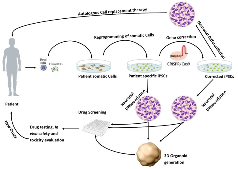
The development of human-based in vitro disease models [24] is an active area of research. One potential source of human in vitro models is the use of human pluripotent stem cells. These models can either be derived from human embryonic stem cells (hESCs) or induced pluripotent stem cells (iPSCs). The iPSCs derived from healthy individuals or patients are a promising approach to developing regenerative therapies as well as in vitro models of pathophysiological mechanisms of diseases (Figure 1) [25].
Figure 1.
Patient-specific stem cell lines for cell replacement therapy and new drug development. Somatic cells from the patient’s skin or blood can be isolated and reprogrammed into iPSCs. These cells have the potential to differentiate into different neuronal populations and can be used to either further study the pathophysiology of the disease or as a drug screening system to test new potential therapies. In addition, these lines can be corrected using CRISPR/Cas9 to generate isogenic controls which provide ideal controls.
These models better recapitulate the complexity of genetic epilepsy. In recent years, stem cell research has focused on using patient-specific hiPSC-derived neurons as in vitro models of epilepsy.
This scoping review aims to assess the application of stem cell-derived in vitro models to model the pathophysiology of epilepsy in the current literature. Specifically, we will determine: (i) what different methodologies have been used to generate in vitro epilepsy models; (ii) which gene variants and specific epileptic phenotypes have been modelled, and (iii) which outcome measures have been investigated.
2. Methods
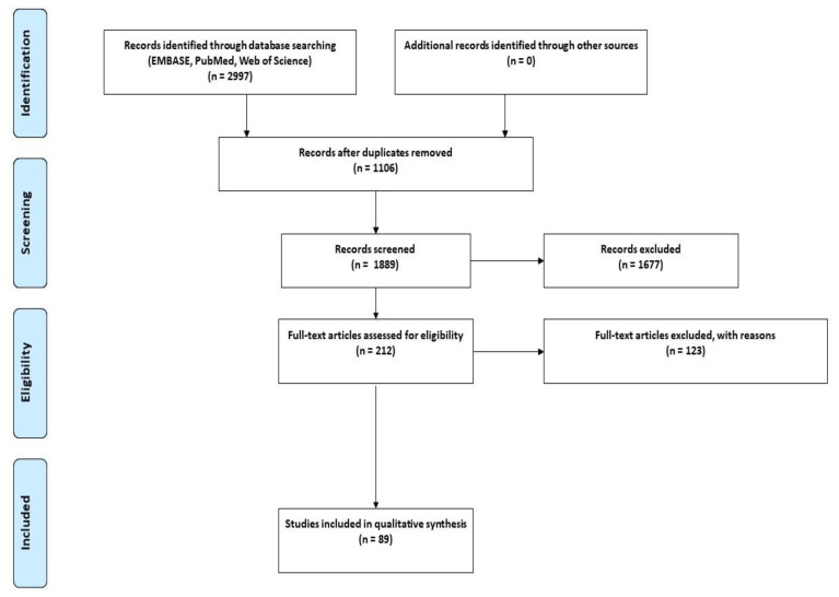
This review was conducted in accordance with the Preferred Reporting Items for Scoping Review (PRISMA-ScR) guidelines (Figure 2) [26]. To minimise the selection bias that is often associated with narrative reviews, we employed the same rigorous methodology used in performing a systematic review.
Figure 2.
PRISMA flow diagram of databases search, two-phase screening, and data extraction workflow. From 2997 studies, 1889 studies were screened for the title and abstracts. Of these, 212 studies underwent a full-text screen which identified the 89 studies that were included in the review.
2.1. Search Strategies
Three electronic databases were searched (Web of Science, PubMed, EMBASE). The predetermined searched terms were as follows:
Search terms for epilepsy:” Epilepsy” or “Refractory epilepsy” or “Seizures” or “Idiopathic generalized epilepsy” or “IGE” or “Frontal lobe epilepsy” or “FLE” or “Temporal lobe epilepsy” or “TLE” or “Autosomal dominant nocturnal frontal temporal lobe epilepsy” or “ADNFLE” or “Familial temporal lobe epilepsy” or “FTLE” or “Genetic Epilepsy” or “Chemically induced epilepsy”.
Search terms for stem cells: “Induced Pluripotent Stem Cells” or “iPSCs” or “Pluripotent Stem Cells” or “Human induced pluripotent stem cells” or “hiPSCs” or “Embryonic stem cells” or “Embryonic derived stem cells” or “Human embryonic stem cells” or “ESCs”. Appropriate search symbols and Boolean operators were used to combine both lists.
2.2. Eligibility Criteria
In this scoping review, all original studies that used stem cells or stem cell-derived cells as disease models to study epilepsy were included. The literature search was performed in 2018 and then updated in 2021. No year limit was set to include all the published studies as the focus was to assess the progress of the stem cell field in epilepsy research. The studies were not limited to a particular outcome measure or the method of neuronal induction. We excluded studies that did not use stem cells, did not assess epilepsy, and did not contain original data, such as reviews, letters, and commentaries.
2.3. Publication Selection
All publications were screened against our predetermined inclusion and exclusion criteria. As per scoping review guidelines, all retrieved publications were screened first by title and abstract followed by a full-text screen, by at least two independent reviewers (MSJ and TT). Any discrepancy between reviewers was resolved by a third reviewer (AAB).
Information was extracted from the selected studies using a standard data extraction template including the epilepsy phenotypes/models of epilepsy based on genetic variants (duplication or deletion of chromosomes), starting cell source/cells type, stem cell types, the cell population used in the study, gene mutation, and the main outcomes of the included studies (Table S1).
2.4. Information Extraction
Data were extracted from the papers included in the scoping review by two independent reviewers (M.S.J. and T.T.) using a data extraction tool developed by the reviewers. A draft extraction form is provided (Table S1). Any disagreements that arose between the reviewers were resolved through discussion, and with an additional reviewer (A.A.B.). The extracted data included specific details about the participants, concept, context, study methods, and key findings relevant to the review question.
2.5. Data Analysis and Presentation
The findings are presented graphically (Figure 3 and Figure 4), diagrammatically (Figure 1), and in tabular form (Table S1). The tabulated and/or charted results were discussed to provide a narrative summary of the review’s question and objective.
Figure 3.
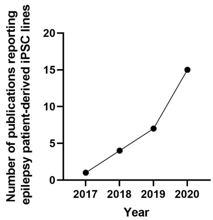
Generation of epileptic patient-specific iPSC lines. The graph indicates the increase in the number of publications reported from 2017 to 2020 that generated iPSC lines from patients with epilepsy.
Figure 4.
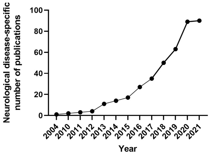
Publications since 2004 in the stem cell field for in vitro epilepsy modelling and drug toxicity testing using patient-derived iPSCs and human ESCs. The graph represents the total number of original research articles published in the field of epilepsy from 2004 to 2021 using stem cells.
3. Results
3.1. Study Selection Process
The major findings of this study are summarized in Table S1. A total of 2997 studies were identified, of which 1106 were removed as duplicates. The remaining 1889 were screened by title and abstract, and 1677 were excluded. The remaining 212 full-text studies were then reviewed, and 123 were excluded based on the inclusion/exclusion criteria. Full-text screening yielded 89 studies that met our pre-specified inclusion criteria (Figure 2). Of the included studies, human embryonic stem cells (hESCs) were used in 16 studies, and human induced pluripotent stem cells (hiPSCs) were used in the remainder (Figure 3).
The studies using hESCs-derived neurons investigated: their differentiation potential to generate specific neuronal subtypes; how genetic variance affects neuronal behaviour and how it influences the development of neuronal diseases such as FOXG1 syndrome and TSC [27,28,29,30,31,32,33,34,35,36,37]. In particular, a study performed by Costa et al, assessed how neurodevelopment and synaptic plasticity were altered in response to mTORC1 inhibition in tuberous sclerosis (TSC) [35]. In other studies, hESCs-derived neurons have been used: to model FOXG1 syndrome to control the endogenous protein dosage of FOXG1 protein in a precise manner which is important for GABA interneuron differentiation [37]; to assess the effect of high glucose concentration in masking the TSC cellular phenotypes using a hESCs-derived TSC model [38]; to investigate genotoxicity of anti-seizure medications [39], and the mode of action of the ASM valproate [40]. Two of these studies also used genetically engineered cells to enhance the release and delivery of adenosine [41,42].
In this review, the studies using hiPSCs were grouped into two major categories. The first group is the studies that described the generation of patient-specific iPSC lines [43,44,45,46,47,48,49,50,51,52,53,54,55,56] that carry genetic variants in genes associated with epilepsy, such as SCN1A, GNB5, LGI1, GRIN2A, KCNC1, or KCNA2 (Figure 3, Table S1) [43,44,45,46,47,48,49,50,51,52,53,54,55,56]. The second group is the studies that assessed the effects of convulsant and anticonvulsant drugs on hiPSC-derived neurons and astrocytes (Figure 4, Table S1) [57,58,59]. Some of these studies used healthy human stem cells to either generate an in vitro disease model by genetically manipulating a gene of interest; or by assessing the effects of different compounds on chemically induced “epilepsy-like” phenotype on otherwise healthy neurons [57,58,59,60]. Others used the iPSCs derived from patients carrying a specific gene variance of interest (Figure 4) [27,36,38,43,44,45,46,47,48,49,50,51,52,53,54,55,56,57,58,60,61,62,63,64,65,66,67,68,69,70,71,72,73,74,75,76,77,78,79,80,81,82,83,84,85,86,87,88,89,90,91,92,93,94,95,96,97,98,99,100,101,102,103,104,105,106,107,108,109,110,111,112].
3.2. Gene Editing Techniques for Disease Modelling and Isogenic Control
The methodology used to induce genetic variance in healthy cells for disease modelling, or to correct patient cells for isogenic control, has evolved over time. Sixteen studies (Table S1) reported the use of gene editing techniques to create isogenic controls or to study the disease-causing genetic variation in patient-derived iPSCs. Transcription Activator-Like Effector Nucleases (TALEN) was one of the first gene-editing techniques used in 2014 to generate the SCN1A mutation in human iPSCs [61] and was subsequently used in two more studies [48,55]. Zinc-Finger Nucleases (ZFNs) is another gene-editing technology and was used to model TSC in 2016 [35]. In addition, virus- or vector-based knock-out and knock-down techniques were also used in a few studies [33,42,95,106]. These methods were subsequently replaced with the more advanced CRISPR/Cas9 method. Unlike its predecessors, TALEN and ZFNs, it is precise, robust, and site-specific with fewer off-target effects [113]. The first use of CRISPR/Cas9 technology in epilepsy was reported in 2016 to generate a loss of function SCN1A mutation in human iPSCs to gain insight into Dravet syndrome [114]. This approach has been widely adopted in recent years with eight additional studies generating disease-specific neurons and isogenic controls using CRISPR/Cas9 [37,44,68,70,74,97,112,114].
3.3. Epilepsy Patient-Specific iPSCs Derived Disease Models
Dravet syndrome was the most commonly studied [60,61,62,63,64,65,67,68,69,114], followed by tuberous sclerosis [35,38,70,71,72], focal cortical dysplasia [82,83,84,85,104], and a few rare epilepsy syndromes [44,45,46,47,53,73,74,76,77,79,80,81,87,115]. These models have the potential to provide useful information about the involvement of a particular gene in disease progression and its anticonvulsant response.
The first in vitro model from a Dravet patient carrying a mutation in the SCN1A gene was generated in 2013. The findings from that study showed that the loss of function in GABAergic inhibition appears to be the main driver in epileptogenesis [62]. Since then, there have been several studies assessing various mutations in the SCN1A gene [48,54,55,56,60,61,62,63,64,65,66,67,68,114]. Neurons derived from these patients exhibit increased sodium currents and hyperexcitability [64], which can be alleviated by treatment with phenytoin [60]. In another study, neurons derived from two patients with Dravet syndrome demonstrated that genetic alterations of SCN1A differentially impacted electrophysiological impairment. The degree of impairment corresponded with the symptomatic severity of the donor from which the iPSCs were derived [63]. Recently, another patient-specific iPSCs-derived neuronal study generated from individuals with SCN1A mutation indicated an imbalance in excitation and inhibition that leads to hyperactivity in the neural network. This study used homozygous and isogenic controls to show the hyperexcitability in the generated neurons [68]. These studies indicated that neurons could recapitulate the neuronal pathophysiology and could potentially be used for screening drugs for personalised therapies [60,64].
The most commonly studied mTORopathies were tuberous sclerosis and focal cortical dysplasia (FCD). A study using neurons derived from a patient with TSC2 mutation reported hyperactivation of mTORC1 pathway [35]. In this model, pharmacological inhibition of mTORC1 with rapamycin reverses developmental abnormalities and synaptic dysfunction during independent developmental stages [35]. In another study, neuronal progenitor cells (NPCs) generated from a patient carrying a heterozygous TSC2 mutation exhibited disrupted neuronal development, potentially contributing to the disease neuropathology. Moreover, NPCs also exhibited activation of mTORC1 downstream signalling and attenuation of PI3K/AKT signalling upstream of TSC [72]. More recently, NPCs generated from the patient carrying TSC germline nonsense mutation in exon 15 of TSC1 showed the influence of TSC1 mutation in the early neurodevelopmental phenotypes, signalling, and gene expression in NPCs compared to the genetically matched wild-type cells [70]. In 2019, Sundberg et al showed that loss of one allele of TSC2 is sufficient to cause some morphological and physiological changes, elevated phosphorylation, and hyperexcitability of mTORC1 in human neurons, but biallelic mutations in TSC2 are necessary to induce gene expression dysregulation seen in cortical tubers. They also found that treatment of TSC2 patient-specific iPSCs-derived neurons with rapamycin reduced neuronal activity and partially reversed gene expression abnormalities [71]. In 2020, Alsaqati used commercially available TSC2 (loss of function mutation) patient-derived iPSCs and reported that the dysfunctional neuronal network behaviour in the differentiated neurons could not be rescued with rapamycin treatment [57]. The difference in response in these two studies is because of two different iPSCs samples carrying different mutations in the TSC2 gene [57,71].
Five studies assessed the FCD-related cortical malformation by generating iPSCs from patients with mutations in genes involved in regulating the mTOR pathway. In 2017, Marinowic et al described the generation of iPSC-based cellular models of refractory epilepsy from the fibroblasts of two refractory epilepsy patients with FCD type IIb, one a 45-year-old male and the other 12-years-old female [84]. Then in 2020, Marinowic published another study using these cells investigating the differences in the migration potential and the expression of genes for cell proliferation, adhesion, and apoptosis. The main finding of the study was that the gene expression was different between the neurons generated from the adult male compared to the child. They concluded that differences in the migration potential of adult cells, and differences in the expression of genes related to the fundamental brain development processes, might be associated with cortical alteration in the two patients with FCD IIb [85]. In 2018, Majolo et al studied the Notch signalling pathway, a pathway involved in cortical development to regulate neuronal differentiation, self-renewal, survival, and neuronal plasticity, using the iPSCs from FCD IIb patients. The study assess the expression of genes involved in Notch signalling and showed that, during embryonic neurogenesis, the neural precursor cells of FCD type IIb individuals exhibited an increase in HEY1 and NOTCH1 genes as well as a decrease in the expression of HES1 and PAX5 genes, compared to the cells from control subjects [82]. In the subsequent study, Majolo et al studied the migration and synaptic aspects of neurons generated using the iPSCs derived from patients with FCD type IIb. Using real-time PCR, the study presented the expression of most of the synaptic and ion channels genes ASCL1, DCX, DLG4, FGF2, NEFL, NEUROD2, NEUROD6, NRCAM, and STX1A in different groups; fibroblasts, iPSCs, differentiated neurons, and brain tissues [83]. This study suggested that the cells derived from FCD patients may have more sensitivity to stimuli resulting in altered cell survival, apoptosis, migration, and morphological development. In 2021, Klofas et al published a study using the FCD patient-derived neurons carrying a heterozygous loss-of-function mutation in the DEPDC5 gene and reported hyperactivation of mTORC1 and enlarged cell somas that were rescued with the inhibition of mTORC1. This study also reported that cell starvation leads to hyperactivation of the mTOR pathway [104] but the exact mechanism is still unclear. None of these FCD studies have performed electrophysiological functional analysis of the generated neurons.
3.4. Outcome Measures
Eighty-eight studies (98%) reported histological, molecular, electrophysiological, or other outcomes to validate the generation of iPSCs or to assess epilepsy-like phenotypes. 87% of studies (Table S1) reported the molecular outcomes to validate the successful generation of patients’ iPSCs and iPSC-derived neuronal disease models, of which 38% (Table S1) also reported the electrical activity of generated neurons using either a patch-clamp or Micro-Electrode Array (MEA) analysis to assess electrophysiological activity.
Electrophysiological analysis was performed using patch-clamp [27,32,33,35,37,60,61,62,63,64,66,67,68,69,77,78,89,90,94,96,97,98,103,107,110,114,116] or MEA [34,36,57,59,69,71,75,86,93,105,108] analysis. Electrophysiology is a preferred method of studying brain activity because it allows the recording of a wide range of neuronal phenomena ranging from the action potential to the network simulation of a neuronal population [27,110,117,118,119]. Patch-clamp or MEA can also be used to measure intracellular voltage [120].
Real-time PCR, western blot, immunoblot, and immunofluorescence were the primary molecular and histological methods applied to validate patient iPSC-derived neuronal disease models. However, in the case of patient-specific iPSC generation carrying a specific genetic variant, Sanger sequencing was the primary measure to confirm the presence of the variant (Table S1). For cell lines, these outcome measures are a standard requirement to publish and report the generation of cell lines. Similarly, for disease modelling research, standardised outcome measures for functional and phenotypical analysis of the generated neurons are needed.
4. Discussion
Identifying and understanding the pathological mechanisms of human diseases such as epilepsy play a crucial role in the development of novel therapeutic approaches. Unfortunately, currently available anti-seizure medications are unable to treat epilepsy in about 30% of patients. The limited efficacy of these ASMs has been, at least partly, attributed to the lack of appropriate pre-clinical models. Most of the in vivo and in vitro disease modelling and drug development is done in animals, and however useful, these models are not ideal to study genetic epilepsy [23]. Some attempts have been made to utilise primary brain tissue from patients, but its limited availability and the difficulty of culturing it makes it challenging to use. Therefore, the ability to utilise patient cells and generate iPSC-derived neurons is of great value to the field of neuroscience. In this review, we have summarised the current literature describing stem cell-derived in vitro epilepsy models.
Using human stem cells to model epilepsy in vitro is a relatively new concept, with the first study published in 2004. Since then, however, the field has exponentially grown with over 25 studies published in the past three years (Table S1). While the early publications reported artificially induced epilepsy-like phenotypes in hESCs, the more recent publications described patient-specific iPSC lines that carry a disease-causing gene variant.
We identified 65 studies describing the establishment of new patient-derived iPSCs lines. While some studies only reported the generation and characterization of these lines, the majority (50 studies) used hPSCs to assess the involvement of a particular gene in disease progression and drug response. These studies have shown that neurons derived from the patient iPSCs are phenotypically and morphologically different compared to healthy neurons or neurons derived from isogenic controls. Moreover, these neurons exhibited delayed differentiation, synaptic abnormalities, and defects in neurite formation and migration [87,95]. They also have a unique electrophysiological signature that differs between both, the individual patients and the controls. Transcriptional changes and disrupted pathways of chromatic modelling specific to a gene variant have also been revealed using patient-specific iPSCs-derived neurons [66,78]. Furthermore, these neurons exhibit bursting impairments leading to hyperpolarization and hyperexcitability which can be altered by the administration of different ASMs. In vitro models generated from patient-derived neurons provide new insight into the disease phenotype, and molecular and cellular mechanisms that underlie epileptogenesis and drug resistance in individual patients, thus identifying crucial pathways of drug screening for the development of novel anti-seizure medications, and precision, and regenerative medicines.
4.1. Limitations of Stem Cell-Derived Epilepsy Models
The iPSCs models hold potential opportunities to enhance our current understanding across a wide range of biological phenomena in epilepsy and beyond, but they come with limitations.
No two iPSC lines or models derived from these lines are the same. They are as unique as the individual from whom they are derived. In addition, protocols used to generate these cells also vary. There is a degree of experimental variability associated with the generation of iPSCs [121], including the source of somatic cells from which the iPSCs were generated, initial cell density used to generate iPSCs, and variability in the reprogramming kits as well as time and duration of each step. To combat these issues, the International Society for Stem Cell Research (ISSCR) has developed a set of guidelines for the generation of new iPSCs lines (https://www.isscr.org/policy/guidelines-for-stem-cell-research-and-clinical-translation/sections/part5. Assessed on 8 August 2022). These guidelines require each newly generated iPSC line to be registered and characterised using pluripotency assays, teratoma formation analysis, germ layer differentiation, donor screening, variant analysis, sequencing, karyotyping, and mycoplasma detection analysis (https://www.journals.elsevier.com/stem-cell-research/lab-resources/scientific-guidelines-for-lab-resources. Assessed on 8 August 2022).
Another source of variability is the protocols used to generate neuronal cultures from these iPSCs. Despite there being only two main methods of neuronal differentiation (either dual SMAD inhibition by using small molecules [122] or the viral vectors method [123]), there are a number of different agents used and each different protocol yields different proportions of neuronal and glial cells. The most physiologically relevant method uses the dual SMAD inhibition which gives rise to both excitatory and inhibitory neuronal populations as well as a small proportion of astrocytes, which are all required for the generation of functional and neuronal networks in vitro. However, this method is time-consuming and costly as it takes several months to generate mature neurons. The alternative is to use viral vector transduction into stem cells to express specific genes for neuronal differentiation. In this method, stem cells are modified to overexpress only one or two neuronal genes, and therefore produce more homogeneous cultures and, in many cases, do not contain astrocytes. As the astrocytes are required for the formation and maintenance of mature functional neuronal networks [124], these methods require co-culturing with an external source of astrocytes. These astrocytes are, however, generated from healthy human brains and do not recapitulate the epileptic phenotype. For the generation of in vitro models that can effectively model epileptogenesis, it is critical to generate both patient-derived excitatory and inhibitory neuronal populations as well as glia.
For the better characterisation and reproducibility of in vitro models, either for the study of the pathophysiology of epilepsy or for drug screening, the methodology of neuronal differentiation and functional assessment need to be standardised. In addition, the sharing of data amongst researchers would allow for the comparison and standardisation of protocols which would maximise reproducibility across different labs. Highly curated and referenced cell lines should be available to the scientific community in the form of cell banks. A few cell banks are: Stem Cells for Biological Assays of Novel Drugs and Predictive Toxicology (StemBANCC) [125], HipSci [126], the European Bank for Induced Pluripotent Stem Cells (EBiSC) [127], and the iPSC Collection for Omic Research (iPSCORE) [128]. A similar standardised depository of neuronal differentiation methods and neuronal functional outcome measures is required to enhance reproducibility and increase the likelihood of developing new and novel therapies.
4.2. Limitations of Our Approach
The major limitation of this study was the sheer magnitude of variability of the studies included. Due to the broad nature of our research questions, the findings of this review are similarly broad. The idea of using stem cell-derived neurons as human in vitro models of epilepsy is a relatively new concept and therefore we did not want to limit our search to a particular treatment or outcome measure. This meant that our included dataset contained a variety of different studies with different study designs and outcome measures.
Due to the lack of standardised reporting of human in vitro models, it was difficult to perform any type of risk of bias assessment. Studies varied in the methods of iPSC generation, neuronal differentiation, and outcome measures. Only thirteen studies assessed the effects of potential novel therapies (Table S1). There have been some efforts made in developing risk bias tools for in vitro studies [129], but for those tools to be applicable for the assessment of in vitro epilepsy models, the methodology and reporting of such models would need to be standardised.
5. Conclusions
The stem cell field has tremendous potential to revolutionise both preclinical and clinical epilepsy research. Advancements in iPSC reprogramming, differentiation, and genome engineering have expanded the use of cell-based models into the mainstream of cellular neuroscience. As the field of in vitro disease modelling evolved, it is critical to develop and standardise methodologies to improve translatability, integrity, and quality [130]. Rigorous optimisation to standardise the approaches may take extra effort, but is an essential requirement to translate the promise of stem cell-based disease models to the development of new therapeutics and therapies. Personalised disease models would allow the neurobiologist to investigate the unknown details of the disease processes by mimicking human pathophysiology, which could lead to major advances in personalised medicines, and would help to address the issue of drug resistance.
Acknowledgments
M.S.J. is supported by Monash Graduate Scholarship and Monash International Tuition Fee Scholarship.
Supplementary Materials
The following supporting information can be downloaded at: https://www.mdpi.com/article/10.3390/cells11243957/s1: Table S1. Data extracted from the included studies to report the progress of the stem cell field in epilepsy research.
Author Contributions
Conceptualisation, M.S.J., A.A.-B., A.A., T.J.O. and P.K.; Screening, M.S.J., T.T., N.D. and A.A.-B.; Methodology and Data Extraction, M.S.J., T.T., N.D., A.A.-B. and A.A.; Result and Discussion, M.S.J., A.A.-B. and A.A.; Supervision and Review, A.A.-B., A.A., T.J.O. and P.K.; Writing and Editing, M.S.J., A.A.-B., A.A., T.J.O. and P.K. All authors have read and agreed to the published version of the manuscript.
Conflicts of Interest
The authors declare no conflict of interest.
Funding Statement
This research received no external funding. The APC was funded by Professors O’Brien and Kwan.
Footnotes
Publisher’s Note: MDPI stays neutral with regard to jurisdictional claims in published maps and institutional affiliations.
References
- 1.Devinsky O., Vezzani A., O’Brien T.J., Jette N., Scheffer I.E., de Curtis M., Perucca P. Epilepsy. Nat. Rev. Dis. Prim. 2018;4:18024. doi: 10.1038/nrdp.2018.24. [DOI] [PubMed] [Google Scholar]
- 2.Perucca P., Perucca E. Identifying mutations in epilepsy genes: Impact on treatment selection. Epilepsy Res. 2019;152:18–30. doi: 10.1016/j.eplepsyres.2019.03.001. [DOI] [PubMed] [Google Scholar]
- 3.Singh N.A., Charlier C., Stauffer D., DuPont B.R., Leach R.J., Melis R., Ronen G.M., Bjerre I., Quattlebaum T., Murphy J.V. A novel potassium channel gene, KCNQ2, is mutated in an inherited epilepsy of newborns. Nat. Genet. 1998;18:25. doi: 10.1038/ng0198-25. [DOI] [PubMed] [Google Scholar]
- 4.Charlier C., Singh N.A., Ryan S.G., Lewis T.B., Reus B.E., Leach R.J., Leppert M. A pore mutation in a novel KQT-like potassium channel gene in an idiopathic epilepsy family. Nat. Genet. 1998;18:53. doi: 10.1038/ng0198-53. [DOI] [PubMed] [Google Scholar]
- 5.Claes L.R., Deprez L., Suls A., Baets J., Smets K., Van Dyck T., Deconinck T., Jordanova A., De Jonghe P. The SCN1A variant database: A novel research and diagnostic tool. Hum. Mutat. 2009;30:E904–E920. doi: 10.1002/humu.21083. [DOI] [PubMed] [Google Scholar]
- 6.Steinlein O.K., Mulley J.C., Propping P., Wallace R.H., Phillips H.A., Sutherland G.R., Scheffer I.E., Berkovic S.F. A missense mutation in the neuronal nicotinic acetylcholine receptor α4 subunit is associated with autosomal dominant nocturnal frontal lobe epilepsy. Nat. Genet. 1995;11:201. doi: 10.1038/ng1095-201. [DOI] [PubMed] [Google Scholar]
- 7.Baybis M., Yu J., Lee A., Golden J.A., Weiner H., McKhann G., Aronica E., Crino P.B. mTOR cascade activation distinguishes tubers from focal cortical dysplasia. Ann. Neurol. Off. J. Am. Neurol. Assoc. Child Neurol. Soc. 2004;56:478–487. doi: 10.1002/ana.20211. [DOI] [PubMed] [Google Scholar]
- 8.Baulac S., Ishida S., Marsan E., Miquel C., Biraben A., Nguyen D.K., Nordli D., Cossette P., Nguyen S., Lambrecq V. Familial focal epilepsy with focal cortical dysplasia due to DEPDC 5 mutations. Ann. Neurol. 2015;77:675–683. doi: 10.1002/ana.24368. [DOI] [PubMed] [Google Scholar]
- 9.D’Gama A.M., Geng Y., Couto J.A., Martin B., Boyle E.A., LaCoursiere C.M., Hossain A., Hatem N.E., Barry B.J., Kwiatkowski D.J. Mammalian target of rapamycin pathway mutations cause hemimegalencephaly and focal cortical dysplasia. Ann. Neurol. 2015;77:720–725. doi: 10.1002/ana.24357. [DOI] [PMC free article] [PubMed] [Google Scholar]
- 10.Lim J.S., Kim W., Kang H.-C., Kim S.H., Park A.H., Park E.K., Cho Y.-W., Kim S., Kim H.M., Kim J.A. Brain somatic mutations in MTOR cause focal cortical dysplasia type II leading to intractable epilepsy. Nat. Med. 2015;21:395. doi: 10.1038/nm.3824. [DOI] [PubMed] [Google Scholar]
- 11.Marsan E., Baulac S. Mechanistic target of rapamycin (mTOR) pathway, focal cortical dysplasia and epilepsy. Neuropathol. Appl. Neurobiol. 2018;44:6–17. doi: 10.1111/nan.12463. [DOI] [PubMed] [Google Scholar]
- 12.Park S.M., Lim J.S., Ramakrishina S., Kim S.H., Kim W.K., Lee J., Kang H.-C., Reiter J.F., Kim D.S., Kim H.H. Brain Somatic Mutations in MTOR Disrupt Neuronal Ciliogenesis, Leading to Focal Cortical Dyslamination. Neuron. 2018;99:83–97.e87. doi: 10.1016/j.neuron.2018.05.039. [DOI] [PubMed] [Google Scholar]
- 13.Scerri T., Riseley J.R., Gillies G., Pope K., Burgess R., Mandelstam S.A., Dibbens L., Chow C.W., Maixner W., Harvey A.S. Familial cortical dysplasia type IIA caused by a germline mutation in DEPDC 5. Ann. Clin. Transl. Neurol. 2015;2:575–580. doi: 10.1002/acn3.191. [DOI] [PMC free article] [PubMed] [Google Scholar]
- 14.Sim J.C., Scerri T., Fanjul-Fernández M., Riseley J.R., Gillies G., Pope K., Van Roozendaal H., Heng J.I., Mandelstam S.A., McGillivray G. Familial cortical dysplasia caused by mutation in the mammalian target of rapamycin regulator NPRL3. Ann. Neurol. 2016;79:132–137. doi: 10.1002/ana.24502. [DOI] [PubMed] [Google Scholar]
- 15.Van Kranenburg M., Hoogeveen-Westerveld M., Nellist M. Preliminary Functional Assessment and Classification of DEPDC 5 Variants Associated with Focal Epilepsy. Hum. Mutat. 2015;36:200–209. doi: 10.1002/humu.22723. [DOI] [PubMed] [Google Scholar]
- 16.Kwan P., Brodie M.J. Early identification of refractory epilepsy. N. Engl. J. Med. 2000;342:314–319. doi: 10.1056/NEJM200002033420503. [DOI] [PubMed] [Google Scholar]
- 17.Kwan P., Arzimanoglou A., Berg A.T., Brodie M.J., Allen Hauser W., Mathern G., Moshé S.L., Perucca E., Wiebe S., French J. Definition of drug resistant epilepsy: Consensus proposal by the ad hoc Task Force of the ILAE Commission on Therapeutic Strategies. Epilepsia. 2010;51:1069–1077. doi: 10.1111/j.1528-1167.2009.02397.x. [DOI] [PubMed] [Google Scholar]
- 18.Kwan P., Schachter S.C., Brodie M.J. Drug-resistant epilepsy. N. Engl. J. Med. 2011;365:919–926. doi: 10.1056/NEJMra1004418. [DOI] [PubMed] [Google Scholar]
- 19.Wiebe S., Blume W.T., Girvin J.P., Eliasziw M. A randomized, controlled trial of surgery for temporal-lobe epilepsy. N. Engl. J. Med. 2001;345:311–318. doi: 10.1056/NEJM200108023450501. [DOI] [PubMed] [Google Scholar]
- 20.Dragunow M. The adult human brain in preclinical drug development. Nat. Rev. Drug Discov. 2008;7:659. doi: 10.1038/nrd2617. [DOI] [PubMed] [Google Scholar]
- 21.Grainger A.I., King M.C., Nagel D.A., Parri H.R., Coleman M.D., Hill E.J. In vitro models for seizure-liability testing using induced pluripotent stem cells. Front. Neurosci. 2018;12:590. doi: 10.3389/fnins.2018.00590. [DOI] [PMC free article] [PubMed] [Google Scholar]
- 22.Kandratavicius L., Balista P.A., Lopes-Aguiar C., Ruggiero R.N., Umeoka E.H., Garcia-Cairasco N., Bueno L.S., Jr., Leite J.P. Animal models of epilepsy: Use and limitations. Neuropsychiatr. Dis. Treat. 2014;10:1693–1705. doi: 10.2147/NDT.S50371. [DOI] [PMC free article] [PubMed] [Google Scholar]
- 23.Shi Y., Inoue H., Wu J.C., Yamanaka S. Induced pluripotent stem cell technology: A decade of progress. Nat. Rev. Drug Discov. 2017;16:115–130. doi: 10.1038/nrd.2016.245. [DOI] [PMC free article] [PubMed] [Google Scholar]
- 24.Easter A., Bell M.E., Damewood J.R., Jr., Redfern W.S., Valentin J.-P., Winter M.J., Fonck C., Bialecki R.A. Approaches to seizure risk assessment in preclinical drug discovery. Drug Discov. Today. 2009;14:876–884. doi: 10.1016/j.drudis.2009.06.003. [DOI] [PubMed] [Google Scholar]
- 25.Dolmetsch R., Geschwind D.H. The human brain in a dish: The promise of iPSC-derived neurons. Cell. 2011;145:831–834. doi: 10.1016/j.cell.2011.05.034. [DOI] [PMC free article] [PubMed] [Google Scholar]
- 26.Tricco A.C., Lillie E., Zarin W., O’Brien K.K., Colquhoun H., Levac D., Moher D., Peters M.D., Horsley T., Weeks L. PRISMA extension for scoping reviews (PRISMA-ScR): Checklist and explanation. Ann. Intern. Med. 2018;169:467–473. doi: 10.7326/M18-0850. [DOI] [PubMed] [Google Scholar]
- 27.Maroof A.M., Keros S., Tyson J.A., Ying S.W., Ganat Y.M., Merkle F.T., Liu B., Goulburn A., Stanley E.G., Elefanty A.G., et al. Directed differentiation and functional maturation of cortical interneurons from human embryonic stem cells. Cell Stem Cell. 2013;12:559–572. doi: 10.1016/j.stem.2013.04.008. [DOI] [PMC free article] [PubMed] [Google Scholar]
- 28.Ahn S., Kim T.-G., Kim K.-S., Chung S. Differentiation of human pluripotent stem cells into Medial Ganglionic Eminence vs. Caudal Ganglionic Eminence cells. Methods. 2016;101:103–112. doi: 10.1016/j.ymeth.2015.09.009. [DOI] [PMC free article] [PubMed] [Google Scholar]
- 29.Vazin T., Ashton R.S., Conway A., Rode N.A., Lee S.M., Bravo V., Healy K.E., Kane R.S., Schaffer D.V. The effect of multivalent Sonic hedgehog on differentiation of human embryonic stem cells into dopaminergic and GABAergic neurons. Biomaterials. 2014;35:941–948. doi: 10.1016/j.biomaterials.2013.10.025. [DOI] [PubMed] [Google Scholar]
- 30.Hartley B.J., Watmuff B., Hunt C.P.J., Haynes J.M., Pouton C.W., Kaur N., Vemuri M.C. Neural Stem Cell Assays. Wiley; Hoboken, NJ, USA: 2015. In vitro Differentiation of Pluripotent Stem Cells towards either Forebrain GABAergic or Midbrain Dopaminergic Neurons; pp. 91–99. [Google Scholar]
- 31.Germain N.D., Banda E.C., Becker S., Naegele J.R., Grabel L.B. Derivation and isolation of NKX2.1-positive basal forebrain progenitors from human embryonic stem cells. Stem Cells Dev. 2013;22:1477–1489. doi: 10.1089/scd.2012.0264. [DOI] [PMC free article] [PubMed] [Google Scholar]
- 32.Nicholas C.R., Chen J., Tang Y., Southwell D.G., Chalmers N., Vogt D., Arnold C.M., Chen Y.-J.J., Stanley E.G., Elefanty A.G., et al. Functional Maturation of hPSC-Derived Forebrain Interneurons Requires an Extended Timeline and Mimics Human Neural Development. Cell Stem Cell. 2013;12:573–586. doi: 10.1016/j.stem.2013.04.005. [DOI] [PMC free article] [PubMed] [Google Scholar]
- 33.Meganathan K., Lewis E.M.A., Gontarz P., Liu S., Stanley E.G., Elefanty A.G., Huettner J.E., Zhang B., Kroll K.L. Regulatory networks specifying cortical interneurons from human embryonic stem cells reveal roles for CHD2 in interneuron development. Proc. Natl. Acad. Sci. USA. 2017;114:E11180–E11189. doi: 10.1073/pnas.1712365115. [DOI] [PMC free article] [PubMed] [Google Scholar]
- 34.Lu C., Shi X., Allen A., Baez-Nieto D., Nikish A., Sanjana N.E., Pan J.Q. Overexpression of NEUROG2 and NEUROG1 in human embryonic stem cells produces a network of excitatory and inhibitory neurons. Faseb J. 2019;33:5287–5299. doi: 10.1096/fj.201801110RR. [DOI] [PMC free article] [PubMed] [Google Scholar]
- 35.Costa V., Aigner S., Vukcevic M., Sauter E., Behr K., Ebeling M., Dunkley T., Friedlein A., Zoffmann S., Meyer C.A. mTORC1 inhibition corrects neurodevelopmental and synaptic alterations in a human stem cell model of tuberous sclerosis. Cell Rep. 2016;15:86–95. doi: 10.1016/j.celrep.2016.02.090. [DOI] [PubMed] [Google Scholar]
- 36.Ishikawa M., Aoyama T., Shibata S., Sone T., Miyoshi H., Watanabe H., Nakamura M., Morota S., Uchino H., Yoo A.S., et al. miRNA-based rapid differentiation of purified neurons from hPSCs advancestowards quick screening for neuronal disease phenotypes in vitro. Cells. 2020;9:532. doi: 10.3390/cells9030532. [DOI] [PMC free article] [PubMed] [Google Scholar]
- 37.Zhu W., Zhang B., Li M., Mo F., Mi T., Wu Y., Teng Z., Zhou Q., Li W., Hu B. Precisely controlling endogenous protein dosage in hPSCs and derivatives to model FOXG1 syndrome. Nat. Commun. 2019;10:928. doi: 10.1038/s41467-019-08841-7. [DOI] [PMC free article] [PubMed] [Google Scholar]
- 38.Rocktäschel P., Sen A., Cader M.Z. High glucose concentrations mask cellular phenotypes in a stem cell model of tuberous sclerosis complex. Epilepsy Behav. 2019;101:106581. doi: 10.1016/j.yebeh.2019.106581. [DOI] [PMC free article] [PubMed] [Google Scholar]
- 39.Kardoost M., Hajizadeh-Saffar E., Ghorbanian M.T., Ghezelayagh Z., Bagheri K.P., Behdani M., Habibi-Anbouhi M. Genotoxicity assessment of antiepileptic drugs (AEDs) in human embryonic stem cells. Epilepsy Res. 2019;158:106232. doi: 10.1016/j.eplepsyres.2019.106232. [DOI] [PubMed] [Google Scholar]
- 40.Da Costa R.F.M., Kormann M.L., Galina A., Rehen S.K. Valproate Disturbs Morphology and Mitochondrial Membrane Potential in Human Neural Cells. Appl. Vitr. Toxicol. 2015;1:254–261. doi: 10.1089/aivt.2015.0016. [DOI] [Google Scholar]
- 41.Poppe D., Doerr J., Schneider M., Wilkens R., Steinbeck J.A., Ladewig J., Tam A., Paschon D.E., Gregory P.D., Reik A. Genome Editing in Neuroepithelial Stem Cells to Generate Human Neurons with High Adenosine-Releasing Capacity. Stem Cells Transl. Med. 2018;7:477–486. doi: 10.1002/sctm.16-0272. [DOI] [PMC free article] [PubMed] [Google Scholar]
- 42.Fedele D.E., Koch P., Scheurer L., Simpson E.M., Mohler H., Brustle O., Boison D. Engineering embryonic stem cell derived glia for adenosine delivery. Neurosci. Lett. 2004;370:160–165. doi: 10.1016/j.neulet.2004.08.031. [DOI] [PubMed] [Google Scholar]
- 43.Kimura Y., Tanaka Y., Shirasu N., Yasunaga S., Higurashi N., Hirose S. Establishment of human induced pluripotent stem cells derived from skin cells of a patient with Dravet syndrome. Stem Cell Res. 2020;47:101857. doi: 10.1016/j.scr.2020.101857. [DOI] [PubMed] [Google Scholar]
- 44.Malerba N., Benzoni P., Squeo G.M., Milanesi R., Giannetti F., Sadleir L.G., Poke G., Augello B., Croce A.I., Barbuti A., et al. Generation of the induced human pluripotent stem cell lines CSSi009-A from a patient with a GNB5 pathogenic variant, and CSSi010-A from a CRISPR/Cas9 engineered GNB5 knock-out human cell line. Stem Cell Res. 2019;40:101547. doi: 10.1016/j.scr.2019.101547. [DOI] [PubMed] [Google Scholar]
- 45.Schwarz N., Uysal B., Rosa F., Löffler H., Mau-Holzmann U.A., Liebau S., Lerche H. Generation of an induced pluripotent stem cell (iPSC) line from a patient with developmental and epileptic encephalopathy carrying a KCNA2 (p. Leu328Val) mutation. Stem Cell Res. 2018;33:6–9. doi: 10.1016/j.scr.2018.08.019. [DOI] [PubMed] [Google Scholar]
- 46.Sun C., Yang M., Qin F., Guo R., Liang S., Hu H. Generation of an induced pluripotent stem cell line SYSUi-003-A from a child with epilepsy carrying GRIN2A mutation. Stem Cell Res. 2020;43:101706. doi: 10.1016/j.scr.2020.101706. [DOI] [PubMed] [Google Scholar]
- 47.Zhang B., Wang Y., Peng J., Hao Y., Guan Y. Generation of a human induced pluripotent stem cell line from an epilepsy patient carrying mutations in the PIK3R2 gene. Stem Cell Res. 2020;44:101711. doi: 10.1016/j.scr.2020.101711. [DOI] [PubMed] [Google Scholar]
- 48.Zhao H., He L., Li S., Huang H., Tang F., Han X., Lin Z., Tian C., Huang R., Zhou P., et al. Generation of corrected-hiPSC (USTCi001-A-1) from epilepsy patient iPSCs using TALEN-mediated editing of the SCN1A gene. Stem Cell Res. 2020;46:101864. doi: 10.1016/j.scr.2020.101864. [DOI] [PubMed] [Google Scholar]
- 49.Arbini A., Gilmore J., King M.D., Gorman K.M., Krawczyk J., McInerney V., O’Brien T., Shen S., Allen N.M. Generation of three induced pluripotent stem cell (iPSC) lines from a patient with developmental epileptic encephalopathy due to the pathogenic KCNA2 variant c.869T>G; p.Leu290Arg (NUIGi052-A, NUIGi052-B, NUIGi052-C) Stem Cell Res. 2020;46:101853. doi: 10.1016/j.scr.2020.101853. [DOI] [PubMed] [Google Scholar]
- 50.Gong P., Jiao X., Zhang Y., Yang Z. Generation of a human iPSC line from an epileptic encephalopathy patient with electrical status epilepticus during sleep carrying KCNA2 (p.P405L) mutation. Stem Cell Res. 2020;49:102080. doi: 10.1016/j.scr.2020.102080. [DOI] [PubMed] [Google Scholar]
- 51.Nengqing L., Dian L., Yingjun X., Yi C., Lina H., Diyu C., Yinghong Y., Bing S., Xiaofang S. Generation of induced pluripotent stem cell GZHMCi001-A and GZHMCi001-B derived from peripheral blood mononuclear cells of epileptic patients with KCNC1 mutation. Stem Cell Res. 2020;47:101897. doi: 10.1016/j.scr.2020.101897. [DOI] [PubMed] [Google Scholar]
- 52.Schwarz N., Uysal B., Rosa F., Loffler H., Mau-Holzmann U.A., Liebau S., Lerche H. Establishment of a human induced pluripotent stem cell (iPSC)line (HIHDNEi002-A) from a patient with developmental and epileptic encephalopathy carrying a KCNA2 (p.Arg297Gln)mutation. Stem Cell Res. 2019;37:101445. doi: 10.1016/j.scr.2019.101445. [DOI] [PubMed] [Google Scholar]
- 53.Tan G.W., Kondo T., Murakami N., Imamura K., Enami T., Tsukita K., Shibukawa R., Funayama M., Matsumoto R., Ikeda A. Induced pluripotent stem cells derived from an autosomal dominant lateral temporal epilepsy (ADLTE) patient carrying S473L mutation in leucine-rich glioma inactivated 1 (LGI1) Stem Cell Res. 2017;24:12–15. doi: 10.1016/j.scr.2017.07.030. [DOI] [PubMed] [Google Scholar]
- 54.Tanaka Y., Higurashi N., Shirasu N., Yasunaga S.i., Moreira K.M., Okano H., Hirose S. Establishment of a human induced stem cell line (FUi002-A) from Dravet syndrome patient carrying heterozygous R1525X mutation in SCN1A gene. Stem Cell Res. 2018;31:11–15. doi: 10.1016/j.scr.2018.06.008. [DOI] [PubMed] [Google Scholar]
- 55.Tanaka Y., Sone T., Higurashi N., Sakuma T., Suzuki S., Ishikawa M., Yamamoto T., Mitsui J., Tsuji H., Okano H. Generation of D1-1 TALEN isogenic control cell line from Dravet syndrome patient iPSCs using TALEN-mediated editing of the SCN1A gene. Stem Cell Res. 2018;28:100–104. doi: 10.1016/j.scr.2018.01.036. [DOI] [PubMed] [Google Scholar]
- 56.Zhao H., Li S., He L., Han X., Huang H., Tang F., Lin Z., Deng S., Tian C., Huang R. Generation of iPSC line (USTCi001-A) from human skin fibroblasts of a patient with epilepsy. Stem Cell Res. 2020;45:101785. doi: 10.1016/j.scr.2020.101785. [DOI] [PubMed] [Google Scholar]
- 57.Alsaqati M., Heine V.M., Harwood A.J. Pharmacological intervention to restore connectivity deficits of neuronal networks derived from ASD patient iPSC with a TSC2 mutation. Mol. Autism. 2020;11:80. doi: 10.1186/s13229-020-00391-w. [DOI] [PMC free article] [PubMed] [Google Scholar]
- 58.Ishii M.N., Yamamoto K., Shoji M., Asami A., Kawamata Y. Human induced pluripotent stem cell (hiPSC)-derived neurons respond to convulsant drugs when co-cultured with hiPSC-derived astrocytes. Toxicology. 2017;389:130–138. doi: 10.1016/j.tox.2017.06.010. [DOI] [PubMed] [Google Scholar]
- 59.Odawara A., Katoh H., Matsuda N., Suzuki I. Physiological maturation and drug responses of human induced pluripotent stem cell-derived cortical neuronal networks in long-term culture. Sci. Rep. 2016;6:26181. doi: 10.1038/srep26181. [DOI] [PMC free article] [PubMed] [Google Scholar]
- 60.Jiao J., Yang Y., Shi Y., Chen J., Gao R., Fan Y., Yao H., Liao W., Sun X.F., Gao S. Modeling Dravet syndrome using induced pluripotent stem cells (iPSCs) and directly converted neurons. Hum. Mol. Genet. 2013;22:4241–4252. doi: 10.1093/hmg/ddt275. [DOI] [PubMed] [Google Scholar]
- 61.Chen W., Liu J., Zhang L., Xu H., Guo X., Deng S., Liu L., Yu D., Chen Y., Li Z. Generation of the SCN1A epilepsy mutation in hiPS cells using the TALEN technique. Sci. Rep. 2014;4:5404. doi: 10.1038/srep05404. [DOI] [PMC free article] [PubMed] [Google Scholar]
- 62.Higurashi N., Uchida T., Lossin C., Misumi Y., Okada Y., Akamatsu W., Imaizumi Y., Zhang B., Nabeshima K., Mori M.X. A human Dravet syndrome model from patient induced pluripotent stem cells. Mol. Brain. 2013;6:19. doi: 10.1186/1756-6606-6-19. [DOI] [PMC free article] [PubMed] [Google Scholar]
- 63.Kim H.W., Quan Z., Kim Y.-B., Cheong E., Kim H.D., Cho M., Jang J., Yoo Y.R., Lee J.S., Kim J.H. Differential effects on sodium current impairments by distinct SCN1A mutations in GABAergic neurons derived from Dravet syndrome patients. Brain Dev. 2018;40:287–298. doi: 10.1016/j.braindev.2017.12.002. [DOI] [PubMed] [Google Scholar]
- 64.Liu Y., Lopez-Santiago L.F., Yuan Y., Jones J.M., Zhang H., O’Malley H.A., Patino G.A., O’Brien J.E., Rusconi R., Gupta A. Dravet syndrome patient-derived neurons suggest a novel epilepsy mechanism. Ann. Neurol. 2013;74:128–139. doi: 10.1002/ana.23897. [DOI] [PMC free article] [PubMed] [Google Scholar]
- 65.Maeda H., Chiyonobu T., Yoshida M., Yamashita S., Zuiki M., Kidowaki S., Isoda K., Yamakawa K., Morimoto M., Nakahata T. Establishment of isogenic iPSCs from an individual with SCN1A mutation mosaicism as a model for investigating neurocognitive impairment in Dravet syndrome. J. Hum. Genet. 2016;61:565. doi: 10.1038/jhg.2016.5. [DOI] [PubMed] [Google Scholar]
- 66.Schuster J., Fatima A., Sobol M., Norradin F.H., Laan L., Dahl N. Generation of three human induced pluripotent stem cell (iPSC) lines from three patients with Dravet syndrome carrying distinct SCN1A gene mutations. Stem Cell Res. 2019;39:101523. doi: 10.1016/j.scr.2019.101523. [DOI] [PubMed] [Google Scholar]
- 67.Sun Y., Dolmetsch R.E. Investigating the therapeutic mechanism of cannabidiol in a human induced pluripotent stem cell (iPSC)-based model of Dravet syndrome. Cold Spring Harb. Symp. Quant. Biol. 2018;83:185–191. doi: 10.1101/sqb.2018.83.038174. [DOI] [PubMed] [Google Scholar]
- 68.Xie Y., Ng N.N., Safrina O.S., Ramos C.M., Ess K.C., Schwartz P.H., Smith M.A., O’Dowd D.K. Comparisons of dual isogenic human iPSC pairs identify functional alterations directly caused by an epilepsy associated SCN1A mutation. Neurobiol. Dis. 2020;134:104627. doi: 10.1016/j.nbd.2019.104627. [DOI] [PubMed] [Google Scholar]
- 69.Fruscione F., Valente P., Sterlini B., Romei A., Baldassari S., Fadda M., Prestigio C., Giansante G., Sartorelli J., Rossi P., et al. PRRT2 controls neuronal excitability by negatively modulating Na+ channel 1.2/1.6 activity. Brain. 2018;141:1000–1016. doi: 10.1093/brain/awy051. [DOI] [PMC free article] [PubMed] [Google Scholar]
- 70.Martin P., Wagh V., Reis S.A., Erdin S., Beauchamp R.L., Shaikh G., Talkowski M., Thiele E., Sheridan S.D., Haggarty S.J. TSC patient-derived isogenic neural progenitor cells reveal altered early neurodevelopmental phenotypes and rapamycin-induced MNK-eIF4E signaling. Mol. Autism. 2020;11:2. doi: 10.1186/s13229-019-0311-3. [DOI] [PMC free article] [PubMed] [Google Scholar]
- 71.Winden K.D., Sundberg M., Yang C., Wafa S.M., Dwyer S., Chen P.-F., Buttermore E.D., Sahin M. Biallelic mutations in TSC2 lead to abnormalities associated with cortical tubers in human iPSC-derived neurons. J. Neurosci. 2019;39:9294–9305. doi: 10.1523/JNEUROSCI.0642-19.2019. [DOI] [PMC free article] [PubMed] [Google Scholar]
- 72.Zucco A.J., Dal Pozzo V., Afinogenova A., Hart R.P., Devinsky O., D’Arcangelo G. Neural progenitors derived from tuberous sclerosis complex patients exhibit attenuated PI3K/AKT signaling and delayed neuronal differentiation. Mol. Cell. Neurosci. 2018;92:149–163. doi: 10.1016/j.mcn.2018.08.004. [DOI] [PMC free article] [PubMed] [Google Scholar]
- 73.Bershteyn M., Nowakowski T.J., Pollen A.A., Di Lullo E., Nene A., Wynshaw-Boris A., Kriegstein A.R. Human iPSC-derived cerebral organoids model cellular features of lissencephaly and reveal prolonged mitosis of outer radial glia. Cell Stem Cell. 2017;20:435–449.e4. doi: 10.1016/j.stem.2016.12.007. [DOI] [PMC free article] [PubMed] [Google Scholar]
- 74.Burnight E.R., Bohrer L.R., Giacalone J.C., Klaahsen D.L., Daggett H.T., East J.S., Madumba R.A., Worthington K.S., Mullins R.F., Stone E.M. CRISPR-Cas9-Mediated correction of the 1.02 kb common deletion in CLN3 in induced pluripotent stem cells from patients with batten disease. CRISPR J. 2018;1:75–87. doi: 10.1089/crispr.2017.0015. [DOI] [PMC free article] [PubMed] [Google Scholar]
- 75.Chia P.H., Zhong F.L., Niwa S., Bonnard C., Utami K.H., Zeng R., Lee H., Eskin A., Nelson S.F., Xie W.H., et al. A homozygous loss-of-function camk2a mutation causes growth delay, frequent seizures and severe intellectual disability. Elife. 2018;7:e32451. doi: 10.7554/eLife.32451. [DOI] [PMC free article] [PubMed] [Google Scholar]
- 76.Chou S.-J., Tseng W.-L., Chen C.-T., Lai Y.-F., Chien C.-S., Chang Y.-L., Lee H.-C., Wei Y.-H., Chiou S.-H. Impaired ROS scavenging system in human induced pluripotent stem cells generated from patients with MERRF syndrome. Sci. Rep. 2016;6:23661. doi: 10.1038/srep23661. [DOI] [PMC free article] [PubMed] [Google Scholar]
- 77.Fink J.J., Robinson T.M., Germain N.D., Sirois C.L., Bolduc K.A., Ward A.J., Rigo F., Chamberlain S.J., Levine E.S. Disrupted neuronal maturation in Angelman syndrome-derived induced pluripotent stem cells. Nat. Commun. 2017;8:15038. doi: 10.1038/ncomms15038. [DOI] [PMC free article] [PubMed] [Google Scholar]
- 78.Germain N.D., Chen P.-F., Plocik A.M., Glatt-Deeley H., Brown J., Fink J.J., Bolduc K.A., Robinson T.M., Levine E.S., Reiter L.T. Gene expression analysis of human induced pluripotent stem cell-derived neurons carrying copy number variants of chromosome 15q11-q13. 1. Mol. Autism. 2014;5:44. doi: 10.1186/2040-2392-5-44. [DOI] [PMC free article] [PubMed] [Google Scholar]
- 79.Gillentine M.A., Yin J., Bajic A., Zhang P., Cummock S., Kim J.J., Schaaf C.P. Functional consequences of CHRNA7 copy-number alterations in induced pluripotent stem cells and neural progenitor cells. Am. J. Hum. Genet. 2017;101:874–887. doi: 10.1016/j.ajhg.2017.09.024. [DOI] [PMC free article] [PubMed] [Google Scholar]
- 80.Guemez-Gamboa A., Çağlayan A.O., Stanley V., Gregor A., Zaki M.S., Saleem S.N., Musaev D., McEvoy-Venneri J., Belandres D., Akizu N. Loss of Protocadherin-12 L eads to D iencephalic-M esencephalic J unction D ysplasia S yndrome. Ann. Neurol. 2018;84:638–647. doi: 10.1002/ana.25327. [DOI] [PMC free article] [PubMed] [Google Scholar]
- 81.Homan C.C., Pederson S., To T.H., Tan C., Piltz S., Corbett M.A., Wolvetang E., Thomas P.Q., Jolly L.A., Gecz J. PCDH19 regulation of neural progenitor cell differentiation suggests asynchrony of neurogenesis as a mechanism contributing to PCDH19 Girls Clustering Epilepsy. Neurobiol. Dis. 2018;116:106–119. doi: 10.1016/j.nbd.2018.05.004. [DOI] [PubMed] [Google Scholar]
- 82.Majolo F., Marinowic D., Machado D., Da Costa J.C. Notch signaling in human iPS-derived neuronal progenitor lines from Focal Cortical Dysplasia patients. Int. J. Dev. Neurosci. 2018;69:112–118. doi: 10.1016/j.ijdevneu.2018.07.006. [DOI] [PubMed] [Google Scholar]
- 83.Majolo F., Marinowic D.R., Palmini A.L.F., DaCosta J.C., Machado D.C. Migration and synaptic aspects of neurons derived from human induced pluripotent stem cells from patients with focal cortical dysplasia II. Neuroscience. 2019;408:81–90. doi: 10.1016/j.neuroscience.2019.03.025. [DOI] [PubMed] [Google Scholar]
- 84.Marinowic D.R., Majolo F., Sebben A.D., Da Silva V.D., Lopes T.G., Paglioli E., Palmini A., Machado D.C., Da Costa J.C. Induced pluripotent stem cells from patients with focal cortical dysplasia and refractory epilepsy. Mol. Med. Rep. 2017;15:2049–2056. doi: 10.3892/mmr.2017.6264. [DOI] [PMC free article] [PubMed] [Google Scholar]
- 85.Marinowic D.R., Majolo F., Zanirati G.G., Plentz I., Neto E.P., Palmini A.L.F., Machado D.C., Da Costa J.C. Analysis of genes involved in cell proliferation, adhesion, and control of apoptosis during embryonic neurogenesis in Induced Pluripotent Stem Cells (iPSCs) from patients with Focal Cortical Dysplasia. Brain Res. Bull. 2020;155:112–118. doi: 10.1016/j.brainresbull.2019.11.016. [DOI] [PubMed] [Google Scholar]
- 86.Pelkonen A., Mzezewa R., Sukki L., Ryynanen T., Kreutzer J., Hyvarinen T., Vinogradov A., Aarnos L., Lekkala J., Kallio P., et al. A modular brain-on-a-chip for modelling epileptic seizures with functionally connected human neuronal networks. Biosens. Bioelectron. 2020;168:112553. doi: 10.1016/j.bios.2020.112553. [DOI] [PubMed] [Google Scholar]
- 87.Shahsavani M., Pronk R.J., Falk R., Lam M., Moslem M., Linker S.B., Salma J., Day K., Schuster J., Anderlid B.M., et al. An in vitro model of lissencephaly: Expanding the role of DCX during neurogenesis. Mol. Psychiatry. 2018;23:1674–1684. doi: 10.1038/mp.2017.175. [DOI] [PubMed] [Google Scholar]
- 88.Yamashita S., Chiyonobu T., Yoshida M., Maeda H., Zuiki M., Kidowaki S., Isoda K., Morimoto M., Kato M., Saitsu H., et al. Mislocalization of syntaxin-1 and impaired neurite growth observed in a human iPSC model for STXBP1-related epileptic encephalopathy. Epilepsia. 2016;57:e81–e86. doi: 10.1111/epi.13338. [DOI] [PubMed] [Google Scholar]
- 89.Khan T., Pasca S. Neuronal Defects in a Human cellular Model of 22q11.2 Deletion Syndrome. Gene Expr. Omn. 2020;26:1888–1898. doi: 10.1038/s41591-020-1043-9. [DOI] [PMC free article] [PubMed] [Google Scholar]
- 90.Dang L.T., Glanowska K.M., Iffland Ii P.H., Barnes A.E., Baybis M., Liu Y., Patino G., Vaid S., Streicher A.M., Parker W.E., et al. Multimodal Analysis of STRADA Function in Brain Development. Front. Cell Neurosci. 2020;14:122. doi: 10.3389/fncel.2020.00122. [DOI] [PMC free article] [PubMed] [Google Scholar]
- 91.Di Matteo F., Pipicelli F., Kyrousi C., Tovecci I., Penna E., Crispino M., Chambery A., Russo R., Ayo-Martin A.C., Giordano M., et al. Cystatin B is essential for proliferation and interneuron migration in individuals with EPM1 epilepsy. EMBO Mol. Med. 2020;12:e11419. doi: 10.15252/emmm.201911419. [DOI] [PMC free article] [PubMed] [Google Scholar]
- 92.Glaß H., Neumann P., Pal A., Reinhardt P., Storch A., Sterneckert J., Hermann A. Combined Dendritic and Axonal Deterioration Are Responsible for Motoneuronopathy in Patient-Derived Neuronal Cell Models of Chorea-Acanthocytosis. Int. J. Mol. Sci. 2020;21:1797. doi: 10.3390/ijms21051797. [DOI] [PMC free article] [PubMed] [Google Scholar]
- 93.Gunnewiek T.M.K., Van Hugte E.J.H., Frega M., Guardia G.S., Foreman K., Panneman D., Mossink B., Linda K., Keller J.M., Schubert D., et al. m.3243A > G-Induced Mitochondrial Dysfunction Impairs Human Neuronal Development and Reduces Neuronal Network Activity and Synchronicity. Cell Rep. 2020;31:107538. doi: 10.1016/j.celrep.2020.107538. [DOI] [PubMed] [Google Scholar]
- 94.Marchetto M.C., Carromeu C., Acab A., Yu D., Yeo G.W., Mu Y., Chen G., Gage F.H., Muotri A.R. A model for neural development and treatment of Rett syndrome using human induced pluripotent stem cells. Cell. 2010;143:527–539. doi: 10.1016/j.cell.2010.10.016. [DOI] [PMC free article] [PubMed] [Google Scholar]
- 95.Patzke C., Südhof T.C. The conditional KO approach: Cre/Lox technology in human neurons. Rare Dis. 2016;4:3560–3571. doi: 10.1080/21675511.2015.1131884. [DOI] [PMC free article] [PubMed] [Google Scholar]
- 96.Quraishi I.H., Stern S., Mangan K.P., Zhang Y., Ali S.R., Mercier M.R., Marchetto M.C., McLachlan M.J., Jones E.M., Gage F.H., et al. An epilepsy-associated KCNT1 mutation enhances excitability of human iPSC-derived neurons by increasing slack KNa currents. J. Neurosci. 2019;39:7438–7449. doi: 10.1523/JNEUROSCI.1628-18.2019. [DOI] [PMC free article] [PubMed] [Google Scholar]
- 97.Simkin D., Marshall K.A., Vanoye C.G., Desai R.R., Bustos B.I., Piyevsky B.N., Ortega J.A., Forrest M., Robertson G.L., Penzes P. Dyshomeostatic modulation of Ca2+-activated K+ channels in a human neuronal model of KCNQ2 encephalopathy. Elife. 2021;10:e64434. doi: 10.7554/eLife.64434. [DOI] [PMC free article] [PubMed] [Google Scholar]
- 98.Tidball A.M., Lopez-Santiago L.F., Yuan Y., Glenn T.W., Margolis J.L., Clayton Walker J., Kilbane E.G., Miller C.A., Martina Bebin E., Scott Perry M. Variant-specific changes in persistent or resurgent sodium current in SCN8A-related epilepsy patient-derived neurons. Brain. 2020;143:3025–3040. doi: 10.1093/brain/awaa247. [DOI] [PMC free article] [PubMed] [Google Scholar]
- 99.Van Diepen L., Buettner F.F.R., Hoffmann D., Thiesler C.T., von Bohlen Und Halbach O., von Bohlen Und Halbach V., Jensen L.R., Steinemann D., Edvardson S., Elpeleg O., et al. A patient-specific induced pluripotent stem cell model for West syndrome caused by ST3GAL3 deficiency. Eur. J. Hum. Genet. 2018;26:1773–1783. doi: 10.1038/s41431-018-0220-5. [DOI] [PMC free article] [PubMed] [Google Scholar]
- 100.Bell S., Hettige N.C., Silveira H., Peng H., Wu H., Jefri M., Antonyan L., Zhang Y., Zhang X., Ernst C. Differentiation of Human Induced Pluripotent Stem Cells (iPSCs) into an Effective Model of Forebrain Neural Progenitor Cells and Mature Neurons. Bio. Protoc. 2019;9:e3188. doi: 10.21769/BioProtoc.3188. [DOI] [PMC free article] [PubMed] [Google Scholar]
- 101.Chi L., Fan B., Zhang K., Du Y., Liu Z., Fang Y., Chen Z., Ren X., Xu X., Jiang C., et al. Targeted Differentiation of Regional Ventral Neuroprogenitors and Related Neuronal Subtypes from Human Pluripotent Stem Cells. Stem Cell Rep. 2016;7:941–954. doi: 10.1016/j.stemcr.2016.09.003. [DOI] [PMC free article] [PubMed] [Google Scholar]
- 102.DeRosa B.A., Belle K.C., Thomas B.J., Cukier H.N., Pericak-Vance M.A., Vance J.M., Dykxhoorn D.M. hVGAT-mCherry: A novel molecular tool for analysis of GABAergic neurons derived from human pluripotent stem cells. Mol. Cell. Neurosci. 2015;68:244–257. doi: 10.1016/j.mcn.2015.08.007. [DOI] [PMC free article] [PubMed] [Google Scholar]
- 103.Inglis G.A.S., Zhou Y., Patterson D.G., Scharer C.D., Han Y., Boss J.M., Wen Z., Escayg A. Transcriptomic and epigenomic dynamics associated with development of human iPSC-derived GABAergic interneurons. Hum. Mol. Genet. 2020;29:2579–2595. doi: 10.1093/hmg/ddaa150. [DOI] [PMC free article] [PubMed] [Google Scholar]
- 104.Klofas L.K., Short B.P., Snow J.P., Sinnaeve J., Rushing G.V., Westlake G., Weinstein W., Ihrie R.A., Ess K.C., Carson R.P. DEPDC5 haploinsufficiency drives increased mTORC1 signaling and abnormal morphology in human iPSC-derived cortical neurons. Neurobiol. Dis. 2020;143:104975. doi: 10.1016/j.nbd.2020.104975. [DOI] [PMC free article] [PubMed] [Google Scholar]
- 105.Kreir M., De Bondt A., Van den Wyngaert I., Teuns G., Lu H.R., Gallacher D.J. Role of Kv7.2/Kv7.3 and M(1) muscarinic receptors in the regulation of neuronal excitability in hiPSC-derived neurons. Eur. J. Pharm. 2019;858:172474. doi: 10.1016/j.ejphar.2019.172474. [DOI] [PubMed] [Google Scholar]
- 106.Nebel R., Zhao D., Pedrosa E., Kirschen J., Lachman H., Zheng D., Abrahams B. Reduced CYFIP1 in iPSC Derived Human Neural Progenitors Results in Donor Specific Dysregulation of Schizophrenia and Epilepsy Genes. Neuropsychopharmacology. 2015;40:S379. doi: 10.1371/journal.pone.0148039. [DOI] [PMC free article] [PubMed] [Google Scholar]
- 107.Ricciardi S., Ungaro F., Hambrock M., Rademacher N., Stefanelli G., Brambilla D., Sessa A., Magagnotti C., Bachi A., Giarda E. CDKL5 ensures excitatory synapse stability by reinforcing NGL-1–PSD95 interaction in the postsynaptic compartment and is impaired in patient iPSC-derived neurons. Nat. Cell Biol. 2012;14:911–923. doi: 10.1038/ncb2566. [DOI] [PMC free article] [PubMed] [Google Scholar]
- 108.Tukker A.M., Van Kleef R., Wijnolts F.M.J., De Groot A., Westerink R.H.S. Towards animal-free neurotoxicity screening: Applicability of hiPSC-derived neuronal models for in vitro seizure liability assessment. Altex. 2020;37:121–135. doi: 10.14573/altex.1907121. [DOI] [PubMed] [Google Scholar]
- 109.Amenduni M., De Filippis R., Cheung A.Y., Disciglio V., Epistolato M.C., Ariani F., Mari F., Mencarelli M.A., Hayek Y., Renieri A., et al. iPS cells to model CDKL5-related disorders. Eur. J. Hum. Genet. 2011;19:1246–1255. doi: 10.1038/ejhg.2011.131. [DOI] [PMC free article] [PubMed] [Google Scholar]
- 110.Miskinyte G., Devaraju K., Grønning Hansen M., Monni E., Tornero D., Woods N.B., Bengzon J., Ahlenius H., Lindvall O., Kokaia Z. Direct conversion of human fibroblasts to functional excitatory cortical neurons integrating into human neural networks. Stem Cell Res. Ther. 2017;8:207. doi: 10.1186/s13287-017-0658-3. [DOI] [PMC free article] [PubMed] [Google Scholar]
- 111.Sakauchi M., Oguni H., Kato I., Osawa M., Hirose S., Kaneko S., Takahashi Y., Takayama R., Fujiwara T. Retrospective multiinstitutional study of the prevalence of early death in Dravet syndrome. Epilepsia. 2011;52:1144–1149. doi: 10.1111/j.1528-1167.2011.03053.x. [DOI] [PubMed] [Google Scholar]
- 112.Fatima A., Schuster J., Akram T., Sobol M., Hoeber J., Dahl N. Generation of a human Neurochondrin deficient iPSC line KICRi002-A-3 using CRISPR/Cas9. Stem Cell Res. 2020;44:101758. doi: 10.1016/j.scr.2020.101758. [DOI] [PubMed] [Google Scholar]
- 113.Heidenreich M., Zhang F. Applications of CRISPR–Cas systems in neuroscience. Nat. Rev. Neurosci. 2016;17:36. doi: 10.1038/nrn.2015.2. [DOI] [PMC free article] [PubMed] [Google Scholar]
- 114.Liu J., Gao C., Chen W., Ma W., Li X., Shi Y., Zhang H., Zhang L., Long Y., Xu H. CRISPR/Cas9 facilitates investigation of neural circuit disease using human iPSCs: Mechanism of epilepsy caused by an SCN1A loss-of-function mutation. Transl. Psychiatry. 2016;6:e703. doi: 10.1038/tp.2015.203. [DOI] [PMC free article] [PubMed] [Google Scholar]
- 115.Gamboa A.G., Rakotomamonjy J., Rylaarsdaam L., Thomas D. Loss of PCDH12 causes cell migration and differentiation defects in human embryonic stem cell-derived neuroprogenitors. J. Neurochem. 2019;150:191. doi: 10.1111/jnc.14777. [DOI] [Google Scholar]
- 116.Giacomoni J., Bruzelius A., Stamouli C.A., Ottosson D.R. Direct conversion of human stem cell-derived glial progenitor cells into GABAergic interneurons. Cells. 2020;9:2451. doi: 10.3390/cells9112451. [DOI] [PMC free article] [PubMed] [Google Scholar]
- 117.Assad J.A., Berdondini L., Cancedda L., De Angelis F., Diaspro A., Dipalo M., Fellin T., Maccione A., Panzeri S., Sileo L. Bioinspired Approaches for Human-Centric Technologies. Springer; Berlin/Heidelberg, Germany: 2014. Brain function: Novel technologies driving novel understanding; pp. 299–334. [Google Scholar]
- 118.Contreras D. Electrophysiological classes of neocortical neurons. Neural Netw. 2004;17:633–646. doi: 10.1016/j.neunet.2004.04.003. [DOI] [PubMed] [Google Scholar]
- 119.Llinás R.R. The intrinsic electrophysiological properties of mammalian neurons: Insights into central nervous system function. Science. 1988;242:1654–1664. doi: 10.1126/science.3059497. [DOI] [PubMed] [Google Scholar]
- 120.Wood C., Williams C., Waldron G.J. Patch clamping by numbers. Drug Discov. Today. 2004;9:434–441. doi: 10.1016/S1359-6446(04)03064-8. [DOI] [PubMed] [Google Scholar]
- 121.Volpato V., Webber C. Addressing variability in iPSC-derived models of human disease: Guidelines to promote reproducibility. Dis. Model. Mech. 2020;13:dmm042317. doi: 10.1242/dmm.042317. [DOI] [PMC free article] [PubMed] [Google Scholar]
- 122.Chambers S.M., Qi Y., Mica Y., Lee G., Zhang X.J., Niu L., Bilsland J., Cao L., Stevens E., Whiting P., et al. Combined small-molecule inhibition accelerates developmental timing and converts human pluripotent stem cells into nociceptors. Nat. Biotechnol. 2012;30:715–720. doi: 10.1038/nbt.2249. [DOI] [PMC free article] [PubMed] [Google Scholar]
- 123.Zhang Y., Pak C., Han Y., Ahlenius H., Zhang Z., Chanda S., Marro S., Patzke C., Acuna C., Covy J., et al. Rapid single-step induction of functional neurons from human pluripotent stem cells. Neuron. 2013;78:785–798. doi: 10.1016/j.neuron.2013.05.029. [DOI] [PMC free article] [PubMed] [Google Scholar]
- 124.Clarke L.E., Barres B.A. Emerging roles of astrocytes in neural circuit development. Nat. Rev. Neurosci. 2013;14:311–321. doi: 10.1038/nrn3484. [DOI] [PMC free article] [PubMed] [Google Scholar]
- 125.Cader Z., Graf M., Burcin M., Mandenius C.-F., Ross J.A. Cell-Based Assays Using iPSCs for Drug Development and Testing. Springer; Berlin/Heidelberg, Germany: 2019. Cell-based assays using differentiated human induced pluripotent cells; pp. 1–14. [DOI] [PubMed] [Google Scholar]
- 126.Leha A., Moens N., Meleckyte R., Culley O.J., Gervasio M.K., Kerz M., Reimer A., Cain S.A., Streeter I., Folarin A. A high-content platform to characterise human induced pluripotent stem cell lines. Methods. 2016;96:85–96. doi: 10.1016/j.ymeth.2015.11.012. [DOI] [PMC free article] [PubMed] [Google Scholar]
- 127.De Sousa P.A., Steeg R., Wachter E., Bruce K., King J., Hoeve M., Khadun S., McConnachie G., Holder J., Kurtz A. Rapid establishment of the European Bank for Induced Pluripotent Stem Cells (EBiSC)-the hot start experience. Stem Cell Res. 2017;20:105–114. doi: 10.1016/j.scr.2017.03.002. [DOI] [PubMed] [Google Scholar]
- 128.Panopoulos A.D., D’Antonio M., Benaglio P., Williams R., Hashem S.I., Schuldt B.M., DeBoever C., Arias A.D., Garcia M., Nelson B.C. iPSCORE: A resource of 222 iPSC lines enabling functional characterization of genetic variation across a variety of cell types. Stem Cell Rep. 2017;8:1086–1100. doi: 10.1016/j.stemcr.2017.03.012. [DOI] [PMC free article] [PubMed] [Google Scholar]
- 129.Tran L., Tam D.N.H., Elshafay A., Dang T., Hirayama K., Huy N.T. Quality assessment tools used in systematic reviews of in vitro studies: A systematic review. BMC Med. Res. Methodol. 2021;21:101. doi: 10.1186/s12874-021-01295-w. [DOI] [PMC free article] [PubMed] [Google Scholar]
- 130.Engle S.J., Blaha L., Kleiman R.J. Best practices for translational disease modeling using human iPSC-derived neurons. Neuron. 2018;100:783–797. doi: 10.1016/j.neuron.2018.10.033. [DOI] [PubMed] [Google Scholar]
Associated Data
This section collects any data citations, data availability statements, or supplementary materials included in this article.


