



Abstract
Aging involves progressive deterioration away from homeostasis. Whereas the healthy adult brain maintains neuroimmune cells in a surveillant and homeostatic state, aged glial cells have a hyperreactive phenotype. These age-related pro-inflammatory biases are driven in part by cell-intrinsic factors, including increased cell priming and pro-inflammatory cell states. In addition, the aged inflammatory milieu is shaped by an altered environment, such as amplified danger signals and cytokines and dysregulated glymphatic function. These cell-instrinsic and environmental factors conspire to heighten the age-related risk for neuroimmune activation and associated pathology. In this review, we discuss cellular and molecular neuroimmune shifts with “healthy” aging; how these age-related changes affect physiology and behavior; and how recent research has revealed neuroimmune pathways and targets for improving health span.
Introduction
The aged brain is a remarkably resilient organ. All body systems exhibit progressive functional decline with age, yet the healthy aged brain is relatively buffered from impairments. For instance, skeletal muscles in humans atrophy substantially between the ages of 30 and 70 years old. This loss of muscle mass is reflected functionally, with fewer and slower 70-year-old marathon runners compared with 30-year-olds. In contrast, a healthy 70-year-old has a similar cognitive function to that of a 30-year-old – in a math competition of long division, you may not even notice a decline in function with healthy aging. So what protects the aged central nervous system (CNS) from the earlier, accelerated deterioration that occurs in the rest of the body? One promising candidate is the brain’s relative “immune privilege”. Whereas the immune system has unfettered access to patrol peripheral organs, immune surveillance and responses in the CNS are restrained. This is important, because aberrant or reactive immune cells in the CNS can cause cascading bystander tissue damage. Therefore, the mammalian CNS’s immune privilege likely protects against aging-related declines in cellular turnover and generation of new cells.
Despite displaying resilience, the aged (vs. younger) CNS does exhibit increased vulnerability to insults. “Inflamm-aging” occurs throughout the body with age; these age-related shifts in immunity also influence the CNS. Glial cells in the aged CNS parenchyma and immune cells in the surrounding meninges become more reactive. Following infection, damage, or stress, the aged CNS can exhibit exaggerated and prolonged pro-inflammatory responses that fail to resolve. Functionally, these increases in inflammation in the brain can underly pathologies such as delirium and cognitive decline. Indeed, in aged animals, infection or even sterile surgery can cause prolonged increases in cytokines in the brain that elicit cognitive deficits [1,2]. In this review, we discuss recent advances in understanding the neuroimmune environment with healthy CNS aging and consider how neuroimmune changes can render aged individuals vulnerable to pathology (Figure 1). Revealing immune-related mechanisms underlying the brain’s resilience and vulnerability with age will help identify strategies for extending physiologic health of the brain and body, with an aim to prolong aging individuals’ health span.
Figure 1.
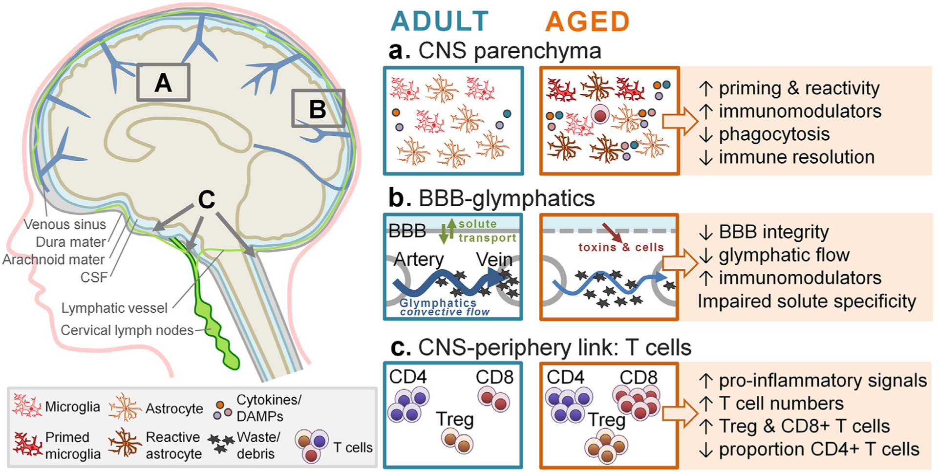
Aging is associated with compensatory shifts in CNS physiology and function, which collaborate to increase neuroimmune priming and pro-inflammatory bias. Left schematic: The neuroimmune system involves several key components: (1) parenchymal neuroimmune cells; (2) glymphatic flow, communication, and clearance via lymphatic vessels, dural venous sinuses, and cervical lymph nodes; and (3) bi-directional communication with the periphery via the vagus nerve, the gut-CNS axis, and other routes (omitted for clarity). Letters A-C highlight representative areas described in the right panel. Letters A-C highlight representative areas described in the right panel. a. The healthy adult CNS parenchyma contains homeostatic microglia and astrocytes, which maintain low baseline inflammatory tone, efficiently phagocytose cells and debris, and resolve sickness and inflammatory responses. The healthy aged CNS parenchyma includes neuroimmune cells with a “primed” phenotype - these cells are hyperreactive to insult, show impaired phagocytic capacity and processing, and have less effective immune resolution ability. The aged CNS also exhibits increased accumulation of inflammatory mediators, such as cytokines and damage-associated molecular patterns (DAMPs). Aging is also associated with aberrant parenchymal accumulation of immune cells typically found in the periphery, such as T cells. b. The healthy adult BBB maintains a cohesive barrier, enabling bi-directional active transport of specific proteins. The glymphatic system clears debris and excess metabolites. In the aged CNS, BBB integrity is reduced, enabling aberrant entry of toxins and cells, in parallel with reduced solute-specific transport. Glymphatic flow is less efficient at clearing debris; this amplifies accumulation of immunomodulatory mediators. c. CNS-periphery communication in the healthy adult contributes to low inflammatory tone at baseline; with aging, increased T cell numbers and shifted T cell proportions contribute to increased pro-inflammatory bias.
CNS-immune communication in adulthood and aging
The CNS and the immune system regulate one another via bidirectional communication. Communication between the immune system and CNS is exemplified by a typical sickness response. Upon sensing an infection, peripheral immune cells such as macrophages, neutrophils, and dendritic cells newly produce proinflammatory cytokines and other mediators. Proinflammatory signaling is communicated to the CNS through three main routes: direct neural signaling via vagal afferents; passive diffusion of cytokines and infiltration of immune cells in the meninges and at circumventricular organs; and tightly regulated transport of mediators and cells across the blood–brain barrier (BBB) (reviewed in Ref. [3]). Once an inflammatory signal reaches the CNS, microglia and other parenchymal neuroimmune cells secrete pro-inflammatory cytokines to propagate transient neuroinflammation and to elicit sickness behaviors [3]. An effective neuroimmune response is adaptive in promoting behavioral changes that improve survival probability and limit sickness spread, but also resolves efficiently to reattain homeostasis and resume crucial daily activities. Indeed, both parenchymal and meningeal immune responses in the healthy adult are biased towards dampened or anti-inflammatory reactivity, thereby supporting efficient immune resolution [4,5].
CNS-immune interfaces have unique anatomical and physiological features that limit excess immune signaling while enabling crosstalk. The BBB is a unique endothelial barrier: its endothelial cells strictly control the passage of cells and solutes via cell-linking tight junctions, expression of transporters for key mediators, low-level expression of adhesion molecules, and absence of fenestrations [6]. The endothelial cell barrier is reinforced by an endothelial basement membrane, pericytes, and surrounding astrocytic endfeet [7]. This barrier is critical in preventing unrestricted access of immune cell to the CNS, yet additional immune cells reside in surrounding CNS spaces. Immune cells in the meningeal compartment – including macrophages, T cells, B cells, dendritic cells, and others – can produce immunomodulators in response to peripheral immune signals [6,8,9]. The meninges consists of an outer layer called dura mater, which contains peripheral cells, related molecules, and meningeal lymphatic vessels; and the more tightly-regulated meningeal layers abutting the CNS called the arachnoid and pia mater, which contain the cerebrospinal fluid (CSF) [7]. Meningeal immune cell populations are maintained during health and bolstered during inflammation via the overlying skull and vertebral bodies, which contain bone marrow [10,11].
The effectiveness of the immune and neuroimmune systems degrades with age. Importantly, this age-related decline typically begins in middle age but progresses more rapidly at late ages (middle age: humans ~38–47 years old, mice 10–15 months old; old age: humans ~56–69 years old; mice ~18–24 months old) (Figure 2) [12]. The aged immune system has impaired sensing and clearance of pathogens: innate immune cells are less sensitive, adaptive immune cells are reduced in number and less diverse, and responses are skewed towards exaggerated pro-inflammatory reactivity [13]. The aged CNS also has shifted immune reactivity. Microglia exhibit neuroimmune priming – this involves reduced neuron-induced dampening of microglial reactivity and heightened expression of proinflammatory machinery. Ultimately, microglia priming leads to pathological hyperreactivity to challenges such as infection or injury [13] (Figure 1a). “Danger signals” and cytokines can also accumulate in the parenchyma to higher levels with age due to impaired glymphatic plumbing [14] (Figure 1b). Further, immune-to-CNS communication is amplified with age, including an aging-induced pro-inflammatory bias in meningeal immune cells and increased leakiness of the BBB [6,15] (Figure 1b,c). For example, T cells can accumulate in the aged CNS parenchyma [16], suggesting that age-related detriments to barrier stability enable aberrant entry of peripheral immune cells.
Figure 2.

Timeline of aging and neuroimmunity in humans and mice. Top: Comparison of life history phases of humans (in years) and mice (in months) – including pre-puberty, young adult, middle aged, and aged phases. The final age of the “aged” phase is a stage at which ~85% of individuals (humans or mice) remain alive. Bottom: Neuroimmune activities across the lifespan. Neuroimmunity through young adulthood is characterized by high inflammatory resolution capacity and phagocytic efficiency, and low baseline inflammatory tone. Middle age is a time of shifting neuroinflammatory milieu. The aged human and mouse has amplified neuroinflammatory priming and reactivity, and impaired ability to phagocytose and resolve inflammation.
Aging-related shifts in neuroimmune reactivity conspire to exaggerate and extend neuroimmune responses. In turn, overzealous pro-inflammatory reactivity with aging can cause prolonged or even permanent detriments on physiologic and behavioral function. Below, we discuss age-related inflammatory changes in CNS immune cells and in signaling from CNS-adjacent compartments.
Glial cells maintain homeostasis and undergo functional changes with age
Aging is associated with widespread shifts in CNS inflammatory state, including increased oxidative stress, reduced neurogenesis, increased risk for region-specific atrophy and neurodegeneration, and a broad increase in inflammatory tone [17]. Various cell types contribute to heightened inflammatory tone in the aging brain. The CNS is composed of neurons, glial cells (such as microglia, oligodendrocytes, ependymal cells, and astrocytes), infiltrating immune cells, as well as nonglial CNS-resident cells (such as perivascular macrophages, pericytes, and endothelial cells). In particular, glial cells shape the shifting neuroimmune environment and undergo stereotypical neuroimmune changes with age – e.g., neuroimmune priming and a pro-inflammatory bias [17]. With healthy aging, these neuroimmune changes may be protective or compensatory mechanisms that are responding to the gradual degradation of the system. Unfortunately, these aging-related neuroimmune shifts can also increase CNS vulnerability and damage.
Microglia: roles in the adult and aged CNS
Microglia are the primary immune patroller of the CNS and are critical for maintaining homeostasis in the brain throughout the lifespan. Under healthy adult conditions, microglia perform key maintenance activities including detecting shifts in inflammatory milieu, engaging in synaptic pruning, providing trophic support to CNS cells, and contributing to BBB function (reviewed in Ref. [13]). Microglia are maintained in a homeostatic, surveillent, and inactivated state by constitutive baseline ligand-receptor signaling with neurons (e.g., CD200-CD200L, CX3CL1-CX3CR1) (reviewed in Ref. [13]). Recent RNA sequencing strategies have enabled the classification of these homeostatic microglia based on their transcriptional profile: there is greater microglia heterogeneity in heathy murine adult brain and microglia express higher levels of genes including Tmem119, P2ry12, and Cx3cr1 [18]. Microglia are dynamic cells that respond to their environment to shift phenotype and function. Upon detecting CNS damage via danger signals and/or proinflammatory cytokines, microglia migrate and proliferate in sites of damage and upregulate inflammatory machinery (e.g., MHCII, scavenger receptors, toll-like receptors, and components of the complement cascade). In the initial phase of an immune response, microglia produce and secrete inflammatory cytokines and chemokines – this period of proinflammatory activity aims to sterilize or kill pathogens. Subsequently, or in parallel, microglia act as a cleanup crew engaging in phagocytic activity – this pro-resolution phase acts to repair tissue and resolve inflammation [3].
With aging, microglia become progressively more proinflammatory and dysfunctional. Aged microglia reduce the expression of homeostatic microglia effectors and increase the expression of inflammatory genes, including Spp1, Itgax, Axl, Lgals3, Clec7a, Trem2, and Cd6818. Several populations of microglia expand with age, including those that express macrophage-specific inflammatory markers (e.g., chemokines Ccl4 and Ccl3) and the inflammatory cytokine IL-1β [17,18]. This increase in chemokine signaling may facilitate homing of immune cell populations to the CNS. Furthermore, with aging, inflammatory microglia have an impaired ability to shift to a pro-resolution cell state – often persisting in a proinflammatory state for much longer than adult microglia [19,20]. For example, following a peripheral E. coli immune challenge, adult rats resolve the CNS inflammatory response within 24 h; however, aged rats exhibit increases in inflammatory cytokines such as IL-1β in the brain for more than a week [1]. This increase in pro inflammatory activity in the aged CNS is primarily driven by microglia.
The ability of microglia to resolve an inflammatory response and engage in phagocytic activity is critical for maintaining CNS homeostasis (for example see Ref. [21]). Therefore, age-related impairments in pro-resolution pathways and phagocytic processing likely exacerbate pathological inflammatory states. Healthy adult microglia engage in homeostatic phagocytosis of excess synapses and cellular debris. During an effective inflammatory response, microglial phagocytosis and processing of extracellular debris shift microglia intracellular signaling towards a resolution cell state. With aging, deficient phagocytic processing precedes microglia-induced neuroinflammation [22]. Aged rodent and human microglia exhibit a striking buildup of lipid droplets, indicative of impaired phagocytic processing [23]. Indeed, the percentage of microglia considered “lipid laden” increases from 12% in young adult mice to 50% in aged mice [23]. Microglia with excess lipid burden have defective ongoing phagocytosis [23]. In addition, lipid droplet-rich microglia have a distinct transcriptome signature compared to microglia from the same animals with a lower lipid content: lipid-laden cells upregulate genes related to the production of nitric oxide, reactive oxygen species, and other pro-inflammatory pathways [23]. This pro-inflammatory, dysfunctional phenotype that occurs in lipid-laden microglia is likely caused by deficits in microglia sterol synthesis, and these pathologic microglial cell states are observed in other contexts, including models of multiple sclerosis [24]. Effective phagocytosis by microglia during aging and pathology is enabled via the immunoglobulin family cell surface receptor, TREM2. TREM2-deficient microglia are unable to adapt to excess cholesterol exposure, form fewer lipid droplets, and build up endoplasmic reticulum stress [21].
Changes in microglia function across the aged brain are heterogeneous. Beginning in middle age in mice (13 months old), microglia exhibit regional variation in their age-associated phenotype [22]. Microglia undergo region-specific expansion with age: the number of microglia increases in the cortex, hippocampus, CNS white matter tracts, and basal ganglia structures [22]. The transcriptional signature of microglia also varies by brain region. Whereas microglia in aged grey matter exist predominantly in homeostatic cell states, microglia in aged white matter develop cell states related to reactivity and phagocytic removal of degenerating myelin [25]. Proteomic analyses further support region-specific vulnerability. Although metabolic changes occur across the entire aging brain [26], the magnitude of metabolic and neuroinflammatory shifts differ regionally. Aging-related changes in microglia function are likely affected by the CNS inflammatory milieu. Indeed, aging alters the proteome of CSF [27], which can influence microglia phenotype and function [28].
Despite the apparent detrimental nature of these microglial-driven pro-inflammatory states, microglia are also crucial for limiting aging-related susceptibility to insult or degeneration. Microglial depletion in aged male mice prior to experimental stroke increased infarct size and infiltration of hematogenous macrophages and neutrophils [29]. In a 5xfAD mouse model of Alzheimer’s disease, microglial depletion caused cerebral amyloid angiopathy, increased hemorrhages, and earlier death [30]. Compared to microglia-competent 5xfAD mice, microglia-depleted 5xfAD mice lacked protective TREM2 expression and microglial-related signaling with endothelial cells and pericytes (e.g., via TGF-β and PDGF-β pathways) [30]. Thus, microglia in the aged brain also have protective roles in maintaining BBB integrity and limiting runaway inflammation.
Astrocytes: roles in the adult and aged CNS
Astrocytes, the most abundant glial cell, are vital for the proper function of the CNS. In the healthy adult, astrocytes maintain CNS homeostasis by providing physical support; reinforcing the BBB; regulating glymphatic function; modulating synaptic transmission; taking up and metabolizing neurotransmitters; and releasing cytokines, chemokines, and neurotrophic factors. As with microglia, astrocyte phenotype and function shift with age. Astrocyte numbers increase with age, which may relate to a neuron-to-glia fate switch that promotes neural stem cells to preferentially differentiate into astrocytes with age [31]. This age-related dysregulation in neuron vs. astrocyte genesis may contribute to reduced hippocampal neurogenesis, which may underlie cognitive deficits with aging [31].
Astrocytes also display regional differences in vulnerability to aging: astrocytes show increased reactive cell states with age, particularly in the hippocampus, hypothalamus, and cerebellum [32–34]. Elevated astrocyte reactivity in mice emerges around 13–14 months old, and further increases by older ages (20 months) [35]. Aged astrocytes (>20 months in mice) appear more similar to those observed in disease states such as Alzheimer’s disease [35]. Aged astrocytes are enriched for immune pathways, and show reductions in pathways related to cholesterol synthesis and synapse elimination [20,33,34]. There is likely heterogeneity in astrocytic changes throughout the brain with age. For example, in response to inflammatory stimuli (e.g., LPS), individual astrocytes undergo distinct inflammatory transitions [36]. Aged astrocytes also show morphological changes, including reduced number and length of astrocytic processes and smaller territorial domains [37] (although this is likely regionally dependent [38]). Functionally, aged astrocytes reduce intercellular coupling, which may reflect a decrease in intercellular connectivity through gap junctions [37]. Further, aged astrocytes have reduced efficiency of K+ clearance and glutamate uptake [37] (although see Ref. [39]). Astrocyte atrophy and reduced activity in the aged brain are associated with impaired synaptic plasticity [37]. Aged astrocytes can also newly secrete toxic lipid meditators to kill nearby neurons and oligodendrocytes, although this occurs mainly in degenerative conditions rather than with healthy aging [40].
Astrocytes and microglia also display shifting intercellular communication with age. Microglial pro-resolution cell states are sustained by astrocyte-derived anti-inflammatory and cholesterol biosynthesis pathways, these neuroimmune-dampening pathways are dysregulated with age [20]. Microglia also drive more reactive phenotypes in astrocytes: blocking microglia-secreted cytokines (e.g., IL-1α, TNFα, and C1q) with genetic or pharmacological methods prevents the development of reactive astrocyte phenotypes [32]. Further, in inflammatory or neurodegenerative conditions, microglia release fragmented mitochondria that induce astrocyte reactivity and propagate neuroinflammation [41].
Sex differences are apparent in the neuroimmune environment with age, with females developing a more pro-inflammatory [42,43] and reduced phagocytic signature [44] at baseline. These sex-specific cellular responses may underlie sex differences in neuroinflammatory disorders. For example, aging females are predisposed to autoimmune disorders, whereas aging males are more susceptible to infectious diseases. Sex differences in neuroimmune aging has been discussed in more detail elsewhere [45,46] – more work is necessary in this area.
In addition to intrinsic shifts in aged CNS cell states, factors extrinsic to CNS cells also promote proinflammatory cell states with age. For example, baseline accumulation of damage-associated molecular patterns, such as HMGB1, can sensitize or “prime” the microglia phenotype in aged rats [19,47]. Similarly, microglial priming or reactivity is driven by myriad additional age-related factors, including proinflammatory-biased peripheral immune responses [48], increased BBB permeability [15], and impaired CNS waste clearance mechanisms [14] (see below). The relative contribution of extrinsic vs. intrinsic factors driving the age-associated phenotype in microglia has recently been addressed using microglia depletion and repopulation strategies. Repopulating CNS microglia in aged mice can ameliorate aspects of inflammatory priming, however, repopulated microglia retain distinct pathologic phenotypes and function from microglia in healthy young adult mice [22,49,50]. This suggests that a combination of environmental and cell-intrinsic factors collaborate to evoke age-related microglial priming.
Taken together, aged CNS cells – particularly microglia and astrocytes – develop primed or reactive cell states that drive exaggerated inflammatory responses. In addition, the parenchyma of the aged CNS accumulates excess danger signals due to impaired glymphatic clearance, and peripheral-to-CNS immune signaling in aging has a pro-inflammatory bias. Thus, CNS cells, extracellular mediators, and peripheral signaling collaborate during aging to amplify immune sensitivity, proinflammatory states, and behavioral detriments. Next, we consider how age-induced changes in communication at the brain-immune interface impact neuroimmune signaling.
Input from surrounding CNS spaces shape the neuroimmune environment with age
The BBB and the blood change with age
The BBB protects the brain from unregulated contact with the blood and its contents: the BBB gates entry of peripheral immune cells and inflammatory signaling molecules into the CNS. The BBB enforces barriers not just at the levels of the vasculature (primarily regulated by tight junctions between endothelial cells) but also in the choroid plexus (regulated by ependymal cells), the circumventricular organs (regulated by tanycytes), and the meninges (primarily regulated by epithelial cells) (recently reviewed in Ref. [15]). BBB endothelial cells prevent leakage between barrier cells through tight junctions, which are protein complexes that bind adjacent barrier cells. Because the BBB prevents the diffusion of blood-borne molecules into the brain, it uses influx and efflux transporters for tasks such as importing nutrients and exporting toxins [15]. Specialized barrier features of the BBB change or degrade with age leading to impaired barrier function. Although BBB morphology remains mostly intact with healthy brain aging [15], the aged BBB shows increased permeability [51,52] and region-specific vulnerability to breakdown (e.g., BBB breakdown typically begins in aged human hippocampus) [53]. There is also a shift in active transport across the BBB with aging. In the healthy adult, specific plasma proteins readily permeate the parenchyma, regulated by transporters in the BBB. With aging, however, plasma protein uptake is diminished in mice and there is a shift in transport from ligand-specific receptor-mediated to non-specific caveolar transcytosis [54]. This shift alters the composition of proteins entering the CNS, enabling excess entry with age of potentially toxic proteins such as albumin, fibrinogen, and autoantibodies that lack canonical transporting receptors [54].
Cells critical to maintaining the BBB shift their cell state with age towards a more senescent phenotype that includes functional deficits related to cellular stress, DNA damage, and replication [55]. With aging, 50% more endothelial cells in mice had a senescent signature (adult 3 month vs. aged 28 month-old: 5% vs. 10% senescent CNS endothelial cells, respectively) [56]. Endothelial cell dysfunction is driven by aging-related loss of a key longevity and stressor response protein, sirtuin-1 [51]. Another regulator of BBB breakdown is the APOE4 ε4 allele, which increases risk for late-onset AD. APOE4 ε4 causes degeneration of capillary-adjacent pericytes and BBB breakdown in the hippocampus and medial temporal lobe, which exacerbate cognitive impairment with aging [57]. APOE4 ε4 accelerates BBB breakdown by unleashing CSF expression of the potent metalloproteinase MMP-9 [57]. Overall, natural and progressive breakdown of the BBB in aged human brain is associated with cognitive impairment [53].
In addition to local CNS changes, the blood contains mediators that regulate aging brain function. For example, reconstituting hematopoietic stem cells in young mice with those from aged mice inhibits hippocampal neurogenesis and impairs cognitive function [58]. Conversely, transferring plasma from aged or young mice that exercise to aged sedentary mice can improve cognition [59]. This may in part be mediated by a liver-derived enzyme called Gpld1, which cleaves glycosylphosphatidylinositol-anchored substrates to initiate potentially CNS-protective signaling cascades [59]. Thus, identifying factors mediating neuroimmune changes with age may aid the development of novel therapeutics.
Aging influences the repertoire of peripheral immune cells that signal to the CNS
Immune cells in spaces surrounding but outside the CNS parenchyma can strongly influence CNS function in animals across the lifespan. Immune cells can reside outside the BBB but within CNS spaces such as the choroid plexus and meninges. Within the parenchyma in healthy adult animals, immune cells are typically limited to resident glia with few peripheral immune cells and a lack of adaptive immune cells. However, the meningeal and choroid plexus compartments contain diverse immune cell types, including monocytes, dendritic cells, T cells, B cells, natural killer cells, and others [6]. Cells in the meningeal space are replenished and recruited from bone marrow in the overlaying skull and vertebral bodies [10,11]. Meningeal immune cells can initiate or propagate neuroimmune signals, and during pathology can infiltrate CNS parenchyma. Given these key roles in regulating neuroimmunity, it follows that peripheral immune cells in the meninges regulate complex behaviors such as cognition and social behavior [60,61]. For example, T cells modulate learning and memory, with depletion of CD4+ Tcells worsening memory and long-term potentiation. T cells mediate these changes through IL-4-dependent signaling interactions with GABAergic neurons [60]. T cell signaling from the meningeal compartment via interferon (IFN)-γ/JAK-STAT regulates neuronal GABAergic neuronal function, driving changes in social behavior [61]. Additionally, mice lacking γδ Tcells or IL-17 exhibit deficits in short-term memory, suggesting that meningeal γδ T cell-derived IL-17 is required for plasticity and short-term memory [62].
In the aged meningeal compartment, T cell numbers increase and T cell subpopulations shift to promote widespread pro-inflammatory bias. T cell numbers in the CNS parenchyma increase modestly in mice from 2 to 18 months of age, and then increase steeply between 18 and 24 months [63]. T cells critically contribute to age-related CNS dysfunction: Rag1−/− mice, which lack T cells, are protected against age-related degeneration and declines in motor and cognitive function. In addition to modulating T cell number, aging shifts the proportions of meningeal T cell subpopulations. Rag1−/− hosts that receive Cd8−/− bone marrow transplant show reduced axon degeneration (vs. WT bone marrow), suggesting that cytotoxic CD8+ T cells drive degeneration and associated behavioral changes with age [63]. Systemic inflammation also fosters CD8+-evoked degeneration in the aged CNS [63]. In addition, the aged mouse meningeal Tcell population is enriched for regulatory T cells (CD4+FoxP3+) [64]. This increase in regulatory T cells may be mediated by age-related decreases in CCR7 signaling: CCR7 deficiency leads to accumulation of regulatory T cells in the meningeal compartment, concomitant with impaired glymphatic function and deficits in learning and memory [64]. Thus, aging causes parallel pro-inflammatory shifts in the CNS and in the peripheral immune compartment, thereby reinforcing CNS and cognitive detriments.
Glymphatic and lymphatic clearance of CNS metabolic waste is impaired with age
A burgeoning area of neuroscience involves the relationship between neuroinflammation and CNS waste clearance. The glymphatic system is essential to CNS nutrient delivery and waste clearance and consists of a network of perivascular tunnels wrapped by astrocytic endfeet. CSF within the subarachoid and cisternal spaces flow into the brain through a periarterial route and exchanges with interstitial fluid (ISF, in the parenchyma) via Aquaporin-4, a water channel on astrocytic endfeet [65]. Flow from the CSF to the ISF likely occurs from cerebral arteries, and efflux of waste and metabolites from the ISF occurs via the cerebral veins (for reviews, see Refs. [6,7]). Effective glymphatic flow and clearance in the adult prevent excess accumulation of macromolecules and potentially toxic mediators, such as danger signals and cytokines. Glymphatic flow drives the collection of ISF in perivenous spaces which then drains to meningeal lymphatic vessels [66]. These vessels exist in the dura mater and track venous sinuses in the brain and exist between vertebrae in the spinal cord [66–69]. The close proximity between lymphatic flow, blood, and dural sinuses support strong communication between immune cells in these sites. T cells and B cells are enriched in dural sinuses [16,70], thereby facilitating bi-directional communication between the CNS and adaptive immune system. Macromolecules and immune cells are cleared via the meningeal lymphatic vessels to the deep cervical lymph nodes [66–68]. Effective and consistent glymphatic and lymphatic draining appears important for maintaining CNS homeostasis [64,71].
In addition to BBB breakdown with aging enabling aberrant entry of inflammatory mediators into the CNS, aging also dysregulates clearance of factors from the CNS. Aging impairs glymphatic system function, which allows the pathologic extracellular accumulation of metabolites, danger signals, and neurodegenerative factors (e.g., tau) [14]. Aquaporin-4 enables elimination of extracellular tau from the brain to CSF and subsequently to deep cervical lymph nodes. In the brains of transgenic mice predisposed to tau pathology, deletion of Aquaporin-4 further amplified tau deposition in CSF, and exacerbated phosphorylated tau accumulation and associated neurodegeneration [72].
Meningeal lymphatic clearance is similarly dysregulated with aging. Impairments in meningeal lymphatic function slow the paravascular efflux of macromolecules from the interstitial fluid in aged mice [71]. This process likely contributes to age-related impairments in cognition: improving meningeal lymphatic drainage of CSF macromolecules by treating aged mice with vascular endothelial growth factor C ameliorates impairments in learning and memory [71]. Disrupting lymphatic vessels can also exacerbate disease pathology in a mouse model of Alzheimer’s disease by leading to access accumulation of amyloid β71. Thus, impaired glymphatic and lymphatic clearance with age drive the accumulation of pathological macromolecules in the CNS and increase the risk of neurodegeneration.
Targeting peripheral-to-CNS immune crosstalk to extend healthy aging
The connection between peripheral immunity and the CNS can be leveraged to modulate neuroinflammatory outcomes with aging. For instance, the gut microbiota influences aging-associated changes in microglia [73]. Modifying the aged gut microbiota with microbiota transfer can improve cognitive function [74,75] and ameliorate age-related changes in the neuroimmune environment [75]. Fetal microbiota transfer may benefit the neuroimmune state by reversing age-associated increases in δ-valerobetaine, a gut microbiota-derived metabolite, in the blood and CNS. Interestingly, the CNS benefits of fetal microbiota transplant can also occur independent of microglia [74]; young microbiota transplant has been shown to ameliorate the probability of CD8+ Tcell activation [75]. In addition to modifying the endogenous microbiome, introducing commensal environmental bacteria can alleviate changes in the neuroimmune environment with age. Bifidobacterium adolescentis supplementation increases catalase activity and host metabolism and improves health span and lifespan in multiple species (Chen et al. Nature aging, 2021). Furthermore, immunizing aged rats with a commensal bacteria, Mycobacterium vaccae, can protect against age-related increases in neuroinflammation and cognitive dysfunction in response to surgery [2,76]. M. vaccae treatment may act through shifting T cell-CNS signaling, thereby reducing microglia priming [2,76]. Taken together, this work demonstrates that “rescuing” peripheral immune signals with aging can ameliorate changes in the neuroimmune environment with age.
Conclusions and implications
The aging CNS exhibits notable resilience compared to other body systems, yet it still displays increased vulnerability over time. One cause of age-related CNS vulnerability is its bias towards a pro-inflammatory phenotype, which occurs due to parallel physiologic shifts. These concomitant age-related changes include neuroimmune priming of CNS cells; increased BBB leakiness; pro-inflammatory state of peripheral signaling; and impaired glymphatic clearance. Age-related priming and pro-inflammatory phenotype render the CNS hypersensitive to secondary challenges, such as infection or damage. Furthermore, the aged CNS is vulnerable to a number of neurodegenerative conditions (recently reviewed in Ref. [17]). Age-related CNS inflammation and barrier breakdown start in crucial regions – such as the hippocampus – so there is a need to understand the mechanisms of initial BBB breakdown and how this can be ameliorated. Further, it is unclear whether age-related neuroinflammation is typically ignited by a specific primary event, or if it is a widespread progressive response to systemic degradation. Overall, revealing beneficial and detrimental aspects of the aged neuroinflammatory milieu could inform novel approaches for improving healthy aging. Future studies should illuminate strategies that dampen age-related neuroimmune pathology and CNS vulnerability, with an overall goal of improving health span.
Acknowledgements
This work was supported by the National Institutes of Health R01AG062716 and R01AG078758 to LKF, and by Mission Connect, a program of the TIRR Foundation (ADG).
Footnotes
Declaration of competing interest
The authors have no conflicts of interest to declare.
Data availability
No data was used for the research described in the article.
References
Papers of particular interest, published within the period of review, have been highlighted as:
• of special interest
•• of outstanding interest
- 1.Barrientos RM, Frank MG, Hein AM, Higgins EA, Watkins LR, Rudy JW, Maier SF: Time course of hippocampal IL-1 beta and memory consolidation impairments in aging rats following peripheral infection. Brain Behav Immun 2009, 23:46–54, 10.1016/j.bbi.2008.07.002. [DOI] [PMC free article] [PubMed] [Google Scholar]
- 2.Fonken LK, Frank MG, D’Angelo HM, Heinze JD, Watkins LR, Lowry CA, Maier SF: Mycobacterium vaccae immunization protects aged rats from surgery-elicited neuroinflammation and cognitive dysfunction. Neurobiol Aging 2018, 71:105–114, 10.1016/j.neurobiolaging.2018.07.012. Epub 2018/08/18 [DOI] [PMC free article] [PubMed] [Google Scholar]
- 3.Buckley MW, McGavern DB: Immune dynamics in the CNS and its barriers during homeostasis and disease. Immunol Rev 2022, 306:58–75, 10.1111/imr.13066. Epub 2022/01/25 [DOI] [PMC free article] [PubMed] [Google Scholar]
- 4.Van Hove H, Martens L, Scheyltjens I, De Vlaminck K, Pombo Antunes AR, De Prijck S, Vandamme N, De Schepper S, Van Isterdael G, Scott CL, Aerts J, Berx G, Boeckxstaens GE, Vandenbroucke RE, Vereecke L, Moechars D, Guilliams M, Van Ginderachter JA, Saeys Y, Movahedi K: A single-cell atlas of mouse brain macrophages reveals unique transcriptional identities shaped by ontogeny and tissue environment. Nat Neurosci 2019, 22:1021–1035, 10.1038/s41593-019-0393-4. Epub 2019/05/08 [DOI] [PubMed] [Google Scholar]
- 5.Dulken BW, Buckley MT, Navarro Negredo P, Saligrama N, Cayrol R, Leeman DS, George BM, Boutet SC, Hebestreit K, Pluvinage JV, Wyss-Coray T, Weissman IL, Vogel H, Davis MM, Brunet A: Single-cell analysis reveals T cell infiltration in old neurogenic niches. Nature 2019, 571:205–210, 10.1038/s41586-019-1362-5. Epub 2019/07/05 [DOI] [PMC free article] [PubMed] [Google Scholar]
- 6.Mastorakos P, McGavern D: The anatomy and immunology of vasculature in the central nervous system. Sci Immunol 2019, 4, 10.1126/sciimmunol.aav0492. Epub 2019/07/14 [DOI] [PMC free article] [PubMed] [Google Scholar]
- 7.Cugurra A, Mamuladze T, Rustenhoven J, Dykstra T, Beroshvili G, Greenberg ZJ, Baker W, Papadopoulos Z, Drieu A, Blackburn S, Kanamori M, Brioschi S, Herz J, Schuettpelz LG, Colonna M, Smirnov I, Kipnis J: Skull and vertebral bone marrow are myeloid cell reservoirs for the meninges and CNS parenchyma. Science 2021, 373, 10.1126/science.abf7844. Epub 2021/06/05 [DOI] [PMC free article] [PubMed] [Google Scholar]
- 8.Herisson F, Frodermann V, Courties G, Rohde D, Sun Y, Vandoorne K, Wojtkiewicz GR, Masson GS, Vinegoni C, Kim J, Kim DE, Weissleder R, Swirski FK, Moskowitz MA, Nahrendorf M: Direct vascular channels connect skull bone marrow and the brain surface enabling myeloid cell migration. Nat Neurosci 2018, 21:1209–1217, 10.1038/s41593-018-0213-2. Epub 2018/08/29 [DOI] [PMC free article] [PubMed] [Google Scholar]
- 9.Salvador AF, de Lima KA, Kipnis J: Neuromodulation by the immune system: a focus on cytokines. Nat Rev Immunol 2021, 21:526–541, 10.1038/s41577-021-00508-z. Epub 2021/03/03 [DOI] [PubMed] [Google Scholar]
- 10.Derecki NC, Quinnies KM, Kipnis J: Alternatively activated myeloid (M2) cells enhance cognitive function in immune compromised mice. Brain Behav Immun 2011, 25:379–385, 10.1016/j.bbi.2010.11.009. [DOI] [PMC free article] [PubMed] [Google Scholar]
- 11.Rua R, Lee JY, Silva AB, Swafford IS, Maric D, Johnson KR, McGavern DB: Infection drives meningeal engraftment by inflammatory monocytes that impairs CNS immunity. Nat Immunol 2019, 20:407–419, 10.1038/s41590-019-0344-y. Epub 2019/03/20 [DOI] [PMC free article] [PubMed] [Google Scholar]
- 12.Dutta S, Sengupta P: Men and mice: relating their ages. Life Sci 2016, 152:244–248, 10.1016/j.lfs.2015.10.025. Epub 2015/11/26 [DOI] [PubMed] [Google Scholar]
- 13.Fonken LK, Frank MG, Gaudet AD, Maier SF: Stress and aging act through common mechanisms to elicit neuroinflammatory priming. Brain Behav Immun 2018:133–138. [DOI] [PMC free article] [PubMed] [Google Scholar]
- 14.Kress BT, Iliff JJ, Xia M, Wang M, Wei HS, Zeppenfeld D, Xie L, Kang H, Xu Q, Liew JA, Plog BA, Ding F, Deane R, Nedergaard M: Impairment of paravascular clearance pathways in the aging brain. Ann Neurol 2014, 76:845–861, 10.1002/ana.24271. [DOI] [PMC free article] [PubMed] [Google Scholar]
- 15.Banks WA, Reed MJ, Logsdon AF, Rhea EM, Erickson MA: Healthy aging and the blood-brain barrier. Nat Aging 2021, 1:243–254, 10.1038/s43587-021-00043-5. Epub 2021/08/10 [DOI] [PMC free article] [PubMed] [Google Scholar]
- 16.Rustenhoven J, Drieu A, Mamuladze T, de Lima KA, Dykstra T, Wall M, Papadopoulos Z, Kanamori M, Salvador AF, Baker W, Lemieux M, Da Mesquita S, Cugurra A, Fitzpatrick J, Sviben S, Kossina R, Bayguinov P, Townsend RR, Zhang Q, Erdmann-Gilmore P, Smirnov I, Lopes MB, Herz J, Kipnis J: Functional characterization of the dural sinuses as a neuroimmune interface. Cell 2021, 184:1000–1016, 10.1016/j.cell.2020.12.040. e27. Epub 2021/01/29 [DOI] [PMC free article] [PubMed] [Google Scholar]
- 17.Scheiblich H, Trombly M, Ramirez A, Heneka MT: Neuroimmune connections in aging and neurodegenerative diseases. Trends Immunol 2020, 41:300–312, 10.1016/j.it.2020.02.002. Epub 2020/03/10 [DOI] [PubMed] [Google Scholar]
- 18.Hammond TR, Dufort C, Dissing-Olesen L, Giera S, Young A, Wysoker A, Walker AJ, Gergits F, Segel M, Nemesh J, Marsh SE, Saunders A, Macosko E, Ginhoux F, Chen J, Franklin RJM, Piao X, McCarroll SA, Stevens B: Single-cell RNA sequencing of microglia throughout the mouse lifespan and in the injured brain reveals complex cell-state changes. Immunity 2019, 50: 253–271, 10.1016/j.immuni.2018.11.004. e6. Epub 2018/11/26 [DOI] [PMC free article] [PubMed] [Google Scholar]
- 19.Fonken LK, Frank MG, Kitt MM, D’Angelo HM, Norden DM, Weber MD, Barrientos RM, Godbout JP, Watkins LR, Maier SF: The alarmin HMGB1 mediates age-induced neuroinflammatory priming. J Neurosci 2016, 36:7946–7956, 10.1523/JNEUROSCI.1161-16.2016. [DOI] [PMC free article] [PubMed] [Google Scholar]
- 20.O’Neil SM, Hans EE, Jiang S, Wangler LM, Godbout JP: Astrocyte immunosenescence and deficits in interleukin 10 signaling in the aged brain disrupt the regulation of microglia following innate immune activation. Glia 2022, 70:913–934, 10.1002/glia.24147. Epub 2022/01/22 [DOI] [PubMed] [Google Scholar]
- 21.Gouna G, Klose C, Bosch-Queralt M, Liu L, Gokce O, Schifferer M, Cantuti-Castelvetri L, Simons M: TREM2-dependent lipid droplet biogenesis in phagocytes is required for remyelination. J Exp Med 2021, 218, 10.1084/jem.20210227. Epub 2021/08/24 No other disclosures were reported. [DOI] [PMC free article] [PubMed] [Google Scholar]
- 22. •.Moca EN, Lecca D, Hope KT, Etienne F, Schaler AW, Espinoza K, Chappell MS, Gray DT, Tweedie D, Sidhu S, Masukawa L, Sitoy H, Mathew R, Saban DR, Greig NH, De Biase LM: Microglia drive pockets of neuroinflammation in middle age. J Neurosci 2022, 42:3896–3918, 10.1523/JNEUROSCI.1922-21.2022. Epub 2022/04/10 [DOI] [PMC free article] [PubMed] [Google Scholar]; This manuscript investigated region-specific changes in microglia across the lifespan in mixed sex C57Bl/6 mice with a focus on the nucleus accumbens and ventral tegmental area (VTA). Age-associated increases in microglia density in brain regions such as the VTA begin as early as 13 months and parallel increases in pro-inflammatory cytokines, whereas brain regions that maintain adult-like microglia density do not exhibit increases in neuroinflammatory markers with age. Microglia responses to aging occur independent of increases in astrocytes or neuronal loss, suggesting that cell intrinsic age-associated changes may occur in microglia. Ablating and allowing microglia to repopulate mitigated region-specific microglia changes in middle aged (13–20 months) mice.
- 23. ••.Marschallinger J, Iram T, Zardeneta M, Lee SE, Lehallier B, Haney MS, Pluvinage JV, Mathur V, Hahn O, Morgens DW, Kim J, Tevini J, Felder TK, Wolinski H, Bertozzi CR, Bassik MC, Aigner L, Wyss-Coray T: Lipid-droplet-accumulating microglia represent a dysfunctional and proinflammatory state in the aging brain. Nat Neurosci 2020, 23:194–208, 10.1038/s41593-019-0566-1. Epub 2020/01/22 [DOI] [PMC free article] [PubMed] [Google Scholar]; This manuscript investigated cytoplasmic content of microglia in adult (3 months) and aged (20 months) male mice (C57Bl/6 background) and determined that lipid droplet-containing microglia were four-fold more prevalent in aged (52%) compared to young (12%) hippocampus. This increase in lipid droplets also occurred in the hippocampus of aged compared to young humans. Microglia with high lipid droplet content exhibited cellular dysfunction including dysregulated phagocytosis and high levels of reactive oxygen species production. A CRISPR-Cas9 screen identified progranulin, a gene associated with neurodegenerative disease risk, as a genetic modifier of lipid droplet formation.
- 24.Berghoff SA, Spieth L, Sun T, Hosang L, Schlaphoff L, Depp C, Duking T, Winchenbach J, Neuber J, Ewers D, Scholz P, van der Meer F, Cantuti-Castelvetri L, Sasmita AO, Meschkat M, Ruhwedel T, Mobius W, Sankowski R, Prinz M, Huitinga I, Sereda MW, Odoardi F, Ischebeck T, Simons M, Stadelmann-Nessler C, Edgar JM, Nave KA, Saher G: Microglia facilitate repair of demyelinated lesions via post-squalene sterol synthesis. Nat Neurosci 2021, 24:47–60, 10.1038/s41593-020-00757-6. Epub 2020/12/23 [DOI] [PMC free article] [PubMed] [Google Scholar]
- 25.Safaiyan S, Besson-Girard S, Kaya T, Cantuti-Castelvetri L, Liu L, Ji H, Schifferer M, Gouna G, Usifo F, Kannaiyan N, Fitzner D, Xiang X, Rossner MJ, Brendel M, Gokce O, Simons M: White matter aging drives microglial diversity. Neuron 2021, 109: 1100–1117, 10.1016/j.neuron.2021.01.027. e10. Epub 2021/02/20 [DOI] [PubMed] [Google Scholar]
- 26.Ding J, Ji J, Rabow Z, Shen T, Folz J, Brydges CR, Fan S, Lu X, Mehta S, Showalter MR, Zhang Y, Araiza R, Bower LR, Lloyd KCK, Fiehn O: A metabolome atlas of the aging mouse brain. Nat Commun 2021, 12:6021, 10.1038/s41467-021-26310-y. Epub 2021/10/17 [DOI] [PMC free article] [PubMed] [Google Scholar]
- 27.Shuken SR, Rutledge J, Iram T, Losada PM, Wilson EN, Andreasson KI, Leib RD, Wyss-Coray T: Limited proteolysis-mass spectrometry reveals aging-associated changes in cerebrospinal fluid protein abundances and structures. Nature Aging 2022, 2:379–388. [DOI] [PMC free article] [PubMed] [Google Scholar]
- 28.Willis CM, Nicaise AM, Krzak G, Ionescu RB, Pappa V, D’Angelo A, Agarwal R, Repolles-de-Dalmau M, Peruzzotti-Jametti L, Pluchino S: Soluble factors influencing the neural stem cell niche in brain physiology, inflammation, and aging. Exp Neurol 2022, 355, 114124, 10.1016/j.expneurol.2022.114124. Epub 2022/06/02 [DOI] [PubMed] [Google Scholar]
- 29.Marino Lee S, Hudobenko J, McCullough LD, Chauhan A: Microglia depletion increase brain injury after acute ischemic stroke in aged mice. Exp Neurol 2021, 336, 113530, 10.1016/j.expneurol.2020.113530. Epub 2020/11/23 [DOI] [PMC free article] [PubMed] [Google Scholar]
- 30.Kiani Shabestari S, Morabito S, Danhash EP, McQuade A, Sanchez JR, Miyoshi E, Chadarevian JP, Claes C, Coburn MA, Hasselmann J, Hidalgo J, Tran KN, Martini AC, Chang Rothermich W, Pascual J, Head E, Hume DA, Pridans C, Davtyan H, Swarup V, Blurton-Jones M: Absence of microglia promotes diverse pathologies and early lethality in Alzheimer’s disease mice. Cell Rep 2022, 39, 110961, 10.1016/j.celrep.2022.110961. Epub 2022/06/16 [DOI] [PMC free article] [PubMed] [Google Scholar]
- 31. •.White CW 3rd, Fan X, Maynard JC, Wheatley EG, Bieri G, Couthouis J, Burlingame AL, Villeda SA: Age-related loss of neural stem cell O-GlcNAc promotes a glial fate switch through STAT3 activation. Proc Natl Acad Sci U S A 2020, 117: 22214–22224, 10.1073/pnas.2007439117. Epub 2020/08/28 [DOI] [PMC free article] [PubMed] [Google Scholar]; This manuscript revealed that age related decreases in neurogenesis are associated with a fate switch of neural stem cells in the hippocampus of male mice (C57BL/6J background). By 6 months of age, mice exhibited a loss of the posttranslational modification, O-linked β-N-acetylglucosamine; this deficit is associated with reduced neurogenesis in favor of increased gliogenesis. RNA-sequencing of neural stem cells revealed that altered STAT3 signaling likely drives these shifts in cellular differentiation.
- 32.Clarke LE, Liddelow SA, Chakraborty C, Munch AE, Heiman M, Barres BA: Normal aging induces A1-like astrocyte reactivity. Proc Natl Acad Sci U S A 2018, 115:E1896–E1905, 10.1073/pnas.1800165115. Epub 2018/02/14 [DOI] [PMC free article] [PubMed] [Google Scholar]
- 33.Boisvert MM, Erikson GA, Shokhirev MN, Allen NJ: The aging astrocyte transcriptome from multiple regions of the mouse brain. Cell Rep 2018, 22:269–285, 10.1016/j.celrep.2017.12.039. Epub 2018/01/04 [DOI] [PMC free article] [PubMed] [Google Scholar]
- 34.Pan J, Ma N, Yu B, Zhang W, Wan J: Transcriptomic profiling of microglia and astrocytes throughout aging. J Neuroinflammation 2020, 17:97, 10.1186/s12974-020-01774-9. Epub 2020/04/03 [DOI] [PMC free article] [PubMed] [Google Scholar]
- 35.Habib N, McCabe C, Medina S, Varshavsky M, Kitsberg D, Dvir-Szternfeld R, Green G, Dionne D, Nguyen L, Marshall JL, Chen F, Zhang F, Kaplan T, Regev A, Schwartz M: Disease-associated astrocytes in Alzheimer’s disease and aging. Nat Neurosci 2020, 23:701–706, 10.1038/s41593-020-0624-8. Epub 2020/04/29 [DOI] [PMC free article] [PubMed] [Google Scholar]
- 36.Hasel P, Rose IVL, Sadick JS, Kim RD, Liddelow SA: Neuroinflammatory astrocyte subtypes in the mouse brain. Nat Neurosci 2021, 24:1475–1487, 10.1038/s41593-021-00905-6. Epub 2021/08/21 [DOI] [PubMed] [Google Scholar]
- 37. •.Popov A, Brazhe A, Denisov P, Sutyagina O, Li L, Lazareva N, Verkhratsky A, Semyanov A: Astrocyte dystrophy in ageing brain parallels impaired synaptic plasticity. Aging Cell 2021, 20, e13334, 10.1111/acel.13334. Epub 2021/03/07 [DOI] [PMC free article] [PubMed] [Google Scholar]; Using two-photon microscopy, this manuscript revealed reduced astrocyte branching and astrocyte-to-astrocyte coupling in the hippocampus of aged (20–24 months) male C57Bl/6J mice. Changes in astrocyte morphology correlated with functional deficits in K+ and glutamate clearance in old astrocytes, which paralleled decreases in synaptic long-term potentiation in hippocampal CA1.
- 38.Bondi H, Bortolotto V, Canonico PL, Grilli M: Complex and regional-specific changes in the morphological complexity of GFAP(+) astrocytes in middle-aged mice. Neurobiol Aging 2021, 100:59–71, 10.1016/j.neurobiolaging.2020.12.018. Epub 2021/01/26 [DOI] [PubMed] [Google Scholar]
- 39.Gomez-Gonzalo M, Martin-Fernandez M, Martinez-Murillo R, Mederos S, Hernandez-Vivanco A, Jamison S, Fernandez AP, Serrano J, Calero P, Futch HS, Corpas R, Sanfeliu C, Perea G, Araque A: Neuron-astrocyte signaling is preserved in the aging brain. Glia 2017, 65:569–580, 10.1002/glia.23112. Epub 2017/01/29 [DOI] [PMC free article] [PubMed] [Google Scholar]
- 40.Guttenplan KA, Weigel MK, Prakash P, Wijewardhane PR, Hasel P, Rufen-Blanchette U, Munch AE, Blum JA, Fine J, Neal MC, Bruce KD, Gitler AD, Chopra G, Liddelow SA, Barres BA: Neurotoxic reactive astrocytes induce cell death via saturated lipids. Nature 2021, 599:102–107, 10.1038/s41586-021-03960-y. Epub 2021/10/08 [DOI] [PMC free article] [PubMed] [Google Scholar]
- 41.Joshi AU, Minhas PS, Liddelow SA, Haileselassie B, Andreasson KI, Dorn GW 2nd, Mochly-Rosen D: Fragmented mitochondria released from microglia trigger A1 astrocytic response and propagate inflammatory neurodegeneration. Nat Neurosci 2019, 22:1635–1648, 10.1038/s41593-019-0486-0. Epub 2019/09/26 [DOI] [PMC free article] [PubMed] [Google Scholar]
- 42.Mangold CA, Wronowski B, Du M, Masser DR, Hadad N, Bixler GV, Brucklacher RM, Ford MM, Sonntag WE, Freeman WM: Sexually divergent induction of microglial-associated neuroinflammation with hippocampal aging. J Neuroinflammation 2017, 14:141, 10.1186/s12974-017-0920-8. Epub 2017/07/25 [DOI] [PMC free article] [PubMed] [Google Scholar]
- 43.Kang SS, Ebbert MTW, Baker KE, Cook C, Wang X, Sens JP, Kocher JP, Petrucelli L, Fryer JD: Microglial translational profiling reveals a convergent APOE pathway from aging, amyloid, and tau. J Exp Med 2018, 215:2235–2245, 10.1084/jem.20180653. Epub 2018/08/08 [DOI] [PMC free article] [PubMed] [Google Scholar]
- 44.Guillot-Sestier MV, Araiz AR, Mela V, Gaban AS, O’Neill E, Joshi L, Chouchani ET, Mills EL, Lynch MA: Microglial metabolism is a pivotal factor in sexual dimorphism in Alzheimer’s disease. Commun Biol 2021, 4:711, 10.1038/s42003-021-02259-y. Epub 2021/06/12 [DOI] [PMC free article] [PubMed] [Google Scholar]
- 45.Bronikowski AM, Meisel RP, Biga PR, Walters JR, Mank JE, Larschan E, Wilkinson GS, Valenzuela N, Conard AM, de Magalhaes JP, Duan JE, Elias AE, Gamble T, Graze RM, Gribble KE, Kreiling JA, Riddle NC: Sex-specific aging in animals: perspective and future directions. Aging Cell 2022, 21, e13542, 10.1111/acel.13542. Epub 2022/01/25 [DOI] [PMC free article] [PubMed] [Google Scholar]
- 46.Darling JS, Sanchez K, Gaudet AD, Fonken LK. The role of microglia in brain aging: a focus on sex differences. Oxford Research Encyclopedia of Neuroscience 2020. [Google Scholar]
- 47.Frank MG, Weber MD, Fonken LK, Hershman SA, Watkins LR, Maier SF: The redox state of the alarmin HMGB1 is a pivotal factor in neuroinflammatory and microglial priming: a role for the NLRP3 inflammasome. Brain Behav Immun 2016, 55: 215–224, 10.1016/j.bbi.2015.10.009. [DOI] [PMC free article] [PubMed] [Google Scholar]
- 48.Fulop T, Larbi A, Dupuis G, Le Page A, Frost EH, Cohen AA, Witkowski JM, Franceschi C: Immunosenescence and inflamm-aging as two sides of the same coin: friends or foes? Front Immunol 2017, 8:1960, 10.3389/fimmu.2017.01960. Epub 2018/01/30 [DOI] [PMC free article] [PubMed] [Google Scholar]
- 49.Elmore MRP, Hohsfield LA, Kramar EA, Soreq L, Lee RJ, Pham ST, Najafi AR, Spangenberg EE, Wood MA, West BL, Green KN: Replacement of microglia in the aged brain reverses cognitive, synaptic, and neuronal deficits in mice. Aging Cell 2018, 17, e12832, 10.1111/acel.12832. Epub 2018/10/03 [DOI] [PMC free article] [PubMed] [Google Scholar]
- 50.O’Neil SM, Witcher KG, McKim DB, Godbout JP: Forced turnover of aged microglia induces an intermediate phenotype but does not rebalance CNS environmental cues driving priming to immune challenge. Acta Neuropathol Commun 2018, 6:129, 10.1186/s40478-018-0636-8. Epub 2018/11/28 [DOI] [PMC free article] [PubMed] [Google Scholar]
- 51. •.Stamatovic SM, Martinez-Revollar G, Hu A, Choi J, Keep RF, Andjelkovic AV: Decline in Sirtuin-1 expression and activity plays a critical role in blood-brain barrier permeability in aging. Neurobiol Dis 2019, 126:105–116, 10.1016/j.nbd.2018.09.006. Epub 2018/09/10 [DOI] [PMC free article] [PubMed] [Google Scholar]; This manuscript evaluated BBB integrity in young (2–6 months), middle aged (6–12 months), and aged (16–22 months) mixed sex C57Bl/6 mice: mice of increasing age had heightened BBB permeability for a range of molecular sized tracers. Age-associated increases in BBB permeability were associated with reduced claudin-5, a major BBB occlusion protein in both mouse and human microvessels. Further, gene array revealed that age-related BBB permeability related to reduced Sirt1; Sirt1 loss- and gain-of-function manipulations support its role in modulating age-associated changes in the BBB.
- 52.Zhao L, Li Z, Vong JSL, Chen X, Lai HM, Yan LYC, Huang J, Sy SKH, Tian X, Huang Y, Chan HYE, So HC, Ng WL, Tang Y, Lin WJ, Mok VCT, Ko H: Pharmacologically reversible zonation-dependent endothelial cell transcriptomic changes with neurodegenerative disease associations in the aged brain. Nat Commun 2020, 11:4413, 10.1038/s41467-020-18249-3. Epub 2020/09/06 [DOI] [PMC free article] [PubMed] [Google Scholar]
- 53.Montagne A, Barnes SR, Sweeney MD, Halliday MR, Sagare AP, Zhao Z, Toga AW, Jacobs RE, Liu CY, Amezcua L, Harrington MG, Chui HC, Law M, Zlokovic BV: Blood-brain barrier breakdown in the aging human hippocampus. Neuron 2015, 85:296–302, 10.1016/j.neuron.2014.12.032. Epub 2015/01/23 [DOI] [PMC free article] [PubMed] [Google Scholar]
- 54.Yang AC, Stevens MY, Chen MB, Lee DP, Stahli D, Gate D, Contrepois K, Chen W, Iram T, Zhang L, Vest RT, Chaney A, Lehallier B, Olsson N, du Bois H, Hsieh R, Cropper HC, Berdnik D, Li L, Wang EY, Traber GM, Bertozzi CR, Luo J, Snyder MP, Elias JE, Quake SR, James ML, Wyss-Coray T: Physiological blood-brain transport is impaired with age by a shift in transcytosis. Nature 2020, 583:425–430, 10.1038/s41586-020-2453-z. Epub 2020/07/03 [DOI] [PMC free article] [PubMed] [Google Scholar]
- 55.Hernandez-Segura A, Nehme J, Demaria M: Hallmarks of cellular senescence. Trends Cell Biol 2018, 28:436–453, 10.1016/j.tcb.2018.02.001. Epub 2018/02/27 [DOI] [PubMed] [Google Scholar]
- 56.Kiss T, Nyul-Toth A, Balasubramanian P, Tarantini S, Ahire C, DelFavero J, Yabluchanskiy A, Csipo T, Farkas E, Wiley G, Garman L, Csiszar A, Ungvari Z: Single-cell RNA sequencing identifies senescent cerebromicrovascular endothelial cells in the aged mouse brain. Geroscience 2020, 42:429–444, 10.1007/s11357-020-00177-1. Epub 2020/04/03 [DOI] [PMC free article] [PubMed] [Google Scholar]
- 57.Montagne A, Nation DA, Sagare AP, Barisano G, Sweeney MD, Chakhoyan A, Pachicano M, Joe E, Nelson AR, D’Orazio LM, Buennagel DP, Harrington MG, Benzinger TLS, Fagan AM, Ringman JM, Schneider LS, Morris JC, Reiman EM, Caselli RJ, Chui HC, Tcw J, Chen Y, Pa J, Conti PS, Law M, Toga AW, Zlokovic BV: APOE4 leads to blood-brain barrier dysfunction predicting cognitive decline. Nature 2020, 581:71–76, 10.1038/s41586-020-2247-3. Epub 2020/05/08 [DOI] [PMC free article] [PubMed] [Google Scholar]
- 58.Smith LK, Verovskaya E, Bieri G, Horowitz AM, von Ungern-Sternberg SNI, Lin K, Seizer P, Passegue E, Villeda SA: The aged hematopoietic system promotes hippocampal-dependent cognitive decline. Aging Cell 2020, 19, e13192, 10.1111/acel.13192. Epub 2020/10/20 [DOI] [PMC free article] [PubMed] [Google Scholar]
- 59.Horowitz AM, Fan X, Bieri G, Smith LK, Sanchez-Diaz CI, Schroer AB, Gontier G, Casaletto KB, Kramer JH, Williams KE, Villeda SA: Blood factors transfer beneficial effects of exercise on neurogenesis and cognition to the aged brain. Science 2020, 369:167–173, 10.1126/science.aaw2622. Epub 2020/07/11 [DOI] [PMC free article] [PubMed] [Google Scholar]
- 60.Herz J, Fu Z, Kim K, Dykstra T, Wall M, Li H, Salvador AF, Zou B, Yan N, Blackburn SM, Andrews PH, Goldman DH, Papadopoulos Z, Smirnov I, Xie XS, Kipnis J: GABAergic neuronal IL-4R mediates T cell effect on memory. Neuron 2021, 109:3609–3618, 10.1016/j.neuron.2021.10.022. e9. Epub 2021/11/19 [DOI] [PMC free article] [PubMed] [Google Scholar]
- 61.Filiano AJ, Xu Y, Tustison NJ, Marsh RL, Baker W, Smirnov I, Overall CC, Gadani SP, Turner SD, Weng Z, Peerzade SN, Chen H, Lee KS, Scott MM, Beenhakker MP, Litvak V, Kipnis J: Unexpected role of interferon-gamma in regulating neuronal connectivity and social behaviour. Nature 2016, 535:425–429, 10.1038/nature18626. Epub 2016/07/15 [DOI] [PMC free article] [PubMed] [Google Scholar]
- 62.Ribeiro M, Brigas HC, Temido-Ferreira M, Pousinha PA, Regen T, Santa C, Coelho JE, Marques-Morgado I, Valente CA, Omenetti S, Stockinger B, Waisman A, Manadas B, Lopes LV, Silva-Santos B, Ribot JC: Meningeal gammadelta T cell-derived IL-17 controls synaptic plasticity and short-term memory. Sci Immunol 2019, 4, 10.1126/sciimmunol.aay5199. Epub 2019/10/13 [DOI] [PMC free article] [PubMed] [Google Scholar]
- 63. ••.Groh J, Knopper K, Arampatzi P, Yuan X, LoBlein L, Saliba A, Kastenmuller W, Martini R: Accumulation of cytotoxic T cells in the aged CNS leads to axon degeneration and contributes to cognitive and motor decline. Nature Aging 2021, 1:357–367. [DOI] [PubMed] [Google Scholar]; Using mixed sex mice with a C57Bl/6 background, this manuscript demonstrated that the aging CNS (optic nerve) exhibits a shift in T cell populations: the proportion of CD8+ T cells moderately increases from 2 to 18 mos, then steeply increases from 18 to 24 mos. This increase in CD8+ T cells is also apparent in aged human CNS tissue. The accumulation of CD8+ T cells is associated with axon damage; absence of mature lymphocytes in Rag1−/− mice attenuated axon loss and motor and cognitive impairments. RNA-sequencing and flow cytometry analyses demonstrated that CD8+ T cells from aged mice (24 months; vs. those from 12 month-old adults) develop a more cytotoxic phenotype, with chronic T cell receptor signaling and activation.
- 64. •.Da Mesquita S, Herz J, Wall M, Dykstra T, de Lima KA, Norris GT, Dabhi N, Kennedy T, Baker W, Kipnis J: Aging-associated deficit in CCR7 is linked to worsened glymphatic function, cognition, neuroinflammation, and beta-amyloid pathology. Sci Adv 2021, 7, 10.1126/sciadv.abe4601. Epub 2021/05/23 [DOI] [PMC free article] [PubMed] [Google Scholar]; Meningeal T cells exhibit reductions in the chemokine CCR7 in aged mice. Inducing a loss of CCR7 in adult mice recapitulated features of the aging brain including an enhanced meningeal Treg response and impairments in cognition. Targeting Tregs in the aged brain with an anti-CD25 antibody improved cognition.
- 65.Iliff JJ, Wang M, Liao Y, Plogg BA, Peng W, Gundersen GA, Benveniste H, Vates GE, Deane R, Goldman SA, Nagelhus EA, Nedergaard M: A paravascular pathway facilitates CSF flow through the brain parenchyma and the clearance of interstitial solutes, including amyloid beta. Sci Transl Med 2012, 4: 147ra11, 10.1126/scitranslmed.3003748. [DOI] [PMC free article] [PubMed] [Google Scholar]
- 66.Louveau A, Smirnov I, Keyes TJ, Eccles JD, Rouhani SJ, Peske JD, Derecki NC, Castle D, Mandell JW, Lee KS, Harris TH, Kipnis J: Structural and functional features of central nervous system lymphatic vessels. Nature 2015, 523:337–341, 10.1038/nature14432. [DOI] [PMC free article] [PubMed] [Google Scholar]
- 67.Aspelund A, Antila S, Proulx ST, Karlsen TV, Karaman S, Detmar M, Wiig H, Alitalo K: A dural lymphatic vascular system that drains brain interstitial fluid and macromolecules. J Exp Med 2015, 212:991–999, 10.1084/jem.20142290. [DOI] [PMC free article] [PubMed] [Google Scholar]
- 68.Ahn JH, Cho H, Kim JH, Kim SH, Ham JS, Park I, Suh SH, Hong SP, Song JH, Hong YK, Jeong Y, Park SH, Koh GY: Meningeal lymphatic vessels at the skull base drain cerebrospinal fluid. Nature 2019, 572:62–66, 10.1038/s41586-019-1419-5.Epub 2019/07/26 [DOI] [PubMed] [Google Scholar]
- 69.Jacob L, Boisserand LSB, Geraldo LHM, de Brito Neto J, Mathivet T, Antila S, Barka B, Xu Y, Thomas JM, Pestel J, Aigrot MS, Song E, Nurmi H, Lee S, Alitalo K, Renier N, Eichmann A, Thomas JL: Anatomy and function of the vertebral column lymphatic network in mice. Nat Commun 2019, 10:4594, 10.1038/s41467-019-12568-w.Epub 2019/10/11 [DOI] [PMC free article] [PubMed] [Google Scholar]
- 70.Schafflick D, Wolbert J, Heming M, Thomas C, Hartlehnert M, Borsch AL, Ricci A, Martin-Salamanca S, Li X, Lu IN, Pawlak M, Minnerup J, Strecker JK, Seidenbecher T, Meuth SG, Hidalgo A, Liesz A, Wiendl H, Meyer Zu Horste G: Single-cell profiling of CNS border compartment leukocytes reveals that B cells and their progenitors reside in non-diseased meninges. Nat Neurosci 2021, 24:1225–1234, 10.1038/s41593-021-00880-y. Epub 2021/07/14 [DOI] [PubMed] [Google Scholar]
- 71.Da Mesquita S, Louveau A, Vaccari A, Smirnov I, Cornelison RC, Kingsmore KM, Contarino C, Onengut-Gumuscu S, Farber E, Raper D, Viar KE, Powell RD, Baker W, Dabhi N, Bai R, Cao R, Hu S, Rich SS, Munson JM, Lopes MB, Overall CC, Acton ST, Kipnis J: Functional aspects of meningeal lymphatics in ageing and Alzheimer’s disease. Nature 2018, 560:185–191, 10.1038/s41586-018-0368-8. Epub 2018/07/27 [DOI] [PMC free article] [PubMed] [Google Scholar]
- 72.Ishida K, Yamada K, Nishiyama R, Hashimoto T, Nishida I, Abe Y, Yasui M, Iwatsubo T: Glymphatic system clears extracellular tau and protects from tau aggregation and neurodegeneration. J Exp Med 2022, 219, 10.1084/jem.20211275. Epub 2022/02/26 [DOI] [PMC free article] [PubMed] [Google Scholar]
- 73.Mossad O, Batut B, Yilmaz B, Dokalis N, Mezo C, Nent E, Nabavi LS, Mayer M, Maron FJM, Buescher JM, de Aguero MG, Szalay A, Lammermann T, Macpherson AJ, Ganal-Vonarburg SC, Backofen R, Erny D, Prinz M, Blank T: Gut microbiota drives age-related oxidative stress and mitochondrial damage in microglia via the metabolite N(6)-carboxymethyllysine. Nat Neurosci 2022, 25:295–305, 10.1038/s41593-022-01027-3. Epub 2022/03/05 [DOI] [PubMed] [Google Scholar]
- 74. •.Mossad O, Nent E, Woltemate S, Folschweiller S, Buescher JM, Schnept D, Erny E, Staeheli P, Bartos M, Szalay A, Stecher B, Vital M, Sauer JF, Lammermann T, Prinz M, Blank T: Microbiota-dependent increase in δ-valerobetaine alters neuronal function and is responsible for age-related cognitive decline. Nature Aging 2021, 1:1127–1136. [DOI] [PubMed] [Google Scholar]; Fecal microbiota transplant (FMT) from young donor (2 months) to aged recipient (15–16 months) male C57Bl/6J mice rescued age-related cognitive decline. Metabolomic analysis of the blood and brain of aged mice and humans established that aging is associated with increases in δ-valerobetaine, a gut microbiota-derived metabolite. Administering intraperitoneal δ-valerobetaine to young adult mice impaired learning and memory and increased inhibitory synaptic transmission. Depleting microglia did not prevent detriments of δ-valerobetaine on learning and memory, implying that this metabolite acts via microglia-independent mechanisms.
- 75.Boehme M, Guzzetta KE, Bastiaanssen TFS, van de Wouw M, Moloney GM, Gual-Grau A, Spichak S, Olavarria-Ramirez L, Fitzgerald P, Morillas E, Ritz NL, Jaggar M, Cowan CSM, Crispie F, Donoso F, Halitzki E, Neto MC, Sichetti M, Golubeva AV, Fitzgerald RS, Claesson MJ, Cotter PD, O’Leary OF, Dinan TG, Cryan JF: Microbiota from young mice counteracts selective age-assocated behavioral deficits. Nature Aging 2021, 1:666–676. [DOI] [PubMed] [Google Scholar]
- 76.Sanchez K, Darling JS, Kakkar R, Wu SL, Zentay A, Lowry CA, Fonken LK: Mycobacterium vaccae immunization in rats ameliorates features of age-associated microglia activation in the amygdala and hippocampus. Sci Rep 2022, 12:2165, 10.1038/s41598-022-05275-y. Epub 2022/02/11 [DOI] [PMC free article] [PubMed] [Google Scholar]
Associated Data
This section collects any data citations, data availability statements, or supplementary materials included in this article.
Data Availability Statement
No data was used for the research described in the article.