PLOS ONE logoLink to PLOS ONE
. 2023 Feb 9;18(2):e0280792. doi: 10.1371/journal.pone.0280792

Mutation of regulatory phosphorylation sites in PFKFB2 does not affect the anti-fibrotic effect of metformin in the kidney

Geoff Harley 1,*, Marina Katerelos 1, Kurt Gleich 1, Mardiana Lee 1, Peter F Mount 1,2, David A Power 1,2
Editor: Partha Mukhopadhyay3
PMCID: PMC9910667  PMID: 36757995

Abstract

The anti-fibrotic effect of metformin has been widely demonstrated. Fibrosis in the kidney after injury is associated with reduced expression of genes involved in both fatty acid and glycolytic energy metabolism. We have previously reported that the anti-fibrotic effect of metformin requires phosphoregulation of fatty acid oxidation by AMP-activated protein kinase (AMPK). To determine whether metformin also acts via regulation of glycolysis, we mutated regulatory phosphosites in the PFKFB2 isoform of 6-phosphofructo-2-kinase/fructose-2,6-biphosphatase (PFKFB2), a key regulator of glycolysis in the kidney. Mice with inactivating knockin (KI) mutations of the phosphorylation sites in PFKFB2 (PFKFB2 KI mice), which reduces the ability to increase the rate of glycolysis following stimulation, were used. Metformin was administered via drinking water to mice with a unilateral ureteric obstruction (UUO) model of renal fibrosis. In the PFKFB2 KI mice treated with metformin, there was decreased fibrosis and macrophage infiltration following UUO as assessed by Western blot for fibronectin and RT-PCR for α-smooth muscle actin, collagen 3, and F4.80, and confirmed by histology. Expression of the inducible PFKFB3 isoform was increased with metformin in UUO in both WT and PFKFB2 KI mice. There was no significant difference between WT and PFKFB2 KI mice treated with metformin in the degree of fibrosis following UUO in any of the Western blot or RT-PCR parameters that were measured. These data show that inhibition of the regulation of glycolysis by PFKFB2 does not diminish the anti-fibrotic effect of metformin in a model of renal fibrosis.

Introduction

The development of renal fibrosis in response to kidney injury is affected by reduced energy generation in tubular epithelial cells [1]. Fatty acid oxidation is the major source of energy in proximal tubular cells, although glycolysis becomes more important in the low oxygen environment of the medulla and distal nephron. Expression of genes involved in fatty acid oxidation in proximal tubules is reduced in human and experimental renal fibrosis [1]. Administration of drugs such as fenofibrate or metformin, which increase fatty acid oxidation, reduce renal fibrosis [2, 3]. We have reported that, in the case of metformin, the anti-fibrotic effect requires phosphorylation of acetyl-CoA-carboxylase (ACC), which controls entry of cytoplasmic long-chain fatty acids into mitochondria for subsequent β-oxidation [2]. Metformin is an indirect activator of the upstream protein kinase AMP-activated protein kinase (AMPK), which then phosphorylates ACC, leading to reduced fatty acid synthesis and increased fatty acid oxidation in mitochondria.

Interestingly, expression of glycolytic pathway genes is also reduced in proximal tubular cells in renal fibrosis [1]. There are three rate-limiting steps in the glycolytic conversion of a six-carbon ring to two 3-carbon pyruvate molecules, namely hexokinase, phosphofructokinase (PFK) and pyruvate kinase. The rate of glycolysis in cells is directly linked to the rate of glucose entry into the cells as well as the activity of these rate-limiting glycolytic enzymes [4].

Regulation of the rate of glycolysis is predominantly via modification of the activity of PFK. The intracellular level of PFK can be regulated by several factors, but one of the most important is the intracellular level of fructose-2,6-bisphosphate (Fru-2,6-P2), a product of the bifunctional enzyme 6-phosphofructo-2-kinase/fructose-2,6-biphosphatase (PFK-2/FBPase-2), which exists as four isoforms designated PFKFB1-4 [5]. In the kidney, PFKFB2 is described as the predominant isoform [6]. Activity of PFKFB2 is increased by C-terminal domain phosphorylation at Ser466 and Ser483. We have previously shown that inactivating mutations of these PFKFB2 phosphorylation sites, which is predicted to reduce the intracellular level of Fru-2,6-P2, reduced the ability to increase glycolysis in renal tubular cells [4].

Metformin is well established as having protective effects against renal fibrosis in mouse models of disease including ischaemia-reperfusion-injury, unilateral ureteric obstruction (UUO), folic acid nephropathy and cisplatin nephrotoxicity [2, 79]. In our previous studies, we have demonstrated that metformin acts, at least in part, by increasing AMPK activation and phosphorylation of ACC, thus stimulating mitochondrial fatty acid oxidation [2]. AMPK is also known to phosphorylate PFKFB2 at its C-terminal end, thereby increasing its activity [5]. In this study, we attempted to determine whether the anti-fibrotic effect of metformin was also dependent on regulation of glycolysis.

Methods

Generation of PFKFB2 KI mice

PFKFB2 KI mice with inactivating mutations of the phosphorylation sites of Ser466 and Ser483 were generated on a C57Bl/6 background by OzGene Pty Ltd, Bentley DC, WA, Australia as previously described [4]. In brief, phosphor-acceptor sites located in exon 15 of the mouse PFK2 gene were mutated to alanine, generating an inactivating mutation. Mouse genotypes were confirmed by PCR.

PFKFB2 KI mice were maintained as a homozygous line on a C57BL/6 background, whereas WT mice were derived from mating of heterozygous PFKFB2 KI mice and maintained as a separate line. As previously reported, there was no difference in plasma glucose or mouse weight between PFKFB2 KI mice and controls, however, the PFKFB2 KI kidneys were smaller and plasma urea was significantly less [4]. In histological studies, there was no abnormality seen in the PFKFB2 KI kidneys [4]. Cultured tubular epithelial cells from PFKFB2 KI mice have impaired glycolysis when analysis on the Seahorse analyser [4].

Animal renal fibrosis model

All experiments received prior approval from the Austin Health Animal Ethics Committee which operates under guidelines prepared by the National Health and Medical Research Council (NHMRC), the Commonwealth Scientific and Industrial Research Organisation (CSIRO) and Animal Welfare Victoria. Since this was an entirely animal study, the requirement for consent was waived. The unilateral ureteric obstruction (UUO) model was used to create renal fibrosis as previously described [10]. In brief, this involved taking male C57Bl/6 mice who were 8–10 weeks old and tying off one of their ureters via a standard surgical technique under isofluorane anaesthesia. Mice were monitored twice daily for weight loss, other signs of distress or evidence of poor health. They received buprenorphine subcutaneous injections for pain in the first three days post-operatively. Seven days later, the mice were sacrificed and a nephrectomy performed. This was done under ketamine anaesthesia with a sufficient dose to provide euthanasia. The obstructed kidney was then used for analysis. In one arm of the study metformin was also added to the mice’s drinking water for three days prior to and during the experiment. Liquid metformin hydrochloride (Focus Pharmaceuticals, London, UK) 0.08mg/mL was added into the drinking water and changed every 48 hours.

Cell culture

Primary cultures of renal tubular epithelial cells (TECs) were prepared by sieving whole mouse kidneys, as we have previously reported [2]. For cell culture stimulation, 4mM metformin (Focus Pharmaceuticals, London, UK) in serum-free media was added to the cells for a period of four hours immediately prior to harvesting. This was based on the experimental design used by Li et al. [7].

Western blot analysis

Kidney lysate preparation and Western blot analysis was performed via standard methods as previously described [11]. Western Blots were analyzed for densitometry using Image J software. The following antibodies were used in the Western Blot analysis: anti-phospho-PFKFB2 Ser483 antibody (Rabbit monoclonal antibody, Cell Signalling Technology, Massachusetts, USA), anti-phospho-ACC Ser79 antibody (ABCAM, Cambridge, UK), anti-hexokinase-1 antibody (Rabbit antibody, Cell Signalling Technology, Massachusetts, USA), anti-PFK1M antibody (Rabbit, Sigma-Aldrich, St Louis, USA), anti-α-Smooth muscle actin–FITC antibody (Sigma Aldrich, St. Louis, USA), anti-Fibronectin antibody (Rabbit monoclonal antibody, Sigma-Aldrich, St Louis, USA), anti-PKM2 antibody (Rabbit monoclonal antibody, Cell Signalling technology, Massachusetts, USA), anti-rabbit Immunoglobulin HRP-linked antibody (Swine polyclonal, Dako, Agilent Pathology Solutions, Santa Clara, USA), anti-Fluorescein-POD Fab fragments (Goat, Roche Applied Science, Indianapolis, USA). Anti-GAPDH (Rabbit monoclonal antibody, Cell Signalling Technology, Massachusetts, USA) was used as a loading control.

Histology

Kidneys were sliced in half transversely and fixed in formalin. Masson’s trichrome staining was performed by the Department of Anatomic Pathology, Austin Health. The percentage area occupied by collagen for Masson’s trichrome-stained sections was measured using Image J software. This quantitative analysis, performed from a set of images that had coverage of the whole renal cortex, was analysed and the values obtained. The amount of fibrosis weas expressed as a percentage of the whole cortical area.

Real-time polymerase chain reaction (qRT-PCR)

Total RNA was purified from whole mouse kidney samples and reverse transcribed for analysis as previously described [11]. This was performed using a Stratagen MX3000 real-time PCR system with Solis Biodyne EvaGreen master mix. The delta-delta Ct method was used to calculate relative expression [12]. Data was expressed as fold expression relative to littermate WT controls.

Statistics

Statistical analyses were performed using Prism version 7.0a for Mac OS X (GraphPad Software, San Diego, CA). Data are presented as mean + SD. Multiple group means were compared by two-way ANOVA followed by a post-hoc test. Comparison of means from two groups was performed by unpaired t-test. P values of <0.05 were considered significant.

Results

Phosphorylation of PFKFB2 and ACC following metformin stimulation in PFKFB2 KI cells

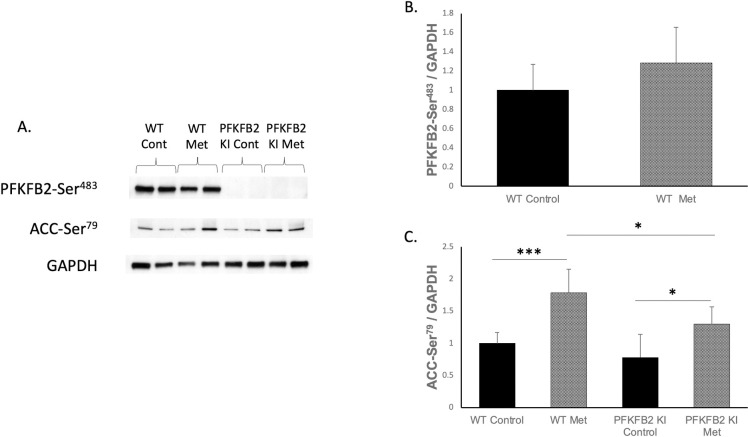
TECs cultured from WT and PFKFB2 KI mice were analyzed via Western blot for expression of phosphorylated PFKFB2 and ACC (Fig 1). As expected, expression of phosphorylated PFKFB2-Ser483 was not detectable in PFKFB2 KI mice (Fig 1A). Expression of phosphorylation at Ser466, the phosphorylation target for AMPK on PFKFB2, cannot be distinguished from a similar site in PFKFB3 using existing antibodies due to sequence overlap, so was not examined. AMPK phosphorylates PFKFB2 at its Ser466 site rather than Ser483 [5], so it was expected that metformin stimulation would not cause a significant increase in phosphorylation of that site, as shown (Fig 1A and 1B). Metformin stimulation of the TEC’s caused a significant increase in expression of phosphorylated ACC-Ser79 seen in both WT and PFKFB2 KI cells (Fig 1A and 1C). Expression of ACC-Ser79 was reduced in the PFKFB2 KI TEC’s compared to their WT counterparts (Fig 1A and 1C). We note that in this analysis, phosphorylated ACC and PFKFB2 are corrected for GAPDH rather than total ACC and PFKFB2, therefore, there is uncertainty as to whether the changes observed here are entirely explained by a change in the relative phosphorylation state, or whether there is also a contribution from a change in overall ACC or PFKFB2 expression.

Fig 1.

Fig 1

Expression of phosphorylation sites in cultured tubular epithelial cells from WT and PFKFB2 KI mice ± metformin stimulation (A-C). The mutated phosphorylation site at PFK-FB2-Ser483 was not detectable in the PFKFB2 KI mice as expected (A). Expression of phospho-Ser483 was not significantly different with metformin stimulation (B). There was increased expression of phosphorylated ACC-Ser79 phosphorylation with metformin stimulation in both WT and PFKFB2 KI cells (A, C ***p<0.001 and *p = 0.01 respectively). Expression of phosphorylated ACC-Ser79 was decreased in PFKFB2 KI metformin cells compared to WT cells (A, C *p = 0.02). Mean + SD.

Renal fibrosis in WT and PFKFB2 KI mice

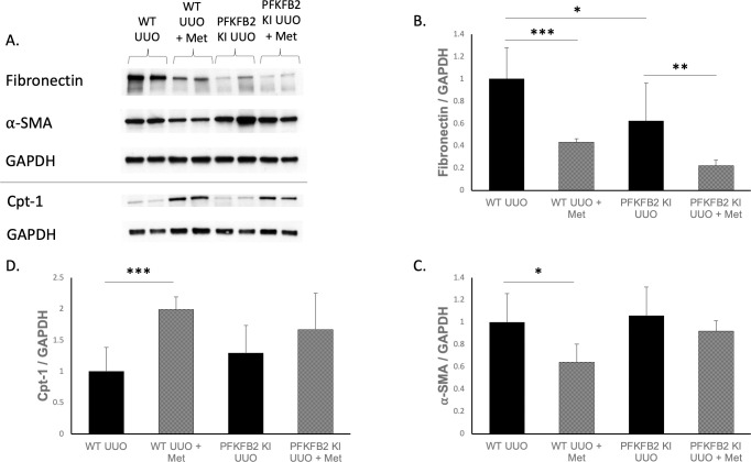
WT and PFKFB2 KI mice underwent UUO and their kidneys were analysed by Western blot (Fig 2), histology (Fig 3) and RT-PCR (Fig 4) for markers of fibrosis. We have previously reported that baseline fibrosis is not different in control PFKFB2 KI mice [4], therefore, sham UUO mice were not included in this study. PFKFB2 KI UUO control kidneys had reduced expression of fibronectin compared to their WT counterparts (Fig 2B) but this was not seen for α-SMA (Fig 2C). Expression of mRNA for α -SMA was reduced in PFKFB2 KI UUO control kidneys compared to their WT counterparts (Fig 4A) but this was not seen for fibronectin (Fig 4B). Cpt-1 expression was also increased in WT UUO + metformin kidneys compared to their WT counterparts (Fig 2D) with trends towards similar in PFKFB2 kidneys, suggestive of increased Cpt-1 activity and fatty acid oxidation with the addition of metformin.

Fig 2.

Fig 2

Western blot markers of fibrosis and metabolism following UUO in WT and PFKFB2 KI ± metformin kidneys (A-D). Analysis showed significantly reduced expression of fibronectin and ⍺-SMA in WT UUO metformin kidneys versus controls (A-C ***p<0.001 and *p = 0.01 respectively). PFKFB2 KI UUO controls had reduced fibronectin (A, B *p = 0.01) but not ⍺-SMA compared to WT (A, C). PFKFB2 KI UUO metformin had reduced fibronectin compared to PFKFB2 KI controls (A, B **p = 0.009) with a trend towards the same in ⍺-SMA (A, C). Expression of Cpt-1 was increased in WT UUO + metformin kidneys compared to WT UUO counterparts (A, D ***p = 0.007). Mean + SD.

Fig 3.

Fig 3

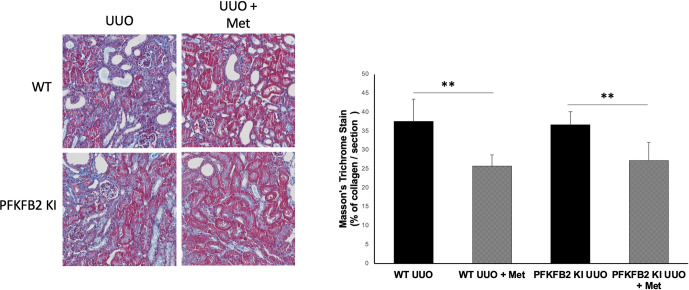
Masson’s trichrome staining of collagen deposition as a marker of fibrosis for WT and PFKFB2 KI UUO kidneys ± metformin showing representative sections (A) with corresponding quantification (B). Mice treated with metformin had less evidence of collagen deposition per section compared to their untreated counterparts for both WT (**p = 0.0025) and PFKFB2 KI mice (**p = 0.0094). There was no significant difference between WT and PFKFB2 groups. Quantification was performed from a set of images that provided coverage of the whole kidney cortex. Mean + SD.

Fig 4.

Fig 4

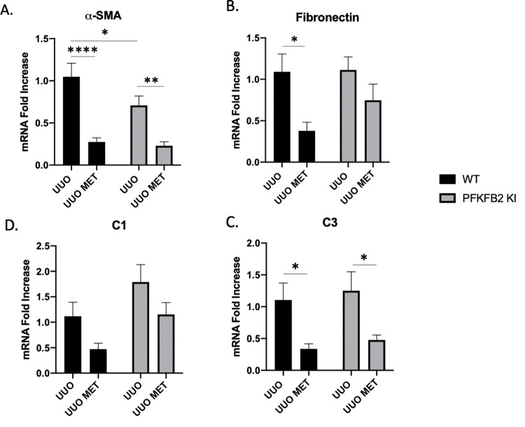
mRNA expression of markers of fibrosis for WT and PFKFB2 KI UUO kidneys ± metformin measured by RT-PCR (A-D). ⍺-smooth muscle actin (⍺-SMA) was significantly lower with the addition of metformin for WT and PFKFB2 KI kidneys (A ****p<0.001 and **p = 0.002 respectively). Similarly, mRNA expression of fibronectin was significantly less in WT UUO kidneys treated with metformin compared to control UUO comparisons (B *p = 0.02) with a trend towards similar effect in PFKFB2 KI kidneys. PFKFB2 KI UUO control kidneys had significantly less expression of ⍺-SMA compared to their WT counterparts (A *p = 0.04). mRNA expression of collagen 3 (C3) was significantly reduced with the addition of metformin (C, p<0.05) with trends towards a similar pattern in Collagen 1 (C1) (D) Mean + SD.

Collagen 1α and collagen 3α chain mRNA, products of the COL1A1 and COL1A3 genes respectively, were not significantly different between WT UUO and PFKFB2 KI UUO control samples (Fig 4C and 4D).

Taken together, we note that our observations using a variety of methods, including Masson Trichrome histology, Western blot (fibronectin and α-SMA), and RT-PCR (fibronectin, α-SMA, collagen 1 and 3), indicate that metformin protects against fibrosis in the UUO model, and that this effect is not altered in the PFKFB2 KI mice.

Effects of metformin in WT and PFKFB2 KI mice

Following metformin treatment, expression of fibronectin and α-SMA protein by Western blot was reduced in WT UUO kidneys (Fig 2A–2C) as well as PFKFB2 KI UUO kidneys for fibronectin (Fig 2A and 2B). When analysing the kidneys for mRNA expression of the same markers via RT-PCR (Fig 4A and 4B), α-SMA and fibronectin were significantly reduced in WT UUO kidneys with the addition of metformin. α-SMA was reduced with metformin in the PFKFB2 KI UUO kidneys (Fig 4A) with a trend towards the same in fibronectin (Fig 4B).

The degree of fibrosis was confirmed using a Masson’s trichrome stain for collagen in the tissues (Fig 3). Analysis demonstrated decreased collagen accumulation per section examined for both WT and PFKFB2 KI kidneys with the addition of metformin.

Collagen 1α and collagen 3α showed similar trends with reduced expression in mice treated with metformin, although only the results for collagen 3α were significant (Fig 4C and 4D).

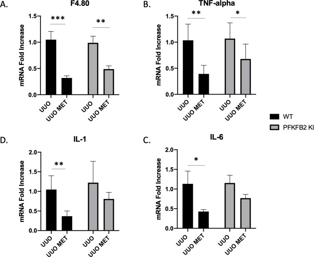
The reduction in macrophage numbers that has been reported with metformin in models of renal fibrosis was seen in both WT and PFKFB2 KI UUO kidneys (Fig 5A) and TNF-α as a marker of inflammation was also similarly reduced (Fig 5B). This is despite an increase in monocyte chemoattractant protein-1 (MCP-1) expression in PFKFB2 KI UUO + metformin kidneys compared to PFKFB2 KI UUO controls and WT UUO + metformin counterparts (S1B Fig). Interleukin 1 and interleukin 6 were also reduced in WT mice treated with metformin and there was a trend for PFKFB2 KI mice (Fig 5C and 5D). There was no significant difference in Sirtuin 3 mRNA expression between groups (S1A Fig).

Fig 5.

Fig 5

Measurement of markers of inflammation via RT-PCR in WT and PFKFB2 KI kidneys subjected to UUO ± metformin (A-D). mRNA expression of F4.80, TNF-α, Interleukin-1 (IL-1) and Interleukin-6 (IL-6) was significantly reduced with the addition of metformin in wild-type mice (A-D ***p = 0.0001, **p = 0.0016, *p = 0.0230, **p = 0.0008 respectively). In the PFKFB2 KI mice, there was decreased mRNA expression of F4.80 and TNF-α with the addition of metformin (A, B **p = 0.0024, *p = 0.0277 respectively). There was no significance difference between WT and PFKFB2 KI mouse groups in any of these parameters measured. Mean + SD.

Considering the lack of an effect of mutation of the regulatory phosphorylation sites of PFKB2 on the anti-fibrotic effect of metformin, we considered the possibility that this lack of effect might be explained by an effect of metformin on the expression of the inducible PFKFB3 isoform. Interestingly, expression of total PFKFB3 was increased in UUO mice treated with metformin for both WT and PFKFB2 KI groups (Fig 6).

Fig 6. Measurement of Western blot expression of total PFKFB3 in whole kidneys for WT and PFKFB2 KI UUO ± metformin.

Fig 6

Expression of Total PFKFB3 was increased in mice treated with metformin for both WT and PFKFB2 KI groups (A, B ***p = 0.0009, ****p<0.0001 respectively). Mean + SD.

Measurement of other rate-limiting steps in glycolysis

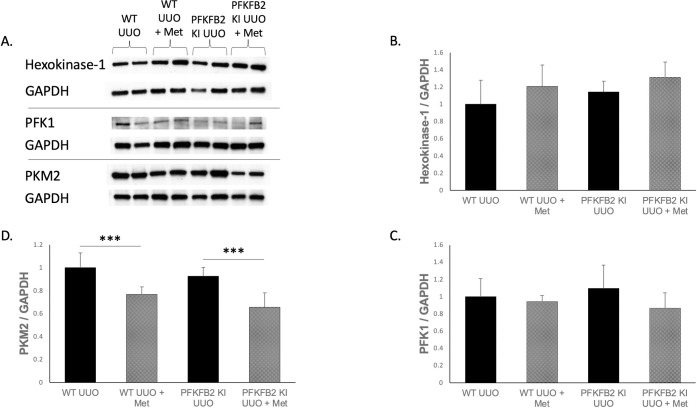
To determine whether other major rate-limiting steps were affected by the mutation of PFKFB2 phosphorylation sites or the addition of metformin, whole kidney lysates from mice were analyzed via Western blot for the expression of key enzymes in the glycolytic pathway (Fig 7). The most prevalent isoforms in the kidney for each of these rate-limiting enzymes was selected for analysis. Expression of Hexokinase-1 and PFK1 were not affected by the knock-in mutation nor stimulation by metformin (Fig 7B and 7C). PKM2 expression decreased with the addition of metformin in both WT and PFKFB2 KI samples (Fig 7D).

Fig 7.

Fig 7

Measurement of the other rate-limiting steps in glycolysis via Western blot for WT and PFKFB2 KI UUO ± metformin kidneys (A-D). The most prevalent form of each of these enzymes in the kidney was selected for analysis. Expression of hexokinase-1 and PFK-1 was not significantly altered by genotype or presence of metformin (B, C). PKM2 expression was significantly reduced in WT and PFKFB2 KI kidneys with the addition of metformin (D ***p = 0.001 and p<0.001 respectively). Mean + SD.

Discussion

In this study we demonstrated that metformin continues to have its protective effects in the UUO model of renal fibrosis in mice with a mutation of a key regulatory control point in glycolysis. When phosphorylated by protein kinases, PFKFB2 increases synthesis of fructose-2,6-bisphosphate, a strong activator of PFK1, the major control point in glycolysis [13]. Mutation of the two phosphorylation sites in its C-terminus reduces the increase in glycolysis seen after stimulation of cells with extracellular glucose [5]. Despite this change, metformin continued to reduce fibrosis in the UUO model. The PFKFB2 isoform was selected for investigation in this study because previous studies have found that it is the most prevalent isoform in the kidney [6]. PFKFB1 is not present in the kidney in detectable amounts, being most predominant in the heart, skeletal muscle and white adipose tissue. PFKFB4, the isoform of PFKFB specific to the testis only, is present in low levels in the kidney but is not regulated by AMPK, so is very unlikely to contribute to the action of metformin [5, 6].

Interestingly, the expression of the PFKFB3 isoform was increased in mice treated with metformin for both WT and PFKFB2 KI UUO groups, which has not previously been described. PFKFB3 is expressed predominantly in brain and placental tissue, and comprises a constitutive as well as an inducible isoform. Expression of the inducible isoform is thought to be low in adult tissues, high in tumour cell lines and increased by pro-inflammatory stimuli [14, 15]. For instance, expression has been shown to be increased in monocytes exposed to lipopolysaccharide as well as in response to hypoxia via the HIF-1 pathway, in addition to stimuli such as progestins, insulin and protein kinase C [1517]. Whilst AMPK-mediated phosphorylation of PFKFB3 is described in the literature, the mechanism of upregulation of total PFKFB3 levels in response to metformin is unknown. The potential effect of this upregulation might be to increase glycolysis, although downstream changes to suggest an increase in glycolysis were not seen; PFK-1 expression was unchanged and PKM2 expression was reduced with the addition of metformin as discussed below. Hence, the significance of this novel effect of metformin on PFKFB3 expression remains uncertain. It is possible that the observed increase in PFKFB3 might be a contributing reason to the lack of effect of mutation of the pFKFB2 phosphosites on the protective action of metformin observed in this study, although this speculation is unproven at the present time.

The protective effects of metformin were demonstrated in multiple markers of fibrosis via Western blot, histology and RT-PCR. The ongoing benefits that were seen in the PFKFB2 KI mice evidently reflect non-PFKFB2 mediated effects of metformin. These other effects of metformin could either be AMPK-mediated or AMPK-independent. Regarding AMPK-mediated effects, these may include metformin causing AMPK-mediated phosphorylation of ACC and consequent increased fatty acid oxidation, as this pathway was unaltered in these mice. This is evidenced by the ongoing ACC-Ser79 activation that was demonstrated with the addition of metformin in the PFKFB2 KI cells. A limitation of our study is that we do not have data on AMPK activity in the kidneys of the metformin treated mice, which was not possible to analyse as we did not employ a free-clamp methodology for harvesting the kidney tissue. Regarding other potential mechanisms of metformin, we noted that Cpt-1 and PFKFB3 expression were increased via Western blot in WT UUO + metformin kidneys with similar changes in the PFKFB2 KI UUO + metformin mice. Given that metformin still displays a protective effect in this PFKFB2 KI model, it would suggest that the PFK pathway and up-regulation of glycolysis for ATP generation is not essential to the anti-fibrotic effects of metformin, which appear to be primarily through fatty acid oxidation.

One unexpected result was reduced expression of the pyruvate kinase isoform PKM2 in both WT and PFKFB2 KI mice receiving metformin compared to those that did not. Previous work has shown increased PKM2 expression in UUO models compared to sham controls for both WT and PFKFB2 KI kidneys [4]. PKM2 expression decreased in UUO mice treated with metformin relative to untreated mice. This suggests that metformin reduced glycolysis, independent of its action on PFKFB2, in mice with fibrosis. This may represent an unexpected and novel action of metformin in disease. However, further studies are required to demonstrate a change in glycolysis. It is also worth noting that PKM2 is active in its tetrameric form, rather than the dimeric form. A limitation of our data is that Western blots are generally unable to distinguish the tetrameric from the dimeric form, so it is unclear whether the reduction in total PKM2 protein represents a change in the number of tetramers [18]. An additional limitation of these data is that glycolysis was not directly measured, which would require either ex vivo studies, or measurement of tissue metabolite levels, which were not available for this study.

In summary, this study shows that metformin continues to have the effect of reducing renal fibrosis in a UUO model, despite the absence of one of the major regulatory mechanisms in control of glycolysis. This indicates that regulation of glycolysis does not play a significant role in mediating the anti-fibrotic effect of metformin.

Supporting information

S1 Fig

Measurement of mRNA expression via RT-PCR of other markers for WT and PFKFB2 KI UUO ± metformin kidneys (A, B). There was no significant difference in expression of Sirtuin 3 between groups (A). Expression of monocyte chemoattractant protein-1 (MCP-1) was increased in PFKFB2 KI UUO + metformin kidneys compared to PFKFB2 KI UUO controls (B *p = 0.0472) and WT UUO + metformin comparators (B **p = 0.0014). Mean + SD.

(TIF)

S1 Raw images

(PDF)

Acknowledgments

We gratefully acknowledge the laboratory of Prof Bruce Kemp at St. Vincent’s Institute of Medical Research who maintained the PFKFB2 KI transgenic line for a period. Parts of this study were presented at the American Society of Nephrology Annual Meeting in 2021.

Data Availability

All relevant data are within the paper and its Supporting Information files.

Funding Statement

G. H. was supported by a postgraduate scholarship from the University of Melbourne. D.P. received a National Health and Medical Research Council (NHMRC) grant. There was no additional external funding received for this study.

References

  • 1.Kang HM, Ahn SH, Choi P. Defective fatty acid oxidation in renal tubular epithelial cells has a key role in kidney fibrosis development. Nature Medicine. 2015;21(1):37. doi: 10.1038/nm.3762 [DOI] [PMC free article] [PubMed] [Google Scholar]
  • 2.Lee M, Katerelos M, Gleich K, Galic S, Kemp B, Mount P, et al. Phosphorylation of Acetyl-CoA Carboxylase by AMPK Reduces Renal Fibrosis and Is Essential for the Anti-Fibrotic Effect of Metformin. JASN. 2018;29(9):2326–36. doi: 10.1681/ASN.2018010050 [DOI] [PMC free article] [PubMed] [Google Scholar]
  • 3.Sohn M, Kim K, Uddin MJ, Lee G, Hwang I, Kang H, et al. Delayed treatment with fenofibrate protects against high-fat diet-induced kidney injury in mice: the possible role of AMPK autophagy. Am J Physiol Renal Physiol. 2017;312:F323–F34. doi: 10.1152/ajprenal.00596.2015 [DOI] [PubMed] [Google Scholar]
  • 4.Lee M, Harley G, Katerelos M, Gleich K, Sullivan M, Laskowski A, et al. Mutation of regulatory phosphorylation sites in PFKFB2 worsens renal fibrosis. Scientific Reports. 2020;10:14531. doi: 10.1038/s41598-020-71475-z [DOI] [PMC free article] [PubMed] [Google Scholar]
  • 5.Rider MH, Bertrand L, Vertommen D, Michels PA, Rousseau GG, Hue L. 6-Phosphofructo-2-kinase/fructose-2,6-bisphosphatase: head-to-head with a bifunctional enzyme that controls glycolysis. Journal of Biochemistry. 2004;381(3):561–79. doi: 10.1042/BJ20040752 [DOI] [PMC free article] [PubMed] [Google Scholar]
  • 6.Minchenko O, Opentanova I, Caro J. Hypoxic regulation of the 6-phosphofructo-2-kinase/fructose-2,6-bisphosphatase gene family (PFKFB-1-4) expression in vivo. FEBS Letters. 2003;554(3):264–70. doi: 10.1016/s0014-5793(03)01179-7 [DOI] [PubMed] [Google Scholar]
  • 7.Li J, Gui Y, Ren J, Liu X, Feng Y, Zeng Z, et al. Metformin protects against cisplatin-induced tubular cell apoptosis and acute kidney injury via AMPKalpha-regulated autophagy induction. Scientific Reports. 2016;6:23975. [DOI] [PMC free article] [PubMed] [Google Scholar]
  • 8.Yi H, Huang C, Shi Y, Cao Q, Zhao Y, Zhang L, et al. Metformin attenuates folic-acid induced renal fibrosis in mice. 233. 2018:7045–54. [DOI] [PubMed] [Google Scholar]
  • 9.Decleves A-E, Sharma K, Satriano J. Beneficial Effects of AMP-Activated Protein Kinase Agonists in Kidney Ischemia-Reperfusion: Autophagy and Cellular Stress Markers. Nephron Exp Nephrol. 2014;128:98–110. doi: 10.1159/000368932 [DOI] [PMC free article] [PubMed] [Google Scholar]
  • 10.Cavaglieri R, Day R, Feliers D, Abboud H. Metformin prevents renal interstitial fibrosis in mice with unilateral ureteral obstruction. Molecular and Cellular Endocrinology. 2015;412:116–22. doi: 10.1016/j.mce.2015.06.006 [DOI] [PubMed] [Google Scholar]
  • 11.Mount P, Gleich K, Tam S. The Outcome of Renal Ischemia-Reperfusion Injury Is Unchanged in AMPK-beta1 Deficient Mice. PLoS One. 2012;7. [DOI] [PMC free article] [PubMed] [Google Scholar]
  • 12.Livak K, Schmittgen T. Analysis of relative gene expression data using real-time quantitative PCR and the 2(-Delta Delta C(T)) Method. Methods. 2001;25(4):402–8. doi: 10.1006/meth.2001.1262 [DOI] [PubMed] [Google Scholar]
  • 13.Mor I, Cheung EC, Vousden KH. Control of Glycolysis through Regulation of PFK1: Old Friends and Recent Additions. Cold Spring Harb Symp Quant Biol. 2011;76:211–6. doi: 10.1101/sqb.2011.76.010868 [DOI] [PubMed] [Google Scholar]
  • 14.Hamilton J, Callaghan M, Sutherland R, Watts C. Identification of PRG1, a novel progestin-responsive gene with sequence homology to 6-phosphofrcuto-2-kinase-/fructose-2,6-bisphosphatase. Mol Endocrinol. 1997;11. [DOI] [PubMed] [Google Scholar]
  • 15.Chesney J, Mitchell R, Benigni F, Bacher M, Spiegel L, Al-Abed Y, et al. An inducible gene product for 6-phospho-fructo-2-kinase with an AU-rich instability element: role in tumor cell glycolysis and the Warburg effect. Proc Natl Sci Acad USA. 1999;96:3047–52. [DOI] [PMC free article] [PubMed] [Google Scholar]
  • 16.Navarro-Sabate A, Manzano A, Riera L, Rosa J, Ventura F, Bartrons R. The human ubiquitous 6-phosphofructo-2-kinase/fructose-2,6-bisphosphatase gene (PFKFB3): promoter characterization and genomic structure. Gene. 2001;264:131–8. doi: 10.1016/s0378-1119(00)00591-6 [DOI] [PubMed] [Google Scholar]
  • 17.Minchenko A, Leshchinsky I, Opentanova I, Sang N, Srinivas V, Armstead V, et al. Hypoxia-inducible factor-1-mediated expression of the 6-phosphofructo-2-kinase/fructose-2,6-bisphosphatase-3 (PFKFB3) gene: its possible role in the Warburg effect. J Biol Chem. 2002;277:6183–7. [DOI] [PMC free article] [PubMed] [Google Scholar]
  • 18.Hsu M, Hung W. Pyruvate kinase M2 fuels multiple aspects of cancer cells: from cellular metabolism, transcriptional regulation to extracellular signaling. Molecular Cancer. 2018;17(35). doi: 10.1186/s12943-018-0791-3 [DOI] [PMC free article] [PubMed] [Google Scholar]

Decision Letter 0

Partha Mukhopadhyay

19 Jan 2022

PONE-D-21-37485Mutation of regulatory phosphorylation sites in PFKFB2 does not affect the anti-fibrotic effect of metformin in the kidneyPLOS ONE

Dear Dr. Harley,

Thank you for submitting your manuscript to PLOS ONE. After careful consideration, we feel that it has merit but does not fully meet PLOS ONE’s publication criteria as it currently stands. Therefore, we invite you to submit a revised version of the manuscript that addresses the points raised during the review process.

Your manuscript was reviewed by two expert investigators and both of them gave valuable feedback. Please address those comments as appropriate.

Please submit your revised manuscript by Mar 03 2022 11:59PM. If you will need more time than this to complete your revisions, please reply to this message or contact the journal office at plosone@plos.org. When you're ready to submit your revision, log on to https://www.editorialmanager.com/pone/ and select the 'Submissions Needing Revision' folder to locate your manuscript file.

Please include the following items when submitting your revised manuscript:

  • A rebuttal letter that responds to each point raised by the academic editor and reviewer(s). You should upload this letter as a separate file labeled 'Response to Reviewers'.

  • A marked-up copy of your manuscript that highlights changes made to the original version. You should upload this as a separate file labeled 'Revised Manuscript with Track Changes'.

  • An unmarked version of your revised paper without tracked changes. You should upload this as a separate file labeled 'Manuscript'.

If you would like to make changes to your financial disclosure, please include your updated statement in your cover letter. Guidelines for resubmitting your figure files are available below the reviewer comments at the end of this letter.

If applicable, we recommend that you deposit your laboratory protocols in protocols.io to enhance the reproducibility of your results. Protocols.io assigns your protocol its own identifier (DOI) so that it can be cited independently in the future. For instructions see: https://journals.plos.org/plosone/s/submission-guidelines#loc-laboratory-protocols. Additionally, PLOS ONE offers an option for publishing peer-reviewed Lab Protocol articles, which describe protocols hosted on protocols.io. Read more information on sharing protocols at https://plos.org/protocols?utm_medium=editorial-email&utm_source=authorletters&utm_campaign=protocols.

We look forward to receiving your revised manuscript.

Kind regards,

Partha Mukhopadhyay, Ph.D.

Academic Editor

PLOS ONE

Journal Requirements:

1. Please ensure that your manuscript meets PLOS ONE's style requirements, including those for file naming. The PLOS ONE style templates can be found at 

https://journals.plos.org/plosone/s/file?id=wjVg/PLOSOne_formatting_sample_main_body.pdf and 

https://journals.plos.org/plosone/s/file?id=ba62/PLOSOne_formatting_sample_title_authors_affiliations.pdf

2.  To comply with PLOS ONE submissions requirements, in your Methods section, please provide additional information regarding the experiments involving animals and ensure you have included details on (1) methods of sacrifice, (2) methods of anesthesia and/or analgesia, and (3) efforts to alleviate suffering.

3. Thank you for stating in your Funding Statement: 

(G.H. was supported by a postgraduate scholarship from the University of Melbourne. We acknowledge the laboratory of Prof Bruce Kemp at St. Vincent’s Institute of Medical Research who maintained the PFKFB2 KI transgenic line for a period. Parts of this study were presented at the American Society of Nephrology Annual Meeting in 2021.)

Please provide an amended statement that declares *all* the funding or sources of support (whether external or internal to your organization) received during this study, as detailed online in our guide for authors at http://journals.plos.org/plosone/s/submit-now.  Please also include the statement “There was no additional external funding received for this study.” in your updated Funding Statement. 

Please include your amended Funding Statement within your cover letter. We will change the online submission form on your behalf.

4.Thank you for stating the following in the Acknowledgments Section of your manuscript: 

(G.H. was supported by a postgraduate scholarship from the University of Melbourne. We acknowledge the laboratory of Prof Bruce Kemp at St. Vincent’s Institute of Medical Research who maintained the PFKFB2 KI transgenic line for a period. Parts of this study were presented at the American Society of Nephrology Annual Meeting in 2021.)

We note that you have provided funding information that is not currently declared in your Funding Statement. However, funding information should not appear in the Acknowledgments section or other areas of your manuscript. We will only publish funding information present in the Funding Statement section of the online submission form. 

Please remove any funding-related text from the manuscript and let us know how you would like to update your Funding Statement. Currently, your Funding Statement reads as follows: 

(D.P. received a National Health and Medical Research Council (NHMRC) grant 

www.nhmrc.gov.au

D.P. was involved in study design, decision to publish and preparation of the manuscript)

Please include your amended statements within your cover letter; we will change the online submission form on your behalf.

5. PLOS ONE now requires that authors provide the original uncropped and unadjusted images underlying all blot or gel results reported in a submission’s figures or Supporting Information files. This policy and the journal’s other requirements for blot/gel reporting and figure preparation are described in detail at https://journals.plos.org/plosone/s/figures#loc-blot-and-gel-reporting-requirements and https://journals.plos.org/plosone/s/figures#loc-preparing-figures-from-image-files. When you submit your revised manuscript, please ensure that your figures adhere fully to these guidelines and provide the original underlying images for all blot or gel data reported in your submission. See the following link for instructions on providing the original image data: https://journals.plos.org/plosone/s/figures#loc-original-images-for-blots-and-gels. 


In your cover letter, please note whether your blot/gel image data are in Supporting Information or posted at a public data repository, provide the repository URL if relevant, and provide specific details as to which raw blot/gel images, if any, are not available. Email us at plosone@plos.org if you have any questions.

6. Please include your full ethics statement in the ‘Methods’ section of your manuscript file. In your statement, please include the full name of the IRB or ethics committee who approved or waived your study, as well as whether or not you obtained informed written or verbal consent. If consent was waived for your study, please include this information in your statement as well. 

[Note: HTML markup is below. Please do not edit.]

Reviewers' comments:

Reviewer's Responses to Questions

Comments to the Author

1. Is the manuscript technically sound, and do the data support the conclusions?

The manuscript must describe a technically sound piece of scientific research with data that supports the conclusions. Experiments must have been conducted rigorously, with appropriate controls, replication, and sample sizes. The conclusions must be drawn appropriately based on the data presented.

Reviewer #1: Yes

Reviewer #2: Partly

**********

2. Has the statistical analysis been performed appropriately and rigorously?

Reviewer #1: Yes

Reviewer #2: Yes

**********

3. Have the authors made all data underlying the findings in their manuscript fully available?

The PLOS Data policy requires authors to make all data underlying the findings described in their manuscript fully available without restriction, with rare exception (please refer to the Data Availability Statement in the manuscript PDF file). The data should be provided as part of the manuscript or its supporting information, or deposited to a public repository. For example, in addition to summary statistics, the data points behind means, medians and variance measures should be available. If there are restrictions on publicly sharing data—e.g. participant privacy or use of data from a third party—those must be specified.

Reviewer #1: Yes

Reviewer #2: Yes

**********

4. Is the manuscript presented in an intelligible fashion and written in standard English?

PLOS ONE does not copyedit accepted manuscripts, so the language in submitted articles must be clear, correct, and unambiguous. Any typographical or grammatical errors should be corrected at revision, so please note any specific errors here.

Reviewer #1: Yes

Reviewer #2: Yes

**********

5. Review Comments to the Author

Please use the space provided to explain your answers to the questions above. You may also include additional comments for the author, including concerns about dual publication, research ethics, or publication ethics. (Please upload your review as an attachment if it exceeds 20,000 characters)

Reviewer #1: In the present manuscript Harley et al. reported the anti-fibrotic effect of metformin in vivo in a mouse model of renal fibrosis induced by unilateral ureteral occlusion (UUO). This anti-fibrotic effect of metformin was independent from the rate of glycolysis. The authors used a 7-day UUO model in both wild-type and genetically modified mice (PFKFB2 KI) lacking a key regulatory point of glycolysis (mutated phosphorylation sites of phosphofructokinase-2) resulting in a reduced ability to increase glycolysis in the kidneys. Despite this change, metformin continued to reduce fibrosis in this UUO model.

The goals are clear, the manuscript is well-written and readable, however, there are some typos that should be corrected in the final version.

Nevertheless, there are major comments need to be addressed:

1. The authors used assessed the severity of renal fibrosis by assessing some key markers of fibrosis including a-SMA or fibronectin at protein or gene expression levels (Figs. 2-3). However, showing representative micrographs (e.g.: Masson’s trichrome or Sirius red staining of the affected kidneys would be a nice addition and improve the quality of the paper.

2. Please put in data from sham operated animals shown in a separate, supplementary figure set.

3. Based on the relevant literature, The mitochondrial deacetylase sirtuin 3 (Sirt3) is involved the stress response activating mitochondrial enzymes involved in fatty acid oxidation, amino acid metabolism, electron transport chain activity, etc. Furthermore, Sirt3 deactivation is a key player in renal fibrosis resulting in epithelial-mesenchymal transition. Did the authors assess the role of Sirt3 in this study? How would a dysregulated glycolysis affect the activity of Sirt3 in the presence or absence of metformin in this UUO model?

4. Did the authors see any changes in parameters describing kidney function (e.g.: NGAL levels reflecting injury or BUN, glomerular filtration rate, etc.) following metformin treatment of either wild-type or PFKFB2 KI mice?

5. Also, please provide information on the levels of other inflammatory cell markers (e.g.: using Ly6C, Ly6G, CD45 immunostainings or RT-PCR) in the injured kidneys.

6. Similarly, tissue levels of other (more conventional) cytokines should be also measured (e.g.: TNFa, IL-1b, IL-17, IL-18, IL-33, MCP-1, MIP-1a).

7. Based on Fig 5, both wild-type and PFKFB2 KI mice represented a reduced expression of pyruvate kinase (PKM2). Therefore, measuring the levels of an upstream metabolite (e.g.:2,3 bis-phosphoglycerate) would determine if glycolysis is downregulated in renal fibrosis.

Reviewer #2: In the present study “Mutation of regulatory phosphorylation sites in PFKFB2 does not affect the anti-fibrotic effect of metformin in the kidney” the authors analyzed the effect of PFKB2 on kidney fibrosis and the contribution to the protective function of metformin. Here are some major concerns to be addressed:

1. The authors need to examine the expression level of PFKFB1, 3 and 4, as well as their phosphorylation levels in the kidneys.

2. The authors need to show the metabolic features of the PFKFB2 KI mice with and without metformin, including glucose, amino acid and fatty acids metabolism.

3. A morphological study should be performed on the kidney samples for fibrosis, such as Masson Trichrome staining or Sirius Rid Staining. A hydroxyproline quantification should also be performed.

4. The authors should show the baseline level of all the parameters in this study by examining and exhibiting the unchallenged WT and PFKFB2 KI mice.

5. AMPK phosphorylation should be examined in this study.

6. To define a negative result, the authors need to also establish a positive control in the same condition. Because the authors’ previous study to determine the function of AMPK-ACC pathway in metformin action on kidney fibrosis is performed on a different kidney fibrosis model, it cannot be directly used as a positive control. The authors need to use the ACC KI mice in the current model to establish a positive control.

7. Blood creatinine and BUN should be tested

8. Why would metformin not increase the phosphorylation of PFKFB2 in wild type mice?

**********

6. PLOS authors have the option to publish the peer review history of their article (what does this mean?). If published, this will include your full peer review and any attached files.

If you choose “no”, your identity will remain anonymous but your review may still be made public.

Do you want your identity to be public for this peer review? For information about this choice, including consent withdrawal, please see our Privacy Policy.

Reviewer #1: No

Reviewer #2: No

[NOTE: If reviewer comments were submitted as an attachment file, they will be attached to this email and accessible via the submission site. Please log into your account, locate the manuscript record, and check for the action link "View Attachments". If this link does not appear, there are no attachment files.]

While revising your submission, please upload your figure files to the Preflight Analysis and Conversion Engine (PACE) digital diagnostic tool, https://pacev2.apexcovantage.com/. PACE helps ensure that figures meet PLOS requirements. To use PACE, you must first register as a user. Registration is free. Then, login and navigate to the UPLOAD tab, where you will find detailed instructions on how to use the tool. If you encounter any issues or have any questions when using PACE, please email PLOS at figures@plos.org. Please note that Supporting Information files do not need this step.

PLoS One. 2023 Feb 9;18(2):e0280792. doi: 10.1371/journal.pone.0280792.r002

Author response to Decision Letter 0


26 Jul 2022

Reviewer #1: In the present manuscript Harley et al. reported the anti-fibrotic effect of metformin in vivo in a mouse model of renal fibrosis induced by unilateral ureteral occlusion (UUO). This anti-fibrotic effect of metformin was independent from the rate of glycolysis. The authors used a 7-day UUO model in both wild-type and genetically modified mice (PFKFB2 KI) lacking a key regulatory point of glycolysis (mutated phosphorylation sites of phosphofructokinase-2) resulting in a reduced ability to increase glycolysis in the kidneys. Despite this change, metformin continued to reduce fibrosis in this UUO model.

The goals are clear, the manuscript is well-written and readable, however, there are some typos that should be corrected in the final version.

Nevertheless, there are major comments need to be addressed:

1. The authors used assessed the severity of renal fibrosis by assessing some key markers of fibrosis including a-SMA or fibronectin at protein or gene expression levels (Figs. 2-3). However, showing representative micrographs (e.g.: Masson’s trichrome or Sirius red staining of the affected kidneys would be a nice addition and improve the quality of the paper.

We have undertaken additional histology work to confirm these findings using quantitative analysis of a Masson’s trichrome stain and added this as Figure 3. In brief, it confirmed the anti-fibrotic effects of metformin in both WT and PFKFB2 KI UUO groups, similar to the existing Western blot and RT-PCR data. We have updated the results section and abstract to include this additional data.

2. Please put in data from sham operated animals shown in a separate, supplementary figure set.

Our group has previously published data regarding the comparison between sham and UUO-operated mice using an identical UUO technique for both WT and PFKFB2 KI mice (Lee et al., 2020) so this was not repeated as part of this paper. We have now explained this point in the results section of the revised manuscript.

3. Based on the relevant literature, The mitochondrial deacetylase sirtuin 3 (Sirt3) is involved the stress response activating mitochondrial enzymes involved in fatty acid oxidation, amino acid metabolism, electron transport chain activity, etc. Furthermore, Sirt3 deactivation is a key player in renal fibrosis resulting in epithelial-mesenchymal transition. Did the authors assess the role of Sirt3 in this study? How would a dysregulated glycolysis affect the activity of Sirt3 in the presence or absence of metformin in this UUO model?

We have measured Sirtuin 3 mRNA expression via RT-PCR and not found it to be significantly different between groups. This data has been added as part of Supplementary Figure 2 and explained in the results section.

4. Did the authors see any changes in parameters describing kidney function (e.g.: NGAL levels reflecting injury or BUN, glomerular filtration rate, etc.) following metformin treatment of either wild-type or PFKFB2 KI mice?

These parameters were not specifically measured. Since the UUO model leaves the contralateral kidney left intact, in previous studies, we have not found whole body markers of renal function to be useful outcome measures.

5. Also, please provide information on the levels of other inflammatory cell markers (e.g.: using Ly6C, Ly6G, CD45 immunostainings or RT-PCR) in the injured kidneys.

These markers were not specifically examined, however, additional RT-PCR analysis of other inflammatory markers has been added to the manuscript as part of the response to the next query.

6. Similarly, tissue levels of other (more conventional) cytokines should be also measured (e.g.: TNFa, IL-1b, IL-17, IL-18, IL-33, MCP-1, MIP-1a).

Additional inflammatory markers have been measured via RT-PCR analysis. These include TNF-alpha, MCP-1 and IL-1. They have been included as part of figure 5 and supplementary figure 2. Similar to the fibrosis markers, the inflammation markers F4.80, TNF-alpha, IL-1 and IL-6 were reduced with metformin and not significantly different between WT and PFKFB2 KI mice. Explanation of these data has been added to the text of the results.

7. Based on Fig 5, both wild-type and PFKFB2 KI mice represented a reduced expression of pyruvate kinase (PKM2). Therefore, measuring the levels of an upstream metabolite (e.g.:2,3 bis-phosphoglycerate) would determine if glycolysis is downregulated in renal fibrosis.

As mentioned in our discussion, this decrease in PKM2 expression may represent downstream reduced glycolysis as an unexpected action of metformin in this model, however, further studies would be needed to confirm this finding, particularly given the issue of dimers and tetramers of PKM2 being relevant to its overall activity. Assuming this change in Western blot expression correlated with overall activity, further upstream metabolites weren’t specifically examined but would be of interest to explore this effect. However, the manuscript isn’t seeking to make a definitive conclusion about metformin’s effects on the rate of glycolysis, only to show that despite absence of one of the major regulatory mechanisms in control of glycolysis, metformin continues to have significant anti-fibrotic effects. We have added explanation in the discussion (second last paragraph), that a limitation of the study is that glycolylysis was not directly measured.

Reviewer #2: In the present study “Mutation of regulatory phosphorylation sites in PFKFB2 does not affect the anti-fibrotic effect of metformin in the kidney” the authors analyzed the effect of PFKB2 on kidney fibrosis and the contribution to the protective function of metformin. Here are some major concerns to be addressed:

1. The authors need to examine the expression level of PFKFB1, 3 and 4, as well as their phosphorylation levels in the kidneys.

As established by the work of Minchenko et al. (Minchenko et al., 2003) and others, PFKFB 1, 3 and 4 exist at lower expression levels in mouse kidney tissue compared with PFKFB2. This was the reason we selected PFKFB2 as the most prevalent isoform. PFKFB1 is not present in the kidney in detectable amounts. PFKFB3 and 4 are present, but we have not attempted further study in view of our focus on the dominant isoform, PFKFB2. Additional information has been added to the first paragraph of the discussion to better explain this point.

2. The authors need to show the metabolic features of the PFKFB2 KI mice with and without metformin, including glucose, amino acid and fatty acids metabolism.

As stated in the manuscript, in the methods section under the “Generation of PFKFB2 KI mice” heading, we have previously published the observed phenotype of the PFKFB2 KI mice (Lee et al., 2020). There was no difference in plasma glucose or mouse weight between PFKFB2 KI mice and controls, however, the PFKFB2 KI kidneys were smaller and plasma urea was significantly less. Furthermore, cultured tubular epithelial cells from PFKFB2 KI mice have impaired glycolysis when analysed on the Seahorse analyser (Lee et al., 2020). We have added this information to this section of the methods.

In this revised manuscript we have added Western blot expression of Cpt-1 (Fig 2D) as a marker of fatty acid metabolism, which interestingly was improved with metformin in WT, and followed a similar trend in the PFKFB2 KI mice. This additional data is not explained in the Results section under the “Renal fibrosis in WT and PFKFB2 KI mice” subheading.

3. A morphological study should be performed on the kidney samples for fibrosis, such as Masson Trichrome staining or Sirius Rid Staining. A hydroxyproline quantification should also be performed.

Please see our response for query 1 by Reviewer #1. Masson’s trichrome staining was performed to quantify the degree of fibrosis and an additional figure has been added to our manuscript (Fig 3). These data are presented in the Results section under the “Effects of metformin in WT and PFKFB2 KI mice” subheading. Hydroxyproline quantification was not performed given these results were consistent with the pattern of Western blot and RT-PCR data presented.

4. The authors should show the baseline level of all the parameters in this study by examining and exhibiting the unchallenged WT and PFKFB2 KI mice.

As discussed above, we have previously published on the phenotype of sham-operated WT and PFKFB2 KI mice using an identical UUO experimental setup (Lee et al., 2020) so that work was not replicated in this paper.

5. AMPK phosphorylation should be examined in this study.

This has been added as part of Supplementary Figure 1. We observed that expression of phosphorylated Thr172, the phosphorylation site on AMPK, was increased in PFKFB2 KI UUO kidneys compared to WT counterparts, a finding of uncertain significance. This additional data has been outlined in the Results section, in the second paragraph of the “Renal fibrosis in WT and PFKFB2 KI mice” section.

6. To define a negative result, the authors need to also establish a positive control in the same condition. Because the authors’ previous study to determine the function of AMPK-ACC pathway in metformin action on kidney fibrosis is performed on a different kidney fibrosis model, it cannot be directly used as a positive control. The authors need to use the ACC KI mice in the current model to establish a positive control.

Our group has used a similar experimental setup in a previous published work in JASN (Lee et al., 2018). In this work the same experimental setup was used – three days of metformin in the drinking water of the mice then a folate nephropathy model and assessment of the degree of kidney fibrosis seven days later. This was done in both WT and ACC1/2 KI mice as a positive control of the anti-fibrotic effects of metformin. Likewise, in this current study, we demonstrated increased ACC-Ser79 activation with metformin even in PFKFB2 KI mice, suggesting ongoing effects on the ACC pathway and leading to our hypothesized explanation of the ongoing protective effects of metformin.

The protective effects of metformin in UUO models of fibrosis have been well established in the past; for instance Cavaglieri et al. used a UUO model in WT C57Bl/6 mice with metformin given one day prior to surgery and the outcome assessed seven days later compared to sham controls (Cavaglieri et al., 2015). They demonstrated a significant protective of metformin based on Western blot and histological measures of fibrosis. The paper by Shen et al. is another example of this (Shen et al., 2016).

7. Blood creatinine and BUN should be tested

These parameters were not specifically measured. Since this is a UUO model with the contralateral kidney left intact, whole body markers of renal function are not useful outcome measures.

8. Why would metformin not increase the phosphorylation of PFKFB2 in wild type mice?

As discussed in the first paragraph of the results section, PFKFB2-Ser483 is not a phosphosite for AMPK, so metformin should have little effect on its phosphorylation. PFKFB2-Ser466 is the active phosphorylation site for AMPK, however, due to sequence homology it cannot be distinguished from a similar site in PFKFB3. Hence, demonstration of increased Western blot expression would be unable to distinguish between increased phosphorylation on PFKFB2 or PFKFB3 so would not be valid.

Amended funding statement

G. H. was supported by a postgraduate scholarship from the University of Melbourne. D.P. received a National Health and Medical Research Council (NHMRC) grant

www.nhmrc.gov.au. There was no additional external funding received for this study.

References:

Cavaglieri, R., Day, R., Feliers, D., & Abboud, H. (2015). Metformin prevents renal interstitial fibrosis in mice with unilateral ureteral obstruction. Molecular and Cellular Endocrinology, 412, 116-122.

Lee, M., Harley, G., Katerelos, M., Gleich, K., Sullivan, M., Laskowski, A., Coughlan, M., Fraser, S., Mount, P., & Power, D. (2020). Mutation of regulatory phosphorylation sites in PFKFB2 worsens renal fibrosis. Scientific Reports, 10, 14531.

Lee, M., Katerelos, M., Gleich, K., Galic, S., Kemp, B., Mount, P., & Power, D. (2018). Phosphorylation of Acetyl-CoA Carboxylase by AMPK Reduces Renal Fibrosis and Is Essential for the Anti-Fibrotic Effect of Metformin. JASN, 29(9), 2326-2336.

Minchenko, O., Opentanova, I., & Caro, J. (2003). Hypoxic regulation of the 6-phosphofructo-2-kinase/fructose-2,6-bisphosphatase gene family (PFKFB-1-4) expression in vivo. FEBS Letters, 554(3), 264-270.

Shen, Y., Miao, N., Xu, J., Gan, X., Xu, D., Zhou, L., Xue, H., Zhang, W., & Lu, L. (2016). Metformin Prevents Renal Fibrosis in Mice with Unilateral Ureteral Obstruction and Inhibits Ang II-Induced ECM Production in Renal Fibroblasts. Int. J. Mol. Sci, 17, 146.

Attachment

Submitted filename: Response to Reviewers.docx

Decision Letter 1

Partha Mukhopadhyay

21 Sep 2022

PONE-D-21-37485R1Mutation of regulatory phosphorylation sites in PFKFB2 does not affect the anti-fibrotic effect of metformin in the kidneyPLOS ONE

Dear Dr. Harley,

Thank you for submitting your manuscript to PLOS ONE. After careful consideration, we feel that it has merit but does not fully meet PLOS ONE’s publication criteria as it currently stands. Therefore, we invite you to submit a revised version of the manuscript that addresses the points raised during the review process.

Please submit your revised manuscript by Nov 05 2022 11:59PM. If you will need more time than this to complete your revisions, please reply to this message or contact the journal office at plosone@plos.org. When you're ready to submit your revision, log on to https://www.editorialmanager.com/pone/ and select the 'Submissions Needing Revision' folder to locate your manuscript file.

Please include the following items when submitting your revised manuscript:

  • A rebuttal letter that responds to each point raised by the academic editor and reviewer(s). You should upload this letter as a separate file labeled 'Response to Reviewers'.

  • A marked-up copy of your manuscript that highlights changes made to the original version. You should upload this as a separate file labeled 'Revised Manuscript with Track Changes'.

  • An unmarked version of your revised paper without tracked changes. You should upload this as a separate file labeled 'Manuscript'.

If you would like to make changes to your financial disclosure, please include your updated statement in your cover letter. Guidelines for resubmitting your figure files are available below the reviewer comments at the end of this letter.

If applicable, we recommend that you deposit your laboratory protocols in protocols.io to enhance the reproducibility of your results. Protocols.io assigns your protocol its own identifier (DOI) so that it can be cited independently in the future. For instructions see: https://journals.plos.org/plosone/s/submission-guidelines#loc-laboratory-protocols. Additionally, PLOS ONE offers an option for publishing peer-reviewed Lab Protocol articles, which describe protocols hosted on protocols.io. Read more information on sharing protocols at https://plos.org/protocols?utm_medium=editorial-email&utm_source=authorletters&utm_campaign=protocols.

We look forward to receiving your revised manuscript.

Kind regards,

Partha Mukhopadhyay, Ph.D.

Section Editor

PLOS ONE

Additional Editor Comments:

Some of you data are inconsistent and one of the reviewer has pointed out clearly. Please address those comments as earliest.

[Note: HTML markup is below. Please do not edit.]

Reviewers' comments:

Reviewer's Responses to Questions

Comments to the Author

1. If the authors have adequately addressed your comments raised in a previous round of review and you feel that this manuscript is now acceptable for publication, you may indicate that here to bypass the “Comments to the Author” section, enter your conflict of interest statement in the “Confidential to Editor” section, and submit your "Accept" recommendation.

Reviewer #1: All comments have been addressed

Reviewer #2: (No Response)

**********

2. Is the manuscript technically sound, and do the data support the conclusions?

The manuscript must describe a technically sound piece of scientific research with data that supports the conclusions. Experiments must have been conducted rigorously, with appropriate controls, replication, and sample sizes. The conclusions must be drawn appropriately based on the data presented.

Reviewer #1: Yes

Reviewer #2: Partly

**********

3. Has the statistical analysis been performed appropriately and rigorously?

Reviewer #1: Yes

Reviewer #2: Yes

**********

4. Have the authors made all data underlying the findings in their manuscript fully available?

The PLOS Data policy requires authors to make all data underlying the findings described in their manuscript fully available without restriction, with rare exception (please refer to the Data Availability Statement in the manuscript PDF file). The data should be provided as part of the manuscript or its supporting information, or deposited to a public repository. For example, in addition to summary statistics, the data points behind means, medians and variance measures should be available. If there are restrictions on publicly sharing data—e.g. participant privacy or use of data from a third party—those must be specified.

Reviewer #1: Yes

Reviewer #2: Yes

**********

5. Is the manuscript presented in an intelligible fashion and written in standard English?

PLOS ONE does not copyedit accepted manuscripts, so the language in submitted articles must be clear, correct, and unambiguous. Any typographical or grammatical errors should be corrected at revision, so please note any specific errors here.

Reviewer #1: Yes

Reviewer #2: Yes

**********

6. Review Comments to the Author

Please use the space provided to explain your answers to the questions above. You may also include additional comments for the author, including concerns about dual publication, research ethics, or publication ethics. (Please upload your review as an attachment if it exceeds 20,000 characters)

Reviewer #1: The authors have improved the paper considerably by incorporating suggestions and remarks of the reviewers and providing appropriate replies to the queries issued by the reviewers.

Reviewer #2: The current version of "Mutation of regulatory phosphorylation sites in PFKFB2 does not affect the anti-fibrotic effect of metformin in the kidney" made much improvement compared to the original version. However, some major concerns still exist:

1. The authors need to show the expression and phosphorylation of PFKFB1, 3 and 4. The present research shows a negative result, attempting to prove that the PFKFB2 function is not important. However, very possibly the other family members have increased their expression and function in compensation. In such case, the conclusion should be rewritten.

2. In Figure 3, the images show much lower fibrosis in PFKFB2 KI UUO mice than WT UUO mice. It is the mildest in fibrosis in all groups. This is inconsistent with the statistical analysis. The authors should better analyze the data and make a more solid conclusion.

3. Because the Masson’s Trichrome stain result shown in Figure 3, the authors need to perform hydroxyproline analysis.

4. In supplemental figure 1, why would not metformin increase the AMPK phosphorylation in any groups? If the authors have trouble with AMPK analysis, this data should not be shown in the study, but discussed. However, at least 2 downstream phosphorylation of AMPK should be presented. The authors already showed ACC. Another downstream of AMPK should be shown. PFKFB family members should suffice.

5. The authors need to use the total protein blotting instead of GAPDH to calculate the phosphorylation level, such as PFKFBs, ACC and AMPK.

**********

7. PLOS authors have the option to publish the peer review history of their article (what does this mean?). If published, this will include your full peer review and any attached files.

If you choose “no”, your identity will remain anonymous but your review may still be made public.

Do you want your identity to be public for this peer review? For information about this choice, including consent withdrawal, please see our Privacy Policy.

Reviewer #1: No

Reviewer #2: No

**********

[NOTE: If reviewer comments were submitted as an attachment file, they will be attached to this email and accessible via the submission site. Please log into your account, locate the manuscript record, and check for the action link "View Attachments". If this link does not appear, there are no attachment files.]

While revising your submission, please upload your figure files to the Preflight Analysis and Conversion Engine (PACE) digital diagnostic tool, https://pacev2.apexcovantage.com/. PACE helps ensure that figures meet PLOS requirements. To use PACE, you must first register as a user. Registration is free. Then, login and navigate to the UPLOAD tab, where you will find detailed instructions on how to use the tool. If you encounter any issues or have any questions when using PACE, please email PLOS at figures@plos.org. Please note that Supporting Information files do not need this step.

PLoS One. 2023 Feb 9;18(2):e0280792. doi: 10.1371/journal.pone.0280792.r004

Author response to Decision Letter 1


7 Dec 2022

1. The authors need to show the expression and phosphorylation of PFKFB1, 3 and 4. The present research shows a negative result, attempting to prove that the PFKFB2 function is not important. However, very possibly the other family members have increased their expression and function in compensation. In such case, the conclusion should be rewritten.

Thank you for this suggestion. In response to this we have undertaken quantification of total PFKFB3 levels by Western blot which, interestingly, showed up-regulation of PFKFB3 expression with metformin – a previously undescribed finding (see new Fig 6). The abstract has been revised to include this observation (see page 2, line 27-28). A description of the observation about increased PFKFB3 has been added to the results section (see page 11, line 281-282). Furthermore, an additional paragraph has been added to the discussion to address this important finding (see pages 13-14). The original figure 6 has been re-ordered as figure 7 to maintain linearity in the manuscript. As outlined in the discussion – PFKFB1 is not present in the kidney in detectable amounts. Likewise, PFKFB4 is present in low levels in the kidney but is not regulated by AMPK, so is unlikely to contribute to the action of metformin (see Minchenko et al. ref 6 of the manuscript). Regarding the interesting point raised here by the reviewer, we have concluded this additional paragraph with an explanation stating that “It is possible that the observed increase in PFKFB3 might be a contributing reason to the lack of effect of mutation of the PFKFB2 phosphosites on the protective action of metformin observed in this study, although this speculation is unproven at the present time”. Finally, we have added three new references to the manuscript (see references 15-17), to assist with placing this additional data about PFKFB3 in context.

2. In Figure 3, the images show much lower fibrosis in PFKFB2 KI UUO mice than WT UUO mice. It is the mildest in fibrosis in all groups. This is inconsistent with the statistical analysis. The authors should better analyze the data and make a more solid conclusion.

Thank you to the reviewer for this useful observation. The quantitative analysis in Fig 3 is based on measurement of a set of images that provide coverage of the whole kidney cortex. The methods section has been revised to make our methodology clearer for the reader (see page 6, line 139-141). Furthermore, the legend for figure 3 has been revised to make this point clearer (see page 10, line 239-240). The reviewer has correctly identified that in the previously submitted version of the manuscript that selected representative images did not well represent our quantification based on the whole kidney cortex. To address this, we have revised figure 3, by selecting images that more accurately represent our overall quantified result (see revised Fig 3).

3. Because the Masson’s Trichrome stain result shown in Figure 3, the authors need to perform hydroxyproline analysis.

Thank you for this suggestion. We have reviewed the method for hydroxyproline analysis as well as commercial kits that are available. This analysis requires untreated frozen tissue. Unfortunately, the unilateral ureteric obstruction model yields a limited amount of tissue which we have already processed for Western Blot, histology and RT-PCR analyses. There is none left for another analysis. However, we do note that we have already quantified fibrosis by two other methods in addition to the Masson Trichrome histology, namely Western blot for fibronectin and α-SMA (see Fig 2) and RT-PCR for fibronectin, α -SMA, collagen 1 and collagen 3 (Fig 4), and the results were all consistent. Our assessment, therefore, is that our study already contains adequate quantification of kidney fibrosis. If we had capacity to add hydroxyproline analysis to our manuscript, this could provide some further confirmation, but our assessment is that it is very unlikely to alter the overall conclusion of our study. To clarify this point clearly for the readers we have added a new sentence in the results section (page 9, line 216-219) explaining that “Taken together, we note that our observations using a variety of methods, including Masson Trichrome histology, Western blot (fibronectin and α-SMA), and RT-PCR (fibronectin, α-SMA, collagen 1 and 3), indicate that metformin protects against fibrosis in the UUO model, and that this effect is not altered in the PFKFB2 KI mice”.

4. In supplemental figure 1, why would not metformin increase the AMPK phosphorylation in any groups? If the authors have trouble with AMPK analysis, this data should not be shown in the study, but discussed. However, at least 2 downstream phosphorylation of AMPK should be presented. The authors already showed ACC. Another downstream of AMPK should be shown. PFKFB family members should suffice.

Thank you for this observation. We agree with the reviewer that it is unusual that AMPK phosphorylation does not increase with metformin. We suspect this may be a methodological issue, as it is well described that AMPK phosphorylation can be influenced by the method of tissue harvesting, and we note that we did not use the freeze-clamp in situ method in this study, which is generally regarded as the gold standard for analysis of AMPK activity in tissue samples. As the reviewer has recommended, because of the uncertainty about the validity of this analysis, we have omitted the AMPK blots from the revised version. We have added explanation of this limitation of our study to the discussion (lines 362-364), where we explain that “A limitation of our study is that we do not have data on AMPK activity in the kidneys of the metformin treated mice, which was not possible to analyse as we did not employ a freeze-clamp methodology for harvesting the kidney tissue”.

Another consideration is that the sequence homology between PFKFB2 and PFKFB3 precludes the ability to quantify their phosphorylated forms in isolation to each other. The PFKFB1 and PFKFB4 isoforms are not significantly expressed in the kidney and not known to be phosphorylated by AMPK.

5. The authors need to use the total protein blotting instead of GAPDH to calculate the phosphorylation level, such as PFKFBs, ACC and AMPK.

Thank you for identifying this issue. As the reviewer has identified, in Fig 1, we corrected for GAPDH instead of the total ACC and PFKFB2. We acknowledge that there is potential inaccuracy to describe this as quantification of phosphorylation (defined as phosphor/total). As the reviewer has identified, what we have measured is the overall expression of phosphorylated protein (phosphor/GAPDH), which could be influenced by both the phosphorylation state and the total abundance of the protein. Unfortunately, we do not have left over samples to repeat the analysis shown in figure 1. To address this concern, we have substantially revised the wording of the results describing the TEC data in figure 1 (both text and figure legend, see changes to pages 7-8). Specifically, we have explained to the reader that “We note that in this analysis, phosphorylated ACC and PFKFB2 are corrected for GAPDH rather than total ACC and PFKFB2, therefore, there is uncertainty as to whether the changes observed here are entirely explained by a change in the relative phosphorylation state, or whether there is also a contribution from a change in overall ACC or PFKFB2 expression” (lines 180-184).

Regarding the AMPK blot (previous Supp Fig 1), as per point 4 above, we have removed this data from the revised version.

Attachment

Submitted filename: PLOS one response reply to reviewers.docx

Decision Letter 2

Partha Mukhopadhyay

10 Jan 2023

Mutation of regulatory phosphorylation sites in PFKFB2 does not affect the anti-fibrotic effect of metformin in the kidney

PONE-D-21-37485R2

Dear Dr. Harley,

We’re pleased to inform you that your manuscript has been judged scientifically suitable for publication and will be formally accepted for publication once it meets all outstanding technical requirements.

Within one week, you’ll receive an e-mail detailing the required amendments. When these have been addressed, you’ll receive a formal acceptance letter and your manuscript will be scheduled for publication.

An invoice for payment will follow shortly after the formal acceptance. To ensure an efficient process, please log into Editorial Manager at http://www.editorialmanager.com/pone/, click the 'Update My Information' link at the top of the page, and double check that your user information is up-to-date. If you have any billing related questions, please contact our Author Billing department directly at authorbilling@plos.org.

If your institution or institutions have a press office, please notify them about your upcoming paper to help maximize its impact. If they’ll be preparing press materials, please inform our press team as soon as possible -- no later than 48 hours after receiving the formal acceptance. Your manuscript will remain under strict press embargo until 2 pm Eastern Time on the date of publication. For more information, please contact onepress@plos.org.

Kind regards,

Partha Mukhopadhyay, Ph.D.

Section Editor

PLOS ONE

Additional Editor Comments (optional):

Reviewers' comments:

Reviewer's Responses to Questions

Comments to the Author

1. If the authors have adequately addressed your comments raised in a previous round of review and you feel that this manuscript is now acceptable for publication, you may indicate that here to bypass the “Comments to the Author” section, enter your conflict of interest statement in the “Confidential to Editor” section, and submit your "Accept" recommendation.

Reviewer #1: All comments have been addressed

Reviewer #2: All comments have been addressed

**********

2. Is the manuscript technically sound, and do the data support the conclusions?

The manuscript must describe a technically sound piece of scientific research with data that supports the conclusions. Experiments must have been conducted rigorously, with appropriate controls, replication, and sample sizes. The conclusions must be drawn appropriately based on the data presented.

Reviewer #1: Yes

Reviewer #2: (No Response)

**********

3. Has the statistical analysis been performed appropriately and rigorously?

Reviewer #1: Yes

Reviewer #2: (No Response)

**********

4. Have the authors made all data underlying the findings in their manuscript fully available?

The PLOS Data policy requires authors to make all data underlying the findings described in their manuscript fully available without restriction, with rare exception (please refer to the Data Availability Statement in the manuscript PDF file). The data should be provided as part of the manuscript or its supporting information, or deposited to a public repository. For example, in addition to summary statistics, the data points behind means, medians and variance measures should be available. If there are restrictions on publicly sharing data—e.g. participant privacy or use of data from a third party—those must be specified.

Reviewer #1: Yes

Reviewer #2: (No Response)

**********

5. Is the manuscript presented in an intelligible fashion and written in standard English?

PLOS ONE does not copyedit accepted manuscripts, so the language in submitted articles must be clear, correct, and unambiguous. Any typographical or grammatical errors should be corrected at revision, so please note any specific errors here.

Reviewer #1: Yes

Reviewer #2: (No Response)

**********

6. Review Comments to the Author

Please use the space provided to explain your answers to the questions above. You may also include additional comments for the author, including concerns about dual publication, research ethics, or publication ethics. (Please upload your review as an attachment if it exceeds 20,000 characters)

Reviewer #1: (No Response)

Reviewer #2: (No Response)

**********

7. PLOS authors have the option to publish the peer review history of their article (what does this mean?). If published, this will include your full peer review and any attached files.

If you choose “no”, your identity will remain anonymous but your review may still be made public.

Do you want your identity to be public for this peer review? For information about this choice, including consent withdrawal, please see our Privacy Policy.

Reviewer #1: No

Reviewer #2: No

**********

Acceptance letter

Partha Mukhopadhyay

31 Jan 2023

PONE-D-21-37485R2

Mutation of regulatory phosphorylation sites in PFKFB2 does not affect the anti-fibrotic effect of metformin in the kidney

Dear Dr. Harley:

I'm pleased to inform you that your manuscript has been deemed suitable for publication in PLOS ONE. Congratulations! Your manuscript is now with our production department.

If your institution or institutions have a press office, please let them know about your upcoming paper now to help maximize its impact. If they'll be preparing press materials, please inform our press team within the next 48 hours. Your manuscript will remain under strict press embargo until 2 pm Eastern Time on the date of publication. For more information please contact onepress@plos.org.

If we can help with anything else, please email us at plosone@plos.org.

Thank you for submitting your work to PLOS ONE and supporting open access.

Kind regards,

PLOS ONE Editorial Office Staff

on behalf of

Dr. Partha Mukhopadhyay

Section Editor

PLOS ONE

Associated Data

    This section collects any data citations, data availability statements, or supplementary materials included in this article.

    Supplementary Materials

    S1 Fig

    Measurement of mRNA expression via RT-PCR of other markers for WT and PFKFB2 KI UUO ± metformin kidneys (A, B). There was no significant difference in expression of Sirtuin 3 between groups (A). Expression of monocyte chemoattractant protein-1 (MCP-1) was increased in PFKFB2 KI UUO + metformin kidneys compared to PFKFB2 KI UUO controls (B *p = 0.0472) and WT UUO + metformin comparators (B **p = 0.0014). Mean + SD.

    (TIF)

    S1 Raw images

    (PDF)

    Attachment

    Submitted filename: Response to Reviewers.docx

    Attachment

    Submitted filename: PLOS one response reply to reviewers.docx

    Data Availability Statement

    All relevant data are within the paper and its Supporting Information files.


    Articles from PLOS ONE are provided here courtesy of PLOS

    RESOURCES