

Abstract
Background
The morbidity and socioeconomic costs of fractures are considerable. The length of time to healing is an important factor in determining a person's recovery after a fracture. Ultrasound may have a therapeutic role in reducing the time to union after fracture by stimulating osteoblasts and other bone‐forming proteins. This is an update of a review previously published in February 2014.
Objectives
To assess the effects of low‐intensity ultrasound (LIPUS), high‐intensity focused ultrasound (HIFUS) and extracorporeal shockwave therapies (ECSW) as part of the treatment of acute fractures in adults.
Search methods
We searched the Cochrane Central Register of Controlled Trials (CENTRAL), MEDLINE, Embase (1980 to March 2022), Orthopaedic Proceedings, trial registers and reference lists of articles.
Selection criteria
We included randomised controlled trials (RCTs) and quasi‐RCTs including participants over 18 years of age with acute fractures (complete or stress fractures) treated with either LIPUS, HIFUS or ECSW versus a control or placebo‐control.
Data collection and analysis
We used standard methodology expected by Cochrane. We collected data for the following critical outcomes: participant‐reported quality of life, quantitative functional improvement, time to return to normal activities, time to fracture union, pain, delayed or non‐union of fracture. We also collected data for treatment‐related adverse events. We collected data in the short term (up to three months after surgery) and in the medium term (later than three months after surgery).
Main results
We included 21 studies, involving 1543 fractures in 1517 participants; two studies were quasi‐RCTs. Twenty studies tested LIPUS and one trial tested ECSW; no studies tested HIFUS. Four studies did not report any of the critical outcomes.
All studies had unclear or high risk of bias in at least one domain. The certainty of the evidence was downgraded for imprecision, risk of bias and inconsistency.
LIPUS versus control (20 studies, 1459 participants)
We found very low‐certainty evidence for the effect of LIPUS on Health‐related quality of life (HRQoL) measured by SF‐36 at up to one year after surgery for lower limb fractures (mean difference (MD) 0.06, 95% confidence interval (CI) ‐3.85 to 3.97, favours LIPUS; 3 studies, 393 participants). This result was compatible with a clinically important difference of 3 units with both LIPUS or control. There may be little to no difference in time to return to work after people had complete fractures of the upper or lower limbs (MD 1.96 days, 95% CI ‐2.13 to 6.04, favours control; 2 studies, 370 participants; low‐certainty evidence).
There is probably little or no difference in delayed union or non‐union up to 12 months after surgery (RR 1.25, 95% CI 0.50 to 3.09, favours control; 7 studies, 746 participants; moderate‐certainty evidence). Although data for delayed and non‐union included both upper and lower limbs, we noted that there were no incidences of delayed or non‐union in upper limb fractures. We did not pool data for time to fracture union (11 studies, 887 participants; very low‐certainty evidence) because of substantial statistical heterogeneity which we could not explain. In upper limb fractures, MDs ranged from 0.32 to 40 fewer days to fracture union with LIPUS. In lower limb fractures, MDs ranged from 88 fewer days to 30 more days to fracture union. We also did not pool data for pain experienced at one month after surgery in people with upper limb fractures (2 studies, 148 participants; very low‐certainty evidence) because of substantial unexplained statistical heterogeneity. Using a 10‐point visual analogue scale, one study reported less pain with LIPUS (MD ‐1.7, 95% CI ‐3.03 to ‐0.37; 47 participants), and the effect was less precise in the other study (MD ‐0.4, 95% CI ‐0.61 to 0.53; 101 participants). We found little or no difference in skin irritation (a possible treatment‐related adverse event) between groups but judged the certainty of the evidence from this small study to be very low (RR 0.94, 95% CI 0.06 to 14.65; 1 study, 101 participants). No studies reported data for functional recovery. Data for treatment adherence were inconsistently reported across studies, but was generally described to be good. Data for costs were reported for one study, with higher direct costs, as well as combined direct and indirect costs, for LIPUS use.
ECSW versus control (1 study, 56 participants)
We are uncertain whether ECSW reduces pain at 12 months after surgery in fractures of the lower limb (MD ‐0.62, 95% CI ‐0.97 to ‐0.27, favours ECSW); the difference between pain scores was unlikely to be clinically important, and the certainty of the evidence was very low. We are also uncertain of the effect of ECSW on delayed or non‐union at 12 months because the certainty of this evidence is very low (RR 0.56, 95% CI 0.15 to 2.01; 1 study, 57 participants). There were no treatment‐related adverse events. This study reported no data for HRQoL, functional recovery, time to return to normal activities, or time to fracture union. In addition, no data were available for adherence or cost.
Authors' conclusions
We were uncertain of the effectiveness of ultrasound and shock wave therapy for acute fractures in terms of patient‐reported outcome measures (PROMS), for which few studies reported data. It is probable that LIPUS makes little or no difference to delayed union or non‐union. Future trials should be double‐blind, randomised, placebo‐controlled trials recording validated PROMs and following up all trial participants. Whilst time to union is difficult to measure, the proportion of participants achieving clinical and radiographic union at each follow‐up point should be ascertained, alongside adherence with the study protocol and cost of treatment in order to better inform clinical practice.
Keywords: Adolescent; Adult; Humans; Extracorporeal Shockwave Therapy; Fractures, Stress; High-Energy Shock Waves; Pain; Randomized Controlled Trials as Topic; Ultrasonography
Plain language summary
Ultrasound and shockwave treatment for recently broken bones in adults
Key messages
‐ The benefits of ultrasound and shockwave treatment in improving people's quality of life after a broken bone are unclear.
‐ Ultrasound therapy probably does not make a difference to how well the bone heals.
‐ Shockwave therapy may very slightly reduce pain one month after injury in people who have a broken bone in their thigh or shin bone. However, it is unlikely that this reduction in pain will be to a meaningful amount.
‐ More well‐designed, large studies are needed to see if ultrasound and shockwave treatment help broken bones to heal.
Why is treating recently broken bones important?
Sometimes, broken bones take longer to heal or may not even fully heal. This can reduce people's quality of life, and increase the time needed to return to their normal activities (such as work). A treatment that can help bone to heal would be beneficial to ensure broken bones heal. Sound waves may help broken bones to form new bone by stimulating the area. People can be treated using sound waves by ultrasound or shockwave therapy. Both treatments involve placing a special device in contact with the skin overlying the fracture site for around 20 minutes on a daily basis. Ultrasound therapy using low‐energy sound waves, compared to shockwave therapy which uses high‐energy sound waves that feel like vibrations on the area that it is applied to.
What did we want to find out?
We wanted to find out if ultrasound or shockwave therapy help recently broken bones to heal more quickly. We also wanted to find out if it improved people's quality of life, and function of the injured bone (for example, whether people are able to perform the same day‐to‐day activities, like walking or brushing their hair, as before their injury), reduced pain and helped people get back to normal activities (such as work) more quickly.
What did we do?
We searched for studies in people who had a recent broken bone. Studies compared:
‐ low or high intensity ultrasound with no treatment or a sham therapy. Sham therapy used a device that looked like ultrasound or shockwave but was not real.
‐ shockwave therapy with no treatment or sham therapy.
We compared and summarised their results, and rated our confidence in the evidence based on factors such as study methods and sizes.
What did we find?
We found 21 studies, including 1517 people with recently broken bones. Twenty studies evaluated low‐intensity ultrasound treatment and one study evaluated shockwave therapy. No studies evaluated high‐intensity ultrasound. The biggest study was in 501 people, with the smallest study in 20 people. Studies were conducted in ten different countries around the world.
Key results
For ultrasound treatment, we are unsure if there is an effect on people's quality of life, time for the broken bone to heal, pain or whether this treatment had any side effects. This treatment probably makes no difference to the number of bones that heal much later than we expect or do not heal at all, and it may not make a difference to the time it takes for people to return to work. We found no ultrasound studies that reported findings for function.
We found that shockwave treatment may very slightly reduce pain in people who had broken bones in their thigh or shin, but not to a meaningful amount. We are unsure if shockwave treatment reduces the number of bones that heal much later than we expect or that do not heal at all. No shockwave studies reported findings for quality of life, function, time to return to work, or time for the broken bone to heal.
Main limitations
Most of the studies were small, and did not report all the findings we were interested in. Many people did not complete the study, and we do not know the results for these missing people. It was possible that some people were aware what treatment they were receiving when a sham device was not used. We also found that there were a lot of differences in findings between different studies. Overall, this meant that we are not confident in most of our findings.
How up to date is this evidence?
This review updates our previous review. The evidence is up to date to March 2022.
Summary of findings
Summary of findings 1. Low‐intensity pulsed ultrasound compared to control for acute fractures in adults.
| LIPUS compared to control for acute fractures in adults | ||||||
| Patient or population: acute fractures in adults; included studies assessed effects in complete upper limb fractures (distal radius, clavicle, scaphoid, mandibular, rib), complete lower limb fractures (fifth metatarsal, tibia, femur, lateral malleolus) and stress fractures Setting: hospitals; included studies were conducted in China, Finland, Germany, India, the Netherlands, Spain, Sweden, USA Intervention: Low intensity pulsed ultrasound (LIPUS) Comparison: control (sham or no sham control) | ||||||
| Outcomes | Anticipated absolute effects* (95% CI) | Relative effect (95% CI) | Number of participants (studies) | Certainty of the evidence (GRADE) | Comments | |
| Risk with control | Risk with LIPUS | |||||
|
Participant‐reported quality of life (medium term) QoL measured using SF‐36 PCS ranging from 0 to 100; high scores indicate better quality of life Follow‐up: time points in the included studies were at 6 months and 1 year |
The mean SF‐36 PCS scores in the control group ranged from 43.1 to 49.3. | MD 0.06 higher (‐3.85 lower to 3.97 higher) | ‐ | 393 (3 studies) | ⊕⊝⊝⊝ Very lowa | Data available only for people with fractures of the lower limb. MCID for SF‐36 PCS ranges from 3 to 5. The MD with LIPUS use is unlikely to be of clinical importance |
|
Quantitative functional improvement Using PROMs |
‐ | Not estimable | ‐ | ‐ | ‐ | No studies reported this outcome |
|
Time to return to normal activities (work) Number of days |
Mean time to return to work in the control group was 10.38 days for upper limb fractures, and 20.7 days for lower limb fractures | MD 1.96 days higher (‐2.13 lower to 6.04 higher) | ‐ | 370 (2 studies) | ⊕⊕⊝⊝ Lowb | Data combined for complete fractures of the upper and lower limb. In addition, data were available by fracture type: Upper limb: MD 1.95 days higher, 95% CI 2.18 lower to 6.08 higher; 1 study, 101 participants Lower limb: MD 2.2 days higher, 95% CI 24.38 lower to 28.78 higher; 1 study, 269 participants |
| Time to fracture union (days) | Mean time to union in the control group ranged from 26.77 days to 70 days for upper limb fractures, and 51.33 days to 190 days for lower limb fractures | See comments | ‐ | 887 (11 studies) | ⊕⊝⊝⊝
Very lowc |
We did not pool data for this outcome because of substantial levels of unexplained heterogeneity. For upper limb fractures, mean differences ranged from 0.32 fewer days to fracture union with LIPUS to 40 days fewer days to fracture union with LIPUS. For lower limb fractures, MDs ranged from 88 fewer days to 30 more days to fracture union with LIPUS |
|
Pain (short term): using VAS (range 0 to 10); higher values indicate worse pain Follow‐up: 1 month |
Mean pain scores in the control group were 3.55 in one study and 3 in the other study | See comments | ‐ | 148 (2 studies) | ⊕⊝⊝⊝ Very lowd |
Data available only for people with fractures of the upper limb. We did not pool data because of substantial levels of unexplained heterogeneity. In 1 study (101 participants), the mean pain score was 0.4 lower with LIPUS (95% CI 0.61 lower to 0.53 higher). In the other study (47 participants), the mean pain score was 1.7 lower with LIPUS (95% CI 3.03 lower to 0.37 lower) |
|
Delayed or non‐union (medium term) Follow‐up: time points in the included studies were at 6 months and 12 months |
Study population | RR 1.25 (0.50 to 3.09) | 746 (7 studies) | ⊕⊕⊕⊝ Moderatee | Data combined for complete fractures of the upper and lower limb. However, studies of upper limb fractures reported no delayed‐ or non‐union. | |
| 40 per 1000 | 50 per 1000 (20 to 123) | |||||
|
Adverse events Reported as skin irritation Follow‐up: 8 weeks |
Study population |
RR 0.94 (0.06 to 14.65) | 101 (1 study) | ⊕⊝⊝⊝ Very lowf |
Data available only for people with fractures of the clavicle | |
| 20 per 1000 | 19 per 1,000 (1 to 299) | |||||
| *The risk in the intervention group (and its 95% confidence interval) is based on the assumed risk in the comparison group and the relative effect of the intervention (and its 95% CI). CI: confidence interval; QoL: quality of life; MCID: minimal clinically important difference; MD: mean difference; PROMS: patient‐reported outcome measures; RR: risk ratio; SF‐36 PCS: Short‐Form 36 Score Physical Component Score; VAS: visual analogue scale | ||||||
| GRADE Working Group grades of evidence High certainty: we are very confident that the true effect lies close to that of the estimate of the effect. Moderate certainty: we are moderately confident in the effect estimate: the true effect is likely to be close to the estimate of the effect, but there is a possibility that it is substantially different. Low certainty: our confidence in the effect estimate is limited: the true effect may be substantially different from the estimate of the effect. Very low certainty: we have very little confidence in the effect estimate: the true effect is likely to be substantially different from the estimate of effect. | ||||||
aWe downgraded by one level due to imprecision because of a wide CI, one level due to unexplained statistical heterogeneity, and one level because of risk attrition bias. bWe downgraded by one level for imprecision due to a wide CI, and one level because of risk attrition bias. cWe downgraded by two levels due to unexplained substantial statistical heterogeneity, and by one level because studies had unclear or high risks of bias dWe downgraded by two levels due to unexplained substantial statistical heterogeneity, and by one level for imprecision because the evidence is from a small number of participants eWe downgraded by one level due to imprecision because a wide CI. fWe downgraded by two level due to imprecision because of a wide CI and because the evidence is from few participants and one level due to the study being at unclear or high risk of bias
Summary of findings 2. Extracorporeal shock wave therapy compared to control for acute fractures in adults.
| ECSW compared to control for acute fractures in adults | ||||||
| Patient or population: adolescents and adults with acute fractures in tibia and femur Setting: hospital, included study was conducted in Taiwan Intervention: Extracorporeal shock wave therapy (ECSW) Comparison: control, included study used no treatment as a control | ||||||
| Outcomes | Anticipated absolute effects* (95% CI) | Relative effect (95% CI) | № of participants (studies) | Certainty of the evidence (GRADE) | Comments | |
| Risk with control | Risk with ECSW | |||||
| Participant‐reported quality of life | ‐ | ‐ | Not estimable | ‐ | ‐ | No studies reported this outcome |
| Quantitative functional improvement | ‐ | ‐ | Not estimable | ‐ | ‐ | No studies reported this outcome |
| Time to return to normal activities | ‐ | ‐ | Not estimable | ‐ | ‐ | No studies reported this outcome |
| Time to fracture union | ‐ | ‐ | Not estimable | ‐ | ‐ | No studies reported this outcome |
|
Pain (medium term): using VAS (range 0 to 10); higher values indicate worse pain Follow‐up: 12 months |
The mean VAS score for the control group was 0.77 | MD 0.62 lower (0.97 lower to 0.27 lower) | ‐ | 57 (1 study) | ⊕⊝⊝⊝ Very lowa | Based on an MCID of 1.4 to 3 points, this was not a clinically important difference |
|
Delayed or non‐union (medium term) Follow‐up: at 12 months |
Study population | RR 0.56 (0.15 to 2.01) | 57 (1 study) | ⊕⊝⊝⊝ Very lowa | ||
| 213 per 1000 | 119 per 1000 (32 to 428) | |||||
| Adverse events | ‐ | ‐ | Not estimable | ‐ | ‐ | There were no treatment‐related adverse events |
| *The risk in the intervention group (and its 95% confidence interval) is based on the assumed risk in the comparison group and the relative effect of the intervention (and its 95% CI). ECSW: extracorporeal shockwave therapy; CI: confidence interval; MCID: minimal clinically important difference; MD: mean difference; RR: risk ratio; VAS: Visual Analogue Scale | ||||||
| GRADE Working Group grades of evidence High certainty: we are very confident that the true effect lies close to that of the estimate of the effect. Moderate certainty: we are moderately confident in the effect estimate: the true effect is likely to be close to the estimate of the effect, but there is a possibility that it is substantially different. Low certainty: our confidence in the effect estimate is limited: the true effect may be substantially different from the estimate of the effect. Very low certainty: we have very little confidence in the effect estimate: the true effect is likely to be substantially different from the estimate of effect. | ||||||
aWe downgraded by one levels due to imprecision due to the evidence being from one study and two levels due to study limitations due to high risk of selection bias because of the quasi‐randomised nature of the trial.
Background
Description of the condition
The morbidity and socioeconomic cost of fractures (broken bones) is considerable. Whilst most fractures unite, between 5% and 10% of long bone fractures are associated with delayed or non‐union, resulting in significant morbidity, loss of independence and loss of productivity (Aaron 2004; Mills 2013). Decreasing time to fracture union would be more cost‐efficient and improve pain and mobility (Bayat 2018). Several interventions, including ultrasound, have been proposed to enhance and accelerate bone healing, and potentially reduce the incidence of the complications associated with fractures and their treatment, whilst accelerating patient recovery (Einhorn 1995; Hadjiargyrou 1998; Harrison 2021; Lai 2021).
Description of the intervention
Ultrasound, comprising high frequency sound waves, is a form of mechanical stimulation that is delivered via a special device to the fracture site. For closed fractures (where the overlying soft tissue envelope remains intact), the device is typically placed in contact with the skin overlying the fracture site and left in position for around 20 minutes on a daily basis.
There are three modalities of ultrasound used in clinical practice.
Low‐intensity pulsed ultrasound (LIPUS)
High‐intensity focused ultrasound (HIFUS)
Extracorporeal shock wave therapy (ECSW)
How the intervention might work
It is known that bone formation and fracture healing are influenced by mechanical factors. It is possible that ultrasound might work by reproducing the effect of functional loading by inducing low level mechanical forces at the fracture site. The mechanisms have not been fully elucidated (Hadjiargyrou 1998), but it is likely that ultrasound influences healing at multiple points during the fracture healing process. In animal studies it has been shown that LIPUS stimulates bone morphogenetic proteins and osteoblasts thus promoting bone healing (Bayat 2018; Lai 2021; Suzuki 2009).
Although it is thought that all three ultrasound modalities work in a similar way in the body, the effectiveness of each modality does appear to be different (Reher 1997; Wang 1994). Thus, these three modalities are considered separately in this review.
Why it is important to do this review
The ability to improve fracture healing would have a large clinical and socioeconomic impact. Whilst there is currently no consensus on the role of ultrasound, its use is becoming increasingly widespread (Victoria 2009). However, at present the use of ultrasound remains controversial with some advocating against its use (Poolman 2017; Schandelmaier 2017). It has been claimed that the effectiveness of LIPUS has been under‐reported as a result of factors relating to attrition of participants and poor adherence to the intervention. (Nakashima 2021). This review updates the summary of the available best evidence on the use of ultrasound for acute fractures in order to inform practice and highlight areas in need of further research.
Objectives
To assess the effects of low‐intensity ultrasound (LIPUS), high‐intensity focused ultrasound (HIFUS) and extracorporeal shockwave therapies (ECSW) as part of the treatment of acute fractures in adults.
Methods
Criteria for considering studies for this review
Types of studies
We included randomised controlled trials (RCTs) and quasi‐RCTs (a method of allocating participants to a treatment which was not strictly random, e.g. by date of birth, hospital record number, alternation) evaluating any type of ultrasound treatment in the management of acute fractures in adults.
Types of participants
We included any skeletally mature adults, over the age of 18 years, with acute traumatic fractures and stress fractures. We excluded trials evaluating treatment for delayed union, non‐union or post‐corticotomy (e.g. distraction osteogenesis).
Types of interventions
Studies evaluating all three types of ultrasound (low‐intensity pulsed ultrasound (LIPUS), high‐intensity focused ultrasound (HIFUS) and extracorporeal shock wave therapy (ECSW)) were eligible provided the treatment was compared with either no additional treatment or a placebo (sham ultrasound). Ultrasound could be the only treatment, but would more usually be an adjunct to a standard‐of‐care treatment applied to all study participants. The standard‐of‐care treatment could be non‐surgical or surgical. We excluded studies comparing ultrasound with other interventions. We considered each modality of ultrasound treatment in a separate comparison group.
Types of outcome measures
Functional recovery, including return to former activities, was the prime focus of the review. However, we anticipated that most trials would not report patient‐reported outcome measures (PROMs), but would focus instead on fracture healing outcomes.
The definition of a healed fracture is contentious. For the purpose of this review we adopted the widely accepted definitions in the literature. A fracture is healed when callus is present bridging three of four cortices on orthogonal radiographs, or there is an absence of pain and movement at the fracture site, or both. It was expected that most studies would report the time to union for each participant. These are the most frequently reported statistics when studies are published in this field. However, it was possible that some studies might have presented a proportional analysis of healed fractures at a number of fixed time points after treatment.
Critical outcomes
Participant‐reported quality of life (QoL) using validated PROMs, such as the EuroQoL‐5D (EQ‐5D) or Short‐Form 36 Item Score (SF‐36)
Quantitative functional improvement using validated PROMs
Time to return to normal activities, including work or activities
Time to fracture union
Pain using validated pain scores, such as a the Visual Analogue Scale (VAS)
Delayed or non‐union
Other important outcomes
Adverse events (including events that were directly related, or likely to be unrelated, to ultrasound treatment or malunion)
Costs
Participant adherence
Timing of outcome assessment
We anticipated that some studies might have reported proportional incidence of union at several time points rather than a time‐to‐event analysis. We planned to group these assessments into three categories: short‐ (up to three months), medium‐ (between three and 12 months) and long‐term follow‐up (greater than one year) (see Unit of analysis issues). These time points were a necessary compromise to encompass data from studies that included different bones with different typical healing times. If studies reported data across several time points, we picked the latest time‐point to correspond with the short‐, medium‐ and long‐term follow‐up (i.e. if a study reported data at 6 and 12 weeks, we would choose the 12 weeks data for the short‐term follow‐up).
Search methods for identification of studies
Electronic searches
We searched the Cochrane Central Register of Controlled Trials (CENTRAL, 18 March 2022 Issue 3) via the Cochrane Register of Studies (CRS‐Web), MEDLINE (Ovid MEDLINE(R) and Epub Ahead of Print, In‐Process & Other Non‐Indexed Citations, Daily and Versions(R) 1946 to 17 March 2022), Embase (1980 to 18 March 2022 week 10) and Orthopaedic Proceedings (18 March 2022). At the time of the search, CENTRAL was fully up‐to‐date with all records from the Bone, Joint and Muscle Trauma Group’s Specialised Register and so it was not necessary to search this separately. There were no constraints based on language.
For this update, we limited the search results to the date of the previous search from 2014 onwards. Details of the search strategies used for previous versions of the review are given in Griffin 2012 and Griffin 2014.
In MEDLINE, we combined the subject‐specific search with the Cochrane Highly Sensitive Search Strategy for identifying randomised trials: sensitivity‐maximising version (Lefebvre 2019). Details of the search strategies can be found in Appendix 1.
We searched the WHO International Clinical Trials Registry Platform Search Portal and ClinicalTrials.gov to identify ongoing and recently completed trials (18 March 2022) (see Appendix 1).
Brief economic commentary
We performed additional searches for the brief economic commentaries (BECs). We searched MEDLINE (Ovid MEDLINE(R) and Epub Ahead of Print, In‐Process & Other Non‐Indexed Citations, Daily and Versions(R) 1946 to 22 March 2022) and Embase (1980 to 22 March 2022) for cost‐of‐illness studies. We searched MEDLINE (2014 to 21 March 2022) and Embase (2010 to 22 March 2022) for economic evaluations. We applied no language restrictions. The dates for the economic evaluations studies were limited to the last date NHS EED stopped including studies from each database.
We combined subject‐specific terms from the original search strategies with filters for cost‐of‐illness and economic evaluations for databases except NHS EED since this database only contains economic evaluation citations. Details of the searches can be found in Appendix 2.
Searching other resources
We searched reference lists of articles retrieved from the electronic search. We contacted experts in the field for any additional or unpublished articles.
Data collection and analysis
Selection of studies
Two review authors (HS, and MW or CC) independently selected the studies for inclusion based upon the criteria defined above. Initially, we screened the titles and abstracts of all the retrieved studies to determine potential eligibility. We then read the full text of each study in this shortlist to determine which studies were eligible for inclusion in the review. We settled any disagreement by consensus between all review authors.
We prepared a PRISMA flow diagram to outline the study selection process, numbers of records at each stage of selection, and reasons for exclusions of full‐text articles (Moher 2009). We reported in the review details of key excluded studies, rather than all studies that were excluded from consideration of full‐text articles.
Data extraction and management
We extracted data from studies using a template that was comparable with the 'Characteristics of included studies' tables in the previous version of the review (Griffin 2014); see Appendix 3 for data extraction template. One review author (HS) extracted data which was checked for accuracy by a second author (CC).
Assessment of risk of bias in included studies
Two review authors (HS and CC) assessed risk of bias in the included studies using the Cochrane risk of bias tool (Higgins 2011). This tool incorporates assessment of the following domains.
Sequence generation (selection bias).
Allocation concealment (selection bias).
Blinding of participants, personnel (performance bias).
Blinding of outcome assessors (detection bias).
Incomplete outcome data (attrition bias).
Selective reporting (reporting bias).
Other risks of bias.
We assessed the risk of bias associated with blinding and incomplete outcome data separately for participant‐reported outcomes and objective outcomes. For each domain, we made judgements using three measures ‐ high, low, or unclear risk of bias ‐ and we recorded these judgements in risk of bias tables.
Measures of treatment effect
We had intended to assess time to fracture union after treatment using a (log) hazard ratio and 95% confidence intervals (CIs). However, as we had anticipated, fracture union was either reported as a proportion of fractures healed at each follow‐up time point or the mean time to union. Where studies reported a proportion of fractures healed, we calculated the mean time to union and standard deviation (SD) assuming that each fracture had healed at the end of the interval between follow‐up time points; in the event that fractures had not healed, we included data reported by study authors for non‐union or delayed union. From the reported and calculated mean times to union, we calculated mean differences (MDs) and 95% CIs. This reflected the widely differing mean times to union in different studies including different bones. Risk ratios (RRs) with 95% CIs were used to express the intervention effect for dichotomous outcomes. For continuous data, such as pain scores, we calculated MDs with 95% CIs.
Unit of analysis issues
It was expected that most studies would report functional improvement scores at a number of follow‐up times; for example, at six and 12 weeks. Dependent on the nature of reporting, we planned to make separate analyses at each of the commonly reported occasions, representing short‐, medium‐ and long‐term follow‐up. If studies included data at multiple time points within one of these categories (e.g. at six and 12 weeks), we selected the latest time point for that category (e.g. 12 weeks for short‐term follow‐up).
It was expected that all studies would report simple parallel group designs. However, if other designs had been reported (e.g. cluster‐randomised designs), we would have used generic inverse variance methods to combine data where appropriate. In the event of multi‐arm trials, we would have reported the data for each intervention study arm separately and split the data from the control group in order to avoid double‐counting of participants. For adverse events, we were careful to ensure that data were reported for the number of participants for each adverse event in order to account for the risk that some participants had more than one adverse event.
Dealing with missing data
We sought additional information from the authors of the included studies where the published information or data were incomplete. Where SDs were not specifically reported, we attempted to determine these, if available, from standard errors (SEs), CIs or exact P values. We did not expect there to be substantial missing data for studies in this research area. Where small amounts of data were missing for proportional outcomes, which could not be reliably determined from the study authors, we then initially classed these outcomes as treatment failures, and we conducted a sensitivity analysis to test the effect of this assumption (see Sensitivity analysis). In our primary analyses, we presented the data as reported by study authors.
Assessment of heterogeneity
The degree of statistical heterogeneity between studies was assessed graphically using the Chi² test and I² statistic (Higgins 2003). We set a conservative P value for Chi² of < 0.1 to indicate significant heterogeneity between studies. Where the heterogeneity statistic indicated significant heterogeneity and one or more studies appeared to be clear outliers, we then carefully checked data for these studies for errors or other methodological reasons why they might differ from the other studies. Where we found good reasons why outlier studies differed from the majority, we removed the study from the pooled analysis; however, we performed all analyses with and without outlier studies where any were excluded (see Sensitivity analysis).
Assessment of reporting biases
We planned to investigate the potential for publication bias and explore possible small‐study biases using funnel plots for when analyses included more than 10 studies (Sterne 2017). Funnel plots were assessed using visual inspection for asymmetry.
To assess outcome reporting bias, we screened clinical trials registers for protocols and registration documents of included studies that were prospectively published, and we sourced all clinical trials register documents that were reported in the study reports of included studies. We used evidence of clinical trials registration to judge whether studies were at risk of selective reporting bias.
Data synthesis
Treatment effects from studies reporting proportional outcomes were summarised using RRs and combined using the Mantel‐Haenszel method. We planned to calculate MDs for continuous outcome measures. However, if studies reported continuous outcome measures using different measurement tools, we calculated standardised mean differences (SMDs) to assess the treatment effect and generic inverse variance methods were used to combine data. We reported CIs at the 95% level. We pooled results of comparable groups of studies using random‐effects models. This choice of the model was chosen after careful consideration of the extent to which any underlying effect could truly be thought to be fixed given the complexity of treatment options and populations included in this review.
Subgroup analysis and investigation of heterogeneity
Although we planned to explore possible sources of heterogeneity between studies (upper versus lower limb fractures; smokers versus non‐smokers), we found insufficient evidence (fewer than 10 studies) to justify formal subgroup analyses for most outcomes. However, we believed it was useful to distinguish between type of fractures, and we therefore presented all findings according to upper or lower limb fractures without including formal tests for subgroup interactions.
Sensitivity analysis
We used sensitivity analysis to explore decisions made during the review process on our critical review outcomes. If pooled analyses had at least two studies, we excluded studies that were:
at high or unclear risk of selection bias (sequence generation);
at high risk of attrition bias;
at high risk of 'other bias';
that were obvious data outliers (which seemed to differ both clinically and statistically from the majority of studies).
We considered possible causes of statistical heterogeneity (when we noted that I2 values were > 75%).
We also performed sensitivity analysis to explore the effects of high rates of attrition using a worst‐case scenario analysis. For continuous measures, in order to determine a conservative estimate of any treatment effect, we assumed that healing times of participants in the treatment group for whom data were missing lay at the extreme of the distribution (two SDs from the reported mean). Conversely, for participants in the control group, we assumed the distribution was unaffected by the missing data.
Summary of findings and assessment of the certainty of the evidence
Two review authors used the GRADE assessment to assess the certainty of evidence associated with the six critical outcomes and for adverse events (Guyatt 2008). The GRADE assessment considers:
risk of bias;
directness of the evidence (indirectness);
heterogeneity of the data (inconsistency);
precision of the effect estimates (imprecision);
risk of publication bias.
We rated certainty of evidence as either high, moderate, low or very low, and we downgraded by one or two levels depending on the assessment in each of the five GRADE domains. We used footnotes to describe reasons for downgrading the certainty of the evidence for each outcome, and we used these judgements when drawing conclusions in the review.
We prepared summary of findings tables using GRADEpro GDT for each comparison with available data (LIPUS, and ECSW). Where data were available for both lower and upper limb fractures, and available at more than one time point, we reported the medium‐term data (combining both fracture types) in the summary of findings table. Where data were available only for upper or lower limb fractures, we prioritised reporting data in the medium‐term for upper limb fractures and in the short term for lower limb fractures. Where data were available with more than one definition for time to return to normal activities (i.e. time to return to work, time to return to leisure activities, and time to return to training), we reported data for time to return to work in the summary of findings table. For adverse events, we selected data for events that were directly related to the device and that were derived from the largest number of participants; for LIPUS, we therefore included data for skin irritation in the summary of findings table.
Results
Description of studies
Results of the search
The search was updated from 2014 to March 2022. We screened a total of 3657 records from the following databases: CENTRAL (845), MEDLINE (624), Embase (1358), the WHO International Clinical Trials Registry Platform (590), ClinicalTrials.gov (204) and Orthopaedic Proceedings (36).
Two of the previously ongoing trials were now completed (Busse 2016; Seifert 2013). We found six new trials. There were four studies awaiting classification, for which the trial registration status was complete, but we have been unsuccessful in contacting the authors for data (KCT0004227; NCT04120662; NCT04518956; PACTR201909505821864). There was one ongoing study (KCT0002591).
We did not identify any additional studies from reference lists or other sources.
Overall, there are now 21 included studies, with no studies excluded from this update after full‐text review, four studies awaiting assessment and one ongoing trial. A summary of the search process is given in Figure 1.
1.

Study flow diagram
Brief economic commentary
We screened a total of 153 records from MEDLINE (24) and Embase (129) for cost‐of‐illness studies.
Included studies
We included 21 studies, involving 1543 fractures in 1517 participants (see Characteristics of included studies). Nineteen of these were RCTs; only two were quasi‐randomised trials (Leung 2004; Wang 2007). We had limited study characteristics for Seifert 2013, as information about this study was gathered from unpublished sources, and for Strauss 1999 as data from this study were only available from a conference poster and abstract.
Participants
Most studies included relatively few participants; Busse 2016 was the largest study in the review.
Busse 2014: 51 participants (23:28, ultrasound:control)
Busse 2016: 501 participants (240:241, ultrasound:control)
Emami 1999: 32 participants (15:17, ultrasound:control)
Gan 2014: 23 participants (10:13, ultrasound:control)
Gopalan 2020: 40 participants (20:20, ultrasound:control)
Handolin 2005: 30 participants (15:15, ultrasound:control)
Handolin 2005a: 22 participants (11:11, ultrasound:control)
Heckman 1994: 97 participants (48:49, ultrasound:control)
Kamath 2020: 60 participants (33:27, ultrasound:control)
Kristiansen 1997: 85 fractures in 83 participants (40:45, ultrasound:control)
Leung 2004: 30 fractures in 28 participants (16:14, ultrasound:control)
Liu 2014: 81 participants (41:40, ultrasound:control)
Lubbert 2008: 120 participants (61:59 ultrasound:control)
Mayr 2000: 30 fractures in 29 participants (15:15, ultrasound:control)
Patel 2015: 28 participants (14:14, ultrasound:control)
Rue 2004: 58 fractures in 40 participants (21:19, ultrasound:control)
Santana‐Rodríguez 2019: 51 participants (25:26, ultrasound:control)
Seifert 2013: 58 participants (32:26, ultrasound:control)
Strauss 1999: 20 participants (10:10, ultrasound:control)
Wang 2007: 59 fractures in 56 participants (28:31, ECSW:control)
Yadav 2008: 67 participants (39:28, ultrasound:control)
Most studies recruited only adults. One study included participants with an age range from 15 to 81 years, but we inferred from the mean age (and SD that the vast majority of participants were likely to be adults (Wang 2007). The majority of studies included participants with conservatively managed fresh fractures; of these, Heckman 1994 reported data from fractures of the tibia, Strauss 1999 fractures of the fifth metatarsal, and the remainder from upper limb fractures (Kristiansen 1997 and Liu 2014: distal radius; Lubbert 2008: clavicle; Mayr 2000: scaphoid). Six studies included participants with operatively managed fractures of the tibia (Busse 2014; Busse 2016; Emami 1999; Leung 2004), or tibia and femur (Kamath 2020; Wang 2007), and two included participants following internal fixation of lateral malleolus (ankle) fractures (Handolin 2005; Handolin 2005a). Two studies included mandibular fractures (Gopalan 2020; Patel 2015) and another included rib fractures (Santana‐Rodríguez 2019). Two studies included participants with acute stress fractures of the tibia (Rue 2004; Yadav 2008), and one study included participants with acute stress fractures of the lower limb, including tibia, fibula, second, third or fourth metatarsal (Gan 2014).
The studies of participants with complete fractures were set in hospital trauma and orthopaedic departments. Rue 2004 included only participants who were American midshipmen with stress fractures presenting to a military clinic. Yadav 2008 included only Indian soldiers with stress fractures presenting to a military clinic. Gan 2014 included participants from a civilian private practice clinic.
These studies were based in a wide variety of countries: Australia (Gan 2014), China (Leung 2004; Liu 2014), Finland (Handolin 2005; Handolin 2005a), Germany (Mayr 2000; Seifert 2013), India (Gopalan 2020; Kamath 2020; Patel 2015; Yadav 2008), the Netherlands (Lubbert 2008), Spain (Santana‐Rodríguez 2019), Sweden (Emami 1999), Taiwan (Wang 2007) and USA (Busse 2014; Busse 2016; Heckman 1994; Kristiansen 1997; Rue 2004; Strauss 1999). Four studies were multicentre studies (Busse 2014; Busse 2016; Kristiansen 1997; Lubbert 2008).
Interventions
All the included studies evaluated the use of LIPUS except Wang 2007, which tested ECSW therapy. The 12 placebo‐controlled LIPUS trials used a deactivated (sham) ultrasound machine in the control group.
The LIPUS treatments were very similar across the included studies. One study applied treatment for 20 minutes twice a day (Strauss 1999), and other individual studies applied treatment each data for 15 minutes (Liu 2014), 10 minutes (Yadav 2008), and five minutes (Patel 2015). The remaining studies applied treatment 20 minutes each day, for a total cumulative time of approximately 24 hours. The ultrasound signal was composed of a 200 µs burst of 1.5 MHz sine waves, with a repetition rate of 1 kHz and a spatial average intensity of 30 mW/cm².
Twelve of the RCTs used a sham treatment as the control (Busse 2014; Busse 2016; Emami 1999; Gan 2014; Handolin 2005; Handolin 2005a; Heckman 1994; Kristiansen 1997; Lubbert 2008; Rue 2004; Santana‐Rodríguez 2019; Yadav 2008). The remaining RCTs did not use placebo controls (Gopalan 2020; Kamath 2020; Liu 2014; Mayr 2000; Patel 2015; Seifert 2013 Strauss 1999); these studies compared the intervention with no additional intervention. Of the quasi‐randomised trials, one used a placebo‐control with a sham device (Leung 2004) and the other study's control group had no additional intervention beyond operative management (Wang 2007).
All 18 studies of participants with complete fractures, apart from Santana‐Rodríguez 2019 investigating rib fractures, used a method of bony stabilisation alongside the intervention and control. In five studies, stabilisation was achieved with either a plaster or a brace (Heckman 1994; Kristiansen 1997; Liu 2014; Lubbert 2008; Mayr 2000; Strauss 1999). Internal fixation was used in the remaining studies.
Outcomes
Four studies did not report any of the critical outcomes (Gan 2014; Gopalan 2020; Kamath 2020; Patel 2015). A mixture of outcomes were reported. In terms of our primary outcomes, the majority of studies reported time to radiographic union using plain radiographs as the primary measure of efficacy. Exceptionally, Mayr 2000 used computed tomography to determine fracture union. Liu 2014 also reported dorsal inclination, decrease of drift angle of ulna and shortening of radius. We considered shortening of radius of >11 mm as malunion.
Six studies reported patient‐reported outcome measures (Busse 2014; Busse 2016; Lubbert 2008; Santana‐Rodríguez 2019; Seifert 2013; Wang 2007). Three studies presented validated quality of life patient‐reported outcomes (Busse 2014; Busse 2016; Seifert 2013). Busse 2016 original primary outcome was Short‐Form 36 Physical Component Score Physical Component Score (SF‐36 PCS), ranging from 0 to 100 with higher scores indicating better health status. However, the United States Food and Drug Administration (US FDA) requesting changing the primary outcome to time to radiographic healing. Both ended up being primary outcomes. They also reported a quality of life outcome Health Utilities Index‐III (HUI‐III), a classification system involving eight components, each with five to six levels of ability: vision, hearing, speech, ambulation, dexterity, emotion, cognition and pain (with a total score of 45, with lower scores being the better quality of life (Horsman 2003)). Busse 2014 also reported data for quality of life using SF‐36 and HUI‐III.
We contacted study authors to gain extra data. For example, Seifert 2013 was a completed study but there were no published data available, however we were able to obtain SF‐36 PCS scores from the study authors. In addition, we contacted Busse 2016 to gain time to event data, including time to radiographic healing, time to return to full weightbearing, time to return to work, time to return to household activities and time to return to leisure activities. We also contacted Busse 2014 for denominators to include patient‐reported outcome measures and time to radiographic union.
Three studies reported validated pain scores using reported pain using Visual Analogue Scale (VAS), a validated pain score ranging from 0 to 10, with a higher score the worse the pain (Lubbert 2008; Santana‐Rodríguez 2019; Wang 2007).
Eight studies reported delayed or non‐union (Busse 2016; Emami 1999; Handolin 2005; Kristiansen 1997; Leung 2004; Lubbert 2008; Mayr 2000; Strauss 1999).
Some papers had outcomes which we could not use in our analyses: one study reported outcomes using mean ranks, and using a non‐validated method we were unsuccessful to gain the mean and SD as well as time to radiographic union (Gopalan 2020); one study did not report time to event, and we were unsuccessful in obtaining these from the authors (Kamath 2020; Santana‐Rodríguez 2019); no raw data available but only scores reported as comparisons to baseline (Patel 2015).
Details about other outcomes measured in each study can be found in the Characteristics of included studies tables.
Funding
Some studies did not report any sources of funding nor any trial protocols to declare funding (Emami 1999; Gopalan 2020; Handolin 2005a; Kamath 2020; Mayr 2000; Patel 2015; Strauss 1999; Yadav 2008). Four studies received funding from both an industry sponsor and independent research foundation (Busse 2014; Busse 2016; Gan 2014; Leung 2004). Six studies received funding from independent research foundations or government bodies alone (Handolin 2005; Liu 2014; Rue 2004; Santana‐Rodríguez 2019; Seifert 2013; Wang 2007). Three studies received funding from an industry sponsor alone (Heckman 1994; Kristiansen 1997; Lubbert 2008)
Seven studies did not report any conflicts of interest (Gopalan 2020; Handolin 2005; Liu 2014; Lubbert 2008; Rue 2004; Wang 2007; Yadav 2008). Six studies reported a declaration of interest such as the receipt of consultancy fees from funders (Busse 2014; Busse 2016; Gan 2014; Heckman 1994; Kristiansen 1997; Santana‐Rodríguez 2019). The remaining studies did not have a statement in regard to conflicts of interest.
Excluded studies
For studies excluded during previous searches, see Griffin 2014. During the updated search, all studies assessed with full‐texts were included; therefore, there are no excluded studies listed in this version of the review.
Studies awaiting classification
Four studies are awaiting classification (KCT0004227; NCT04120662; NCT04518956; PACTR201909505821864). These studies are listed in clinical trials registers as completed, however we have been unable to source a published report of their findings and attempts at contacting study investigators was unsuccessful; we await publication of their full study reports for inclusion in future updates of the review. KCT0004227 investigated the efficacy of LIPUS versus sham treatment in tibial shaft fractures, aiming to enrol 10 participants. NCT04120662 investigated the use of LIPUS alone versus intramedullary screw fixation alone in fifth metatarsal fractures in soccer players, with enrolment of 30 participants. NCT04518956 investigated the efficacy of intermaxillary fixation plus ECSW versus intermaxillary fixation and LIPUS in mandibular fractures, with enrolment of 21 participants. PACTR201909505821864 investigated the assessing the use of ultrasound versus sham treatment in patients presenting with lower limb fractures, with anticipated enrolment of 115 participants.
Ongoing studies
We identified one study (KCT0002591). This study is investigating the use of ultrasound in adults aged 65 to 85 years of age who have had surgery for intertrochanteric hip fractures, comparing 20 minutes of ultrasound and 20 minutes of conventional treatment twice a day for four weeks compared to a control of 20 minutes of conventional treatment twice a day for four weeks.
Risk of bias in included studies
A summary of the assessment of the risk of bias in each study can be found in Figure 2.
2.
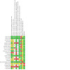
Risk of bias summary: review authors' judgements about each risk of bias item for each included study
(Empty cells = not applicable as no participant‐reported outcomes in study)
Allocation
Sequence generation and methods of allocation were poorly reported in older studies; an absence of details of methods resulted in a judgement of unclear risk for one or both domains. We judged 10 studies to be at low risk of selection bias (Busse 2014; Busse 2016; Gopalan 2020; Heckman 1994; Kristiansen 1997; Liu 2014; Lubbert 2008; Mayr 2000; Santana‐Rodríguez 2019; Yadav 2008). We judged nine studies to be at unclear risk of selection bias relating to sequence generation due to lack of information as to the randomisation process (Emami 1999; Gan 2014; Handolin 2005; Handolin 2005a; Kamath 2020; Patel 2015; Rue 2004; Seifert 2013 Strauss 1999). We judged both quasi‐randomised trials to be at high risk of selection for both sequence generation and allocation concealment owing to their study designs (Leung 2004; Wang 2007).
Only six studies used methods which we believed were likely to conceal allocation of the randomisation sequence (Busse 2014; Busse 2016; Heckman 1994; Kristiansen 1997; Lubbert 2008; Santana‐Rodríguez 2019). It was not possible to conceal allocation in the quasi‐randomised trials and we therefore judged both of these to be at high risk of selection bias for allocation concealment (Leung 2004; Wang 2007). Other studies reported insufficient information about this methodology and risk of bias was therefore unclear.
Blinding
For blinding, we made judgements according to the type of outcome: participant‐reported measures or objectives measures.
Participants and personnel
Eight studies reported participant‐reported measures. Of these, we judged four studies to be at low risk of performance bias because methods were used to disguise the intervention and control treatments (Busse 2014; Busse 2016; Lubbert 2008; Santana‐Rodríguez 2019). Four studies were at high risk of bias because the control group had no treatment or because no attempts were made to disguise the intervention and control treatments (Gopalan 2020; Heckman 1994; Patel 2015; Wang 2007). We judged risk of bias in Seifert 2013 to be unclear because we had insufficient information.
All studies reported at least one objective outcome measure. Eight studies were at high risk of bias (Gopalan 2020; Kamath 2020; Leung 2004; Liu 2014; Mayr 2000; Patel 2015; Strauss 1999; Wang 2007). Of those deemed at high risk of bias, one study used a sham device that was dissimilar to the intervention unit and therefore the blinding in the study may have been compromised (Leung 2004), whilst the other seven used no additional intervention as a control. We judged risk of bias in Seifert 2013 to be unclear because we had insufficient information, and we judged risk of performance bias for objective measures to be low in the remaining studies.
Blinding of outcome assessment
We judged four of the eight studies that reported participant‐reported measures to be at low risk of detection bias (Busse 2014; Busse 2016; Lubbert 2008; Santana‐Rodríguez 2019) because the intervention and control were identical, participants were unlikely to know their treatment allocation when reporting their outcome information. For the three studies in which treatment and control allocation was known to the participants, we judged detection bias to be at high risk (Gopalan 2020; Patel 2015; Wang 2007). Again, we judged risk of detection bias for participant‐reported measures to be unclear in Seifert 2013.
For objective outcome measures, we judged risk of detection bias to be unclear in only four studies because of lack of information (Patel 2015; Rue 2004; Seifert 2013; Strauss 1999). The remaining studies were at low risk of detection bias because treatments were disguised throughout the trial or independent assessors were used to collect outcome data.
Incomplete outcome data
We were successful in contacting study author of five trials (Busse 2014; Busse 2016; Heckman 1994; Kristiansen 1997; Lubbert 2008) for missing data. In addition, we sourced unpublished data from Seifert 2013. Most studies reported data for all randomised participants, or reported very few losses, and we judged risk of attrition bias to be low. However, we judged10 studies to be at high risk of attrition bias because of large numbers of participant loss, or loss that was unexplained or not balanced between groups (Busse 2014; Busse 2016; Gan 2014; Handolin 2005; Handolin 2005a; Heckman 1994; Kristiansen 1997; Lubbert 2008; Rue 2004; Seifert 2013).
Selective reporting
Four studies were registered with a clinical trials register (Busse 2016; Gopalan 2020; Santana‐Rodríguez 2019; Seifert 2013). Of these, we judged only Busse 2016 to be at low risk of selective reporting bias; although this prospectively registered study made changes to the outcomes; this was adequately explained in the published study report. We judged the risk of selective reporting bias to be unclear in Gopalan 2020 because this study was registered retrospectively and it was not feasible to use the clinical trials registration documents to assess the risk of selective reporting. Santana‐Rodríguez 2019 was also retrospectively registered but we noted that one outcome measure was listed in the clinical trials register but not reported in the published report and we could not rule out the possibility of selective reporting bias; we therefore judged the risk in this study to be high. Seifert 2013 was prospectively registered, but with no formal trial report we are unsure of any selective reporting bias.
We were unable to judge risk of selective reporting bias in the remaining studies because these studies did not report a protocol or registration with a clinical trials register.
Other potential sources of bias
We judged two studies to be at high risk of other bias (Strauss 1999; Seifert 2013). For Strauss 1999, we only used data from a poster abstract which was limited and we expected that these data were not peer‐reviewed. Similarly, data for Seifert 2013 were from personal communication only rather than from a peer‐reviewed published report.
Effects of interventions
Low‐intensity pulsed ultrasound (LIPUS) versus control (20 studies, 1459 participants)
Critical outcomes
Health‐related quality of life (HRQoL)
Busse 2014 and Busse 2016 reported this outcome using Health Utility Index‐III scores and Short‐Form 36 Physical Component Scores (SF‐36‐PCS); we used the data from SF‐36‐PCS scores as this measurement tool is more widely used. Seifert 2013 also reported SF‐36 PCS scores. We used data for these outcomes reported in the short term at three months, and in the medium term at one year for Busse 2014 and Busse 2016, and six months for Seifert 2013; all three studies included fractures in the lower limbs.
We found no evidence of a difference in HRQoL in the short term (mean difference (MD) 0.82, 95% confidence interval (CI) ‐0.67 to 2.31, favours low‐intensity pulsed ultrasound (LIPUS); 3 studies, 540 participants; moderate‐certainty evidence; Analysis 1.1) or medium‐term (MD 0.06, 95% CI ‐3.85 to 3.97, favours LIPUS; 3 studies, 393 participants; very low‐certainty evidence; Analysis 1.1). This point estimate unlikely to be of clinical importance as studies report a minimal clinically important differences (MCID) in orthopaedic‐related problems ranging for SF‐36 physical component score (PCS) of 3 to 5 points (Busse 2016; McHorney 1994). We recognise, however, that the 95% CI includes the possibility of both clinical improvement and reduction in quality of life. We downgraded both the short‐term and medium‐term evidence by one level due to imprecision because of a wide CI. We also downgraded the medium‐term evidence by one level owing to unexplained statistical heterogeneity (I2 = 52%) and one level for risk of attrition bias.
1.1. Analysis.
Comparison 1: LIPUS versus control, Outcome 1: Health‐related quality of life (lower limb)
Quantitative functional improvement
No studies reported quantitative functional improvement using validated patient‐reported outcome measures (PROMs).
Time to return to normal activities
Complete fractures
Busse 2016 and Lubbert 2008 provided data on return to work. For the pooled data for upper and lower limb fractures, there was little or no difference between treatments (MD 1.96 days, 95% CI ‐2.13 to 6.04, favours control; 2 studies, 370 participants; low‐certainty evidence; Analysis 1.2). We downgraded by one level for imprecision due to a wide CI and one level for risk of attrition bias. There was evidence of little or no difference in time to return to work after upper limb fractures (MD 1.95 days, 95% CI ‐2.18 to 6.08, favours control; 1 study, 101 participants; Analysis 1.2) ,and for people with lower limb fractures (MD 2.20 days, 95% CI ‐24.38 to 28.78, favours control; 1 study, 269 participants; Analysis 1.2).
1.2. Analysis.
Comparison 1: LIPUS versus control, Outcome 2: Time to return to work complete fractures (days)
In addition, Busse 2016 reported time to return to leisure activities and we found little or no difference according to whether LIPUS was used (MD ‐10.90 days, 95% CI ‐33.98 to 12.18, favours LIPUS; 1 study, 321 participants; low‐certainty evidence; Analysis 1.3). We downgraded by one level for imprecision due to very wide CI and by one level because the study was at high risk of attrition bias. This study also reported time to weightbearing and time to return to household activities which we have not included in this review.
1.3. Analysis.
Comparison 1: LIPUS versus control, Outcome 3: Time to return to normal activities (days)
Although Handolin 2005 reported no significant difference in the Olerud‐Molander score between treatment and control groups in 16 participants (53% of the 30 randomised participants) at 18 months follow‐up, we did not include data as they were reported incompletely and efforts to contact the study authors were unsuccessful.
Stress fractures
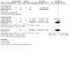
Rue 2004 and Yadav 2008 both reported time to return to training or duty in 40 midshipmen and 67 military recruits, respectively. There was no evidence of a difference between treatments of stress fractures of the tibia (MD ‐8.55 days, 95% CI ‐22.71 to 5.61; favours LIPUS; 2 studies, 93 participants; very low‐certainty evidence; see Analysis 1.4). We downgraded the evidence by two levels due to unexplained considerable heterogeneity (I² = 78%), by one level for imprecision for a wide CI and by one level due to the studies having unclear risks of bias.
1.4. Analysis.
Comparison 1: LIPUS versus control, Outcome 4: Time to return to training/duty after stress fracture (days)
Time to fracture union
Although time to union data were available in most studies, the definition of union, timing of assessment and statistical analysis were variable. Study data were reported where time to union or proportion of those who achieved union at each follow‐up point were available or were provided upon successful contact with authors. It was not possible to calculate an overall time to fracture union for Santana‐Rodríguez 2019 due to unclear reporting of loss to follow‐up. We noted the following data for Santana‐Rodríguez 2019: at one month 2/20 participants had callus that was formed or remodelled, at three months 13/19 participants had callus that was formed or remodelled, and six months 12/16 participants had callus that formed or remodelled.
Seven studies of 617 participants defined union radiographically (Busse 2014; Busse 2016; Emami 1999; Handolin 2005; Handolin 2005a; Kristiansen 1997; Mayr 2000). Where data were presented from surgeons and radiologists, we report only those based upon radiologists' opinions. Three studies, which included 289 participants, defined union as a combined clinical and radiographic finding with similar definitions of radiographic union (Heckman 1994; Leung 2004; Liu 2014). Lubbert 2008 defined union based upon participants' self‐reports.
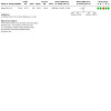
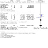
Each of the studies reporting this outcome, apart from Liu 2014 where 100% completed the trial, only reported a per‐protocol analysis, where the reported data are for those participants who complied with the protocol, including follow‐up. We contacted the study authors who explained that such an analysis was necessary because the data were missing due to the haphazard follow‐up of some participants. We did not pool data due to substantial, unexplained heterogeneity (I2 = 90%; very low‐certainty evidence; Analysis 1.5). In addition, we did not analyse data according to upper or lower limb fracture because of substantial, unexplained heterogeneity for upper limbs (I2 = 92%) and lower limbs (I2 = 88%). For upper limb fractures, MDs ranged from 0.32 to 40 fewer days to fracture union for participants treated with LIPUS. For lower limb fractures, MDs ranged from 88 fewer days to 30 more days for participants treated with LIPUS.
1.5. Analysis.
Comparison 1: LIPUS versus control, Outcome 5: Time to fracture union (days)
Because data were available from 11 studies for this outcome, we used formal tests for subgroup interactions according to upper and lower limb fractures but we found no evidence that heterogeneity was explained by type of fracture. Studies reported insufficient information for us to also test the impact of smoking status on the results. We downgraded the evidence by two levels due to substantial heterogeneity and by one level due to studies having unclear or high risks of bias.
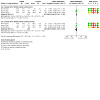
We created funnel plots for this analysis. We could not rule out a possibility of publication bias or small‐study effects (Figure 3).
3.
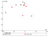
Funnel plot for Analysis 1.5 Time to fracture union
Pain
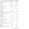
Lubbert 2008 reported pain in 101 participants in the short term (at one month) using a visual analogue scale (VAS); lower values in this 10‐point scale indicate less pain. Santana‐Rodríguez 2019 also reported VAS scores at one month for 47 participants. Data from both studies were for participants who had upper limb fractures. We did not pool data due to substantial, unexplained heterogeneity (I2 = 80%; very low‐certainty evidence Analysis 1.6). In one study, participants reported less pain after LIPUS (MD ‐1.7, 95% CI ‐3.03 to ‐0.37; 47 participants). However, the effect estimate was less precise in the other study (MD ‐0.4, 95% CI ‐0.61 to 0.53; 101 participants). We downgraded the evidence by two levels due to unexplained substantial statistical heterogeneity, and by one level for imprecision because the evidence is from a small number of participants
1.6. Analysis.

Comparison 1: LIPUS versus control, Outcome 6: Pain (short‐term)
Santana‐Rodríguez 2019 reported medium term (at six months) VAS for pain from 47 participants showing little evidence of a difference between either intervention (MD ‐0.50, 95% CI ‐1.02 to 0.02; favours LIPUS; 1 study, 47 participants; low‐certainty evidence; Analysis 1.7). This is unlikely to be of clinical importance as literature has reported a MCID of between 1.4 and 3 on this scale (Copay 2018; Tashjian 2009). We downgraded by two levels for imprecision because the evidence is from a small number of participants.
1.7. Analysis.
Comparison 1: LIPUS versus control, Outcome 7: Pain scores (medium‐term)
Delayed union and non‐union
We found no evidence of a difference between interventions in the short term (RR 0.77, 95% CI 0.15 to 3.83; favours LIPUS; 3 studies, 139 participants; very low‐certainty of evidence; Analysis 1.8). Lubbert 2008 reported no non‐unions in the short term (two months) for upper‐limb fractures, and this effect estimate was primarily derived of two studies in lower‐limb fractures at three months (Handolin 2005; Handolin 2005a). We downgraded by one level for moderate heterogeneity (I2 = 35%), by one level for imprecision due to a wide CI, and by two levels due to the studies being at unclear or high risk of bias.
1.8. Analysis.
Comparison 1: LIPUS versus control, Outcome 8: Delayed or non‐union (short‐term)
We found little or no difference between either intervention in the medium term (RR 1.25, 95% CI 0.50 to 3.09, favours control; 7 studies, 746 participants; moderate‐certainty evidence; Analysis 1.9). Kristiansen 1997 and Mayr 2000 reported no non‐unions for upper‐limb fractures at four months and 12 months, respectively, and this effect estimate was primarily derived from studies of lower‐limb fractures at six and 12 months for non‐union (Busse 2016; Strauss 1999), and delayed union (Emami 1999; Leung 2004). We downgraded by one level for imprecision due to a wide CI.
1.9. Analysis.
Comparison 1: LIPUS versus control, Outcome 9: Delayed or non‐union (medium‐term)
Sensitivity analysis
We found insufficient studies to conduct sensitivity analysis on the following outcomes: time to return to work (Analysis 1.2), time to return to normal activities (Analysis 1.3), pain‐scores (medium‐term) (Analysis 1.7), delayed or non‐union (Analysis 1.8).
High or unclear risk of selection bias (for sequence generation)
HRQoL: we excluded Seifert 2013. This did not alter our interpretations of the effect for the short‐ and medium‐term analyses.
Time to return to training/duty after stress fractures: we did not perform a sensitivity analysis as both studies were at unclear risk of bias
Time to fracture union: we excluded Emami 1999, Gan 2014, Handolin 2005, Handolin 2005a and Leung 2004. Heterogeneity remained at considerable levels (I2 = 88%) and we did not pool the data for the remaining studies.
Delayed or non‐union: we excluded Emami 1999, Leung 2004 and Strauss 1999. This did not alter our interpretation of the effect for this outcome.
High risk of attrition bias
HRQoL (short‐term) and HRQoL (medium‐term): we excluded Busse 2016 and Seifert 2013. This did not alter our interpretations of the effect for the short‐ and medium‐term analyses.
Time to return to training/duty after stress fractures: we excluded Rue 2004. We found that the analysis favoured LIPUS (MD ‐14.38 days, 95% CI ‐16.7 to ‐12.06; 1 study, 67 participants)
Time to fracture union: only two studies had a low risk of attrition bias for upper limb fractures (Liu 2014; Mayr 2000), and two studies had low risk of attrition bias for lower limb fractures (Emami 1999; Leung 2004). We found no differences in the interpretation of the effects when excluding studies at high risk of bias.
Pain scores (short‐term): we excluded Lubbert 2008. We found that the analysis favoured LIPUS (MD ‐1.70, 95% CI ‐3.03 to ‐0.37; 1 study, 47 participants)
High risk of 'other' bias
HRQoL: we excluded Seifert 2013. This did not alter our interpretations of the effect for the short‐ and medium‐term analyses.
Time to fracture union: we excluded Heckman 1994; Kristiansen 1997; Leung 2004. Whilst the I2 value of heterogeneity remained high for upper limb fractures (I2 = 83%), this was no longer the case for fractures in the lower limbs. In this group of participants, we noted that there was little or no difference between treatments in time to fracture union (MD ‐1.32, 95% CI ‐8.79 to 6.15; 6 studies, 516 participants).
Obvious data outliers
Time to fracture union: we excluded Emami 1999 as this study reported a greater tendency towards improvement in time to fracture union in the control group than any of the other studies, as well as excluding Heckman 1994 and Leung 2004 due to appearing to be significant outliers favouring LIPUS. Without these studies, there was no longer evidence of statistical heterogeneity, with little or no difference between treatments in time to fracture union (MD ‐2.09, 95% CI ‐9.65 to 5.47; 5 studies, 494 participants).
Additional sensitivity analysis
Time points in analysis: we performed a sensitivity analysis on HRQoL medium term at comparable time points, including Busse 2016 data at 38 weeks to compare to Busse 2014 and Seifert 2013 data at six months. This did not alter our interpretation.
Missing data: to explore the impact of missing data, we calculated 'worst‐case' analyses for those outcomes in which data were missing (Sensitivity analysis). For HRQoL, sensitivity analysis did not alter our interpretation. For time to return to work and time to return to normal activities, sensitivity analyses favoured the control group (time to return to work: MD 125.63 days, 95% CI 106.18 to 145.08; time to return to normal activities: MD 62.0 days, 95% CI 43.52 to 80.48). For time to fracture union, heterogeneity remained at considerable levels (I2 = 92%) and we did not pool the data in this sensitivity analysis. See Appendix 4.
Other important outcomes
Adverse events
We report adverse events in Analysis 1.10. Thirteen studies reported on adverse events (Busse 2014; Busse 2016; Emami 1999; Gan 2014; Handolin 2005; Handolin 2005a; Heckman 1994; Kamath 2020; Kristiansen 1997; Leung 2004; Lubbert 2008; Patel 2015; Santana‐Rodríguez 2019). Most adverse events were not related to the study device. Three studies reported a low incidence of self‐resolving conditions (muscle cramping, skin irritation, erythema and swelling), which did not lead to any trial protocol violations (Heckman 1994; Leung 2004; Lubbert 2008). The swelling reported in one study was at six‐week follow‐up but resolved at future follow‐up points (Heckman 1994). Patel 2015 reported that one participant had subperiosteal bone formation in mandibular fractures involving the developing tooth germ (aggregation of cells that form a tooth) where the LIPUS was administered; this self‐resolved without active treatment. Patel 2015 also reported that one participant in the control group developed fibrous ankylosis but was lost to follow‐up.
1.10. Analysis.
Comparison 1: LIPUS versus control, Outcome 10: Adverse events
Cost
One study conducted an economic evaluation of LIPUS as part of their trial (Busse 2016). Findings indicated that cost was higher with LIPUS use, both in terms of cost of the device (mean increase of USD 3647, 95% CI USD 3244 to USD 4070; P < 0.001), and from the societal perspective which includes both direct and indirect costs (mean increase of USD 3422, 95% CI USD 1568 to USD 5283; P < 0.001); see Tarride 2017.
Adherence
Seven studies commented on adherence (Busse 2014; Busse 2016; Emami 1999; Handolin 2005; Heckman 1994; Kristiansen 1997; Santana‐Rodríguez 2019). Adherence was either reported using internal timers contained within devices or from participant treatment diaries. Emami 1999 reported good adherence to the trial protocol, with no significant difference between the treatment and placebo groups' usage or diary records, both of which closely matched the protocol requirements (ultrasound: mean (SD) 23.4 (± 0.8) hours; placebo: mean (SD) 22.3 (± 1.0) hours; participant diary: mean 24.6 hours). Kristiansen 1997 reported similar findings (ultrasound: mean 62 hours; placebo 64 hours), which compared favourably with the trial protocol requirement. Two studies did not report data, but stated that reported adherence less formally but did highlight good participant compliance (Handolin 2005; Heckman 1994). Handolin 2005 reported comparable duration of use of the ultrasound device (mean: 40.7 days versus 39.9 days), whereas Heckman 1994 stated only comparable usage of the devices. Participants of Rue 2004 were administered treatments by trial personnel so that adherence was easily determined. Both LIPUS and control groups missed a similar proportion of treatments, which was less than approximately 20% of all treatments in each group. Santana‐Rodríguez 2019 reported that there was full compliance with the protocol. Busse 2016 tracked compliance reported compliance for 424 participants, with 189 reporting ≥ 75% compliance, and 119 reporting between 50% to 75% compliance. There were no significant differences between the two treatment groups. Busse 2014 reported that 76% of participants reported full compliance and 24% registered more than 50% compliance.
Extracorporeal shock wave therapy (ECSW) versus control (1 study, 56 participants)
ECSW was tested only in Wang 2007, which compared ECSW with no ECSW in 56 participants with 59 fractures of the tibia or femur. Results in this trial were reported for fractures instead of participants; it was not possible to correct for the unit of analysis discrepancy.
We judged the certainty of the evidence for all outcomes to be very low. We downgraded by one level because the evidence was derived from only one small study, and by two levels because this quasi‐randomised study was at high risk of selection bias.
Critical outcomes
HRQoL, quantitative functional improvement, time to return to normal activities, and time to fracture union
Wang 2007 did not report any data for these outcomes.
Pain
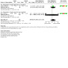
Wang 2007 reported VAS scores at one week, three months, six months and 12 months. We used data for three months for short term and 12 months for medium term. We found a small difference in pain scores at short term (MD ‐0.87, 95% CI ‐1.31 to ‐0.43, favours ECSW; Analysis 2.1) and medium term (MD ‐0.62, 95% CI ‐0.97 to ‐0.27; very low‐certainty evidence; Analysis 2.1). However, these differences are unlikely to be of clinical importance as this pain scale has MCID of between 1.4 and 3 (Copay 2018; Tashjian 2009).
2.1. Analysis.
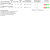
Comparison 2: ECSW versus control, Outcome 1: Pain (VAS: 0 no pain to 10 severe pain)
Delayed union and non‐union
Wang 2007 reported non‐union at 12 months and found there was no evidence of a difference in delayed union or non‐union rates of ECSW at 12 months (RR 0.56, 95% CI 0.15 to 2.01, favours ECSW; 1 study, 57 participants; very low‐certainty evidence; Analysis 2.2). All incidences of delayed or non‐union were in fractures of the femur.
2.2. Analysis.
Comparison 2: ECSW versus control, Outcome 2: Delayed or non‐union (medium‐term)
Sensitivity analysis
Two participants with two fractures (one each in the control and intervention group) were excluded from the analysis in Wang 2007. We performed a 'worst‐case' analysis which did not alter our interpretation of the evidence for short‐ and medium‐term pain nor for delayed union and non‐union.
Other important outcomes
Adverse events
Wang 2007 reported one case of deep infection and osteomyelitis in each group (both participants were excluded from the final analyses) and five cases of superficial infection (2/27 versus 3/30), all of which resolved with antibiotics and wound care. There were no other complications, including those directly related to shockwave treatment.
Cost and adherence
Wang 2007 did not report any data for these outcomes.
Discussion
Summary of main results
The review presented evidence from 21 trials comparing low‐intensity pulsed ultrasound (LIPUS) versus control, and one trial comparing extracorporeal shock wave therapy (ECSW) versus control. We found no trials evaluating high‐intensity focused ultrasound. The included trials form a clinically heterogeneous group of studies, which included participants with a range of acute fractures, treated in a variety of ways. The fractures were complete fractures in 18 trials and stress fractures in three trials.
LIPUS versus control
No studies investigated quantitative functional improvement. Of the studies that reported health‐related quality of life (HRQoL), these only included lower limb fractures and we found moderate‐certainty evidence of little to no change in HRQoL in the short term (at three months) and very low‐certainty evidence of little to no change in the medium term (at six to 12 months); the change was below that of previously reported levels to indicate a minimal clinically important difference (MCID). There was low‐certainty evidence of little or no difference in medium‐term pain scores at six months and the change was below literature reported MCID. There was low‐certainty evidence of little or no difference in time to return to work for complete fracture and time to return to normal activities, and very low‐certainty evidence of no change in time to return to training/duties after stress fractures.
More studies reported data for delayed/non‐union and we found that LIPUS probably makes no difference to this outcome. We were unable to pool data on short‐term pain scores at one month or time to fracture union due to substantial statistical heterogeneity and we judged this evidence to be very low certainty. Levels of statistical heterogeneity for time to fracture union scores only reduced when excluding studies at high risk of 'other' bias; when pooled in sensitivity analysis, there was little or no difference in time to fracture union overall and in upper limbs, there was improvement in lower limbs.
For studies that reported adherence, compliance tended to be commented upon as being compliant with protocol requirements, rather than formal data presented. Adverse effects directly associated with treatment use (or associated devices) were found to be few and minor. Data for costs were reported for one study, with higher direct costs, as well as combined direct and indirect costs, for LIPUS use.
ECSW versus control
The small quasi‐randomised trial evaluating ECSW for tibia and femur fractures did not report on functional outcomes nor time to union. There was very‐low certainty evidence of a small difference in pain in the short term at three months and medium term at 12 months, but the difference was below the MCID. There was very‐low certainty of evidence of little to no difference in delayed or non‐unions. The only reported complication was infection, with no significant differences between the two groups. There were no data on cost or adherence.
Overall completeness and applicability of evidence
This review includes data from 21 studies, conducted in eight countries with ages ranging from 15 to 81 years. Nineteen studies tested the use of LIPUS in acute fractures, three of which reported outcomes with stress fractures. One study tested the use of ECSW in complete fractures. No studies evaluated high‐intensity focused ultrasound. Two studies were quasi‐randomised trials; whilst the remaining studies were RCTs of which, five studies used no placebo controls in the form of a 'sham' probe device. Most settings were typical hospital settings. The participants included those with fractures of the upper or lower limbs, which were treated either surgically or conservatively. Although these populations were highly heterogeneous, they are still representative of the type of fracture populations, generally at higher risk of delayed healing and non‐union, for which treatment adjuncts might be considered. The included studies reported the use of ultrasound in a wide variety of settings and participants.
We found only limited data investigating ECSW with very low‐certainty evidence of decreased pain in the short term and medium term; further studies investigating ECSW are needed to increase the certainty of the effect estimates. There was no evidence to suggest ECSW reduced delayed union or non‐union rates
For the studies investigating LIPUS, four studies did not report on any of the critical outcome measures. No studies reported on the critical outcome of quantitative functional outcome. Only two studies reported HRQoL, measured using SF‐36 PCS, and this included data from an unpublished study which reported data for 540 participants in the short‐term and 393 participants in the medium term (Seifert 2013). Additionally, we were unable to pool analysis from the two studies that reported validated pain scores due to considerable unexplained heterogeneity.
We were unable to pool data for the critical outcome of time to fracture union. Data were difficult to ascertain as typically participants were assessed at fixed follow‐up intervals that varied between studies, whilst some data were missing for time to union. Moreover, the definition of fracture healing is variably defined in the literature, with studies defining healing clinically and radiographically. This reflects the difficulty in assessing this outcome as it impossible to assess healing in each participant every day, inevitably leading to a lack of precision in estimates of healing times. However, we see no reason why this process should have differed between treatment groups in any study, so would not expect there to be any bias in estimates for the treatment effects. However, this may, at least in part, explain the significant heterogeneity in observed healing times between studies which meant we could not pool the data.
Only 11 studies reported adverse events, and of those that appeared directly related to the device, these all self‐resolved. It is unclear if there is under‐reporting or a positive safety profile for the intervention. Additionally, participant adherence was inconsistently reported in studies.
Clinical practice varies worldwide but LIPUS remains a specialist treatment usually only considered for, or administered to, people with fractures at risk of delayed union or non‐union. Thus, studies involving this type of participant would be best to determine if there is an effectiveness of LIPUS (Harrison 2021; Puts 2021).
Certainty of the evidence
We used GRADE to assess the certainty of evidence for each critical outcome, which ranged from very low‐ to low‐certainty evidence.
We downgraded the evidence if we judged that risks of bias in the included studies may have impacted the results. We noted that some studies were at high risk of performance and detection bias because they did not use a placebo control, and that some studies had a notably large rate of attrition. Effect estimates were often imprecise with wide confidence intervals (CIs) and it is likely that this is a reflection of the small studies in this review, which were likely to be underpowered. We also found moderate to substantial levels of statistical heterogeneity in the evidence for some outcomes which we were unable to explain and we therefore also downgraded the certainty of some evidence for inconsistency. We could not rule out the possibility of publication bias in any outcomes. We prepared a funnel plot for the analysis of time to fracture union (which had 11 studies) which was asymmetrical indicating possible publication bias; we did not explore this further or downgrade the certainty of the evidence for publication bias. We did not downgrade any evidence for indirectness.
Potential biases in the review process
We conducted this review following the Cochrane Methodological Expectations for Cochrane Intervention Reviews (MECIR). We have transparently reported any changes to the Methods since the previous version of this review in Differences between protocol and review. These changes were minimal and included the re‐organisation of the outcomes into 'critical' and 'other important' outcomes and making a greater distinction between quality of life and return to normal activities and other functional outcomes. We did not conduct subgroup analysis when evidence included fewer than 10 studies, however we ensured that data were reported separately for upper and lower limbs. We also conducted additional sensitivity analysis according to risk of bias decisions. We did not expect any of these changes to impact the evidence in this review. We note that we did not consider alternative choices for post‐hoc subgroup analyses which may have explained the high levels of heterogeneity in some of our findings. We attempted to contact the authors of included studies to retrieve missing data whenever possible. This update also includes summary of findings tables and an assessment of the certainty of the evidence.
Agreements and disagreements with other studies or reviews
We identified nine systematic reviews evaluating the use of ultrasound and shockwave therapy for acute fracture.
Our review agreed with the outcomes reported in most of the reviews that there was little or no difference in time to return to work (Lou 2017; Rutten 2016; Schandelmaier 2017; Sijie 2021), time to weightbearing (Lou 2017; Schandelmaier 2017), time to radiographic healing (Ebrahim 2014; Schandelmaier 2017), pain (Schandelmaier 2017; Sijie 2021), non‐union rates (Hannemann 2014; Lou 2017; Sijie 2021) or quality of life (Sijie 2021). One review agreed that there were no differences in LIPUS on return to duty on stress fractures (Busse 2009).
Similar to this review, many review analyses' had considerable levels of statistical heterogeneity (I2 ranging from 69% to 98%). There were some disagreements with other reviews. One review found an LIPUS improved quality of life (Lou 2017). Five reviews found that LIPUS reduces time to fracture union (Busse 2009; Hannemann 2014; Lou 2017; Rutten 2016; Tajali 2012). One review found mild improvement in time to third cortical healing in acute fractures (Tajali 2012). Three of these reviews performed subgroup analysis which showed LIPUS improved time to radiographic union in non‐operatively managed fractures (Busse 2009; Hannemann 2014; Rutten 2016), and also two reviews found reduced time to clinical healing (Hannemann 2014; Rutten 2016). However, both reviews' analyses had considerable levels of statistical heterogeneity. One review also found that LIPUS improved time to clinical healing in lower limbs and diaphyseal fractures, but this analysis included considerable levels of statistical heterogeneity (I2 = 97%) (Hannemann 2014).
Another systematic review highlighted the need for a robust, double‐blind, randomised, placebo‐controlled trial (Puts 2021).
We note that our assessment of the certainty of the evidence differed from decisions reached in Schandelmaier 2017 which may be explained by different methodological approaches to the reviews.
Authors' conclusions
Implications for practice.
For people who have an acute fracture, there is currently insufficient evidence on the effectiveness of low‐intensity pulsed ultrasound (LIPUS) on patient‐reported outcome measures (PROMs), such as quality of life, for people who have had complete or stress fractures. However, more studies reported delayed or non‐union and we found that LIPUS probably makes no difference to this clinical measure. Similarly, evidence for extracorporeal shock wave therapy (ECSW )was limited to one study but did show some evidence of a reduction in pain but it is likely this is below clinical significance. No studies investigated high‐intensity focused ultrasound.
Implications for research.
Any future research investigating the use of ultrasound for acute fractures should involve secure randomisation and placebo controls with appropriate 'sham' probe controls. Trials should be prospectively registered and conform to reported standards set out in the CONSORT statement (Boutron 2008). Participant loss was high in this review, with particular impact on the evidence for health‐related quality of life (HRQoL); we encourage investigators to put measures in place to improve study follow‐up with an aim to report the results of all trial participants. Compliance should be measured and reported so that the effect of ultrasound is not influenced by poor compliance. Given that LIPUS is usually considered for people at risk of delayed or non‐union, studies should prioritise these study participants. Outcome measures should focus on PROMs to determine if the possible benefit of ultrasound in terms of fracture healing translates into a tangible benefit, and these measures should focus on quality of life as well as functional outcomes. We identified one ongoing study in this review which should contribute additional data in future updates of this review (KCT0002591. This study is investigating the effectiveness of ultrasound therapy in hip fracture and includes PROMs of quality of life and pain. We note, however, that this study does not compare the intervention with a sham treatment and we anticipate a high risk of performance and detection bias in this study.
Feedback
Issues concerning choice of analysis, 12 December 2014
Summary
Comment: This review contains several errors, some of them serious. As a result, the treatment effect calculated for low‐intensity pulsed ultrasound (LIPUS) is inconsistent with the real effect shown in the reported data. 1. In an effort to eliminate bias, the authors rejected the results of published fresh‐fracture studies on LIPUS. Instead, they re‐analyzed data from each paper based on their own criteria. As part of this re‐analysis, they inserted outcomes data for patients lost to follow‐up (‘Data collection and analysis’; ‘Dealing with missing data’). This might have been acceptable if the authors had treated patients equally in the active and placebo groups, but they did not. In the LIPUS group, patients lost to follow‐up were assigned a time‐to‐heal equal to two standard deviations greater than the mean for that group. In the placebo group, however, missing patients were assumed to have healed normally and were assigned a time‐to‐heal equal to the group mean. The authors called this a “worst case” analysis, and it effectively increased heal times in the LIPUS group by an average of 9%. Based on the skewed data, it was concluded that LIPUS and control groups were not significantly different (Abstract). This conclusion is not supported by the actual data. When Griffin et al. analyzed the literature without unequal imputation of heal rates, LIPUS was shown to significantly accelerate fracture healing (text and Figure 3; p=0.03). However, this “as reported” analysis was neither included in the abstract nor discussed in detail in the text. The authors also asserted, incorrectly, that the results of the “worst case” and “as reported” analyses were similar (‘Risk of bias in included studies’; ‘Incomplete outcome data’). Both the biased analysis and its burial deep within the text are concerning. Unless readers purchase and closely review the full text, they will remain unaware that the authors’ “worst case” analysis changed the data and, indeed, the main finding of the study. 2. Other errors in the manuscript include: a) Incomprehension of normal fracture‐healing heterogeneity. It is universally recognized that different bones heal at different rates. Thus, variation is unavoidable when comparing healing times at different fracture locations. The broad generalizations applied by the authors to probe this variation, such as upper limb vs. lower, or smoker vs. nonsmoker, are inadequate. b) Mischaracterization of the normal heterogeneity in healing by bone. The authors misinterpreted the inherent heterogeneity of fracture healing as evidence of bias, which was then used to justify the rejection of “as reported” heal‐rate data from the literature (‘Effects of interventions’). c) Unacknowledged, unsupported, a priori assumptions that all forms of ultrasound are comparable, and all low‐intensity ultrasound is equivalent. d) Unmerited inclusion of extracorporeal shockwave treatment (1 study) and high‐intensity focused ultrasound (0 studies) in the review. e) Inappropriate analysis of the evidence for delayed union and nonunion (Figure 6). By design, the authors’ search criteria only identified acute‐fracture studies (‘Methods’: ‘Types of participants’). Having excluded delayed‐union and nonunion studies at the start, no valid analysis was possible for this clinical population. f) Inappropriate inclusion of the study by Lubbert et al., which lacked radiographic outcome data, in analyses of time to radiographic union (Figures 3 and 4). g) Unspecified criteria for the weighting of results from different studies (Figures 3‐6). This practice was not discussed in the methods or body of the paper, and no explanation or algorithm was presented. The given weights were not based on the number of patients per study or other obvious criteria. h) Unrealistic criteria for radiological review. In 6 of 7 LIPUS studies (Heckman 1994, Kristiansen 1997, Emami 1999, Mayr 2000, Leung 2004, Handolin 2005a), radiographs were assessed by multiple, blinded reviewers. In 5 of 7 studies, the review team included both surgeons and radiologists. The authors’ criticism that “none of the included studies used a panel of independent radiologists to assess radiographic union,” (‘Discussion’; ‘Quality of the evidence’) represents an unrealistic standard that is not demonstrably superior to the joint efforts of surgeons and radiologists. The fact that radiographs in Mayr et al. 2000 actually were reviewed by a panel of independent, blinded radiologists, suggests an inadequate review of the literature. In light of these serious issues, we recommend withdrawal of the current review and publication of a revised version in which these errors are corrected. Conflict of interest statement: Both authors are affiliated with Bioventus LLC*, Durham, NC.
* Bioventus is the manufacturer of Exogen® device, which was tested in several trials in this review.
Reply
We thank Drs Heeckt and Brodie for their interest and careful consideration of our Cochrane Review.
1. We agree that there are multiple ways to report the pooled data and that our ‘worst case’ analysis sets a higher standard than an ‘as reported’ analysis. The ‘worst case’ analysis is however important so that readers can discern one possible and conservative interpretation that is consistent with the data. In this case, it shows that the data could be consistent with no treatment effect if the missing data in each study were not missing at random. In essence, the variation in estimates between the ‘worst case’ and ‘as reported’ analyses simply highlights the critical importance of good follow‐up in clinical studies. Our approach to handling these types of data issues, and their effect on study outcomes, are not novel.(1) A fuller discussion of the effects and means of handling missing data can be found in the Cochrane Handbook.(2)
We agree that it is also important to describe the ‘as reported’ analysis and we already do this in the ‘Effects of interventions’ section as well as, as you point out, presenting the data in Figure 3 and Analysis 1.3.
The statement that reads ‘the proportion of missing data was sufficiently low, that “as reported” and “worst case” analyses were similar’ is incomplete. We recognise that whilst the effect is significant in the ‘as reported’ analysis, the effect estimate and confidence intervals are approximately comparable. We have removed this statement to avoid any confusion.
Both the methods and the abstract were clear that our review was designed to report, and draw conclusions from, the ‘worst case’ analysis. Importantly, the abstract also stated that the likely impact of this design was to give “more conservative estimates of treatment effects for time to fracture union”. We believe that this should be sufficient to alert readers to look deeper within the article for a more in depth analysis.
2. With regards to the other observations:
a) We certainly agree that the rate of fracture healing varies between anatomical sites. However, this should not have biased the within‐study estimates of treatment effect because participants within each trial were drawn from the same fracture populations. The intervention and control groups should therefore include a similar number of tibial, fifth metatarsal, scaphoid fractures etc. We recognise that the effect may not necessarily be linearly related to control healing times, and we sought to explore the observed heterogeneity between studies with some pre‐specified subgroup analyses. Whilst the categories for these subgroups were coarsely defined the source studies did not report baseline demographics in sufficient detail to facilitate a more in‐depth analysis.
b) As discussed earlier our preference for a ‘worst case’ analysis is due to the important possibility of bias from attrition. Heterogeneity is not relevant here. Our division of the fractures into upper and lower limb subgroups was a means to try to explore possible causes for the observed heterogeneity.
c) It is certainly possible that different forms of low‐intensity ultrasound vary in effectiveness. However, this raises a more general criticism of pooling data from multiple studies. This is why, for example, we determined a priori to analyse LIPUS, HIFUS, and ECSW separately.
d) It is not clear why the inclusion of HIFUS and ECSW should be ‘unmerited’ as the review was designed to consider all ultrasound technologies, not simply LIPUS. In any event, as you point out, there was only one ECSW trial and none investigating HIFUS. The ECSW trial data was analysed, reported, and discussed separately from the data concerning LIPUS.
e) We agree that it would have been improper for us to draw conclusions about the role of ultrasound for treating delayed and/or non‐unions. This is because our review only included studies of patients with acute fractures. However, non‐union is an important outcome of acute fracture and it was appropriate for us to comment on whether ultrasound reduced the risk of delayed and/or non‐union in this population.
f) We recognise the difficulty of including data from Lubbert 2008 in pooled analyses from other studies. We defined union a priori as radiographic or clinical or both (see types of outcome measures). We have modified the figure legends (Figures 3 and 4) to more accurately reflect our pre‐specified methodology.
g) The weighting of studies is a feature of the Mantel‐Haenszel method of producing a pooled estimate of the effect. This is the default method for pooling data within Cochrane Reviews where data are sparse due to small study size or low event rates or both. A fuller discussion can be found in the Cochrane Handbook.(3)
h) We agree that a ‘panel of independent radiologists’ is not in itself demonstrably superior to surgeons and radiologists. However, radiologists are more likely to be more removed from the study in other ways, e.g. not also performing the clinical assessment of fracture union in the same patient.
The important details are really whether appropriate assessors (surgeons or radiologists) were multiple, independent, and blinded. We do not think that this would be an unfairly high standard against which to hold a modern randomised controlled trial. These three standards were not met in a number of cases, e.g. Leung 2004 did not blind assessors and Enami 1999 did not make pooled results available for analysis.
We also recognise that Mayr 2000 made efforts to assess outcome more formally: blinded, independent radiologists and a surgeon assessed CTs for fracture union. We have added further detail to the Characteristics of Included Studies table for Mayr 2000 and expanded our discussion to highlight this aspect of the study.
References
1. Akl EA, Briel M, You JJ, Sun X, Johnston BC, Busse JW et al. Potential impact on estimated treatment effects of information lost to follow‐up in randomised controlled trials (LOST‐IT): systematic review. BMJ 2012;344:e2809
2. Higgins JPT, Deeks JJ, Altman DG (editors). Chapter 16: Special topics in statistics. In: Higgins JPT, Green S (editors), Cochrane Handbook for Systematic Reviews of Interventions Version 5.1.0 (updated March 2011). The Cochrane Collaboration, 2011. Available from www.cochrane‐handbook.org.
3. Deeks JJ, Higgins JPT, Altman DG (editors). Chapter 9: Analysing data and undertaking meta‐analyses. In: Higgins JPT, Green S (editors). Cochrane Handbook for Systematic Reviews of Interventions Version 5.1.0 (updated March 2011). The Cochrane Collaboration, 2011. Available from www.cochrane‐handbook.org.
Contributors
Comment from Peter Heeckt and Christopher Brodie, Bioventus LLC Reply from Xavier Griffin, Contact Author, on behalf of the review team, and Helen Handoll, acting Feedback Editor, Cochrane Bone, Joint and Muscle Trauma Group
What's new
| Date | Event | Description |
|---|---|---|
| 2 March 2023 | New citation required but conclusions have not changed | With the new studies, we were able to incorporate more data into our outcomes. However, conclusion remains unchanged that we are uncertain of the effectiveness of ultrasound therapy in acute fractures, and that further trials are needed. |
| 2 March 2023 | New search has been performed | New literature search and data extraction completed. Work was delayed due to Covid‐19 pandemic. Nine new studies were identified. Two previously ongoing studies were now completed, with four studies awaiting classification and one study ongoing. Four new authors were added to the byline and three were removed. |
History
Protocol first published: Issue 7, 2010 Review first published: Issue 2, 2012
| Date | Event | Description |
|---|---|---|
| 22 January 2015 | Feedback has been incorporated | Prompted by feedback, received 12 December 2014, minor amendments made as detailed in the reply (Feedback 1). |
| 2 June 2014 | New search has been performed | New search. No additional studies included. Since the original review, one potentially eligible study has been completed and is awaiting publication. Another is completed but the data are not yet available for analysis. Review edited to provide more information about included trials. |
| 2 June 2014 | New citation required but conclusions have not changed | No additional studies included and no changes made to the conclusions. |
Acknowledgements
We would like to thank Jason Busse, Julia Siefert and Professor Dirk Stengel who responded to our requested for further information about their studies. We would also like to thank Maria Clarke for help in our literature searches, as well as the editorial staff team from Cochrane Bone, Joint and Muscle Trauma Group who supported the review authors in the development, in particular Dr Joanne Elliott.
Editorial and peer‐reviewer contributions
Cochrane Bone, Joint and Muscle Trauma Group supported the authors in the development of this review. Xavier Griffin and Sharon Lewis are members of the Cochrane Bone, Joint and Muscle Trauma Group, but were not involved in the editorial process or decision‐making for this review.
The following people conducted the editorial process for this article:
Sign‐off Editor (final editorial decision); Toby Lasserson, Cochrane Deputy Editor in Chief,
Managing Editor (selected peer reviewers, collated peer‐reviewer comments, provided editorial guidance to authors, edited the article): Joanne Elliott, Bone, Joint and Muscle Trauma Group;
Information Specialist (ran databases searches and advised on search methods): Maria Clarke; Bone, Joint and Muscle Trauma Group;
Methodological Editor (advised on methodology and review content): Rachel Richardson, Network Support Fellow, Cochrane;
Copy Editor (copy‐editing and production): Heather Maxwell, Central Production Service, Cochrane;
Peer‐reviewers (provided comments and recommended an editorial decision): Jason Busse and Rudolf Poolman (clinical review).
This project was supported by the National Institute for Health Research (NIHR) via Cochrane Infrastructure funding to the Cochrane Bone, Joint and Muscle Trauma Group. The views and opinions expressed herein are those of the review authors and do not necessarily reflect those of the Systematic Reviews Programme, NIHR, National Health Service or the Department of Health.
Acknowledgements from the previous version of this review
We would like to thank the trial authors Jason Busse, Joan McCabe, James Heckman, Pieter Lubbert and Julia Seifert, each of whom responded positively to our requests for further information about their studies. We would also like to thank Anette Bluemle and Juliane Ried from the German Cochrane Centre for their help in translation of one of the studies.
The authors would like to thank Prof William Gillespie, Dr Helen Handoll, Dr James D Heckman, Prof Peter Herbison and Dr Vicki Livingstone for valuable comments on the protocol and review. We also acknowledge the help of Mrs Lesley Gillespie and Dr Joanne Elliott in developing the search strategies, and the editorial base staff Lindsey Elstub and Laura MacDonald for their help in the processes of writing the protocol and review. We would also thank Dr Nick Smith for previous contributions to earlier versions of the review.
Appendices
Appendix 1. Search strategies (2014 to April 2021)
The Cochrane Central Register of Controlled Trials (CRS Online)
The CENTRAL search was run in three stages: the first search was run in December 2019, top‐up searches were run in April 2021 and March 2022.
Search 1
1 MESH DESCRIPTOR Ultrasonics AND CENTRAL: TARGET (306) 2 MESH DESCRIPTOR Ultrasonic Therapy AND CENTRAL: TARGET (774) 3 MESH DESCRIPTOR High‐Energy Shock Waves AND CENTRAL: TARGET (162) 4 (ultraso* OR LIPUS OR HIPUS OR HIFU* OR shock wave* OR shockwave* OR ESWT): AB,EH,KW,KY,MC,MH,TI,TO AND CENTRAL: TARGET (40887) 5 #1 OR #2 OR #3 OR #4 (40887) 6 MESH DESCRIPTOR Fractures, Bone EXPLODE ALL AND CENTRAL: TARGET (5580) 7 MESH DESCRIPTOR Fracture Healing AND CENTRAL: TARGET (518) 8 MESH DESCRIPTOR Bone Remodeling EXPLODE ALL AND CENTRAL: TARGET (2613) 9 MESH DESCRIPTOR Bony Callus AND CENTRAL: TARGET (23) 10 fractur*: AB,EH,KW,KY,MC,MH,TI,TO AND CENTRAL: TARGET (19062) 11 #6 OR #7 OR #8 OR #9 OR #10 (21816) 12 #5 AND #11 (799) 13 01/05/2014_TO_10/12/2019:CRSCREADTED AND CENTRAL: TARGET (828225) 14 #12 AND #13 (522)
Search 2
#13 10/12/2019_TO_13/04/2021:CRSCREATED AND CENTRAL:TARGET (209736) #14 #12 AND #13 (199)
Search 3
#13 13/04/2021_TO_18/03/22:CRSCREATED AND CENTRAL:TARGET (113442) #14 #12 AND #13 (124)
MEDLINE (Ovid Online)
The MEDLINE search was run in three stages: the first search was run in December 2019, top‐up searches were run in April 2021 and March 2022.
Search 1
1 Ultrasonics/ or Ultrasonic Therapy/ or High‐Energy Shock Waves/ (33799) 2 (ultraso* or LIPUS or HIPUS or HIFU* or shock wave* or shockwave* or ESWT).tw. (367638) 3 1 or 2 (375533) 4 exp Fractures, Bone/ or Fracture Healing/ or exp Bone Remodeling/ or Bony Callus/ (237089) 5 fractur*.tw. (246688) 6 4 or 5 (344669) 7 3 and 6 (5837) 8 Randomized controlled trial.pt. (495639) 9 Controlled clinical trial.pt. (93449) 10 randomized.ab. (462556) 11 placebo.ab. (203220) 12 Drug therapy.fs. (2161472) 13 randomly.ab. (322828) 14 trial.ab. (485825) 15 groups.ab. (1982955) 16 or/ 8‐15 (4582257) 17 exp Animals/ not Humans.sh. (4648908) 18 16 not 17 (3968997) 19 7 and 18 (1034) 20 (201405* or 201406* or 201407* or 201408* or 201409* or 201410* or 201411* or 201412* or 2015* or 2016* or 2017* or 2018* or 2019*).ed,dt. (7356637) 21 19 and 20 (361)
Search 2
20 (201912* or 2020* or 2021*).ed,dt. (2736338) 21 19 and 20 (139)
Search 3
20 (202104* or 202105* or 202106* or 202107* or 202108* or 202109* or 202110* or 202111* or 202112* or 2022*).ed,dt. (2102197) 21 19 and 20 (124)
Embase (Ovid Online)
The Embase search was run in three stages: the first search was run in December 2019, top‐up searches were run in April 2021 and March 2022.
Search 1
1 Ultrasound/ or Ultrasound Therapy/ or extracorporeal shock wave lithotripsy/ (180012) 2 (ultraso* or LIPUS or HIPUS or HIFU* or shock wave* or shockwave* or ESWT).tw. (522561) 3 1 or 2 (554309) 4 exp Fracture/ or Fracture Treatment/ or Bone Remodeling/ (286591) 5 fractur*.tw. (275729) 6 4 or 5 (368256) 7 3 and 6 (9175) 8 (dental or tooth or oral).mp. (1804735) 9 7 not 8 (8453) 10 Randomized controlled trial/ (576959) 11 Controlled clinical study/ (462364) 12 Random$.ti,ab. (1464142) 13 randomization/ (84881) 14 intermethod comparison/ (253944) 15 placebo.ti,ab. (292090) 16 (compare or compared or comparison).ti. (473874) 17 ((evaluated or evaluate or evaluating or assessed or assess) and (compare or compared or comparing or comparison)).ab. (2013987) 18 (open adj label).ti,ab. (75571) 19 ((double or single or doubly or singly) adj (blind or blinded or blindly)).ti,ab. (218494) 20 double blind procedure/ (164503) 21 parallel group$1.ti,ab. (24578) 22 (crossover or cross over).ti,ab. (98859) 23 ((assign$ or match or matched or allocation) adj5 (alternate or group$1 or intervention$1 or patient$1 or subject$1 or participant$1)).ti,ab. (314779) 24 (assigned or allocated).ti,ab. (369805) 25 (controlled adj7 (study or design or trial)).ti,ab. (330960) 26 (volunteer or volunteers).ti,ab. (233967) 27 trial.ti. (279696) 28 or/10‐27 (4434066) 29 (exp animal/ or animal.hw. or nonhuman/) not (exp human/ or human cell/ or (human or humans).ti.) (5844438) 30 28 not 29 (3826919) 31 (2014* or 2015* or 2016* or 2017* or 2018* or 2019*).dc,yr. (9741679) 32 9 and 30 and 31 (795)
Search 2
31 (2019* or 2020* or 2021*).dc,yr. (4712088) 32 9 and 30 and 31 (318)
Search 3
31 (2021* or 2022*).dc,yr. (2798195) 32 9 and 30 and 31 (245)
Orthopaedic Proceedings
Fracture and ultrasound 2014‐2022 (33)
Fracture and shockwave 2014‐2022 (3)
WHO International Clinical Trials Registry Platform
fracture* and Ultraso* (577)
fracture* and shockwave (13)
Clinicaltrials.gov
195 Studies found for: Fracture and ultrasound | First posted from 10/01/2013 to 03/18/2022
9 Studies found for: Fracture and shockwave | First posted from 10/01/2013 to 03/18/2022
Appendix 2. Search strategies for brief economic commentary
MEDLINE (OVID Online)
We used two different search strategies for the MEDLINE search. We combined the subject‐specific terms from the original search strategy with a filter for cost‐of‐illness and for economic evaluation.
The Medline search was run in three stages: the first search was run in December 2019, top‐up searches were run in April 2021 and March 2022.
Cost‐of‐illness ‐ Search 1
1 Ultrasonics/ or Ultrasonic Therapy/ or High‐Energy Shock Waves/ (33799) 2 (ultraso* or LIPUS or HIPUS or HIFU* or shock wave* or shockwave* or ESWT).tw. (367711) 3 1 or 2 (375606) 4 exp Fractures, Bone/ or Fracture Healing/ or exp Bone Remodeling/ or Bony Callus/ (237089) 5 fractur*.tw. (246728) 6 4 or 5 (344709) 7 3 and 6 (5838) 8 (cost? adj2 (illness or disease or sickness)).tw. (3803) 9 (burden? adj2 (illness or disease? or condition? or economic*)).tw. (34581) 10 ("quality‐adjusted life years" or "quality adjusted life years" or QALY?).tw. (11835) 11 Quality‐adjusted life years/ (11605) 12 "cost of illness"/ (26049) 13 Health expenditures/ (19469) 14 (out‐of‐pocket adj2 (payment? or expenditure? or cost? or spending or expense?)).tw. (4417) 15 (expenditure? adj3 (health or direct or indirect)).tw. (8357) 16 ((adjusted or quality‐adjusted) adj2 year?).tw. (19581) 17 or/8‐16 (103622) 18 7 and 17 (19)
Search 2
19 (201912* or 2020* or 2021*).ed,dt. (2736338) 20 18 and 19 (3)
Search 3
19 (202104* or 202105* or 202106* or 202107* or 202108* or 202109* or 202110* or 202111* or 202112* or 2022*).ed,dt. (2284400) 20 18 and 19 (2)
Economic evaluation ‐ Search 1
1 Ultrasonics/ or Ultrasonic Therapy/ or High‐Energy Shock Waves/ (33799) 2 (ultraso* or LIPUS or HIPUS or HIFU* or shock wave* or shockwave* or ESWT).tw. (367711) 3 1 or 2 (375606) 4 exp Fractures, Bone/ or Fracture Healing/ or exp Bone Remodeling/ or Bony Callus/ (237089) 5 fractur*.tw. (246728) 6 4 or 5 (344709) 7 3 and 6 (5838) 8 Economics/ (27102) 9 exp "costs and cost analysis"/ (230537) 10 Economics, Dental/ (1908) 11 exp economics, hospital/ (24061) 12 Economics, Medical/ (9041) 13 Economics, Nursing/ (3996) 14 Economics, Pharmaceutical/ (2898) 15 (economic$ or cost or costs or costly or costing or price or prices or pricing or pharmacoeconomic$).ti,ab. (755439) 16 (expenditure$ not energy).ti,ab. (28561) 17 value for money.ti,ab. (1623) 18 budget$.ti,ab. (28179) 19 or/8‐18 (904726) 20 ((energy or oxygen) adj cost).ti,ab. (3992) 21 (metabolic adj cost).ti,ab. (1360) 22 ((energy or oxygen) adj expenditure).ti,ab. (24249) 23 or/20‐22 (28635) 24 19 not 23 (898138) 25 letter.pt. (1053234) 26 editorial.pt. (510790) 27 historical article.pt. (355472) 28 or/25‐27 (1900407) 29 24 not 28 (862804) 30 exp animals/ not humans/ (4648908) 31 29 not 30 (808282) 32 bmj.jn. (78247) 33 "cochrane database of systematic reviews".jn. (14718) 34 health technology assessment winchester england.jn. (1281) 35 or/32‐34 (94246) 36 31 not 35 (802129) 37 limit 36 to yr="2014 ‐Current" (290132) 38 7 and 37 (88)
Search 2
37 7 and 36 (305) 38 (201912* or 2020* or 2021*).ed,dt. (2736338) 39 37 and 38 (29)
Search 3
37 7 and 36 (323) 38 (202104* or 202105* or 202106* or 202107* or 202108* or 202109* or 202110* or 202111* or 202112* or 2022*).ed,dt. (2284400) 39 37 and 38 (28)
Embase (OVID Online)
We used two different search strategies for the Embase search. We combined the subject‐specific terms from the original search strategy with a filter for cost‐of‐illness and for economic evaluation.
The Embase search was run in three stages: the first search was run in December 2019, top‐up searches were run in April 2021 and March 2022.
Cost‐of‐illness ‐ Search 1
1 Ultrasound/ or Ultrasound Therapy/ or extracorporeal shock wave lithotripsy/ (180012) 2 (ultraso* or LIPUS or HIPUS or HIFU* or shock wave* or shockwave* or ESWT).tw. (522561) 3 1 or 2 (554309) 4 exp Fracture/ or Fracture Treatment/ or Bone Remodeling/ (286591) 5 fractur*.tw. (275729) 6 4 or 5 (368256) 7 3 and 6 (9175) 8 (dental or tooth or oral).mp. (1804735) 9 7 not 8 (8453) 10 (cost? adj2 (illness or disease or sickness)).tw. (5804) 11 (burden? adj2 (illness or disease? or condition? or economic*)).tw. (54330) 12 ("quality‐adjusted life years" or "quality adjusted life years" or QALY?).tw. (21010) 13 Quality‐adjusted life years/ (25013) 14 "cost of illness"/ (18744) 15 exp "health care cost"/ (279524) 16 (out‐of‐pocket adj2 (payment? or expenditure? or cost? or spending or expense?)).tw. (6150) 17 (expenditure? adj3 (health or direct or indirect)).tw. (10612) 18 ((adjusted or quality‐adjusted) adj2 year?).tw. (28239) 19 or/10‐18 (369152) 20 9 and 19 (101)
Search 2
21 (2019* or 2020* or 2021*).dc,yr. (4712088) 22 20 and 21 (14)
Search 3
21 (2021* or 2022*).dc,yr. (2825302) 22 20 and 21 (14)
Economic evaluation ‐ Search 1
1 Ultrasound/ or Ultrasound Therapy/ or extracorporeal shock wave lithotripsy/ (180012) 2 (ultraso* or LIPUS or HIPUS or HIFU* or shock wave* or shockwave* or ESWT).tw. (522561) 3 1 or 2 (554309) 4 exp Fracture/ or Fracture Treatment/ or Bone Remodeling/ (286591) 5 fractur*.tw. (275729) 6 4 or 5 (368256) 7 3 and 6 (9175) 8 (dental or tooth or oral).mp. (1804735) 9 7 not 8 (8453) 10 Health Economics/ (28181) 11 exp Economic Evaluation/ (293665) 12 exp Health Care Cost/ (279524) 13 pharmacoeconomics/ (7150) 14 or/10‐13 (511861) 15 (econom$ or cost or costs or costly or costing or price or prices or pricing or pharmacoeconomic$).ti,ab. (986012) 16 (expenditure$ not energy).ti,ab. (37385) 17 (value adj2 money).ti,ab. (2301) 18 budget$.ti,ab. (35722) 19 or/15‐18 (1019530) 20 14 or 19 (1233572) 21 letter.pt. (1054275) 22 editorial.pt. (622628) 23 note.pt. (770862) 24 or/21‐23 (2447765) 25 20 not 24 (1134261) 26 (metabolic adj cost).ti,ab. (1413) 27 ((energy or oxygen) adj cost).ti,ab. (3991) 28 ((energy or oxygen) adj expenditure).ti,ab. (30356) 29 or/26‐28 (34722) 30 25 not 29 (1127333) 31 animal/ (1316400) 32 exp animal experiment/ (2332067) 33 nonhuman/ (5987832) 34 (rat or rats or mouse or mice or hamster or hamsters or animal or animals or dog or dogs or cat or cats or bovine or sheep).ti,ab,sh. (5176431) 35 or/31‐34 (8198293) 36 exp human/ (19818940) 37 human experiment/ (471929) 38 36 or 37 (19820196) 39 35 not 38 (5947723) 40 30 not 39 (1021744) 41 0959‐8146.is. (52565) 42 (1469‐493X or 1366‐5278).is. (22401) 43 1756‐1833.en. (30850) 44 or/41‐43 (95626) 45 40 not 44 (1014915) 46 conference abstract.pt. (3645465) 47 45 not 46 (823635) 48 limit 47 to yr="2010 ‐Current" (415205) 49 9 and 48 (162)
Search 2
48 9 and 47 (407) 49 (2019* or 2020* or 2021*).dc,yr. (4712088) 50 48 and 49 (47)
Search 3
48 9 and 47 (433) 49 (2021* or 2022*).dc,yr. (2825302) 50 48 and 49 (46)
Appendix 3. Template data extraction form
| Methods | Type of study design (e.g. randomised, quasi‐randomised) |
| Participants | Setting: Size (total number of randomised participants, number randomised to each group): Baseline characteristics: Inclusion criteria: Exclusion criteria: |
| Interventions | General surgical details: Intervention details: Control details: |
| Outcomes | Reported outcomes and time points |
| Notes | Funding: Conflicts of interest: Notes: |
[Enter text here]
Appendix 4. Sensitivity analyses: worst case analyses for missing data
| Outcome | Effect estimate (worst case analysis) | Studies (participants) | Interpretation (compared to primary analyses) |
| HRQoL (lower limb) Short term |
MD ‐0.83, 95% CI ‐2.23 to 0.57 | 3 studies, 612 participants | Compared with Analysis 1.1, the point estimate favours the alternative group (LIPUS). But the CI is wide and infers benefit and harm for intervention and control groups. This does not alter our interpretation. |
| HRQoL (lower limb) Medium term |
MD ‐4.44, 95% CI ‐5.81 to ‐3.06 | 3 studies, 612 participants | Compared with Analysis 1.1, the effect estimate indicates that HRQoL is improved with LIPUS. The primary analysis was imprecise with a wide CI. |
| Time to return to work (lower limb) | MD 125.63 days, 95% CI 106.18 to 145.08 | 1 study, 501 participants | Compared with Analysis 1.2, the effect estimate indicates that people who received the control treatment returned to work much earlier than those who received LIPUS. The primary analysis was imprecise with a wide CI. |
| Time to return to normal activities (lower limb) | MD 62 days, 95% CI 43.52 to 80.48 | 1 study, 501 participants | Compared with Analysis 1.3, the effect estimate indicates that people who received the control treatment returned to normal activities much earlier than those who received LIPUS. The primary analysis was imprecise with a wide CI. |
| Time to fracture union | ‐ | 10 studies, 1028 participants | Heterogeneity was at considerable levels (I2 = 92%) and we did not pool data in this sensitivity analysis. This was similar to the primary analysis, Analysis 1.5 |
CI: confidence interval; HRQoL: health‐related quality of life; LIPUS: low‐intensity pulsed ultrasound; dMD: mean difference
Data and analyses
Comparison 1. LIPUS versus control.
| Outcome or subgroup title | No. of studies | No. of participants | Statistical method | Effect size |
|---|---|---|---|---|
| 1.1 Health‐related quality of life (lower limb) | 3 | Mean Difference (IV, Random, 95% CI) | Subtotals only | |
| 1.1.1 Lower limb: SF‐36 scores (short‐term) | 3 | 540 | Mean Difference (IV, Random, 95% CI) | 0.82 [‐0.67, 2.31] |
| 1.1.2 Lower limb: SF‐36 scores (medium‐term) | 3 | 393 | Mean Difference (IV, Random, 95% CI) | 0.06 [‐3.85, 3.97] |
| 1.2 Time to return to work complete fractures (days) | 2 | 370 | Mean Difference (IV, Fixed, 95% CI) | 1.96 [‐2.13, 6.04] |
| 1.2.1 Resumption of work upper limb (as reported) | 1 | 101 | Mean Difference (IV, Fixed, 95% CI) | 1.95 [‐2.18, 6.08] |
| 1.2.2 Resumption of work lower limb (as reported) | 1 | 269 | Mean Difference (IV, Fixed, 95% CI) | 2.20 [‐24.38, 28.78] |
| 1.3 Time to return to normal activities (days) | 1 | Mean Difference (IV, Fixed, 95% CI) | Totals not selected | |
| 1.4 Time to return to training/duty after stress fracture (days) | 2 | 93 | Mean Difference (IV, Random, 95% CI) | ‐8.55 [‐22.71, 5.61] |
| 1.5 Time to fracture union (days) | 11 | Mean Difference (IV, Random, 95% CI) | Totals not selected | |
| 1.5.1 Upper limb | 4 | Mean Difference (IV, Random, 95% CI) | Totals not selected | |
| 1.5.2 Lower limb | 7 | Mean Difference (IV, Random, 95% CI) | Totals not selected | |
| 1.6 Pain (short‐term) | 2 | Mean Difference (IV, Fixed, 95% CI) | Totals not selected | |
| 1.6.1 Upper limb | 2 | Mean Difference (IV, Fixed, 95% CI) | Totals not selected | |
| 1.7 Pain scores (medium‐term) | 1 | Mean Difference (IV, Fixed, 95% CI) | Totals not selected | |
| 1.7.1 Upper limb | 1 | Mean Difference (IV, Fixed, 95% CI) | Totals not selected | |
| 1.8 Delayed or non‐union (short‐term) | 3 | 139 | Risk Ratio (M‐H, Random, 95% CI) | 0.77 [0.15, 3.83] |
| 1.8.1 Upper limb | 1 | 91 | Risk Ratio (M‐H, Random, 95% CI) | Not estimable |
| 1.8.2 Lower limb | 2 | 48 | Risk Ratio (M‐H, Random, 95% CI) | 0.77 [0.15, 3.83] |
| 1.9 Delayed or non‐union (medium‐term) | 7 | 746 | Risk Ratio (M‐H, Random, 95% CI) | 1.25 [0.50, 3.09] |
| 1.9.1 Upper limb | 2 | 112 | Risk Ratio (M‐H, Random, 95% CI) | Not estimable |
| 1.9.2 Lower limb | 5 | 634 | Risk Ratio (M‐H, Random, 95% CI) | 1.25 [0.50, 3.09] |
| 1.10 Adverse events | 11 | Risk Ratio (M‐H, Random, 95% CI) | Totals not selected | |
| 1.10.1 Compartment syndrome | 2 | Risk Ratio (M‐H, Random, 95% CI) | Totals not selected | |
| 1.10.2 Deep infection | 3 | Risk Ratio (M‐H, Random, 95% CI) | Totals not selected | |
| 1.10.3 Deep vein thrombosis | 2 | Risk Ratio (M‐H, Random, 95% CI) | Totals not selected | |
| 1.10.4 Pulmonary embolus | 1 | Risk Ratio (M‐H, Random, 95% CI) | Totals not selected | |
| 1.10.5 Death | 1 | Risk Ratio (M‐H, Random, 95% CI) | Totals not selected | |
| 1.10.6 Superficial infection | 2 | Risk Ratio (M‐H, Random, 95% CI) | Totals not selected | |
| 1.10.7 Digestive problems secondary to analgesia | 1 | Risk Ratio (M‐H, Random, 95% CI) | Totals not selected | |
| 1.10.8 Muscle cramping | 1 | Risk Ratio (M‐H, Random, 95% CI) | Totals not selected | |
| 1.10.9 Swelling | 1 | Risk Ratio (M‐H, Random, 95% CI) | Totals not selected | |
| 1.10.10 Swelling and erythema | 1 | Risk Ratio (M‐H, Random, 95% CI) | Totals not selected | |
| 1.10.11 Skin irritation | 1 | Risk Ratio (M‐H, Random, 95% CI) | Totals not selected | |
| 1.10.12 Subperistoeal bone formation | 1 | Risk Ratio (M‐H, Random, 95% CI) | Totals not selected | |
| 1.10.13 Hardware removal | 1 | Risk Ratio (M‐H, Random, 95% CI) | Totals not selected | |
| 1.10.14 Irrigation and debridement | 1 | Risk Ratio (M‐H, Random, 95% CI) | Totals not selected | |
| 1.10.15 Deep vein thrombosis and pulmonary embolism | 1 | Risk Ratio (M‐H, Random, 95% CI) | Totals not selected | |
| 1.10.16 Neurapraxia | 1 | Risk Ratio (M‐H, Random, 95% CI) | Totals not selected | |
| 1.10.17 Pneumonia/pneumonia‐like symptoms | 1 | Risk Ratio (M‐H, Random, 95% CI) | Totals not selected |
Comparison 2. ECSW versus control.
| Outcome or subgroup title | No. of studies | No. of participants | Statistical method | Effect size |
|---|---|---|---|---|
| 2.1 Pain (VAS: 0 no pain to 10 severe pain) | 1 | Mean Difference (IV, Fixed, 95% CI) | Totals not selected | |
| 2.1.1 Short‐term (3 months) | 1 | Mean Difference (IV, Fixed, 95% CI) | Totals not selected | |
| 2.1.2 Medium‐term (12 months) | 1 | Mean Difference (IV, Fixed, 95% CI) | Totals not selected | |
| 2.2 Delayed or non‐union (medium‐term) | 1 | Risk Ratio (M‐H, Fixed, 95% CI) | Totals not selected | |
| 2.2.1 As reported analysis | 1 | Risk Ratio (M‐H, Fixed, 95% CI) | Totals not selected | |
| 2.2.2 Sensitivity analysis | 1 | Risk Ratio (M‐H, Fixed, 95% CI) | Totals not selected |
Characteristics of studies
Characteristics of included studies [ordered by study ID]
Busse 2014.
| Study characteristics | ||
| Methods | Multicentre pilot RCT, parallel design | |
| Participants |
Setting: 6 Canadian trauma centres Study dates: March 2006 to June 2007 Size: 51 participants in total, with 23 randomised to intervention arm and 28 randomised to sham. Baseline characteristics (overall):
Baseline characteristics (intervention group):
Baseline characteristics (control group):
Inclusion criteria: men and women aged > 18 years with an open Gustilo and Anderson grade I‐IIIB open fracture or closed Tscherne Grade 0‐3 tibial fracture treated with intramedullary nailing; treatment starting within 14 days of intramedullary nailing. Exclusion criteria: people with: circumferential, open wounds that precluded placement of an ultrasound device at the fracture site, general wound care that precluded ultrasound skin contact, pilon fractures, tibial fractures that extended into the knee or ankle joint and required reduction, pathologic fractures, bilateral tibial fractures, segmental fractures, spiral fractures more than 3 inches in length, concomitant injuries which, in the opinion of the attending surgeon, were likely to impair function for at least as long as the patient’s tibial fracture, or tibial fractures that showed less than 25% cortical contact and more than a 1 cm gap following intramedullary nail fixation. Women who were pregnant or nursing or who planned to become pregnant over the course of treatment, people with active implantable devices such as cardiac pacemakers, those with cognitive impairment or language difficulties that might impede the valid completion of questionnaires, and those who were likely to have problems with maintaining follow‐up |
|
| Interventions |
General surgical details: for closed fractures, antibiotic treatment for 24 hours. For open fractures, antibiotic treatment for 72 hours. Cortical contact of bone ends guided weightbearing post‐operatively. Intervention details: once daily treatment of LIPUS for 20 minutes until radiographic evidence of bridging at all four cortices or until 52 week follow‐up Control details: once daily treatment of sham with similar visual, tactile and auditory signals for 20 minutes until radiographic evidence of bridging at all four cortices or until 52 week follow‐up |
|
| Outcomes |
Reported outcomes and time points: Follow‐up schedule: discharge, 6 weeks, 3,4,5,6,9 and 12 months postoperatively Primary outcomes: SF‐36 PCS, device‐related adverse events, unplanned secondary procedures Secondary outcomes: radiographic healing, rates of malunion and nonunion, rates of secondary procedures (operative and non‐operative), SMFA dysfunction index, HUI‐III |
|
| Notes |
Funding: industry‐partnered research grant from Canadian Institutes of Health Research and Smith & Nephew. The industry funder reviewed initial drafts of the protocol and negotiation and agreement ws reached with the trial Steering Committee. In addition, Smith & Nephew reviewed the early drafts of the manuscript. Conflict of interest: some study authors have received consulting fees and/or royalties from Smith & Nephew |
|
| Risk of bias | ||
| Bias | Authors' judgement | Support for judgement |
| Random sequence generation (selection bias) | Low risk | Quote: "Participating investigators randomized patients by accessing a 24‐hour toll‐free remote telephone randomization system that ensured concealment. Randomization was stratified by center and by severity of soft‐tissue injury (open or closed) in randomly permuted blocks. Patients and clinicians were unaware of block sizes." |
| Allocation concealment (selection bias) | Low risk | Quote:"Participating investigators randomized patients by accessing a 24‐hour toll‐free remote telephone randomization system that ensured concealment. Randomization was stratified by center and by severity of soft‐tissue injury (open or closed) in randomly permuted blocks. Patients and clinicians were unaware of block sizes." |
| Blinding of participants and personnel (performance bias) Participant‐reported outcome | Low risk | Quote: "Patients, surgeons, and other clinicians, data collectors, outcome adjudicators, and data analysts were blinded to treatment allocation until the data analysis was complete" |
| Blinding of participants and personnel (performance bias) Objective measures | Low risk | Quote:"Patients, surgeons, and other clinicians, data collectors, outcome adjudicators, and data analysts were blinded to treatment allocation until the data analysis was complete" |
| Blinding of outcome assessment (detection bias) Participant‐reported measures | Low risk | Quote: "The active and placebo treatment devices were identical in every way with the exception of the administration of ultrasound, in that they had the same visual, tactile, and auditory signals." Comment: in addition to being blinded, the active and sham devices were identical thus detection bias would be low risk |
| Blinding of outcome assessment (detection bias) Objective measures | Low risk | Quote: "The active and placebo treatment devices were identical in every way with the exception of the administration of ultrasound, in that they had the same visual, tactile, and auditory signals." Comment: the data collectors and surgeons were blinded and the devices looked identical, therefore detection bias would be low risk. |
| Incomplete outcome data (attrition bias) | High risk | 8/51 participants lost to follow‐up, with 6 lost in the control group and 2 in the intervention group |
| Selective reporting (reporting bias) | Unclear risk | The study authors report that a protocol was "developed, but did not register" a protocol prior to conduct of the study |
| Other bias | Low risk | No other risk of bias was identified |
Busse 2016.
| Study characteristics | ||
| Methods | Concealed, blinded, sham control RCT | |
| Participants |
Setting: 43 North American trauma centres Study dates: October 2008 to March 2013 Size: 501 participants in total, with 250 randomised to intervention arm and 251 randomised to sham arm Baseline characteristics (overall):
Baseline characteristics (intervention):
Baseline characteristics (control):
Inclusion criteria: men or women aged ≥ 18 years open (Gustilo type I‐IIIB) or closed (Tscherne grade 0‐3) tibial fracture within 14 days of intramedullary nail Exclusion criteria: people with wound care that precluded ultrasound skin contact, tibial fracture associated with a vascular injury requiring repair, pilon fractures, tibial fractures that extend into the joint and require reduction, pathologic fractures, bilateral tibial fractures. In addition, any those who were likely to be unable to maintain follow‐up such as those with no fixed address, those with cognitive impairment or language difficulties that would impede the valid completion of questionnaires, women who were pregnant or nursing or planned to become pregnant during their treatment period, or participants with osteobiologic implants at the site of their tibial fracture or with active implanted devices such as cardiac pacemakers, any additional injuries that were likely to impair function and tibial fractures that had 1 cm or more gap after surgical fixation. |
|
| Interventions |
General surgical details: surgery, and pre‐ and postoperative care was performed by trauma‐fellowship trained orthopaedic surgeons. Each participant underwent intramedullary nailing. The decision of reamed versus unreamed, and the number of interlocking screws was at the discretion of the surgeon. Antibiotic treatment for closed fractures continued for 24 hours after, open fractures was continued for 72 hours. Cortical contact of bone ends guided weightbearing post‐operatively. Intervention details: once daily treatment of LIPUS (ultrasound signal 30 mW/cm2) for 20 minutes until radiographic evidence of bridging at all four cortices or until 52 week follow‐up Control details: once daily treatment of sham with similar visual, tactile and auditory signals for 20 minutes until radiographic evidence of bridging at all four cortices or until 52 week follow‐up |
|
| Outcomes |
Reported outcomes and time points: Follow‐up schedule: discharge, 6, 12, 18, 26, 38, and 52 weeks postoperatively. Primary outcomes: time to radiographic union; SF‐36 PCS Secondary outcomes: return to work for those employed; return to household activities without limitations; return to at least 80% of function from before injury; return to leisure activities without limitations; time to full weightbearing; and scores on the HUI‐III; adverse events relating to device |
|
| Notes |
Funding: Canadian Institutes of Health Research and an industry sponsor (Smith & Nephew) Conflict of interest: study author(s) on the study have declared conflict of interest through relationship with Smith & Nephew and McMaster University Notes: The paper reflects that FDA requested a change to primary and secondary outcome. Originally, primary outcome was SF‐36 score with secondary outcome rate of non‐union. This was modified to the above primary and secondary outcomes. The industry sponsor withdrew funding from study when interim analysis demonstrated no benefit. We managed to contact the study authors in order to receive data for time to event (such as time to radiographic healing, time to return to leisure activities, time to return to work and time to return to household activities). The data given had different denominators for each time to event. |
|
| Risk of bias | ||
| Bias | Authors' judgement | Support for judgement |
| Random sequence generation (selection bias) | Low risk | Remote telephone randomisation system |
| Allocation concealment (selection bias) | Low risk | Telephone system ensured concealment of allocation |
| Blinding of participants and personnel (performance bias) Participant‐reported outcome | Low risk | Quote: "Patients, surgeons and other clinicians, data collectors, outcome adjudicators, data analysts, and the industry sponsor were blind to treatment allocation" |
| Blinding of participants and personnel (performance bias) Objective measures | Low risk | Quote: "Patients, surgeons and other clinicians, data collectors, outcome adjudicators, data analysts, and the industry sponsor were blind to treatment allocation" |
| Blinding of outcome assessment (detection bias) Participant‐reported measures | Low risk | Quote: "Patients, surgeons and other clinicians, data collectors, outcome adjudicators, data analysts, and the industry sponsor were blind to treatment allocation until the data analysis was complete." |
| Blinding of outcome assessment (detection bias) Objective measures | Low risk | Quote: "A central adjudication committee, comprising orthopedic trauma surgeons who were blind to device allocation, independently adjudicated patient eligibility, time to radiographic healing (bridging of three cortices), non‐union, secondary procedures, and adverse events related to the fracture." |
| Incomplete outcome data (attrition bias) | High risk | Variable loss of follow‐up for each outcome ranging from 4% to 27% |
| Selective reporting (reporting bias) | Low risk | This trial was registered with a national trials register. Rationale for change to protocol explained in paper, and all outcomes reported. |
| Other bias | Low risk | No other bias identified |
Emami 1999.
| Study characteristics | ||
| Methods | RCT, parallel design. | |
| Participants |
Setting: Uppsala University Hospital, Sweden. Study dates: May 1995 to January 1997 Size: 51 participants in total, with 15 in intervention arm. Baseline characteristics (overall):
Inclusion criteria: men and women aged > 16 years with a closed or Gustilo and Anderson grade I open fracture of the tibial diaphysis treated with closed reduction and fixation with a reamed, intra‐medullary, locked nail. Exclusion criteria: if the radiographs showed severe comminution at the fracture site or open physes, if the fracture was a Gustilo‐type Grade II or III open fracture, multiple fractures, or other injuries, history of alcohol or drug dependency; current steroid, anticoagulant, NSAID or bisphosphonate use; past medical history of neuropathy, arthritis, malignant disease; radiographs that showed severe comminution or open physes. |
|
| Interventions |
General surgical details: participants underwent closed reduction and reamed, intramedullary nailing of the fracture. Surgery was performed by 1 of 6 experienced trauma surgeons. The fracture site was marked with a permanent skin marker. Intervention details: ultrasound treatment was started within 3 days of fixation and was continued for 75 days. The treatment consisted of 1 x 20‐minute period daily with a maximum exposure of 25 hours. The transducer head was coupled to the skin with a standard gel. The ultrasound signal was composed of a 200 µs burst of 1.5 MHz sine waves, with a repetition rate of 1 kHz and a spatial average intensity of 30 mW/cm². Control details: sham ultrasound treatment was started within 3 days of fixation and was continued for 75 days. The treatment consisted of one 20‐minute period daily with a maximum exposure of 25 hours. The sham device was a deactivated, identical model to that provided to the test group. |
|
| Outcomes |
Reported outcomes and time points: Follow‐up schedule: every third week until union. Additional follow‐up at 26 and 52 weeks irrespective of union status. Primary outcome: time to radiographic union (by above monitoring schedule) Secondary outcomes: time to first radiographic evidence of callus; proportion of fractures united at six months; adverse events. |
|
| Notes |
Conflict of interest: no statement of conflict of interest in study manuscript. Funding: no funding information provided in manuscript. No trial registration. Notes: outcomes were assessed by a single‐blinded radiologist and an orthopaedic surgeon independently, but were not pooled. The data used in this review are derived from the single independent radiologist. |
|
| Risk of bias | ||
| Bias | Authors' judgement | Support for judgement |
| Random sequence generation (selection bias) | Unclear risk | Quote: "The study was ... randomized" Comment: No specific report of how the sequence was generated. |
| Allocation concealment (selection bias) | Unclear risk | The method to conceal allocation was not reported |
| Blinding of participants and personnel (performance bias) Objective measures | Low risk | Quotes: "All devices were coded, and the study fulfilled the criteria for being double blinded with placebo controls." "The codes were not broken for any device until the radiographic reviews for all patients had been completed." "...devices were identical in every way..." |
| Blinding of outcome assessment (detection bias) Objective measures | Low risk | Quote: "All radiographs were assessed separately in independent blind reviews by a musculoskeletal radiologist and an orthopaedic trauma surgeon". |
| Incomplete outcome data (attrition bias) | Low risk | Quote: "In one patient, it became obvious during the course of the study that he did not fulfil the inclusion/exclusion criteria." Comment: this single participant was excluded from the study. The remaining 32 participants completed the study for duration of follow‐up |
| Selective reporting (reporting bias) | Unclear risk | This study was not registered with a clinical trials register and no protocol available. |
| Other bias | Low risk | No other bias detected. |
Gan 2014.
| Study characteristics | ||
| Methods | Prospective, double‐blinded, placebo‐controlled RCT, parallel design | |
| Participants |
Setting: private practice in Sydney, Australia. Study dates: not reported Size: 30 participants initially recruited with 23 participants included in the final analysis. 10 treatment (7 female), 13 placebo (12 female) Baseline characteristics (overall):
Baseline characteristics (intervention):
Baseline characteristics (control):
Inclusion criteria: people who had grade II‐IV bone stress injury diagnosed on MRI of either the postero‐medial tibia, fibula or second, third, or fourth metatarsal. Subjects of all levels of sporting activity were included in the study. Exclusion criteria: people with other lower limb BSI such as the navicular, fifth metatarsal, anterior tibia, femoral neck, or pubic ramus. |
|
| Interventions |
Intervention details: use of LIPUS device for 4 weeks, 20 minutes daily (frequency of 1.5 % ± 5 MHz, modulating signal burst width of 200 % ± 10 ms at a repetition rate of 1.0 % ± 10kHz with ERA of 3.88 % ± 1cm2, which delivered a temporal average power of 117 % ± 30 % mW or spatial average–temporal average of 30 % ± 30 % mW/cm2). Control details: use of placebo device identical in weight and appearance for 4 weeks, 20 minutes daily |
|
| Outcomes |
Reported outcomes and time points: Follow‐up schedule: baseline, 4, 8, 10 and 12 weeks from time of initial intervention Primary outcomes: clinical outcome, comprised of 6 recorded clinical parameters (night pain, pain at rest, pain on walking, pain with running, tenderness, and pain with single leg hop). These were recorded as either yes or no. Radiological outcome: MRI grade and bone marrow edema size of each BSI. |
|
| Notes |
Funding: Australian Sports Commission, Surgical Synergies Pty Ltd, I‐MED Network Radiology, New South Wales Institute of Sport, Sydney. Sports Medicine Centre, Narrabeen Sports Medicine Centre, and North Sydney Sports Medicine Centre. Pulsed ultrasound equipment was loaned from Surgical Synergies Pty Ltd. Conflicts of interest: 1 study author receives fees from one of the industry sponsors |
|
| Risk of bias | ||
| Bias | Authors' judgement | Support for judgement |
| Random sequence generation (selection bias) | Unclear risk | No method of randomisation reported |
| Allocation concealment (selection bias) | Unclear risk | There was no detail as to how the sequence generation was concealed when randomising participants. |
| Blinding of participants and personnel (performance bias) Objective measures | Low risk | Quote: "The subjects, clinical assessor, and reporting radiologist were blinded to the allocation of treatment or placebo devices." |
| Blinding of outcome assessment (detection bias) Objective measures | Low risk | Quote: "The subjects, clinical assessor, and reporting radiologist were blinded to the allocation of treatment or placebo devices." |
| Incomplete outcome data (attrition bias) | High risk | Quote: "Subjects who did not complete the study were excluded on initial examination or withdrew because of personal reasons." Comment: 7/30 (23%) recruited participants were not included in the analysis and it is not clear if these losses were balanced between groups |
| Selective reporting (reporting bias) | Unclear risk | No clinical trials registration or protocol available |
| Other bias | Low risk | No other sources of bias identified |
Gopalan 2020.
| Study characteristics | ||
| Methods | RCT | |
| Participants |
Setting: Department of Oral and Maxillofacial Surgery, SRM Dental College and Hospital, Chennai, India Study dates: not reported Size: 40 participants in total, with 20 in each arm Baseline characteristics (overall):
Baseline characteristics(intervention):
Baseline characteristics (control):
Inclusion criteria: people aged 20 to 40 years presenting with a mandibular fracture involving the anterior mandible (symphysis, parasymphysis, body) and ASA Grade II from smoking or drinking Exclusion criteria: pathological fractures involving the angle, ramus, or condyle; fractures not requiring ORIF; ASA Class I; non‐smokers; non‐drinkers; participants with any systemic condition or disease |
|
| Interventions |
General surgical details: participants underwent open reduction and internal fixation by a single surgeon. All received the same pre‐ and post‐operative care with regimen of cefotaxime 1g twice daily for 5 days, IV acetaminophen 1g twice daily for 2 days, followed by oral paracetamol 650 mg for 3 days. Intervention details: LIPUS (1.5 MHz, 30 mW/cm2) performed at 4, 8, 14, and 20 days postoperatively. It was applied for 20 minutes. Control details: no additional post‐operative treatment |
|
| Outcomes |
Reported outcomes and time points: Follow‐up schedule: clinical follow‐up at 5, 9, 15 and 21 day post‐operatively. Radiographic follow‐up pre‐, 4, 8 and 12 weeks Primary outcome: fracture healing assessed using ultrasound and orthopantomogram Secondary outcomes: pain score (VAS), assessment of wound healing (Modified Landry's Wound Healing Index) and teeth mobility |
|
| Notes |
Funding: no funding information stated in manuscript and no trial protocol registered. Conflict of interest: study authors state no conflict of interest Notes: fracture healing was assessed using ultrasound and orthopantomogram, which were both blindly reviewed by 1 study author. Speed of fracture healing not an outcome listed, but commented on in the discussion: “Also, our results have clearly demonstrated the accelerated fracture healing potential. This was shown by 60% of the patients in the study group demonstrating complete fracture healing by 12 weeks postoperatively compared with only 15% of the control group.” Data in paper were presented as mean ranks with no raw data for pain or radiographic assessments. We contacted study authors to request data, but we received no response. No outcome measures were available for use in our analyses. |
|
| Risk of bias | ||
| Bias | Authors' judgement | Support for judgement |
| Random sequence generation (selection bias) | Low risk | Randomisation by lottery method |
| Allocation concealment (selection bias) | Unclear risk | There was no information provided on whether sequence generation code was concealed to those allocating participants to treatment group |
| Blinding of participants and personnel (performance bias) Participant‐reported outcome | High risk | Control group received no additional treatment other than surgery and therefore they were not blinded to treatment allocation |
| Blinding of participants and personnel (performance bias) Objective measures | High risk | Control group received no additional treatment other than surgery |
| Blinding of outcome assessment (detection bias) Participant‐reported measures | High risk | Participants self‐reported their own pain and were not blinded to the treatment. |
| Blinding of outcome assessment (detection bias) Objective measures | Low risk | Quote: "Fracture healing was assessed using orthopantomogram and ultrasonography performed at 4, 8, and 12 weeks postoperatively. The orthopantomograms were assessed by D.G. 2 and the ultrasound scans were evaluated by G.A., both of whom were unaware of which patients were in the study and control groups." Comment: blinded analysis |
| Incomplete outcome data (attrition bias) | Low risk | All participants completed follow‐up |
| Selective reporting (reporting bias) | Unclear risk | Whilst all outcomes stated in trial registry were reported in trial report, the trial was registered retrospectively and we could not feasibly use this document to assess risk of selective reporting bias |
| Other bias | Low risk | No other risk of bias identified |
Handolin 2005.
| Study characteristics | ||
| Methods | Placebo‐controlled RCT, parallel design | |
| Participants |
Setting: Helsinki University Central Hospital, Finland. Study dates: October 2001 to September 2002 Size: 30 participants in total, 15 in each arm. Baseline characteristics (intervention):
Baseline characteristics (control):
Inclusion criteria: people aged between 18 and 65 years with displaced Weber B fractures of the lateral malleolus. Exclusion criteria: widening of the distal tibiofibular joint; open fracture; inability to co‐operate with the requirements of the trial. |
|
| Interventions |
General surgical details: participants underwent open reduction and internal fixation with a 4.5 mm self‐reinforced poly‐L‐lactic acid screw. Surgery was carried out by one of two surgeons. The fracture was approached through a lateral incision. Postoperatively the ankle was immobilised for 6 weeks with a removable Soft Cast brace. Partial weightbearing was allowed at 2 weeks and full weightbearing at 4 weeks. Intervention details: participants self‐administered daily ultrasound treatment for 20 minutes from the 3rd to 9th postoperative weeks directly over the fracture marked with an intraoperatively placed marker. Appropriate contact between the probe and the skin was maintained with standard ultrasound coupling gel. The ultrasound signal was composed of a 200 µs burst of 1.5 MHz sine waves, with a repetition rate of 1 kHz and a spatial average intensity of 30 mW/cm². Control details: participants in the control group were given a similar treatment regimen but had an externally similar sham machine instead. |
|
| Outcomes |
Reported outcomes and time points: Follow‐up schedule: 2, 6, 9 and 12 weeks and, in a separate publication, 18 months. Primary outcome: plain radiographic assessment at 2, 6, 9 and 12 weeks and at 18 months. MDCT at 18 months and DEXA scan post‐operatively and at 18 months. At 18 months, the clinical outcome was assessed using the Olerud‐Molander scoring as well as clinical examination; this was reported in a separate article for 16 (8 versus 8) participants. |
|
| Notes |
Funding: Foundation for Orthopaedical and Traumatological Research in Finland, Helsinki University Central Hospital, and the Academy of Finland. Conflicts of interest: 2 study authors performed the operations. There are no conflicts of interest formally declared in the manuscript. Note: based on overlapping, but not matching, dates of recruitment we have assumed that an associated publication for this study reporting 18‐month results for 16 participants is a long‐term follow‐up of this trial. These reports share a common methodology and reporting framework. Efforts to contact the authors were unsuccessful. |
|
| Risk of bias | ||
| Bias | Authors' judgement | Support for judgement |
| Random sequence generation (selection bias) | Unclear risk | Quote: "...prospective, randomised ... study." Comment: The method of sequence generation was not reported. |
| Allocation concealment (selection bias) | Unclear risk | The method of allocation concealment was not reported |
| Blinding of participants and personnel (performance bias) Objective measures | Low risk | Quote: "the ultrasound exposures were performed randomly and double‐blind; half of the devices were active (15 patients in the US group) and half were sham (15 patients in the non‐US group). The codes were broken after all the assessments were performed" |
| Blinding of outcome assessment (detection bias) Objective measures | Low risk | Quote: "the ultrasound exposures were performed randomly and double‐blind; half of the devices were active (15 patients in the US group) and half were sham (15 patients in the non‐US group). The codes were broken after all the assessments were performed" |
| Incomplete outcome data (attrition bias) | High risk | Some participant loss in both groups In this small study, we judged that the risk of attrition bias was high |
| Selective reporting (reporting bias) | Unclear risk | No prospective registration or trial protocol available |
| Other bias | Low risk | No other risk of bias was identified |
Handolin 2005a.
| Study characteristics | ||
| Methods | Placebo‐controlled RCT, parallel design. | |
| Participants |
Setting: Helsinki University Central Hospital, Finland. Study dates: September 2002 to March 2003. Size: 22 participants, 11 in each arm. Baseline characteristics (intervention):
Baseline characteristics (control):
Inclusion criteria: people aged between 18 and 65 years with displaced Weber B fractures of the lateral malleolus. Exclusion criteria: widening of the distal tibiofibular joint; open fracture; inability to co‐operate with the requirements of the trial. |
|
| Interventions |
General surgical details: participants underwent open reduction and internal fixation with a 4.5 mm self‐reinforced poly‐L‐lactic acid screw. 6‐week period of immobilisation with a removable soft cast. At 2 weeks, partial weightbearing was allowed with full weightbearing at 4 weeks. Intervention details: participants self‐administered daily ultrasound treatment for 20 minutes from the third to ninth postoperative weeks directly over the fracture marked with an intra‐operatively placed marker. Appropriate contact between the probe and the skin was maintained with standard ultrasound coupling gel. The ultrasound signal was composed of a 200 µs burst of 1.5 MHz sine waves, with a repetition rate of 1 kHz and a spatial average intensity of 30 mW/cm². Control details: participants in the control group were given a similar treatment regimen but had an externally similar sham machine instead. |
|
| Outcomes |
Reported outcomes and time points: Follow‐up schedule: baseline, 2, 6, 9 and 12 weeks postoperatively Primary outcome: fracture healing using line visualisation and callus formation on plain radiographs Secondary outcomes: endosteal united fracture line. In addition, fracture healing was assessed by multiplanar CT. |
|
| Notes |
Funding: no funding declared Conflicts of interest: no conflict of interest reported |
|
| Risk of bias | ||
| Bias | Authors' judgement | Support for judgement |
| Random sequence generation (selection bias) | Unclear risk | Quote: "prospective, randomized, double‐blind and placebo controlled study". Comment: study authors do not report on the methods of sequence generation |
| Allocation concealment (selection bias) | Unclear risk | The method of allocation concealment is not reported |
| Blinding of participants and personnel (performance bias) Objective measures | Low risk | Quote: "The patients were randomly provided with either an active or sham ultrasound device in a double‐blind manner". Comment: likely to be the same device but placebo devices were deactivated. |
| Blinding of outcome assessment (detection bias) Objective measures | Low risk | Quote: "All clinical and radiographic analyses were performed by observers blinded to the ultrasound treatment; the code indicating whether it was an active or a sham device being used was broken after word" |
| Incomplete outcome data (attrition bias) | High risk | Some participant loss in both groups In this small study, we judged that the risk of attrition bias was high |
| Selective reporting (reporting bias) | Unclear risk | No protocol or clinical trials register available. |
| Other bias | Low risk | No additional biases identified |
Heckman 1994.
| Study characteristics | ||
| Methods | Placebo‐controlled RCT, parallel design . | |
| Participants |
Setting: University of Texas Health Science Centre, USA. Study dates: September 1986 to December 1990. Size: 97 participants were enrolled. Of the 48 participants in the test group, 11 violated the protocol and 4 were lost to follow‐up, leaving 33 participants completing the study. Of the 49 participants in the control group, 6 violated the protocol and 9 were lost to follow‐up, leaving 34 participants completing the study. Baseline characteristics (intervention):
Baseline characteristics (control):
Inclusion criteria: skeletally mature men and non‐pregnant women aged < 76 years with closed or grade I open, transverse or short oblique/spiral, fractures of the tibial diaphysis that could be treated with closed reduction and cast immobilisation. Exclusion criteria: post‐reduction findings of long oblique/spiral fracture, length of fracture line greater than twice the diameter of the diaphysis; fracture displacement > 50 %; fracture gap > 0.5 cm or persistent shortening; persistent angulation > 10 degrees; metaphyseal fracture; large butterfly fragment; pathological fracture; comminution; participant inability to comply with trial procedures; current prescription of NSAID, calcium channel blockers, bisphosphonates; history of thrombophlebitis, vascular insufficiency, alcoholism or nutritional deficiency. |
|
| Interventions |
General details: participants were treated with closed reduction and above‐knee casting. An alignment window was placed in the cast at the level of the fracture over the antero‐medial aspect of the leg. Reduction of the casting to a below‐knee cast, any subsequent splintage and weight bearing status was at the discretion of the clinician. Intervention details: participants underwent ultrasound treatment for 20 minutes each day from the second to twentieth week, or earlier if the clinician believed there was adequate evidence of union. The ultrasound signal was composed of a 200 µs burst of 1.5 MHz sine waves, with a repetition rate of 1 kHz and a spatial average intensity of 30 mW/cm². Control details: participants in the control group were given a similar treatment regimen but had an externally similar sham machine instead. |
|
| Outcomes |
Follow‐up schedule: plain radiographs at 4, 6, 8, 10, 12, 14, 20, 33 and 52 weeks. Clinical examination at times of cast change and at the time of union. Primary outcomes: time to combined radiographic and clinical union. |
|
| Notes |
Funding: industry sponsor (Exogen) Conflict of interest: at least authors receive benefits from a "commercial party" in relation to the contents of the article. Notes: the weightbearing status of the participants was strictly described initially but subsequently handed over to the discretion of the treating clinician part way through the trial. It was confirmed in personal communication with James Heckman that there was no time to union data on participants who violated protocol. Cook 1997 describes a subgroup analysis of the study by Heckman 1994. Smoking status was collected prospectively during the study for half the participants and retrospectively for the other half. There were 33 participants in the active group and 34 in the control group. These numbers correspond with the numbers of participants that successfully completed the study by Heckman 1994. Of these, smoking status was not determined in 7 participants due to loss to follow‐up. |
|
| Risk of bias | ||
| Bias | Authors' judgement | Support for judgement |
| Random sequence generation (selection bias) | Low risk | Quote: "...predetermined computer generated code." Comment: likely to have been a robust method. |
| Allocation concealment (selection bias) | Low risk | Quote: "...the patients were randomized, in groups of four, at each study site..." Comment: it is likely that the sequence was held centrally and allocations were given to the distant study centres. |
| Blinding of participants and personnel (performance bias) Participant‐reported outcome | High risk | Only data from 67 fractures were presented, which represents a loss to follow‐up of 31%. Previous correspondence with the lead study author stated that 13 participants were lost to follow‐up. It was unclear if this was balanced between groups. In addition, 17 participants were excluded from analysis because of deviations from protocol |
| Blinding of participants and personnel (performance bias) Objective measures | Low risk | Quote: "The active and placebo devices were identical in every way..." |
| Blinding of outcome assessment (detection bias) Objective measures | Low risk | Radiographs were assessed in "independent, blind reviews" |
| Incomplete outcome data (attrition bias) | High risk | Only data from 67 fractures were presented, which represents a loss to follow‐up of 31%. Previous correspondence with the lead study author stated that: 13 participants were lost to follow‐up. It was unclear if this was balanced between groups. In addition, 17 participants were excluded because of protocol deviations |
| Selective reporting (reporting bias) | Unclear risk | No protocol or clinical trials registration available. |
| Other bias | Low risk | No other risk of bias identified |
Kamath 2020.
| Study characteristics | ||
| Methods | RCT, parallel design. | |
| Participants |
Setting: Kasturba Medical College, India. Study dates: October 2009 to April 2012. Size: 60 participants in total, with 33 in treatment arm and 27 in control arm. Baseline characteristics (overall):
Baseline characteristics (intervention):
Baseline characteristics (control):
Inclusion criteria: people aged 20 to 60 years old treated with closed diaphyseal fracture of tibia and femur treated with reamed intramedullary nail fixation. Exclusion criteria: severe comminution (Muller type B, C); people with segmental fractures; open fractures; pathological fractures; multiple fractures; patients with head injury. |
|
| Interventions |
General surgical details: fracture fixation was performed with intramedullary nail fixation. Intervention details: LIPUS daily for 20 minutes for a total of 30 days. Control details: no further treatment. |
|
| Outcomes |
Reported outcomes and time points Follow‐up schedule: every fourth week for 16 weeks. Follow‐up included sonographic and radiographic assessment. Primary outcome: fracture healing as assessed radiographically and sonographically. |
|
| Notes |
Funding: no mention of funding sources in manuscript and no trial protocol to compare. Conflicts of interest: no statement of conflict of interest in manuscript. Notes: primary and secondary outcome measures were not specifically indicated. The study comments on speed of fracture healing in the discussion, however this was not commented on as an outcome in the methods. We attempted contact with study authors for raw data on the time to fracture healing as this was not reported in the manuscript. We were unsuccessful. |
|
| Risk of bias | ||
| Bias | Authors' judgement | Support for judgement |
| Random sequence generation (selection bias) | Unclear risk | No method of randomisation described |
| Allocation concealment (selection bias) | Unclear risk | The method of allocation concealment is not reported |
| Blinding of participants and personnel (performance bias) Objective measures | High risk | Control treatment was no additional intervention, and the trial was conducted in hospital. It is possible that participants were not blinded and that control participants receiving no additional treatment may lead to performance bias. |
| Blinding of outcome assessment (detection bias) Objective measures | Low risk | Fracture union scores were assessed by surgeons who were "blinded about the study" |
| Incomplete outcome data (attrition bias) | Low risk | All participants completed follow‐up |
| Selective reporting (reporting bias) | Unclear risk | No clinical trials registration or protocol available |
| Other bias | Low risk | No other risk of bias was identified |
Kristiansen 1997.
| Study characteristics | ||
| Methods | Placebo‐controlled RCT, parallel design | |
| Participants |
Setting: multicentre trial, USA. Study dates: not reported. Size: a total of 85 fractures in 83 participants. Of the 40 fractures in the test group, there were 10 withdrawn, leaving 30. Of the 45 fractures in the control group, 3 were lost to follow‐up and 11 were withdrawn, leaving 31. Baseline characteristics (intervention):
Baseline characteristics (control):
Inclusion criteria: any adult with fracture of the distal aspect of the radius with dorsal angulation. Exclusion criteria: fracture extending beyond 4 cm proximally from the tip of the radial styloid, failure to satisfactorily reduce closed and immobilise in a below elbow cast, requirement for additional reduction after ultrasound treatment had begun, associated fracture of the ulnar shaft, current prescription of steroids or anticoagulant, any medical history of thrombophlebitis or vascular insuffiencey of the upper limb, current nutritional deficiency or alcohol dependency. |
|
| Interventions |
General surgical details: participants underwent closed reduction and immobilisation of the limb in a cast with volar flexion and ulnar deviation. A window was created on the dorsal aspect of the cast overlying the fracture and a retaining alignment fixture was placed in the window. The participants were given a device within 7 days of the fracture, were told to use it for 20 minutes a day, until their 10‐week appointment. Clinical examination was performed and radiographs were made at 1, 2, 3, 4, 5, 6, 8, 10, 12 and 16 weeks after the fracture by each site investigator. Intervention details: ultrasound probe that fitted into the retaining fixture was given to each participant. The ultrasound signal was composed of a 200 µs burst of 1.5 MHz sine waves, with a repetition rate of 1 kHz and a spatial average intensity of 30 mW/cm². Control details: a visually and audibly similar device was given to each participant. |
|
| Outcomes |
Reported outcomes and time points: Follow‐up schedule: weekly until week 6 and then 8, 10, 12 and 16 weeks. End point was defined as combined clinical and radiographic healing. Primary outcome: time to radiographic union. Secondary outcomes: time to early trabecular healing, time to cortical bridging, percentage of organised trabecular healing, loss of reduction. |
|
| Notes |
Funding: Exogen Conflicts of interest: study authors received "benefits" from a "Commercial party for the article. Notes: the protocol specified combined clinical and radiographic healing, but investigators were reluctant to remove casts, therefore no clinical data are reported and radiographic union was used as the primary outcome measure. It was confirmed in personal communication with Joan McCabe that multiple reports with similar titles were all from the same study. Cook 1997 describes a subgroup analysis of the study by Kristiansen 1997. Smoking status before and during the study was retrospectively collected. There were 30 participants in the active group and 31 in the control group. These numbers correspond with the numbers of participants that successfully completed the study by Kristiansen 1997. There were 10 participants who could not be located for a retrospective analysis of smoking status. We noted that 2 participants had bilateral fractures and they were treated with alternate devices. These fractures were analysed as independent events. |
|
| Risk of bias | ||
| Bias | Authors' judgement | Support for judgement |
| Random sequence generation (selection bias) | Low risk | Quote: "Randomly assigned...according to a computer generated code, developed by an independent consultant". |
| Allocation concealment (selection bias) | Low risk | Randomisation code developed by an independent consultant |
| Blinding of participants and personnel (performance bias) Objective measures | Low risk | Quote: "The placebo device...was identical to the active unit". |
| Blinding of outcome assessment (detection bias) Objective measures | Low risk | Quote: "The principal investigator and the independent radiologist...were blinded...performed independent central assessments...of the radiographic parameters of union." |
| Incomplete outcome data (attrition bias) | High risk | All participants who were lost to follow‐up were accounted for but we noted that there was a loss of approximately 30%. 15 participants lost were in the placebo group and 11 were in the intervention group |
| Selective reporting (reporting bias) | Unclear risk | No protocol or clinical trials registration available. |
| Other bias | Low risk | No other sources of bias identified |
Leung 2004.
| Study characteristics | ||
| Methods | Quasi‐randomised, placebo‐controlled study, parallel design. | |
| Participants |
Setting: Chinese University of Hong Kong, China. Study dates: September 1999 to April 2002. Size: a total of 30 fractures in 28 participants. Baseline characteristics (overall):
Baseline characteristics (treatment):
Baseline characteristics (control):
Inclusion criteria: people with open or comminuted tibial fractures. Exclusion criteria: simple fractures, fractures of sites other than the tibia. |
|
| Interventions |
General surgical details: participants with closed fractures or Gustilo grade 1 or 2 open fractures in the diaphysis underwent fixation with reamed, locked intramedullary nail. Participants with fractures in the metaphysis or Gustilo grade 3 open fractures were treated with an external fixator. All open fractures were treated with emergency debridement and delayed closure. Intervention details: LIPUS machine was given to the participants as soon as the soft tissues were closed. The ultrasound signal was composed of a 200 µs burst of 1.5 MHz sine waves, with a repetition rate of 1 kHz and a spatial average intensity of 30 mW/cm² and was given for 20 minutes a day, for 90 days using coupling gel applied directly over the fracture site. Control details: a sham device that was externally identical to the LIPUS machine was given to the participants as soon as the soft tissues were closed. |
|
| Outcomes |
Reported outcomes and time points: Follow‐up schedule: every 3 weeks for the first 3 months, every 6 weeks for the following 3 months and every 8 weeks for the last 6 months. End point was combined clinical and radiographic union. Primary outcome: time to union. Secondary outcomes: bone mineral density and plasma bone specific alkaline phosphatase, adverse events. |
|
| Notes |
Funding: study funded by two industry sponsors (Exogen and Smith & Nephew) as well as the Hong Kong Research Grant Council Conflict of Interest: no statement of conflict of interest in study manuscript. Note: some participants had 2 fractures, and we could not be certain from the study report whether these were randomised separately. |
|
| Risk of bias | ||
| Bias | Authors' judgement | Support for judgement |
| Random sequence generation (selection bias) | High risk | Quote: "...assigned...according to the sequence of admission". Comments: Quasi‐randomised. |
| Allocation concealment (selection bias) | High risk | Quote: "...assigned...according to the sequence of admission". Comments: No list provided. Quasi‐randomised. |
| Blinding of participants and personnel (performance bias) Objective measures | High risk | Quote: "Control group were given a dummy machine". Comments: photos of the dummy machine show that the machine was not identical to the intervention machine |
| Blinding of outcome assessment (detection bias) Objective measures | Low risk | Radiographs were assessed by independent surgeons |
| Incomplete outcome data (attrition bias) | Low risk | The complete dataset was presented |
| Selective reporting (reporting bias) | Unclear risk | No protocol or clinical trials register available |
| Other bias | Low risk | No other sources of bias identified |
Liu 2014.
| Study characteristics | ||
| Methods | RCT, parallel design. | |
| Participants |
Setting: Institute of Shuguang Hospital Affiliated to Shanghai TCM University, Shanghai, China. Study dates: October 2005 to March 2008. Size: 81 participants in total, with 41 in treatment group and 40 in control group. Baseline characteristics (intervention):
Baseline characteristics (control):
Inclusion criteria: people with fractures involving radiocarpal joint or distal ulnoradial joint; people with fractures involving ulnar styloid process. Exclusion criteria: Smith's or Barton's fracture; those with systemic disease. |
|
| Interventions |
General surgical details: all participants went under closed reduction and immobilised in a cast in the position of palmar flexion. Intervention details: LIPUS was administered through a 2.5 cm diameter 'window' applied to the dorsal side of the fracture site for 15 minutes a day for at least 12 weeks. First administration was by doctors in hospital, and then it was self‐administered. 2 weeks later, participants were immobilised with below‐elbow plaster support. Control details: immobilised with plaster support and cast until clinical union. |
|
| Outcomes |
Reported outcomes and time points Follow‐up schedule: participants were followed up every week until union. Union was classified when participants could raise 1 kg levelly for 1 minute and faint fracture line on X‐ray. Primary outcomes: time to fracture healing, degree of dorsal inclination, decrease of drift angle of ulna, and shortening of radius. |
|
| Notes |
Funding: funding from the following sources: "National TCM Traumatology and Orthopedics Key Discipline, Shanghai Leading Talent Project, Key Project of Shanghai Science and Technology Commission, Shanghai Health Bureau in Pharmaceutical Research Special, The Shanghai TCM genre the Shi’s SHANGKE heritage base project." Conflicts of Interest: study authors declare that they have no conflict of interest. |
|
| Risk of bias | ||
| Bias | Authors' judgement | Support for judgement |
| Random sequence generation (selection bias) | Low risk | Randomisation performed by random number table |
| Allocation concealment (selection bias) | Unclear risk | The method of allocation concealment is not reported |
| Blinding of participants and personnel (performance bias) Objective measures | High risk | In the intervention group a "‘window’ of 2.5 cm in diameter was cut on the dorsal side of fracture site" and then treated in below‐elbow cast after 2 weeks. Whereas the control group were treated with plaster support and cast. Therefore blinding was not possible |
| Blinding of outcome assessment (detection bias) Objective measures | Low risk | Outcome assessors were "blinded to treatment method" |
| Incomplete outcome data (attrition bias) | Low risk | All participants completed follow‐up and all outcomes fully reported |
| Selective reporting (reporting bias) | Unclear risk | No clinical trial registration or protocol available |
| Other bias | Low risk | No other risk of bias was identified |
Lubbert 2008.
| Study characteristics | ||
| Methods | RCT, parallel design. | |
| Participants |
Setting: multicentre trial, the Netherlands. Study dates: March 2001 to 31 December 2003. Size: 120 participants. Of the 61 in the test group, 9 were lost to follow‐up, leaving 52 participants. Of the 59 in the control group, 7 were lost to follow‐up and 3 did not complete the intervention, leaving 49 participants. Baseline characteristics (overall):
Baseline characteristics (intervention):
Baseline characteristics (control):
Inclusion: people aged > 18 years, diaphyseal fracture of the clavicle (Allman group 1), treatment begun within 5 days of trauma. Exclusion: those with multiple trama, re‐fracture, pathological fracture, open fracture or threatened soft tissue envelope, metaphyseal fracture. |
|
| Interventions |
General details: all participants were treated non‐operatively with a collar and cuff sling for symptom control. Free arm movements within a range allowed by pain were allowed from day 1. Participants maintained a treatment diary. Intervention details: a LIPUS machine was given to the participants at the first visit. The ultrasound signal was given for 20 minutes a day, for 28 days using coupling gel applied directly over the fracture site. The unit was an Exogen 2000 battery powered Main Operating Unit and a Smith & Nephew Treatment Head Module transducer that delivered an ultrasound signal composed of a 200 µs burst of 1.5 MHz sine waves, with a repetition rate of 1 kHz and a spatial average intensity of 30 mW/cm². Control details: a sham device that was externally identical to the LIPUS machine was given to the participants with similar instructions for use. |
|
| Outcomes |
Reported outcomes and time points: Follow‐up schedule: 1, 2, 4, 6, 8 weeks. Primary outcome: fracture healing (days). Secondary outcomes: surgical procedures (number/ group), surgical procedures (days after trauma), number of painkiller tablets (tablets/ 28days), VAS, adverse events, resumption of household activities (days), resumption of professional work (days), resumption of sport (days). |
|
| Notes |
Funding: industry sponsor (Smith & Nephew) Conflicts of Interest: study authors declare that they have no conflict of interest Notes: data from the participants excluded from the study was provided by Pieter Lubbert in personal communication; these allowed an intention‐to‐treat analysis. |
|
| Risk of bias | ||
| Bias | Authors' judgement | Support for judgement |
| Random sequence generation (selection bias) | Low risk | Quote: "For each participating hospital consecutive numbered transducers were delivered in packs of four." Quote:"Randomisation took place at the site of the manufacturer." Comment: distant block randomisation. |
| Allocation concealment (selection bias) | Low risk | Allocation was concealed at a distant site (equipment manufacturer). |
| Blinding of participants and personnel (performance bias) Participant‐reported outcome | Low risk | Quote: "The placebo transducers looked identical..." |
| Blinding of participants and personnel (performance bias) Objective measures | Low risk | Quote: "The placebo transducers looked identical..." |
| Blinding of outcome assessment (detection bias) Participant‐reported measures | Low risk | Participants reported their own symptoms in a diary. Because participants were unaware of their treatment allocation, detection bias had low risk. |
| Blinding of outcome assessment (detection bias) Objective measures | Low risk | Study authors do not describe how physicians who assessed participant outcome, or the lead author who reviewed medical records and x‐rays, were blinded. However, given that the equipment was identical and the manufacturer concealed the equipment we have inferred detection bias to be at low risk |
| Incomplete outcome data (attrition bias) | High risk | Although reasons for losses were explained in the study report, we judged that these losses were high and included some participant loss from analysis owing to side effects of the intervention |
| Selective reporting (reporting bias) | Unclear risk | No protocol or clinical trials registration available |
| Other bias | Low risk | No other biases identified |
Mayr 2000.
| Study characteristics | ||
| Methods | RCT, parallel design | |
| Participants |
Setting: German emergency outpatient department. Single‐centre study. Study dates: October 1996 to April 1998 Size: 29 participants, 30 fractures; 15 fractures in each group. Baseline characteristics (overall):
Inclusion criteria: skeletally mature adults with a fresh stable scaphoid fracture (AO B1 and B2). Exclusion criteria: unstable fractures, generalised skeletal disease, pathological fracture, fracture > 10 days old at diagnosis. |
|
| Interventions |
General details: a forearm plaster splint was applied to include the thumb to the interphalangeal joint, followed by a circular restraining forearm bandage. Intervention details: after appliance of the circular immobilising forearm bandage, daily 20‐minute pulsed low‐intensity ultrasound treatment (Exogen: frequency: 1.5 MHz, pulsed with 1 kHz, signal length: 200 µsec, intensity: 30 mW/cm²) was conducted. Control details: no additional placebo treatment. |
|
| Outcomes |
Reported outcomes and time points Follow‐up schedule: CT at 6 weeks and then every 2 weeks until union. Primary outcome: time to union by CT assessment of fracture union. Secondary outcome: percentage of ossification of the fracture gap. |
|
| Notes |
Funding: no funding details were available. Conflicts of Interest: no statement on conflicts of interest declared. Notes: the follow‐up schedule was changed after 6 participants had been scanned at 6 weeks, when 3 participants had already achieved union. From that point onwards in the trial, first follow‐up was at 4 weeks. Translated from German. |
|
| Risk of bias | ||
| Bias | Authors' judgement | Support for judgement |
| Random sequence generation (selection bias) | Low risk | Sequence generated by a random number generator. |
| Allocation concealment (selection bias) | Unclear risk | Not reported. |
| Blinding of participants and personnel (performance bias) Objective measures | High risk | Control group received casting only compared to intervention group who received casting and ultrasound treatment |
| Blinding of outcome assessment (detection bias) Objective measures | Low risk | CT scans were blinded before reporting by a panel of independent radiologists and surgeons |
| Incomplete outcome data (attrition bias) | Low risk | No loss of outcome data |
| Selective reporting (reporting bias) | Unclear risk | No protocol available or clinical trials registration available. |
| Other bias | Low risk | No other biases identified |
Patel 2015.
| Study characteristics | ||
| Methods | RCT, parallel design. | |
| Participants |
Setting: India. Study dates: not reported. Size: 28 participants in total, with 14 in each arm. Baseline characteristics (intervention):
Baseline characteristics (control):
Inclusion criteria: aged 15 to 35 years with no prior medical condition that may contraindicate treatment with IMF, unilateral or bilateral parasymphysis and undisplaced or minimally displaced angle fracture. Those with injuries in which only reduction could be easily achieved with the IMF. Fracture no more than 1 week old. Individuals with no fracture or excessive morbidity of the tooth in fracture line. Exclusion criteria: not stated. |
|
| Interventions |
General surgical details: all participants were treated with IMF. Intervention details: LIPUS received for 5 minutes on alternate‐day basis for 24 days. Control details: no further therapy. |
|
| Outcomes |
Reported outcomes and time points: Follow‐up schedule: weekly for 5 weeks. Primary outcome: radiographic density was assessed using digital orthopantomogram taken before and after IMF weekly for 5 weeks. Secondary outcomes: pain score was assessed using VAS weekly, and reported as change in pain score. Clinical mobility of fracture site by digital manipulation pre‐IMF and after 3 weeks. Complications were also collected. |
|
| Notes |
Funding: no funding source stated in manuscript and no trial registration. Conflicts of interest: no statement on conflict of interest in manuscript. Notes: we attempted to contact study authors for raw data for mean scores and time to radiographic union, but we were unsuccessful in receiving these. |
|
| Risk of bias | ||
| Bias | Authors' judgement | Support for judgement |
| Random sequence generation (selection bias) | Unclear risk | Randomisation process was not specified |
| Allocation concealment (selection bias) | Unclear risk | Not reported. |
| Blinding of participants and personnel (performance bias) Participant‐reported outcome | High risk | Control group received operative management only with no sham device |
| Blinding of participants and personnel (performance bias) Objective measures | High risk | Control group received operative management only with no sham device |
| Blinding of outcome assessment (detection bias) Participant‐reported measures | High risk | The control group received no sham device and this may have influenced the outcome data. |
| Blinding of outcome assessment (detection bias) Objective measures | Unclear risk | There is no mention of blinding or independent assessment of fracture healing |
| Incomplete outcome data (attrition bias) | Low risk | All participants completed follow‐up |
| Selective reporting (reporting bias) | Unclear risk | No clinical trial registration or protocol was available |
| Other bias | Low risk | No other risk of bias was identified |
Rue 2004.
| Study characteristics | ||
| Methods | RCT, parallel design | |
| Participants |
Setting: US Naval Academy. Study dates: June 2000 to August 2000 Size: 40 midshipmen with 58 stress fractures; data reported for 26 (14 in the treatment group and 12 in the control group) midshipmen with tibial stress fractures. Baseline characteristics (overall):
Baseline characteristics (intervention):
Baseline characteristics (control):
Inclusion Criteria: new midshipmen sustaining stress fractures diagnosed on radiographic and scintigraphic examinations during initial training. Informed consent. Exclusion Criteria: none. |
|
| Interventions |
General surgical details: while not stated explicitly it is likely that all participants received the standard‐of‐care treatment that included protected weightbearing if normal walking reproduced symptoms, alternative aerobic exercise, a daily multivitamin and calcium supplementation (twice daily 500 mg). Intervention details: daily 20‐minute LIPUS treatment (Exogen Inc, Piscataway, NJ) administered by sports medicine personnel until stress fracture had healed. Control details: similar protocol with a sham unit. |
|
| Outcomes |
Reported outcomes and time points: Follow‐up schedule: daily treatments until fit to return to duty (work) defined as no pain on palpation, the ability to do a single leg hop on the affected side without pain and radiographic evidence of healing. Primary outcome: time to return to duty (work). Secondary outcome: adherence. |
|
| Notes |
Funding: study was sponsored by The Chief, Navy Bureau of Medicine and Surgery, Washington, DC, Clinical Investigation Program. Conflicts of interest: no conflict of interest declared in the manuscript. Notes: although 40 participants were enrolled with a variety of injured bones, only 33 were able to comply with the protocol for a variety of reasons. Of these 33, 7 further participants were excluded from the analysis as only those with fractures of the tibia were analysed (total attrition: 14 of 40). The 26 participants had 43 tibial stress fractures ‐ time to return to duty was based on stress fracture site with the longest duration of symptoms. |
|
| Risk of bias | ||
| Bias | Authors' judgement | Support for judgement |
| Random sequence generation (selection bias) | Unclear risk | Quote: "...and were randomized into one of two treatment protocols..." Comment: No description of sequence generation. |
| Allocation concealment (selection bias) | Unclear risk | Quote: "...and were randomized into one of two treatment protocols..." Comment: No description of allocation concealment. |
| Blinding of participants and personnel (performance bias) Objective measures | Low risk | Quote: "The placebo group underwent the identical protocol, except that the stimulator unit was non‐functional." |
| Blinding of outcome assessment (detection bias) Objective measures | Unclear risk | Although the study states it "was a double‐blind, placebo‐controlled investigation", no mention is made of whether the assessors were independent or blinded to the treatment outcome |
| Incomplete outcome data (attrition bias) | High risk | 14 participants of the original 40 recruited were lost to follow‐up. 2 did not wish to be in the study, 2 did not return to clinic after diagnosis, 1 left the armed forces, 2 were not able to have ultrasound performed due to health or location of injury issues. A further 7 were not included if they did not have tibial stress fractures. It was not stated if these losses were balanced between groups |
| Selective reporting (reporting bias) | Unclear risk | No protocol or clinical trials registration available. |
| Other bias | Low risk | No other biases identified |
Santana‐Rodríguez 2019.
| Study characteristics | ||
| Methods | Double‐blind RCT, parallel design. | |
| Participants |
Setting: Spain. Study dates: February 2012 to March 2015. Size: 51 participants in total, 25 in ultrasound arm and 26 in control arm. Baseline characteristics (intervention):
Baseline characteristics (control):
Inclusion criteria: people aged ≥ 18 years with unilateral rib fractures within the previous 7 days. Exclusion criteria: people aged < 18 years; those with impaired cognitive capacity or low level of consciousness; presence of complications: pneumothorax, hemothorax, lung contusion or where the transducer could not be applied, such as chest wall ulceration, open wound or infection; those with sepsis; those with recent local tumour pathology or active distant tumour disease; numbness or insensitivity on the affected area. |
|
| Interventions |
General details: each participant received treatment with analgesia, respiratory physiotherapy. The test and control group each received the ultrasound therapy was started during the first 24 hours after recruitment and the duration of treatment was 20 consecutive days. The time of treatment was proportional to the surface area of the injury. Intervention details: ultrasound emission at a frequency of 1Mhz, intensity of 0.5 W/cm2, 10 % pulse (50 mW/cm2) for 1 min/cm2. Control details: pulsed ultrasound procedure "without" ultrasound emission for 1 min/cm2. |
|
| Outcomes |
.Reported outcomes and time points Follow‐up schedule: baseline, 1, 3 and 6 months. Primary outcome: pain decrease > 1.5 to 2 points at 1 month using VAS, 25 % increase in bone callus healing rate at 3 months Secondary outcomes: pain decrease > 1.5 to 2 points using VAS at 3 months, 25 % increase in bone callus healing rate at 6 months, improvement in quality of life assessed by EuroQol questionnaire, return to physical activity, work activity, pain medication, adverse events. |
|
| Notes |
Funding: supported by Sociedad Espanola de Neumologıa y Cirugıa Toracica. Conflicts of interest: 1 study author was funded by the sponsor and 1 study author has a patent for telemedicine use of controlled ultrasound. The remainder of the authors stated no conflict of interest. Notes: we attempted to contact study authors for data for time to return to physical activity, time to physical activity or work, time to radiographic healing, and EuroQol questionnaire results, but we were unsuccessful. The EuroQol questionnaire responses were also not reported in the main text. |
|
| Risk of bias | ||
| Bias | Authors' judgement | Support for judgement |
| Random sequence generation (selection bias) | Low risk | Quote: "Randomisation sequences were computer‐generated by an independent monitor" |
| Allocation concealment (selection bias) | Low risk | Quote: "Randomisation sequences were computer‐generated by an independent monitor" |
| Blinding of participants and personnel (performance bias) Participant‐reported outcome | Low risk | Control group received a sham device. Treatment was administered by quote: "two physiotherapists who did not receive specific training applied the assigned treatment to each patient." |
| Blinding of participants and personnel (performance bias) Objective measures | Low risk | Control group received a sham device. Treatment was administered by quote: "two physiotherapists who did not receive specific training applied the assigned treatment to each patient." |
| Blinding of outcome assessment (detection bias) Participant‐reported measures | Low risk | Participants received identical devices and were blinded |
| Blinding of outcome assessment (detection bias) Objective measures | Low risk | Quote: "Thoracic surgeons responsible for follow‐up evaluations and radiologists were blinded to the allocation process and treatment." |
| Incomplete outcome data (attrition bias) | Low risk | Only 2 participants were lost to follow‐up |
| Selective reporting (reporting bias) | High risk | Registration for the trial was done retrospectively. EuroQol questionnaire was included as an outcome in the trial registration but was not reported in the manuscript. |
| Other bias | Low risk | No other sources of bias were identified |
Seifert 2013.
| Study characteristics | ||
| Methods | Multicentre RCT, parallel design. | |
| Participants |
Setting: hospitals in Germany. Study dates: not reported. Size: intended target of 250. Baseline characteristics: not available. Inclusion criteria: adults with closed or type I open fractures of the tibia that had been treated by reamed or unreamed locking intramedullary nails < 10 days prior to randomisation. Adults with fractures of the lateral malleolus, fixed by plates, as well as adults with minor concomitant injuries (bruises, sprains) were offered trial participation. Exclusion criteria: multiple injuries/polytrauma, > I° open fractures, pregnant or breastfeeding women, pathological fractures. |
|
| Interventions |
General surgical details: tibial fractures underwent operative treatment with intramedullary nail. Intervention details: pulsed, low‐energetic ultrasound (Exogen, Smith & Nephew), applied daily for 3 months. Control details: standard of care. |
|
| Outcomes |
Reported outcomes and time points Follow‐up: follow‐up schedule not stated but lasted up to 1 year after randomisation. Primary outcome: bone union 3 months (± 1 week) after randomisation, as assessed on plain radiographs by independent blinded raters. Secondary outcomes: delayed union and non‐union rates, health‐related quality of life using SF‐36 and EQ‐5D, functional outcomes (WOMAC), duration of sick leave, cost‐utility, serious adverse events. |
|
| Notes |
Funding: government sponsor (German Employer's Liability Insurance for the Administrative Professions (Verwaltungs‐Berufsgenossenschaft)). Conflict of interest: no statement of conflict of interest declared in trial registration. Notes: study authors contacted for provisional data ‐ only EQ‐5D and SF‐36 scores provided. Trial registration identified after preparation of the review. Indicated as a completed trial (01/10/2008 to 01/10/2010). Efforts to learn its current status were unsuccessful for the first version of this review but Dr Seifert indicated that data were under analysis during the update of the review. (Seifert 2013 B). Contact: Dr Julia Seifert, Berlin (julia.seifert@ukb.de) We also found that the results may have been presented in an oral presentation but have not obtained a copy of this: Froese E, Umbre D, Stengel D. Pulsed ultrasound to speed healing after internal fixation of tibia fractures‐ Results from the randomised PUSH‐IT trial (ISRCTN90844675). 12th Congress European Forum For Research In Rehabilitation; 11‐14 September 2013, Istanbul Turkey. |
|
| Risk of bias | ||
| Bias | Authors' judgement | Support for judgement |
| Random sequence generation (selection bias) | Unclear risk | Randomisation process not stated in trial registration |
| Allocation concealment (selection bias) | Unclear risk | Allocation concealment not stated in trial registration |
| Blinding of participants and personnel (performance bias) Participant‐reported outcome | Unclear risk | Blinding not stated in trial registration. |
| Blinding of participants and personnel (performance bias) Objective measures | Unclear risk | Blinding not stated in trial registration |
| Blinding of outcome assessment (detection bias) Participant‐reported measures | Unclear risk | Blinding of outcome assessment not stated in trial registration |
| Blinding of outcome assessment (detection bias) Objective measures | Unclear risk | Blinding of outcome assessment not stated in trial registration |
| Incomplete outcome data (attrition bias) | High risk | We noted a large number of drop‐outs in the study data provided by the study authors. |
| Selective reporting (reporting bias) | Unclear risk | Trial was prospectively registered, with some data available via personal communication with the study author. We could not be certain of selective reporting bias because a full manuscript with all outcomes has not been published |
| Other bias | High risk | The data presented were only available from personal communication with the author. Therefore this study has not been peer‐reviewed |
Strauss 1999.
| Study characteristics | ||
| Methods | RCT, parallel design. | |
| Participants |
Setting: USA hospital. Study dates: not reported. Size: 20 participants, 20 fractures; 10 fractures in each group. Baseline characteristics: not reported. Inclusion criteria: people with a fracture of the fifth metatarsal (zone II). Exclusion criteria: not stated. |
|
| Interventions |
General details: all fractures were initially treated with short leg cast and weightbearing as tolerated for a mean of 10 days. All casts were converted to a hinged ankle foot orthosis and participants continued with weightbearing until fracture union. Intervention details: participants were given LIPUS therapy for 20 minutes twice each day. Control details: participants were given no additional placebo treatment. |
|
| Outcomes |
Reported outcomes and time points Follow‐up schedule: not reported. Primary outcome: time to clinical and radiographic union. Secondary outcome: proportion of union within 20 weeks. |
|
| Notes |
Funding: no sources of funding identified in study, and no trial registration. Conflict of interest: no statement of conflict of interest in study manuscript. Note: inadequate data were presented to include the primary outcome in the analysis in this review. |
|
| Risk of bias | ||
| Bias | Authors' judgement | Support for judgement |
| Random sequence generation (selection bias) | Unclear risk | Quote: "...were studied in a prospective randomized setting. The twenty fractures were randomly divided..." Comment: Method of randomisation is unclear. |
| Allocation concealment (selection bias) | Unclear risk | Not reported |
| Blinding of participants and personnel (performance bias) Objective measures | High risk | Control group received no sham LIPUS machine. |
| Blinding of outcome assessment (detection bias) Objective measures | Unclear risk | Although abstract states the study was double‐blind, no mention of independent assessment of radiographs or how this was achieved |
| Incomplete outcome data (attrition bias) | Low risk | All participants were followed up to the final time point of the study |
| Selective reporting (reporting bias) | Unclear risk | No protocol or clinical trials registration available. |
| Other bias | High risk | This study was only reported as a poster abstract. The detail contained within this report is minimal, not peer‐reviewed, and evaluation of the risk of bias is extremely limited |
Wang 2007.
| Study characteristics | ||
| Methods | Quasi‐randomised controlled trial. | |
| Participants |
Setting: Taiwan. Study dates: January 2004 to October 2004. Size: a total of 59 fractures in 56 participants. There was one exclusion in each group, leading to 27 fractures in 27 participants in the test and 30 fractures in 27 participants in the control. Baseline characteristics (intervention):
Baseline characteristics (control):
Inclusion criteria: people with acute, displaced, high energy trauma diaphyseal fractures of the femur and tibia that required reduction and internal or external fixation. Exclusion criteria: pathological fracture, active infection, coagulopathy, immunosuppression, pregnancy, cardiac pacemaker, skeletal immaturity, poor compliance. |
|
| Interventions |
General surgical details: all closed fractures were treated with open or closed reduction and internal fixation with intra‐medullary nailing or plate fixation. Participants with type III‐C open fractures were initially treated with surgical debridement of the wounds and external fixator for fracture stabilisation. Delayed open or closed reduction and internal fixation was performed when the soft tissues were optimised. All other open fractures were treated with primary open reduction and internal fixation. Postoperative management included early ambulation with no weightbearing allowed through the affected limb; quadriceps and hamstring and lower limb joint range of motion exercises. Intervention details: participants in the study group received shockwave treatment immediately after surgery under the same anaesthesia. For participants with type III‐C open fractures, shockwave treatment was performed after delayed open reduction and internal fixation for the fractures. The source of shockwaves was from an OssaTron (High Medical Technology, Kreulingen, Switzerland). Shockwaves were performed with participants on the fracture table. The fracture site was verified with C‐arm X‐rays, and the depth of treatment was confirmed with the control guide of the device under C‐arm imaging. Surgical lubrication gel was applied to the area of skin in direct contact with the shockwave tube. Each fracture site was treated with 6,000 impulses of shockwave at 28 kV (equivalent to 0.62 mJ/mm² energy flux density). Shockwaves were applied in two planes with equal dosage in each plane as a single session. Control details: participants in the control group received open reduction and internal fixation without shockwave treatment after surgery. |
|
| Outcomes |
Reported outcomes and time points Follow‐up schedule: 1, 3, 6 and 12 months. Primary outcome: proportion of union at 12 months. Secondary outcome: proportion of union at earlier time points, fracture alignment, pain (VAS), weightbearing status, adverse events. |
|
| Notes |
Funding: National Science Council, National Health Research Institute and Chang Gung Research Fund Conflicts of interest: authors declared no conflict of interest Note: some participants had more than one fracture and study authors did not report whether randomisation was at the participant or fracture level. |
|
| Risk of bias | ||
| Bias | Authors' judgement | Support for judgement |
| Random sequence generation (selection bias) | High risk | Quote: "[The study group] who had surgery on odd days of the week, and the control group ... who had surgery performed on even days of the week" Comment: quasi‐randomised study. |
| Allocation concealment (selection bias) | High risk | Quote: "[The study group] who had surgery on odd days of the week, and the control group..who had surgery performed on even days of the week" Comment: it would be easy to identify pattern using this quasi‐randomised approach. |
| Blinding of participants and personnel (performance bias) Participant‐reported outcome | High risk | Control group did not receive shockwave treatment and therefore participants and personnel were not blinded. Control group did not receive shockwave treatment and therefore participants and personnel were not blinded. |
| Blinding of participants and personnel (performance bias) Objective measures | High risk | Control group did not receive shockwave treatment and therefore participants and personnel were not blinded |
| Blinding of outcome assessment (detection bias) Participant‐reported measures | High risk | The control group did not received shockwave treatment and this may have influenced participants' data |
| Blinding of outcome assessment (detection bias) Objective measures | Low risk | Quote: "An independent examiner blinded to the nature of the study protocol performed the examination."..."A radiologist blinded to the nature of the study protocol performed the radiographic examinations." |
| Incomplete outcome data (attrition bias) | Low risk | Quote: "Two patients were excluded from the final analysis because of postoperative deep infection and osteomyelitis." Comment: this was one participant each in the control and intervention group. Whilst this was consistent with the eligibility criteria, we note that it unusual to exclude participants because of adverse events |
| Selective reporting (reporting bias) | Unclear risk | No protocol or clinical trials registration available. |
| Other bias | Low risk | No other sources of bias identified |
Yadav 2008.
| Study characteristics | ||
| Methods | RCT, parallel design. | |
| Participants |
Setting: Indian military recruits in training. Study dates: not reported Size: 67 participants with stress fracture; with 39 in the treatment group and 28 in the control group. Baseline characteristics (intervention):
Baseline characteristics (group):
Inclusion criteria: history and examination consistent with a diagnosis of stress fracture. Exclusion criteria: none stated. |
|
| Interventions |
General details: all participants were managed non‐operatively and prescribed paracetamol and ice‐packs. Intervention details: treated with 10 min/day using an ultrasound probe emitting a 3 MHz, 1 W/cm² ultrasound signal pulsed with a duty cycle of 50 %. Control details: similar treatment with a sham unit which was identical to the test unit. |
|
| Outcomes |
Reported outcomes and time points Follow‐up schedule: no formal follow‐up schedule. Participants were followed up until they returned to training Primary outcome: time to return to training. |
|
| Notes |
Funding: no funding source reported Conflict of Interest: study authors declared no conflict of interest |
|
| Risk of bias | ||
| Bias | Authors' judgement | Support for judgement |
| Random sequence generation (selection bias) | Low risk | Quote: "...were randomly assigned ... by chit method." Comment: we judged this method to be adequate. |
| Allocation concealment (selection bias) | Unclear risk | Quote: "...were randomly assigned ... by chit method." Comment: it is not clear whether this was done on or off site and who had access to the results. |
| Blinding of participants and personnel (performance bias) Objective measures | Low risk | Quote: "... nonfunctioning unit identical in appearance." |
| Blinding of outcome assessment (detection bias) Objective measures | Low risk | Quote: "... patients ... study's researchers were blinded..." |
| Incomplete outcome data (attrition bias) | Low risk | No loss to follow‐up |
| Selective reporting (reporting bias) | Unclear risk | No protocol or clinical trials registration available. |
| Other bias | Low risk | No other biases identified |
ASA: American Society of Anesthesiologists; BSI: bone stress injury; CT: computed tomography; DEXA: dual‐energy X‐ray absorptiometry; ERA: effective radiating area; EQ‐5D: EuroQoL 5 dimension instrument; ECSW: extracorporeal shockwave; FDA: Food and Drug Administration; HUI‐III: Health Utilities Index‐III; IMF: intermaxillary fixation; IV: intravenous(ly; LIPUS: low‐intensity pulsed ultrasound; MDCT: multi‐detector computed tomography; M/F: male/female; MRI: magnetic resonance imaging; NSAID: non‐steroidal anti‐inflammatory drug; n: number of participants; ORIF: open reduction and internal fixation; RCT: randomised controlled trial; R/L: right/left; SD: standard deviation; SF‐36 (PCS)::short‐form 36 (physical component score); SMFA: Short Musculoskelletal Function Assessment; US: ultrasound; VAS: visual analogue scale; WOMAC: Western Ontario and McMaster Universities Osteoarthritis Index; Y/N: yes/no
Characteristics of studies awaiting classification [ordered by study ID]
KCT0004227.
| Methods | Randomised, control trial |
| Participants |
Estimated enrolment: 10 Inclusion criteria: adult men and women (aged 19 and over) who underwent internal fixation of the tibial shaft fracture and deemed to be suitable as subject of screen test Exclusion criteria: participants who take steroids, anticoagulants, calcium channel blockers, those who are pregnant, breastfeeding or planning pregnancy, those who have evidence of thrombophlebitis or lack of vascular function, those with recent alcoholism or malnutrition history, those who have a smoking history of more than 1 pack/day, those who are applying other clinical trial drugs or medical devices within 30 days prior to commencement of clinical trial, those who do not agree with the written consent, a person who is judged to be unfair as a participant. |
| Interventions |
Intervention details: LIPUS for 20 minutes per day (frequency 1.5 MHz, 0.75 MHz, intensity 30, 40, 60mW/cm2) Control details: sham treatment |
| Outcomes |
Follow‐up schedule: not formally stated in registration Primary outcome: evaluation of the formation of a bone bridge at the fracture site Secondary outcome: evaluation of the degree of bone healing by radiography |
| Notes |
NCT04120662.
| Methods | Randomised, single‐blind, control trial |
| Participants |
Enrolment: 30 participants Inclusion criteria: male soccer players aged over 18 years with a diagnosis of proximal fifth metatarsal stress fracture, according to clinical signs and symptoms and to radiologic findings, fracture occurred during soccer practice Exclusion criteria: anyone under 18 years old, traumatic fracture, fracture occurred out of soccer practice, patients with metatarsal shaft, neck or head fracture, patients with contraindication to receive surgical treatment, patients with contraindication to receive shock wave treatment, patients that refuse the informed consent |
| Interventions |
Intervention details: 3 weekly session, one per week, of focused shock waves, using an electro hydraulic device set to an energy flux density (EFD) of 0.21 mJ/mm2 and 2000 impulses. Control details: intramedullary screw fixation |
| Outcomes |
Follow‐up schedule: monthly until return to play up to a maximum of 6 months Primary outcome: evaluation of healing on X‐Ray, time to return to play Secondary outcome: pain assessment using VAS, American Orthopaedic Foot and Ankle Score, evaluation of Tegner activity scale |
| Notes |
NCT04518956.
| Methods | Randomised, controlled trial |
| Participants |
Inclusion criteria: patients aged 20‐40 with acute mandibular fracture amenable to closed reduction Exclusion criteria: patients with scars, burns and infection in the skin |
| Interventions |
Intervention arm 1 details: inter‐maxillary fixation and shockwave therapy (1500‐4000 pulses per session) for 5‐10 minutes every day from second day after operation Intervention arm 2 details: inter‐maxillary fixation and LIPUS (three times weekly for six weeks from second day after operation) Control: inter‐maxillary fixation |
| Outcomes |
Follow‐up schedule: at 24 hours, 1 week, 6 weeks and 12 weeks Primary outcome: change in pain scale (rated from 0 to 10), change in bone density as measured by CT |
| Notes | Whilst the trial registration status is complete, we were unsuccessful in contacting the authors for available data and are awaiting data to be published. |
PACTR201909505821864.
| Methods | Randomised, controlled trial |
| Participants |
Inclusion criteria: all patients admitted with lower limb fractures of the lower limbs Exclusion criteria: patients with multiple fractures or who were not local to trial region |
| Interventions |
Intervention: LIPUS 20 minutes on alternate days Control: sham treatment for 20 minutes on alternate days |
| Outcomes |
Follow‐up schedule: 6 weeks, 12 weeks, 18 weeks Primary outcome: fracture healing assessed by radiographs Secondary outcome: cortical bridging assessments by radiographs |
| Notes | Whilst the trial registration status is complete, we were unsuccessful in contacting the authors for available data and are awaiting data to be published. |
LIPUS = low‐intensity pulsed ultrasound; VAS = visual analogue scale.
Characteristics of ongoing studies [ordered by study ID]
KCT0002591.
| Study name | KCT0002591 |
| Methods | Randomised, controlled trial |
| Participants |
Inclusion criteria: participants aged 65 to 85 years following hip surgery with intertrochanteric fracture, ability to understand and follow simple explanations and commands Exclusion criteria: recent medical emergency treatments, knee joint replacement surgery, anatomical fracture of corresponding side, severe physical conditions of upper and lower extremities, treatment of steroid medicine, treatment of anticoagulant medicine, other severe conditions not able to participate in the treatments, currently participating other clinical trial test |
| Interventions |
Intervention details: 20 minutes of ultrasound therapy and conventional treatment Control details: 20 minutes of conventional treatment for 4 weeks (twice/day). |
| Outcomes |
Follow‐up schedule: preoperative, postoperative and at 4 weeks Primary outcome: bony union as graded by CT Secondary outcome: bony union as graded by X‐ray, VAS, SF‐12 |
| Starting date | |
| Contact information | |
| Notes |
CT: computed tomography; VAS: Visual Analogue Scale.
Differences between protocol and review
Differences since previous version of the review
Review information
The following new review authors worked on this review update: Henry Searle, Matthew Welch, Conor Coyle, and Sharon Lewis. Three review authors did not complete work on this update and were removed from the author by‐line (Matthew Costa, David Metcalfe and Nick Parsons).
Objectives
We edited the objectives to provide a single brief statement.
Methods
Types of participants: the previous version of the review included participants with stress fractures as well as complete fractures. In this update, we edited the Methods section to make clear that both types of fractures were included.
Types of outcomes: we changed the previous primary and secondary outcomes to critical and other important outcomes, and we separated quality of life and return to normal activities from other functional measures. We also reported data with the latest time point to correspond with the short‐, medium‐ and long‐term follow‐up (i.e. if a study reported data at six and 12 weeks, we chose the 12 weeks data for the short‐term follow‐up). In order to provide a comprehensive report of adverse events, we also include events which were unlikely to be related to treatment.
Electronic searches: in this update, we also searched Brief Economic Commentaries in an attempt to capture data for costs.
Data extraction and management: the data extraction template that was previously used was no longer available. We therefore adopted a new template (added to Appendix 3) using comparable fields to those in the previous 'Characteristics of included studies' tables. To these tables, we also added study dates, funding sources and declarations of interest.
Assessment of risk of bias in included studies: we did not separately assess risk of bias caused by baseline imbalances (age, sex, smoking status) because this was already considered during assessment of selection bias.
Assessment of reporting bias: we used additional methods to manage reporting bias, making risk of bias assessments based on whether studies were prospectively registered with a clinical trials register and whether reported outcomes were consistent with prespecified outcomes.
Unit of analysis issues: we added extra information in this section to clarify. Although we did not identify any studies with multiple intervention arms, we described how we would this in anticipation of future updates of the review. We also described how we managed the risk of unit of analysis errors when collecting data for adverse events.
Data synthesis: rather than using the fixed‐effect model unless we noted statistical heterogeneity, we opted to use the random‐effects model whenever we pooled data. This accounted for the potential variation in populations and their treatment management in this review.
Subgroup analysis and investigation of heterogeneity: we were unable to perform formal subgroup analyses owing to insufficient studies. In order to make the distinction between upper and lower limb fractures, we presented our findings separately according to these two fracture groups.
Sensitivity analyses: we added additional sensitivity analyses to test the robustness of our findings against decisions made during the review process. We therefore excluded all studies at high or unclear risk of selection bias, attrition bias, and 'other bias'. We also conducted sensitivity analyses in the previous review, including removal of individual studies that had outlying data and conducting a worst‐case scenario analysis to address missing data.
Summary of findings: we assessed the certainty of the evidence in this review, following guidance in the Cochrane Handbook (Higgins 2011).
Contributions of authors
HS extracted study database searching, interpreted findings, writing and editing of the review.
SL is involved in the writing of the review and editing.
CC is involved in the database searching, screening and writing and editing of the review.
MW extracted study data, database searching and updating the review.
For previous contributions, see Griffin 2014.
Sources of support
Internal sources
No sources of support provided
External sources
No sources of support provided
Declarations of interest
HS: none known
SL (review author and Deputy Co‐ordinating Editor of the Bone, Joint and Muscle Trauma group): none known. SL was not involved in the editorial process
CC: none known
MW: none known
XG: (guarantor and Co‐ordinating Editor of the Cochrane Bone, Joint and Muscle Trauma Group): co‐editor of Trauma & Orthopaedics Group. His institution receives funds for his expert consultancy with several companies; none involve the development of ultrasound or shockwave therapy for acute fractures in adults. XG was not involved in the editorial process
New search for studies and content updated (no change to conclusions)
References
References to studies included in this review
Busse 2014 {published data only}
- Busse J. TRUST pilot data [personal communication]. Email to: J Busse 30 October 2022.
- Busse JW, Bhandari M, Einhorn TA, Heckman JD, Leung K-S, Schemitsch E, et al. Trial to re-evaluate ultrasound in the treatment of tibial fractures (TRUST): a multicenter randomized pilot study. Trials 2014;15(206):1-8. [PMID: ] [DOI] [PMC free article] [PubMed] [Google Scholar]
Busse 2016 {published data only}
- Busse J. Time to return original data [personal communication]. Email to: J Busse 3 November 2021.
- Busse JW, Bhandari M, Einhorn TA, Schemitsch E, Heckman JD, Tornetta P, et al. Re-evaluation of low intensity pulsed ultrasound in treatment of tibial fractures (TRUST): randomized clinical trial. BMJ 2016;355:1-7. [PMID: ] [DOI] [PMC free article] [PubMed] [Google Scholar]
- NCT00667849. Trial to evaluate ultraSound in the treatment of tibial fractures (TRUST). clinicaltrials.gov/ct2/show/NCT00667849 (first received 28 April 2008).
Emami 1999 {published data only}
- Emami A, Petren-Mallmin M, Larsson S. No effect of low-intensity ultrasound on healing time of intramedullary fixed tibial fractures. Journal of Orthopaedic Trauma 1999;13(4):252-7. [PMID: 10342350]10342350 [Google Scholar]
Gan 2014 {published data only}
- Gan TY, Kuah D, Graham KS, Markson G. Low-intensity pulsed ultrasound in lower limb bone stress injuries: a randomized controlled trial. Clinical Journal of Sports Medicine 2014;24(6):457-60. [PMID: ] [DOI] [PubMed] [Google Scholar]
Gopalan 2020 {published data only}
- CTRI/2016/11/007467. Evaluation of efficacy of low intensity pulsed ultrasound in facilitating mandibular fracture healing – a single blinded randomized controlled clinical trial. ctri.nic.in/Clinicaltrials/pdf_generate.php?trialid=15408&EncHid=&modid=&compid=%27,%2715408det%27 (first received 16 November 2016).
- Gopalan A, Panneerselvam E, Doss GT, Ponvel K, Raja K. Evaluation of efficacy of low intensity pulsed ultrasound in facilitating mandibular fracture healing - a blinded randomized controlled clinical trial. American Association of Oral and Maxillofacial Surgeons 2020;78(6):997.E1-997.E7. [PMID: ] [DOI] [PubMed] [Google Scholar]
Handolin 2005 {published data only (unpublished sought but not used)}
- Handolin L, Kiljunen V, Arnala I, Kiuru MJ, Pajarinen J, Partio EK, et al. No long-term effects of ultrasound therapy on bioabsorbable screw-fixed lateral malleolar fracture. Scandinavian Journal of Surgery 2005;94(3):239-42. [PMID: ] [DOI] [PubMed] [Google Scholar]
- Handolin L, Kiljunen V, Arnala I, Pajarinen J, Partio EK, Rokkanen P. The effect of low intensity ultrasound and bioabsorbable self-reinforced poly-L-lactide screw fixation on bone in lateral malleolar fractures. Archives of Orthopaedic and Trauma Surgery 2005;125(5):317-21. [PMID: ] [DOI] [PubMed] [Google Scholar]
Handolin 2005a {published data only (unpublished sought but not used)}
- Handolin L, Kiljunen V, Arnala I, Kiuru M, Pajarinen J, Partio E, et al. The effects of low-intensity ultrasound on 22 bioabrorbable self-reinforced poly-L-lactic acid (SR-PLLA) screw fixed lateral malleolar fracture. Suomen Ortopedia ja Traumatologia 2004;27(4):236-8. [Google Scholar]
- Handolin L, Kiljunen V, Arnala I, Kiuru MJ, Pajarinen J, Partio EK, et al. Effect of ultrasound therapy on bone healing of lateral malleolar fractures of the ankle joint fixed with bioabsorbable screws. Journal of Orthopaedic Science 2005;10(4):391-5. [PMID: ] [DOI] [PubMed] [Google Scholar]
Heckman 1994 {published and unpublished data}
- Cook S, Ryaby JP, Heckmann JD, Kristiansen TK. Low-intensity pulsed ultrasound accelerates tibia and distal radius fracture healing in smokers. Hefte zur der Unfallchirurg 1996;262:336. [Google Scholar]
- Cook SD, Ryaby JP, McCabe J, Frey JJ, Heckman JD, Kristiansen TK. Acceleration of tibia and distal radius fracture healing in patients who smoke. Clinical Orthopaedics & Related Research 1997;337:198-207. [PMID: ] [DOI] [PubMed] [Google Scholar]
- Cook SD, Ryaby JP, McCabe J, Frey JJ, Heckman JD, Kristiansen TK. Low intensity pulsed ultrasound accelerates tibia and distal radius fracture healing in smokers [abstract]. Orthopaedic Transactions 1996;20(1):56. [Google Scholar]
- Heckman JD, Ryaby JP, McCabe J, Frey JJ, Kilcoyne RF. Acceleration of tibial fracture-healing by non-invasive, low-intensity pulsed ultrasound. Journal of Bone and Joint Surgery - American Volume 1994;76(1):26-34. [PMID: ] [DOI] [PubMed] [Google Scholar]
- Heckman JD. Further details of study data [personal communication]. Email to: JD Heckman 2 September 2010.
Kamath 2020 {published data only}
- Kamath JB, Jayasheelan N, Reddy B, Muhammed S, Savur A. The effect of low-intensity pulsed ultrasound therapyon fracture healing. Muller Journal of Medical Sciences and Research 2015;6(1):49-53. [Google Scholar]
Kristiansen 1997 {published and unpublished data}
- Cook S, Ryaby JP, Heckmann HD, Kristiansen TK. Low-intensity pulsed ultrasound accelerates tibia and distal radius fracture healing in smokers. Hefte zur der Unfallchirurg 1996;262:336. [Google Scholar]
- Cook SD, Ryaby JP, McCabe J, Frey JJ, Heckman JD, Kristiansen TK. Acceleration of tibia and distal radius fracture healing in patients who smoke. Clinical Orthopaedics and Related Research 1997;337:198-207. [DOI] [PubMed] [Google Scholar]
- Cook SD, Ryaby JP, McCabe J, Frey JJ, Heckman JD, Kristiansen TK. Low intensity pulsed ultrasound accelerates tibia and distal radius fracture healing in smokers [abstract]. Orthopaedic Transactions 1996;20(1):56. [Google Scholar]
- Kristiansen TK, Ryaby JP, McCabe J, Frey J. Controlling loss of reduction in distal radius fractures in an randomized, double-blind study using low-intensity ultrasound. In: Hefte zur der Unfallchirurg. Vol. 262. 1996:369.
- Kristiansen TK, Ryaby JP, McCabe J, Frey J. Controlling loss of reduction in distal radius fractures with low intensity pulsed ultrasound [abstract]. Orthopaedic Transactions 1997;21(1):141. [Google Scholar]
- Kristiansen TK, Ryaby JP, McCabe J, Frey JJ, Roe LR. Accelerated healing of distal radial fractures with the use of specific, low-intensity ultrasound: a multicenter, prospective, randomized, double-blind, placebo-controlled study. Journal of Bone and Joint Surgery - American Volume 1997;79(7):961-73. [DOI] [PubMed] [Google Scholar]
- Kristiansen TK. The effect of low power specifically programmed ultrasound on the healing time of fresh fractures using a Colles model [abstract]. Journal of Orthopaedic Trauma 1990;4(2):227. [Google Scholar]
- McCabe J. Further details of study data [personal communication]. Email to: J McCabe 22 September 2010.
Leung 2004 {published data only}
- Leung KS, Lee WS, Tsui HF, Liu PP, Cheung WH. Complex tibial fracture outcomes following treatment with low-intensity pulsed ultrasound. Ultrasound in Medicine and Biology 2004;30(3):389-95. [PMID: ] [DOI] [PubMed] [Google Scholar]
Liu 2014 {published data only}
- LiuI Y, WeiII X, KuangIII Y, ZhengIV Y, GuV X, ZhanVI H, et al. Ultrasound treatment for accelerating fracture healing of the distal radius. A control study. Acta Cirúrgica Brasileira 2014;29(11):765-70. [PMID: ] [DOI] [PubMed] [Google Scholar]
Lubbert 2008 {published and unpublished data}
- Lubbert PH, Rijt RH, Hoorntje LE, Werken C. Low-intensity pulsed ultrasound (LIPUS) in fresh clavicle fractures: a multi-centre double blind randomised controlled trial. Injury 2008;39(12):1444-52. [PMID: ] [DOI] [PubMed] [Google Scholar]
- Lubbert PH. Further details of study data [personal communication]. Email to: PH Lubbert 7 November 2010.
Mayr 2000 {published data only}
- Mayr E, Rudzki M, Borchardt B, Ruter A. Accelerated healing of scaphoid fractures - a randomized study [abstract]. Journal of Orthopaedic Trauma 1999;13(4):310. [Google Scholar]
- Mayr E, Rudzki M-M, Rudzki M, Borchardt B, Hausser H, Ruter A. Does pulsed low-Intensity ultrasound accelerate healing of scaphoid fractures? [Beschleunigt niedrig intensive, gepulster Ultraschall die Heilung von Skaphoidfrakturen?]. Handchirurgie Mikrochirurgie Plastische Chirurgie 2000;32(2):115-22. [DOI] [PubMed] [Google Scholar]
- Mayr E. Accelerated healing of scaphoid fracture - a randomized study [abstract]. Journal of Bone and Joint Surgery - British Volume 1999;81 (Suppl 2):206. [Google Scholar]
Patel 2015 {published data only}
- Patel K, Kumar S, Kathiriya N, Madan S, Shah A, Venkataraghavan K, et al. An evaluation of the effect of therapeutic ultrasound on healing of mandibular fracture. Craniomaxillofacial Trauma & Reconstruction 2015;8(4):299-306. [PMID: ] [DOI] [PMC free article] [PubMed] [Google Scholar]
Rue 2004 {published data only}
- Rue JP, Armstrong DW 3rd, Frassica FJ, Deafenbaugh M, Wilckens JH. The effect of pulsed ultrasound in the treatment of tibial stress fractures. Orthopedics 2004;27(11):1192-5. [PMID: ] [DOI] [PubMed] [Google Scholar]
Santana‐Rodríguez 2019 {published data only}
- researchregistry1928. Clinical assessment of the effect of pulsed ultrasounds on rib fractures healing. researchregistry.com/browse-the-registry#home/registrationdetails/5841d7b8843718642fa4880b/ (first received 2 December 2016).
- Santana-Rodríguez, N, Clavo, B, Llontop P, Fiuza MD, Calatayud-Gastardi J, López D, et al. Pulsed ultrasounds reduce pain and disability, increasing rib fracture healing, in a randomized controlled trial. Pain Medicine 2019;20(10):1980-8. [PMID: ] [DOI] [PubMed] [Google Scholar]
Seifert 2013 {unpublished data only}ISRCTN90844675
- Seifert J. Original data in trial [personal communication]. Email to: J Seifert 30 October 2013.
- Seifert J. Original trail data [personal communication]. Email to: J Seifert 24 January 2021.
- Seifert J. Pulsed ultrasound to speed-up healing after intramedullary nailing of tibia fractures (PUSH-IT). controlled-trials.com/ISRCTN90844675/ISRCTN90844675 (accessed 9 December 2011).
Strauss 1999 {published data only (unpublished sought but not used)}
- Strauss E, McCabe J. Treatment of Jones fractures of the foot with adjunctive use of pulsed low-intensity ultrasound stimulation [abstract]. In: American Academy of Orthopaedic Surgeons Annual Meeting; 1998 Mar 13-14; New Orleans. 1998.
- Strauss E, Ryaby JP, McCabe J. Treatment of Jones' fractures of the foot with adjunctive use of low-pulsed ultrasound stimulation. Journal of Orthopaedic Trauma 1999;13(4):310. [Google Scholar]
Wang 2007 {published data only}
- Liu HC, Fu TH. Effects of shockwave on acute high-energy fractures of the femur and tibia [abstract]. In: American Academy of Orthopaedic Surgeons Annual Meeting; 2007 Feb 14–18; San Diego. 2007.
- Wang CJ, Liu HC, Fu TH. The effects of extracorporeal shockwave on acute high-energy long bone fractures of the lower extremity. Archives of Orthopaedic and Trauma Surgery 2007;127(2):137-42. [PMID: ] [DOI] [PubMed] [Google Scholar]
Yadav 2008 {published data only}
- Yadav YK, Salgotra KR, Banerjee A. Role of ultrasound therapy in the healing of tibial stress fractures. Medical Journal Armed Forces India 2008;64(3):234-6. [DOI] [PMC free article] [PubMed] [Google Scholar]
References to studies awaiting assessment
KCT0004227 {published data only}
- KCT0004227. A single-site, double-blind, randomized study to evaluate the efficacy and safety of fracture treatment after using a low-intensity pulsed ultrasound stimulator (LIPUS, SMARTSOL ™) in fracture patients. cris.nih.go.kr/cris/search/detailSearch.do?seq=14212&search_page=L&search_lang=E&lang=E&latest=Y (first received 17 July 2019).
NCT04120662 {unpublished data only}
- NCT04120662. Effectiveness of shockwave treatment for proximal fifth metatarsal stress fracture in soccer players. clinicaltrials.gov/ct2/show/NCT04120662 (first received 9 October 2019).
NCT04518956 {published data only}
- NCT04518956. Shockwave therapy versus LIPUS on mandibular fracture bone healing. clinicaltrials.gov/ct2/show/NCT04518956 (first received 19 August 2020).
PACTR201909505821864 {published data only}
- PACTR201909505821864. Efficacy of low-intensity pulsed ultrasound in healing of lower limb fractures. pactr.samrc.ac.za/TrialDisplay.aspx?TrialID=9390 (first received 1 January 2017).
- PACTR201909505821864. Efficacy of low-intensity pulsed ultrasound in healing of lower limb fractures. trialsearch.who.int/?TrialID=PACTR201909505821864 (first received 11 September 2019).
References to ongoing studies
KCT0002591 {published data only}
- KCT0002591. Ultrasound stimulating treatment for intertrochanteric fracture. cris.nih.go.kr/cris/search/detailSearch.do?seq=9426&search_page=L&search_lang=E&lang=E (first received 19 September 2020).
Additional references
Aaron 2004
- Aaron RK, Ciombor DM, Simon BJ. Treatment of nonunions with electric and electromagnetic fields. Clinical Orthopaedics & Related Research 2004;(419):21-9. [PMID: ] [DOI] [PubMed] [Google Scholar]
Bayat 2018
- Bayat M, Virdi A, Jalalifirouzkouhi R, Rezaei F. Comparison of effects of LLLT and LIPUS on fracture healing in animal models and patients: A systematic review. Progress in Biophysics and Molecular Biology 2018;132:3-22. [DOI] [PubMed] [Google Scholar]
Boutron 2008
- Boutron I, Moher D, Altman DG, Schulz KF, Ravaud P, CONSORT Group. Extending the CONSORT statement to randomized trials of nonpharmacologic treatment: explanation and elaboration. Annals of Internal Medicine 2008;148(4):295-309. [DOI] [PubMed] [Google Scholar]
Busse 2009
- Busse JW, Kaur J, Mollon B, Bhandari M, Tornetta P 3rd, Schunemann HJ, et al. Low intensity pulsed ultrasonography for fractures: systematic review of randomised controlled trials. BMJ 2009;338:b351. [DOI] [PMC free article] [PubMed] [Google Scholar]
Cook 1997
- Cook SD, Ryaby JP, McCabe J, Frey JJ, Heckman JD, Kristiansen TK. Acceleration of tibia and distal radius fracture healing in patients who smoke. Clinical Orthopaedics & Related Research 1997;(337):198-207. [DOI] [PubMed] [Google Scholar]
Copay 2018
- Copay AG, Chung AS, Eyberg B, Olmscheid N, Chutkan N, Spangehl MJ . Minimum clinically important difference: current trends in the Orthopaedic literature, part I: upper extremity. Journal of Bone & Joint Surgery 2018;6(9):e1. [DOI] [PubMed] [Google Scholar]
Ebrahim 2014
- Ebrahim S, Mollon B, Bance S, Busse JW, Bhandari M. Low-intensity pulsed ultrasonography versus electrical stimulation for fracture healing: a systematic review and network meta-analysis. Canadian Journal of Surgery 2014;57(3):E105. [DOI] [PMC free article] [PubMed] [Google Scholar]
Einhorn 1995
- Einhorn TA. Enhancement of fracture healing. Journal of Bone & Joint Surgery - American Volume 1995;77(6):940-56. [MEDLINE: ] [DOI] [PubMed] [Google Scholar]
GRADEpro GDT [Computer program]
- GRADEpro GDT. McMaster University (developed by Evidence Prime). Hamilton (ON): McMaster University (developed by Evidence Prime), 2015, Version accessed 28 February 2022.
Guyatt 2008
- Guyatt GH, Oxman AD, Vist GE, Kunz R, Falck-Ytter Y, Alonso-Coello P, et al. GRADE: an emerging consensus on rating quality of evidence and strength of recommendations. British Medical Journal 2008;336(7650):924-6. [PMID: ] [DOI] [PMC free article] [PubMed] [Google Scholar]
Hadjiargyrou 1998
- Hadjiargyrou M, McLeod K, Ryaby JP, Rubin C. Enhancement of fracture healing by low intensity ultrasound. Clinical Orthopaedics & Related Research 1998;(355 Suppl):S216-29. [PMID: ] [DOI] [PubMed] [Google Scholar]
Hannemann 2014
- Hannemann PF, Mommers EH, Schots JP, Brink PR, Poeze M. The effects of low-intensity pulsed ultrasound and pulsed electromagnetic fields bone growth stimulation in acute fractures: a systematic review and meta-analysis of randomized controlled trials. Archives of Orthopaedic and Trauma Surgery 2014;134(8):1093-106. [DOI] [PubMed] [Google Scholar]
Harrison 2021
- Harrison A, Alt V. Low-intensity pulsed ultrasound (LIPUS) for stimulation of bone healing – a narrative review. Injury 2021;52(Supplement 2):S91-6. [DOI] [PubMed] [Google Scholar]
Higgins 2003
- Higgins JP, Thompson SG, Deeks JJ, Altman DG. Measuring inconsistency in meta-analyses. BMJ 2003;327(7414):557-60. [MEDLINE: ] [DOI] [PMC free article] [PubMed] [Google Scholar]
Higgins 2011
- Higgins JP, Altman DG. Chapter 8: Assessing risk of bias in included studies. In: Higgins JP, Green S, editor(s). Cochrane Handbook for Systematic Reviews of Interventions Version 5.1.0 (updated March 2011). The Cochrane Collaboration, 2011. Available from handbook.cochrane.org.
Horsman 2003
- Horsman J, Furlong W, Feeny D, Torrance G. The Health Utilities Index (HUI®): concepts, measurement properties and applications. Health and Quality of Life Outcomes 2003;1(54):1-13. [DOI] [PMC free article] [PubMed] [Google Scholar]
Lai 2021
- Lai WC, Iglesias BC, Mark BJ, Wang D. Low-intensity pulsed ultrasound augments tendon, ligament, and bone–soft tissue healing in preclinical animal models: a systematic review. Arthroscopy: the Journal of Arthroscopic & Related Surgery 2021;37(7):2318-33. [DOI] [PubMed] [Google Scholar]
Lefebvre 2019
- Lefebvre C, Glanville J, Briscoe S, Littlewood A, Marshall C, Metzendorf M-I, et al. Chapter 4: Searching for and selecting studies. In: Higgins JP, Thomas J, Chandler J, Cumpston M, Li T, Page MJ, Welch VA (editors). Cochrane Handbook for Systematic Reviews of Interventions version 6.0 (updated July 2019). Cochrane, 2019.. Available from www.training.cochrane.org/handbook.
Lou 2017
- Lou S, Lv H, Li Z, Zhang L, Tang P. The effects of low-intensity pulsed ultrasound on fresh fracture: a meta-analysis. Medicine 2017;96(39):e8181. [DOI] [PMC free article] [PubMed] [Google Scholar]
McHorney 1994
- McHorney CA, Ware JE Jr, Lu JF, Sherbourne CD. The MOS 36-itemShort-Form Health Survey (SF-36): III. Tests of data quality, scaling assumptions, and reliability across diverse patient groups. Medical Care 1994;32(1):40-66. [DOI] [PubMed] [Google Scholar]
Mills 2013
- Mills LA, Simpson AH. The relative incidence of fracture non-union in the Scottish population (5.17 million): a 5-year epidemiological study. BMJ Open 2013;3(2):e002276. [DOI] [PMC free article] [PubMed] [Google Scholar]
Moher 2009
- Moher D, Liberati A, Tetzlaff J, Altman DG, PRISMA Group. Preferred reporting items for systematic reviews and meta-analyses: the PRISMA statement. PLoS Medicine 2009;6(7):e1000097. [PMID: ] [DOI] [PMC free article] [PubMed] [Google Scholar]
Nakashima 2021
- Nakashima M, Saito T, Takahashi T, Matsumura T, Takeshita K. Younger age is a significant factor for poorer adherence in fracture patients who received low-intensity pulsed ultrasound: A retrospective study. Journal of Orthopaedic Science 2021;S0949-2658(21):00352-3. [DOI] [PubMed] [Google Scholar]
Poolman 2017
- Poolman RW, Agoritsas T, Siemieniuk RA, Harris IA, Schipper IB, Mollon B, et al. Low intensity pulsed ultrasound (LIPUS) for bone healing: a clinical practice guideline. BMJ 2017;356:j576. [DOI] [PubMed] [Google Scholar]
Puts 2021
- Puts R, Vico L, Beilfuß N, Shaka M, Padilla F, Raum K. Pulsed ultrasound for bone regeneration - outcomes and hurdles in the clinical application: a systematic review. European Cells & Materials 2021;42:281-311. [DOI] [PubMed] [Google Scholar]
Reher 1997
- Reher P, Elbeshir el-NI, Harvey W, Meghji S, Harris M. The stimulation of bone formation in vitro by therapeutic ultrasound. Ultrasound in Medicine & Biology 1997;23(8):1251-8. [PMID: ] [DOI] [PubMed] [Google Scholar]
Rutten 2016
- Rutten S, Van Den Bekerom MP, Sierevelt IN, Nolte PA. Enhancement of bone-healing by low-intensity pulsed ultrasound: a systematic review. Journal of Bone and Joint Surgery Reviews 2016;4(3):e6. [DOI] [PubMed] [Google Scholar]
Schandelmaier 2017
- Schandelmaier S, Kaushal A, Lytvyn L, Heels-Ansdell D, Siemieniuk RA, Agoritsas T, et al. Low intensity pulsed ultrasound for bone healing: systematic review of randomized controlled trials. BMJ 2017;356:j656. [DOI] [PMC free article] [PubMed] [Google Scholar]
Seifert 2013 B
- Seifert J. Personal communication. 30 October 2013.
Sijie 2021
- Sijie X, Qi L. Low-intensity pulsed ultrasound in treatment of fractures: a systematic review and meta-analysis. Chinese Journal of Tissue Engineering Research 2021;25(12):1944-50. [Google Scholar]
Sterne 2017
- Sterne JA, Egger M, Moher D, Boutron I (editors). Chapter 10: Addressing reporting biases. In: Higgins JP, Churchill R, Chandler J, Cumpston MS (editors), Cochrane Handbook for Systematic Reviews of Interventions Version 5.2.0 (updated June 2017), Cochrane, 2017. Available from www.training.cochrane.org/handbook.
Suzuki 2009
- Suzuki A, Takayama T, Suzuki N, Kojima T, Ota N, Asano S, et al. Daily low-intensity pulsed ultrasound stimulates production of bone morphogenetic protein in ROS 17/2.8 cells. Journal of Oral Science 2009;51(1):29-36. [DOI] [PubMed] [Google Scholar]
Tajali 2012
- Tajali SB, Houghton P, MacDermid JC, Grewal R. Effects of low-intensity pulsed ultrasound therapy on fracture healing: a systematic review and meta-analysis. American Journal of Physical Medicine & Rehabilitation 2012;91(4):349-67. [DOI] [PubMed] [Google Scholar]
Tarride 2017
- Tarride JE, Hopkins RB, Blackhouse G, Burke N, Bhandari M, Johal H, et al. Low-intensity pulsed ultrasound for treatment of tibial fractures: an economic evaluation of the TRUST study. Bone and Joint Journal 2017;99-B(11):1526-32. [DOI] [PubMed] [Google Scholar]
Tashjian 2009
- Tashjian RZ, Deloach J, Porucznik CA, Powell AP. Minimal clinically important differences (MCID) and patient acceptable symptomatic state (PASS) for visual analog scales (VAS) measuring pain in patients treated for rotator cuff disease. Journal of Shoulder and Elbow Surgery December 2009;18(6):927-32. [DOI] [PubMed] [Google Scholar]
Victoria 2009
- Victoria G, Petrisor B, Drew B, Dick D. Bone stimulation for fracture healing: What's all the fuss? Indian Journal of Orthopaedics 2009;43(2):117-20. [DOI] [PMC free article] [PubMed] [Google Scholar]
Wang 1994
- Wang SJ, Lewallen DG, Bolander ME, Chao EY, Ilstrup DM, Greenleaf JF. Low intensity ultrasound treatment increases strength in a rat femoral fracture model. Journal of Orthopaedic Research 1994;12(1):40-7. [PMID: ] [DOI] [PubMed] [Google Scholar]
References to other published versions of this review
Griffin 2012
- Griffin XL, Smith N, Parsons N, Costa ML. Ultrasound and shockwave therapy for acute fractures in adults. Cochrane Database of Systematic Reviews 2012, Issue 2. Art. No: CD008579. [DOI: 10.1002/14651858.CD008579.pub2] [DOI] [PubMed] [Google Scholar]
Griffin 2014
- Griffin XL, Parsons N, Costa ML, Metcalfe. Ultrasound and shockwave therapy for acute fractures in adults. Cochrane Database of Systematic Reviews 2014, Issue 6. Art. No: CD008579. [DOI: 10.1002/14651858.CD008579.pub3] [DOI] [PMC free article] [PubMed] [Google Scholar]