

Abstract
Recombinant proteins are being evaluated as smallpox and monkeypox vaccines because of their perceived safety compared to live vaccinia virus. Previously, we demonstrated that three or more injections of a Ribi-type adjuvant with a combination of three proteins from the outer membranes of intracellular (L1 protein) and extracellular (A33 and B5 proteins) forms of vaccinia virus protected mice against a lethal intranasal challenge with vaccinia virus. Here, we compared several adjuvants and found that QS-21 and to a lesser extent alum plus CpG oligodeoxynucleotides accelerated and enhanced neutralizing antibody responses to a mixture of L1 and A33 proteins, provided the highest ratio of IgG2a to IgG1 isotype response, and protected mice against disease and death after only two immunizations three weeks apart. In addition, sera of monkeys immunized with recombinant vaccinia virus proteins and QS-21 neutralized monkeypox virus in vitro and reduced monkeypox virus load, skin lesions, and morbidity compared to the non-immunized group following challenge.
Keywords: smallpox, monkeypox, vaccinia virus
1. Introduction
The eradication of smallpox, through the administration of a vaccine comprised of live vaccinia virus (VACV), saved many millions of lives [1]. In addition to ending the mortality and morbidity of smallpox, eradication of this disease permitted the cessation of vaccination and the associated expense and adverse reactions, which can be life threatening particularly for the immunocompromised [2]. Nevertheless, having stopped vaccination, the human population is now more susceptible to smallpox, as well as monkeypox, than it was 30 years ago [3]. The decline in immunity and apprehension regarding the reintroduction of variola virus, the causative agent of smallpox, as a biological weapon have stimulated efforts to modernize and stockpile the conventional smallpox vaccine and to develop safer vaccine candidates. Highly attenuated strains of VACV and recombinant proteins and DNA provide the basis for alternative smallpox vaccines that might be used for those most vulnerable to the side effects of the standard vaccine.
The rational selection of immunogens for recombinant vaccines is dependent on knowledge of poxvirus structure and function [4]. Several studies demonstrated that antibodies to the extracellular (EV) form of VACV, in addition to the intracellular mature form (MV), provide superior protection to orthopoxviruses in small animal models [5-7]. MVs comprise the most basic form of the infectious particle and are released as such upon cell lysis. EVs, which have undergone exocytosis from cells at the plasma membrane to enhance spread within the host, are essentially MVs with an additional membrane [8-10]. Importantly, the viral protein constituents of the MV and outer EV membrane are entirely different and therefore present different immune targets. Several MV and EV proteins have been identified as protective immunogens in orthopoxvirus infections of experimental animals. Individual recombinant A27, L1 and H3 MV proteins [11-13] and A33 and B5 EV proteins [11, 14, 15] can induce partial protection. However, both DNA [16] and protein [11] immunization studies indicated that multicomponent vaccines eliciting antibodies to MV and EV proteins provided better protection against VACV than single component vaccines. In addition, monkeypox virus (MPXV) DNA priming and protein boosting provided better protection in a MPXV model than either alone [17]. Results obtained by passive administration of polyclonal or monoclonal IgGs to A33, B5 and L1 suggested that antibodies are important for the protection achieved by protein vaccines [18, 19].
Immunostimulatory adjuvants have been developed to enhance immune responses to weak protein immunogens. In our previous study [11] we used a non-toxic derivative of the gram negative bacterial lipopolysaccharide monophosphoryl lipid A (MPL), which retains the ability to stimulate the innate immune response via the toll-like receptor TLR4 [20, 21], in conjunction with trehalose dicoyrnomycolate (TDM) from the cord factor of the tubercle bacillus to enhance the adjuvant effect [22]. MPL has been safely used as a vaccine adjuvant in animal models and in human clinical trials against several infectious diseases and has been effective in shifting immune responses to some antigens from a Th2-dominant to a Th1-dominant response [23]. The combination of MPL with TDM is often used as an alternative to Freund’s complete adjuvant. Despite the use of this potent adjuvant system, three or four immunizations were needed to obtain good antibody responses to the VACV proteins and protection against VACV challenge [11]. Goals of the present study were to accelerate and enhance the immune response in order to reduce the number of immunizations and recombinant proteins necessary for full protection and to extend the work to non-human primates.
To achieve our goals, we compared the efficacy of the adjuvant system used in our prior study with several others. QS-21 is a water-soluble saponin extracted from the bark of the Quillaja saponaria molina tree that has been developed as an adjuvant [24]. QS-21 can enhance both humoral and cell-mediated immune responses and has been used in human clinical trials [25-27]. Another emerging adjuvant strategy employs synthetic oligodeoxynucleotides (ODNs) with unmethylated CpG motifs. Bacterial DNA contains a high frequency of unmethylated CpG dinucleotides, which have been shown to stimulate the innate immune response through recognition by the TLR 9 receptor [28-30]. CpG ODNs have been used in experimental vaccines and can induce a shift towards Th1-polarized responses in both animal models and humans [31] and can be combined with both mineral-based adjuvants like aluminum hydroxide gel (alum) as well as emulsion adjuvants like MPL. Protein-alum complexes form a depot at the site of injection, which enhances uptake by antigen presenting cells [32] and activates cytokines and specific T-cell subpopulations [33, 34].
In the present study, we compared the immunogenicity and protection induced by two recombinant VACV proteins A33 and L1 without adjuvant or combined with alum, alum + CpG ODNs, MPL + TDM or QS-21 in the VACV murine pneumonia model [35, 36]. We also describe an initial determination of the protective immunity induced by recombinant VACV proteins in conjunction with QS-21, determined to be the most effective of the adjuvant formulations in mice, in a MPXV cynomolgous monkey model [37].
2. Materials and Methods
2.1 Viruses and Cells
BS-C-1 monolayer cells (ATCC CCL-26) were maintained at 37°C and 5% CO2in modified Eagle’s minimal essential medium (EMEM) supplemented with 10% heat-inactivated fetal bovine serum (FBS, Hyclone, Logan, UT), 2 mM L-glutamine(Invitrogen, Carlsbad, CA), 10 U/ml penicillin and 10 μg/ml streptomycin (Invitrogen). HeLa S3 suspension cells (ATCC CCL-2.2) were maintained at 37°C in modified Eagle medium for spinner cells supplemented with 5% heat-inactivated equine serum (Hyclone). VACV strain Western Reserve (WR) (ATCC VR-1354), VV-NP-SIIINFEKL-EGFP [38, 39], and IHD-J (from S. Dales, Rockefeller University, NY), were grown in HeLa S3 cells, purified by sucrose density centrifugation, and titered by plaque assay on BS-C-1 cells [40].
MPXV strain Zaire 79 (V-79-I-005) originally isolated from the scab of an infected human by incubation in LLC-MK2 cells and passaged twice in BS-C-40 cells was obtained from J. Esposito (Centers for Disease Control and Prevention, Atlanta, GA) and propagated in MA-104 cells. A titered clarified lysate was used for the virus challenge.
2.2 Recombinant proteins and ODNs
Soluble forms of the VACV proteins A33, B5, and L1 were prepared in insect cells infected with recombinant baculoviruses and purified from the medium by nickel affinity chromatography as previously described [41, 42]. A mixture of two CpG ODNs (GCTAGACGTTAGCGT and TCAACGTTGA) with phosphorothioate backbones were used as vaccine adjuvants [28]. Neither endotoxin (measured by chromogenic Limulus amoebocyte lysate assay) nor protein (measured by bicinchoninic acid protein assay kit, Pierce Chemicals) was detected in the ODN preparations.
2.3 Mouse immunization and challenge protocol
5 to 6 week old female BALB/c mice (n = 4-5 mice/group) were purchased from Taconic (Germantown, NY) and were maintained in a pathogen-free environment in sterile microisolator cages at an NIAID animal facility. Mice were immunized subcutaneously and boosted three weeks later with 10 μg each of A33 and L1 proteins in phosphate buffered saline (PBS) or with alum, alum and 50 μg of phosphorothioate ODNs containing CpG motifs, a Ribi-adjuvant system (MPL+TDM; Sigma-Aldrich, St.Louis, MO), or a saponin adjuvant QS-21 (Antigenics Inc., New York, NY). Proteins or proteins and CpG ODNs were adsorbed to alum (protein/alum ratio = 2:1 w/w) by vortexing tubes containing immunogens while adding alum in a dropwise manner and then adding PBS to dilute mixtures to the appropriate concentration. MPL+TDM was solubilized in PBS to 2x concentration and combined with immunogens and PBS and vortexed to create a stable oil-in-water emulsion. QS-21 adjuvant (2 mg/ml stock in sterile water) was diluted with proteins and PBS to a final concentration of 15 μg/ml. All immunization mixtures were administered subcutaneously at a final volume of 100 μl. Mice were bled one day prior to each immunization and prior to challenge by tail bleed for serological analysis.
Three to four weeks following the second immunization, mice were challenged intranasally with VACV WR as previously described [11]. Briefly, a thawed aliquot of sucrose gradient purified VACV WR was sonicated and diluted in PBS to a final concentration of 106 pfu/20 μl. Mice were anesthetized with isoflurane and inoculated intranasally with 20 μl of the VACV preparation. The mice were weighed and observed daily for two weeks. Mice were terminated if they lost 30% of their initial weight according to a protocol approved by the NIAID Animal Care and Use Committee.
2.4 Monkey immunization and challenge protocol
Three female cynomolgous monkeys were immunized intramuscularly with 100 μg each of A33, B5, and L1 proteins combined with 50 μg of QS-21 adjuvant on days 0, 28, 57 and 251 of the study. At the same times, one monkey was immunized with 50 μg of QS-21 alone and two monkeys remained unimmunized. One day prior to each immunization or challenge, monkeys were bled and serum was obtained for analysis. One month after the fourth immunization, monkeys were challenged intravenously with 5 × 10⁷ pfu of MPXV and monitored daily for signs of illness. Supportive care including subcutaneous fluids was provided as needed. Every three or four days following challenge, blood was collected for analysis. Monkeys were housed at Bioqual, Inc. (Rockville, MD) during the immunization period and transferred to US Army Research Institute of Infectious Diseases (USAMRIID, Ft. Detrick, Frederick, MD) at the time of challenge. The USAMRIID Animal Care and Use Committee approved the protocols.
2.5 Enzyme-linked immunosorbent assay (ELISA)
Polystyrene 96-well round bottom plates (model 3799, Corning, Corning, NY) were coated with recombinant A33, B5 or L1 proteins or a VACV-infected cell lysate as previously described [11]. Serum was heat-inactivated at 56°C for 30 min prior to analysis and reciprocal endpoint titers were determined by serial two-fold dilution of individual or pooled mouse serum or individual monkey serum samples. Total mouse IgG was detected by addition of anti-mouse (γ-chain) horseradish peroxidase (HRP)-conjugated antibody (Roche Diagnostics, GmbH, Mannheim, Germany) and isotype-specific antibodies were distinguished by using horseradish peroxidase-conjugated antibodies against murine IgG1 or IgG2a (BD Pharmingen, San Diego, CA). Monkey antibodies were detected with an anti-monkey Fc-specific peroxidase-conjugated antibody used at a 1:4000 dilution (Nordic Immunology, Tilburg, The Netherlands). A ready-to-use solution of soluble 3,3′,5,5′-tetramethylbenzidine (BM Blue, POD substrate; Roche Diagnostics) was added to plates after removal of HRP-conjugated antibody and the A370 nm and A492 nm were measured with a spectrophotometer after incubation for 30 min at room temperature. Reciprocal endpoint titers were determined for mouse samples as the dilution with an absorbance of 0.1 after subtraction of background absorbance of serum samples incubated on plates not coated with protein or lysate. Titers of monkey samples were determined as the dilution with an absorbance two standard deviations above that measured in wells not treated with serum.
2.6 VACV MV Neutralization and comet reduction assays
Two types of MV neutralization assays were performed. For the flow cytometric assay, HeLa S3 cells were infected with VV-NP-siiinfekl-EGFP in the presence of cytosine arabinoside and treated with serial two-fold dilutions of mouse or monkey serum. The cells were analyzed 18 h later for green fluorescence by flow cytometry in order to determine the 50% inhibitory concentration of each sample as previously described [43].
For the plaque reduction assay, a 96-well U-bottom cluster plate (Corning) was coated with 0.1% FBS in PBS (0.1 ml/well) and incubated overnight at 4°C. The coating solution was removed and duplicate 2-fold serial dilutions of serum were made in Dulbecco’s modified EMEM supplemented with l-glutamine, antibiotics and 2.5% FBS in a final volume of 0.1 ml/well. A purified virus stock of VACV WR was diluted in the same medium and 200 pfu (in 0.1 ml) was mixed with diluted serum and incubated for 1 hour at 37°C. Confluent Vero cells were infected with the virus/serum mixtures (0.1 ml virus/serum and 0.4 ml of Dulbecco’s modified EMEM/2.5% FBS) for 2 h at 37°C, and cells were overlaid with EMEM/2% FBS/0.5% methyl cellulose following removal of the virus inoculum. Cells were incubated for two days at 37°C and plaques were visualized by staining with crystal violet.
For the VACV comet reduction assay, six-well plates of confluent BS-C-1 cells were infected with 80 pfu/well of VACV IHD-J strain for 2 h at 37°C. The virus inoculum was removed and cells were overlaid with EMEM supplemented with 2.5% fetal bovine serum, 2 mM L-glutamine, antibiotics and a dilution of mouse or monkey serum. Plates were incubated for 40 h at 37°C and stained with crystal violet to enumerate plaques.
2.7 MPXV neutralization and comet reduction assays
For the plaque reduction neutralization assay, 50 pfu of MPXV strain Zaire 79 (V-79-I-005) was incubated at 35.5ºC for 3 h with dilutions of monkey sera in RPMI containing 2% FBS. Confluent monolayers of E6 cells were infected with the virus/serum mixtures in 6 well plates and incubated at 35.5ºC and 6% CO2 for 1 h. The inoculum was removed and the cells were incubated at 35.5ºC and 6% CO2 for 3 days in RPMI containing 2% FBS. Plaques were visualized by staining with crystal violet.
For the comet reduction assay, confluent monolayers of BS-C-40 cells in 6-well cell culture plates were infected with MPXV strain Zaire 79 (V-79-I-005) at 50 pfu per well in RPMI medium containing 2% FBS. Plates were incubated at 35.5ºC and 6% CO2 for 1 h and rocked every 15 minutes to ensure even distribution of inoculum. Medium was aspirated; cells were washed twice and overlaid with RPMI containing 2% FBS and a dilution of heat-inactivated monkey serum. Each treatment was performed in duplicate Plates were incubated at a fixed angle for 3 days at 35.5ºC and 6% CO2 Cells were fixed in 10% phosphate buffered formalin and treated with polyclonal rabbit anti-variola antibody [44]. Following incubation with peroxidase labeled goat anti-rabbit IgG (KPL 074-1506), comets were visualized by addition of TrueBlue peroxidase substrate (KPL 50-78-02).
2.8 Determination of MPXV genomes in blood
Viral DNA was extracted from whole blood using the QIAGEN QIAamp DNA Mini Kit. A quantitative TaqMan-Minor Groove Binder polymerase chain reaction was set up with a pan-orthopoxvirus probe as previously described [37]. Each sample was run in duplicate and the limit of detection for this assay was 200 genomes/ml of blood.
2.9 Statistical analysis
The mouse weight loss data collected following intranasal challenge were analyzed statistically. To compare treatment groups, the area under the curve corrected for the follow-up period was calculated for each mouse for days 2 through 14 post-infection as a summary statistic with a trapezoidal rule using all available measurements [45]. Area under the curve values were compared between all treatment groups with the non-parametric Wilcoxon rank sum test adjusting p values according to Holm [46] to control family wise error rate in the multiple tests. Monkey viral load and lesion count data were analyzed similarly for days 0 through 28 except that a t test was used to compare animal groups. Significance was set at a p-value < 0.05.
3. Results
3.1 Effect of adjuvants on antibody responses to VACV A33 and L1 proteins
We previously reported that a vaccine, composed of recombinant forms of the L1 MV protein and the A33 and B5 EV proteins secreted from insect cells, combined with MPL + TDM, administered 3 or 4 times protected mice against a lethal VACV intranasal challenge [11]. An objective of the present study was to determine if protection of mice from disease and death could be attained with fewer immunizations and proteins than were previously required. The number of protein immunogens was reduced from 3 to 2 as protection with L1 and A33 was nearly as good as that achieved with the combination of L1, A33 and B5 [11]. In addition, we thought that differences in adjuvants might be more easily discerned with fewer immunogens as well as fewer vaccinations. Female BALB/c mice were immunized subcutaneously and boosted three weeks later with recombinant forms of the VACV proteins A33 and L1 (the combination henceforth abbreviated as AL) with or without adjuvant. The following adjuvants were used: alum, alum+CpG ODNs, MPL+TDM or QS-21. An additional group of mice was immunized with alum+CpG ODNs without AL as a negative control. Serum samples were collected prior to each immunization to determine A33 and L1 binding antibodies. A strong anti-A33 response after a single vaccination was only obtained using QS-21 as the adjuvant for AL, and this response was boosted 3 weeks later (Fig. 1A). The latter titer was comparable to what was achieved after 3 immunizations with MPL+TDM in a previous study [11]. The boosted A33 titers obtained with AL plus alum+CpG ODNs or MPL+TDM were similar to each other and higher than the titers achieved with AL and alum or no adjuvant but less than with AL and QS-21 (Fig. 1A). Although L1 is less immunogenic than A33 [11], significant antibody titers were achieved after boosting and were highest with AL and QS-21 or alum+CpG ODNs, somewhat lower with MPL+TDM, and undetectable with alum or no adjuvant (Fig. 1B). Overall, the effectiveness of the adjuvants in inducing antibody responses to AL was QS-21 > alum+CpG ODNs > MPL+TDM > alum = no adjuvant. The antibody titers following challenge, which appear in the non-shaded areas of Figs. 1A and 1B, will be discussed in section 3.3.
Figure 1. ELISA values of pooled mouse sera following immunizations with A33 and L1 proteins combined with different adjuvants.
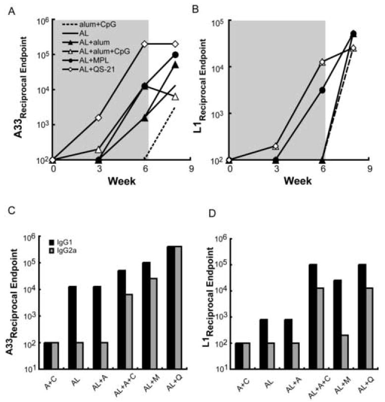
Mice (n = 5) were immunized twice with a mixture containing 10 μg each of A33 and L1 proteins (AL) alone or combined with alum, alum+CpG, MPL+TDM (MPL) or QS-21 and challenged with VACV strain WR at 3 weeks after the last immunization. Serum was collected prior to immunization (week 0), three weeks following immunizations 1 and 2 (shaded area), and two weeks after challenge (unshaded area). Antibodies to A33 (A) and L1 (B) were determined on pooled sera by ELISA and reciprocal endpoint values are plotted. In panel B, the plots for AL plus QS-21 and for AL plus alum+CpG are superimposed. Serum collected three weeks after the second immunization was re-analyzed for IgG1 or IgG2a isotype antibodies to A33 (C) and L1 (D). In the latter panels, alum and CpG are abbreviated A and C, respectively.
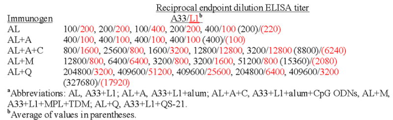
The data shown in Fig. 1 were obtained with sera pooled from animals within each group. We also analyzed the sera from individual mice collected after the boost in order to perform a statistical analysis (Table 1). The A33 ELISA titers of sera from mice immunized with AL plus QS-21 were significantly higher than the titers of mice of any other group (p<0.00002). The titers obtained after immunization with AL plus alum and CpG ODNs or with AL plus MPL+TDM were significantly higher than after AL without adjuvant or with alum but did not differ significantly from each other. Similar results were obtained from the L1 ELISA determinations, except that the difference between the titers obtained with AL plus QS-21 or with AL and alum plus CpG ODNs did not reach statistical significance,
Table 1.
Reciprocal endpoint dilution ELISA titers to A33 and L1 of sera from individual mice immunized with AL plus various adjuvants
|
Immunizations with protein, in contrast to live virus, typically induce a predominant Th2 response with IgG1 as the dominant antibody isotype in Balb/c mice. However, the Th1 response as revealed by IgG2a antibody can be enhanced by some adjuvants. Figs. 1C and 1D show the results of isotype-specific ELISAs that detect antibodies against A33 and L1 in sera collected three weeks after the second immunization. The anti-A33 antibodies from mice immunized with AL alone or with alum were exclusively IgG1 as the IgG2a titers were no higher than the adjuvant alone control. Some IgG2a in addition to IgG1 was made after AL plus alum+CpG ODNs or MPL+TDM (Fig. 1C). However, the highest total IgG2a as well as the highest ratio of IgG2a to IgG1 occurred with AL plus QS-21. Isotype-specific titers against L1 were IgG1-dominant in all groups, but IgG2a titers above the control were observed with sera from mice immunized with AL plus QS-21 or alum+CpG ODNs (Fig. 1D). These results suggested that the magnitude and isotype of the antibody responses were influenced by both the nature of the protein as well as the adjuvant. Overall with the two protein immunogens, QS-21 induced the most IgG2a with alum+CpG ODNs and MPL+TDM next.
3.2 Induction of VACV neutralizing and comet-reducing antibodies
A flow cytometry-based GFP assay was used to detect MV-neutralizing antibody induced by L1. Neutralizing antibody was not detected after the primary immunization but was present after the boost. The highest neutralizing titers were measured in the AL plus QS-21 and the AL plus alum+CpG ODNs groups (Fig. 2). These neutralization values were similar to that of vaccinia immune globulin, although the latter targeted additional MV proteins (data not shown). Significant neutralization was achieved with sera from mice immunized with AL and MPL+TDM but not with AL and alum or no adjuvant (Fig. 2). The post-challenge titers will be discussed in Section 3.3.
Figure 2. Induction of neutralizing antibody.
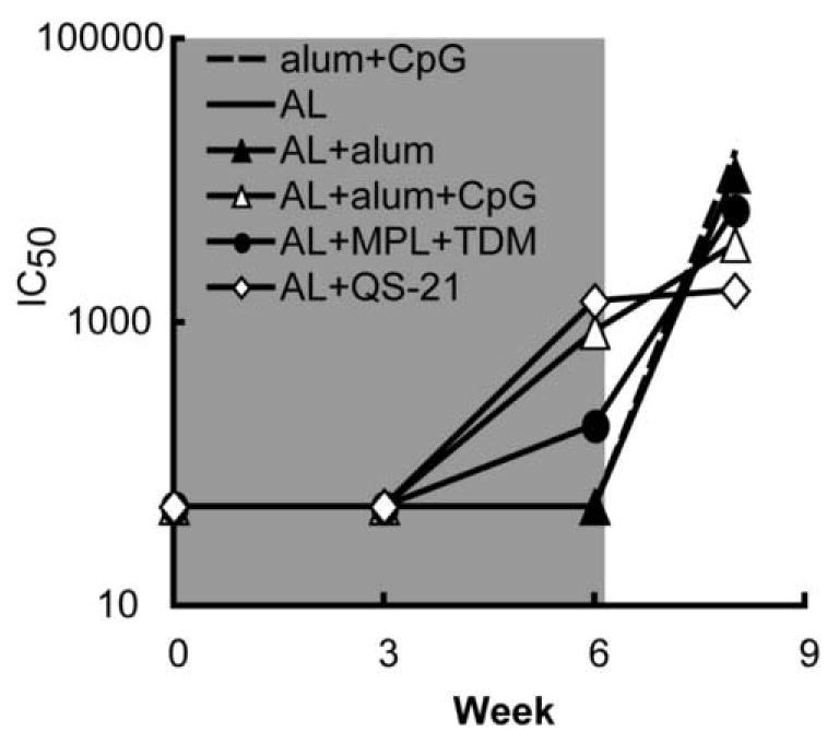
The sera described in Figure 1 obtained from mice immunized with AL plus the indicated adjuvants were used. MV neutralizing antibodies were measured with a flow cytometry-based GFP assay and the 50% inhibitory concentration (IC50) was determined for each pool of mouse sera
The IHD-J strain of VACV produces large numbers of satellite plaques known as comets when cells are infected in liquid medium. Antibodies to certain EV proteins such as A33 but not MV proteins such as L1 can suppress the formation of comets. Pre-challenge sera from animals receiving alum+CpG alone or proteins without adjuvant had no evident effect on comet size, whereas pre-challenge sera from animals immunized with proteins and adjuvants reduced the sizes of comets to various extents (Fig. 3). The greatest reduction occurred with AL plus QS-21, consistent with the highest anti-A33 ELISA titer (Fig. 1A). The comet-reducing activity of post-challenge sera will be discussed in Section 3.3.
Figure 3. Induction of comet-reducing antibody.
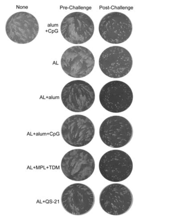
The sera described in Figure 1 obtained from mice immunized with A33 and L1 (AL) plus the indicated adjuvants were used to detect antibodies that inhibit the formation satellite plaques due to spread of EV in liquid medium. BS-C-1 cells were infected with VACV strain IHD-J (80 pfu/well), overlaid with medium containing a 1:50 dilution of pooled mouse serum, and 40 h later stained with crystal violet. The column labeled pre-challenge represents samples collected three weeks after the second immunization and one day prior to intranasal virus challenge. The post-challenge column shows samples collected from surviving mice two weeks following challenge. The well shown in the upper left corner shows the typical formation of comet-shaped plaques in the absence of serum
3.3 Effects of adjuvants on the induction of protective immune responses to A33 and L1 proteins
The protective effects of immunization with AL combined with different adjuvants were determined in mice that were infected intranasally with the WR strain of VACV [35, 36]. Weight loss, which occurs during the first week following challenge, is directly correlated with virus replication in the lung and provides an objective, non-invasive way of following disease [7, 47]. According to our animal protocol, mice were terminated if they lost greater than 30% of their initial weight. Three to four weeks following the second protein immunization with various adjuvants, mice were challenged intranasally with 5 LD50 of VACV WR. The majority of mice died or was sacrificed by ten days post-infection in the control group immunized with adjuvant without protein or with AL in the absence of adjuvant (Fig. 4A). Half of the mice immunized with AL and alum and 80% of those immunized with AL plus MPL + TDM survived. Most impressively, all mice immunized with AL combined with either QS-21 or alum+CpG ODNs survived challenge (Fig. 4A). Dramatic weight loss of surviving mice was observed over the first six days following challenge in most groups, except for mice immunized with AL plus QS-21 (Fig. 4B). The AL plus QS-21 group showed little fluctuation in weight and no visible signs of illness throughout the observation period. Mice immunized with AL and alum+CpG ODNs were next best and showed less weight loss overall than the other immunized groups. It is important to note that Rees et al. [48] reported that CpG ODNs alone could protect against VACV challenge through the upper respiratory route by stimulating innate immunity. However the protective action of CpG ODNs was short lived and gone by 21 days, which was the minimum time that we waited before challenge. Moreover, our control group received alum+CpG ODNs.
Figure 4. Survival and weight changes in mice immunized with AL proteins and different adjuvants followed by intranasal VACV challenge.
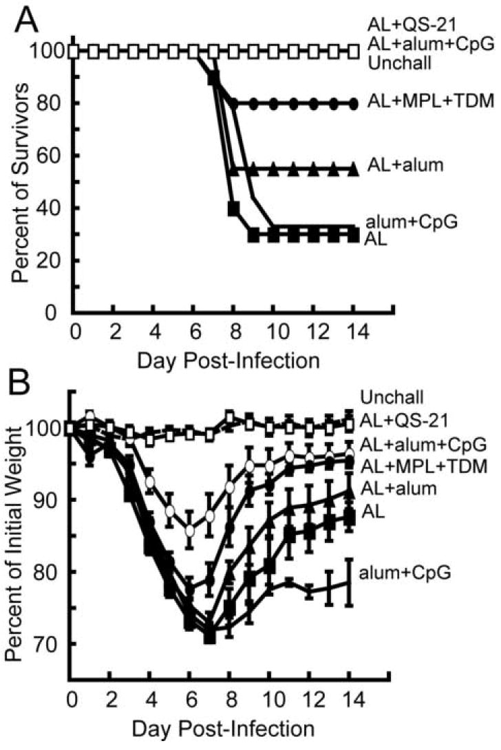
Mice were immunized as described in Fig. 1, and three weeks following the second immunization, were challenged intranasally with 106 pfu of VACV strain WR. The unchallenged (Unchall) mice were not immunized or challenged. The alum+CpG control group received no recombinant protein prior to challenge. Mice were weighed daily for two weeks and sacrificed if their weight fell below 70% of the initial value. The percent of survivors (A) and the percent of initial weight of surviving mice (B) are shown for each group. The data shown here represents two independent experiments and each group had 4-5 mice/group. Each data point is the average weight +/−SEM of mice in each group from the two challenge experiments
Statistical analysis of the weight loss data was achieved by calculating the area under the curve as a summary statistic for each animal and using the nonparametric Wilcoxon rank sum test with the Holm p-value adjustment method for multiple tests to compare the animal groups. The resulting p-values are shown in Table 2. Challenged mice that were immunized with AL plus QS-21 lost significantly less weight than mice given any of the other immunizations; furthermore the weight loss in this group was not significantly different from unchallenged mice. Mice given AL and alum+CpG ODNs had significantly less weight loss than mice immunized with AL alone or with alum alone. Mice immunized with AL and MPL+TDM showed significantly less weight loss than mice immunized with AL alone.
Table 2.
p-values calculated with area under the curve analysis of weight loss data in Fig. 4B followed by the Wilcoxan rank sum test using the Holm p-value adjustment
| alum + CpG | AL | AL + alum | AL + alum + CpG | AL + MPL + TDM | |
|---|---|---|---|---|---|
| ALa + alum | 0.4 | 0.4 | - | - | - |
| AL + alum + CpG | 0.002b | 0.002 | 0.02 | - | - |
| AL + MPL +TDM | 0.02 | 0.02 | 0.4 | 0.4 | - |
| AL + QS21 | 0.0005 | 0.0003 | 0.0005 | 0.0008 | 0.0003 |
aAL, A33 plus L1 proteins
b Bold numbers indicate significant p-values
This statistical analysis confirmed that mice immunized with AL and QS-21 were the best protected, in addition to inducing the highest overall antibody response. Binding the proteins to alum did not significantly enhance protection, but addition of CpG ODNs to AL plus alum provided significant protection from weight loss. The weight loss data paralleled the number of survivors, since the groups with more survivors showed less weight loss.
Boosting of antibody to the viral immunogens following challenge is an indirect measure of virus replication. Hence, surviving animals that were least well protected were anticipated to show highest boosting. Convalescent serum, collected from surviving mice two weeks after challenge, showed a boost in A33 antibody except for the AL plus QS-21 or alum+CpG ODNs groups (Fig. 1A), which had been the best protected. The same two groups showed the least boosting of anti-L1 antibody after challenge (Fig. 1B). Although prior to challenge, MV neutralization and EV comet reduction were exclusively due to anti-L1 and anti-A33 antibodies, respectively, antibodies to additional proteins may have contributed to inhibition after challenge. Thus sera from all groups, except those receiving AL plus QS-21, showed a dramatic rise in neutralizing antibodies two weeks post challenge (Fig. 2), likely targeting additional MV proteins. Similarly, all of the post-challenge sera showed strong comet-inhibiting activity (Fig. 3).
3.4 Binding and VACV neutralizing antibodies following immunization of cynomolgous monkeys with recombinant A33, B5, and L1 combined with QS-21
Prior to the adjuvant comparison studies described above, we initiated an experiment to test the efficacy of a recombinant protein vaccine in a non-human primate. Three cynomolgous monkeys (030, 770, 974) were immunized three times at one-month intervals with 100 μg each of recombinant A33, B5 and L1 proteins with QS-21 adjuvant. One control monkey (026) was immunized with the QS-21 adjuvant alone and two additional controls (398, 419) received neither adjuvant nor protein. The average ELISA titers for the three immunized monkeys are shown at each time point (Fig. 5A). Antibodies specific to A33, B5, and L1 were detected one month after the first immunization and were boosted by the second immunization. The titers fell somewhat between the second and third immunizations but were boosted again by the latter. The reciprocal ELISA titers declined gradually over the next 6 months and at the end of this time were more than a log lower than the peak values. However, the titers rebounded again after another immunization. A similar response pattern was obtained using a VACV-infected cell lysate for the ELISA in order to detect antibodies capable of recognizing non-recombinant VACV antigens (Fig 5B).
Figure 5. Analysis of monkey sera following immunizations with A33, B5 and L1combined with QS-21.
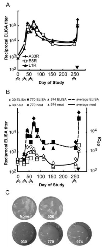
Cynomolgous monkeys were immunized with 100 μg each of recombinant A33, B5 and L1 proteins combined with the adjuvant QS-21 at days 0, 28,57 and 251 as indicated by double arrows below the x-axis. Four weeks after the fourth immunization, monkeys were challenged intravenously with 5 × 10⁷ pfu of MPXV as indicated by the solid black triangle above the x-axis. A. Averages of the ELISA values specific for A33, B5, and L1 are shown. B. Reciprocal ELISA values determined against an infected cell lysate and flow cytometry VACV neutralization titers of individual monkeys and averages are presented. C. The presence of EV-neutralizing antibodies in sera collected prior to challenge was determined using the comet reduction assay. Key: None, no serum; 026, adjuvant only monkey serum; 030, 770, 974, sera from immunized monkeys.
Neutralizing antibodies were detected using the flow cytometry assay following protein immunization and were boosted following each immunization as shown in Fig. 5B. Only L1-specific antibodies neutralize MVs since antibodies against A33 and B5 only target the EV form of the virus. IC50 titers fell to background levels between the third and the fourth immunization, but were boosted to peak levels after the fourth. The sera obtained after the fourth immunization were also tested by a VACV plaque-reduction assay, which gives lower titers than the flow cytometry assay (Fig. 6A).
Figure 6. VACV and MPXV plaque reduction and MPXV comet inhibition.
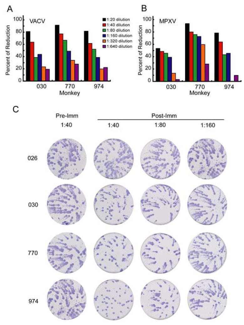
A. Pre-challenge serum samples were tested in a VACV plaque reduction assay. Data are from three separate analyses each done in duplicate and percentage reductions were determined with respect to serum from adjuvant-only monkey (026). B. Pre-challenge serum samples were tested in a MPXV plaque reduction assay. Analyses were done in duplicate and the percentage reduction determined as above. C. Sera (1:40 dilution) obtained prior to immunization (Pre-Imm) and after the final immunization (Post-imm) were tested in a MPXV comet reduction assay. The plates were stained with anti-variola rabbit antibody followed by peroxidase-labeled goat anti-rabbit IgG; comets were visualized by adding peroxidase substrate accounting for their dark color. Experiment was done in duplicate and photographs of one set are shown.
Antibodies to both A33 and B5 can reduce the release of extracellular virus from cells, which is responsible for comet formation. Pre-immune sera (not shown) and serum from the control monkey receiving adjuvant alone (026) did not cause any comet reduction compared to the no serum control. Serum from each of the three immunized monkeys prior to challenge dramatically reduced comets as shown in the bottom three wells of Figure 5C.
3.5 MPXV neutralizing antibodies following immunization of cynomolgous monkeys with recombinant A33, B5, and L1 combined with QS-21
Sera from the immunized monkeys also neutralized MPXV. The MPXV plaque reduction titer (Fig. 6B) was similar to the titer determined with VACV (Fig. 6A), consistent with the conserved protein sequences. The immune sera also inhibited MPXV comet formation whereas the serum from the monkey receiving adjuvant alone (026) did not (Fig. 6C). Thus, antibodies to VACVL1 neutralized MPXV MVs and antibody toVACV B5 or A33 prevented spread of MPXV.
3.6 Protection of monkeys from severe disease and death following an intravenous MPXV challenge
Although we had originally intended to challenge the monkeys with MPXV after the third immunization, logistical problems prevented this. An opportunity to challenge arose about 6-months later leading us to give an additional protein immunization. Four weeks after that, the monkeys receiving proteins and QS-21 (030, 770, 974), QS-21 alone (026) and no vaccine (398, 419) were challenged intravenously with 5 X 10⁷ pfu of MPXV. The monkeys were monitored for one month following challenge during which the immunized monkeys appeared healthy. In contrast, the non-immunized monkeys were severely ill with fever and weight loss as previously described [37] and one (026) died on day 12, despite supportive efforts including subcutaneous fluids. The number of virus-induced skin lesions was counted every three to four days. The monkeys that were not immunized with protein developed 575 to 820 skin lesions each, which peaked at twelve days post-infection (Fig. 7A). There were fewer lesions in immunized monkeys (maximum of 65 to 140 each) and they were generally smaller and atypical compared to those observed in naïve monkeys, developed less synchronously, and healed rapidly. The difference in lesion number between immunized and control animals was statistically significant (p=0.02).
Figure 7. Protein immunization reduces skin lesions and circulating virus in macaques.
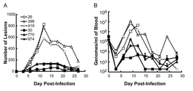
(A) Skin lesions. Monkeys immunized as described in the Legend to Fig. 5 were challenged intravenously with 5 × 10⁷ pfu MPXV and skin lesions were counted at 3 to 4 day intervals. (B) Blood samples were collected at 3 to 4 day intervals and the number of viral genomes was determined in duplicate by real-time quantitative PCR. The average +/− standard deviation is shown for each time point. The limit of sensitivity was 200 genomes/ml. Key to monkeys: 026 (QS21 adjuvant only); 398 and 419 (no immunization); 030, 770 and 974 (QS21 and A33, B5 and L1 proteins). Note that the dashed and solid lines are from control and protein immunized animals, respectively
A real-time PCR assay detected viral genomes in the blood of all monkeys immediately after challenge (day 0), followed by a decline (Fig. 7B). On day 6, however, an increase in viral genomes was detected with peak values generally present at day 9. The viral loads were higher in the controls than in the immunized monkeys with an average difference of about 2.5 logs, which was statistically significant (p=0.04) despite the small number of animals.
4. Discussion
An important role of antibody in protection against secondary orthopoxvirus infections has been well documented. Studies using B- and T-cell depletion [49, 50] or gene knockouts [51] demonstrated the pivotal role of the CD4(+) T cell-driven antibody response for protection against VACV infection in mice. The fundamental role of B-cells was also shown in ectromelia virus infections of mice [15, 52] and in a macaque MPXV model [53]. These findings suggest that a protein subunit vaccine, which primarily induces an antibody response, may be sufficient to defend against smallpox, if the animal models are predictive of disease protection in humans.
Our previous study [11] showed that a combination of soluble recombinant forms of VACV MV and EV membrane proteins could protect as well as live VACV in the VACV WR mouse pneumonia model. However, the requirement for 3 or 4 protein immunizations would be impractical for vaccine delivery and we therefore investigated the use of other adjuvants. We now show that the combination of QS-21 with only the A33 and L1 proteins completely prevented weight loss as well as death after only 2 vaccinations at 3-week intervals. Using the same vaccination regimen, the combination of the 2 proteins with alum+CpG ODNs was next best, followed by MPL+TDM. Alum+CpG ODNs was clearly superior to alum alone. The degree of protection in the mouse model with different adjuvants correlated with antibody levels, whether determined by ELISA, neutralization of MVs, or reduction in spread of satellite plaques.
Analysis of convalescent serum from surviving mice showed a consistent pattern with respect to ELISA and neutralizing antibody titers. Groups of mice that had higher titers against A33 and L1 prior to challenge and the best protection, namely those receiving AL plus QS-21 or AL plus alum+CpG ODNs, showed the lowest boosting of A33 and L1 antibodies. Conversely, groups that had the lowest A33 and L1 titers prior to challenge and the poorest protection had the greatest boosting of antibody responses following challenge. This trend was observed with both MV and EV antigens, and likely reflects the degree of replication of the challenge virus in poorly immunized animals.
Protein immunogens typically induce a predominantly Th2-type response that is characterized by activation of B-cells and production of antibodies, especially IgG1. QS-21 and to a lesser extent alum+CpG ODNs and MPL+TDM increased the IgG2a antibody, indirectly suggesting an enhancement of the Th1 response. This effect of immunostimulatory adjuvants has been noted previously for other immunogens. Both QS-21 [54-58] and CpG ODNs [59, 60] can also augment the production of antigen-specific cytotoxic T cells, though this was not evaluated in the present study.
The monkey study described here provides a preliminary evaluation of the immune response to recombinant VACV proteins in a primate and provides evidence that they can induce protection against severe disease following a MPXV challenge. This study mimicked our earlier mouse experiments [11], which used B5 in addition to A33 and L1. Recombinant B5 protein [11, 14] as well as antibodies to B5 [18, 19] can provide protection in the VACV mouse pneumonia model. In addition, B5 is the major target of EV neutralizing antibody in human VACV immune globulin [61], though antibody to B5 is not necessarily the most protective in vivo. Also, VACV B5 has a slightly greater amino acid identity (96%) to the MPXV homolog than VACV A33, which has 93% identity. L1 is the most highly conserved with 98% identity between the VACV and MPXV homologs. Therefore, we anticipated that antibodies to these VACV proteins would cross react with the MPXV homologs and provide at least partial protection. We chose to use QS-21, based on our previous experience with recombinant HIV proteins in a monkey model [62], even though we had not yet determined the superiority of this adjuvant for inducing VACV protection in mice. The recombinant VACV proteins were immunogenic in monkeys and binding antibodies were detected after the first immunization and boosted by a second, at which time VACV neutralizing and comet-reducing antibodies were also found. Between immunizations, the antibody levels dropped but were boosted again even after 7 months. At this time, we also demonstrated MPXV neutralizing and comet-reducing antibodies. A previously described intravenous MPXV challenge was used because of its consistency [37], which was particularly important with a small number of animals. There were several indicators of protection: each unimmunized animal developed approximately 700 typical pustular skin lesions, whereas the vaccinated ones developed about 100 smaller atypical lesions; the virus load was reduced by about 2.5 logs; and most importantly the vaccinated animals appeared healthy whereas the unvaccinated were gravely ill and one died. Based on the number of skin lesions, the protection was less than achieved with modified VACV Ankara or the licensed smallpox vaccine [37] and similar to that obtained with a 4-component DNA vaccine (VACV L1R, A27L. A33R, and B5R) administered percutaneously with a gene gun [63]. In a recently published study [17], it was reported that a 4-component DNA vaccine composed of MPXV orthologs of the same VACV genes administered intramuscularly did not induce neutralizing antibody or protect monkeys against a MPXV challenge. In contrast, the corresponding recombinant MPXV proteins with alum or CPG ODNs provided partial protection, which was enhanced by prior DNA vaccinations. Although VACV and MPXV membrane proteins are very closely related to the variola virus orthologs, it would seem that the latter would be most appropriate for a smallpox vaccine.
Acknowledgments
This work was done to partially fulfill the Ph.D. thesis requirements of C.F. at the University of Maryland. The authors would like to thank Gary Cohen and Roselyn Eisenberg for purified proteins, the production of which was supported by USPHS Grant NIH RCE-U54-AI57168 from the NIAID, NIH. Antigenics, Inc. kindly provided QS-21 adjuvant, and Norman Cooper supplied cells and virus stocks. Thanks also to the NIAID Animal Care Branch. The study was partially supported by intramural funds from the NIAID, NIH and the Office for Chemical and Biological Defense of DTRA
Footnotes
Publisher's Disclaimer: This is a PDF file of an unedited manuscript that has been accepted for publication. As a service to our customers we are providing this early version of the manuscript. The manuscript will undergo copyediting, typesetting, and review of the resulting proof before it is published in its final citable form. Please note that during the production process errorsmaybe discovered which could affect the content, and all legal disclaimers that apply to the journal pertain.
References
- 1.Fenner F, Henderson DA, Arita I, Jezek Z, Ladnyi ID. Smallpox and itseradication. First. World Health Organization; Geneva: 1988. [Google Scholar]
- 2.Fulginiti VA, Papier A, Lane JM, Neff JM, Henderson DA. Smallpox vaccination: A review, part II Adverse events. Clin Inf Dis. 2003 Jul 15;37(2):251–71. doi: 10.1086/375825. [DOI] [PubMed] [Google Scholar]
- 3.Henderson DA. The looming threat of bioterrorism. Science. 1999 Feb 26;283(5406):1279–82. doi: 10.1126/science.283.5406.1279. [DOI] [PubMed] [Google Scholar]
- 4.Moss B. Poxviridae: the viruses and their replication. In: Knipe DM, Howley PM, editors. Fields Virology. 4. Vol. 2001. Philadelphia: Lippincott Williams & Wilkins; pp. 2849–83. [Google Scholar]
- 5.Boulter EA, Zwartouw HT, Titmuss DHJ, Maber HB. The nature of the immune state produced by inactivated vaccinia virus in rabbits. Am J Epidemiol. 1971;94:612–20. doi: 10.1093/oxfordjournals.aje.a121360. [DOI] [PubMed] [Google Scholar]
- 6.Boulter EA, Appleyard G. Differences between extracellular and intracellular forms of poxvirus and their implications. Prog Med Virol. 1973;16:86–108. [PubMed] [Google Scholar]
- 7.Law M, Putz MM, Smith GL. An investigation of the therapeutic value of vaccinia-immune IgG in a mouse pneumonia model. J Gen Virol. 2005 Apr;86(Pt 4):991–1000. doi: 10.1099/vir.0.80660-0. [DOI] [PubMed] [Google Scholar]
- 8.Payne LG. Significance of extracellular virus in the in vitro and in vivo dissemination of vaccinia virus. J Gen Virol. 1980;50:89–100. doi: 10.1099/0022-1317-50-1-89. [DOI] [PubMed] [Google Scholar]
- 9.Blasco R, Moss B. Role of cell-associated enveloped vaccinia virus in cell-to-cell spread. J Virol. 1992;66(7):4170–9. doi: 10.1128/jvi.66.7.4170-4179.1992. [DOI] [PMC free article] [PubMed] [Google Scholar]
- 10.Smith GL, Vanderplasschen A, Law M. The formation and function of extracellular enveloped vaccinia virus. J Gen Virol. 2002 Dec;83(Pt 12):2915–31. doi: 10.1099/0022-1317-83-12-2915. [DOI] [PubMed] [Google Scholar]
- 11.Fogg C, Lustig S, Whitbeck JC, Eisenberg RJ, Cohen GH, Moss B. Protective immunity to vaccinia virus induced by vaccination with multiple recombinant outer membrane proteins of intracellular and extracellular virions. J Virol. 2004 Oct;78(19):10230–7. doi: 10.1128/JVI.78.19.10230-10237.2004. [DOI] [PMC free article] [PubMed] [Google Scholar]
- 12.Lai CF, Gong SC, Esteban M. The purified 14-kilodalton envelope protein of vaccinia virus produced in Escherichia coli induces virus immunity in animals. J Virol. 1991;65(10):5631–5. doi: 10.1128/jvi.65.10.5631-5635.1991. [DOI] [PMC free article] [PubMed] [Google Scholar]
- 13.Davies DH, McCausland MM, Valdez C, Huynh D, Hernandez JE, Mu YX, et al. Vaccinia virus H3L envelope protein is a major target of neutralizing antibodies in humans and elicits protection against lethal challenge in mice. J Virol. 2005 Sep;79(18):11724–33. doi: 10.1128/JVI.79.18.11724-11733.2005. [DOI] [PMC free article] [PubMed] [Google Scholar]
- 14.Galmiche MC, Goenaga J, Wittek R, Rindisbacher L. Neutralizing and protective antibodies directed against vaccinia virus envelope antigens. Virology. 1999;254(1):71–80. doi: 10.1006/viro.1998.9516. [DOI] [PubMed] [Google Scholar]
- 15.Fang M, Cheng H, Dai ZP, Bu ZM, Sigal LJ. Immunization with a single extracellular enveloped virus protein produced in bacteria provides partial protection from a lethal orthopoxvirus infection in a natural host. Virology. 2006 Feb;345(1):231–43. doi: 10.1016/j.virol.2005.09.056. [DOI] [PubMed] [Google Scholar]
- 16.Hooper JW, Custer DM, Thompson E. Four-gene-combination DNA vaccine protects mice against a lethal vaccinia virus challenge and elicits appropriate antibody responses in nonhuman primates. Virology. 2003 Feb 1;306(1):181–95. doi: 10.1016/S0042-6822(02)00038-7. [DOI] [PMC free article] [PubMed] [Google Scholar]
- 17.Heraud JM, Edghill-Smith Y, Ayala V, Kalisz I, Parrino J, Kalyanaraman VS, et al. Subunit recombinant vaccine protects against monkeypox. J Immunol. 2006 Aug 15;177(4):2552–64. doi: 10.4049/jimmunol.177.4.2552. [DOI] [PubMed] [Google Scholar]
- 18.Lustig S, Fogg C, Whitbeck JC, Eisenberg RJ, Cohen GH, Moss B. Combinations of polyclonal or monoclonal antibodies to proteins of the outer membranes of the two infectious forms of vaccinia virus protect mice against a lethal respiratory challenge. J Virol. 2005;79:13454–62. doi: 10.1128/JVI.79.21.13454-13462.2005. [DOI] [PMC free article] [PubMed] [Google Scholar]
- 19.Chen ZC, Earl P, Americo J, Damon I, Smith SK, Zhou YH, et al. Chimpanzee/human mAbs to vaccinia virus B5 protein neutralize vaccinia and smallpox viruses and protect mice against vaccinia virus. Proc Natl Acad Sci USA. 2006 Feb;103(6):1882–7. doi: 10.1073/pnas.0510598103. [DOI] [PMC free article] [PubMed] [Google Scholar]
- 20.Poltorak A, Smirnova I, He X, Liu MY, Van Huffel C, McNally O, et al. Genetic and physical mapping of the Lps locus: identification of the toll-4 receptor as a candidate gene in the critical region. Blood Cells Mol Dis. 1998 Sep;24(3):340–55. doi: 10.1006/bcmd.1998.0201. [DOI] [PubMed] [Google Scholar]
- 21.Takayama K, Ribi E, Cantrell JL. Isolation of a nontoxic lipid A fraction containing tumor regression activity. Cancer Res. 1981 Jul;41(7):2654–7. [PubMed] [Google Scholar]
- 22.Masihi KN, Lange W, Brehmer W, Ribi E. Immunobiological activities of nontoxic lipid A: enhancement of nonspecific resistance in combination with trehalose dimycolate against viral infection and adjuvant effects. Int J Immunopharmacol. 1986;8(3):339–45. doi: 10.1016/0192-0561(86)90116-5. [DOI] [PubMed] [Google Scholar]
- 23.Evans JT, Cluff CW, Johnson DA, Lacy MJ, Persing DH, Baldridge JR. Enhancement of antigen-specific immunity via the TLR4 ligands MPL adjuvant and Ribi.529. Expert Rev Vaccines. 2003 Apr;2(2):219–29. doi: 10.1586/14760584.2.2.219. [DOI] [PubMed] [Google Scholar]
- 24.Liu G, Anderson C, Scaltreto H, Barbon J, Kensil CR. QS-21 structure/function studies: effect of acylation on adjuvant activity. Vaccine. 2002 Jun 21;20(2122):2808–15. doi: 10.1016/s0264-410x(02)00209-8. [DOI] [PubMed] [Google Scholar]
- 25.Evans TG, McElrath MJ, Matthews T, Montefiori D, Weinhold K, Wolff M, et al. QS-21 promotes an adjuvant effect allowing for reduced antigen dose during HIV-1 envelope subunit immunization in humans. Vaccine. 2001 Feb 28;19(1516):2080–91. doi: 10.1016/s0264-410x(00)00415-1. [DOI] [PubMed] [Google Scholar]
- 26.Livingston PO, Adluri S, Helling F, Yao TJ, Kensil CR, Newman MJ, et al. Phase 1 trial of immunological adjuvant QS-21 with a GM2 ganglioside-keyhole limpet haemocyanin conjugate vaccine in patients with malignant melanoma. Vaccine. 1994 Nov;12(14):1275–80. doi: 10.1016/s0264-410x(94)80052-2. [DOI] [PubMed] [Google Scholar]
- 27.Kashala O, Amador R, Valero MV, Moreno A, Barbosa A, Nickel B, et al. Safety, tolerability and immunogenicity of new formulations of the Plasmodium falciparum malaria peptide vaccine SPf66 combined with the immunological adjuvant QS-21. Vaccine. 2002 May 22;20(1718):2263–77. doi: 10.1016/s0264-410x(02)00115-9. [DOI] [PubMed] [Google Scholar]
- 28.Klinman DM, Yi AK, Beaucage SL, Conover J, Krieg AM. CpG motifs present in bacteria DNA rapidly induce lymphocytes to secrete interleukin 6, interleukin 12, and interferon gamma. Proc Natl Acad Sci USA. 1996 Apr 2;93(7):2879–83. doi: 10.1073/pnas.93.7.2879. [DOI] [PMC free article] [PubMed] [Google Scholar]
- 29.Krieg AM, Yi AK, Matson S, Waldschmidt TJ, Bishop GA, Teasdale R, et al. CpG motifs in bacterial DNA trigger direct B-cell activation. Nature. 1995 Apr 6;374(6522):546–9. doi: 10.1038/374546a0. [DOI] [PubMed] [Google Scholar]
- 30.Yamamoto S, Yamamoto T, Shimada S, Kuramoto E, Yano O, Kataoka T, et al. DNA from bacteria, but not from vertebrates, induces interferons, activates natural killer cells and inhibits tumor growth. Microbiol Immunol. 1992;36(9):983–397. doi: 10.1111/j.1348-0421.1992.tb02102.x. [DOI] [PubMed] [Google Scholar]
- 31.Klinman DM. Immunotherapeutic uses of CpG oligodeoxynucleotides. Nat Rev Immunol. 2004 Apr;4(4):249–58. doi: 10.1038/nri1329. [DOI] [PubMed] [Google Scholar]
- 32.Bomford R. Aluminium salts: perspectives in their use as adjuvants. In: Gregoriadis GAA, Poste G, editors. NATO Advanced Study Institute on Immunological Adjuvants and Vaccines. New York: Plenum Press; 1988. pp. 35–41. [Google Scholar]
- 33.Jordan MB, Mills DM, Kappler J, Marrack P, Cambier JC. Promotion of B cell immune responses via an alum-induced myeloid cell population. Science. 2004 Jun 18;304(5678):1808–10. doi: 10.1126/science.1089926. [DOI] [PubMed] [Google Scholar]
- 34.Lindblad EB. Aluminium compounds for use in vaccines. Immunol Cell Biol. 2004 Oct;82(5):497–505. doi: 10.1111/j.0818-9641.2004.01286.x. [DOI] [PubMed] [Google Scholar]
- 35.Turner GS. Respiratory infection of mice with vaccinia virus. J Gen Virol. 1967;1(3):399–402. doi: 10.1099/0022-1317-1-3-399. [DOI] [PubMed] [Google Scholar]
- 36.Williamson JD, Reith RW, Jeffrey LJ, Arrand JR, Mackett M. Biological characterization of recombinant vaccinia viruses in mice infected by the respiratory route. J Gen Virol. 1990 NOV;71:2761–7. doi: 10.1099/0022-1317-71-11-2761. [DOI] [PubMed] [Google Scholar]
- 37.Earl PL, Americo JL, Wyatt LS, Eller LA, Whitbeck JC, Cohen GH, et al. Immunogenicity of a highly attenuated MVA smallpox vaccine and protection against monkeypox. Nature. 2004;428:182–5. doi: 10.1038/nature02331. [DOI] [PubMed] [Google Scholar]
- 38.Anton LC, Schubert U, Bacik I, Princiotta MF, Wearsch PA, Gibbs J, et al. Intracellular localization of proteasomal degradation of a viral antigen. J Cell Biol. 1999 Jul 12;146(1):113–24. doi: 10.1083/jcb.146.1.113. [DOI] [PMC free article] [PubMed] [Google Scholar]
- 39.Norbury CC, Malide D, Gibbs JS, Bennink JR, Yewdell JW. Visualizing priming of virus-specific CD8(+) T cells by infected dendritic cells in vivo. Nature Immunol. 2002 Mar;3(3):265–71. doi: 10.1038/ni762. [DOI] [PubMed] [Google Scholar]
- 40.Earl PL, Cooper N, Wyatt S, Moss B, Carroll MW. Preparation of cell cultures and vaccinia virus stocks. In: Ausubel FM, Brent R, Kingston RE, Moore DD, Seidman JG, Smith JA, et al., editors. Current Protocols in Molecular Biology. Vol. 16. New York: John Wiley and Sons; 1998. pp. 1–3. [Google Scholar]
- 41.Aldaz-Carroll L, Whitbeck JC, Ponce de Leon M, Lou H, Hirao L, Isaacs SN, et al. Epitope-mapping studies define two major neutralization sites on the vaccinia virus extracellular enveloped virus glycoprotein B5R. J Virol. 2005 May;79(10):6260–71. doi: 10.1128/JVI.79.10.6260-6271.2005. [DOI] [PMC free article] [PubMed] [Google Scholar]
- 42.Aldaz-Carroll L, Whitbeck JC, Ponce de Leon M, Lou H, Pannell LK, Lebowitz J, et al. Physical and immunological characterization of a recombinant secreted form of the membrane protein encoded by the vaccinia virus L1R gene. Virology. 2005;341(1):59–71. doi: 10.1016/j.virol.2005.07.006. [DOI] [PubMed] [Google Scholar]
- 43.Earl PL, Americo JL, Moss B. Development and use of a vaccinia virus neutralization assay based on flow cytometric detection of green fluorescent protein. J Virol. 2003 Oct;77(19):10684–8. doi: 10.1128/JVI.77.19.10684-10688.2003. [DOI] [PMC free article] [PubMed] [Google Scholar]
- 44.Yang HL, Kim SK, Kim M, Reche PA, Morehead TJ, Damon IK, et al. Antiviral chemotherapy facilitates control of poxvirus infections through inhibition of cellular signal transduction. J Clin Invest. 2005 Feb;115(2):379–87. doi: 10.1172/JCI23220. [DOI] [PMC free article] [PubMed] [Google Scholar]
- 45.Journot V, Chene G, Joly P, Saves M, Jacqmin-Gadda H, Molina JM, et al. Viral load as a primary outcome in human immunodeficiency virus trials: a review of statistical analysis methods. Control Clin Trials. 2001 Dec;22(6):639–58. doi: 10.1016/s0197-2456(01)00158-1. [DOI] [PubMed] [Google Scholar]
- 46.Holm S. A simple sequentialrejective multiple test procedure. Scand, J Statistics. 1979;6:65–70. [Google Scholar]
- 47.Luker KE, Hutchens M, Schultz T, Pekosz A, Luker GD. Bioluminescence imaging of vaccinia virus: Effects of interferon on viral replication and spread. Virology. 2005 Oct 25;341(2):284–300. doi: 10.1016/j.virol.2005.06.049. [DOI] [PubMed] [Google Scholar]
- 48.Rees DGC, Gates AJ, Green M, Eastaugh L, Lukaszewski RA, Griffin KF, et al. CpG-DNA protects against a lethal orthopoxvirus infection in a murine model. Antiviral Research. 2005 Feb;65(2):87–95. doi: 10.1016/j.antiviral.2004.10.004. [DOI] [PubMed] [Google Scholar]
- 49.Belyakov IM, Earl P, Dzutsev A, Kuznetsov VA, Lemon M, Wyatt LS, et al. Shared modes of protection against poxvirus infection by attenuated and conventional smallpox vaccine viruses. Proc Natl Acad Sci USA. 2003 Aug 5;100(16):9458–63. doi: 10.1073/pnas.1233578100. [DOI] [PMC free article] [PubMed] [Google Scholar]
- 50.Xu R, Johnson AJ, Liggitt D, Bevan MJ. Cellular and humoral immunity against vaccinia virus infection of mice. J Immunol. 2004 May 15;172(10):6265–71. doi: 10.4049/jimmunol.172.10.6265. [DOI] [PubMed] [Google Scholar]
- 51.Wyatt LS, Earl PL, Eller LA, Moss B. Highly attenuated smallpox vaccine protects mice with and without immune deficiencies against pathogenic vaccinia virus challenge. Proc Nat Acad Sci USA. 2004;101:4590–5. doi: 10.1073/pnas.0401165101. [DOI] [PMC free article] [PubMed] [Google Scholar]
- 52.Panchanathan V, Chaudhri G, Karupiah G. Interferon function is not required for recovery from a secondary poxvirus infection. Proc Natl Acad Sci USA. 2005 Sep 6;102(36):12921–6. doi: 10.1073/pnas.0505180102. [DOI] [PMC free article] [PubMed] [Google Scholar]
- 53.Edghill-Smith Y, Golding H, Manischewitz J, King LR, Scott D, Bray M, et al. Smallpox vaccine-induced antibodies are necessary and sufficient for protection against monkeypox virus. Nature Med. 2005 Jul;11(7):740–7. doi: 10.1038/nm1261. [DOI] [PubMed] [Google Scholar]
- 54.Mikloska Z, Ruckholdt M, Ghadiminejad I, Dunckley H, Denis M, Cunningham AL. Monophosphoryl lipid A and QS21 increase CD8 T lymphocyte cytotoxicity to herpes simplex virus-2 infected cell proteins 4 and 27 through IFN-gamma and IL-12 production. J Immunol. 2000 May 15;164(10):5167–76. doi: 10.4049/jimmunol.164.10.5167. [DOI] [PubMed] [Google Scholar]
- 55.Moore A, McCarthy L, Mills KH. The adjuvant combination monophosphoryl lipid A and QS21 switches T cell responses induced with a soluble recombinant HIV protein from Th2 to Th1. Vaccine. 1999 Jun 4;17(2021):2517–27. doi: 10.1016/s0264-410x(99)00062-6. [DOI] [PubMed] [Google Scholar]
- 56.Newman MJ, Wu JY, Gardner BH, Munroe KJ, Leombruno D, Recchia J, et al. Saponin adjuvant induction of ovalbumin-specific CD8+ cytotoxic T lymphocyte responses. J Immunol. 1992 Apr 15;148(8):2357–562. [PubMed] [Google Scholar]
- 57.Wu JY, Gardner BH, Murphy CI, Seals JR, Kensil CR, Recchia J, et al. Saponin adjuvant enhancement of antigen-specific immune responses to an experimental HIV-1 vaccine. J Immunol. 1992 Mar 1;148(5):1519–25. [PubMed] [Google Scholar]
- 58.Newman MJ, Munroe KJ, Anderson CA, Murphy CI, Panicali DL, Seals JR, et al. Induction of antigen-specific killer T lymphocyte responses using subunit SIVmac251 gag and env vaccines containing QS-21 saponin adjuvant. AIDS Res Hum Retroviruses. 1994 Jul;10(7):853–61. doi: 10.1089/aid.1994.10.853. [DOI] [PubMed] [Google Scholar]
- 59.Davis HL, Weeratna R, Waldschmidt TJ, Tygrett L, Schorr J, Krieg AM. CpG DNA is a potent enhancer of specific immunity in mice immunized with recombinant hepatitis B surface antigen. J Immunol. 1998 Jan 15;160(2):870–6. [PubMed] [Google Scholar]
- 60.Oxenius A, Martinic MM, Hengartner H, Klenerman P. CpG-containing oligonucleotides are efficient adjuvants for induction of protective antiviral immune responses with T-cell peptide vaccines. J Virol. 1999 May 5;73:4120–6. doi: 10.1128/jvi.73.5.4120-4126.1999. [DOI] [PMC free article] [PubMed] [Google Scholar]
- 61.Bell E, Shamim M, Whitbeck JC, Sfyroera G, Lambris JD, Isaacs SN. Antibodies against the extracellular enveloped virus B5R protein are mainly responsible for the EEV neutralizing capacity of vaccinia immune globulin. Virology. 2004 Aug 1;325(2):425–31. doi: 10.1016/j.virol.2004.05.004. [DOI] [PubMed] [Google Scholar]
- 62.Earl PL, Sugiura W, Montefiori DC, Broder CC, Lee SA, Wild C, et al. Immunogenicity and protective efficacy of oligomeric human immunodeficiency virus type 1 gp140. J Virol. 2001;75(2):645–53. doi: 10.1128/JVI.75.2.645-653.2001. [DOI] [PMC free article] [PubMed] [Google Scholar]
- 63.Hooper JW, Thompson E, Wilhelmsen C, Zimmerman M, Ichou MA, Steffen SE, et al. Smallpox DNA vaccine protects nonhuman primates against lethal monkeypox. J Virol. 2004 May;78(9):4433–43. doi: 10.1128/JVI.78.9.4433-4443.2004. [DOI] [PMC free article] [PubMed] [Google Scholar]