

Abstract
Myiasis is defined as infestation of a mammal by fly larvae. It may occur on either living tissues (primary myiasis) or dead tissues (secondary myiasis). In this report, we present a patient with myiasis with an extremely rare clinical manifestation and severe allergic reaction, and we review the literature in order to reveal the current status. A 20-year-old female patient was admitted to our emergency department due to rush on face, cough and shortness of breath. The maggot came out of her nose was identified as Oestrus ovis. With a diagnosis of severe allergic reaction due to myiasis, she was treated diphenhidramine, prednisone and inhale albuterol in the emergency department. After treatment and further investigation, she was discharged with full recovery. Myiasis is a rare cause for severe allergic reaction in patients with definite diagnosis. Immediate diagnosis and treatment are milestones in preventing bad outcomes.
Keywords: Allergy/immunology, epidemiology/public health, infectious diseases
Introduction
Myiasis is a term defining condition characterized by infestation of a mammal by fly larvae in order to feed on its tissues.1 While, in primary, myiasis larvae feed on living tissues, in secondary, myiasis larvae feed on dead tissues.2 Human myiasis is often classified according to the area infested such as cutaneous, oral, ocular, nasal, urogenital and gastrointestinal myiasis. Presentations on the skin are localized furuncular myiasis, creeping dermal myiasis, as well as wound and body cavity myiasis.3
When nostrils are invaded by dipteran larvae, it is called nasal myiasis.4 Nasal myiasis is thought to be underreported in tropical countries. However, it is endemic due to warm and humid environment. The condition is commonly seen in older female people due to decreased ability to ward off the flies themselves. It is common in low socioeconomic classes that suffer from poor nasal hygienic conditions.5,6
Nasal myiasis may cause embarrassment and stress to the patient, relatives and health care providers. In addition, it has a wide range of symptoms from mild to severe such as foreign body sensation, itching, nasal discharge, sneezing and even more severe respiratory manifestations and penetration of larvae into the brain.7 The most useful method for preventing the myiasis is extracting the flies.8 In this report, we present you a rare case of myiasis resulting in severe allergic reaction and aim to clarify the current status of myiasis in Turkey in the light of the literature.
Case report
A 20-year-old female patient was admitted to our emergency department (ED) due to rash on face, cough and shortness of breath. From the anamnesis, it was understood that she was lying near a tree on the grass in a park. Suddenly, she felt itching on her nose. After a sneeze, she recognized that a maggot has blown out of her nose on the handkerchief she used. And then, she suddenly developed rash, dyspnoea and shortness of breath. On admission to our ED, she was hypotensive (90/60 mmHg) with a heart rate of 90 beats/min and a saturation of 96% by probe. She had a slightly altered mental status. On medical history, she had allergy to an unknown origin and she was under improper medication. After the maggot on the handkerchief was investigated by a parasitologist, a prediagnosis of severe allergic reaction due to myiasis was made (see Figure 1 for segmented larva with oral hooks in the first segment). On auscultation, she had stridor and wheezing. Blood tests were obtained and they did not reveal any abnormalities. A sinus graphy and a brain computed tomography for a possible sinus and brain infestation were performed, and they also did not reveal any abnormalities. She was treated by 50 mg diphenhidramine IV, 60 mg prednisone IV and 2.5 mg inhale albuterol. Following treatment, shortness of breath and rush improved. After her symptoms improved, a consultation with ear–nose–throat specialist was performed, and the patient was evaluated via rigid nasal endoscopy. Any residual larvae could not be determined. After follow-up, the patient was discharged with full recovery.
Figure 1.
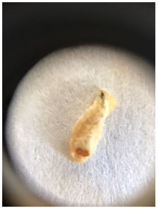
Oestrus ovis as a cause of nasal myiasis.
Discussion
Myiasis takes root from the ancient Greek word ‘myia’ which means fly. It is known to be an infestation of the tissues and organs caused by fly (diptera) larvea.9
Some fly species lay their eggs on intact or damaged tissues of humans. When the eggs convert to larvae following hatching, myiasis develops. Usually, the predisposing factor in the occurrence of the disease is lack of hygiene.10 In our case, socioeconomical status of the patient was high, which means that the disease is a threat for everyone in the society regardless of personal hygiene or social status.
Nasal myiasis is known to be a self-limiting infestation.11 In a report of nasal infestation of Oestrus ovis, it was stated that the disease was characterized by local inflammation of the mucosa and by sneezing or by manual extraction of the larvae from the cavity, and the larvae could be eliminated spontaneously usually without leaving sequelae.12,13 Nasal myiasis in our case also had O. ovis infestation; however, severe allergic reaction has developed in our patient. If the treatment had delayed, it was possible to have bad outcomes for the patient.
Commonly, the infestation lasts 2 weeks and causes moderate-to-severe nasal discomfort with obstruction, rhinorrhoea and a burning sensation. Allergic reaction due to nasal O. ovis infestation requiring hospitalization is known to be an unusual presentation.11 In our case, severe allergic reaction accompanied with asthma-like symptoms occurred and aggressive treatment was required.
The clinical and pathological infestation of myiasis in human depends on the type of flies, the mode of invasion by the larvae, the degree and type of migration after invasion, the stage in the life cycle of the fly, including the type and number of larval molts in the host, and the topographical site of invasion. These flies can cause disease in humans, with living on rotten tissue (maggots) or larvae crawl under the skin. Myiasis treatment, according to its type, is to be performed as soon as possible, with application of lateral pressure and suffocation by occlusion of the punctum with mineral oils, petroleum jelly or pork fat. Also, more-invasive interventions, such as surgical excision, may be necessary.14 In our case, any residual larvae could not be determined. Besides, in our case, myiasis has developed on healthy nasal cavity tissue instead of a wound.
For prevention from the disease, environmental health is important. In order to control myiasis, population of the flies must be controlled both in the open areas and health facilities. Enhancing environmental health standards in living and working places by public education about myiasis and the role of flies in the pathogenesis of the disease may play an important role in prevention and management of myiasis.4
Although generally the parasite is known to be seen in patients with mental retardation and/or accompanying psychiatric disorders, elderly individuals, those with poor self-care and hygiene and those with immune system disorders,15 our report revealed that the disease may also be seen in healthy subjects. Data on myiasis in the literature are mainly based on case reports in Turkey. It is known that aural, ophthalmic and wound myiases are the most commonly seen types of myiasis reported in Turkey. The most common species was Wohlfahrtia magnifica.10,15–17 İnce et al.18 reported a case of respiratory myiasis following aspiration of the larvae.
O. ovis is known to be the most common reason for ophthalmomyiasis externa. Koylu et al. reported a case of ophthalmomyiasis externa – the infestation of conjunctiva by the larvae of O. ovis – and proposed a novel method of treatment by 1% drop of cyclopentolate – a parasympatholytic anticholinergic agent – which might make the collection of larvae easier.19–23 The disease may be confused with bacterial conjunctivitis causing delays in treatment.24
Demirel-Kaya et al.25 presented a case of a patient with a wound due to squamous cell carcinoma from whom 200 larvae were collected and identified as Lucilia sericata. In another report by Bayindir et al.,26 a wound of the head and neck region due to squamous cell carcinoma presented with cutaneous myiasis, and was treated with antisepsis, larval removal and general care preventions. It is also known that L. sericata is also used for maggot debridement therapy after sterile maggots are produced in university laboratories and by industrial institutions. The therapy is based on the wound debridement and disinfection of the wound.27
Demir et al. reported a case of a case of urogenital myiasis in a 10-year-old girl who was admitted due to pruritus and dysuria. Psychoda albipennis was the causative agent which is responsible for most of the cases of myiasis reported from Turkey.26,28–31
Myiasis that develops in a hospital setting is referred to as nosocomial myiasis. It is mostly seen in intensive care patients with hypoesthesia or disturbed consciousness, preventing the patient from sensing contact from the fly.32,33 Ergün et al.34 presented a case of nosocomial wound myiasis by Sarcophaga species in patient with biliary tract injury caused by laparoscopic cholecystectomy. Polat et al.35 reported two otomyiasis cases caused by Sarcophaga species.
In a report, it was stated that a patients with preseptal cellulitis presented with a larva on the eyelid.36 Also, a gingival myiasis in a 2-year-old child was reported by Arslan et al.37
Conclusion
Physicians must be aware of myiasis particularly in patients with poor hygienic condition and persistent infections. Besides, it should be remembered that not only people with open wound or predisposing factors living in rural or tropical areas but also healthy subjects living in city centers are under risk of myiasis.
Footnotes
Declaration of conflicting interests: The author(s) declared no potential conflicts of interest with respect to the research, authorship and/or publication of this article.
Ethical approval: Our institution does not require ethical approval for reporting individual cases or case series.
Funding: The author(s) received no financial support for the research, authorship and/or publication of this article.
Informed consent: Written informed consent was obtained from the patient(s) for their anonymized information to be published in this article.
ORCID iDs: Ali Kemal Erenler  https://orcid.org/0000-0002-2101-8504
Ayşegül Taylan Özkan
https://orcid.org/0000-0001-8421-3625
References
- 1. Landehag J, Skogen A, Asbakk K, et al. Human myiasis caused by the reindeer warble fly, Hypoderma tarandi, case series from Norway, 2011 to 2016. Euro Surveill 2017; 22(29): 30576. [DOI] [PMC free article] [PubMed] [Google Scholar]
- 2. Sheikh S, Pallagatti S, Singla I, et al. Oral myiasis: a review. J Clin Exp Dent 2011; 3(5): e465–e468. [Google Scholar]
- 3. Lachish T, Marhoom E, Mumcuoglu KY, et al. Myiasis in travelers. J Travel Med 2015; 22(4): 232–236. [DOI] [PubMed] [Google Scholar]
- 4. Babamahmoudi F, Rafinejhad J, Enayati A. Nasal myiasis due to Lucilia sericata (Meigen, 1826) from Iran: a case report. Trop Biomed 2012; 29(1): 175–179. [PubMed] [Google Scholar]
- 5. Sharma H, Dayal D, Agrawal SP. Nasal myia-sis: review of 10-year experience. J Laryngol Otol 1989; 103(5): 489–491. [DOI] [PubMed] [Google Scholar]
- 6. Yaghoobi R, Bagherani N. Chrysomya bezziana infestation in a neglected squamous cell carcinoma on the face. Indian J Dermatol Venereol Leprol 2009; 75(1): 81–82. [DOI] [PubMed] [Google Scholar]
- 7. Kim JS, Seo PW, Kim JW, et al. A nasal myiasis in a 76-year-old female in Korea. Korean J Parasitol 2009; 47(4): 405–407. [DOI] [PMC free article] [PubMed] [Google Scholar]
- 8. Dokur M, Eroglu F, Ipek DN, et al. Two different myiasis cases in southeast of Turkey: ophthalmomyiasis and cutaneous myiasis. Parasitol Res 2015; 114(7): 2767–2770. [DOI] [PubMed] [Google Scholar]
- 9. Hope FW. On insects and their larvae occasionally found in the human body. Trans R Entomol Soc Lond 1840; 2: 256-271. [Google Scholar]
- 10. Özkol HU, Calka O. Furuncle persistent to long-term antibiotic therapy in a non-tropical region: a diagnosis that must not be overlooked: furuncular cutaneous myiasis. Turkiye Parazitol Derg 2014; 38(2): 138–140. [DOI] [PubMed] [Google Scholar]
- 11. Uriarte FJ, Ell SR. Doctor, there are maggots in my nose. J R Soc Med 1997; 90(11): 634–635. [DOI] [PMC free article] [PubMed] [Google Scholar]
- 12. Hoyer P, Williams RR, Lopez M, et al. Human nasal myiasis caused by Oestrus ovis in the highlands of Cusco, Peru: report of a case and review of the literature. Case Rep Infect Dis 2016; 2016: 2456735. [DOI] [PMC free article] [PubMed] [Google Scholar]
- 13. Blaizot R, Vanhecke C, Le Gall P, et al. Furuncular myiasis for the Western dermatologist: treatment in outpatient consultation. Int J Dermatol 2018; 57(2): 227–230. [DOI] [PubMed] [Google Scholar]
- 14. Boggild AK, Keystone JS, Kain KC. Furuncular myiasis: a simple and rapid method for extraction of intact Dermatobia hominis larvae. Clin Infect Dis 2002; 35(3): 336–338. [DOI] [PubMed] [Google Scholar]
- 15. Cevik C, Kaya OA, Akbay E, et al. An unusual Wohlfahrtia magnifica myiasis case localized in cutaneous and subcutaneous tissues in a patient with head-neck cancer. Turkiye Parazitol Derg 2014; 38(2): 135–137. [DOI] [PubMed] [Google Scholar]
- 16. Çetin Özdemir E, Ekşi F, Şenyurt SZ, et al. A case of gingival myiasis caused by Wohlfahrtia magnifica. Mikrobiyol Bul 2014; 48(3): 512–517. [PubMed] [Google Scholar]
- 17. Mengi E, Demirhan E, Arslan IB. Aural myiasis: case report. North Clin Istanb 2015; 241(3): 175–177. [DOI] [PMC free article] [PubMed] [Google Scholar]
- 18. İnce E, Oğuzkurt P, Gezer HÖ, et al. Aspiration of an interesting foreign body: myiasis. Turk J Pediatr 2015; 57(6): 621–623. [PubMed] [Google Scholar]
- 19. Koylu MT, Gokce G, Uysal Y, et al. Unexpected cause of conjunctival injection. Saudi Med J 2015; 36(4): 502–503. [DOI] [PMC free article] [PubMed] [Google Scholar]
- 20. Özyol P, Özyol E, Sankur F. External ophthalmomyiasis: a case series and review of ophthalmomyiasis in Turkey. Int Ophthalmol 2016; 36(6): 887–891. [DOI] [PubMed] [Google Scholar]
- 21. Calışkan S, Ugurbaş SC, Sağdık M. Ophthalmomyiasis externa: three cases caused by Oestrus ovis larvae in Turkey. Trop Doct 2014; 44(4): 230–232. [DOI] [PubMed] [Google Scholar]
- 22. Istek Ş. Ophthalmomyiasis externa from Hakkari, the south east border of Turkey. BMJ Case Rep 2014; 2014: bcr2013201226. [DOI] [PMC free article] [PubMed] [Google Scholar]
- 23. Yar K, Özcan AA, Koltaş İS. External ophthalmomyiasis: case reports. Turkiye Parazitol Derg 2011; 35(4): 224–226. [DOI] [PubMed] [Google Scholar]
- 24. Akdemir MO, Ozen S. External ophthalmomyiasis caused by Oestrus ovis misdiagnosed as bacterial conjunctivitis. Trop Doct 2013; 43(3): 120–123. [DOI] [PubMed] [Google Scholar]
- 25. Demirel-Kaya F, Orkun Ö, Çakmak A, et al. A case of extensive wound myiasis caused by Lucilia sericata (Diptera: Calliphoridae) in a patient with maxillary sinus squamous cell carcinoma, in Turkey. J Arthropod Borne Dis 2016; 10(2): 267–270. [PMC free article] [PubMed] [Google Scholar]
- 26. Bayindir T, Cicek MT, Atambay M, et al. Cutaneous myiasis in a malignant wound of the head and neck region. J Craniofac Surg 2012; 23(1): e19–e20. [DOI] [PubMed] [Google Scholar]
- 27. Mumcuoğlu KY, Taylan Ozkan A. The treatment of suppurative chronic wounds with maggot debridement therapy. Turkiye Parazitol Derg 2009; 33(4): 307–315. [PubMed] [Google Scholar]
- 28. Demir AD, Iraz M, İpek DN. Urogenital myiasis caused by Psychoda albipennis in a child. Turk Pediatri Ars 2015; 50(1): 65–68. [DOI] [PMC free article] [PubMed] [Google Scholar]
- 29. Beyhan YE, Yılmaz H, Baran Aİ, et al. Urogenital myiasis caused by Psychoda albipennis (Diptera: Psychodidae) in a woman in siirt. Turkiye Parazitol Derg 2015; 39(4): 316–318. [DOI] [PubMed] [Google Scholar]
- 30. Çiçek M, Diker AI, Ipek DN, et al. Urogenital myiasis caused by Psychoda albipennis. Turkiye Parazitol Derg 2012; 36(1): 51–53. [DOI] [PubMed] [Google Scholar]
- 31. Taylan-Ozkan A, Babur C, Kilic S, et al. Urogenital myiasis caused by Psychoda albipennis (Diptera: Nematocera) in Turkey. Int J Dermatol 2004; 43(12): 904–905. [DOI] [PubMed] [Google Scholar]
- 32. Hira PR, Assad RM, Okasha G, et al. Myiasis in Kuwait: nosocomial infections caused by Lucilia sericata and Megaselia scalaris. Am J Trop Med Hyg 2004; 70(4): 386–389. [PubMed] [Google Scholar]
- 33. Türk M, Afsar I, Ozbel Y, et al. A case of nasomyiasis whose agent was Sarcophaga sp. Turkiye Parazitol Derg 2006; 30(4): 330–332. [PubMed] [Google Scholar]
- 34. Ergün S, Akıncı O, Sirekbasan S, et al. Postoperative wound myiasis caused by Sarcophaga carnaria. Turkiye Parazitol Derg 2016; 40(3): 172–175. [DOI] [PubMed] [Google Scholar]
- 35. Polat E, Sirekbasan S, İnan HC. Two cases of myiasis of middle ear caused by Sarcophaga. Turkiye Parazitol Derg 2016; 40(3): 176–178. [DOI] [PubMed] [Google Scholar]
- 36. Karadag-Oncel E, Cengiz AB, Ozer-Bekmez B. Persistent eyelid swelling in a child: lest myiasis be forgot. Eur J Pediatr 2014; 173(12): 1649. [DOI] [PubMed] [Google Scholar]
- 37. Arslan S, Islamoğlu A, Çobanoğlu B. A rare case of gingival myiasis in a 2-year-old child. Int J Paediatr Dent 2013; 23(5): 387–388. [DOI] [PubMed] [Google Scholar]