

Summary
We present a case of a primigravida in her 30s who had a caesarean delivery of dichorionic diamniotic twins at 33 weeks of gestation. Her postpartum course was complicated by a herpes simplex virus (HSV) infection of her nipple, found after her neonates were diagnosed with HSV encephalitis. She was evaluated at her 3-week postpartum visit and reported that her neonates were concurrently admitted to the neonatal intensive care unit with disseminated neonatal HSV-1. The patient and her partner were in a monogamous relationship with no known history of HSV. Physical examination demonstrated a vertical fissure on the face of her right nipple and a small cluster of vesicles on her left hand. PCR swabs of the lesions were positive for HSV-1 at both locations. The patient was started on oral valacyclovir 1000 mg two times per day, topical acyclovir ointment applied 4–6 times per day and mupirocin ointment applied 3 times per day to her breast with resolution of her breast lesions. She was able to continue expressing her breastmilk with the help of a pump and then resumed breastfeeding once her infection was cleared. Her infants recovered after prolonged parenteral antiviral therapy with age-appropriate development at follow-up.
Keywords: Infections, Skin, Neonatal intensive care, Infant nutrition (including breastfeeding), Herpes simplex virus
Background
Within the Herpesviridae family, there are nine herpes viral subtypes that are known to primarily infect humans, including human herpes virus 1 (HHV-1) through HHV-8. HHV-1 to HHV-6 are more commonly implicated in fetal infections leading to miscarriages, congenital infections, intrapartum transmission and postnatal acquisition if exposed to an infected individual.1 Herpes simplex 1 (HSV-1/HHV-1) and herpes simplex 2 (HSV-2/HHV-2) are the most common viruses from this family with a prevalence estimated to be >50% of adults affected in the USA.2 Estimates of neonatal herpes have an incidence in the USA of 1.0–76.2 per 100 000 live births, with a concern that this is increasing in incidence.3 In 2016, globally an estimated 491.5 million people were living with HSV-2 infection, equivalent to 13.2% of the world’s population. An estimated 3752.0 million people had HSV-1 infection at any site, with a global prevalence of 66.6%.4
HSV-1 is classically associated with orofacial infections and HSV-2 is typically associated with genital HSV. There, however, is considerable overlap in disease manifestations secondary to exogenous inoculation. HSV-1 is commonly implicated in extragenital areas resulting in herpetic whitlow or HSV breast infections and is now known to cause 30%–50% of neonatal HSV infections.3 5 6 This contrasts with HSV-2 previously being the primary cause of neonatal HSV.
Infants and/or toddlers can become infected via three distinct routes: in utero infection, intrapartum and postnatal acquisition.7 Neonatal HSV infections are classified as either disseminated disease (25%), central nervous system disease and local/limited disease of the skin, eyes and/or mouth (35%–85%).8 A rare extra genitourinary site for HSV-1 in adults is the breast. Acquisition is usually via the following routes: infant-maternal transmission during breastfeeding, autoinoculation or by sexual contact. Most reported cases of herpes nipple infection are in women who are currently breastfeeding. Routes of infection in lactating parents can be transmitted from neonate to mother or more commonly from mother to neonate.59,12
It is important to identify active herpetic lesions of the breast in a lactating person as this is a contraindication to breastfeeding. However, in patients with a prior history of HSV, if there are no active lesions on the breast, then direct breastfeeding is considered safe. In those who develop herpes nipple infections, limited data are available regarding the optimal treatment of the breast lesions and the timing of resuming breastfeeding. Current recommendations for mothers who wish to continue breastfeeding with an active herpes infection of the breast would be to maintain expressing/pumping and discarding the milk until the infection has been cleared.
Here, we present the work-up and management of a primigravida in her 30s who presented to our institution’s postpartum clinic approximately 3 weeks after delivery via low transverse caesarean section. She delivered dichorionic diamniotic twins at 33 weeks of gestation and reported a painful cracked nipple at this visit. She had no known history of HSV, but her twin neonates were admitted to the neonatal intensive care unit (ICU) for disseminated HSV prior to the onset of visible maternal lesions.
Case presentation
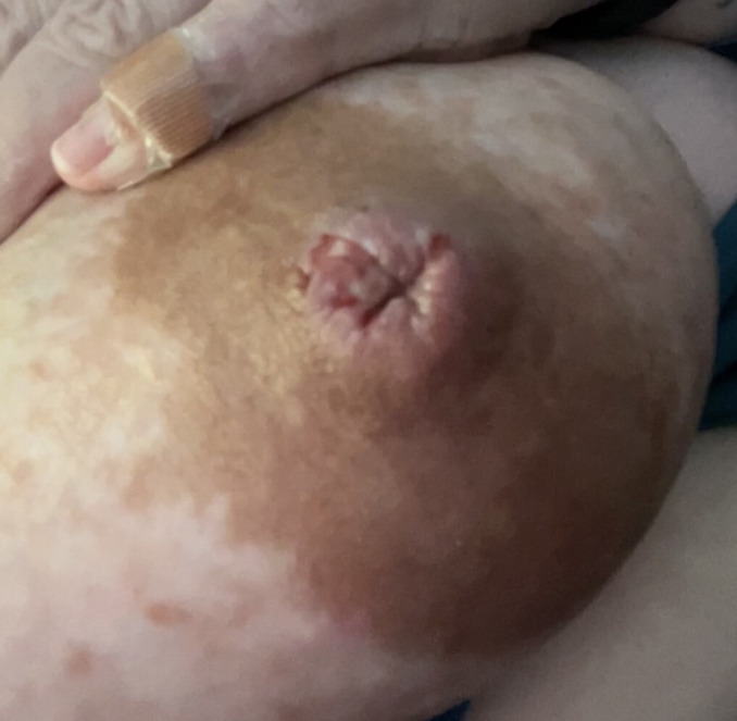
We present a case of a primigravida in her 30s with dichorionic diamniotic twins who underwent an uncomplicated primary low transverse caesarean section due to preterm labour and fetal malpresentation at 33 weeks of gestation. Her pregnancy was complicated by cervical insufficiency requiring an exam-indicated McDonald cerclage, obesity and gestational diabetes. She had no prior history of sexually transmitted infections, and the amniotic membrane was intact at the time of her caesarean section. Her twins were admitted to the neonatal ICU after birth due to prematurity. Initially, they progressed appropriately, but on days 11–12 of life, they began to experience frequent episodes of apnea, isolated fever, seizures and lethargy, and then they developed elevated transaminases at day 13 of life which prompted a sepsis work-up including blood cultures and lumbar puncture. The infants were started on intravenous ampicillin, gentamicin and acyclovir for broad spectrum coverage. On day 14 of life, testing of cerebrospinal fluid resulted in revealing HSV-1 for both infants, antibiotics were discontinued and acyclovir was continued. At the mother’s postpartum visit, she reported a painful, cracked right nipple that developed 1 week earlier (figure 1). She attributed the pain and fissure to pumping with a wrongly sized pump flange. A larger-sized flange was subsequently used, but her nipple had still not healed. She also reported a small cluster of vesicles on her left hand she believed arose after her infants started to decompensate. She had no other signs/symptoms or any systemic infection. She was in a monogamous relationship with her partner, and neither the patient nor her partner had any history of genital or orolabial lesions. They both denied kissing the babies. Physical examination showed a deep vertical fissure in her right nipple (figure 1) with some erythema, without any vesicles or ulcers, as well as several healing 2–3 mm grouped ulcers on the left hand.
Figure 1. Photograph of right nipple with fissure and ulcerations.
Investigations
At her 3-week postpartum clinical visit, the lesions on the patient’s left hand and right nipple were swabbed and PCR testing for HSV-1 and HSV-2 was performed. Both swabs returned as positive for HSV-1 and negative for HSV-2. A serum level of HSV IgM was also ordered at this visit and was found to be elevated at 3.80 IV (<0.89 IV indicates a negative result), which was consistent with a recent infection.
Differential diagnosis
Nipple dermatitis and pain are common postpartum issues with several aetiologies. Symptoms may be due to modifiable factors such as improper latch techniques or flange size. Other causes include dermatitis, eczema, psoriasis or infectious aetiologies such as HSV, bacterial infections or candida.13 After a thorough history is obtained, a thorough physical exam including a breast exam is necessary to identify any swelling, erythema, ulcers, fissures, vesicles or masses.
The most common cause of nipple fissure is poor latch technique, which includes suboptimal infant positioning and either excessive or inadequate sucking by the neonate.14 Similarly, incorrect use of a breast pump or a poorly fitted flange can cause nipple trauma and pain, which can be addressed with the help of lactation specialists. Our patient had preterm infants and had attempted direct breastfeeding during her hospital course but was primarily pumping to express breastmilk, putting her at increased risk of nipple trauma.
Pre-existing skin breakdown at the nipple can serve as the entry point for pathogens; conversely, inflammation and irritation due to infection can precipitate nipple cracking. Our patient had some erythema at the site of the fissure, but in the absence of streaking erythema, purulent discharge or systemic symptoms, secondary or bacterial infection was lower on the differential. Further, her symptoms were not consistent with candidiasis since the patient did not have burning or constant breast pain, no recent antibiotic use and no history of recent vaginal thrush.15
Other alternative diagnoses to consider in breastfeeding mothers who present with breast pain would include local manifestations of dermatosis such as eczema or psoriasis or Paget disease of the breast.16 These dermatoses can present as a cracked, pruritic and burning nipple, but are usually more gradual. They are most common in women who have a pre-existing skin condition or because of nipple irritation. Physical examination typically reveals a red, scaly rash of the areola and nipple. These dermatoses may present similar to herpes simplex breast lesions. However, this patient’s breast symptoms were isolated to the single nipple fissure (figure 1), came on quickly, had no evidence of breast skin thickening, and she did not have any known pre-existing skin conditions.
In our case, we believed the most likely diagnosis was going to be HSV nipple infection. The classic presentation of an HSV nipple infection reveals a crusted nipple with miniature vesicles on an erythematous base that is very tender to touch and typically limited to the nipple or areola.12 13 Extensive erythema or skin changes outside the areola are not common. Our patient had a single vertical fissure through her right nipple face. The nipple fissure and grouped vesicular lesions on her left hand and disseminated neonatal infection of her twins raised suspicion for either maternal to neonatal HSV transmission or the converse. We believed it unlikely that another HHV infection was responsible for the grouped maternal lesions and illness of the neonates due to other HHV infections not presenting with group vesicular lesions, no suspicious findings on antepartum ultrasound showing any signs of congenital infections prior to birth and no other highly suspicious findings on examination or lab values of the neonates.
Treatment
The patient was empirically prescribed oral valacyclovir 1000 mg two times per day at her routine 3-week postpartum visit for a presumed HSV outbreak. To prevent secondary bacterial infection and promote healing, she was prescribed a 2% mupirocin nipple ointment along with topical acyclovir ointment applied 4–6 times per day.
When the PCR resulted as positive for HSV-1, the patient was instructed to take valacyclovir two times per day for 10 days for treatment and once daily thereafter for suppression. She continued to express breastmilk but was advised to abstain from using the milk pumped from her right breast until the lesion healed. She was advised that the milk pumped from the left breast was safe in accordance with current guidelines regarding postnatal HSV infection and was advised to avoid cross-contamination.
Outcome and follow-up
18 days after her initial postpartum visit, she presented to the clinic for follow-up. The lesion on her right nipple was almost completely healed aside from a 1–2 mm area of skin breakage. She was still taking the oral valacyclovir and applying the topical acyclovir daily. Both breasts were swabbed for HSV, and the right breast was found to still be positive for HSV-1. The following month, she went to the clinic for a repeat exam and was found to have a completely healed nipple. Swabs obtained at this visit showed negative HSV results from both breasts. She was advised that she could begin giving her infants milk from both breasts. Her infants recovered after 21 days of intravenous acyclovir and were discharged home with oral acyclovir for 6 months. The patient and her family moved to a different area of the country within 1 month of discharge from the hospital. The infants had difficulties latching once recovered, and the patient exclusively used a breast pump for several weeks until she decided to wean. As of 1 year post partum, the infants were meeting appropriate developmental milestones without concern per the patient’s report.
Discussion
Diagnosing HSV infection poses a clinical challenge as it can be an overlooked differential, but herpes infection of the breast should be included in the differential diagnosis for breast pain during lactation and should be treated prophylactically in a lactating person if highly suspected to reduce the risk of disseminated neonatal HSV. Gold standard diagnosis of HSV requires unroofing a fluid-filled vesicle and performing PCR to detect viral DNA and viral subtype.16 Treatment should be started as soon as possible prior to the results of PCR returning if there is any clinical suspicion of infection in vulnerable populations.
Postnatal acquisition is less common and occurs because of direct contact with an HSV-infected person usually from a mucocutaneous source. The most commonly reported cases of HSV nipple lesions are in women who are currently breastfeeding and can be transmitted from the neonate to the mother.9,12 Case reports documenting extragenital HSV spread from lactating mother to child are less common and can result in child death if not properly diagnosed.5 Due to most cases of neonatal HSV being transmitted from mother to neonate, we consider this the most likely aetiology in our case; however, the true source is unclear, and it is possible another care provider or third party such as hospital staff could have been the source.
Once HSV nipple infection is diagnosed, if there are no active HSV lesions on or around the nipple, there are no contraindications to breastfeeding. Further, this patient’s case demonstrates that with local wound care and continued pumping, breastmilk production can be maintained and continued breastfeeding can be achieved after resolution of the herpetic lesions. Initiating treatment as soon as possible is the best treatment strategy to ensure the best outcomes for infants and affected moms.
Learning points.
Herpes nipple infection is uncommon but is an important cause of maternal to neonatal herpes simplex virus infections.
Herpes nipple infection should be included in the differential diagnosis of breast lesions in all lactating mothers due to the high potential neonatal morbidity from maternal-infant transmission.
Continued milk expression with concurrent local wound care can allow for wound healing and preservation of milk production if the parent has a goal of returning to breastfeeding.
Footnotes
Funding: The authors have not declared a specific grant for this research from any funding agency in the public, commercial or not-for-profit sectors.
Case reports provide a valuable learning resource for the scientific community and can indicate areas of interest for future research. They should not be used in isolation to guide treatment choices or public health policy.
Provenance and peer review: Not commissioned; externally peer reviewed.
Patient consent for publication: Consent obtained directly from patient(s).
Author note: Rebecca Joseph helped with manuscript edits prior to the manuscript being resubmitted but was not directly involved with this patient's care.
Contributor Information
Stephanie Stokes, Email: stephaniestokes2020@gmail.com.
Carolyn Zahler-Miller, Email: czahlermiller@augusta.edu.
Katherine Dunn, Email: katdunn@augusta.edu.
References
- 1.Auriti C, De Rose DU, Santisi A, et al. Pregnancy and viral infections: mechanisms of fetal damage, diagnosis and prevention of neonatal adverse outcomes from cytomegalovirus to SARS-CoV-2 and Zika virus. Biochim Biophys Acta Mol Basis Dis. 2021;1867:166198. doi: 10.1016/j.bbadis.2021.166198. [DOI] [PMC free article] [PubMed] [Google Scholar]
- 2.Products - data briefs - number 304 - February 2018. 2019. [18-Nov-2023]. https://www.cdc.gov/nchs/products/databriefs/db304.htm Available. Accessed.
- 3.Slutsker JS, Schillinger JA. Assessing the burden of infant deaths due to herpes simplex virus, human immunodeficiency virus, and congenital syphilis: United States, 1995 to 2017. Sex Transm Dis. 2021;48:S4–10. doi: 10.1097/OLQ.0000000000001458. [DOI] [PMC free article] [PubMed] [Google Scholar]
- 4.James C, Harfouche M, Welton NJ, et al. Herpes simplex virus: global infection prevalence and incidence estimates, 2016. Bull World Health Organ. 2020;98:315–29. doi: 10.2471/BLT.19.237149. [DOI] [PMC free article] [PubMed] [Google Scholar]
- 5.Field SS. Fatal neonatal herpes simplex infection likely from unrecognized breast lesions. J Hum Lact. 2016;32:86–8. doi: 10.1177/0890334415596987. [DOI] [PubMed] [Google Scholar]
- 6.Arnold KC, Flint CJ, Arnold KC, et al. Management of herpes in pregnancy. Obstet Essent Quest-Based Rev. 2017:53–8. doi: 10.1007/978-3-319-57675-6. [DOI] [Google Scholar]
- 7.Simmons A. Clinical manifestations and treatment considerations of herpes simplex virus infection. J Infect Dis. 2002;186:S71–7. doi: 10.1086/342967. [DOI] [PubMed] [Google Scholar]
- 8.De Rose DU, Bompard S, Maddaloni C, et al. Neonatal herpes simplex virus infection: from the maternal infection to the child outcome. J Med Virol. 2023;95:e29024. doi: 10.1002/jmv.29024. [DOI] [PubMed] [Google Scholar]
- 9.Toussaint A, Simonson C, Valla C. Herpes mastitis: diagnosis and management. Breast J. 2016;22:335–8. doi: 10.1111/tbj.12579. [DOI] [PubMed] [Google Scholar]
- 10.Barrett ME, Heller MM, Stone HF, et al. Primary herpes simplex virus infection of the nipple in a breastfeeding woman. Cutis. 2016;97:E10–1. [PubMed] [Google Scholar]
- 11.Gong P. Cytopathological diagnosis of herpes simplex viral mastitis: three rare cases and a review of the literature. J Cytol. 2020;37:200–3. doi: 10.4103/JOC.JOC_139_19. [DOI] [PMC free article] [PubMed] [Google Scholar]
- 12.Gupta S, Malhotra A, Dash S. Child to mother transmission of herpes simplex virus-1 infection at an unusual site. Acad Dermatol Venereol. 2008;22:878–9. doi: 10.1111/j.1468-3083.2007.02461.x. [DOI] [PubMed] [Google Scholar]
- 13.Heller MM, Fullerton-Stone H, Murase JE. Caring for new mothers: diagnosis, management and treatment of nipple dermatitis in breastfeeding mothers. Int J Dermatol. 2012;51:1149–61. doi: 10.1111/j.1365-4632.2011.05445.x. [DOI] [PubMed] [Google Scholar]
- 14.Buck ML, Amir LH, Cullinane M, et al. Nipple pain, damage, and vasospasm in the first 8 weeks postpartum. Breastfeed Med. 2014;9:56–62. doi: 10.1089/bfm.2013.0106. [DOI] [PMC free article] [PubMed] [Google Scholar]
- 15.Amir LH, Baeza C, Charlamb JR, et al. Identifying the cause of breast and nipple pain during lactation. BMJ. 2021;374:1628. doi: 10.1136/bmj.n1628. [DOI] [PubMed] [Google Scholar]
- 16.Melvin AJ, Mohan KM, Vora SB, et al. Neonatal herpes simplex virus infection: epidemiology and outcomes in the modern era. J Pediatr Infect Dis Soc. 2022;11:94–101. doi: 10.1093/jpids/piab105. [DOI] [PMC free article] [PubMed] [Google Scholar]