Abstract
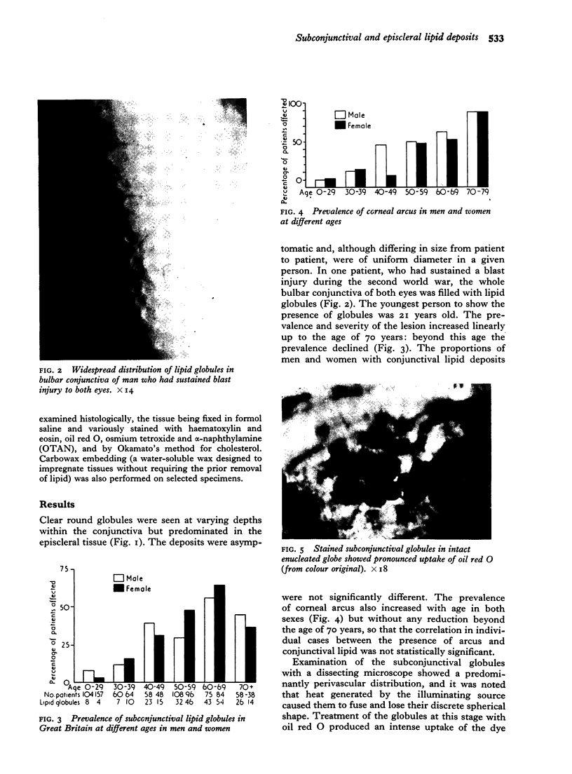
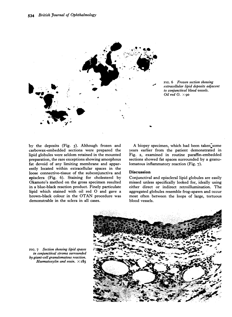
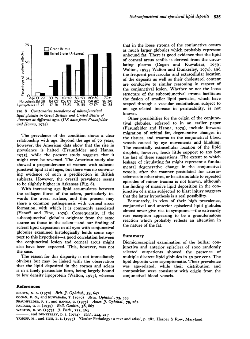
Biomicroscopical examination of the bulbar conjunctiva and anterior episclera of 1000 randomly selected outpatients showed the presence of multiple discrete lipid globules in 30 per cent. The lipid deposits were asymptomatic. Their prevalence was age-related, while their distribution and composition were consistent with origin from the conjunctival blood vessels.
Full text
PDF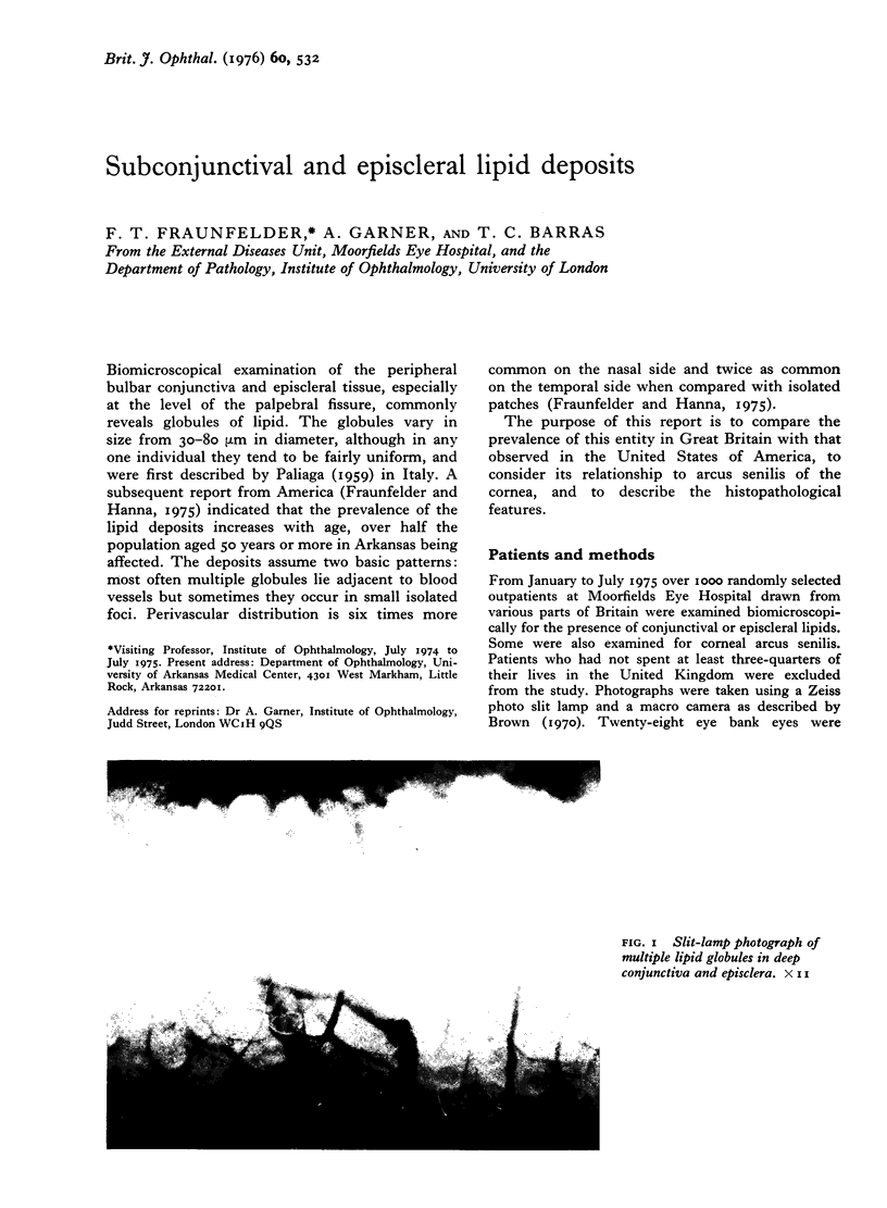
Images in this article
Selected References
These references are in PubMed. This may not be the complete list of references from this article.
- Fraunfelder F. T., Hanna C. Subconjunctival and episcleral lipid globules. Am J Ophthalmol. 1975 Feb;79(2):262–270. doi: 10.1016/0002-9394(75)90080-x. [DOI] [PubMed] [Google Scholar]
- PALIAGA G. P. [Biomicroscopic and histological study of particular formations contained among the meanders of the anterior ciliary arteries]. Boll Ocul. 1959 Nov;38:867–871. [PubMed] [Google Scholar]
- Walton K. W., Dunkerley D. J. Studies on the pathogenesis of corneal arcus formation II. Immunofluorescent studies on lipid deposition in the eye of the lipid-fed rabbit. J Pathol. 1974 Dec;114(4):217–229. doi: 10.1002/path.1711140406. [DOI] [PubMed] [Google Scholar]
- Walton K. W. Studies on the pathogenesis of corneal arcus formation. I. The human corneal arcus and its relation to atherosclerosis as studied by immunofluorescence. J Pathol. 1973 Dec;111(4):263–274. doi: 10.1002/path.1711110407. [DOI] [PubMed] [Google Scholar]


