Abstract
Although progestin has been used to treat endometrial hyperplasia and endometrial carcinoma (EC), its therapeutic efficacy is limited. In order to improve this, the underlining mechanisms of the effects of progestin need to be elucidated in more detail. In the present study, we examined the involvement of mitogen-inducible gene-6 (MIG6), a negative regulator of the EGF receptor, in the progestin-mediated growth suppression of endometrial epithelia. The immunohistochemical expression of MIG6 was elevated in the early to mid-secretory phases of normal endometrium and also with endometrial hyperplasia after medroxyprogesterone acetate (MPA) therapy. The addition of progesterone (P4) to progesterone receptor (PR)-positive EC cells reduced the viability and induced MIG6 messenger RNA (mRNA) and protein expression. The silencing of MIG6 using siRNA eliminated the P4-mediated reduction of EC cell viability, indicating that MIG6 is an essential downstream component of PR-mediated growth suppression. In order to enhance PR-driven signals, we examined the effects of histone deacetylase (HDAC) inhibitors because histone acetylation has been shown to increase the expression of PR. The addition of three HDAC inhibitors (panobinostat, LBH589; trichostatin A, TSA; suberoylanilide hydroxamic acid, SAHA) decreased the viability of EC cells and up-regulated the expression of PR and MIG6, and these effects were the strongest with LBH589. The addition of LBH589 and MPA synergistically decreased the viability and increased apoptosis in EC cells. These results indicate that LBH589 has potential as an enhancer of progestin therapy via the up-regulation of PR and MIG6.
Electronic supplementary material
The online version of this article (doi:10.1007/s12672-017-0295-4) contains supplementary material, which is available to authorized users.
Keywords: Progesterone Receptor, Endometrial Carcinoma, HDAC Inhibitor, Endometrial Hyperplasia, Ishikawa Cell
Introduction
Endometrial carcinoma (EC) is the most common gynecological malignancy and fourth most common cancer in women worldwide [1]. The number of EC patients in Japan has markedly increased in recent decades, and there were approximately 14,800 new cases in 2013 [2]. Although patients younger than 40 years old account for approximately 5% of all EC patients, the number of young patients has also increased with elevation in the total number of EC patients. Changes in lifestyle such as older ages at marriage and childbearing decreases in the number of children delivered, and increases in the intake of a high-calorie diet, may also contribute to increasing young EC patients [3]. Accordingly, demands for fertility-sparing therapy for this malignancy are growing.
Medroxyprogesterone acetate (MPA) has been widely used to treat atypical endometrial hyperplasia (AEH) and low-grade EC. The complete response rates of AEH and EC to MPA therapy were found to be 72–94.1 and 41.7–100%, respectively, whereas recurrence rates were 0–40.0% and 18.2–100%, respectively. Pregnancy rates among patients with AEH and EC were 33.3–45.5 and 6.3–58.3%, respectively [4]. Although MPA therapy represents the first-line treatment for EC in young women, previous findings have indicated insufficient treatment outcomes. Several drugs have been tested in an attempt to enhance the therapeutic efficacy of MPA including metformin [5, 6], selective estrogen receptor modulators (i.e., tamoxifen), and gonadotropin-releasing hormone agonists [7]; however, no drug has been reported to achieve sufficient therapeutic efficacy [8]. Therefore, the development of novel drugs to reinforce MPA therapy is needed.
In order to improve the effects of progestin therapy, the mechanisms underlying the progestin-induced inhibition of endometrial cell growth required to be elucidated in more detail. Although the involvement of several molecules responsible for progestin-mediated growth suppression such as p21 [9], p27 [10], and FOXO1 [11] has been reported, the mechanisms responsible remain unclear. Mitogen-inducible gene-6 (MIG6) is a cytoplasmic protein and is also known as ERBB receptor feedback inhibitor 1 (ERRFI1) which functions as a negative regulator of the epidermal growth factor receptor (EGFR) family by interfering with its dimerization. Therefore, MIG6 is a tumor-suppressing gene that is associated with several kinds of human cancers [12–15]. Recent studies [16, 17] demonstrated that the uterus-specific conditional knockout mouse of Mig6 resulted in endometrial hyperplasia and cancer, suggesting that Mig6 is an essential component in the progesterone-mediated suppression of endometrial cell growth. Therefore, further analyses of the expression and function of MIG6 in normal and neoplastic endometrial cells in humans may provide insights to the role of progestin as a tumor suppressor.
Another mechanism underlining MPA resistance is the reduced expression of the progesterone receptor (PR) in AEH and EC. The expression of PR in EC is suppressed by a number of mechanisms including epigenetic regulation such as promoter methylation and histone acetylation and miRNA-mediated down-regulation at the post-transcriptional level [18]. Histone deacetylase (HDAC) inhibitors have been reported to recover the expression of PR [19]. HDAC inhibitors are known to be potent anticancer agents because they accelerate histone acetylation, which modulates the expression of genes involved in cancer cell growth and survival pathways [20]. Furthermore, HDAC inhibitors have been shown to increase the expression of MIG6 in human lung cancer cell lines [21].
Therefore, we herein examined the expression of MIG6 in the normal cyclic endometrium and MPA-treated neoplastic endometrial cells and compared with that of PR in order to clarify the role of MIG6 in progesterone-induced growth inhibition. We also investigated whether HDAC inhibitors exert synergistic effects with progestin therapy on EC cells by elevating the expression of PR and enhancing the functional involvement of MIG6.
Materials and Methods
Collection of Normal and Neoplastic Endometrial Tissues
Formalin-fixed, paraffin-embedded tissues of EC (44 cases), AEH (18 cases), and the normal endometrium (23 cases) were collected from the pathology files of patients who underwent biopsy or hysterectomy at Shinshu University Hospital. This study was approved by the ethics committee of the institution, and written informed consent was provided by each patient before the collection of tissues.
Agents
MPA and trichostatin A (TSA) were purchased from Wako Pure Chemical Industries (Osaka, Japan) and eluted with ethanol. Suberoylanilide hydroxamic acid (SAHA) (Cayman Chemical Company, Ann Arbor, MI, USA) and panobinostat (LBH589) (Selleck Chemicals LLC, Houston, TX) were eluted in dimethyl sulfoxide (DMSO).
Cell Cultures and Reagents
Cultured normal endometrial glandular (NEG) cells were isolated from patients who underwent hysterectomy for benign diseases (e.g., leiomyoma) as described previously [10]. NEG cells were cultured in HAM-F12 medium with 15% charcoal-filtered FCS on collagen type IV-coated dishes.
The EC cell line Ishikawa 3-H-12 (Ishikawa) was directly gifted by Dr. Nishida (Kasumigaura Medical Center, Ibaraki, Japan) who established this cell line. This cell line was authenticated using polymorphic short tandem repeat (STR) loci examined by Takara Bio Inc. (Shiga, Japan). HHUA was provided by the RIKEN BioResource Center (Ibaraki, Japan), HEC108, HEC151, and HEC265 were supplied by the JCRB Cell Bank (Osaka, Japan), and HEC1A, HEC1B, ECC1, AN3CA, and KLE were purchased from the ATCC (Manassas, VA). RIKEN, JCRB, and ATCC use STR analysis for molecular authentication of cell lines. The passage numbers of cell lines used in this study were less than 20. Cells were maintained in medium supplemented with fetal bovine serum according to the providers’ instructions. Incubation was conducted at 37 °C under 5% CO2 in the air.
Immunohistochemistry
Four-micrometer-thick sections of formalin-fixed and paraffin-embedded tissues were deparaffinized, rehydrated, and boiled in 0.01 M citrate buffer (pH 6.0) for 20 min in a microwave oven. They were then treated with 0.3% hydrogen peroxide for 15 min to block endogenous peroxidase activity and incubated with the primary antibody at 4 °C overnight. As the primary antibody, rabbit anti-ERRFI1 (MIG6) antibody (product number HPA027206, Sigma, St. Louis, MO, USA) and mouse monoclonal anti-progesterone receptor antibody (product number MA1-12626, Thermo Fisher Scientific Anatomical Pathology, Cheshire, WA, UK), which recognizes PR-A and B, were used. After sections had been washed in phosphate-buffered saline, they were incubated with Histofine MAX-PO (Rabbit/Mouse) (Nichirei, Tokyo, Japan) at room temperature for 30 min and then stained with diaminobenzidine and 0.15% hydrogen peroxidase. Counterstaining with hematoxylin was subsequently performed. Staining without the primary antibody was used as a negative control. Immunoreactivity for the expression of PR and MIG6 in NEG, AEH, and EC tissues was evaluated in 100 arbitrarily selected cells in five high-power fields in each section and described as the immunoreactive score (IRS). The IRS was calculated by multiplying the quantity score and staining intensity score as described [22]. The quantity score was rated as follows: no staining was scored as 0, 1–10% of cells stained was scored as 1, 11–50% as 2, 51–80% as 3, and 81–100% as 4. Staining intensity was rated on a scale from 0 to 3, with 0 being negative, 1 weak, 2 moderate, and 3 strong.
Real-Time Quantitative Reverse Transcriptase-Polymerase Chain Reaction
Total RNA from cultured cells was extracted using TRIzol reagent (Life Technologies, Inc., Rockville, MD, USA), and its concentration was measured by the NanoDrop-2000 spectrophotometer (Thermo Fisher Scientific, Wilmington, DE). Real-time quantitative reverse transcriptase-polymerase chain reaction (RT-PCR) was performed using a One Step SYBR Primescript RT-PCR Kit (Perfect Real Time) (Takara) in LightCycler 480 system II (Roche Diagnostics GmbH, Mannheim, Germany) according to the manufacturer’s instructions. MIG6 and PR messenger RNA (mRNA) expression were quantitated using β-actin (ACTB) as an internal control gene. Each experiment for real-time RT-PCR was independently repeated three times with three replicates. The primer sequences used were as follows: PR forward 5′-ATGTGGCAGATCCCACAGGAGTTT-3′, PR reverse 5′-ACTGGGTTTGACTTCGTAGCCCTT-3′, MIG6 forward 5′-CTGGAGCAGTCGCAGTGAG-3′, MIG6 reverse 5′-GCCATTCATCGGAGCAGATTTG-3′, ACTB forward 5′-GACAGGATGCAGAAGGAGATTACT-3′, and ACTB reverse 5′-TGATCCACATCTGCTGGAAGGT-3′.
Western Blotting
Proteins from cultured cells were lysed in a RIPA lysis buffer (Millipore Corporation, Billerica, MA) containing the protease inhibitor cocktail Complete Mini (Roche Diagnostics GmbH, Mannheim, Germany). Lysates were centrifuged 15,000×g for 5 min and then collected supernatant and measured protein concentration. Ten micrograms of protein of each sample was electrophoresed on NuPAGE Bis-Tris Gels in NuPAGE MES SDS Running Buffer and transferred to PVDF blotting membranes (Thermo Fisher Scientific, Wilmington, DE). Membranes were blotted with the following primary antibodies: a rabbit polyclonal anti-MIG6 antibody (Cell Signaling Technology, Beverly, MA, USA), rabbit polyclonal anti-PR A/B antibody (clone C89F7, Cell Signaling Technology, Beverly, MA, USA), and mouse monoclonal anti-ACTB antibody (clone AC-15, Sigma, St. Louis, MO, USA), at 4 °C overnight and then with a ECL anti-mouse/rabbit IgG, horseradish peroxidase-linked species-specific whole antibody (Amersham, Piscataway, NJ) at room temperature for 1 h. Bound antibodies were then visualized using the ECL Western blot detection reagent (Amersham, Piscataway, NJ).
WST-1 Assay
Cell viability was evaluated using WST-1 reagent (Roche Diagnostics GmbH, Mannheim, Germany) according to the manufacturer’s instructions. On the first day, 5 × 10³ cells/well were plated onto 96-well plates. After being incubated for 24 h, the medium was renewed, and HDAC inhibitors and/or MPA were added. After 72 h, the medium was changed to 10% WST-1 reagent in phenol red-free Dulbecco’s modified Eagle’s medium (Sigma, St. Louis, MO). After 1.5 h, A450 wavelength light was measured using a Synergy HT Microplate Reader (Bio-Tek Instruments, Winooski, VT, USA). Each result was obtained from three independent experiments with eight replicates. The synergistic effects of MPA and HDAC inhibitors were evaluated by coefficient of drug interaction (CDI) methods; CDI value was calculated with the equation CDI = AB / (A × B) (AB, relative cell viability of the combination; A or B, relative cell viability of the single-agent groups). CDI values were interpreted as follows: CDI < 1 indicates a synergistic effect, CDI = 1 indicates an additive effect and CDI > 1 indicates an antagonistic effect.
Transfection
A pSG5-PR-B transient expression vector and the empty vector pSG5, a kind gift from Dr. Kato K at Kyushu University with the permission from Dr. Chambon P at Pasteur University, were transfected into Ishikawa cells to establish PR-overexpressing Ishikawa cells. In order to obtain MIG6-overexpressing cells, a human ERRFI1 cDNA clone (pCMV6-ERRFI1) and the empty vector pCMV6 were transfected into Ishikawa cells, HEC1B cells, and AN3CA cells. The knockdown of MIG6 was performed by transfecting ERRFI1 (MIG6) Trilencer-27 Human siRNA (three types; MIG6 silencing (si-MIG6) (A)–(C)) and scrambled negative control siRNA (SR30004) (Origene Technologies, Rockville, MD, USA) into Ishikawa and HEC1B cells expressing high levels of MIG6. The knockdown of PR was performed by transfecting PR Trilencer-27 Human siRNA (three types; siPR (A)–(C)) (Origene Technologies) into Ishikawa, HEC1B, and AN3CA cells. All transfections were conducted by Lipofectamine 2000 (Invitrogen, Carlsbad, CA) following manufacturer’s instructions. The medium was changed 12–24 h after transfection, and cells were used in each experiment.
Annexin V-PI Staining
HEC1B cells were cultured on chamber slides and treated with 10 nM of LBH589 and/or 100 nM of MPA. After being incubated for 48 h, cells were stained with Annexin V and propidium iodide (PI) according to the manufacturer’s protocol (Annexin-V-FLUOS Staining Kit, Roche Diagnostics GmbH, Mannheim, Germany). Apoptotic cells were counted as a combination of Annexin V+/PI− (early apoptotic) and Annexin V+/PI+ (late apoptotic/necrotic) cells. The experiment was repeated three times, and results were shown as mean percentages ± SD of Annexin V+ cells.
PI Staining
HEC1B cells were treated with 10 nM of LBH589 and/or 100 nM of MPA for 48 h. DNA contents were measured using PI staining and a flow cytometry analysis using FACSCANTO II (Becton Dickinson, San Jose, CA, USA).
Statistical Analysis
Statistical analyses were conducted using the Student’s t test, paired t test, and Mann-Whitney test for comparisons of two groups. Differences were considered significant at P < 0.05. These analyses were performed using SPSS version 14 (SPSS Inc., Chicago, IL, USA).
Results
The Expression of MIG6 Was Increased by Progestins in Normal and Neoplastic Endometria
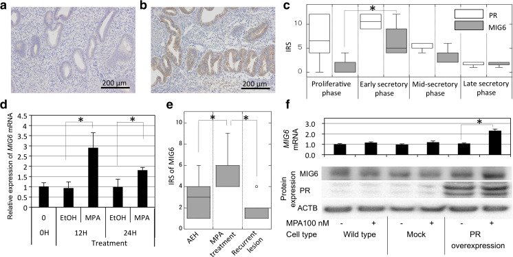
Immunostaining for MIG6, which was stained in the cytoplasm, indicated that the expression of MIG6 in NEG cells was weak in the proliferative phase (2 ± 1.58, median IRS ± SD) and became stronger in the early secretory phase (5 ± 3.79, P = 0.006). This difference of MIG6 expression may be associated with the endogenous progesterone (P4) level which is much higher in the secretory phase than in the proliferative phase. In association with the weaker expression of PR in the mid to late secretory phases, that of MIG6 also decreased (Fig. 1a–c). The expression of MIG6 mRNA was increased 3.09-folds at 24 h and 1.83-folds at 48 h after the addition of 100 nM of MPA (Fig. 1d).
Fig. 1.
a, b Immunohistochemical expression of MIG6 in the normal endometrium in the proliferative phase (a) and secretory phase (b). Positive staining was observed in the secretory phase. c Graphical demonstration of the expression of the progesterone receptor (PR) and MIG6 in the proliferative phase, early, mid, and late secretory phases. Data are presented in the box plot as the median and 25–75 percentile of the IRS. MIG6 expression was stronger in the early to mid-secretory phases when PR was positive. d Relative expression of MIG6 mRNA in cultured normal endometrial glandular (NEG) cells after the addition of MPA at 100 nM. Data are presented as the mean ± SD of three independent experiments. e The immunohistochemical expression of MIG6 in atypical endometrial hyperplasia (AEH), in AEH after the MPA treatment, and in recurrent tumors after the MPA treatment. The box plot indicates median and 25–75 percentile. f The expression of MIG6 mRNA, MIG6, and PR protein in Ishikawa cells transfected with PR (PR overexpression) or empty vector (mock) with or without MPA. The cells were harvested at 24 h after addition of MPA or vehicle. *P < 0.01, IRS immunoreactive score, which was calculated by multiplying the quantity score (no staining as 0, 1–10% as 1, 11–50% as 2, 51–80% as 3, and 81–100% as 4) and staining intensity score (negative as 0, weak as 1, moderate as 2, and strong as 3)
The expression of MIG6 in AEH treated with oral MPA (600 mg daily, n = 18) was then examined. The IRS for MIG6 before the treatment was 3 ± 1.79 (median ± SD), and it increased during MPA therapy to 4 ± 1.38. In 10 tissues collected from recurrent cases, only the weak expression of MIG6 was observed (Fig. 1e) (1 ± 1.00).
In order to evaluate the effects of MPA on low-grade endometrioid adenocarcinoma, 100 nM of MPA was added to Ishikawa cells. The expression of MIG6 mRNA in wild-type Ishikawa cells was not significantly affected by the addition of MPA. After the transfection of PR cDNA into Ishikawa cells, the expression of MIG6 was enhanced 2.14-folds by the treatment with MPA, suggesting the involvement of PR in the induction of MIG6 (Fig. 1f).
The Expression of MIG6 in EC Tissues and Cell Lines
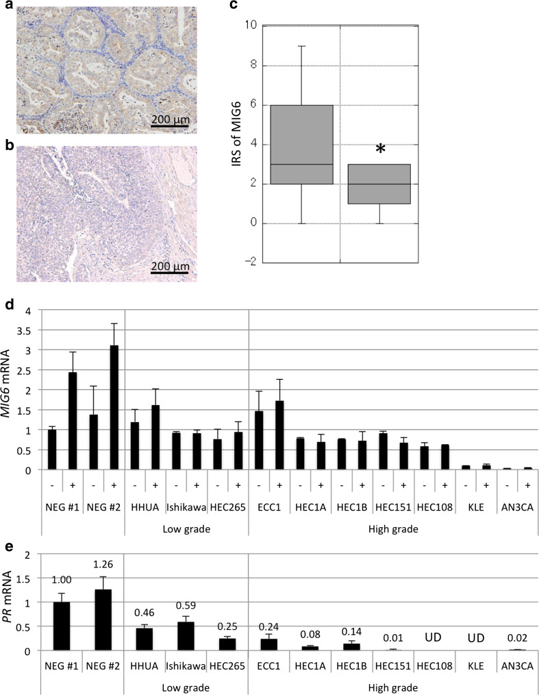
The expression of MIG6 in EC tissues was evaluated using immunohistochemistry, and the results obtained indicated that the IRS in grade 2 and 3 (2 ± 0.93, 1–3; median ± SD, 25–75 percentile) endometrioid adenocarcinoma were significantly lower than that in grade 1 (3 ± 2.5, 2–6, P = 0.001) (Fig. 2a–c). Although there are some exceptions (such as ECC1), real-time RT-PCR revealed that MIG6 mRNA levels were lower in cell lines derived from high-grade endometrioid adenocarcinoma (ECC1, HEC1A, HEC1B, HEC151, HEC108, KLE, and AN3CA) than in NEG cells or low-grade endometrioid adenocarcinoma cell lines (HHUA, Ishikawa, and HEC265). The PR mRNA levels were also lower in high-grade cell lines. The addition of 100 nM of MPA increased MIG6 mRNA expression levels (2.44-folds in NEG no. 1 and 2.26-folds in NEG no. 2) in NEG cells, whereas no or minimal effects were observed in EC cell lines (Fig. 2d).
Fig. 2.
a–c Results of immunostaining for MIG6 in grade 1 (a) and grade 3 (b) endometrial carcinomas and its graphical demonstration (the box plot indicating median and 25–75 percentile) (c). The expression of MIG6 decreased in poorly differentiated tumors. d, e The results of real-time PCR indicating the expression of MIG6 (d) and PR (e) mRNA in two NEG cells (NEG no.1 and 2) and various endometrial carcinoma cell lines before and at 24 h after the addition of MPA. The expression of MIG6 was increased by MPA in NEG and HHUA cells but was absent in other cell lines. *P < 0.01, IRS immunoreactive score
Involvement of MIG6 in the MPA-Mediated Reduction of EC Cell Viability
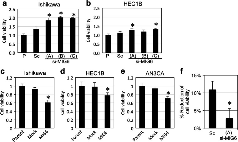
MIG6 mRNA expression in Ishikawa and HEC1B cells was knocked down by siRNA (Suppl. Fig. 1a–d). si-MIG6 increased cell viability compared with scrambled siRNA in Ishikawa (1.38–1.50-folds, Fig. 3a) and HEC1B (1.06–1.19-folds, Fig. 3b). The effect of MIG6-silencing in HEC1B is modest and not convincing (Fig. 3b), suggesting that the relation between MIG6 expression and cell viability/growth is weaker in HEC1B than that in Ishikawa. Otherwise, the basic expression level of MIG6 in HEC1B may be too low to suppress the cell viability/growth. MIG6 cDNA was transfected into Ishikawa, HEC1B, and AN3CA cells (Suppl. Fig. 1e–g), and the forced expression of MIG6 attenuated cell viability compared with mock (0.66-folds in Ishikawa, 0.79-folds in HEC1B and 0.75-folds in AN3CA) (Fig. 3c–e). The viability of PR-overexpressing Ishikawa cells was also decreased by the addition of 100 nM of MPA, whereas the knockdown of MIG6 suppressed this effect (Fig. 3f, 10.9 ± 2.3 and 2.9 ± 2.6%, respectively).
Fig. 3.
a–e Effects of MIG6 silencing on cell viability in Ishikawa (a) and HEC1B (b) cells and those of MIG6 overexpression in Ishikawa (c), HEC1B (d), and AN3CA (e) cells. The WST-1 assay showed that the knockdown of MIG6 increased and with its transfection decreased the viability of these cells. f Effects of MIG6 silencing on MPA responses. MIG6 silencing decreased the MPA-induced reduction of PR-transfected Ishikawa cell viability. The WST-1 assay was performed after 72 h of incubation. P or Parent parent cells without transfection, si-MIG6 MIG6 siRNA-transfected cells, Sc scrambled negative control siRNA transfected cells, mock empty vector-transfected cells, MIG6 MIG6 cDNA-transfected cells. Asterisk: significant difference (P < 0.01) compared with Sc or mock
LBH589 Enhanced MPA Effects by Increasing the Expression of PR and MIG6
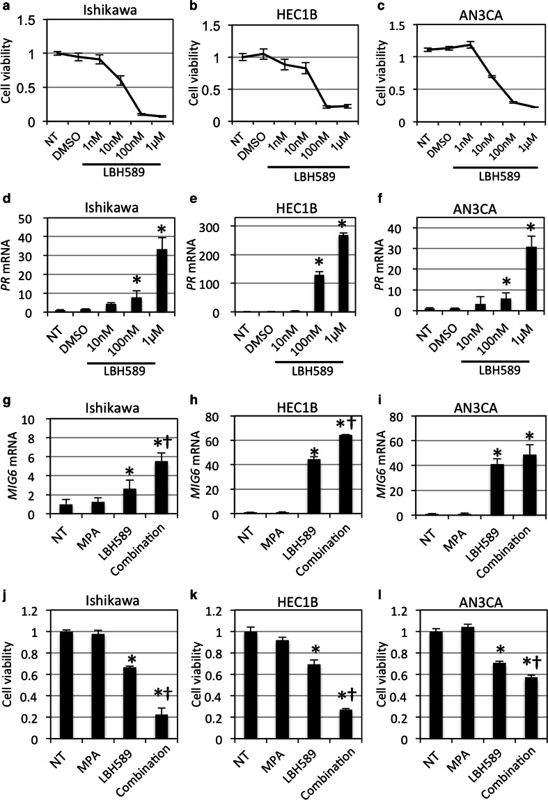
Three HDAC inhibitors, TSA, SAHA, and LBH589, exerted cytotoxic effects to varying degrees (Suppl. Fig. 2a–f and Fig. 4a–c). Among the three drugs tested, LBH589 had the strongest effects on the survival of cells. LBH589 also dose-dependently enhanced the expression of PR in Ishikawa, HEC1B, and AN3CA cells (Fig. 4d–f), whereas TSA and SAHA failed to enhance the expression of PR in AN3CA cells (Suppl. Fig. 3). Enhanced expression of PR protein in Ishikawa cells by LBH589 was also confirmed by western blotting (Suppl. Fig. 4).
Fig. 4.
a–c Relative reduction of Ishikawa (a), HEC1B (b), and AN3CA (c) cell viability by the WST-1 assay at 72 h after the treatment with 1 nM to 1 μM of LBH589. LBH589 reduced the viability of these cells. Data were shown as a ratio to the no treatment (NT). d–f Relative expression of PR mRNA at 24 h after the treatment with 10 nM of LBH589. Data were shown as a ratio to the no treatment (NT). LBH589 increased the expression of PR. g–i Relative expression of MIG6 mRNA by the treatment of MPA (100 nM, for 24 h), LBH589 (10 nM, for 48 h) or their combination. Data were shown as a ratio to the no treatment (NT). LBH589 increased the expression of MIG6 mRNA. j–l Cell viability after the treatment with 10 nM of LBH589 and/or 100 nM of MPA in the same cells. Data were shown as a ratio to the no treatment (NT). The addition of LBH589 to MPA decreased viability. NT no treatment, DMSO treated with DMSO (the solvent of LBH589), Combination treated with LBH589 and MPA. Asterisk: significant difference (P < 0.05) compared with NT. Dagger: significant difference (P < 0.05) compared with LBH589
In order to establish whether the LBH589-induced expression of PR affects that of MIG6 expression, 10 nM of LBH589 and/or 100 nM of MPA were added to EC cell lines. While the single use of MPA failed to induce the expression of MIG6, MPA in combination with LBH589 markedly up-regulated the expression of MIG6 (4.38-folds in Ishikawa, 60.7-folds in HEC1B, and 49.9-folds in AN3CA). The single use of LBH589 also enhanced the expression of MIG6 (2.08-folds in Ishikawa, 41.9-folds in HEC1B, and 42.1-folds in AN3CA) (Fig. 4g–i). This synergistic effect of MPA on up-regulation of MIG6 mRNA was canceled by PR-silencing using PR-siRNA (siPR (A)–(C)) (Suppl. Fig. 5). In addition, since MPA is clinically used but known to have androgen effect, purer progestins such as P4 and promegestone were used to further clarify the progestin effect on the MIG6 expression. These progestins significantly enhanced the expression of MIG6 in LBH589-treated EC cell lines compared with the cells treated without progestins (P < 0.05) (Suppl. Fig. 6). These results suggest that LBH589 increases the sensitivity of EC cells to MPA and other progestins. TSA and SAHA exerted similar effects in combination with MPA (Suppl. Fig. 7). The effect of 10 nM of LBH589 and/or 100 nM of MPA on cell viability was then assessed using the WST-1 assay. The results indicated that LBH589 and MPA synergistically decreased cell viability in the three cell lines tested (respective fold changes by LBH589 single and LBH589/MPA combination as 0.68 and 0.23-folds in Ishikawa, 0.75 and 0.29-folds in HEC1B, and 0.68 and 0.55-folds in AN3CA) (Fig. 4j–l) because CDI values were less than 1 (Ishikawa = 0.34, HEC1B = 0.42, AN3CA = 0.77).
LBH589 and MPA Synergistically Induced Apoptosis in HEC1B Cells via the MIG6 Signaling Pathway
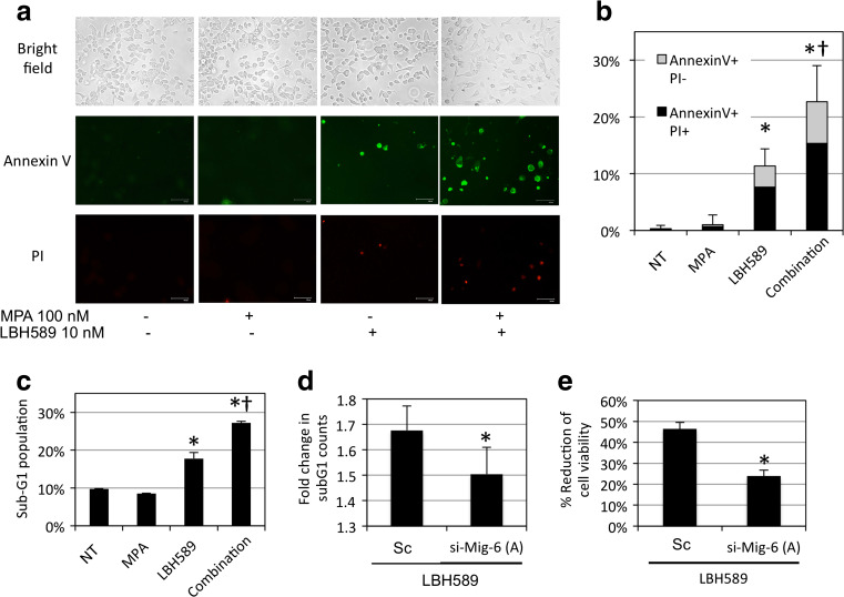
The effects of LBH589 and MPA on apoptosis were then examined using Annexin V-PI staining. Although MPA alone failed to induce apoptosis, the single use of LBH589 increased 34.3-folds of early and late phase apoptosis/necrosis. In addition, the combination of LBH589 and MPA synergistically increased 68.7-folds of apoptosis/necrosis (Fig. 5a, b). Apoptosis was also evaluated by PI staining followed by flow cytometry. The sub-G1 population was increased 1.82-folds by the single use of LBH589 and 2.80-folds by the combined use of LBH589 and MPA exerted synergistic effects (Fig. 5c and Suppl. Fig. 8).
Fig. 5.
a, b Effects of MPA and LBH589 on the survival of HEC1B cells (a) and its graphical demonstration (b). Fluorescent staining was performed at 48 h after the addition of LBH589 and/or MPA. Middle panel: Annexin V (green). Lower panel: PI (red). MPA with LBH589 increased the number of apoptotic cells. c Effects of MPA and LBH589 on the cell cycle. MPA with LBH589 increased the sub-G1 population. Flow cytometry was performed at 48 h after the addition of LBH589 and/or MPA. d, e Effects of MIG6 silencing on the LBH589-induced sub-G1 population (d) and viability reduction (e). The numerical values were acquired by comparing with NT. MIG6 silencing eliminated the LBH589-mediated sub-G1 population and viability reduction. NT no treatment, Combination treated with LBH589 and MPA, si-MIG6 (A) MIG6 siRNA (A) transfected cells, Sc scrambled negative control siRNA transfected cells. Asterisk: significant difference (P < 0.05) compared with NT or Sc. Dagger: significant difference (P < 0.05) compared with LBH589
The role of MIG6 in LBH589-induced apoptosis was then investigated using siRNA. MIG6 silencing resulted in reduced the fold change of sub-G1 counts (Fig. 5d) (1.68 ± 0.1 and 1.5 ± 0.1, mean fold change ± SD, respectively, p = 0.01). The effects of LBH589 on the reduction of cell viability were attenuated by the knockdown of MIG6 (Fig. 5e) (46.5 ± 3.1% in scrambled and 23.9 ± 2.9% in si-MIG6, mean ± SD, P < 0.001). These results suggest that MIG6 mediates the effects of LBH589 and indicates that MIG6 plays an important role in the PR-induced suppression of neoplastic EG cells.
Discussion
MIG6 directly binds to the kinase domain of EGFR and suppresses EGFR-related signals leading to cell proliferation and motility. MIG6 has also been shown to alleviate excessive EGF-driven signals and function as an inducible feedback inhibitor [15, 23, 24]. Our present study revealed that MIG6 expression was regulated by progestin and PR. However, MIG6 have never reported as the factor directly transcribed by PR, suggesting that MIG6 is the downstream factor of PR and the expression of MIG6 might be regulated by other PR-responsive genes. In the present study, the forced expression of MIG6 reduced, while the silencing of MIG6 accelerated EC cell viability, indicating that MIG6 also acts as a tumor suppressor in this malignancy. Recent studies reported anti-oncogenic roles for MIG6 in tumors in the breast [13] and brain [15]. Jeong et al. demonstrated that the expression of MIG6 in EC was P4-dependent, and conditional knockout mice lacking MIG6 in the uterus (PR cre/+ Mig6 f/f) had greater uterine weights and endometrial hyperplasia [16]. The same group reported that MIG6 is a critical tumor suppressor that mediates the function of P4 to prevent the development of EC using another type of conditional MIG6 knockout mouse (Wnt7a cre/+ Mig6 f/f) [17]. These findings suggest that MIG6 is a pivotal molecule in the progestin-mediated growth suppression of endometrial epithelia.
The present immunohistochemical study on the normal endometrium demonstrated that the expression of MIG6 in endometrial epithelium was closely correlated with that of PR; the expression of MIG6 was stronger in NEG cells in the secretory phase than in the proliferative phase. Furthermore, the expression of MIG6 was weaker in the late secretory phase, as was that of PR. These results are consistent with the progesterone-dependent nature of MIG6 expression. A previous study reported that the expression of MIG6 in glandular cells was increased in the early secretory phase, which is in agreement with our results [16]. The immunostaining of EC tissues showed that MIG6 protein expression was reduced in less differentiated carcinomas. This result may be attributed to the expression of PR being stronger in well-differentiated EC, which generally has a favorable prognosis [25]. These findings indicate that progestin-induced, MIG6-related growth suppression is a feature of well-differentiated EC tissues.
The results of the present study indicate that MIG6 is a pivotal molecule in the progestin-induced suppression of endometrial epithelial cell growth. We demonstrated that extrinsic progestin induced the expression of MIG6 in patients with endometrial hyperplasia. This result is reversely supported by the reduced expression of MIG6 in progestin-resistant recurrent cases. We also found that MIG6 mRNA and protein expression in Ishikawa cell with forced expression of PR was increased by the addition of MPA, whereas viability was decreased and apoptosis was induced. Xu et al. reported that the forced expression of MIG6 in Ishikawa cells enhanced P4-induced apoptosis, growth suppression, and the suppression of migration by 39.4, 37.9, and 48.9%, respectively [26]. Our results demonstrated that the knockdown of MIG6 eliminated the antiproliferative effects of MPA in PR-positive Ishikawa cells. This study is the first to show the direct contribution of MIG6 to progestin-related growth suppression.
Several progestin-induced tumor suppressors have been identified in endometrial epithelia. We previously reported the progestin-induced expression of p27, a tumor suppressor of cyclin-cdk complexes, in EC cells [10]. p27 knockout mice exhibited multiorgan hyperplasia, and abnormal ovarian function, whereas uterus and the endometrium were morphologically normal [27]. Kyo et al. reported the progestin-mediated up-regulation of the forkhead transcription factor FOXO1, which is known to be involved in decidualization, using microarrays [11]. They suggested that FOXO1 is a direct target of progestin because progestin markedly induced FOXO1 gene expression and the siRNA inhibition of FOXO1 significantly attenuated the ability of progestin to inhibit endometrial epithelial cell growth; however, the role of FOXO1 in uterine tumorigenesis is still elusive. Collectively, MIG6 appears to be the critical downstream component as shown by knockout mouse models [16, 17].
Although the P4-PR-MIG6 axis is considered to be essential in the growth suppression of normal and neoplastic endometrial epithelia, the expression of MIG6 is weak in histologically high-grade tumors, with approximately 30% of endometrial hyperplasia and low-grade EC are refractory to MPA [28]. Thus, elevations in the expression of MIG6 appear to be necessary for obtaining better treatment responses. One of these approaches is to increase the expression of PR. In the present study, the effects of three HDACs (TSA, SAHA, and LBH589) on PR expression were examined, and we revealed that LBH 589 was the strongest inducer of PR expression in a dose-dependent manner, which is consistent with previous findings [29]. Regarding the mechanisms underlying the HDAC inhibitor-mediated recovery of PR, Yang et al. reported that the histone in the promoter region of the PR gene was acetylated by HDAC inhibitors, resulting in the dissociation of the SUZ12 polycomb repressive complex 2 subunit, which suppresses the transcription of PR from the promoter [29]. This study also indicated that these HDAC inhibitors exerted cytotoxicity and antiproliferative effects against EC cells. We previously reported that the HDAC inhibitors TSA, apicidin, and SAHA suppressed the proliferation of a number of EC cells and ovarian carcinoma cells [30, 31], and the mechanisms responsible were the induction of cell cycle arrest to G1/S or G2/M via the up-regulation of CDKN1A, p27, and p15 and down-regulation of cyclin D and A [32].
Moreover, 10 nM of LBH589 increased the expression of MIG6. Although the mechanism underlying the LBH589-induced up-regulation of MIG6 currently remains unknown, Zhang et al. reported that the addition of TSA did not alter the histone acetylation of the MIG6 promoter region in the lung cancer cell line A427, the expression of MIG6 in which was induced by TSA [21]. The same group revealed that a 50-bp region in exon 1 of the MIG6 gene was a TSA-response element using a luciferase assay containing reporters of various sizes.
The results of the present study indicate that the silencing of MIG6 reduced apoptosis in HEC1B cells, which was caused by the concurrent use of MPA and LBH589. This result clearly suggests that LBH589 synergistically decreases the viability of and accelerates apoptosis in EC cells with MPA via the up-regulation of PR and MIG6. Therefore, activation of the MIG6 pathway appears to be promising as a therapeutic strategy for EC. LBH589 recovered the expression of PR and increased that of MIG6, and this study is the first to reveal the potential of LBH589 as an enhancer of MPA in EC cells. Further research is needed in order to elucidate the effects and safety of the combined use of LBH589 and MPA in animal models.
Electronic Supplementary Material
A, B: Results of the transfection of MIG6 siRNA into Ishikawa cells. (A: MIG6 mRNA, B: protein) C, D: Results of the transfection of MIG6 siRNA into HEC1B cells. (C: MIG6 mRNA, D: protein) E, F: Results of the transfection of MIG6 cDNA into Ishikawa cells (E) and HEC1B cells (F). Cells were harvested at 24 h after transfection. P or Parent: parent cells without transfection, si-MIG6 (A)-(C): MIG6 siRNA (A)-(C) transfected cells, Sc: scrambled negative control siRNA transfected cells, Mock: empty vector transfected cells, MIG6: MIG6 cDNA transfected cells. (PDF 277 kb)
A-C: Effects of TSA on the viability of Ishikawa (A), HEC1B (B), and AN3CA (C) cells examined by the WST-1 assay. D-E: Effects of SAHA on the viability of Ishikawa (D), HEC1B (E), and AN3CA (F) cells. The WST-1 assay was performed at 72 h after the treatment. NT: no treatment, EtOH: treated with ethanol (solvent of TSA), DMSO: treated with DMSO (solvent of SAHA). (PDF 142 kb)
A-C: Effects of TSA on the expression of PR mRNA in Ishikawa (A), HEC1B (B), and AN3CA (C) cells examined by RT-PCR. D-E: Effects of SAHA on the expression of PR mRNA in Ishikawa (D), HEC1B (E), and AN3CA (F) cells. Cells were harvested at 24 h after the treatment. NT: no treatment, EtOH: treated with ethanol (solvent of TSA), DMSO: treated with DMSO (solvent of SAHA). (PDF 196 kb)
The expression of PR protein in Ishikawa cells at 24 h after the treatment of LBH589. The expression of PR protein was enhanced by 10 nM of LBH589. Control: no treatment, DMSO: treated with DMSO (solvent of LBH589). (PDF 61 kb)
Effects of PR-silencing by PR-siRNA (siPR(A)-(C)) on the expression of MIG6 mRNA in Ishikawa cells treated by LBH589 (10 nM, for 48 h) and MPA (100 nM, for 24 h). The synergistic effect of the combination treatment with LBH589 and MPA was canceled by PR-silencing. Control: no treatment, DMSO: treated with DMSO (solvent of LBH589). (PDF 74 kb)
Effects of progesterone (P4, 100 nM, for 24 h) or Promegestone (100 nM, for 24 h) on the expression of MIG6 mRNA in Ishikawa (A), HEC1B (B), and AN3CA (C) treated by LBH589 (10 nM, for 48 h). Control: no treatment, EtOH: treated with ethanol (solvent of progestins). (PDF 106 kb)
A-C: Effects of TSA and/ or MPA on the expression of MIG6 mRNA in Ishikawa (A), HEC1B (B), and AN3CA (C) cells examined by RT-PCR. D-E: Effects of SAHA and/or MPA on the expression of MIG6 mRNA in Ishikawa (D), HEC1B (E), and AN3CA (F) cells. NT: no treatment. (PDF 134 kb)
Representative results of flow cytometry of propidium iodide (PI) staining in HEC1B cells treated with 10 nM of LBH589 and/or 100 nM of MPA for 48 h. P1 indicates sub-G1 fraction. (PDF 284 kb)
Acknowledgments
The authors are grateful to Fumi Tsunoda and Eiji Uchida (Research Assistants, Department of Obstetrics and Gynecology, Shinshu University School of Medicine) for their excellent technical assistance. This work was supported by Grants-in-Aid for Scientific Research (KAKENHI) from Japan Society for the Promotion of Science (JSPS), Grant Number 16K20183.
Compliance with Ethical Standards
Conflict of Interest
The authors declare that they have no conflict of interest.
References
- 1.Ferlay J, Shin HR, Bray F, Forman D, Mathers C, Parkin DM. Estimates of worldwide burden of cancer in 2008: GLOBOCAN 2008. Int J Cancer. 2010;127:2893–2917. doi: 10.1002/ijc.25516. [DOI] [PubMed] [Google Scholar]
- 2.Katanoda K, Hori M, Matsuda T, Shibata A, Nishino Y, Hattori M, Soda M, Ioka A, Sobue T, Nishimoto H. An updated report on the trends in cancer incidence and mortality in Japan, 1958-2013. Jpn J Clin Oncol. 2015;45:390–401. doi: 10.1093/jjco/hyv002. [DOI] [PubMed] [Google Scholar]
- 3.Bongaarts J, Blanc AK. Estimating the current mean age of mothers at the birth of their first child from household surveys. Popul Health Metr. 2015;13:25. doi: 10.1186/s12963-015-0058-9. [DOI] [PMC free article] [PubMed] [Google Scholar]
- 4.Ohyagi-Hara C, Sawada K, Aki I, Mabuchi S, Kobayashi E, Ueda Y, Yoshino K, Fujita M, Tsutsui T, Kimura T. Efficacies and pregnant outcomes of fertility-sparing treatment with medroxyprogesterone acetate for endometrioid adenocarcinoma and complex atypical hyperplasia: our experience and a review of the literature. Arch Gynecol Obstet. 2015;291:151–157. doi: 10.1007/s00404-014-3417-z. [DOI] [PubMed] [Google Scholar]
- 5.Hawkes AL, Quinn M, Gebski V, Armes J, Brennan D, Janda M, feMME Trial Committee & Obermair A Improving treatment for obese women with early stage cancer of the uterus: rationale and design of the levonorgestrel intrauterine device ± metformin ± weight loss in endometrial cancer (feMME) trial. Contemp Clin Trials. 2014;39:14–21. doi: 10.1016/j.cct.2014.06.014. [DOI] [PubMed] [Google Scholar]
- 6.Tabrizi AD, Melli MS, Foroughi M, Ghojazadeh M, Bidadi S. Antiproliferative effect of metformin on the endometrium—a clinical trial. Asian Pac J Cancer Prev. 2014;15:10067–10070. doi: 10.7314/APJCP.2014.15.23.10067. [DOI] [PubMed] [Google Scholar]
- 7.Pronin SM, Novikova OV, Andreeva JY, Novikova EG. Fertility-sparing treatment of early endometrial cancer and complex atypical hyperplasia in young women of childbearing potential. Int J Gynecol Cancer. 2015;25:1010–1014. doi: 10.1097/IGC.0000000000000467. [DOI] [PubMed] [Google Scholar]
- 8.Umene K, Banno K, Kisu I, Yanokura M, Nogami Y, Tsuji K, Masuda K, Ueki A, Kobayashi Y, Yamagami W, Tominaga E, Susumu N, Aoki D. New candidate therapeutic agents for endometrial cancer: potential for clinical practice (review) Oncol Rep. 2013;29:855–860. doi: 10.3892/or.2013.2221. [DOI] [PMC free article] [PubMed] [Google Scholar]
- 9.Dai D, Wolf DM, Litman ES, White MJ, Leslie KK. Progesterone inhibits human endometrial cancer cell growth and invasiveness: down-regulation of cellular adhesion molecules through progesterone B receptors. Cancer Res. 2002;62:881–886. [PubMed] [Google Scholar]
- 10.Shiozawa T, Horiuchi A, Kato K, Obinata M, Konishi I, Fujii S, Nikaido T. Up-regulation of p27Kip1 by progestins is involved in the growth suppression of the normal and malignant human endometrial glandular cells. Endocrinology. 2001;142:4182–4188. doi: 10.1210/endo.142.10.8455. [DOI] [PubMed] [Google Scholar]
- 11.Kyo S, Sakaguchi J, Kiyono T, Shimizu Y, Maida Y, Mizumoto Y, Mori N, Nakamura M, Takakura M, Miyake K, Sakamoto M, Inoue M. Forkhead transcription factor FOXO1 is a direct target of progestin to inhibit endometrial epithelial cell growth. Clin Cancer Res. 2011;17:525–537. doi: 10.1158/1078-0432.CCR-10-1287. [DOI] [PubMed] [Google Scholar]
- 12.Amatschek S, Koenig U, Auer H, Steinlein P, Pacher M, Gruenfelder A, Dekan G, Vogl S, Kubista E, Heider KH, Stratowa C, Schreiber M, Sommergruber W. Tissue-wide expression profiling using cDNA subtraction and microarrays to identify tumor-specific genes. Cancer Res. 2004;64:844–856. doi: 10.1158/0008-5472.CAN-03-2361. [DOI] [PubMed] [Google Scholar]
- 13.Anastasi S, Sala G, Huiping C, Caprini E, Russo G, Iacovelli S, Lucini F, Ingvarsson S, Segatto O. Loss of RALT/MIG-6 expression in ERBB2-amplified breast carcinomas enhances ErbB-2 oncogenic potency and favors resistance to Herceptin. Oncogene. 2005;24:4540–4548. doi: 10.1038/sj.onc.1208658. [DOI] [PubMed] [Google Scholar]
- 14.Ruan DT, Warren RS, Moalem J, Chung KW, Griffin AC, Shen W, Duh QY, Nakakura E, Donner DB, Khanafshar E, Weng J, Clark OH, Kebebew E. Mitogen-inducible gene-6 expression correlates with survival and is an independent predictor of recurrence in BRAF (V600E) positive papillary thyroid cancers. Surgery. 2008;144:908–913. doi: 10.1016/j.surg.2008.07.028. [DOI] [PubMed] [Google Scholar]
- 15.Ying H, Zheng H, Scott K, Wiedemeyer R, Yan H, Lim C, Huang J, Dhakal S, Ivanova E, Xiao Y, Zhang H, Hu J, Stommel JM, Lee MA, Chen AJ, Paik JH, Segatto O, Brennan C, Elferink LA, Wang YA, Chin L, DePinho RA. Mig-6 controls EGFR trafficking and suppresses gliomagenesis. Proc Natl Acad Sci U S A. 2010;107:6912–6917. doi: 10.1073/pnas.0914930107. [DOI] [PMC free article] [PubMed] [Google Scholar]
- 16.Jeong JW, Lee HS, Lee KY, White LD, Broaddus RR, Zhang YW, Vande Woude GF, Giudice LC, Young SL, Lessey BA, Tsai SY, Lydon JP, DeMayo FJ. Mig-6 modulates uterine steroid hormone responsiveness and exhibits altered expression in endometrial disease. Proc Natl Acad Sci U S A. 2009;106:8677–8682. doi: 10.1073/pnas.0903632106. [DOI] [PMC free article] [PubMed] [Google Scholar]
- 17.Kim TH, Lee DK, Cho SN, Orvis GD, Behringer RR, Lydon JP, Ku BJ, McCampbell AS, Broaddus RR, Jeong JW. Critical tumor suppressor function mediated by epithelial Mig-6 in endometrial cancer. Cancer Res. 2013;73:5090–5099. doi: 10.1158/0008-5472.CAN-13-0241. [DOI] [PMC free article] [PubMed] [Google Scholar]
- 18.Yang S, Thiel KW, Leslie KK. Progesterone: the ultimate endometrial tumor suppressor. Trends Endocrinol Metab. 2011;22:145–152. doi: 10.1016/j.tem.2011.01.005. [DOI] [PMC free article] [PubMed] [Google Scholar]
- 19.Yang S, Xiao X, Jia Y, Liu X, Zhang Y, Wang X, Winters CJ, Devor EJ, Meng X, Thiel KW, Leslie KK. Epigenetic modification restores functional PR expression in endometrial cancer cells. Curr Pharm Des. 2014;20:1874–1880. doi: 10.2174/13816128113199990532. [DOI] [PMC free article] [PubMed] [Google Scholar]
- 20.Gołąbek K, Strzelczyk JK, Wiczkowski A, Michalski M. Potential use of histone deacetylase inhibitors in cancer therapy. Contemp Oncol. 2015;19:436–440. doi: 10.5114/wo.2015.51824. [DOI] [PMC free article] [PubMed] [Google Scholar]
- 21.Zhang YW, Staal B, Dykema KJ, Furge KA, Vande Woude GF. Cancer-type regulation of MIG-6 expression by inhibitors of methylation and histone deacetylation. PLoS One. 2012;7:e38955. doi: 10.1371/journal.pone.0038955. [DOI] [PMC free article] [PubMed] [Google Scholar]
- 22.Remmele W, Stegner HE. Recommendation for uniform definition of an immunoreactive score (IRS) for immunohistochemical estrogen receptor detection (ER-ICA) in breast cancer tissue. Pathologe. 1987;8:138–140. [PubMed] [Google Scholar]
- 23.Anastasi S, Baietti MF, Frosi Y, Alemà S, Segatto O. The evolutionarily conserved EBR module of RALT/MIG6 mediates suppression of the EGFR catalytic activity. Oncogene. 2007;26:7833–7846. doi: 10.1038/sj.onc.1210590. [DOI] [PubMed] [Google Scholar]
- 24.Ferby I, Reschke M, Kudlacek O, Knyazev P, Pantè G, Amann K, Sommergruber W, Kraut N, Ullrich A, Fässler R, Klein R. Mig6 is a negative regulator of EGF receptor-mediated skin morphogenesis and tumor formation. Nat Med. 2006;12:568–573. doi: 10.1038/nm1401. [DOI] [PubMed] [Google Scholar]
- 25.Zhang Y, Zhao D, Gong C, Zhang F, He J, Zhang W, Zhao Y, Sun J. Prognostic role of hormone receptors in endometrial cancer: a systematic review and meta-analysis. World J Surg Oncol. 2015;13:208. doi: 10.1186/s12957-015-0619-1. [DOI] [PMC free article] [PubMed] [Google Scholar]
- 26.Xu W, Zhu S, Zhou Y, Jin Y, Dai H, Wang X. Upregulation of mitogen-inducible gene 6 triggers antitumor effect and attenuates progesterone resistance in endometrial carcinoma cells. Cancer Gene Ther. 2015;22:536–541. doi: 10.1038/cgt.2015.52. [DOI] [PubMed] [Google Scholar]
- 27.Nakayama K, Ishida N, Shirane M, Inomata A, Inoue T, Shishido N, Horii I, Loh DY, Nakayama K. Mice lacking p27Kip1 display increased body size, multiorgan hyperplasia, retinal dysplasia, and pituitary tumors. Cell. 1996;85:707–720. doi: 10.1016/S0092-8674(00)81237-4. [DOI] [PubMed] [Google Scholar]
- 28.Ushijima K, Yahata H, Yoshikawa H, Konishi I, Yasugi T, Saito T, Nakanishi T, Sasaki H, Saji F, Iwasaka T, Hatae M, Kodama S, Saito T, Terakawa N, Yaegashi N, Hiura M, Sakamoto A, Tsuda H, Fukunaga M, Kamura T. Multicenter phase II study of fertility-sparing treatment with medroxyprogesterone acetate for endometrial carcinoma and atypical hyperplasia in young women. J Clin Oncol. 2007;25:2798–2803. doi: 10.1200/JCO.2006.08.8344. [DOI] [PubMed] [Google Scholar]
- 29.Yang S, Jia Y, Liu X, Winters C, Wang X, Zhang Y, Devor EJ, Hovey AM, Reyes HD, Xiao X, Xu Y, Dai D, Meng X, Thiel KW, Domann FE, Leslie KK. Systematic dissection of the mechanisms underlying progesterone receptor downregulation in endometrial cancer. Oncotarget. 2014;5:9783–9797. doi: 10.18632/oncotarget.2392. [DOI] [PMC free article] [PubMed] [Google Scholar]
- 30.Fakhry H, Miyamoto T, Kashima H, Suzuki A, Ke H, Konishi I, Shiozawa T. Immunohistochemical detection of histone deacetylases in endometrial carcinoma: involvement of histone deacetylase 2 in the proliferation of endometrial carcinoma cells. Hum Pathol. 2010;41:848–858. doi: 10.1016/j.humpath.2009.11.012. [DOI] [PubMed] [Google Scholar]
- 31.Hayashi A, Horiuchi A, Kikuchi N, Hayashi T, Fuseya C, Suzuki A, Konishi I, Shiozawa T. Type-specific roles of histone deacetylase (HDAC) overexpression in ovarian carcinoma: HDAC1 enhances cell proliferation and HDAC3 stimulates cell migration with downregulation of E-cadherin. Int J Cancer. 2010;127:1332–1346. doi: 10.1002/ijc.25151. [DOI] [PubMed] [Google Scholar]
- 32.Manal M, Chandrasekar MJN, Gomathi Priya J, Nanjan MJ. Inhibitors of histone deacetylase as antitumor agents: a critical review. Bioorg Chem. 2016;67:18–42. doi: 10.1016/j.bioorg.2016.05.005. [DOI] [PubMed] [Google Scholar]
Associated Data
This section collects any data citations, data availability statements, or supplementary materials included in this article.
Supplementary Materials
A, B: Results of the transfection of MIG6 siRNA into Ishikawa cells. (A: MIG6 mRNA, B: protein) C, D: Results of the transfection of MIG6 siRNA into HEC1B cells. (C: MIG6 mRNA, D: protein) E, F: Results of the transfection of MIG6 cDNA into Ishikawa cells (E) and HEC1B cells (F). Cells were harvested at 24 h after transfection. P or Parent: parent cells without transfection, si-MIG6 (A)-(C): MIG6 siRNA (A)-(C) transfected cells, Sc: scrambled negative control siRNA transfected cells, Mock: empty vector transfected cells, MIG6: MIG6 cDNA transfected cells. (PDF 277 kb)
A-C: Effects of TSA on the viability of Ishikawa (A), HEC1B (B), and AN3CA (C) cells examined by the WST-1 assay. D-E: Effects of SAHA on the viability of Ishikawa (D), HEC1B (E), and AN3CA (F) cells. The WST-1 assay was performed at 72 h after the treatment. NT: no treatment, EtOH: treated with ethanol (solvent of TSA), DMSO: treated with DMSO (solvent of SAHA). (PDF 142 kb)
A-C: Effects of TSA on the expression of PR mRNA in Ishikawa (A), HEC1B (B), and AN3CA (C) cells examined by RT-PCR. D-E: Effects of SAHA on the expression of PR mRNA in Ishikawa (D), HEC1B (E), and AN3CA (F) cells. Cells were harvested at 24 h after the treatment. NT: no treatment, EtOH: treated with ethanol (solvent of TSA), DMSO: treated with DMSO (solvent of SAHA). (PDF 196 kb)
The expression of PR protein in Ishikawa cells at 24 h after the treatment of LBH589. The expression of PR protein was enhanced by 10 nM of LBH589. Control: no treatment, DMSO: treated with DMSO (solvent of LBH589). (PDF 61 kb)
Effects of PR-silencing by PR-siRNA (siPR(A)-(C)) on the expression of MIG6 mRNA in Ishikawa cells treated by LBH589 (10 nM, for 48 h) and MPA (100 nM, for 24 h). The synergistic effect of the combination treatment with LBH589 and MPA was canceled by PR-silencing. Control: no treatment, DMSO: treated with DMSO (solvent of LBH589). (PDF 74 kb)
Effects of progesterone (P4, 100 nM, for 24 h) or Promegestone (100 nM, for 24 h) on the expression of MIG6 mRNA in Ishikawa (A), HEC1B (B), and AN3CA (C) treated by LBH589 (10 nM, for 48 h). Control: no treatment, EtOH: treated with ethanol (solvent of progestins). (PDF 106 kb)
A-C: Effects of TSA and/ or MPA on the expression of MIG6 mRNA in Ishikawa (A), HEC1B (B), and AN3CA (C) cells examined by RT-PCR. D-E: Effects of SAHA and/or MPA on the expression of MIG6 mRNA in Ishikawa (D), HEC1B (E), and AN3CA (F) cells. NT: no treatment. (PDF 134 kb)
Representative results of flow cytometry of propidium iodide (PI) staining in HEC1B cells treated with 10 nM of LBH589 and/or 100 nM of MPA for 48 h. P1 indicates sub-G1 fraction. (PDF 284 kb)


