European Journal of Translational Myology logoLink to European Journal of Translational Myology
. 2023 Apr 27;33(2):11427. doi: 10.4081/ejtm.2023.11427

2023 Padua Days of Muscle and Mobility Medicine: post meeting book of abstracts

Sandra Zampieri 1,2,3,4,5,6,, Ines Bersch 7, Helmut Kern 5,6, Nejc Sarabon 8, Riccardo Rosati 9, Nathan K LeBrasseur 10, Christiaan Leeuwenburgh 11, Ugo Carraro 2,3,4
PMCID: PMC10388625  PMID: 37114363

Abstract

The 2023 Padua Days of Muscle and Mobility Medicine (Pdm3) were held from March 29th to April 1st, 2023. Most of the abstracts were published electronically in the European Journal of Translational Myology (EJTM) 33(1) 2023. Here we report the complete book of abstracts that confirms the interest of more than 150 scientists and clinicians from Austria, Bulgaria, Canada, Denmark, France, Georgia, Germany, Iceland, Ireland, Italy, Mongolia, Norway, Russia, Slovakia, Slovenia, Spain, Switzerland, The Netherlands and USA to gather to the Hotel Petrarca of Thermae of Euganean Hills, Padua, Italy for contributing and attending the Pdm3 (https://www.youtube.com/watch?v=zC02D4uPWRg). The 2023 Pdm3 started March 29th in the historic Aula Guariento of thePadua Galilean Academy of Letters, Arts and Sciences with the Lecture of Prof. Carlo Reggiani and ended in the late afternoon with the Lecture of Professor Terje Lømo after introductory words of Professor Stefano Schiaffino. The program followed in the Hotel Petrarca Conferenece Halls from March 30 to April 1, 2023. The extended topic interests of specialists in basic myology sciences and clinicians, collected under the umbrella neologism of Mobility Medicine, is stressed also by expansion of Sections of the EJTM Editorial Board (https://www.pagepressjournals.org/index.php/bam/board). We hope that Speakers of the 2023 Pdm3 and readers of EJTM will submit “EJTM Communications” to the European Journal of Translational Myology (PAGEpress, Pavia, Italy) by May 31, 2023 and/or invited review and original articles for the 2023 special issue: "Pdm3" of Diagnostics, MDPI, Basel, Switzerland due September 30, 2023.

Key Words: Padua Days of Muscle and Mobility Medicine (Pdm3), European Journal of Translational Myology and Mobility Medicine, PAGEpress, Pavia, Italy

Ethical Publication Statement

We confirm that we have read the Journal’s position on issues involved in ethical publication and that this report is consistent with those guidelines.

The Padua Muscle Days (PMDs), i.e., meetings on biology, physiology, pathology, physical medicine and rehabilitation of skeletal muscle in health and diseases started in 1985 to provide advice on Translational Myology. Year after year interests broadened to complementary disciplines and neologisms were introduced. Finally, from 2021 the Conference was renamed Padua Days on Muscle and Mobility Medicine (Pdm3). The program of the 2023 Pdm3 was confirmed in autumn 2022 with Scientific Sessions to occur over four full days at either the Guariento Hall of the Padua Galilean Academy of Arts, Letters and Sciences (March 29, 2023) and at the Conference Halls of the Hotel Petrarca, Thermae of Euganean Hills (Padua), Italy.

For a short promo of the 2023 Pdm3 link to: https://www.youtube.com/watch?v=zC02D4uPWRg Collected during autumn and winter 2022 and 2023, titles and abstracts of more than 90 oral presentations were listed in evolving preliminary schedules.1,2

Here we add a few more last-minute abstracts, bringing the final Program to about 120 Presentations.

The four days of 2023 Pdm3 included senior and junior speakers (basic myologists and clinicians) from Austria, Bulgaria, Canada, Denmark, France, Georgia, Germany, Iceland, Ireland, Italy, Mongolia, Norway, Russia, Slovakia, Slovenia, Spain, Switzerland, The Netherlands and USA.

The Final Program and Book of Abstracts are here published in electronic format in the issue 33(2) 2023 of the European Journal of Translational Myology (EJTM).

The heterogeneity of specialists, gathered under the umbrella neologism of Mobility Medicine, is underlined by the new Sections of the 2023 Editorial Board of EJTM (www.pagepressjournals.org/index.php/bam/board).

Some blank abstracts appear in the Book of 105 abstracts with only authors' names and affiliations, due to the request of Authors not to disclose their unpublished results. This is a pity, but at the same time it is very strong evidence of the actuality and originality of the 2023 Pdm3 Program. At least 140 attendees (more than half were young basic scientists and clinicians) gathered in the rinascimental frescos-rich Aula Guariento of the Padua Galilean Academy of Arts, Letters and Sciences and in the Conference Halls of the Hotel Petrarca Thermae of Euganean Hills (Padua) to get a preview of original results of the best world laboratories performing translational research on muscle and mobility medicine. The 2023 Pdm3 started in the morning of March 29th with the Lecture of Prof. Carlo Reggiani of the University of Padua, (Italy). After introduction of Professor Stefano Schiaffino of the University of Padua (Italy), the Lecture of Professor Terje Lømo of the University of Oslo (Norway) closed the program of the first day.

In the following days that we spent in the Conference Hall of Hotel Petrarca, the following program lists the Lectures of: H. Lee Sweeney of the University of Florida, Gainesville, FL (USA), Jonathan Jarvis of the Liverpool John Moores University (UK), Paolo Gargiulo of the University of Rejkyavik (Iceland), Feliciano Protasi of the University of Chieti (Italy), Helmut Kern, LBI Rehabilitation Research, Vienna (Austria), and long- or short-Oral presentations of more than 100 Speakers.

The novelties of the 2023 Pdm3 were two Practical Courses: the one on the Functional analysis of the stomatognathic system, organized by Claudia Dellavia and Riccardo Rosati of the University of Milan (Italy) and the Practical Activities on European Medical Thermalism organized by Ugo Carraro of the University of Padua (Italy) during a working lunch at the Medical Hotel Ermitage in Teolo, Euganean Hills (Padua), Italy). Both events were appreciated by dozens of participants.

We thank those organizers that were able to attract excellent young, senior and old speakers, specifically: Elisabeth R. Barton, Ines Bersch, Paolo Gargiulo, Elena P. Ivanova, Christiaan Leeuwenburgh, Marco V. Narici, Riccardo Rosati, Piera Smeriglio, Carla Stecco, Daniela Tavian and H. Lee Sweeney.

Following Table lists the best Organizer, Conferences (tied), Sessions (tied), Speaker and Young Speakers (tied).

The Best of the 2023 Pdm3

Organizer: Ugo Carraro

Lecturers (equal in merit): Helmuth Kern; Carlo Reggiani

Sessions (equal in merit): Senescence & Rejuvenation; FES managements of acquired muscle diseases

Speaker: Nathan K. LeBrasseur

Young Speakers (equal in merit): Agnese De Mario, Maria Chiara Maccarone, Maira Rossi and Ester Tommasini

In conclusion, the 2023 Pdm3 was even more interesting than the successful events of the last years.3-10

Acknowledgments

The authors thank Organizers, Chairs, Speakers, and Attendees of the Pdm3.

List of acronyms

EJTM

European Journal of Translational Myology

MDPI

Molecular Diversity Preservation International

Pdm3

Padua Days on Muscle and Mobility Medicine

PMD

Padua Muscle Days

Funding Statement

Funding: The 2023 Pdm3 are supported by the University of Florida Myology Institute and Wellstone Center, Gainesville, FL, USA (H. Lee Sweeney, Gainesville, FL, USA) and by Physiko- & Rheuma-therapie, Institute for Physical Medicine and Rehabilitation, St. Pölten, Austria, Centre of Active Ageing - Competence Centre for Health, Prevention and Active Ageing, St. Pölten, Austria and Ludwig Boltzmann Institute for Rehabilitation Research, St. Pölten, Austria (Helmut Kern, Wien, Austria).

Contributor Information

Ines Bersch, Email: ines.bersch@paraplegie.ch.

Helmut Kern, Email: info@active-ageing.eu.

Nejc Sarabon, Email: nejc.sarabon@fvz.upr.si.

Riccardo Rosati, Email: riccardo@riccardorosati.eu.

Nathan K. LeBrasseur, Email: LeBrasseur.Nathan@mayo.edu.

Christiaan Leeuwenburgh, Email: cleeuwen@ufl.edu.

Ugo Carraro, Email: ugo.carraro@unipd.it.

References

  • 1.Zampieri S, Narici MV, Gargiulo P, Carraro U. Abstracts of the 2023 Padua Days of Muscle and Mobility Medicine (2023Pdm3) to be held March 29 - April 1 at the Galileian Academy of Padua and at the Petrarca Hotel, Thermae of Euganean Hills, Padua, Italy. Eur J Transl Myol. 2023. Feb 10. doi: 10.4081/ejtm.2023.11247. Epub ahead of print. PMID: 36786151. [DOI] [PMC free article] [PubMed] [Google Scholar]
  • 2.Zampieri S, Stecco C, Narici M, Masiero S, Carraro U. 2023. On-site Padua Days on Muscle and Mobiliy Medicine: Call for speakers. Eur J Transl Myol. 2022. Dec 13. doi: 10.4081/ejtm.2022.11071. Online ahead of print. PMID: 36511885 Free article. [DOI] [PMC free article] [PubMed] [Google Scholar]
  • 3.Carraro U, Bittmann F, Ivanova E, Jónsson H Jr, Kern H, Leeuwenburgh C, Mayr W, Scalabrin M, Schaefer L, Smeriglio P, Zampieri S. Post-meeting report of the 2022 On-site Padua Days on Muscle and Mobility Medicine, March 30 - April 3, 2022, Padua, Italy. Eur J Transl Myol. 2022. Apr 13;32(2):10521. doi: 10.4081/ejtm.2022.10521. PMID: 35421919; PMCID: PMC9295170. [DOI] [PMC free article] [PubMed] [Google Scholar]
  • 4.Sweeney HL, Masiero S, Carraro U. The 2022 On-site Padua Days on Muscle and Mobility Medicine hosts the University of Florida Institute of Myology and the Wellstone Center, March 30 - April 3, 2022 at the University of Padua and Thermae of Euganean Hills, Padua, Italy: The collection of abstracts. Eur J Transl Myol. 2022. Mar 10;32(1):10440. doi: 10.4081/ejtm.2022.10440. PMID: 35272451; PMCID: PMC8992680. [DOI] [PMC free article] [PubMed] [Google Scholar]
  • 5.Carraro U, Yablonka-Reuveni Z. Translational research on Myology and Mobility Medicine: 2021 semi-virtual PDM3 from Thermae of Euganean Hills, May 26-29, 2021. Eur J Transl Myol. 2021. Mar 18;31(1):9743. doi: 10.4081/ejtm.2021.9743. PMID: 33733717; PMCID: PMC8056169. [DOI] [PMC free article] [PubMed] [Google Scholar]
  • 6.Carraro U. Thirty years of translational research in Mobility Medicine: Collection of abstracts of the 2020 Padua Muscle Days. Eur J Transl Myol. 2020. Apr 1;30(1):8826. doi: 10.4081/ejtm.2019.8826. PMID: 32499887; PMCID: PMC7254447 [DOI] [PMC free article] [PubMed] [Google Scholar]
  • 7.Carraro U. 2019. Spring PaduaMuscleDays: Translational Myology and Mobility Medicine. Eur J Transl Myol. 2019. Feb 21;29(1):8105. doi: 10.4081/ejtm.2019.8105. PMID: 31019665; PMCID: PMC6460213 [DOI] [PMC free article] [PubMed] [Google Scholar]
  • 8.Carraro U. Exciting perspectives for Translational Myology in the Abstracts of the 2018Spring PaduaMuscleDays: Giovanni Salviati Memorial - Chapter I - Foreword. Eur J Transl Myol. 2018. Feb 20;28(1):7363. doi: 10.4081/ejtm.2018.7363. PMID: 29686822; PMCID: PMC5895991. [DOI] [PMC free article] [PubMed] [Google Scholar]
  • 9.Carraro U. 2017. Spring PaduaMuscleDays, roots and byproducts. Eur J Transl Myol. 2017. Jun 27;27(2):6810. doi: 10.4081/ejtm.2017.6810. PMID: 28713538; PMCID: PMC5505085. [DOI] [PMC free article] [PubMed] [Google Scholar]
Eur J Transl Myol. 2023 Apr 27;33(2):11427.

Session I: Adaptations to Physical Exercise in Aging: from cell to functioning: Marco V. Narici, Roberto Bottinelli, Chairs: 2023Pdm3 March 29 - Abstract 1 LECTURE Single fibers as a tool in aging muscle research

Carlo Reggiani 1,

Abstract

Skeletal muscle is a highly heterogeneous tissue, composed of a variety of cells and different myofiber types, mostly classified as slow and fast, the latter divided in two subgroups fast 2A and fast 2X. The functional properties of a muscle are determined not only by its myofibers with their contractile and metabolic features but also by all cellular components. With aging, progressive alterations of all cellular and molecular components lead to skeletal muscle atrophy and loss of performance. Although the final functional output of a muscle depends on many factors from neural drive to metabolic support, muscle fibers are the only component able to generate force and movement. The study of the structural, molecular and physiological characteristics of individual single fibers can provide important clues to understand which changes are more relevant to the age-dependent impairment of muscle function and, to some extent, to design possible countermeasures aimed to slow down the aging process Key Words: aging; skeletal muscle; fiber types; force generation; fiber size regulation.

References

  • 1.Murgia M, Toniolo L, Nagaraj N, Ciciliot S, Vindigni V, Schiaffino S, Reggiani C, Mann M. Single Muscle Fiber Proteomics Reveals Fiber-Type-Specific Features of Human Muscle Aging. Cell Rep. 2017. Jun 13;19(11):2396-2409. doi: 10.1016/j.celrep.2017.05.054. PMID: 28614723 [DOI] [PubMed] [Google Scholar]
  • 2.Aas SN, Breit M, Karsrud S, Aase OJ, Rognlien SH, Cumming KT, Reggiani C, Seynnes O, Rossi AP, Toniolo L, Raastad T. Musculoskeletal adaptations to strength training in frail elderly: a matter of quantity or quality? J Cachexia Sarcopenia Muscle. 2020. Jun;11(3):663-677. doi: 10.1002/jcsm.12543. Epub 2020 Feb 24. PMID: 32091670; PMCID: PMC7296272 [DOI] [PMC free article] [PubMed] [Google Scholar]
  • 3.Venturelli M, Saggin P, Muti E, Naro F, Cancellara L, Toniolo L, Tarperi C, Calabria E, Richardson RS, Reggiani C, Schena F. In vivo and in vitro evidence that intrinsic upper- and lower-limb skeletal muscle function is unaffected by ageing and disuse in oldest-old humans. Acta Physiol (Oxf). 2015. Sep;215(1):58-71. doi: 10.1111/apha.12524. Epub 2015 May 28. PMID: 25965867; PMCID: PMC4516639. [DOI] [PMC free article] [PubMed] [Google Scholar]
  • 4.Rejc E, Floreani M, Taboga P, Botter A, Toniolo L, Cancellara L, Narici M, Šimunič B, Pišot R, Biolo G, Passaro A, Rittweger J, Reggiani C, Lazzer S. Loss of maximal explosive power of lower limbs after 2 weeks of disuse and incomplete recovery after retraining in older adults. J Physiol. 2018. Feb 15;596(4):647-665. doi: 10.1113/JP274772. Epub 2018 Jan 19. PMID: 29266264; PMCID: PMC5813607. [DOI] [PMC free article] [PubMed] [Google Scholar]
  • 5.Marcucci L, Reggiani C. Increase of resting muscle stiffness, a less considered component of age-related skeletal muscle impairment. Eur J Transl Myol. 2020. Jun 17;30(2):8982. doi: 10.4081/ejtm.2019.8982. PMID: 32782762; PMCID: PMC7385684. [DOI] [PMC free article] [PubMed] [Google Scholar]
Eur J Transl Myol. 2023 Apr 27;33(2):11427.

2023Pdm3 March 29 - Abstract 2 Skeletal muscle plasticity to resistance training in pre-frail/frail elderly women

Russell T Hepple 1

Abstract

On-site presentation of unpublished results. The author denied Zoom circulation, recording and post-meeting dissemination by YOUTUBE

Eur J Transl Myol. 2023 Apr 27;33(2):11427.

2023Pdm3 March 29 - Abstract 3 Improving muscle power through exercise in old age

Julian Alcázar Caminero 1,2,

Abstract

Muscle power is defined as the rate at which mechanical work is performed or the product of force and velocity. Notably, muscle power is one of the first capabilities declining with age, making the loss of muscle power an important hallmark of aging. The loss of muscle power with age is also one of the main determinants of impaired functional ability in older people. Specifically, relative muscle power (i.e., normalized to body mass) has been found to be more strongly associated to physical performance in older people in comparison with absolute muscle power, handgrip strength and sarcopenia. This would be due to most of the activities of daily living being weight bearing tasks (e.g., walking, chair rising or stair climbing). Nevertheless, the lack of feasible tests and normative data for older people has prevented the widespread testing of muscle power in older people in the past. Recently, a feasible test to evaluate relative lower-limb muscle power in older people has been validated: the sit-to-stand muscle power test (1). In addition, normative data, cut-off points and an operational algorithm to diagnose low relative muscle power in older people has been provided (2). Importantly, older people having low relative muscle power presented a higher risk of mobility limitations, frailty, disability, hospitalization, and all-cause mortality. Fortunately, power-oriented resistance training performed throughout a relative wide range of loads has been provided as an effective treatment to revert low muscle power in older people (3). With the advent of increasingly aging societies, and with main healthy aging goal of maintaining functional ability, the monitoring and treatment of low relative muscle power through exercise in older people should be a public health priority. Finally, and with this purpose, a free-cost smartphone app (PowerFrail App®) that integrates the sit-to-stand muscle power test is available.

graphic file with name ejtm-33-2-11427-g001.jpg

Key Words: Mechanical power, aging, power training, functional ability, sarcopenia

References

  • 1.Alcazar J, Losa-Reyna J, Rodriguez-Lopez C, Alfaro-Acha A, Rodriguez-Mañas L, Ara I, García-García FJ, Alegre LM. The sit-to-stand muscle power test: An easy, inexpensive and portable procedure to assess muscle power in older people. Exp Gerontol. 2018. Oct 2;112:38-43. doi: 10.1016/j.exger.2018.08.006. Epub 2018 Sep 1. PMID: 30179662. [DOI] [PubMed] [Google Scholar]
  • 2.Alcazar J, Alegre LM, Van Roie E, Magalhães JP, Nielsen BR, González-Gross M, Júdice PB, Casajús JA, Delecluse C, Sardinha LB, Suetta C, Ara I. Relative sit-to-stand power: aging trajectories, functionally relevant cut-off points, and normative data in a large European cohort. J Cachexia Sarcopenia Muscle. 2021. Aug;12(4):921-932. doi: 10.1002/jcsm.12737. Epub 2021 Jul 3. PMID: 34216098; PMCID: PMC8350203. [DOI] [PMC free article] [PubMed] [Google Scholar]
  • 3.Rodriguez-Lopez C, Alcazar J, Sanchez-Martin C, Baltasar-Fernandez I, Ara I, Csapo R, Alegre LM. Neuromuscular adaptations after 12 weeks of light- vs. heavy-load power-oriented resistance training in older adults. Scand J Med Sci Sports. 2022. Feb;32(2):324-337. doi: 10.1111/sms.14073. Epub 2021 Oct 16. PMID: 34618979. [DOI] [PubMed] [Google Scholar]
Eur J Transl Myol. 2023 Apr 27;33(2):11427.

2023Pdm3 March 29 - Abstract 4 Home-based aerobic exercise training improves skeletal muscle oxidative metabolism in old people

Simone Porcelli 1,2,, Mauro Marzorati 2, Lucrezia Zuccarelli 3, Roberto Bottinelli 1,4, Bruno Grassi 4

Abstract

The world population is ageing, and adults more than 65 years old are expected to double in number to about 1.5 billion in 2050. Although the rate of change is heterogeneous, the human ageing is characterized by inevitable and progressive reduction of physiological functions leading to exercise intolerance, defined as the incapacity to produce/maintain adequate muscle force or power to accomplish activities of daily living. In aged adults slower gait speed, lower muscle strength and changes in muscle quality have been correlated to impaired skeletal muscle oxidative capacity. Elderly, compared to young, show lower O2 consumption at peak exercise (VO2peak) and impaired submaximal muscle oxidative function as well as decreases in mitochondrial content and reduced function in mitochondrial respiration and mitochondrial enzymatic activities. High-level of habitual physical activity and exercise training may slower VO2peak decline and preserve mitochondrial function in elderly. This presentation will focus on methods and tools capable to objectively identify and quantify, noninvasively, changes in O2 delivery and O2 uptake within skeletal muscle in response to endurance training when home-based aerobic training sessions are prescribed to older adults and patients with metabolic myopathies. The importance of exercise intensity for individualized training prescription will be also highlighted together with potential pitfalls of prescribing exercise at home based on specific heart rate (HR) values as textbook and guidelines suggest.

graphic file with name ejtm-33-2-11427-g002.jpg

Key Words: Activities of daily living, metabolic myopathies, chronic heart failure, NIRS, high-resolution respirometry

References

  • 1.Harridge SD, Lazarus NR. Physical Activity, Aging, and Physiological Function. Physiology (Bethesda). 2017. Mar;32(2):152-161. doi: 10.1152/physiol.00029.2016. [DOI] [PubMed] [Google Scholar]
  • 2.Gonzalez-Freire M, Scalzo P, D'Agostino J, Moore ZA, Diaz-Ruiz A, Fabbri E, Zane A, Chen B, Becker KG, Lehrmann E, Zukley L, Chia CW, Tanaka T, Coen PM, Bernier M, de Cabo R, Ferrucci L. Skeletal muscle ex vivo mitochondrial respiration parallels decline in vivo oxidative capacity, cardiorespiratory fitness, and muscle strength: The Baltimore Longitudinal Study of Aging. Aging Cell. 2018. Apr;17(2):e12725. doi: 10.1111/acel.12725. [DOI] [PMC free article] [PubMed] [Google Scholar]
  • 3.Cartee GD, Hepple RT, Bamman MM, Zierath JR. Exercise Promotes Healthy Aging of Skeletal Muscle. Cell Metab. 2016. Jun 14;23(6):1034-1047. doi: 10.1016/j.cmet.2016.05.007. PMID: 27304505; PMCID: PMC5045036. [DOI] [PMC free article] [PubMed] [Google Scholar]
  • 4.Porcelli S, Marzorati M, Morandi L, Grassi B. Home-based aerobic exercise training improves skeletal muscle oxidative metabolism in patients with metabolic myopathies. J Appl Physiol (1985). 2016. Sep 1;121(3):699-708. doi: 10.1152/japplphysiol.00885.2015. Epub 2016 Jul 21. PMID: 27445303. [DOI] [PubMed] [Google Scholar]
  • 5.Zuccarelli L, Porcelli S, Rasica L, Marzorati M, Grassi B. Comparison between Slow Components of HR and V˙O2 Kinetics: Functional Significance. Med Sci Sports Exerc. 2018. Aug;50(8):1649-1657. doi: 10.1249/MSS.0000000000001612. PMID: 29570539. [DOI] [PubMed] [Google Scholar]
Eur J Transl Myol. 2023 Apr 27;33(2):11427.

2023Pdm3 March 29 Abstract 5 Differential muscle adaptations to concentric and eccentric resistance training in older people

Martino Franchi 1,

Abstract

On-site presentation of unpublished results. The author denied Zoom circulation, recording and post-meeting dissemination by YOUTUBE

Eur J Transl Myol. 2023 Apr 27;33(2):11427.

2023Pdm3 March 29 Abstract 6 The neuroprotective effects of exercise in older age

Marco V Narici 1,

Abstract

On-site presentation of unpublished results. The author denied Zoom circulation, recording and post-meeting dissemination by YOUTUBE

Eur J Transl Myol. 2023 Apr 27;33(2):11427.

2023Pdm3 March 29 Abstract 7 Nutrient-exercise interaction on muscle mass and function in aging

Gianni Biolo 1,

Abstract

On-site presentation of unpublished results. The author denied Zoom circulation, recording and post-meeting dissemination by YOUTUBE

Eur J Transl Myol. 2023 Apr 27;33(2):11427.

2023Pdm3 March 29 Abstract 8 Nutrient-exercise interaction on muscle mass and function in aging

G Valli 1,, F Sarto 1, F Negro 1, E Monti 1, G Sirago 1, S Zampieri 1, M Narici 1, G De Vito 1

Abstract

Motor Unit conduction velocity (MUCV) relates to muscle fiber diameter and is considered a size principle parameters.1,2 In the present study, the MUCV response to 10 days of unilateral limb suspension (ULLS), was investigated using high-density surface electromyography (HDsEMG). In addition, muscle biopsies were collected. All measures were performed at baseline (LS0) and after the ULLS (LS10). Ten healthy young male volunteers underwent, over two sessions, HDsEMG recording during trapezoidal isometric contractions at 25 and 50% of maximal voluntary contraction (MVC) using a 64 electrodes grid positioned over the vastus lateralis muscle. MUCV and action potential amplitude were computed at recruitment on the double differential derivation of MUs action potential waveforms along the electrode columns as previously described2. MUs action potential amplitude was calculated as the root mean square of the waveforms (RMS) on the same channels used for MUCV calculation. Muscle fibre diameter was estimated from serial cross-sections stained for myofibrillar ATPase. Correlations between MUCV and muscle fiber diameter were computed at LS0 and LS10 and were reported as coefficient of determination (R2). Preliminary results for a subgroup of 7 participants out of 10 showed decreased MUCV at LS10 from 4.40 ± 0.13 m/s to 4.07 ± 0.13 m/s at 25% MVC (p<0.001) and from 4.64 ± 0.13 to 4.30 ± 0.14 m/s at 50% MVC (p<0.001). MUs RMS was not affected by the intervention (from 41.70 ± 7.13 to 44.70 ± 7.17 mV at 25% MVC (p=0.269) and from 59.10 ± 8.06 to 66.50 ± 8.60 mV at 50% MVC(p=0.119)). MUCV showed a strong correlation with muscle fibers diameter at LS0 (R2=0.78 at 25% MVC and R2=0.64 at 50% MVC). However, this correlation was disrupted by the intervention (at LS10, R2=0.22 at 25% MVC and R2=0.30 at 50% MVC). This last finding suggests that other non-structural factors, such as ion channels dysregulation (e.g., calcium handling), could be responsible for the observed reduction in MUCV and lack of correlation between MUCV and estimated fiber diameter after 10 days of unilateral limb suspension.

Key Words: motor unit, high density electromyography, muscle fiber conduction velocity, muscle fiber diameter

References

  • 1.Andreassen S, Arendt-Nielsen L. Muscle fibre conduction velocity in motor units of the human anterior tibial muscle: a new size principle parameter. J Physiol. 1987. Oct;391:561-71. doi: 10.1113/jphysiol.1987.sp016756. PMID: 3443957; PMCID: PMC1192232. [DOI] [PMC free article] [PubMed] [Google Scholar]
  • 2.Casolo A, Maeo S, Balshaw TG, Lanza MB, Martin NRW, Nuccio S, Moro T, Paoli A, Felici F, Maffulli N, Eskofier B, Kinfe TM, Folland JP, Farina D, Vecchio AD. Non-invasive estimation of muscle fibre size from high-density electromyography. J Physiol. 2023. Mar 16. doi: 10.1113/JP284170. Epub ahead of print. PMID: 36929484. [DOI] [PubMed] [Google Scholar]
Eur J Transl Myol. 2023 Apr 27;33(2):11427.

2023Pdm3 March 29 - Abstract 9 Combating muscle weakness in bed-ridden eldery with Home-based Full-Body in-Bed Gym (hbFBiBG): Basics, Implementation and Preliminary Results of the Padua Initiative

Maria Chiara Maccarone 1,, Barbara Ravara 2,3,4,5, Walter Giuriati 2, Stefano Masiero 1,3, Ugo Carraro 2,3,5

Abstract

Mobility-impaired subjects, either very old or younger but suffering with systemic neuromuscular disorders or chronic organ failures, spend small amounts of time for daily physical activity, contributing to aggravate their poor mobility by resting muscle atrophy.1,2 Among these patients at risk of developing muscle atrophy and skeletal muscle weakness, also patients suffering from COVID-19 syndrome or Long Covid outcomes can be included.3 In addition to the management of psychological symptoms, it is mandatory to offer to these patients physical rehabilitation approaches easy to learn and to self-manage at home. Inspired by the proven capability to recover skeletal muscle contractility and strength by home-based volitional exercises and functional electrical stimulation, we suggest a 10-20 min-long daily routine of easy and safe physical exercises that can activate, and recover from weakness, skeletal muscles employed in every-day mobility activities. Most of these exercises can be performed in bed (Full-Body in-Bed Gym) and hospitalized patients can learn this light training before leaving the hospital, representing an extension of the traditional cardiovascular-respiratory rehabilitation training.1-5 We started collecting preliminary data from patients enrolled in the Full-Body in-Bed Gym program at the Rehabilitation Section of the University of Padua, Italy. Blood pressure readings demonstrated a transient decrease in peripheral resistance due to the increased blood flow in the main activated muscles. Patients’ good compliance to the treatment and satisfaction were also collected. Preliminary data suggest that Full-Body in- Bed Gym, performed regularly and continued over time, may help maintaining independence of frail subjects, including patients suffering from COVID-19 pandemic outcomes. We hope that our preliminary experience in the future will be extended with further results from an international clinical trial. Interested colleagues are invited to join our project.

graphic file with name ejtm-33-2-11427-g003.jpg

graphic file with name ejtm-33-2-11427-g004.jpg

Key Words: skeletal muscle weakness, home-based Full-Body in-Bed Gym, older olds, mobility impaired persons

References

  • 1.Gava P, Kern H, Carraro U. Age-associated power decline from running, jumping, and throwing male masters world records. Exp Aging Res. 2015;41(2):115-35. doi: 10.1080/0361073X.2015. 1001648. [DOI] [PubMed] [Google Scholar]
  • 2.Carraro U, Gava K, Musumeci A, Baba A, Piccione F, Marcante A. Safe Antiaging Full-Body In-Bed Gym and FES for Lazy Persons: Home In-Bed Exercises for Fighting Muscle Weakness in Advanced Age. In: Rehabilitation Medicine for Elderly Patients, Masiero S, Carraro U, Eds., pag. 43-52. ISBN 978-3-319-57405-9 ISBN 978-3-319-57406-6 (eBook). doi 10.1007/978-3-319-57406-6. [Google Scholar]
  • 3.Carraro U, Marcante A, Ravara B, Albertin G, Maccarone MC, et al. Skeletal muscle weakness in older adults home-restricted due to COVID-19 pandemic: a role for full-body in-bed gym and functional electrical stimulation. Aging Clin Exp Res. 2021;33(7):2053-2059. doi:10.1007/s40520-021-01885-0 [DOI] [PMC free article] [PubMed] [Google Scholar]
  • 4.Carraro U, Gava K, Baba A, Marcante A, Piccione F. To Contrast and Reverse Skeletal Muscle Atrophy by Full-Body In-Bed Gym, a Mandatory Lifestyle for Older Olds and Borderline Mobility-Impaired Persons. Adv Exp Med Biol. 2018;1088:549-560. doi: 10.1007/978-981-13-1435-3_25. [DOI] [PubMed] [Google Scholar]
  • 5.Berger D, Bloechlinger S, von Haehling S, Doehner W, Takala J, Z'Graggen WJ, Schefold JC. Dysfunction of respiratory muscles in critically ill patients on the intensive care unit. J Cachexia Sarcopenia Muscle. 2016. Sep;7(4):403-12. doi: 10.1002/jcsm.12108. Epub 2016. Mar 9. [DOI] [PMC free article] [PubMed] [Google Scholar]
Eur J Transl Myol. 2023 Apr 27;33(2):11427.

2023Pdm3 March 29 - Abstract 10 Blood contamination, a lucky chance to analyze non-invasively Myokines in mouth fluids

Ugo Carraro 1,2,3,

Abstract

Use of saliva in clinical studies are increasing to identify methods less invasive than blood sampling in search for systemic changes of biomarkers related to physical activity, aging, late aging and rehabilitation. The consensus is that the diagnostic value of whole saliva is compromised by the presence of blood, but we are looking at the contamination as a major opportunity for non-invasive analyses of serological biomarkers. The aim of this preliminary study was to evaluate the presence of serum in mouth fluids of healthy seniors and the eventual changes after a modest trauma, i.e., tooth brushing. Seven heathy persons, aged more than 65 years, drooling saliva in a test tube provided the fluids for the analyses. After low speed centrifugation, small aliquots of supernatants were frozen in liquid nitrogen and stored at -80° until use. Aliquots were thawed and used for quantification by the Lowry method of total proteins and by colorimetric ELISA of serum albumin, fibrinogen and lysozyme. Hemoglobin content was quantified by Spectrophotometry. Adjustment of saliva dilution, after a preliminary test, increased the homogeneity of the analytes' content determined by colorimetric ELISA. The control reference to judge the quantity of serum in saliva was a pool of sera from age-matched healthy persons. Saliva collected from the seven healthy elderly person before and after tooth-and-gum, brushing presented measurable amount of the analytes, including fibrinogen, a minor component of the pooled sera. Tooth brushing did not induced statistically significant difference in analytes' contents, suggesting that a measurable blood contamination is a frequent event in elderly persons. In conclusion, fibrinogen analysis in saliva is a promising approach to quantify serological biomarkers by a non-invasive procedure that will increase acceptability and frequency of analyses during follow-up in aging and rehabilitation

graphic file with name ejtm-33-2-11427-g005.jpg

Key Words: saliva, blood contamination, fibrinogen, non-invasive analyses of plasm proteins

References

  • 1.Ravara B, Zampieri S, Kern H, Carraro U. Blood contamination, a problem or a lucky chance to analyze non-invasively Myokines in mouth fluids? Eur J Transl Myol. 2019. Dec 10;29(4):8713. doi: 10.4081/ejtm.2019.8713. PMID: 31908751; PMCID: PMC6926435. [DOI] [PMC free article] [PubMed] [Google Scholar]
  • 2.Kang JH, Kho HS. Blood contamination in salivary diagnostics: current methods and their limitations. Clin Chem Lab Med. 2019. Jul 26;57(8):1115-1124. doi: 10.1515/cclm-2018-0739. PMID: 30511922 [DOI] [PubMed] [Google Scholar]
  • 3.Parkin GM, Kim S, Mikhail A, Malhas R, McMillan L, Hollearn M, Granger DA, Mapstone M, Yassa MA, Thomas EA. Associations between saliva and plasma cytokines in cognitively normal, older adults. Aging Clin Exp Res. 2023. Jan;35(1):117-126. doi: 10.1007/s40520-022-02292-9. Epub 2022 Nov 1. PMID: 36319939 [DOI] [PMC free article] [PubMed] [Google Scholar]
  • 4.Forcina L, Miano C, Scicchitano BM, Musarò A. Signals from the Niche: Insights into the Role of IGF-1 and IL-6 in Modulating Skeletal Muscle Fibrosis. Cells. 2019. Mar 11;8(3):232. doi: 10.3390/cells8030232. PMID: 30862132; PMCID: PMC6468756. [DOI] [PMC free article] [PubMed] [Google Scholar]
Eur J Transl Myol. 2023 Apr 27;33(2):11427.

2023Pdm3 March 29 Abstract 11 Salivary irisin is induced by strenuous exercise stimulus and correlates with circulating irisin

Ester Tommasini 1,2,, Sara Missaglia 1,3, Paola Vago 1,3, Claudio Pecci 4, Christel Galvani 2, Andrea Silvestrini 5, Alvaro Mordente 5, Daniela Tavian 1,3

Abstract

Physical activity can increase the circulating level of irisin, fostering several beneficial effects on health.1,2 This exercise-induced myokine is generally analyzed in blood, but its collection is invasive and exposes to potential risks. Substitution of saliva sample for blood would represent a less invasive method for irisin detection. However, a limited number of studies evaluated the amount of irisin in saliva samples.3,4 Our purpose was to verify whether saliva could represent an alternative sample in which detecting irisin changes induced by an exogenous stimulus. Five active healthy adults (age: 29±14yrs; mean body mass index: 22,9±3,7kg/m2; mean peak oxygen uptake 54,0±16,5ml/kg/min) performed an incremental exercise until exhaustion on a cycle ergometer and their blood and saliva samples were collected before the exercise, 15min, 24h, 48h post-exhaustion. Irisin levels were determined using ELISA Assay Kit (#EK-067-29). Incremental exercise has determined consistent changes in irisin levels. Indeed, serum and salivary irisin levels increased from baseline to 24h post-exercise, and then returned to basal level after 48h of rest. A significant difference of serum irisin levels at 15min (p≤0,01) and 24h (p≤0,001) compared to baseline was found. Moreover, a significant correlation (r=0,92, P<0,05) was found between irisin percentage change in serum and saliva from baseline to 24h post-exercise.5 The findings, while preliminary, indicate that collecting saliva samples represents a valid and sensitive method of detecting irisin level changes in response to exercise.

graphic file with name ejtm-33-2-11427-g006.jpg

Key Words: irisin, saliva sample, serum sample, physical exercise

References

  • 1.Liu S, Cui F, Ning K, Wang Z, Fu P, Wang D, Xu H. Role of irisin in physiology and pathology. Front Endocrinol. 2022; 13:962968. doi: 10.3389/fendo.2022.962968. [DOI] [PMC free article] [PubMed] [Google Scholar]
  • 2.Bao JF, She QY, Hu PP, Jia N, Li A. Irisin, a fascinating field in our times. Trends Endocrinol Metab. 2022; 33(9):601-613. doi: 10.1016/j.tem.2022.06.003. [DOI] [PubMed] [Google Scholar]
  • 3.Aydin S, Aydin S, Kobat MA, Kalayci M, Eren MN, Yilmaz M, Kuloglu T, Gul E, Secen O, Alatas OD, Baydas A. Decreased saliva/serum irisin concentrations in the acute myocardial infarction promising for being a new candidate biomarker for diagnosis of this pathology. Peptides. 2014; 56:141-145.doi: 10.1016/j.peptides.2014.04.002. [DOI] [PubMed] [Google Scholar]
  • 4.Khan SU, Ghafoor S, Khaliq S, Syed AR. Salivary Irisin and periodontal clinical parameters in patients of chronic periodontitis and healthy individuals: A novel salivary myokine for periodontal disease. J Pak Med Assoc. 2022; 72(1):27-33.doi: 10.47391/JPMA.01367. [DOI] [PubMed] [Google Scholar]
  • 5.Missaglia S, Tommasini E, Vago P, Pecci C, Galvani C, Silvestrini A, Mordente A, Tavian D. Salivary and serum irisin in healthy adults before and after exercise. European Journal of Translational Myology. 2023. 10.4081/ejtm. 2023.11093 [DOI] [PMC free article] [PubMed] [Google Scholar]
Eur J Transl Myol. 2023 Apr 27;33(2):11427.

SESSION II: Skeletal Muscle Epigenetics and the dark side of the genome: Piera Smeriglio, Marco Sandri, Chairs: 2023Pdm3 March 29 - Abstract 12 Discovering novel longevity genes by looking at the dark side of the genome

Marco Sandri 1

Abstract

On-site presentation of unpublished results. The authors denied Zoom recording, circulation and post-meeting dissemination by YOUTUBE

Eur J Transl Myol. 2023 Apr 27;33(2):11427.

2023Pdm3 March 29 - Abstract 13 Chromatin conformation of muscle stem cells in physiological and pathological muscular aging

Philina Santarelli 1, Emanuele di Patrizio Soldateschi 2, Federica Lucini 3, Margherita Mutarelli 4, Valentina Rosti 2, Cristiano Petrini 3, Elisa Salviato 3, Francesco Ferrari 3, Chiara Lanzuolo 1,2,

Abstract

The correct 3D organization of the genome is known to influence the spatiotemporal expression of lineage-specific genes during stem cell differentiation and aging processes. We introduce a novel evolution of the SAMMY-seq technique to precisely map genomic regions separated by their biochemical properties. This single-handedly technique enables the identification of heterochromatic and euchromatic domains and their compartmentalization in the nuclear space. Crucial practical advantages of this method include: its applicability on as little as 10K cells; reduced costs; few manipulation steps and short execution time. In postnatal Muscle Stem Cells (MuSCs) we observed a reproducible distribution of euchromatic and heterochromatic genomic domains, in line with known epigenetic signatures. Our findings highlight how MuSCs over life exhibit a global steady chromatin organization, accompanied by solubility changes that favour processes such as MuSCs activation but may become obstacles during aging for proper pool maintenance. Still, we describe environmental alterations of the muscle niche, emphasizing a supportive population of MuSCs, namely FAPs, which exhibit dramatic transcriptional alterations during aging. Our extensive characterization of the environment and chromatin organization in MuSCs expands our understanding of quiescence, activation and aging processes, laying the groundwork for the study of the role of the epigenome in pathological conditions.

graphic file with name ejtm-33-2-11427-g007.jpg

Key Words: Muscle stem cells, epigenetics, chromatin compartments, muscle aging

References

  • 1.Rosti V., Gorini F., Santarelli P., Sarnicola M.L., Magnani S., Lanzuolo C. “Polycomb bodies detection in murine fibromuscular stroma from skin, skeletal muscles and aortic tissues” Methods Mol Biol. In press. [DOI] [PubMed] [Google Scholar]
  • 2.Sebestyén E, Marullo F, Lucini F, Petrini C, Bianchi A, Valsoni S, Olivieri I, Antonelli L, Gregoretti F, Oliva G, Ferrari F, Lanzuolo C. SAMMY-seq reveals early alteration of heterochromatin and deregulation of bivalent genes in Hutchinson-Gilford Progeria Syndrome. Nat Commun. 2020. Dec 8;11(1):6274. doi: 10.1038/s41467-020-20048-9. PMID: 33293552; PMCID: PMC7722762. [DOI] [PMC free article] [PubMed] [Google Scholar]
  • 3.Bianchi A, Mozzetta C, Pegoli G, Lucini F, Valsoni S, Rosti V, Petrini C, Cortesi A, Gregoretti F, Antonelli L, Oliva G, De Bardi M, Rizzi R, Bodega B, Pasini D, Ferrari F, Bearzi C, Lanzuolo C. Dysfunctional polycomb transcriptional repression contributes to lamin A/C-dependent muscular dystrophy. J Clin Invest. 2020. May 1;130(5):2408-2421. doi: 10.1172/JCI128161. PMID: 31999646; PMCID: PMC7190994. [DOI] [PMC free article] [PubMed] [Google Scholar]
Eur J Transl Myol. 2023 Apr 27;33(2):11427.

2023Pdm3 March 29 - Abstract 14 Epigenetic modifiers role in modulating muscle stem cell plasticity

Isabella Scionti 1,

Abstract

On-site presentation of unpublished results. The authors denied Zoom recording, circulation and post-meeting dissemination by YOUTUBE

Eur J Transl Myol. 2023 Apr 27;33(2):11427.

2023Pdm3 March 29 - Abstract 15 Understanding epigenetics in spinal muscle atrophy: how the yin and yang of genomic regulation contribute to the disease

Piera Smeriglio 1,

Abstract

On-site presentation of unpublished results. The author denied Zoom circulation, recording and post-meeting dissemination by YOUTUBE

graphic file with name ejtm-33-2-11427-g008.jpg

Eur J Transl Myol. 2023 Apr 27;33(2):11427.

2023Pdm3 March 29 - Abstract 16 Epigenetic targeting of BET proteins rewire metabolism in the aged skeletal muscle

Lorenzo Nevi 1, Cinzia Bottino 1, Ummu Guven 1, Giulio Pavesi 1, Marc Beltrà 2, Fabio Penna 2, Giuseppina Caretti 1,

Abstract

Aging is associated with a progressive decline of muscle mass and strength that is observed among healthy adults, with an acceleration in the rate of decline past middle age. The pathological loss of muscle mass associated with aging, known as sarcopenia, negatively affects the quality of life and leads to increased occurrence of falls, hospitalization, and to decreased independence. Previous reports from our group showed that the epigenetic factor bromodomain and extra-terminal domain (BET) protein BRD4 plays a role in promoting muscle wasting in experimental models of cancer cachexia and muscular dystrophy. Here, we evaluated the impact of pharmacological blockade of BET proteins in the skeletal muscle of 24-month-old mice. Mice were treated with the BET inhibitor JQ1+ (20mg/kg) or the inactive enantiomer JQ1- daily, for 24 days. During treatment, mice were weighed, and muscle performance was evaluated through the treadmill and grip tests. After sacrifice, different muscles and several tissues were isolated and collected for morphological and molecular analysis, including RNA-seq, Western Blot, and IHC. Our data show that JQ1 treatment induced weight loss in old mice and BET blockade also displayed a beneficial effect on muscle performance, and it was associated with a marked reduction in fibrosis. Following JQ1 treatment, RNA-seq assays highlighted an enrichment in the level of key transcripts involved in fatty acid oxidation in skeletal muscle. Metabolomic and immunoblot analysis revealed a reduced reliance on glycolysis and an increase in fatty acid oxidation. In conclusion, our data suggest that JQ1+ treatment ameliorates mitochondrial fatty acid metabolism in old mice, improves muscle function and it may be beneficial in the treatment of sarcopenia.

Key Words: BET proteins, sarcopenia, lipid metabolism

References

  • 1.von Haehling S, Morley JE, Anker SD. An overview of sarcopenia: facts and numbers on prevalence and clinical impact. J Cachexia Sarcopenia Muscle. 2010. Dec;1(2):129-133. doi: 10.1007/s13539-010-0014-2. Epub 2010 Dec 17. PMID: 21475695; PMCID: PMC3060646. [DOI] [PMC free article] [PubMed] [Google Scholar]
  • 2.Segatto M, Fittipaldi R, Pin F, Sartori R, Dae Ko K, Zare H, Fenizia C, Zanchettin G, Pierobon ES, Hatakeyama S, Sperti C, Merigliano S, Sandri M, Filippakopoulos P, Costelli P, Sartorelli V, Caretti G. Epigenetic targeting of bromodomain protein BRD4 counteracts cancer cachexia and prolongs survival. Nat Commun. 2017. Nov 22;8(1):1707. doi: 10.1038/s41467-017-01645-7. PMID: 29167426; PMCID: PMC5700099. [DOI] [PMC free article] [PubMed] [Google Scholar]
  • 3.Segatto M, Szokoll R, Fittipaldi R, Bottino C, Nevi L, Mamchaoui K, Filippakopoulos P, Caretti G. BETs inhibition attenuates oxidative stress and preserves muscle integrity in Duchenne muscular dystrophy. Nat Commun. 2020. Nov 30;11(1):6108. doi: 10.1038/s41467-020-19839-x. PMID: 33257646; PMCID: PMC7705749. [DOI] [PMC free article] [PubMed] [Google Scholar]
Eur J Transl Myol. 2023 Apr 27;33(2):11427.

2023Pdm3 March 29 - Abstract 17 Facioscapulohumeral Dystrophy: Incomplete Repression of the Cleavage Stage Transcription Factor DUX4 in Skeletal Muscle

Silvère van der Maarel 1,

Abstract

Facioscapulohumeral dystrophy (FSHD) is characterized by progressive weakness and wasting of the facial, shoulder girdle and upper arm muscles. FSHD is caused by incomplete repression of the cleavage stage transcription factor DUX4 in skeletal muscle. A copy of this DUX4 gene is located in each unit of the D4Z4 macrosatellite repeat in the subtelomere of chromosome 4q, which adopts a repressive chromatin structure preventing DUX4 expression in somatic cells. In the population, the polymorphic D4Z4 repeat varies between 8-100 units and most often, DUX4 expression in skeletal muscle of FSHD patients is caused by D4Z4 chromatin relaxation as a consequence of a contraction of the repeat to a size of 1-10 units (FSHD1). However, with the advances in genome technologies, new genetic causes for FSHD have been uncovered over the past decade. In addition to genomic rearrangements to D4Z4 other than repeat contractions such as translocations and duplications, also mutations in chromatin factors that are necessary to establish or maintain a repressive D4Z4 chromatin structure (SMCHD1, DNMT3B and LRIF1) have been recognized to cause FSHD (FSHD2). However, rather than a dichotomy between both disease forms, FSHD1 and FSHD2 should rather be considered a continuum in which the reduced D4Z4 repeat size and a partial failure in establishing and maintaining a repressive D4Z4 chromatin structure proportionally contribute to the derepression of DUX4 in skeletal muscle.

Key Words: Facioscapulohumeal dystrophy (FSHD), D4Z4, epigenetics, chromatin, DUX4

References

  • 1.Tihaya MS, Mul K, Balog J, de Greef JC, Tapscott SJ, Tawil R, Statland JM, van der Maarel SM. Facioscapulohumeral muscular dystrophy: the road to targeted therapies. Nat Rev Neurol. 2023. Jan 10. doi: 10.1038/s41582-022-00762-2. Epub ahead of print. PMID: 36627512. [DOI] [PMC free article] [PubMed] [Google Scholar]
Eur J Transl Myol. 2023 Apr 27;33(2):11427.

SESSION III: Adaptations in Aging: from molecules to functioning Amber L. Pond, Rosanna Piccirillo, Chairs: 2023Pdm3 March 29 - Abstract 18 The HERG K+ Channel increases Intracellular Calcium in myotubes by modulation of Calsequestrin

Amber Pond 1,, Shalini Guha 1, Emily LaVigne 2, Natalie McClure 1, Jennifer Koran 3, Gregory H Hockerman 2

Abstract

The ERG1A K+ channel upregulates protein degradation in skeletal muscle atrophying in response to disuse1 and is found upregulated in muscle atrophying in response to cancer1 and denervation2. We have shown that over-expression of the HERG channel increases basal intracellular calcium concentration ([Ca2+]i) and calpain activity in C2C12 myotubes3; however, it is not known how HERG modulates [Ca2+]i. Indeed, we find that L-type calcium current is not changed by HERG expression. Thus, to explore this mechanism, we increased [Ca2+]i by depolarization with 100 mM KCl and used Fura2 dyes and immunoblot to reveal that HERG does not alter myotube calcium levels by affecting L-type calcium channels. Instead, using the SERCA blocking agent thapsigargin with our Fura2 assay, we discovered that the HERG-mediated increase in calcium occurs through modulation of intracellular calcium stores4. Therefore, we hypothesized that HERG may be modulating[Ca2+]i by modulation of ryanodine receptor (RyR1) activity. To investigate this, we transduced myotubes in a 96-well plate with either a control or HERG-encoded adenovirus and after 48 hours loaded these with QBT Fura (Molecular Devices; San Jose, CA). At 1 hour post-loading, we treated the myotubes with either ryanodine (90 uM to block RyR1 receptors) or vehicle for 30 minutes. We then treated the cells with caffeine (5 mM) to activate ryanodine receptors and evaluated the [Ca2+]i over time by fluorescence (340 and 380 excitement; 508 nm emission). The 340/380 ratios were determined and normalized to baseline. The ratios were plotted over time, the area under the curve (AUC) was calculated, and these were analyzed by ANOVA with means separated by Tukey’s test. Interestingly, in response to caffeine, the significant increase in [Ca2+]i was similar in control (73.3%, p<0.005) and HERG-expressing (71.7%, p<.001) myotubes. However, when the cells were treated with ryanodine to block RyR1, the [Ca2+]i increase was lower in the HERG-expressing (46.4%, p<0.02) relative to control (24.9%, p<0.8) myotubes. The data suggest that the increase in [Ca2+]i in the HERG-expressing cells is, at least in part, a result of RyR1 activation. Because calsequestrin 1 (CaSeq1) is an integral part of RyR1 modulation of [Ca2+]i and was reported mildly downregulated in HERG-expressing myotubes by Next Generation Sequencing,5 we performed an immunoblot on control and HERG-expressing myotubes (n=8, 4 replicates each HERG and control), in which we detected a full length CaSeq ~63 kD protein along with ~50 kD and ~40 kD CaSeq proteins which appear to be CaSeq1 degradation products. Each protein was decreased in the HERG-expressing myotubes relative to control cells: ~63 kD decreased 47.2%, p<0.001; ~50 kD decreased 44.9%, p<0.01; and ~40 kD decreased 49.8%, p<0.01 (Student’s t-test). CaSeq1 is known to function as a calcium buffer in skeletal muscle sarcoplasmic reticulum (SR), storing calcium when the concentration is high. Thus, we suggest that HERG expression produces a decrease in CaSeq protein, which removes this calcium binding protein and increases free calcium in intracellular calcium stores (Figure). Dysregulation of normal calcium buffering is known to contribute to pathologies in both skeletal muscle and heart. This is an exciting finding which merits further exploration.

graphic file with name ejtm-33-2-11427-g009.jpg

Key Words: HERG, calcium signaling, ryanodine receptors, calsequestrin, myotubes

References

  • 1.Pond AL, Whitmore C, Thimmapuram J, Hockerman GH. The ERG1A potassium channel induces expression of genes related to skeletal muscle atrophy in C2C12 myotubes. FASEBJ 2021;35(S1):1. 10.1096/fasebj.2021.35.S1.04316. [DOI] [Google Scholar]
  • 2.Wang X, Hockerman GH, Green HW 3rd, Babbs CF, Mohammad SI, Gerrard D, Latour MA, London B, Hannon KM, Pond AL. Merg1a K+ channel induces skeletal muscle atrophy by activating the ubiquitin proteasome pathway. FASEB J. 2006. Jul;20(9):1531-3. doi: 10.1096/fj.05-5350fje. Epub 2006 May 24. PMID: 16723379. [DOI] [PubMed] [Google Scholar]
  • 3.Anderson LB, Ravara B, Hameed S, Latour CD, Latour SM, Graham VM, Hashmi MN, Cobb B, Dethrow N, Urazaev AK, Davie JK, Albertin G, Carraro U, Zampieri S, Pond AL. MERG1A Protein Abundance Increases in the Atrophied Skeletal Muscle of Denervated Mice, But Does Not Affect NFκB Activity. J Neuropathol Exp Neurol. 2021. Sep 10;80(8):776-788. doi: 10.1093/jnen/nlab062. PMID: 34363662. [DOI] [PubMed] [Google Scholar]
  • 4.Whitmore C, Pratt EPS, Anderson L, Bradley K, Latour SM, Hashmi MN, Urazaev AK, Weilbaecher R, Davie JK, Wang WH, Hockerman GH, Pond AL. The ERG1a potassium channel increases basal intracellular calcium concentration and calpain activity in skeletal muscle cells. Skelet Muscle. 2020. Jan 16;10(1):1. doi: 10.1186/s13395-019-0220-3. PMID: 31948476; PMCID: PMC6966811. [DOI] [PMC free article] [PubMed] [Google Scholar]
  • 5.Pond AL, Pratt E, Whitmore C, Rantz E, Salyer A, Carraro U, Zampieri S, Cheatwood J, Hockerman GH. 2018. Investigation of the mechanism by which the ERG1a K+ channel increases intracellular calcium. Presented at 2018 Spring Padua Muscle Days, March 15-17; University of Padua, Italy. [Google Scholar]
  • 6.Pond AL, Whitmore C, Thimmapuram J, Hockerman GH. The ERG1A potassium channel induces expression of genes related to skeletal muscle atrophy in C2C12 myotubes. FASEBJ 2021;35(S1):1. 10.1096/fasebj.2021.35.S1.04316. Presented Virtually at the 2021 Experimental Biology Conference on April 27, 2021. [DOI] [Google Scholar]
Eur J Transl Myol. 2023 Apr 27;33(2):11427.

2023Pdm3 March 29 - Abstract 19 Fascia; facts and fantasies

Hans Hoppeler 1,

Abstract

Classical anatomy of academic medicine describes bones (osteology), joints (arthrology) and muscle (myology). There is no specific category which describes “fascia” as an independent anatomical entity. Likewise, academic medicine does not recognize fascia as a pathogenetic entity other than when considered as connective tissue under the broad heading of rheumatic diseases. This is different in osteopathic medicine. The founder of osteopathic medicine, Still, A.T. (1828-1917) viewed the human organism very much as a mechanical contraption that needed to be balanced to function properly. As a MD, trained by his father, he abhorred the drug medications of his time, which he (rightfully) considered dangerous. Instead, he corrected the mechanical causes for disordered bodily functions by adjusting the bony framework. For Still, the power to cure depended on the knowledge of the right or normal position and the skill to adjust bones, muscles and ligaments accordingly. As broadly speaking connective tissue (ligaments, fascia, tendons, connective interstitial tissue) is responsible for the connectivity and the positioning of the body elements in 3D space, it follows that these elements have become a major focus in osteopathic medicine today. Academic medicine sees connective tissue as composed of collagen, elastin and matrix with mechanical properties depending on the quantity and arrangement of these elements harbouring various sensory elements and important parts of the immune system and response. However, academic medicine does not see somatic dysfunctions in general related to myofascial function or dysfunction. Osteopathic medicine has a different perspective and sees fascia as a key mediator in somatic disease and pain conditions. Over the last 20 years a sizeable osteopathic literature has developed on these issues. This merits a closer look at fascia function and malleability in view of their potentially overlooked relevance for pathological processes in human disease.

graphic file with name ejtm-33-2-11427-g010.jpg

Key Words: Osteopatic medicine, fascia, connective tissue, mechanical signaling

References

  • 1.Still AT. Osteopathy, Research and Practice. 1910, Past Masters Press,. 2020, ISBN 978-1-396-31698-2. [Google Scholar]
  • 2.Liem T, Tozzi P, Chila A. Fascia in the Osteopathic Field. 2017, Handspring, ISBN 9781909141278. [Google Scholar]
Eur J Transl Myol. 2023 Apr 27;33(2):11427.

2023Pdm3 March 29 - Abstract 20 MyoRep: a novel reporter system to detect early muscle atrophy in vivo

Andrea Re Cecconi 1, Nicoletta Rizzi 1, Mara Barone 1, Mara Forti 1, Michela Chiappa 1, Adriana Maggi 1, Paolo Ciana 1, Lorena Zentilin 1, Rosanna Piccirillo 1,

Abstract

On-site presentation of unpublished results. The authors denied Zoom circulation, recording and post-meeting dissemination by YOUTUBE

Eur J Transl Myol. 2023 Apr 27;33(2):11427.

2023Pdm3 March 29 - Abstract 21 Selective autophagy and ER dynamics during muscle differentiation

Paolo Grumati 1,

Abstract

On-site presentation of unpublished results. The author denied Zoom circulation, recording and post-meeting dissemination by YOUTUBE

Eur J Transl Myol. 2023 Apr 27;33(2):11427.

2023Pdm3 March 29 - Abstract 22 The role of Akt-mTORC1 signaling in regulating muscle mass and function

Bert Blaauw 1,

Abstract

On-site presentation of unpublished results. The author denied Zoom circulation, recording and post-meeting dissemination by YOUTUBE

Eur J Transl Myol. 2023 Apr 27;33(2):11427.

2023Pdm3 March 29 - Abstract 23 Circulating muscle-derived miR-206 links skeletal muscle dysfunction to heart sympathetic denervation

Marco Ronfini 1,2, V Di Mauro 3, V Prando 1,2, A Franco-Romero 1,2, Antonio Mazzaro 4, G Favaro 2, F Lo Verso 2, L Dokshokova 1,2, A Armani 1,2, Gianni Sorarù 5, M Guescini 6, D Catalucci 3, Marco Mongillo 1,2, Marco Sandri 1,2, Tania Zaglia 1,2,

Abstract

On-site presentation of unpublished results. The author denied Zoom circulation, recording and post-meeting dissemination by YOUTUBE

Eur J Transl Myol. 2023 Apr 27;33(2):11427.

2023Pdm3 March 29 – Abstract 24 Sympathetic neuropathology is revealed in muscles affected by Amyotrophic Lateral Sclerosis

Antonio Mazzaro 1,2, Veronica Vita 1,2, Marco Ronfini 2,3, Irene Casola 4, Arianna Klein 2, Gabriella Dobrowolny 4, Gianni Sorarù 5, Antonio Musarò 4,6, Marco Mongillo 2,3,7, Tania Zaglia 2,3,

Abstract

On-site presentation of unpublished results. The author denied Zoom circulation, recording and post-meeting dissemination by YOUTUBE

Eur J Transl Myol. 2023 Apr 27;33(2):11427.

2023Pdm3 March 29 - Abstract 25 LECTURE Body temperature regulation by muscle tone

Terje Lømo 1,, Arild Njå 1

Abstract

Experiments were done on adult, male, Wistar rats chronically implanted with (i) EMG electrodes for recording motor unit activity, (ii) thermistors for recording core body and muscle temperatures, and (iii) a tube in the jugular vein for infusion of drugs. Each rat was placed in a climate chamber and video recorded at temperatures ranging from +4 to +34°C. Four springs supported the floor under which a force transducer recorded all movements such that each movement and the rest/sleep period that followed, could be monitored consecutively for the duration of the experiment. Previous work had shown (i) that during rest/sleep, tonic motor unit activity in several deep muscles increases linearly when the ambient temperature falls from 32°C to less than 7°C and (ii) that overt shivering first occurs when the temperature reaches about 7°C (Lømo et al., 2020). Here we show, first, that in adductor longus, a red muscle that lies close to the large femoral vein, the amount of tonic activity increases progressively when it gets colder, confirming our view that such heat-producing activity in deep muscles warms up the blood in nearby veins before it enters the body’s cavities. Second, we show that iliopsoas, a large muscle that we term internal because it lies inside the abdominal and pelvic cavities, may generate substantial amounts of tonic motor unit activity during rest/sleep. But, unlike all deep external muscles that we record from, this tonic activity is independent of the ambient temperature. Furthermore, simultaneous EMG recordings from iliopsoas and different deep external muscles revealed occasional brief peaks of tonic activity in iliopsoas that coincided with similarly brief periods of no activity (atonia) in the external muscles. The duration and spacing of these peaks/troughs suggested some relationship to REM sleep, which accompanies such muscle atonia. Third, and again unexpectedly, we observed massive tonic motor unit activity in iliopsoas when the rat recovered from general anaesthesia, using ZRF i.v. (14.7 mg zolazepam, 14.7 mg tiletamine, 1.77 mg xylazine and 10 μg fentanyl per ml 0.9% NaCl). Following ZRF, all motor unit activity stopped, and the body temperature fell rapidly. During recovery, tonic activity started first in iliopsoas, then in deep external muscles, and, at about the same time, the body temperature began to rise. Only much later did the rat begin to wake up and start moving. This suggested that iliopsoas may generate heat-producing tonic activity in an emergency that threatens the body’s core temperature without participating in the daily moment to moment temperature control. The results bear on the relative roles of brown adipose tissue (BAT) and tonic motor unit activity in controlling the body temperature and will be discussed. The relevant literature is confusing. Muscle tone and burst activity (overt shivering) are both described as forms of shivering, the continuous form often presented as pre-shivering tone or as a pre-stage to overt shivering. In general, these forms are not explicitly distinguished and are often treated as different expressions of the same underlying phenomenon. Although they both produce heat to protect the body against hypothermia, they should not be conflated because they arise from entirely different mechanism and cover strikingly different ambient temperature ranges (Lømo et al., 2020).

References

Eur J Transl Myol. 2023 Apr 27;33(2):11427.

09:00 AM SESSION IV: FES managements of acquired muscle diseases: Ines Bersch, Helmut Kern, Chairs: 2023Pdm3 March 30 - Abstract 26 Electrical stimulation in lower motoneuron lesions, from scientific evidence to clinical practice – a successful transition, a Physioterapy experience and research perspective

Ines Bersch 1,, Winfried Mayr 2

Abstract

Long pulse stimulation and its beneficial effect on lower motor neuron (LMN) damage have long been underestimated in daily rehabilitation practice in Switzerland/worldwide. The underlying reason may be the lack of knowledge about the neurophysiological changes of muscle after LMN in the acute phase after injury, the incorrect selection of stimulation parameters, namely pulse width, frequencies and amplitudes, the lack of perseverance in the application in the chronic phase after injury or the unavailability of appropriate stimulators. In addition, the aetiology of the damage to the LMN may vary. The lesion might directly occur in the anterior horn, as it is the case in spinal cord injury (SCI), or it might result from peripheral nerve injuries, which can affect upper or lower extremities as a result of cuts, fractures or disc herniations. Due to the large heterogeneity of the aetiologies and the small population in the individual groups, the aim of the present observational study was to extract the number of patients, the type of LMN and form of stimulation from data collected in 2022 and consequently to substantiate these with the scientific evidence of the expected stimulation effect. Data collection included a pool of 128 patients seen in the year 2022. Only those who consulted the outpatient service for the first time seeking to conduct long pulse stimulation in the domestic setting were included for data analysis. The individuals were grouped in stimulation of the upper or lower extremities as well as in damage affecting the anterior horn or the peripheral nerve. 100 new patient cases were evaluated in 2022. 38% showed an upper motor neuron lesion, 49% a LMN lesion and 13% a combined lesion (Fig.1). In 57% the lower limbs and in 43% the upper limbs were affected, respectively (Fig. 2). The heterogeneity of the various types of LMN is reflected in Figure 3. Six patients that conducted long pulse stimulation at home are presented as cases with the physiological background and the illustration of their functional improvement. FES of denervated muscles is an early treatment option in temporary and potentially chronic peripheral denervation. The best effects are accomplished as long as the muscle is intact or just slightly atrophied and not yet undergoes degenerative developments.1-4 In cases with perspective of recovery of nerve supply, the method is capable of preserving the reinnervation target in near-normal state. If denervation is permanent, FES of denervated muscle is the only option to maintain muscle tissue and metabolic functioning in the anatomical region.5,6 This is important for prevention, e.g. pressure injuries, various degenerative developments, and eventual future novel therapy options. Long-term degeneration results in definitely irreversible conditions; nevertheless there is evidence, that even then tissue morphology and metabolic processes can be positively affected.6 The spectrum of available stimulators is still limited. There are several handy stimulators on the market, that allow shorter “long-duration” stimuli – shorter means duration per phase of 15 to 20ms in biphasic pulses, which is a precondition to accomplish fused contractions via pulse trains with frequencies of 20 Hz or more, in principle. This is essential for building of muscle volume, force and endurance training, and functional use. A limitation of most available stimulators is in low deliverable maximum amplitude and minimum of inter-pulse pauses being longer than the pulse itself. Currently, the “Stimulette RISE”, offered by the Viennese family enterprise Schuhfried, is the only certified medical product capable of delivering stimuli with the necessary reduced inter-pulse pauses, down to 10ms, which are required for eliciting fused contractions, and an intensity reserve sufficient for activating larger muscles. What remains critical, is handling and placement of electrodes. As excessive local current density can result in skin injury, care must be taken to assure full surface skin contact with evenly distributed contact pressure and so, well distributed current density. Longstanding denervation and in particular large muscles require the use of conductive polymer electrodes in wet foam pockets or with gel as contact medium. Biphasic rectangular pulses are first choice and most effective in eliciting muscle contractions. For cases, where intact sensory nerves or motor nerves are unintentionally co-activated, ramp-shaped pulse forms can shift activation thresholds of neurons higher than those of muscle fibers, based on accommodation effects in the nerve fiber membrane with a higher excitability. In case of partly denervation of a muscle, which is seen in practice rather often, accommodating pulses can be useful for recruiting the denervated fiber population with some selectivity. This is useful for estimating the degree of denervation in single muscles as well as for focused conditioning of the denervated fiber population.

graphic file with name ejtm-33-2-11427-g011.jpg

Key Words: long pulse stimulation, denervation, lower motoneuron damage

References

  • 1.Alberty M, Mayr W, Bersch I. Electrical Stimulation for Preventing Skin Injuries in Denervated Gluteal Muscles-Promising Perspectives from a Case Series and Narrative Review. Diagnostics (Basel). 2023. Jan 6;13(2):219. doi: 10.3390/diagnostics13020219. PMID: 36673029; PMCID: PMC9857944. [DOI] [PMC free article] [PubMed] [Google Scholar]
  • 2.Bersch I, Fridén J. Electrical stimulation alters muscle morphological properties in denervated upper limb muscles. EBioMedicine. 2021. Dec;74:103737. doi: 10.1016/j.ebiom.2021.103737. Epub 2021 Dec 9. PMID: 34896792; PMCID: PMC8671101. [DOI] [PMC free article] [PubMed] [Google Scholar]
  • 3.Bersch I, Fridén J. Upper and lower motor neuron lesions in tetraplegia: implications for surgical nerve transfer to restore hand function. J Appl Physiol (1985). 2020. Nov 1;129(5):1214-1219. doi: 10.1152/japplphysiol.00529.2020. Epub 2020 Sep 24. PMID: 32969783. [DOI] [PubMed] [Google Scholar]
  • 4.Chandrasekaran S, Davis J, Bersch I, Goldberg G, Gorgey AS. Electrical stimulation and denervated muscles after spinal cord injury. Neural Regen Res. 2020. Aug;15(8):1397-1407. doi: 10.4103/1673-5374.274326. PMID: 31997798; PMCID: PMC7059583. [DOI] [PMC free article] [PubMed] [Google Scholar]
  • 5.Kern H, Salmons S, Mayr W, Rossini K, Carraro U. Recovery of long-term denervated human muscles induced by electrical stimulation. Muscle Nerve. 2005. Jan;31(1):98-101. doi: 10.1002/mus.20149. PMID: 15389722. [DOI] [PubMed] [Google Scholar]
  • 6.Kern H, Carraro U. Home-Based Functional Electrical Stimulation of Human Permanent Denervated Muscles: A Narrative Review on Diagnostics, Managements, Results and Byproducts Revisited 2020. Diagnostics (Basel). 2020. Jul 29;10(8):529. doi: 10.3390/diagnostics10080529. PMID: 32751308; PMCID: PMC7460102. [DOI] [PMC free article] [PubMed] [Google Scholar]
Eur J Transl Myol. 2023 Apr 27;33(2):11427.

2023Pdm3 March 30 - Abstract 27: Home-Based Electrical Stimulation Training for SCI Persons with Lower Motor Neuron Injury

Ashraf S Gorgey 1,2,, Ahmad M Azaam 1, Refka E Khalil 1

Abstract

Spinal cord injury (SCI) results in skeletal muscle atrophy and dramatic changes in body composition. Lower motor neuron (LMN) injury results in denervation of the lower extremity muscles which exacerbates muscle atrophy.1,2 Our recent unpublished work demonstrated that compared to innervated group with SCI (Figure), LMN resulted in remarkably lower cross-sectional area of the whole thigh and knee extensor muscles. This is accompanied with increased intramuscular fat and decreased leg lean mass.

Furthermore, knee bone mineral density of the distal femur and proximal tibia is severely impacted following LMN. In the RISE project, long pulse width stimulation (LPWS) has shown promising outcomes in stimulation of the denervated muscles.1,2 However, applications of LPWS are clearly limited in North America. We have previously shown that using testosterone treatment in conjunction with surface neuromuscular resistance training augmented muscle hypertrophy and enhanced metabolic profile after SCI.3,4 We, hereby, present our preliminary evidence about conducting home-based and lab-based trial of combining TT + either short or LPWS stimulation in SCI persons with LMN. The overall goal is to increase skeletal muscle size, leg lean mass and to improve metabolic health in this sub-population with SCI https://clinicaltrials.gov/ct2/show/NCT03345576. The-12 month trial demonstrated the safety and feasibility of using LPWS with TT in restoring muscle size after chronic LMN. Furthermore, home-based approach is likely to provide an opportunity for long-term commitment and adherence for this population; especially during the era of COVID-19 pandemic.

graphic file with name ejtm-33-2-11427-g012.jpg

Key Words: Denervation, muscle cross sectional area, intramuscular fat, long pulse width stimulation, testosterone, spinal cord injury

References

  • 1.Gorgey AS, Khalil RE, Alrubaye M, Gill R, Rivers J, Goetz LL, Cifu DX, Castillo T, Caruso D, Lavis TD, Lesnefsky EJ, Cardozo CC, Adler RA. Testosterone and long pulse width stimulation (TLPS) for denervated muscles after spinal cord injury: a study protocol of randomised clinical trial. BMJ Open. 2022. Oct 5;12(10):e064748. doi: 10.1136/bmjopen-2022-064748. [DOI] [PMC free article] [PubMed] [Google Scholar]
  • 2.Chandrasekaran S, Davis J, Bersch I, Goldberg G, Gorgey AS. Electrical stimulation and denervated muscles after spinal cord injury. Neural Regen Res. 2020. Aug;15(8):1397-1407. doi: 10.4103/1673-5374.274326. PMID: 31997798. [DOI] [PMC free article] [PubMed] [Google Scholar]
  • 3.Gorgey AS, Khalil RE, Gill R, Gater DR, Lavis TD, Cardozo CP, Adler RA. Low-Dose Testosterone and Evoked Resistance Exercise after Spinal Cord Injury on Cardio-Metabolic Risk Factors: An Open-Label Randomized Clinical Trial. J Neurotrauma. 2019. Sep 15;36(18):2631-2645. doi: 10.1089/neu.2018.6136. [DOI] [PubMed] [Google Scholar]
  • 4.Gorgey AS, Graham ZA, Chen Q, Rivers J, Adler RA, Lesnefsky EJ, Cardozo CP. Sixteen weeks of testosterone with or without evoked resistance training on protein expression, fiber hypertrophy and mitochondrial health after spinal cord injury. J Appl Physiol (1985). 2020. Jun 1;128(6):1487-1496. doi: 10.1152/japplphysiol.00865.2019. [DOI] [PubMed] [Google Scholar]
Eur J Transl Myol. 2023 Apr 27;33(2):11427.

2023Pdm3 March 30 - Abstract 28 Skin improvements by home-based Functional Electrical Stimulation (hbFES)

Giovanna Albertin 1,2,, Andrea Porzionato 1,2, Helmut Kern 3,4, Ugo Carraro 2,5,6

Abstract

The epidermis represents the first line of defense of our body against the outside world. The skin is a complex sensory organ that protects the internal body from chemical, physical, and biological insults despite its apparently simple structure. Dermatological complications may develop after spinal cord injury (SCI) due to higher vulnerability to hypoxia, injury and infection (1). Despite the clinical relevance of dermatological complications, information on the histological changes that occur in skin of SCI persons is scarce, mainly because skin biopsies are usualy not performed except for major dermatological problems, such as pressure sore complications(2). Through the EU Program RISE 3,4, SCI patients suffering with complete conus and cauda equina syndrome (Conus-Cauda Syndrome), and thus with permanent denervation and degeneration of muscles (DDM),, were subjected to home-based functional electrical stimulation (h-bFES) of atrophic quadriceps muscles. It was observed impressive positive changes in the stimulated thigh muscles over a period of two years with use of very large electrodes in contact with the skin and a new electrical stimulator designed in Vienna. Because tissue biopsies harvested for muscle's analysis included the skin, we had extended quantitative histologic analyses of 52 skin biopsies of both legs of 13 SCI patients. We stressed that the tissue biopsies were collected only from SCI patients who did not present with any local or general dermatological complications. The aim of this study was to verify the influence of h-bFES on skin. We have proven that 2 years of h-bFES reversed the process of skin atrophy and the flattening that occurs between 1 and 8 years post-SCI, that were the different period from spinal cord injury of the subjects that we have analyzed. We provided evidence that h-bFES produced an increased epidermal thickness and dermal-epidermal complexity (5), both of which are important mechanisms of skin resistance to physical, chemical and biological insult. The skin epidermis has shown an increase of thickness from 46.3 μm ± 9.6 μm before electrical stimulation to 64.3 μm ± 12.9 μm after 2 years of daily electrical stimulation Times different from the spinal damage allowed us to highlight that it was possible to act on the process of skin atrophy even though several years had passed since the spinal damage. Our results provided the evidence to justify further testing of electrical stimulation as an important contributory mechanism to prevent or manage skin disorders, in particular incoming pressure sores in SCI, metabolic diseases and late aging.

graphic file with name ejtm-33-2-11427-g013.jpg

Key Words: Epidermis thickness, electrostimulation, hbFES, spinal cord injury

References

  • 1.Marbourg JM, Bratasz A, Mo X, Popovich PG. Spinal Cord Injury Suppresses Cutaneous Inflammation: Implications for Peripheral Wound Healing. J Neurotrauma. 2017. Mar 15;34(6):1149-1155. doi: 10.1089/neu.2016.4611. Epub 2016 Oct 17. PMID: 27650169; PMCID: PMC5359642. [DOI] [PMC free article] [PubMed] [Google Scholar]
  • 2.Hitzig SL, Eng JJ, Miller WC, Sakakibara BM; SCIRE Research Team. An evidence-based review of aging of the body systems following spinal cord injury. Spinal Cord. 2011. Jun;49(6):684-701. doi: 10.1038/sc.2010.178. Epub 2010 Dec 14. PMID: 21151191; PMCID: PMC3181216. [DOI] [PMC free article] [PubMed] [Google Scholar]
  • 3.Kern H, Carraro U, Adami N, Biral D, Hofer C, Forstner C, Mödlin M, Vogelauer M, Pond A, Boncompagni S, Paolini C, Mayr W, Protasi F, Zampieri S. Home-based functional electrical stimulation rescues permanently denervated muscles in paraplegic patients with complete lower motor neuron lesion. Neurorehabil Neural Repair. 2010. Oct;24(8):709-21. doi: 10.1177/1545968310366129. Epub 2010 May 11. PMID: 20460493. [DOI] [PubMed] [Google Scholar]
  • 4.Carraro U, Kern H, Gava P, Hofer C, Loefler S, Gargiulo P, Mosole S, Zampieri S, Gobbo V, Ravara B, Piccione F, Marcante A, Baba A, Schils S, Pond A, Gava F. Biology of Muscle Atrophy and of its Recovery by FES in Aging and Mobility Impairments: Roots and By-Products. Eur J Transl Myol. 2015. Aug 25;25(4):221-30. doi: 10.4081/ejtm.2015.5272. PMID: 26913160; PMCID: PMC4748978. [DOI] [PMC free article] [PubMed] [Google Scholar]
  • 5.Kern H, Hofer C, Loefler S, Zampieri S, Gargiulo P, Baba A, Marcante A, Piccione F, Pond A, Carraro U. Atrophy, ultra-structural disorders, severe atrophy and degeneration of denervated human muscle in SCI and Aging. Implications for their recovery by Functional Electrical Stimulation, updated 2017. Neurol Res. 2017. Jul;39(7):660-666. doi: 10.1080/01616412.2017.1314906. Epub 2017 Apr 13. PMID: 28403681. [DOI] [PubMed] [Google Scholar]
  • 6.Ravara B, Hofer C, Kern H, Guidolin D, Porzionato A, De Caro R, Albertin G. Dermal papillae flattening of thigh skin in Conus Cauda Syndrome. Eur J Transl Myol. 2018. Dec 13;28(4):7914. doi: 10.4081/ejtm.2018.7914. PMID: 30662702; PMCID: PMC6317141. [DOI] [PMC free article] [PubMed] [Google Scholar]
  • 7.Kern H, Carraro U. Home-Based Functional Electrical Stimulation of Human Permanent Denervated Muscles: A Narrative Review on Diagnostics, Managements, Results and Byproducts Revisited 2020. Diagnostics (Basel). 2020. Jul 29;10(8):529. doi: 10.3390/diagnostics10080529. PMID: 32751308 Free PMC article. Review. [DOI] [PMC free article] [PubMed] [Google Scholar]
Eur J Transl Myol. 2023 Apr 27;33(2):11427.

2023Pdm3 March 30 - Abstract 29 40 years of basic and applied myology for hbDDM FES in 20 slides

Ugo Carraro 1,2,3,

Abstract

I will begin by describing my research activities in 1979 and the serendipitous events of my dedication to denervation-reinnervation of muscles and their electrical stimulation in animal models,1-4 and years later in patients.5-11 After many years of basic research, I found myself conducting human studies to apply our findings to human mobility disorders, including those of aging. My contributions have been light and ultrastructural microscopy and molecular approaches, in particular on isomyosins and other muscle-type markers, but above all I have had the good fortune to attract brilliant young collaborators,7 and to propose them to apply to skeletal muscle approaches that had proven to be effective in clinical cardiology.9 Along the way, a few scientists and clinicians (particularly physical medicine and rehabilitation specialists) with similar interests approached me.5-11 Some collaborations yielded exciting results,4-11 most frustrations. However, this is the normal ratio in translational studies from basic science to medicine: many exciting preliminary results end in failure, especially the most original and promising ones. Fortunately, after so many years in which we were the only research team to publish good results in the field of electrical stimulation of chronically denervated muscles, independent groups are following our lead, obtaining promising results.12,13 I have to admit that most of my projects have ended in disappointments, but I still think it's more than enough to have dreams and the great luck to test them with rigorous scientific approaches, sharing emotions and frustrations with brilliant young collaborators.

graphic file with name ejtm-33-2-11427-g014.jpg

Key Words: Permanent muscle denervation, electrical stimulation, hbFES for DDM, countermeasures for aging and preliminary aging

References

  • 1.Carraro U. Translational mobility medicine: Dreams, hopes, frustrations. CLEUP, Padua, Italy. ISBN 978 88 5495 609 4. [Google Scholar]
  • 2.Carraro U, Catani C, Biral D. Selective maintenance of neurotrophically regulated proteins in denervated rat diaphragm. Exp Neurol. 1979. Mar;63(3):468-75. doi: 10.1016/0014-4886(79)90165-1. PMID: 155010. [DOI] [PubMed] [Google Scholar]
  • 3.Carraro U, Morale D, Mussini I, Lucke S, Cantini M, Betto R, Catani C, Dalla Libera L, Danieli Betto D, Noventa D. Chronic denervation of rat hemidiaphragm: maintenance of fiber heterogeneity with associated increasing uniformity of myosin isoforms. J Cell Biol. 1985. Jan;100(1):161-74. doi: 10.1083/jcb.100.1.161. PMID: 3965469 Free PMC article. [DOI] [PMC free article] [PubMed] [Google Scholar]
  • 4.Carraro U, Catani C, Belluco S, Cantini M, Marchioro L. Slow-like electrostimulation switches on slow myosin in denervated fast muscle. Exp Neurol. 1986. Dec;94(3):537-53. doi: 10.1016/0014-4886(86)90236-0. PMID: 3780906. [DOI] [PubMed] [Google Scholar]
  • 5.Kern H, Boncompagni S, Rossini K, Mayr W, Fanò G, Zanin ME, Podhorska-Okolow M, Protasi F, Carraro U. Long-term denervation in humans causes degeneration of both contractile and excitation-contraction coupling apparatus, which is reversible by functional electrical stimulation (FES): a role for myofiber regeneration? J Neuropathol Exp Neurol. 2004. Sep;63(9):919-31. doi: 10.1093/jnen/63.9.919. PMID: 15453091. [DOI] [PubMed] [Google Scholar]
  • 6.Boncompagni S. Severe muscle atrophy due to spinal cord injury can be reversed in complete absence of peripheral nerves. Eur J Transl Myol 2012;22:161–200. 10.4081/bam.2012.4.161 – DOI [Google Scholar]
  • 7.Squecco R, Carraro U, Kern H, Pond A, Adami N, Biral D, Vindigni V, Boncompagni S, Pietrangelo T, Bosco G, Fanò G, Marini M, Abruzzo PM, Germinario E, Danieli-Betto D, Protasi F, Francini F, Zampieri S. A subpopulation of rat muscle fibers maintains an assessable excitation-contraction coupling mechanism after long-standing denervation despite lost contractility. J Neuropathol Exp Neurol. 2009. Dec;68(12):1256-68. doi: 10.1097/NEN.0b013e3181c18416. PMID: 19915489. [DOI] [PubMed] [Google Scholar]
  • 8.Kern H, Carraro U, Adami N, Biral D, Hofer C, Forstner C, Mödlin M, Vogelauer M, Pond A, Boncompagni S, Paolini C, Mayr W, Protasi F, Zampieri S. Home-based functional electrical stimulation rescues permanently denervated muscles in paraplegic patients with complete lower motor neuron lesion. Neurorehabil Neural Repair. 2010. Oct;24(8):709-21. doi: 10.1177/1545968310366129. Epub 2010 May 11. PMID: 20460493. [DOI] [PubMed] [Google Scholar]
  • 9.Gargiulo P, Reynisson PJ, Helgason B, Kern H, Mayr W, Ingvarsson P, Helgason T, Carraro U. Muscle, tendons, and bone: structural changes during denervation and FES treatment. Neurol Res. 2011. Sep;33(7):750-8. doi: 10.1179/ 1743132811Y.0000000007. PMID: 21756556. [DOI] [PubMed] [Google Scholar]
  • 10.Kern H, Carraro U. Home-Based Functional Electrical Stimulation of Human Permanent Denervated Muscles: A Narrative Review on Diagnostics, Managements, Results and Byproducts Revisited 2020. Diagnostics (Basel). 2020. Jul 29;10(8):529. doi: 10.3390/diagnostics10080529. PMID: 32751308 Free PMC article. Review. [DOI] [PMC free article] [PubMed] [Google Scholar]
  • 11.Albertin G, Ravara B, Kern H, Zampieri S, Loefler S, Hofer C, Guidolin D, Messina F, De Caro R, Alaibac M, Carraro U. Trauma of Peripheral Innervation Impairs Content of Epidermal Langerhans Cells. Diagnostics (Basel). 2022. Feb 23;12(3):567. doi: 10.3390/diagnostics12030567. PMID: 35328120 Free PMC article. [DOI] [PMC free article] [PubMed] [Google Scholar]
  • 12.Bersch I, Fridén J. Electrical stimulation alters muscle morphological properties in denervated upper limb muscles. EBioMedicine. 2021. Dec;74:103737. doi: 10.1016/j.ebiom.2021. 103737. Epub 2021 Dec 9. [DOI] [PMC free article] [PubMed] [Google Scholar]
  • 13.Gorgey AS, Khalil RE, Alrubaye M, Gill R, Rivers J, Goetz LL, Cifu DX, Castillo T, Caruso D, Lavis TD, Lesnefsky EJ, Cardozo CC, Adler RA. Testosterone and long pulse width stimulation (TLPS) for denervated muscles after spinal cord injury: a study protocol of randomised clinical trial. BMJ Open. 2022. Oct 5;12(10):e064748. doi: 10.1136/bmjopen-2022-064748. PMID: 36198461 Free PMC article. [DOI] [PMC free article] [PubMed] [Google Scholar]
Eur J Transl Myol. 2023 Apr 27;33(2):11427.

2023Pdm3 March 30 - Abstract 30 Neuromuscular electrical stimulation of hand in post-COVID syndrome

Giovanni Pegoraro 1,, Alessandro Ambrosi 1

Abstract

The global pandemic of coronavirus disease 2019 (COVID-19) has revealed a surprising number of extra-pulmonary manifestations of the SARS-CoV-2 infection. There have been emerging reports related to COVID-19 infection and disease course including myositis, neuropathy, arthropathy, and soft tissue abnormalities in lower and upper limb too. NMES can be used to induce a muscle contraction when it is difficult or impossible for the person to achieve this voluntarily thereby allowing effective exercise and the strengthening of muscles observed that usual programs for amyotrophy couldn’t provide any muscular contraction. So we propose specific neuro muscular electrostimulation (NMES) parameters at low frequency to increase upper limb muscles strength. We studied 10 patients who developed diffuse and symmetrical muscle weakness after a long stay in the intensive care unit (ICU). We applied 30 minutes a day for two months three different frequency electrostimulation to:

  1. Arm extensor muscles because they provide help to the control of the trunk in sitting and standing position

  2. Intrinsic muscles because of their severe ipotrophy.

We applied the following parameters of frequency:

  1. Amyotrophy frequency 30-40 hz, 250 μs

  2. Study frequency 1-2 hz, 250 μs

  3. Triangular stimulation 0,2 hz, 250 μs

Muscle contractions have been detected by visualization and a biofeedback EMG device. In the arm extensor at the beginning only low Frequency and high intensive stimulation could provide muscle contraction. After two months of high intensive stimulation, and physiotherapy, muscle normal excitability seemed to be restored. About hand intrinsic muscle, triangle impulse resulted the best parameters. We treated very severe weak muscle in patient after long covid. In conclusion, to reanimate muscular function of arm extensor muscles, hypotrophy muscle stimulation tetanic parameters have not provide any muscular activity. Only low frequency (1-2 hz) high intensity stimulation (80-120 mAmp) could achieve muscular contraction.

Key Words: Skeletal muscle weakness, COVID-19 pandemic syndrome, electrical stimulation parameters

References

  • 1.Segers J, Hermans G, Bruyninckx F, Meyfroidt G, Langer D, Gosselink R. Feasibility of neuromuscular electrical stimulation in critically ill patients. J Crit Care. 2014. Dec;29(6):1082-8. doi: 10.1016/j.jcrc.2014.06.024. Epub 2014 Jun 30. PMID: 25108833. [DOI] [PubMed] [Google Scholar]
  • 2.Routsi C, Gerovasili V, Vasileiadis I, Karatzanos E, Pitsolis T, Tripodaki E, Markaki V, Zervakis D, Nanas S. Electrical muscle stimulation prevents critical illness polyneuromyopathy: a randomized parallel intervention trial. Crit Care. 2010;14(2):R74. doi: 10.1186/cc8987. Epub 2010 Apr 28. PMID: 20426834; PMCID: PMC2887197. [DOI] [PMC free article] [PubMed] [Google Scholar]
Eur J Transl Myol. 2023 Apr 27;33(2):11427.

SESSION Va: Genetic muscle diseases: Elisabeth R. Barton, H. Lee Sweeney, Chairs: 2023Pdm3 March 30 - Abstract 31 LECTURE Improving upon AAV.micro-dystrophin gene therapy for DMD

H Lee Sweeney 1,

Abstract

On-site presentation of unpublished results. The author denied Zoom circulation, recording and post-meeting dissemination by YOUTUBE

Eur J Transl Myol. 2023 Apr 27;33(2):11427.

2023Pdm3 March 30 - Abstract 32 Proteomic profiling of reactive myofibrosis in the aged and dystrophic diaphragm

Kay Ohlendieck 1,2,, Stephen Gargan 1,2, Dieter Swandulla 3, Paul Dowling 1,2

Abstract

The membrane cytoskeletal protein dystrophin and its associated sarcolemmal protein complex are involved in contractile fibre stability, lateral force transmission and cellular signaling mechanisms in skeletal muscle tissues. Primary abnormalities in the DMD gene result in the almost complete loss of the Dp427-M isoform of dystrophin and cause progressive skeletal muscle wasting in association with fat substitution and chronic inflammation. A striking pathophysiological hallmark of X-linked muscular dystrophy is reactive myofibrosis that results in fibre scarring and the loss of muscle elasticity during excitation-contraction relaxation cycles. In order to study changes in the extracellular matrix in association with Duchenne muscular dystrophy, our laboratories have carried out mass spectrometry-based proteomic surveys of the dystrophic and aged diaphragm muscle from the mdx-4cv model of dystrophinopathy. A drastic increase in components of the matrisome, including various collagens, proteoglycans, fibronectin and the matricellular protein periostin, were identified. Changes in protein abundance related to the extracellular matrix were confirmed by comparative immunoblotting and immunofluorescence microscopy. We are currently evaluating the suitability of fibrotic markers to characterize patient biopsy specimens. Novel proteomic markers can now be tested for their suitability to improve diagnostic procedures, prognosis, and therapeutic monitoring, as well as for being used for the identification of new therapeutic targets.

graphic file with name ejtm-33-2-11427-g015.jpg

References

  • 1.Ohlendieck K, Swandulla D. Complexity of skeletal muscle degeneration: multi-systems pathophysiology and organ crosstalk in dystrophinopathy. Pflugers Arch. 2021. Dec;473(12):1813-1839. doi: 10.1007/s00424-021-02623-1. Epub 2021 Sep 22. PMID: 34553265; PMCID: PMC8599371. [DOI] [PMC free article] [PubMed] [Google Scholar]
  • 2.Dowling P, Gargan S, Murphy S, Zweyer M, Sabir H, Swandulla D, Ohlendieck K. The Dystrophin Node as Integrator of Cytoskeletal Organization, Lateral Force Transmission, Fiber Stability and Cellular Signaling in Skeletal Muscle. Proteomes. 2021. Feb 2;9(1):9. doi: 10.3390/proteomes9010009. PMID: 33540575; PMCID: PMC7931087. [DOI] [PMC free article] [PubMed] [Google Scholar]
  • 3.Zweyer M, Sabir H, Dowling P, Gargan S, Murphy S, Swandulla D, Ohlendieck K. Histopathology of Duchenne muscular dystrophy in correlation with changes in proteomic biomarkers. Histol Histopathol. 2022. Feb;37(2):101-116. doi: 10.14670/HH-18-403. Epub 2021 Dec 7. PMID: 34873679. [DOI] [PubMed] [Google Scholar]
  • 4.Dowling P, Gargan S, Swandulla D, Ohlendieck K. Proteomic profiling of impaired excitation-contraction coupling and abnormal calcium handling in muscular dystrophy. Proteomics. 2022. Dec;22(23-24):e2200003. doi: 10.1002/pmic.202200003. Epub 2022 Aug 8. PMID: 35902360. [DOI] [PMC free article] [PubMed] [Google Scholar]
  • 5.Gargan S, Dowling P, Zweyer M, Henry M, Meleady P, Swandulla D, Ohlendieck K. Proteomic Identification of Markers of Membrane Repair, Regeneration and Fibrosis in the Aged and Dystrophic Diaphragm. Life (Basel). 2022. Oct 22;12(11):1679. doi: 10.3390/life12111679. PMID: 36362832; PMCID: PMC9696191. [DOI] [PMC free article] [PubMed] [Google Scholar]
  • 6.Dowling P, Gargan S, Zweyer M, Sabir H, Henry M, Meleady P, Swandulla D, Ohlendieck K. Proteomic profiling of the interface between the stomach wall and the pancreas in dystrophinopathy. Eur J Transl Myol. 2021. Mar 26;31(1):9627. doi: 10.4081/ejtm.2021.9627. PMID: 33709651; PMCID: PMC8056161. [DOI] [PMC free article] [PubMed] [Google Scholar]
Eur J Transl Myol. 2023 Apr 27;33(2):11427.

2023Pdm3 March 30 - Abstract 33 To unravel immune response in Duchenne Muscular Dystrophy

Marina Bouchè 1,

Abstract

On-site presentation of unpublished results. The author denied Zoom circulation, recording and post-meeting dissemination by YOUTUBE

Eur J Transl Myol. 2023 Apr 27;33(2):11427.

2023Pdm3 March 30 - Abstract 34 Postural control impairments in Fabry disease

Philippe Perrin 1,2,3,, Roland Jaussaud 1,4, Laetitia Peultier-Celli 1,2, Joëlle Deibener-Kaminsky 4, François Feillet 5, Pierre Kaminsky 1,4

Abstract

Fabry disease (FD) is a rare inherited lysosomal storage disorder caused by the deficiency of the enzyme alpha-galactosidase A. This deficiency leads to an accumulation of glycosphingolipids leading to progressive and multisystemic disease, including renal, cardiac, and neurological damages. Neuro-otological and visual impairments can generate postural control impairments, inner ear, and vision being involved in this function (1-4). Fourteen adult patients (8 men/6 women, mean age = 37.6 ± 11.4 years) and two children (mean age = 11 years) with FD, and 19 healthy adults (12 men/7 women, mean age = 36.5 ± 16.9 years) and two healthy children (mean age = 10.5 years) took part in this study to assess the impact of FD on postural control. The sensory organization test (SOT) (EquiTest, NeuroCom, Clackamas, OR, USA) was used to assess overall balance and the use of specific sensory inputs to maintain postural control. An environmental conflict (visual and/or somatosensory) was created combining three visual situations (eyes open, eyes closed, and sway referenced visual surround motion) with two platform situations (stable platform and sway referenced platform motion), aiming to calculate a composite equilibrium score (CES), a high score being representative of good postural control. Somatosensory (RSOM), visual (RVIS), and vestibular (RVEST) contributions to postural control were calculated, a low score reflecting a poor use of the indicated sensory input. The CES (p < 0.001), RVIS (p = 0.001) and RVEST (p = 0.003) were lower in adult patients with FD compared with the healthy subjects, whereas no difference in RSOM was observed. Inner ear and visual pathologies associated with the central nervous system impairments are factors of postural control impairments. Understanding the specific balance control deficits in FD, determining the appropriate modalities of visuo-vestibular rehabilitation and quantifiably assessing its effect on improving postural stability and quality of life, could help to provide better balance-oriented programs (5). Physical activities, which can also be rehabilitative, by maintaining or increasing the weight of proprioception, may help diminish dependency on altered sensorial inputs, with the particular attempt of preventing falls.

graphic file with name ejtm-33-2-11427-g016.jpg

Key Words: Fabry disease, postural control, posturography, rehabilitation, cochleo-vestibular disorders

References

  • 1.Peultier-Celli L, Jaussaud R, Kaminsky P, Deibener-Kaminsky J, Feillet F, Perrin P. Balance control impairments in Fabry disease. Front Neurol. 2022. Sep 30;13:856946. doi: 10.3389/fneur.2022.856946. [DOI] [PMC free article] [PubMed] [Google Scholar]
  • 2.Germain DP. Fabry disease. Orphanet J Rare Dis. 2010. Nov 22;5:30. doi: 10.1186/1750-1172-5-30. [DOI] [PMC free article] [PubMed] [Google Scholar]
  • 3.Lidove O, Kaminsky P, Hachulla E, Leguy-Seguin V, Lavigne C, Marie I, et al.; FIMeD investigators. Fabry disease 'The New Great Imposter': results of the French Observatoire in Internal Medicine Departments (FIMeD). Clin Genet. 2012. Jun;81(6):571-7. doi: 10.1111/j.1399-0004.2011.01718.x [DOI] [PubMed] [Google Scholar]
  • 4.Cammarata G, Fatuzzo P, Rodolico MS, Colomba P, Sicurella L, Iemolo F, Zizzo C, Alessandro R, Bartolotta C, Duro G, Monte I. High variability of Fabry disease manifestations in an extended Italian family. Biomed Res Int. 2015;2015:504784. doi: 10.1155/2015/504784. [DOI] [PMC free article] [PubMed] [Google Scholar]
  • 5.Buatois S, Gauchard GC, Aubry C, Benetos A, Perrin P. Current physical activity improves balance control during sensory conflicting conditions in older adults. Int J Sports Med. 2007. Jan;28(1):53-8. doi: 10.1055/s-2006-924054. [DOI] [PubMed] [Google Scholar]
Eur J Transl Myol. 2023 Apr 27;33(2):11427.

SESSION Vb: Genetic muscle diseases: Capucine Trollet, H. Lee Sweeney, Chairs: 2023Pdm3 March 30 - Abstract 35 Novel role of store operated Ca2+ entry in Limb-Girdle Muscular Dystrophy 2A

Katelyn Villani 1, Renjia Zhong 1, Zachary Brandt 1, C Spencer Henley-Beasley 1, Lan Wei-LaPierre 1, Elisabeth Barton 1,

Abstract

Limb-Girdle Muscular Dystrophy 2A (LGMD2A) is an autosomal recessive muscle disease characterized by progressive weakness of pelvic and scapular muscles. LGMD2A results from loss-of-function mutations in Calpain-3 (CAPN3), a calcium-dependent, non-lysosomal protease strictly expressed in skeletal muscle (1). Although LGMD2A is the most prevalent of the LGMDs, the the exact contribution of CAPN3 loss to the variable pathology remains unclear. Initially, CAPN3 was found to maintain sarcomere integrity by regulating sarcomere remodeling through its localization on Titin (2). However, recent evidence suggests CAPN3 is also localized at the triad and may contribute to Ca2+ regulation and signaling during excitation-contraction coupling. In particular, the main SR Ca2+ pump, sarco/endo-plasmic reticulum Ca2+ ATPase (SERCA) activity is significantly reduced in calpain-3 knockout (C3KO) mice, which may lead to SR store depletion and the activation of store operated Ca2+ entry to replensish the SR Ca2+ store (3). SOCE activity was assessed at baseline then subsequently evoked with a treadmill running protocol in C57BL/6J (C57) and Calpain-3 knockout (C3KO) mice at pre-symptomatic (8 weeks old) and symptomatic (6-months old) ages (4). Muscle bundles from Extensor Digitorum Longus (EDL) muscles, selected due to their high SOCE activity, were analyzed using immunohistochemistry for proteins in the triad associated with SOCE activity. SOCE activity was measured in Flexor Digitorum Brevis fibers via Indo-1 Ca2+ measurements. Surprisingly, muscles from C3KO mice showed evidence of SOCE activity at rest, which persisted following treadmill running. This was in distinct contrast to muscles from C57 mice, where no SOCE occurred at rest, but was apparent after treadmill running. Thus, constitutively active SOCE in C3KO muscles is a newly identified mechanism underlying LGMD2A pathology of LGMD2A and supports a role of CAPN3 in Ca2+ homeostasis and handling Immunostaining of ORAI1 and RYR show absence of Orai1 doublet in resting C3KO muscle.

graphic file with name ejtm-33-2-11427-g017.jpg

Key Words: Orai1, Stim 1, SOCE

References

  • 1.Sorimachi H, Kimura S, Kinbara K, Kazama J, Takahashi M, Yajima H, Ishiura S, Sasagawa N, Nonaka I, Sugita H, Maruyama K, Suzuki K. Structure and physiological functions of ubiquitous and tissue-specific calpain species. Muscle-specific calpain, p94, interacts with connectin/titin. Adv Biophys. 1996;33:101-22. doi: 10.1016/s0065-227x(96)90026-x. PMID: 8922106. [DOI] [PubMed] [Google Scholar]
  • 2.Kramerova I, Kudryashova E, Tidball JG, Spencer MJ. Null mutation of calpain 3 (p94) in mice causes abnormal sarcomere formation in vivo and in vitro. Hum Mol Genet. 2004. Jul 1;13(13):1373-88. doi: 10.1093/hmg/ddh153. Epub 2004 May 11. PMID: 15138196. [DOI] [PubMed] [Google Scholar]
  • 3.Lyfenko AD, Dirksen RT. Differential dependence of store-operated and excitation-coupled Ca2+ entry in skeletal muscle on STIM1 and Orai1. J Physiol. 2008. Oct 15;586(20):4815-24. doi: 10.1113/jphysiol.2008.160481. Epub 2008 Sep 4. PMID: 18772199; PMCID: PMC2614059. [DOI] [PMC free article] [PubMed] [Google Scholar]
  • 4.Boncompagni S, Michelucci A, Pietrangelo L, Dirksen RT, Protasi F. Exercise-dependent formation of new junctions that promote STIM1-Orai1 assembly in skeletal muscle. Sci Rep. 2017. Oct 27;7(1):14286. doi: 10.1038/s41598-017-14134-0. Erratum in: Sci Rep. 2018. Nov 27;8(1): 17463. PMID: 29079778; PMCID: PMC5660245. [DOI] [PMC free article] [PubMed] [Google Scholar]
  • 5.Michelucci A, Boncompagni S, Pietrangelo L, García-Castañeda M, Takano T, Malik S, Dirksen RT, Protasi F. Transverse tubule remodeling enhances Orai1-dependent Ca2+ entry in skeletal muscle. Elife. 2019. Oct 28;8:e47576. doi: 10.7554/eLife.47576. PMID: 31657717; PMCID: PMC6837846. [DOI] [PMC free article] [PubMed] [Google Scholar]
Eur J Transl Myol. 2023 Apr 27;33(2):11427.

2023Pdm3 March 30 - Abstract 36 Cell and molecular actors of fibrosis in muscle diseases

Capucine Trollet 1,, Mona Bensalah 1, Laura Muraine 1, Alexis Boulinguiez 1, Paul Dowling 2, Jean Lacau-St Guily 1,3, Sophie Perie 4, Kay Ohlendieck 2, Vincent Mouly 1, Gillian Butler-Browne 1, Anne Bigot 1, Elisa Negroni 1

Abstract

Fibrosis is described in many organs as an excessive accumulation of extracellular matrix (ECM) proteins that replace tissue and alter its function. In skeletal muscle, fibrosis is a pathological feature common to many myopathies, such as Duchenne muscular dystrophy (DMD), oculopharyngeal muscular dystrophy (OPMD) or inclusion body myositis (IBM). Excessive accumulation of ECM alters the muscular function and the potential innovative therapeutic strategies. Several cellular actors are known to be implicated in the establishment and the maintenance of the fibrosis: macrophages, fibroadipogenic progenitors (FAPS) as well as satellite cells. The ECM, apart from its essential role as an architectural scaffold, has also a pivotal role in this process influencing muscle-resident cells through biochemical and biomechanical signals. Combining mass cytometry, transcriptome profiling, secretome analysis, in vitro co-culture experiments and in vivo transplantation in immunodeficient mice, we first investigated the role and nature of FAPs from human fibrotic muscles and compared them to FAPs from healthy muscle. Our results show that human FAPs from fibrotic muscles display a strikingly different profile than FAPs from non fibrotic muscles; fibrotic FAPs show an exacerbated proliferation and ECM secretion, and when activated, have a detrimental effect on muscle differentiation. In pharyngeal muscles, we also demonstrated the role of endothelin, a new targetable regulator involved in this process. Then using mass spectrometry, we characterized the ECM composition of DMD, OPMD and IBM human skeletal muscle biopsies. We identified a few shared ECM protein components as well as many specific ones for each pathology, highlighting differences in the amount and nature of ECM components. This work on human muscle biopsies provides a better understanding (a) of the key role of FAPs and their cross-talk with muscle cells through a paracrine signaling pathway and (b) of the ECM proteome of the muscle in pathological conditions. Altogether these data will lead the way to the identification of key components and targetable pathways for anti-fibrosis therapies.

graphic file with name ejtm-33-2-11427-g018.jpg

Key Words: Skeletal muscle, fibrosis, FAPs, cell-cell communication, ECM, myopathies

References

  • 1.Muscle fibro-adipogenic progenitors from a single-cell perspective: Focus on their "virtual" secretome. Negroni E, Kondili M, Muraine L, Bensalah M, Butler-Browne GS, Mouly V, Bigot A, Trollet C. Front Cell Dev Biol. 2022. Sep 19;10:952041. doi: 10.3389/fcell.2022.952041. [DOI] [PMC free article] [PubMed] [Google Scholar]
  • 2.A negative feedback loop between fibroadipogenic progenitors and muscle fibres involving endothelin promotes human muscle fibrosis. Bensalah M, Muraine L, Boulinguiez A, Giordani L, Albert V, Ythier V, Dhiab J, Oliver A, Hanique V, Gidaro T, Perie S, Lacau St-Guily J, Corneau A, Butler-Browne G, Bigot A, Mouly V, Negroni E, Trollet C. J Cachexia Sarcopenia Muscle. 2022. Jun;13(3):1771-1784. doi: 10.1002/jcsm.12974. [DOI] [PMC free article] [PubMed] [Google Scholar]
  • 3.Combined methods to evaluate human cells in muscle xenografts. Bensalah M, Klein P, Riederer I, Chaouch S, Muraine L, Savino W, Butler-Browne GS, Trollet C, Mouly V, Bigot A, Negroni E. PLoS One. 2019. May 2;14(5):e0211522. doi: 10.1371/journal.pone. 0211522. [DOI] [PMC free article] [PubMed] [Google Scholar]
  • 4.Update on anti-fibrotic pharmacotherapies in skeletal muscle disease. Muraine L, Bensalah M, Butler-Browne G, Bigot A, Trollet C, Mouly V, Negroni E. Curr Opin Pharmacol. 2022. Dec 23;68:102332. doi: 10.1016/j.coph.2022.102332. [DOI] [PubMed] [Google Scholar]
Eur J Transl Myol. 2023 Apr 27;33(2):11427.

2023Pdm3 March 30 - Abstract 37 Investigating pathogenic mechanisms in FSHD myogenesis

Massimo Ganassi 1,, Peter S Zammit 1

Abstract

Muscle strength is essential for efficient movement and muscle weakness affects function to rapidly impinge on the quality of life. Facioscapulohumeral muscular dystrophy (FSHD) is usually characterized by weakness and wasting of specific muscle groups and disease can progress and compromise self-sufficiency. FSHD pathogenesis is associated with epigenetic derepression at chromosome 4q35, unleashing expression of the transcription factor DUX4, whose accumulation hampers myogenesis and induces cell-death. Why FSHD muscle progressively weakens is still unclear, and is likely not solely caused by DUX4. Efficient muscle function is usually maintained by a specialized group of proteins that remove and replace worn components of the sarcomere, the force-generating apparatus, to assure its working integrity throughout life. Little is known on muscle integrity in FSHD, but sarcomeric dysfunction contributes to muscle weakness and reduced growth (1), suggesting that suppression of factors involved in sarcomere homeostasis may participate to FSHD pathogenesis. Our transcriptomic analysis (2) revealed suppression of genes involved in sarcomere homeostasis in FSHD muscles. Specifically, we found severely reduced level of a factor involved in sarcomere maintenance and regulating crucial myogenic signals during myogenesis. Here we show how manipulation of this factor in muscle cells affects FSHD myogenesis and modulate DUX4-induced signals (3) indicating its contribution FSHD muscle dysfunction. Ongoing epigenomics analysis will clarify upstream regulation of sarcomere integrity and expand understanding of FSHD pathogenesis.

graphic file with name ejtm-33-2-11427-g019.jpg

Key Words: FSHD, DUX4, sarcomere, myotubes, muscular dystrophy

References

  • 1.Lassche S, Stienen GJ, Irving TC, van der Maarel SM, Voermans NC, Padberg GW, Granzier H, van Engelen BG, Ottenheijm CA. Sarcomeric dysfunction contributes to muscle weakness in facioscapulohumeral muscular dystrophy. Neurology. 2013. Feb 19;80(8):733-7. doi: 10.1212/WNL.0b013e318282513b. Epub 2013 Jan 30. PMID: 23365058; PMCID: PMC3589299. [DOI] [PMC free article] [PubMed] [Google Scholar]
  • 2.Banerji CRS, Panamarova M, Pruller J, Figeac N, Hebaishi H, Fidanis E, Saxena A, Contet J, Sacconi S, Severini S, Zammit PS. Dynamic transcriptomic analysis reveals suppression of PGC1α/ERRα drives perturbed myogenesis in facioscapulohumeral muscular dystrophy. Hum Mol Genet. 2019. Apr 15;28(8):1244-1259. doi: 10.1093/hmg/ddy405. [DOI] [PMC free article] [PubMed] [Google Scholar]
  • 3.Ganassi M, Figeac N, Reynaud M, Ortuste Quiroga HP, Zammit PS. Antagonism Between DUX4 and DUX4c Highlights a Pathomechanism Operating Through β-Catenin in Facioscapulohumeral Muscular Dystrophy. Front Cell Dev Biol. 2022. Sep 7;10:802573. doi: 10.3389/fcell.2022.802573. [DOI] [PMC free article] [PubMed] [Google Scholar]
  • 4.Ganassi M, Zammit PS. Involvement of muscle satellite cell dysfunction in neuromuscular disorders: Expanding the portfolio of satellite cell-opathies. Eur J Transl Myol. 2022. Mar 18;32(1):10064. doi: 10.4081/ejtm.2022.10064. [DOI] [PMC free article] [PubMed] [Google Scholar]
Eur J Transl Myol. 2023 Apr 27;33(2):11427.

02:00 PM - SESSION VI: Twenty Years of AIM: Daniela Tavian, Corrado Angelini, Chairs: 2023Pdm3 March 30 - Abstract 38 Phenotype variabilities of laminopathies

Gabriele Siciliano 1, Gabriele Vadi 1,, Francesca Torri 1, Mariaconcetta Rende 1, Giovanna Lattanzi 2, Giulia Ricci 1

Abstract

LMNA-related disorders, or laminopathies, are rare diseases caused by mutations in the LMNA gene, which encodes for the nuclear envelope proteins, lamin A and C, via alternative splicing. The main functions of lamins are to provide structural support to the nucleus, maintenance of nuclear shape and spacing of nuclear pore complexes. Over the years, numerous reports have suggested that lamins also take part in other process: chromatin organization, DNA replication, epigenetics, transcription, cell cycle regulation, cell development and differentiation, nuclear migration, and apoptosis. Lamins together with integral membrane proteins of nuclear envelope and associated proteins participate in the regulation of chromatin organization and formation of chromatin microdomains associated with nuclear envelope. Laminopathies are associated with a wide range of disease phenotypes, including neuromuscular, cardiac, metabolic disorders and premature aging syndromes. Different phenotypes with skeletal muscle involvement have been linked to LMNA mutations: limb-girdle muscular dystrophy type 1B (LGMD1B, old nomenclature); autosomal dominant Emery-Dreifuss muscular dystrophy (EDMD2) and the rarer recessive form (EDMD3), a form of congenital muscular dystrophy, the LMNA-related congenital muscular dystrophy (L-CMD). Although lamins A/C are expressed in almost all cells and tissues, the high degree of tissue-specificity (i.e., skeletal muscle, cardiac muscle, adipose tissue, peripheral nerve) and the resulting different phenotypes observed in laminopathies are not completely elucidated. Moreover, therapeutic approaches to laminopathies are as yet symptomatic and no cure is currently available for any of these diseases. These considerations provide evidence of the extent to which an Italian Network for Laminopathies (NIL), involving centers spread throughout Italy and involved in clinics, research, industry and patients and their associations can help in addressing the study of those diseases and finding a therapeutic strategy. The use of corticosteroids to improve muscle power in of patients withL-CMD has been proposed, despite to date there is no clear indications to support the use of corticosteroids in these patients and reliable biomarkers to demonstrate (or monitor) the effectiveness of the treatment is not available. Corticosteroids have anti-inflammatory activity and are able to modify the secretion of some cytokines. A study in a cohort of patients affected by muscular laminopathies found that TGF beta 2 secretion is consistently elevated in subjects with EDMD2 or other muscular laminopathies, except in L-CMD. Interestingly, our studies indicate that patients affected by L-CMD have a strongly altered profile of inflammatory cytokine secretion. This profile is also very different from that observed in patients affected by EDMD2, a form with later onset and slow progression of muscle disease. In fact, in L-CMD patients we observed an increase of interleukin 17, interleukin 9 and interleukin 1 receptor, whereas in patients with EDMD2 we reported a constant increase in TGFbeta 2 which was normal in L-CMD children. It is therefore reasonable to suppose that a different profile of the secretome may be related to the greater severity of the clinical picture of L-CMD. Starting from the collaborative approach and the multidisciplinary effort of the Italian Network for Laminopathies, we are performing an open-label prospective cohort pilot study to evaluate the effect of the treatment with Deflazacort in a cohort of clinically and molecularly well-characterized L-CMD patients.

graphic file with name ejtm-33-2-11427-g020.jpg

Key Words: Lamin A/C, laminopathies, limb-girdle muscular dystrophy type 1B, Emery-Dreifuss muscular dystrophy (EDMD2), LMNA-related congenital muscular dystrophy (L-CMD)

References

  • 1.Kang SM, Yoon MH, Park BJ. Laminopathies; Mutations on single gene and various human genetic diseases. BMB Rep. 2018. Jul;51(7):327-337. doi: 10.5483/bmbrep.2018.51.7.113. PMID: 29764566; PMCID: PMC6089866. [DOI] [PMC free article] [PubMed] [Google Scholar]
  • 2.Montano V, Mancuso M, Simoncini C, Torri F, Chico L, Ali G, Rocchi A, Baldinotti F, Caligo MA, Lattanzi G, Mattioli E, Cenacchi G, Barison A, Siciliano G, Ricci G. A Single mtDNA Deletion in Association with a LMNA Gene New Frameshift Variant: A Case Report. J Neuromuscul Dis. 2022;9(3):457-462. doi: 10.3233/JND-220802. PMID: 35466949. [DOI] [PubMed] [Google Scholar]
  • 3.Cappelletti C, Tramacere I, Cavalcante P, Schena E, Politano L, Carboni N, Gambineri A, D'Amico A, Ruggiero L, Ricci G, Siciliano G, Boriani G, Mongini TE, Vercelli L, Biagini E, Ziacchi M, D'Apice MR, Lattanzi G, Mantegazza R, Maggi L, Bernasconi P. Cytokine Profile in Striated Muscle Laminopathies: New Promising Biomarkers for Disease Prediction. Cells. 2020. Jun 23;9(6):1532. doi: 10.3390/cells9061532. PMID: 32585971; PMCID: PMC7348753. [DOI] [PMC free article] [PubMed] [Google Scholar]
  • 4.Maggi L, D'Amico A, Pini A, Sivo S, Pane M, Ricci G, Vercelli L, D'Ambrosio P, Travaglini L, Sala S, Brenna G, Kapetis D, Scarlato M, Pegoraro E, Ferrari M, Toscano A, Benedetti S, Bernasconi P, Colleoni L, Lattanzi G, Bertini E, Mercuri E, Siciliano G, Rodolico C, Mongini T, Politano L, Previtali SC, Carboni N, Mantegazza R, Morandi L. LMNA-associated myopathies: the Italian experience in a large cohort of patients. Neurology. 2014. Oct 28;83(18):1634-44. doi: 10.1212/WNL.0000000000000934. Epub 2014 Oct 1. PMID: 25274841. [DOI] [PubMed] [Google Scholar]
  • 5.Bernasconi P, Carboni N, Ricci G, Siciliano G, Politano L, Maggi L, Mongini T, Vercelli L, Rodolico C, Biagini E, Boriani G, Ruggiero L, Santoro L, Schena E, Prencipe S, Evangelisti C, Pegoraro E, Morandi L, Columbaro M, Lanzuolo C, Sabatelli P, Cavalcante P, Cappelletti C, Bonne G, Muchir A, Lattanzi G. Elevated TGF β2 serum levels in Emery-Dreifuss Muscular Dystrophy: Implications for myocyte and tenocyte differentiation and fibrogenic processes. [DOI] [PMC free article] [PubMed] [Google Scholar]
Eur J Transl Myol. 2023 Apr 27;33(2):11427.

2023Pdm3 March 30 - Abstract 39 Pharmacological profile of the most promising CFTR corrector for sarcoglycanopathy treatment

Alberto Benetollo 1,, Martina Scano 1, Sofia Parrasia 2, Lucia Biasutto 1,3, Francesco Dalla Barba 1, Paola Caccin 1, Marcello Carotti 1, Nogara Leonardo 1, Bert Blaauw 1, Dorianna Sandonà 1

Abstract

Sarcoglycanopathies (LGMDR3-6) are the most severe forms of autosomal recessive limb-girdle muscular dystrophies. Sarcoglycanopathies, due to mutations in sarcoglycan (SG) genes, are characterized by progressive weakness of the shoulder and pelvic muscles. Most of these genetic defects, accounting for about the 65%, are missense mutations. They lead to a non-properly folded, even though potentially functional protein that is removed from the cellular context through the quality control (QC) system. Treatments with a small molecule called C17, belonging to the CFTR modulator family, resulted in the effective rescue of the mutant R98H-alpha-SG in vivo in a mouse model characterized by “humanized hind-limbs” expressing the mutated-SG. In particular, this compound produced a general amelioration of the pathological phenotype, and, most importantly, the recovery of the muscle force. These very promosing results moved us toward an in-depth investigation of the pharmacological properties of this compound, performing preliminary ADME (adsorption, distribution, metabolism and elimination) studies. We assessed the concentration-time (C-T) kinetic relationship, the biodistribution, the formation of drug metabolites in vitro and in vivo, and the elimination from the body. The in vitro drug biotransformation studies resulted in no formation of C17 derivatives using both human and mouse hepatic fractions (microsomes and S9 fraction). On the other hand, the analysis of urine and faeces of C17-treated mice evealed the presence of 2 metabolites in urine and 5 metabolites in faeces. These results could suggest that C17 is not metabolized at the level of liver, but rather through the activity of intestinal drug metabolizing enzymes. The analysis of gut content of C17-treated mice is ongoing. The concentration-time kinetic relationship revealed that C17 reaches the maximum concentration in the plasma after 2 hours from the administration and it is well distributed in all the mouse body compartments. Through the C17 steady-state study, we observed that C17 achieves the pharmacological plateau after 3 days. It was interesting to observe that at the level of the brain, heart and particularly skeletal muscle, our target-tissue, the pattern is compatible with the way of administration and the concentration-time kinetic of the plasma. After 48 hours from the injection, the C17 corrector is still detectable in skeletal muscles, and this information allowed us to perform a new chronic treatment with a lengthened regimen of administration that resulted, as in Scano et al. (2022), in an effective recovery of the tibialis anterior force as the rescue of the sarcoglycans mutant and the sarcoglycan complex at the sarcolemma.

graphic file with name ejtm-33-2-11427-g021.jpg

Key Words: Sarcoglycanopathies, CFTR correctors, pharmacokinetics, in vitro drug biotransformation

References

  • 1.Mariz Vainzof, Lucas S. Souza, Juliana Gurgel- Giannetti, Mayana Zatz, Sarcoglycanopathies: an update, Neuromuscular Disorders, Volume 31, Issue 10,. 2021, Pages 1021-1027, ISSN 0960-8966. [DOI] [PubMed] [Google Scholar]
  • 2.Martina Scano, Alberto Benetollo, Leonardo Nogara, Michela Bondì, Francesco Dalla Barba, Michela Soardi, Sandra Furlan, Eylem Emek Akyurek, Paola Caccin, Marcello Carotti, Roberta Sacchetto, Bert Blaauw, Dorianna Sandonà. CFTR corrector C17 is effective in muscular dystrophy in vivo proof of concept in LGMDR3 Human Molecular Genetics, Volume 31, 4, 15 February. 2022, Pages 499–509. [DOI] [PMC free article] [PubMed] [Google Scholar]
  • 3.Schrag M., Regal K., Chapter 4 - Pharmacokinetics and Toxicokinetics, Editor(s): Ali Said Faqi, A Comprehensive Guide to Toxicology in Nonclinical Drug Development (Second Edition), Academic Press,. 2013, Pages 69-106, ISBN 9780128036204. [Google Scholar]
  • 4.Vrbanac J., Slauter R., Chapter 3 - ADME in Drug Discovery, Editor(s): Ali Said Faqi, A Comprehensive Guide to Toxicology in Nonclinical Drug Development (Second Edition), Academic Press,. 2017, Pages 39-67, ISBN 9780128036204. [Google Scholar]
  • 5.Lu F.C., 1996. Biotransformation of toxicants, in: Basic Toxicology: Fundamentals, Target Organs and Risk Assessment, third ed., Taylor and Francis: Washington, DC, pp. 27–39. [Google Scholar]
Eur J Transl Myol. 2023 Apr 27;33(2):11427.

2023Pdm3 March 30 - Abstract 40 Zebrafish and sarcoglycanopathies: characterization of models suitable for phenotype-based screening of drugs

Francesco Dalla Barba 1,, Marcello Carotti 1, Alberto Benetollo 1, Martina Scano 1, Paola Caccin 1, Francesco Argenton 2, Dorianna Sandonà 1

Abstract

Sarcoglycanopathies (LGMDR3-6) are four severe autosomal recessive limb-girdle muscular dystrophies caused by mutations in the SGCA, SGCB, SGCG and SGCD genes, resulting in the strong reduction of alpha, beta, gamma and delta-sarcoglycan (SG) proteins. The consequence is the disruption of a key complex protecting striated muscle membrane (sarcolemma) from contraction stress. The typical clinical phenotype is characterized by early onset and includes progressive weakness of the proximal pelvic girdle and shoulder muscles, with variable cardiac and respiratory involvement (1). For the in vivo modelling of sarcoglycanopathies, different KI and KO mouse lines have been generated so far, while in zebrafish only morpholino KD models have been produced. Several are the advantages of using zebrafish as animal model, especially considering the similarity of the skeletal muscle structure and function with those of mammals. In addition, the conservation of several components of the DGC and sarcoglycan proteins (particularly beta- and delta- SG) prompted us to focus on zebrafish for modelling LGMDR4 and LGMDR5(2).

We exploited the CRISPR/Cas9 technology to generate two KO zebrafish mutants, the beta-SG-KO and delta-SG-KO lines (3). Despite the absence of a sarcoglycan subunit, the characterization of the two single KO mutants during the first 6dpf revealed a mild phenotype. We observed a small reduction in the embryo dimension, as well as a slight alteration in the organization of the skeletal muscle fibers (4). At resting conditions, KO embryos performed like wild type zebrafish and only if subjected to stressful conditions, it was possible to highlight a slight reduction in the swimming performance.

The advantage of using zebrafish, especially for drug screening, lies in the possibility of performing experiments during the early stages of the fish's life.

Therefore, in order to accelerate the onset of the disease traits, we decided to use two approaches:

  • Induce the manifestation of the dystrophic features by growing from 2 to 5dpf delta-SG-KO and wild type zebrafish in a viscous medium (fish water added with 1% methyl cellulose)(5). By swimming in a high-density water since from hatching, muscles experiment a greater than normal effort. If the sarcolemma of delta-SG-KO zebrafish is more fragile than the wild type one, the expected outcome is the appearance of muscle damage.

  • Production of a double knock out zebrafish (bd-SG-DKO), by breeding beta-SG-KO with delta-SG-KO. In this case, a reduced dimension of the body length was clear evident as early as 3dpf, as well as the presence of larvae with altered phenotype. The swimming ability was severely impaired as expected by the presence of damaged skeletal muscle fibers.

In both cases the phenotype of the larvae closely resembles the characteristics observed in sarcoglycanopathies, suggesting them as useful tools for the study of the disease and for development of in vivo phenotype-based screening of new drugs.

graphic file with name ejtm-33-2-11427-g022.jpg

Key Words: Sarcoglycanophaties, CRISPR/Cas9, zebrafish model, drug screening

References

  • 1.Vainzof M, Souza LS, Gurgel-Giannetti J, Zatz M. Sarcoglycanopathies: an update. Neuromuscul Disord. 2021. Oct;31(10):1021-1027. doi: 10.1016/j.nmd.2021.07.014. Epub 2021 Jul 28. PMID: 34404573. [DOI] [PubMed] [Google Scholar]
  • 2.Cheng L, Guo XF, Yang XY, Chong M, Cheng J, Li G, Gui YH, Lu DR. Delta-sarcoglycan is necessary for early heart and muscle development in zebrafish. Biochem Biophys Res Commun. 2006. Jun 16;344(4):1290-9. doi: 10.1016/j.bbrc.2006.03.234. Epub 2006 Apr 19. PMID: 16650823. [DOI] [PubMed] [Google Scholar]
  • 3.Andrew P.W. Gonzales, Jing-Ruey Joanna Yeh, Chapter Eighteen - Cas9-Based Genome Editing in Zebrafish, Editor(s): Jennifer A. Doudna, Erik J. Sontheimer, Methods in Enzymology, Academic Press, Volume 546,. 2014, Pages 377-413, ISSN 0076-6879, ISBN 9780128011850, https://doi.org/10.1016/B978-0-12-801185-0.00018-0. [DOI] [PubMed] [Google Scholar]
  • 4.Smith L. L., Beggs A. H., Gupta V. A. Analysis of Skeletal Muscle Defects in Larval Zebrafish by Birefringence and Touch-evoke Escape Response Assays. J. Vis. Exp. (82), e50925, doi:10.3791/50925 (2013). [DOI] [PMC free article] [PubMed] [Google Scholar]
  • 5.Ruparelia AA, Oorschot V, Vaz R, Ramm G, Bryson-Richardson RJ. Zebrafish models of BAG3 myofibrillar myopathy suggest a toxic gain of function leading to BAG3 insufficiency. Acta Neuropathol. 2014. Dec;128(6):821-33. doi: 10.1007/s00401-014-1344-5. Epub 2014 Oct 2. PMID: 25273835 [DOI] [PubMed] [Google Scholar]
Eur J Transl Myol. 2023 Apr 27;33(2):11427.

2023Pdm3 March 29 - Abstract 41 Exploring triheptanoin as treatment for neutral lipid storage disease with myopathy

Sara Missaglia 1,, Daniela Tavian 1, Nelida Inés Noguera 1, Corrado Angelini 1, Francesca Cortese 1, Massimiliano Filosto 1, Matteo Garibaldi 1, Ariela Smigliani 1, Alessandra Zaza 1, Elena Maria Pennisi 1

Abstract

On-site presentation of unpublished results. The authors denied Zoom circulation, recording and post-meeting dissemination by YOUTUBE

Eur J Transl Myol. 2023 Apr 27;33(2):11427.

2023Pdm3 March 29 - Abstract 42 New avenues for treatment of Facioscapulohumeral Muscular Dystrophy

Giulia Ricci 1,, Francesca Torri 1, Beatrice Ciurli 1, Gabriele Siciliano 1

Abstract

Facioscapulohumeral muscular dystrophy (FSHD) is one of the most common forms of muscular dystrophy overall. Symptoms can present during childhood up until late adulthood, with an average age at onset of 30 years; a selective pattern of skeletal muscle weakness and a wide spectrum of disease expression are commonly observed, also within the families. The pathogenic molecular mechanism is believed to reside in an aberrant expression of the DUX4 gene due to abnormal levels of hypomethylation in the D4Z4 region on chromosome 4q35. DUX4 is a double-homeobox transcription factor that is normally expressed during em-bryogenesis and activates a core set of genes involved in zygotic development; it is then silenced in most adult tissues except thymus and testis. While evidence is growing about phenotypical variability and possible dif-ferent disease courses, knowledge of pathophysiologic mechanisms underlying those differences is still far from comprehensive. As clinical trials are finally approaching also for FSHD, understanding of the interplaying factors determining disease course, phenotypic characterization of patients, and choosing appropriate outcome measures is crucial for correct selection of molecules, trials’ design and selection of endpoints. To date, there are no approved treatments for FSHD and standard of care only implies personalized physical therapy and management of motor disability. In the last years we observed the evolution of FSHD clinical trials from non-specific anabolic or anti-inflammatory/oxidant strategies, to cutting-edge molecular therapies targeting DUX4. The accepted hypothesis involving aberrant DUX4 expression in skeletal muscle has guided the research pipeline for targeted therapies, in particular treatments reducing DUX4 gene expression. Among them, Losmapimod, a p38-mitogen activated protein kinase inhibitor that has been studied in multiple fields from oncology to cardiovascular medicine, has been proposed as a novel treatment for FSHD and a phase III clinical trial is currently recruiting patients.

graphic file with name ejtm-33-2-11427-g023.jpg

graphic file with name ejtm-33-2-11427-g024.jpg

Key Words: Facioscapolohumeral Muscular Dystrophy (FSHD), D4Z4, DUX4, D4Z4 methylation, epigenetic modifiers

References

  • 1.Wang LH, Tawil R. Current Therapeutic Approaches in FSHD. J Neuromuscul Dis. 2021;8(3):441-451. doi: 10.3233/JND-200554. PMID: 33579868; PMCID: PMC8203219. [DOI] [PMC free article] [PubMed] [Google Scholar]
  • 2.Caputo V, Megalizzi D, Fabrizio C, Termine A, Colantoni L, Caltagirone C, Giardina E, Cascella R, Strafella C. Update on the Molecular Aspects and Methods Underlying the Complex Architecture of FSHD. Cells. 2022. Aug 29;11(17):2687. doi: 10.3390/cells11172687. PMID: 36078093; PMCID: PMC9454908. [DOI] [PMC free article] [PubMed] [Google Scholar]
  • 3.Mocciaro E, Runfola V, Ghezzi P, Pannese M, Gabellini D. DUX4 Role in Normal Physiology and in FSHD Muscular Dystrophy. Cells. 2021. Nov 26;10(12):3322. doi: 10.3390/cells10123322. PMID: 34943834; PMCID: PMC8699294. [DOI] [PMC free article] [PubMed] [Google Scholar]
  • 4.Nikolic A, Jones TI, Govi M, Mele F, Maranda L, Sera F, Ricci G, Ruggiero L, Vercelli L, Portaro S, Villa L, Fiorillo C, Maggi L, Santoro L, Antonini G, Filosto M, Moggio M, Angelini C, Pegoraro E, Berardinelli A, Maioli MA, D'Angelo G, Di Muzio A, Siciliano G, Tomelleri G, D'Esposito M, Della Ragione F, Brancaccio A, Piras R, Rodolico C, Mongini T, Magdinier F, Salsi V, Jones PL, Tupler R. Interpretation of the Epigenetic Signature of Facioscapulohumeral Muscular Dystrophy in Light of Genotype-Phenotype Studies. Int J Mol Sci. 2020. Apr 10;21(7):2635. doi: 10.3390/ijms21072635. PMID: 32290091; PMCID: PMC7178248. [DOI] [PMC free article] [PubMed] [Google Scholar]
  • 5.Ricci G, Ruggiero L, Vercelli L, Sera F, Nikolic A, Govi M, Mele F, Daolio J, Angelini C, Antonini G, Berardinelli A, Bucci E, Cao M, D'Amico MC, D'Angelo G, Di Muzio A, Filosto M, Maggi L, Moggio M, Mongini T, Morandi L, Pegoraro E, Rodolico C, Santoro L, Siciliano G, Tomelleri G, Villa L, Tupler R. A novel clinical tool to classify facioscapulohumeral muscular dystrophy phenotypes. J Neurol. 2016. Jun;263(6):1204-14. doi: 10.1007/s00415-016-8123-2. Epub 2016 Apr 28. PMID: 27126453; PMCID: PMC4893383. [DOI] [PMC free article] [PubMed] [Google Scholar]
Eur J Transl Myol. 2023 Apr 27;33(2):11427.

2023Pdm3 March 30 - Abstract 43 Two plasma circulating-miRs for the diagnosis of idiopathic inflammatory myopathies

Roberta Costa 1, Cristina Morsiani 2, Giovanni Merola 1, Rita Rinaldi 3, Erika Ciurca 2, Federica Longo 2, Miriam Capri 2,4, Giovanna Cenacchi 1,

Abstract

Idiopathic inflammatory myopathies (IIMs) are a group of rare diseases characterized by muscle pain and weakness associated with lymphocyte infiltration in the muscle tissue. Etiopathogenesis is not yet fully understood and the diagnosis is long-lasting and costly, often leading to a delay in the therapy. The diagnosis requires several steps, including an accurate clinical and laboratory investigation, EMG and muscle biopsy. The latter permits to define the diagnosis, distinguishing between IIM and hereditary myopathies, but it is an invasive procedure. The identification of new biomarkers able to differentiate patients with myositis by simple, low cost and non-invasive tests could improve the diagnostic efficiency, reducing the time to diagnosis/handling patients, and the costs generated by wrongly or untreated patients. MicroRNAs (miRs) are post transcriptional regulators of gene expression and the circulating ones are appealing non-invasive potential biomarkers. We have analyzed the expression of plasma circulating-miRs (c-miRs) in patients with or without IIM, compared with healthy subjects, and we have identified an index, based on the ratio between two c-miRs, which is able to distinguish with high specificity and sensitivity among patients with IIM or with other neuromuscular disorders with clinical signs similar to myositis. The study has been conducted in blind and confirmed by standard diagnostic tools. The index, based on a simple and rapid method of analysis, is patent protected and has a great potential as non-invasive diagnostic biomarkers for IIMs. Current experiments are focused on the identification of molecular mechanisms underpinning the alteration of the two identified c-miRs.

graphic file with name ejtm-33-2-11427-g025.jpg

Key Words: Idiopathic inflammatory myopathies, microRNA, biomarkers

References

  • 1.Lundberg IE, Tjärnlund A, Bottai M, et al. 2017 European League Against Rheumatism/American College of Rheumatology classification criteria for adult and juvenile idiopathic inflammatory myopathies and their major subgroups. Ann Rheum Dis. 2017;76(12):1955-1964. doi:10.1136/annrheumdis-2017-211468. [DOI] [PMC free article] [PubMed] [Google Scholar]
  • 2.Leclair V, Lundberg IE. New Myositis Classification Criteria-What We Have Learned Since Bohan and Peter. Curr Rheumatol Rep. 2018. Mar 17;20(4):18. doi: 10.1007/s11926-018-0726-4. [DOI] [PMC free article] [PubMed] [Google Scholar]
  • 3.Olivieri F, Rippo MR, Monsurrò V, Salvioli S, Capri M, Procopio AD, Franceschi C. MicroRNAs linking inflamm-aging, cellular senescence and cancer. Ageing Res Rev. 2013. Sep;12(4):1056-68.doi: 10.1016/j.arr.2013.05.001. [DOI] [PubMed] [Google Scholar]
  • 4.Georgantas RW, Streicher K, Greenberg SA, Greenlees L, Zhu W, Brohawn P, Higgs BW, Czapiga M, Morehouse C, Amato A, Richman L, Jallal B, Yao Y, Ranade K. Inhibition of myogenic MicroRNAs-1, 133, and 206 by inflammatory cytokines links inflammation and muscle degeneration in adult inflammatory myopathies. Arthritis Rheumatol. 2014. Apr;66(4):1022-33. doi: 10.1002/art.38292. [DOI] [PubMed] [Google Scholar]
Eur J Transl Myol. 2023 Apr 27;33(2):11427.

2023Pdm3 March 30 - Abstract 44 Morpho-functional characterization of Transportin3 in myogenic differentiation of a cell model of LGMD D2

Roberta Costa 1,2, Maria Teresa Rodia 1,2, Serafina Pacilio 1,2, Claudia Zacchini 1,2, Matteo Bergonzoni 1, Martina Fazzina 3, Flavia Frabetti 3, Monica Borgatti 4, Spartaco Santi 5, Giovanna Cenacchi 1,2

Abstract

Limb Girdle Muscular Dystrophy D2 (LGMD D2) is caused by a heterozygous mutation in the termination codon of the TNPO3 gene. This mutation gives a protein which is 15-aminoacids longer in its C-terminal domain. TNPO3 gene encodes for TNPO3, which normally mediates the translocation to the nucleus of SR proteins, a family of splicing factors and other proteins related to RNA metabolism. Recently a relationship among TNPO3 mutation and alteration in myogenic pathways has been suggested.

The goal of this work was to investigate the pathogenetic mechanism of LGMD D2 creating a cell model of disease in which would be possible to study the role of TNPO3 in the myogenic process and in possible muscle-specific molecular pathways. Murine C2C12 myoblasts were transfected with a plasmid carring respectively the wild type (WT) or the mutated (MUT) sequence of TNPO3. We monitored the gene and protein expression profiles of TNPO3, of myogenic regulatory factors (MRFs), myomiRNA and muscle-specific proteins. Preliminary data suggest morphological and expression changes of genes and proteins involved in myogenic differentiation in comparison to the C2C12 control line.

The approach used is a first step to understand the role of TNPO3 in muscle physiology and in the pathogenetic mechanism underlying LGMD D2 which is still unknown.

Key Words: LGMD D2, TNPO3, myogenesis, myogenic regulatory factors: myomiRNA

References

  • 1.Torella A, Fanin M, Mutarelli M, et al. Next-generation sequencing identifies transportin 3 as the causative gene for LGMD1F. PLoS One. 2013;8(5):e63536. Published 2013 May 7. doi:10.1371/journal.pone.0063536. [DOI] [PMC free article] [PubMed] [Google Scholar]
  • 2.Costa R, Rodia MT, Vianello S, et al. Transportin 3 (TNPO3) and related proteins in limb girdle muscular dystrophy D2 muscle biopsies: A morphological study and pathogenetic hypothesis. Neuromuscul Disord. 2020;30(8):685-692. doi:10.1016/j.nmd.2020.05.006. [DOI] [PubMed] [Google Scholar]
  • 3.Costa R, Rodia MT, Zini N, et al. Morphological study of TNPO3 and SRSF1 interaction during myogenesis by combining confocal, structured illumination and electron microscopy analysis [published online ahead of print, 2021 Jan 15]. Mol Cell Biochem. 2021;10.1007/s11010-020-04023-y. doi:10.1007/s11010-020-04023-y. [DOI] [PMC free article] [PubMed] [Google Scholar]
  • 4.Costa R, Rodia MT, Pacilio S, Angelini C, Cenacchi G. LGMD D2 TNPO3-Related: From Clinical Spectrum to Pathogenetic Mechanism. Front Neurol. 2022. Mar 4;13:840683. doi: 10.3389/fneur.2022.840683. [DOI] [PMC free article] [PubMed] [Google Scholar]
  • 5.Pegoraro V, Angelini C. Circulating miR-206 as a Biomarker for Patients Affected by Severe Limb Girdle Muscle Dystrophies. Genes (Basel). 2021. Jan 12;12(1):85. doi: 10.3390/genes12010085. [DOI] [PMC free article] [PubMed] [Google Scholar]
Eur J Transl Myol. 2023 Apr 27;33(2):11427.

2023Pdm3 March 29 - Abstract 45 N-glycosylation inhibition impairs C2C12 and L6 myoblast differentiation and IGF-1 signalling

Giosuè Annibalini 1, Matteo Bocconcelli 1, Roberta Saltarelli 1, Giacomo Valli 2, Laura Barberi 3, Antonio Musarò 3, Elena Barbieri 1

Abstract

Several muscular diseases are associated with aberrant protein glycosylation, suggesting essential glycan-mediated functions in myogenesis and muscle development (1). However, the role played by protein N-glycosylation in the process of muscle differentiation remains poorly characterized. Here, we used C2C12 and L6 muscle cell cultures to investigate the effects of pharmacological inhibition of N-glycosylation by Tunicamycin (TUN) on myoblast differentiation.

Non-toxic doses of TUN (0.01μg/ml) inhibited C2C12 and L6 myoblast fusion and disrupted the coordinated temporal expression of myogenic regulator genes Ccnd1, MyoD, Myogenin and Mrf4. C2C12 control myotubes also showed an increase lectin binding (ConA; PHA-L and AAL) compared to myoblasts, while lectin reactivities decreased in TUN-treated myotubes indicating a N-glycosylation deficiency. Intrestingly, similar resulst were obtained by genetic knockdown of phosphomannomutase 2 (PMM2) gene in C2C12 cells, which encode an enzyme essential for catalysing an early step of the N-glycosylation pathway (2). Finally, TUN treatment decreased the IGF-1R level and markedly attenuated the IGF-1-induced ERK-1/2 and Akt phosphorylation (3). These results suggest that impaired myoblast differentiation could be a key factor in the pathophysiology of muscle-related manifestations commonly found in congenital disorders of N-glycosylation. Our data form a valuable resource to further understand the glycobiology of myogenesis and will aid to explain the Association between abnormal N-glycosylation and defects in muscle development and regeneration commonly found in individuals with Congenital disorders of glycosylation (CDG), such as PMM2-CDG, and other disease associated with protein hypo-Nglycosylation.

PMM2, Phosphomannomutase 2; Ccnd1, Cyclin D1; PCNA, Proliferating Cell Nuclear Antigen; MyoD, Myogenic differentiation 1; Mrf4, Myogenic Factor 6; Con A, Concanavalin A; PHA-L, Phaseolus vulgaris leucoagglutinin; AAL, Aleuria Aurantia (AAL); IGF-1R, Insulin-like growth factor-1 receptor; AKT, Serine/threonine protein kinase B; ERK1/2 Extracellular signal-regulated kinase 1/2.

graphic file with name ejtm-33-2-11427-g026.jpg

Key Words: Myoblast differentiation, N-glycosylation, IGF-1 pathway, PMM2, Congenital Disorders of Glycosylation

References

  • 1.Blazev R, Ashwood C, Abrahams JL, Chung LH, Francis D, Yang P, Watt KI, Qian H, Quaife-Ryan GA, Hudson JE, Gregorevic P, Thaysen-Andersen M, Parker BL. Integrated Glycoproteomics Identifies a Role of N-Glycosylation and Galectin-1 on Myogenesis and Muscle Development. Mol Cell Proteomics. 2021;20:100030. doi: 10.1074/mcp.RA120.002166. Epub 2020 Dec 19. PMID: 33583770; PMCID: PMC8724610. [DOI] [PMC free article] [PubMed] [Google Scholar]
  • 2.Freeze HH, Jaeken J, Matthijs G. CDG or not CDG. J Inherit Metab Dis. 2022. May;45(3):383-385. doi: 10.1002/jimd.12498. Epub 2022 Apr 1. PMID: 35338706; PMCID: PMC9121739. [DOI] [PMC free article] [PubMed] [Google Scholar]
  • 3.Di Patria L, Annibalini G, Morrone A, Ferri L, Saltarelli R, Galluzzi L, Diotallevi A, Bocconcelli M, Donati MA, Barone R, Guerrini R, Jaeken J, Stocchi V, Barbieri E. Defective IGF-1 prohormone N-glycosylation and reduced IGF-1 receptor signaling activation in congenital disorders of glycosylation. Cell Mol Life Sci. 2022. Feb 24;79(3):150. doi: 10.1007/s00018-022-04180-x. PMID: 35211808; PMCID: PMC8873121. [DOI] [PMC free article] [PubMed] [Google Scholar]
Eur J Transl Myol. 2023 Apr 27;33(2):11427.

2023Pdm3 March 30 - Abstract 46 Clinical and genetic characterization of Neutral lipid storage disease with myopathy (NLSDM)

Massimiliano Filosto 1,2,, Stefano Cotti Piccinelli 1,2, Filomena Caria 2, Barbara Risi 2, Beatrice Labella 1,3, Loris Poli 3, Alessandro Padovani 1,3

Abstract

Neutral lipid storage disease with myopathy (NLSD-M) is an autosomal recessive muscle disorder characterized by triglyceride-containing cytoplasmic droplets in leukocytes and muscle tissue. It presents with adult onset slowly progressive proximal muscle weakness often associated with hypertrophic cardiomyopathy. Here, we report a 42-year-old patient who presented progressive proximal upper limb weakness since age 38. Clinical examination showed asymmetrical (right> left) proximal upper limb weakness and muscle hypotrophy, mild weakness of both orbicularis oculi muscles and difficulty in walking on heels. Diffuse accumulation of lipid droplets in muscle cells was detected on muscle biopsy and Jordan anomaly, a well-known NLSD marker, was revealed by peripheral blood smear. Genetic analysis displayed a novel homozygote deletion in the PNPLA2 gene (exon2: c [45_47de]) causing the in frame p.[Gly16Del]. Both unaffected parents harbored the heterozygote deletion. Upper-limb muscle MRI showed atrophy of the supraspinatus and infraspinatus muscles, especially on the right side. Cardiac focal inferolateral intramural fibrosis was noted by MRI as a possible manifestation of lipid accumulation. However, no cardiac involvement was clinically detected. Our study confirms that 1) distal muscle involvement can be a feature of NLSD-M, 2) in Italian patients cardiac involvement is usually mild, differently from Far East subjects in which it is frequent and often leads to heart transplantation, 3) infraspinatus muscle is one of the most radiologically affected muscles and 4) Italian families usually harbor private mutations. Moreover, our patient presents with orbicularis oculi muscle weakness, therefore indicating that facial muscles can also be involved and confirming clinical heterogeneity of this disease.

Key Words: Skeletal muscle weakness, lipid storage myopathy, NLSD-M

References

  • 1.Pennisi EM, Arca M, Bertini E, Bruno C, Cassandrini D, D'amico A, Garibaldi M, Gragnani F, Maggi L, Massa R, Missaglia S, Morandi L, Musumeci O, Pegoraro E, Rastelli E, Santorelli FM, Tasca E, Tavian D, Toscano A, Angelini C; Italian NLSD Group. Neutral Lipid Storage Diseases: clinical/genetic features and natural history in a large cohort of Italian patients. Orphanet J Rare Dis. 2017. May 12;12(1):90. doi: 10.1186/s13023-017-0646-9. PMID: 28499397; PMCID: PMC5427600. [DOI] [PMC free article] [PubMed] [Google Scholar]
Eur J Transl Myol. 2023 Apr 27;33(2):11427.

2023Pdm3 March 30 - Abstract 47 Estrogens recover muscle regeneration impaired by the pathogenic gene, DUX4, in orthotopic human xenograft

Silvia Maiullari 1,2, Giorgia di Blasio 1,2, Giada Mele 1,2, Isabella Manni 3, Emanuela Teveroni 1, Patrizia Calandra 1, Ludovica Giorgini 1,2, Fabio Mancino 2, Luca Proietti 2, Fabio Maiullari 4,5, Roberto Rizzi 4, Enzo Ricci 2, Cesare Gargioli 5, Alfredo Pontecorvi 2, Siro Luvisetto 1, Giancarlo Deidda 1, Fabiola Moretti 1,

Abstract

Facioscapulohumeral dystrophy (FSHD) is an autosomal dominant muscular dystrophy and one of the more frequent hereditary myopathy. The pathology shows a wide range of clinical signs, with modifying factors contributing to this variability. Among these factors, the beneficial activity of estrogen hormones is still controversial. We investigated the effect of the estrogens 17β-estradiol (E2) and the 5α-dihydrotestosterone-derived 3β-androstenediol (3β-diol) on muscle regeneration. To recapitulate human cell hormone sensitivity, we exploited a humanized heterokaryon FSHD mouse model, constituted by engrafting of human primary muscle mesenchymal stroma cells with perivascular cells (PVCs)-like phenotype in surgery-treated murine muscle. Lentiviral expression of the pathogenic FSHD gene, DUX4, in these cells impaired muscle structural and functional recovery. Notably, both hormones counteracted DUX4 activity and rescued structural and functional muscle performance impaired by DUX4 expression. Interestingly, E2 and 3β-diol act differently on muscle recovery by reducing muscle fibrosis and improving muscle differentiation, respectively. These results demonstrate that estrogens recover murine muscle regeneration reduced by DUX4 expression and support the hypothesis of their beneficial activity on human FSHD muscle.

graphic file with name ejtm-33-2-11427-g027.jpg

Key Words: DUX4, Estrogen, FSHD, Muscle regeneration, muscle mesenchymal stroma cells (MMSC)

References

  • 1.Teveroni E, Pellegrino M, Saccon S, Calandra P, Cascino I, Farioli-Vecchioli S, Puma A, Garibaldi M, Morosetti R, Tasca G, Ricci E, Trevisan CP, Galluzzi G, Pontecorvi A, Crescenzi M, Deidda G, Moretti F. Estrogens enhance myoblast differentiation in facioscapulohumeral muscular dystrophy by antagonizing DUX4 activity. J. Clin. Invest. 2017; 127:1531-1545, doi: 10.1172/JCI89401, PMID: 28263188. [DOI] [PMC free article] [PubMed] [Google Scholar]
  • 2.Mul K, Horlings CGC, Voermans NC, Schreuder THA, van Engelen BGM. Lifetime endogenous estrogen exposure and disease severity in female patients with facioscapulohumeral muscular dystrophy. Neuromuscul Disord. 2018. Jun;28(6):508-511. doi: 10.1016/j.nmd.2018.02.012. Epub 2018 Mar 8. PMID: 29655530. [DOI] [PubMed] [Google Scholar]
  • 3.Zatz M, Marie SK, Cerqueira A, Vainzof M, Pavanello RC, Passos-Bueno MR. The facioscapulohumeral muscular dystrophy (FSHD1) gene affects males more severely and more frequently than females. Am J Med Genet. 1998. May 1;77(2):155-61. PMID: 9605290. [PubMed] [Google Scholar]
  • 4.Ricci E, Galluzzi G, Deidda G, Cacurri S, Colantoni L, Merico B, Piazzo N, Servidei S, Vigneti E, Pasceri V, Silvestri G, Mirabella M, Mangiola F, Tonali P, Felicetti L. Progress in the molecular diagnosis of facioscapulohumeral muscular dystrophy and correlation between the number of KpnI repeats at the 4q35 locus and clinical phenotype. Ann Neurol. 1999. Jun;45(6):751-7. [DOI] [PubMed] [Google Scholar]
Eur J Transl Myol. 2023 Apr 27;33(2):11427.

2023Pdm3 March 30 - Abstract 48 Perturbations of cholesterol metabolism in the dystrophic muscle in DMD

Abbass Jaber 1,2, Ai Vu Hong 1,2, Nathalie Bourg 1,2, Isabelle Richard 1,2, David Israeli 1,2

Abstract

Duchenne muscular dystrophy (DMD), an X-linked progressive muscular dystrophy, is characterized by muscle wasting, fibrosis, fat accumulation, loss of motor functions and cardio-respiratory failure. DMD is caused by an absent or a dysfunctional dystrophin. Dystrophin is thought to stabilize the sarcolemma, with suggestions for additional biochemical functions. Incomplete understanding of dystrophin’s functions is a barrier for the development of improved therapeutic approaches. Previously, we profiled miRNA in the plasma of DMD patients and found high level of miRNA dysregulation (Amor et al. 2021), which predicted mitochondrial dysfunction (Sanson et al. 2020; Vu Hong et al. 2022) and perturbations of cholesterol metabolism (Amor et al. 2021; Israeli et al. 2022).

The goal of the present study is the characterization of lipid metabolism perturbations in the dystrophic muscle, with the prospect of development of improved therapy (Bourg et al. 2022). Our preliminary data show that cholesterol accumulates in the endolysosomal system in the dystrophic muscle of the mdx mouse. This accumulation may affect intracellular trafficking. We are also developing an in vitro screening system for the identification of compounds to accelerate cholesterol removal from the endolysosomal system. A progress in the understanding of the partipation of the endolysosomal system in DMD pathophysiology may offer new therapeutic perspective in muscular dystrophy.

graphic file with name ejtm-33-2-11427-g028.jpg

Key Words: Duchenne Muscular Dystrophy, cholesterol, lysosome, combined therapy, miRNA

References

  • 1.Amor F, Vu Hong A, Corre G, Sanson M, Suel L, Blaie S, Servais L, Voit T, Richard I, Israeli D, et al. 2021. Cholesterol metabolism is a potential therapeutic target in Duchenne muscular dystrophy. J Cachexia Sarcopenia Muscle 12: 677–693. https://onlinelibrary.wiley.com/doi/10.1002/jcsm.12708. [DOI] [PMC free article] [PubMed] [Google Scholar]
  • 2.Bourg N, Hong AV, Lostal W, Jaber A, Guerchet N, Tanniou G, Bordier F, Bertil-Froidevaux E, Georger C, Daniele N, et al. 2022. Co-Administration of Simvastatin Does Not Potentiate the Benefit of Gene Therapy in the mdx Mouse Model for Duchenne Muscular Dystrophy. Int J Mol Sci 2022, Vol 23, Page 2016 23: 2016. https://www.mdpi.com/1422-0067/23/4/2016.. [DOI] [PMC free article] [PubMed] [Google Scholar]
  • 3.Israeli D, Vu Hong A, Corre G, Miagoux Q, Richard I. 2022. Deciphering the Molecular Mechanism of Incurable Muscle Disease by a Novel Method for the Interpretation of miRNA Dysregulation. Non-coding RNA 8: 48. https://pubmed-ncbi-nlm-nih-gov.proxy.insermbiblio.inist.fr/35893231/ [DOI] [PMC free article] [PubMed] [Google Scholar]
  • 4.Sanson M, Hog Vu A, Massourides E, Bourg N, Suel L, Amor F, Corre G, Bénit P, Barthelemy I, Blot S, et al. 2020. miR-379 links glucocorticoid treatment with mitochondrial response in Duchenne muscular dystrophy. Sci Rep 10: 9139. http://www.nature.com/articles/s41598-020-66016-7. [DOI] [PMC free article] [PubMed] [Google Scholar]
  • 5.Vu Hong A, Bourg N, Sanatine P, Poupiot J, Charton K, Gicquel E, Massourides E, Spinazzi M, Richard I, Israeli D. 2022. Dlk1-Dio3 cluster miRNAs regulate mitochondrial functions in the dystrophic muscle in Duchenne muscular dystrophy. Life Sci alliance 6: e202201506. https://pubmed.ncbi.nlm.nih.gov/36265896/ [DOI] [PMC free article] [PubMed] [Google Scholar]
Eur J Transl Myol. 2023 Apr 27;33(2):11427.

2023Pdm3 March 30 - Abstract 49 Modulation of vesicles’ secretion by EPS in an in vitro muscle model

Luca Giacomelli 1, Rachele Agostini 1, Paola Ceccaroli 1, Emanuela Polidori 1, Michela Battistelli 1, Michele Guescini 1,

Abstract

Skeletal muscle (SkM) has emerged as an important secretory organ releasing myokines and, as demonstrated in recent literature, also extracellular vesicles (EVs) during aerobic exercise, which contributes mainly to exercise adaptations in an autocrine, paracrine or endocrine manner.1 There is a substantial interest in how acute and chronic exercise can promote EV release and its role in mediating the systemic effects of skeletal muscle activity. Nevertheless, most previous studies about EV secretion in acute and chronic exercise have focused on circulating EVs, which comprise a mixture of vesicles derived from circulating cells and other secretory tissues besides skeletal muscle. For these reasons,2 the present study aims to clarify if a prolonged acute session of electric pulse stimulation (EPS) in differentiated C2C12 myocytes, reproducing a high-intensity exercise bout, can promote the secretion of both large and small extracellular vesicles. Subsequently, large and small EVs from contracting myocytes have been defined in particle concentration and vesicle marker content. To compare the impact of different muscle stress levels on EV secretion, we performed EPS sessions using two pulse width conditions, simulating non-damaging (1 ms) and damaging (10 ms) contraction conditions, respectively. Finally, the influence of stimulated muscle-derived EVs on SkM microenvironment has been investigated. Notably, we found that 10-ms EVs stimulated the expression of higher levels of IL-1beta mRNA than 1-ms EVs in RAW-264.7 macrophages. Altogether, these data suggest that EVs could have a role in regulating physical adaptations to high-intensity exercise.

graphic file with name ejtm-33-2-11427-g029.jpg

Key Words: Extracellular vesicles, electric pulse stimulation (EPS), myokines, inflammation

References

  • 1.Joshua P Nederveen, Geoffrey Warnier, Alessia Di Carlo, Mats I Nilsson, Mark A. Tarnopolsky Extracellular Vesicles and Exosomes: Insights From Exercise Science. Front Physiol . 2021. Feb 1;11:604274. doi: 10.3389/fphys.2020.604274 [DOI] [PMC free article] [PubMed] [Google Scholar]
  • 2.Martin Whitham, Benjamin L Parker, Martin Friedrichsen, Janne R Hingst, Marit Hjorth, William E Hughes, Casey L Egan, Lena Cron, Kevin I Watt, Rhiannon P Kuchel, Navind Jayasooriah, Emma Estevez, Tim Petzold, Catherine M Suter, Paul Gregorevic, Bente Kiens, Erik A Richter, David E James, Jørgen FP Wojtaszewski, Mark A Febbraio. Extracellular Vesicles Provide a Means for Tissue Crosstalk during Exercise. Cell Metab 2018. Jan 9;27(1):237-251.e4. doi: 10.1016/j.cmet.2017. 12.001. [DOI] [PubMed] [Google Scholar]
Eur J Transl Myol. 2023 Apr 27;33(2):11427.

2023Pdm3 March30 - Abstract 50 Activation of muscle-specific Akt1 reverts cancer-dependent muscle wasting and reduces tumor mass

Alessia Geremia 1,

Abstract

On-site presentation of unpublished results. The authors denied also Zoom circulation, recording and post-meeting dissemination by YOUTUBE

Eur J Transl Myol. 2023 Apr 27;33(2):11427.

2023Pdm3 March 30 Abstract 51 Salivary irisin is induced by strenuous exercise stimulus and correlates with circulating irisin

Ester Tommasini 1,2,, Sara Missaglia 1,3, Paola Vago 1,3, Claudio Pecci 4, Christel Galvani 2, Andrea Silvestrini 5, Alvaro Mordente 5, Daniela Tavian 1,3

Abstract

Physical activity can increase the circulating level of irisin, fostering several beneficial effects on health.1,2 This exercise-induced myokine is generally analyzed in blood, but its collection is invasive and exposes to potential risks. Substitution of saliva sample for blood would represent a less invasive method for irisin detection. However, a limited number of studies evaluated the amount of irisin in saliva samples.3,4 Our purpose was to verify whether saliva could represent an alternative sample in which detecting irisin changes induced by an exogenous stimulus. Five active healthy adults (age: 29±14yrs; mean body mass index: 22,9±3,7kg/m2; mean peak oxygen uptake 54,0±16,5ml/kg/min) performed an incremental exercise until exhaustion on a cycle ergometer and their blood and saliva samples were collected before the exercise, 15min, 24h, 48h post-exhaustion. Irisin levels were determined using ELISA Assay Kit (#EK-067-29). Incremental exercise has determined consistent changes in irisin levels. Indeed, serum and salivary irisin levels increased from baseline to 24h post-exercise, and then returned to basal level after 48h of rest. A significant difference of serum irisin levels at 15min (p≤0,01) and 24h (p≤0,001) compared to baseline was found. Moreover, a significant correlation (r=0,92, P<0,05) was found between irisin percentage change in serum and saliva from baseline to 24h post-exercise.5 The findings, while preliminary, indicate that collecting saliva samples represents a valid and sensitive method of detecting irisin level changes in response to exercise.

graphic file with name ejtm-33-2-11427-g030.jpg

Key Words: irisin, saliva sample, serum sample, physical exercise

References

  • 1.Liu S, Cui F, Ning K, Wang Z, Fu P, Wang D, Xu H. Role of irisin in physiology and pathology. Front Endocrinol. 2022; 13:962968. doi: 10.3389/fendo.2022.962968. [DOI] [PMC free article] [PubMed] [Google Scholar]
  • 2.Bao JF, She QY, Hu PP, Jia N, Li A. Irisin, a fascinating field in our times. Trends Endocrinol Metab. 2022; 33(9):601-613. doi: 10.1016/j.tem.2022.06.003. [DOI] [PubMed] [Google Scholar]
  • 3.Aydin S, Aydin S, Kobat MA, Kalayci M, Eren MN, Yilmaz M, Kuloglu T, Gul E, Secen O, Alatas OD, Baydas A. Decreased saliva/serum irisin concentrations in the acute myocardial infarction promising for being a new candidate biomarker for diagnosis of this pathology. Peptides. 2014; 56:141-145.doi: 10.1016/j.peptides.2014.04.002. [DOI] [PubMed] [Google Scholar]
  • 4.Khan SU, Ghafoor S, Khaliq S, Syed AR. Salivary Irisin and periodontal clinical parameters in patients of chronic periodontitis and healthy individuals: A novel salivary myokine for periodontal disease. J Pak Med Assoc. 2022; 72(1):27-33.doi: 10.47391/JPMA.01367. [DOI] [PubMed] [Google Scholar]
  • 5.Missaglia S, Tommasini E, Vago P, Pecci C, Galvani C, Silvestrini A, Mordente A, Tavian D. Salivary and serum irisin in healthy adults before and after exercise. Eur J Transl Myol. 2023. https://doi.org/10.4081/ejtm.2023.11093 [DOI] [PMC free article] [PubMed] [Google Scholar]
Eur J Transl Myol. 2023 Apr 27;33(2):11427.

2023Pdm3 March 30 - Abstract 52 Lecture Transcriptomic and growth responses to programmed resistance training in mouse, rat and human

Jonathan C Jarvis 1,, Mark Viggars 1,2, Hazel Sutherland 1, Lauren Harlow 3, Christopher Cardozo 3

Abstract

Mice are commonly used to investigate muscle biology and disease, but many labs have found it difficult to produce whole muscle hypertrophy without chronic overload or very long periods of training. We have established a model of muscle growth in the rat using programmed contractions in which the plantar flexors are used to resist the dorsiflexors of the ankle [1, 2]. We will present data from experiments in the rat that show a marked increase of muscle mass, showing how the acute transcriptomic response 1 hour after an exercise session changes as a muscle adapts to daily exercise over 30 days. We will also show that a similar degree of hypertrophy is achieved after 30 days with exercise every three days, and that the transcriptomic response in this case shows less change towards an ‘endurance’ expression profile. We will also present evidence from experiments using the same approach in mice, comparing the transcriptomic response of mouse muscle and rat muscle to the same protocol. In mice we see similar changes in gene expression but no increase in muscle mass. Future comparison of the transcriptomic and protein synthetic and degradative mechanisms in these models will be informative.

Key Words: Skeletal muscle hypertrophy, skeletal muscle transcriptome, exercise response, muscle adaptation

References

  • 1.Viggars MR, Sutherland H, Lanmüller H, Schmoll M, Bijak M, Jarvis JC. Adaptation of the transcriptional response to resistance exercise over 4 weeks of daily training. FASEB J. 2023. Jan;37(1):e22686. doi: 10.1096/fj.202201418R. PMID: 36468768. [DOI] [PubMed] [Google Scholar]
  • 2.Schmoll M, Unger E, Sutherland H, Haller M, Bijak M, Lanmüller H, Jarvis JC. SpillOver stimulation: A novel hypertrophy model using co-contraction of the plantar-flexors to load the tibial anterior muscle in rats. PLoS One. 2018. Nov20;13(11):e0207886. doi: 10.1371/journal.pone.0207886. PMID: 30458051; PMCID: PMC6245836. [DOI] [PMC free article] [PubMed] [Google Scholar]
Eur J Transl Myol. 2023 Apr 27;33(2):11427.

09:00 AM SESSION VII: Senescence&Rejuvenation Nathan K. LeBrasseur, Christiaan Leeuwenburgh, Chairs: 2023Pdm3 March 31 - Abstract 53 Cellular senescence as a driver of skeletal muscle aging

Nathan K LeBrasseur 1,

Abstract

Senescence is a cell fate in response to various stressors that has been implicated in the pathogenesis of multiple aging-related conditions. Despite profound age-associated changes in skeletal muscle, the extent to which senescence may impact its constituent cells has not been methodically examined. This lecture will highlight our recent work (Zhang, Nature Aging, 2022) using single cell and bulk RNA-sequencing and complementary imaging methods on skeletal muscle of young and old mice that demonstrated that a subpopulation of old fibroadipogenic progenitors (FAPs) highly expresses p16Ink4a in concert with multiple senescence-related genes and, in parallel, exhibits DNA damage and chromatin reorganization. It will also review our analysis of isolated myofibers, as we observed a senescence phenotype within a subset of old cells, governed instead by p21cip1. New unpublished and published (e.g., Englund, Molecular Metabolism, 2023) insights into the contributions of senescent FAPs and p21-expressing myofibers to skeletal muscle aging will be shared, as will our efforts to discover senotherapeutic interventions to target these specific cell populations. Collectively, this lecture will provide compelling evidence for cellular senescence as a hallmark and potentially tractable mediator of skeletal muscle aging.

Key Words: Cellular senescence, skeletal muscle, aging

References

  • 1.Zhang X, Habiballa L, Aversa Z, Ng YE, Sakamoto AE, Englund DA, Pearsall VM, White TA, Robinson MM, Rivas DA, Dasari S, Hruby AJ, Lagnado AB, Jachim SK, Granic A, Sayer AA, Jurk D, Lanza IR, Khosla S, Fielding RA, Nair KS, Schafer MJ, Passos JF, LeBrasseur NK. Characterization of cellular senescence in aging skeletal muscle. Nat Aging. 2022. Jul;2(7):601-615. doi: 10.1038/s43587-022-00250-8. Epub 2022 Jul 15. PubMed PMID: 36147777; PubMed Central PMCID: PMC9491365. [DOI] [PMC free article] [PubMed] [Google Scholar]
  • 2.Englund DA, Jolliffe A, Aversa Z, Zhang X, Sturmlechner I, Sakamoto AE, Zeidler JD, Warner GM, McNinch C, White TA, Chini EN, Baker DJ, van Deursen JM, LeBrasseur NK. p21 induces a senescence program and skeletal muscle dysfunction. Mol Metab. 2023. Jan;67:101652. doi: 10.1016/j.molmet.2022.101652. Epub 2022 Dec 9. PMID: 36509362; PMCID: PMC9800630. [DOI] [PMC free article] [PubMed] [Google Scholar]
  • 3.Fielding RA, Atkinson EJ, Aversa Z, White TA, Heeren AA, Achenbach SJ, Mielke MM, Cummings SR, Pahor M, Leeuwenburgh C, LeBrasseur NK. Associations between biomarkers of cellular senescence and physical function in humans: observations from the lifestyle interventions for elders (LIFE) study. Geroscience. 2022. Dec;44(6):2757-2770. doi: 10.1007/s11357-022-00685-2. Epub 2022 Nov 11. PMID: 36367600; PMCID: PMC9768064 [DOI] [PMC free article] [PubMed] [Google Scholar]
Eur J Transl Myol. 2023 Apr 27;33(2):11427.

2023Pdm3 March 31 - Abstract 54 Impact of age and sex on lysosomes and mitophagy during muscle use and disuse

David A Hood 1,

Abstract

Mitochondria are dynamic organelles that are constantly being synthesized (biogenesis) and recycled. This “turnover” promotes the maintenance of an optimally functioning pool of mitochondria.1 While considerable work has documented the processes of organelle biogenesis, less is understood about the recycling of mitochondria, termed mitophagy. In particular, mitochondrial content is diminished with age, suggesting that biogenesis could be impaired, and/or that mitophagy is accelerated.2 Our work has shown that mitophagy is accelerated up to the point of the lysosome, the organelle responsible for the terminal degradation of mitochondria. However, evidence exists for lysosomal dysfunction in aging muscle. In addition, there has been very little reported on possible biological sex differences in lysosomal content or function in muscle. Our recent research illustrates that lysosomal content is elevated in muscle of female mice, and that this persists and is exaggerated with age, suggesting that females have an enhanced capacity to clear dysfunctional organelles.3,4 This is evidenced by a higher mitophagy flux in female muscle. Remarkably, this is unaffected by the loss of the transcription factor TFE3, which is purported to be an important regulator of lysosomal gene expression.4 However, when the expression of both TFE3 and its family member TFEB is reduced, mitophagy flux is attenuated and lysosomal function is impaired.5 This seminar will focus on how exercise can be used as a stimulus for both mitochondrial targeting via mitophagy as well as lysosomal biogenesis, and how it can serve as a useful behavioural therapeutic modality to reverse impairments in the mitophagy pathway that arise with age and muscle disuse.

Key Words: Mitochondria, muscle adaptations, biological sex differences, lysosomal biogenesis, aging

References

  • 1.Hood DA, Memme JM, Oliveira AN, Triolo M. Maintenance of Skeletal Muscle Mitochondria in Health, Exercise, and Aging. Annu Rev Physiol. 2019. Feb 10;81:19-41. doi: 10.1146/annurev-physiol-020518-114310. Epub 2018 Sep 14. PMID: 30216742. [DOI] [PubMed] [Google Scholar]
  • 2.Carter HN, Chen CC, Hood DA. Mitochondria, muscle health, and exercise with advancing age. Physiology (Bethesda). 2015. May;30(3):208-23. doi: 10.1152/physiol.00039.2014. PMID: 25933821. [DOI] [PubMed] [Google Scholar]
  • 3.Triolo M, Oliveira AN, Kumari R, Hood DA. The influence of age, sex, and exercise on autophagy, mitophagy, and lysosome biogenesis in skeletal muscle. Skelet Muscle. 2022. Jun 11;12(1):13. doi: 10.1186/s13395-022-00296-7. PMID: 35690879; PMCID: PMC9188089. [DOI] [PMC free article] [PubMed] [Google Scholar]
  • 4.Oliveira A.N., Memme J.M., Wong J., Hood D.A.. Dimorphic effect of TFE3 in determining mitochondrial and lysosomal content in muscle following denervation. J. Cachexia, Sarcopenia Muscle (in revision, 2023). [DOI] [PMC free article] [PubMed] [Google Scholar]
  • 5.Oliveira A.N., Tamura Y., Memme J.M., Hood D.A.. Role of TFEB and TFE3 in mediating lysosomal and mitochondrial adaptations to contractile activity in muscle cells. Autophagy Reports (in revision, 2023). [Google Scholar]
Eur J Transl Myol. 2023 Apr 27;33(2):11427.

2023Pdm3 March 31 - Abstract 55 Inflammation, mitochondrial dysfunction senescense in skeletal muscle with aging and in peripheral artery disease

Sunil K Saini 1, Christiaan Leeuwenburgh 2,

Abstract

Aging and diseases such as cancer, peripheral artery disease (PAD) are associated with skeletal muscle inflammation, oxidative stress, increased senescense, altered muscle metabolism; and mitochondrial dysfunction [1-3]. Many of the biological changes in calf skeletal muscle have been associated with functional impairment and mobility loss [2]. We will discuss several recent studies which show that specific genes, miRNA, and biological pathways in skeletal muscle are tightly associated with functional performance in aging and clinical progression of PAD [3-6]. We found damage in the genes encoding polypeptides for the electron transport chain (ETC) in skeletal muscle was associated with walking performance in people with and without PAD [5]. We also found marked changes in cellular pathways in subjects with peripheral artery disease progression in specific genes and pathways involve in cellular senescense, inflammation and muscle contraction. Identifying what biological pathways are specifically associated with impaired skeletal muscle function may lead to new effective strategies that can reverse muscle dysfunction and in turn, can improve mobility.

Key Words: Aging, peripheral artery disease, inflammation, senescense, muscle, miRNA, mitochondria

References

  • 1.Zhang D, et al. Analysis of Biological Aging and Risks of All-Cause and Cardiovascular Disease-Specific Death in Cancer Survivors. JAMA Netw Open. 2022. PMID: 35731518 [DOI] [PMC free article] [PubMed] [Google Scholar]
  • 2.McDermott M.M., et al., Skeletal Muscle Pathology in Peripheral Artery Disease: A Brief Review. Arterioscler Thromb Vasc Biol, 2020. 40(11): p. 2577-2585. [DOI] [PMC free article] [PubMed] [Google Scholar]
  • 3.Picca A, et al. Altered Expression of Mitoferrin and Frataxin, Larger Labile Iron Pool and Greater Mitochondrial DNA Damage in the Skeletal Muscle of Older Adults. 2022, Cells. PMID: 33276460 [DOI] [PMC free article] [PubMed] [Google Scholar]
  • 4.Saini SK, et al. Dysregulated Genes, MicroRNAs, Biological Pathways, and Gastrocnemius Muscle Fiber Types Associated With Progression of Peripheral Artery Disease: A Preliminary Analysis. 2022, JAHA.PMID: 36300658 [DOI] [PMC free article] [PubMed] [Google Scholar]
  • 5.Saini SK, et al. Mitochondrial DNA damage in calf skeletal muscle and walking performance in people with peripheral artery disease. Free Radic Biol Med. 2020, PMID: 32911084. [DOI] [PMC free article] [PubMed] [Google Scholar]
  • 6.Saini SK. Time-Restricted Eating Regimen Differentially Affects Circulatory miRNA Expression in Older Overweight Adults. Nutrients. 2022. PMID: 35565812. [DOI] [PMC free article] [PubMed] [Google Scholar]
Eur J Transl Myol. 2023 Apr 27;33(2):11427.

2023Pdm3 March 31 - Abstract 56 Pharmacological modulation of MCU in skeletal muscle

Agnese De Mario 1,, Ilaria Piazza 1, Rosario Rizzuto 1, Cristina Mammucari 1,2

Abstract

Skeletal muscle mitochondria readily accumulate Ca2+ in response to SR store-releasing stimuli thanks to the activity of the mitochondrial calcium uniporter (MCU), the highly selective channel responsible for mitochondrial Ca2+ (mitCa2+) uptake. In skeletal muscle, MCU-dependent mitCa2+ positively regulates myofiber size by impinging on PGC1α4 and IGF1-AKT/PKB pathways.1 While the genetic modulation of the MCU has been widely applied, small molecules able to increase mitCa2+ uptake are rare. Spermine and related polyamines lower the threshold of mitCa2+ uptake.2 The p38 mitogen-activated protein kinase inhibitor SB202190 modulates mitCa2+ with a mechanism independent of p38 activity.3 Several natural plant flavonoids increase MCU activity independently of their antioxidant activity.4 Agonists and antagonists of the estrogen receptor as 4,4′,4″-(4-propyl-[1 h]-pyrazole-1,3,5-triyl)trisphenol (PPT), diethylstilbestrol, and 17-β-estradiol modulate the activity of the uniporter. By using a well-established methodology based on Aequorin, a calcium-sensitive probe that emits light upon Ca2+ binding, we screened a library of 1,600 FDA-approved drugs for their ability to modulate mitCa2+ uptake in living cells. We identified Amorolfine as a positive MCU modulator.5 Amorolfine is a morpholine antifungal drug that inhibits enzymes of the fungal sterol synthesis pathway and it is indicated for the topic treatment of mycoses. Amorolfine increases mitCa2+ uptake in Hela, C2C12 cells and adult isolated myofibers without affecting cytCa2+ and mitochondrial membrane potential. In agreement with the role of MCU in triggering hypertrophy, Amorolfine increases the size of C2C12 myotubes in an MCU-dependent manner Moreover Amorolfine significantly increases the cross-sectional area of TA and EDL muscle fibers when injected for one week in TA muscles. In light of the role of mitCa2+ uptake in sustaining aerobic metabolism the positive modulation of MCU by Amorolfine increases oxygen consumption rate in flexor digitorum brevis (FDB)-isolated myofibers.5 Thus, the positive modulation of MCU by Amorolfine could be useful to sustain muscle trophsim in different conditions characterized by muscle atrophy.

graphic file with name ejtm-33-2-11427-g031.jpg

Key Words: Skeletal muscle hypertrophy, mitochondrial calcium, positive MCU modulators, Amorolfine

References

  • 1.Mammucari C, Gherardi G, Zamparo I, Raffaello A, Boncompagni S, Chemello F, Cagnin S, Braga A, Zanin S, Pallafacchina G, Zentilin L, Sandri M, De Stefani D, Protasi F, Lanfranchi G, Rizzuto R. The mitochondrial calcium uniporter controls skeletal muscle trophism in vivo. Cell Rep. 2015. Mar 3;10(8):1269-79. doi: 10.1016/j.celrep.2015.01.056. Epub 2015 Feb 26. PMID: 25732818; PMCID: PMC4351162. [DOI] [PMC free article] [PubMed] [Google Scholar]
  • 2.Nicchitta CV, Williamson JR. Spermine. A regulator of mitochondrial calcium cycling. J Biol Chem. 1984. Nov 10;259(21):12978-83. PMID: 6238031.. [PubMed] [Google Scholar]
  • 3.Montero M, Lobaton CD, Moreno A, Alvarez J. A novel regulatory mechanism of the mitochondrial Ca2+ uniporter revealed by the p38 mitogen-activated protein kinase inhibitor SB202190. FASEB J. 2002. Dec;16(14):1955-7. doi: 10.1096/fj.02-0553fje. Epub 2002 Oct 4. PMID: 12368236. [DOI] [PubMed] [Google Scholar]
  • 4.Montero M, Lobatón CD, Hernández-Sanmiguel E, Santodomingo J, Vay L, Moreno A, Alvarez J. Direct activation of the mitochondrial calcium uniporter by natural plant flavonoids. Biochem J. 2004. Nov 15;384(Pt 1):19-24. doi: 10.1042/BJ20040990. PMID: 15324303; PMCID: PMC1134084. [DOI] [PMC free article] [PubMed] [Google Scholar]
  • 5.De Mario A, Tosatto A, Hill JM, Kriston-Vizi J, Ketteler R, Vecellio Reane D, Cortopassi G, Szabadkai G, Rizzuto R, Mammucari C. Identification and functional validation of FDA-approved positive and negative modulators of the mitochondrial calcium uniporter. Cell Rep. 2021. Jun 22;35(12):109275. doi: 10.1016/j.celrep.2021. 109275. PMID: 34161774; PMCID: PMC8242467. [DOI] [PMC free article] [PubMed] [Google Scholar]
Eur J Transl Myol. 2023 Apr 27;33(2):11427.

2023Pdm3 March 31 - Abstract 57 Nitrate supplementation promotes an anabolic response and attenuates neuromuscular alterations in 24-months old male mice

Maira Rossi 1,, Lucrezia Zuccarelli 2, Lorenza Brocca 1, Cristiana Sazzi 1, Simone Porcelli 1, Bruno Grassi 2, Roberto Bottinelli 1,3,4, Maria Antonietta Pellegrino 1,3

Abstract

Aging is associated with a progressive loss of muscle mass and strength, defined as sarcopenia. Neuromuscular junction (NMJ) deterioration plays a key role in age-related musculoskeletal impairment. Moreover, loss of nitric oxide synthase in skeletal muscle is associated with defects at NMJ,1 and, together with a reduced age-related nitric oxide bioavailability,2 is causative of muscle wasting3 and harmed mitochondria.4 Here we investigated the ability of chronic nitrate supplementation to counteract neuromuscular alterations during aging. 22-months-old C57BL/6 male mice were assigned as follows: old controls (Old-CTRL) and old supplemented with 1.5 mM inorganic nitrate in drinking water for two months (Old-N). All mice were sacrificed at 24 months and skeletal muscles dissected for ex-vivo determinations. A cohort of young male mice (7 months) was also evaluated to quantify age-related changes.Compared to Old-CTRL, treated mice showed a positive remodelling of the NMJ morphology (evaluated through the confocal microscopy) and an improvement of fibers innervation status supported by a decreased postsynaptic fragmentation and a reduced number of NCAM‐positive fibers, respectively. Moreover, Old-N mice showed an improvement in mitochondrial respiratory function (assessed through the High-Resolution Respirometry), despite no change in mitochondrial dynamics. Furthermore, nitrate treatment induced an anabolic response through higher activation of factors involved in protein synthesis (P70S6K and S6), consistent with higher Gastrocnemius fibers cross-sectional area found in treated animals in comparison to Old-CTRL. The present data indicate that nitrate supplementation has the potential to counteract age-related neuromuscular deterioration.

graphic file with name ejtm-33-2-11427-g032.jpg

Key Words: Neuromuscular junction, aging, nitrate supplementation, muscle wasting

References

  • 1.Adams ME, Anderson KN, Froehner SC. The alpha-syntrophin PH and PDZ domains scaffold acetylcholine receptors, utrophin, and neuronal nitric oxide synthase at the neuromuscular junction. J Neurosci. 2010. Aug 18;30(33):11004-10. doi: 10.1523/JNEUROSCI.1930-10.2010 [DOI] [PMC free article] [PubMed] [Google Scholar]
  • 2.Di Massimo C, Scarpelli P, Di Lorenzo N, Caimi G, di Orio F, Ciancarelli MG. Impaired plasma nitric oxide availability and extracellular superoxide dismutase activity in healthy humans with advancing age. Life Sci. 2006. Feb 9;78(11):1163-7. doi: 10.1016/j.lfs.2005.06.037. Epub 2005 Oct 7 [DOI] [PubMed] [Google Scholar]
  • 3.Samengo G, Avik A, Fedor B, Whittaker D, Myung KH, Wehling-Henricks M, Tidball JG. Age-related loss of nitric oxide synthase in skeletal muscle causes reductions in calpain S-nitrosylation that increase myofibril degradation and sarcopenia. Aging Cell. 2012. Dec;11(6):1036-45. doi: 10.1111/acel.12003. Epub 2012 Oct 4 [DOI] [PMC free article] [PubMed] [Google Scholar]
  • 4.Nisoli E, Clementi E, Paolucci C, Cozzi V, Tonello C, Sciorati C, Bracale R, Valerio A, Francolini M, Moncada S, Carruba MO. Mitochondrial biogenesis in mammals: the role of endogenous nitric oxide. Science. 2003. Feb 7;299(5608):896-9. doi: 10.1126/science.1079368 [DOI] [PubMed] [Google Scholar]
Eur J Transl Myol. 2023 Apr 27;33(2):11427.

2023Pdm3 March 31 - Abstract 58 Insulin resistance modification during bed rest: relationship with circulating and muscular MMP and TIMPs

Nicola Fiotti 1,, Filippo Giorgio di Girolamo 1, Emiliana Giacomello 1, Rado Pisot 2, Gianni Biolo 1

Abstract

Physical inactivity or bed rest increase insulin resistance and predispose to diabetes. Matrix MetalloProteinases (MMPs), a class of extra and intracellular endoproteases, and their inhibitors, Tissue Inhibitors of MmPs (TIMPs) play a role in muscle remodeling but limited evidence exists on their relationship with metabolic changes following muscular inactivity.1-3 We looked for an association between modifications of muscle and plasma MMPs and TIMPs with changes in glucose tolerance during experimental microgravity. In ten young healthy males (18-33 yr) undergoing 10 days of horizontal bed rest, muscle expression and plasma levels of MMP-1, -2, -8, -9 and -13 and of their inhibitors TIMP-1 and TIMP-2 have been compared with changes in fasting glucose, insulin and HOMA index. At baseline, cytoplasmic and pericellular MMP13 positively correlated with circulating glucose, while MMP-8 and -9 plasma levels positively correlate with Homa index and insulin levels (P<.008). These MMP13 and MMP8 correlations with the glucose tolerance test were lost by day 5 and that of MMP9 by the end (day 10) of bed rest. During bed rest, changes (i.e. final/initial) in cytoplasm TIMP1 expression negatively correlated with those of HOMA index (-.721, P=0.019) and insulin (-.697, P= 0.025). In detail, Homa index and insulin increased substantially (>15% from baseline) in 6 subjects and remained stable in 4 (insulin: +57 Vs -1.5 and HOMA-IR: + 47% Vs -7%, respectively, P=0.01). The six subjects with worsening of glucose tolerance also had a stable or reduced cytoplasm TIMP1 (-27 %, range -80-0%) during bed rest, while it remained increased in the other 4 (+37% range 7-78%), as in Figure. Homa index and Insulin reached and AUC of 1.0 (p=.011) on ROC curve when compared according to TIMP1 pattern (stabe or reduced Vs increased). In conclusions, short term glucose levels regulation seems associated with cellular and pericellular MMP-13, and MMP-8 and -9 at baseline. Worsening of glucose tolerance during bed rest is observed in subjects with lowering of TIMP-1 cytoplasm muscle content during physical inactivity.

graphic file with name ejtm-33-2-11427-g033.jpg

Key Words: Insulin resistance; bed rest; TIMP-1, immunofluorescence, Physical inactivity

References

  • 1.Kologrivova IV, et al. System of matrix metalloproteinases and cytokine secretion in type 2 diabetes mellitus and impaired carbohydrate tolerance associated with arterial hypertension. Bull Exp Biol Med. 2014. Mar;156(5):635-8. [DOI] [PubMed] [Google Scholar]
  • 2.Kadoglou NP, et al. Exercise ameliorates serum MMP-9 and TIMP-2 levels in patients with type 2 diabetes. Diabetes Metab. 2010. Apr;36(2):144-51. [DOI] [PubMed] [Google Scholar]
  • 3.Hopps E, et al. Gelatinases and their tissue inhibitors in a group of subjects with metabolic syndrome. J Investig Med. 2013. Aug;61(6):978-83. [DOI] [PubMed] [Google Scholar]
Eur J Transl Myol. 2023 Apr 27;33(2):11427.

2023Pdm3 March 31 - Abstract 59 Instrumental evaluations of the stomatognathic apparatus: static and dynamic tests

Claudia Dellavia 1,, Riccardo Rosati 1

Abstract

The stomatognathic apparatus constantly performs complex activities, such as deglutition, mastication, speech, suction. The muscles that move mandibula, tongue, hyoid, soft palate and lips generate mechanical forces dissipated by the hard tissues (bones, temporomandibular joint, teeth). Several muscle groups are involved and coordinated by the central nervous system under the influence of peripheral inputs. Surface electromyography (sEMG) is a non-invasive low-cost method usable in research and in dental clinical practice for the quantitative and qualitative analysis of head and neck muscles. sEMG does not directly assess the muscular fibers then, to remove (or reduce) technical and biological artefacts, a well-standardized protocol should coordination or to reduce the muscular adaption to new occlusal conditions.

graphic file with name ejtm-33-2-11427-g034.jpg

Key Words: sEMG masticatory muscle, dental occlusion, dental rehabilitation, swallowing, neck muscles

References

  • 1.Dong Y, Wang XM, Wang MQ, Widmalm SE. Asymmetric muscle function in patients with developmental mandibular asymmetry. J Oral Rehabil. 2008;35(1):27-36. [DOI] [PubMed] [Google Scholar]
  • 2.Ferrario VF, Tartaglia GM, Luraghi FE, Sforza C. The use of surface electromyography as a tool in differentiating temporomandibular disorders from neck disorders. Man Ther. 2007;12(4):372-379. [DOI] [PubMed] [Google Scholar]
  • 3.Armijo–Olivo S, Magee DJ. Electromyographic activity of the masticatory and cervical muscles during resisted jaw opening movement. J Oral Rehabil. 2007;34(3):184-194. [DOI] [PubMed] [Google Scholar]
  • 4.Botelho AL, Gentil FH, Sforza C, da Silva MA. Standardization of the electromyographic signal through the maximum isometric voluntary contraction. Cranio. 2011;29(1):23-31. [DOI] [PubMed] [Google Scholar]
  • 5.Ferrario VF, Sforza C. Biomechanical model of the human mandible in unilateral clench: distribution of temporomandibular joint reaction forces between working and balancing sides. J Prosthet Dent. 1994;72(2):169-176. [DOI] [PubMed] [Google Scholar]
Eur J Transl Myol. 2023 Apr 27;33(2):11427.

10:30 AM SESSION VIII: Muscle Fascia, biology and pathology Carla Stecco, Alessandro Martini, Chairs: 2023Pdm3 March 31 - Abstract 60 Fascia and aging

Carla Stecco 1,, Chenglei Fan 1, Carmelo Pirri 1, Caterina Fede 1, De Caro Raffaele 1

Abstract

Extracellular matrix (ECM) of intramuscular connective tissue (IMCT) play critical role not only in maintaining integrity of muscle, but also in providing mechanical properties and local signals to modulate muscle cell fusion and regeneration. The aim of this study was to investigate whether the contents of ECM of IMCT change with aging. Age-related changes of ECM in human quadriceps femoris were compared in 10 young men (37.5±9.0y), 12 elderly men (79.0±12.4y) patients with traumatic fracture (Studio 3027P/AO/13). Age-related ECM alterations in mice hindlimb were compared in 6-weeks puberty (group A); 8-months middle age (group B) and 2-years old (group C) C57BL/6J male mice. Hematoxylin Eosin, Picrosirius-red, the collagen type I (COLI), III (COLIII) antibody, and biotinylated hyaluronan binding protein (HABP) immunohistochemistry staining were used to evaluate the morphology, collagen content, COLI, COLIII and HA both in human and mice muscle cross-section. Alcian Blue, Weigert Van Gieson staining were used to evaluate the glycosaminoglycans and elastic fiber components in human male specimens. Age-related alterations of HA concentrations were evaluated both in human and mice specimens using Purple-Jelley HA assay. The collagen contents in IMCT were also significantly increased in mice with aging, above all due to an increase in COLI both in the human (P =0.001) and mice specimens, while there were no significantly differences in COLIII. In addition, the area percentage of elastic fibers in perimysium was significantly lower (P<0.01) in the elderly men group. The HA content was significantly decreased in the elderly men compare to the young men (P=0.04). In addition, the amount of HA in mouse hindlimb muscle was also significantly decreased with health aging. The accumulation of collagen content and decreased HA in ECM of IMCT, the decreased relative elastic fibers in perimysium may cause the IMCT stiffer and reduce its’ adaptability, changing gliding of the IMCT and influence the function of muscle fibers. These alterations of ECM in IMCT properly can partly explain the peripheral mechanisms of the decline of age-related locomotor ability

graphic file with name ejtm-33-2-11427-g035.jpg

Key Words: Aging, intramuscular connective tissue, collagen, hyaluronan

References

  • 1.Fede C, Fan C, Pirri C, Petrelli L, Biz C, Porzionato A, Macchi V, De Caro R, Stecco C. The Effects of Aging on the Intramuscular Connective Tissue. Int J Mol Sci. 2022. Sep 21;23(19):11061. doi: 10.3390/ijms231911061. [DOI] [PMC free article] [PubMed] [Google Scholar]
  • 2.Fan C, Pirri C, Fede C, Guidolin D, Biz C, Petrelli L, Porzionato A, Macchi V, De Caro R, Stecco C. Age-Related Alterations of Hyaluronan and Collagen in Extracellular Matrix of the Muscle Spindles. J Clin Med. 2021. Dec 24;11(1):86. doi: 10.3390/jcm11010086. [DOI] [PMC free article] [PubMed] [Google Scholar]
  • 3.Gaudreault N, Benoît-Piau J, van Wingerden JP, Stecco C, Daigle F, Léonard G. An Investigation of the Association between Transversus Abdominis Myofascial Structure and Activation with Age in Healthy Adults using Ultrasound Imaging. Int J Sports Phys Ther. 2021. Aug 1;16(4):1093-1103. doi: 10.26603/001c.25168. [DOI] [PMC free article] [PubMed] [Google Scholar]
  • 4.Pavan P, Monti E, Bondí M, Fan C, Stecco C, Narici M, Reggiani C, Marcucci L. Alterations of Extracellular Matrix Mechanical Properties Contribute to Age-Related Functional Impairment of Human Skeletal Muscles. Int J Mol Sci. 2020. Jun 2;21(11):3992. doi: 10.3390/ijms21113992. [DOI] [PMC free article] [PubMed] [Google Scholar]
  • 5.Wilke J, Macchi V, De Caro R, Stecco C. Fascia thickness, aging and flexibility: is there an association? J Anat. 2019. Jan;234(1):43-49. doi: 10.1111/joa.12902. Epub 2018 Nov 11. [DOI] [PMC free article] [PubMed] [Google Scholar]
Eur J Transl Myol. 2023 Apr 27;33(2):11427.

2023Pdm3 March 31 – Abstract 61 Tensor Tympani: unveiling a hidden muscle

Diego Cazzador 1,, Valerio Maria Di Pasquale Fiasca 1, Davide Brotto 1, Giovanna Albertin 3, Laura Astolfi 2, Elisabetta Zanoletti 1, Alessandro Martini 2

Abstract

The Tensor Tympani muscle (TTM), a small muscle placed in the medial-anterior portion of the middle ear cavity, represents a subject of lively debate. Its function and activity have been investigated for decades, but still remain unclear. Aiming to collect the available knowledge regarding this structure, the authors performed a review of the literature. Particular attention was dedicated to its anatomy and physiology. It is known that the TTM reacts to different stimuli, both auditory and non-auditory. It was hypothesized its role in the middle ear reflex to protect the cochlea from high-intensity noises, in synergy with the stapedius muscle. Moreover, the activity of the TTM was demonstrated to determine a conductive hearing loss by stiffening the ossicular chain. Other triggers for TTM contraction were described, such as vocalization, swallowing and chewing. A relationship was proposed between the TTM and the tensor veli palatini, two muscles both developing form the first pharyngeal arch, and other masticatory muscles. Some other non-auditory triggers can activate the TTM, including anticipation of loud sounds, orbital air jet and tactile stimulation of the face. Several theories regarding the function of TTM are still being postulated. Its neural connection with the brain and other muscles are still object of study. Its relationship with the eustachian tube could provide explanation to different pathological processes. Indeed, the function and activity of TTM have been speculated for decades, and nowadays it is not possible to state a definitive answer to its physiological role.

Key Words: Fascia, Tensor Tympani, Middle ear muscles

References

  • 1.Fournier P, Paquette S, Paleressompoulle D, Paolino F, Devèze A, Noreña A. Contraction of the stapedius and tensor tympani muscles explored by tympanometry and pressure measurement in the external auditory canal. Hear Res. 2022. Jul;420:108509. doi: 10.1016/j.heares.2022.108509. Epub 2022 Apr 27. PMID: 35568596. [DOI] [PubMed] [Google Scholar]
  • 2.Stach BA, Jerger JF, Jenkins HA. The human acoustic tensor tympani reflex. A case report. Scand Audiol. 1984;13(2):93-9. doi: 10.3109/01050398409043046. PMID: 6463558 [DOI] [PubMed] [Google Scholar]
  • 3.Rodríguez-Vázquez JF, Sakiyama K, Abe H, Amano O, Murakami G. Fetal Tendinous Connection Between the Tensor Tympani and Tensor Veli Palatini Muscles: A Single Digastric Muscle Acting for Morphogenesis of the Cranial Base. Anat Rec (Hoboken). 2016. Apr;299(4):474-83. doi: 10.1002/ar.23310. Epub 2016 Jan 27. PMID: 26744237 [DOI] [PubMed] [Google Scholar]
Eur J Transl Myol. 2023 Apr 27;33(2):11427.

2023Pdm3 March 31 - Abstract 62 Ultrasound imaging and fasciae

Carmelo Pirri 1,, Veronica Macchi 1, Carla Stecco 1, Raffaele De Caro 1

Abstract

Ultrasound (US) imaging has assumed an important role in the soft tissue assessments. The fascia is actually one of those interesting structures, and its pathologic characteristics are worth discussing with regard to our daily practice. Fasciae are extensive connective tissues that envelope our muscles, nerves, organs, and viscera. Actually, many histologic and macroscopic studies have described the same fascial organization in all regions of the body, with few regional specializations. The knowledge about these fasciae can help the sonographer easily recognize them throughout the whole body—for a better orientation as well as prompt diagnosis. On US imaging the superficial fascia is a thin hyperchoic layer in the subcutaneous tissue, whereas the deep/muscular fascia is a thick hyperchoic layer in closed relationship with the muscles. Some studies investigated the value of US examination of fasciae in different topographical regions. Varoius US parameters were evaluated in the last years, such as thickness and echogenicity. The latter two, in particular the thickness, showed a variability among the various topographical regions. Moreover, the reliability of US imaging for measuring these structures has also been assessed in various other studies, showing a good/optimal intra- and inter-rater reliability. US imaging is a reliable tool for assessing the fasciae, providing an excellent anatomical definition that accurately corresponds to histological findings.

graphic file with name ejtm-33-2-11427-g036.jpg

Key Words: Ultrasound imaging, deep fascia, superficial fascia, thickness, echogenicity, reliability

References

  • 1.Özçakar L, Kara M, Chang KV, Çarl AB, Akkaya N, Tok F, Chen WS, Wang TG, Tekin L, Ulaşl AM, Chen CP, Çapkn E, De Muynck M. Nineteen reasons why physiatrists should do musculoskeletal ultrasound: EURO-MUSCULUS/USPRM recommendations. Am J Phys Med Rehabil. 2015. Jun;94(6):e45-9. doi: 10.1097/PHM.0000000000000223. [DOI] [PubMed] [Google Scholar]
  • 2.Stecco C, Macchi V, Porzionato A, Duparc F, De Caro R. The fascia: the forgotten structure. Ital J Anat Embryol. 2011;116(3):127-38. [PubMed] [Google Scholar]
  • 3.Pirri C, Stecco C, Fede C, Macchi V, Özçakar L. Ultrasound Imaging of the Fascial Layers: You See (Only) What You Know. J Ultrasound Med. 2020. Apr;39(4):827-828. doi: 10.1002/jum.15148. Epub 2019 Oct 23. [DOI] [PubMed] [Google Scholar]
  • 4.Pirri C, Guidolin D, Fede C, Macchi V, De Caro R, Stecco C. Ultrasound Imaging of Brachial and Antebrachial Fasciae. Diagnostics (Basel). 2021. Dec 2;11(12):2261. doi: 10.3390/diagnostics11122261. [DOI] [PMC free article] [PubMed] [Google Scholar]
  • 5.Pirri C, Pirri N, Porzionato A, Boscolo-Berto R, De Caro R, Stecco C. Inter- and Intra-Rater Reliability of Ultrasound Measurements of Superficial and Deep Fasciae Thickness in Upper Limb. Diagnostics (Basel). 2022. Sep 9;12(9):2195. doi: 10.3390/diagnostics12092195. [DOI] [PMC free article] [PubMed] [Google Scholar]
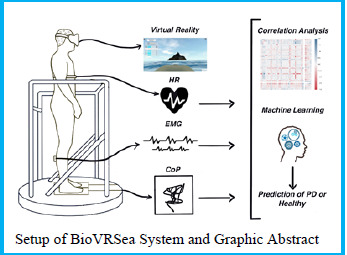
Eur J Transl Myol. 2023 Apr 27;33(2):11427.

2023Pdm3 March 31 - Abstract 63 How sex hormones can affect the fasciae

Caterina Fede 1,, Carla Stecco 1

Abstract

It is well established that the prevalence rate of musculoskeletal pain is significantly higher in women with respect to males, with an even greater onset of chronic conditions (Ceccarelli et al., 2021). It is recently reported that fascia can be a possible source of pain: if the connective tissue is altered, the behaviour of the fascial tissue and the underlying muscle may become compromised causing myofascial pain. Sex hormone disorders can dysregulate the fascial tissue: the comprehension of this mechanism is an important step for understanding gender differences in myofascial pain, helping clinicians to diagnose and treat patients (Fede et al., 2022). Sex hormone receptors are expressed by fascial fibroblasts, with a lower expression with the decrease in hormone levels in post-menopausal women (Fede et al., 2016). The cells of the fascia can moreover modulate the synthesis of extracellular matrix components depending on hormone levels: when β-estradiol levels are low, fascial tissue becomes enriched in collagen-I (from 5.2% of control sample to 8.4%), with a parallel decrease in collagen-III (from 2.4% to 1.5%) and elastic fibres (from 0.5% to 0.2%) (Fede et al., 2019). Consequently, the tissue becomes less elastic and more rigid, something that normally occurs during menopause. Conversely, when hormone levels are high, as they normally are during the ovulatory peak or during pregnancy, the opposite takes place: collagen-III rises to 6.8% during ovulation and 6.7% during pregnancy as does Fibrillin-1 (from 0.2% in menopause to 3.6% during pregnancy) while collagen-I falls to 1.9% (Fede et al., 2019). The result is softer, more elastic tissue. These results highlight how hormonal disorders in women can dysregulate the extracellular matrix synthesis (Figure), modifying the properties of tissue and evoking the sensitization of fascial nociceptors. Further studies on multifactorial effects of sex hormones on fasciae and pain mechanisms will permit to correlate any dysfunctions in hormonal levels linked to pathologies, ageing and period of the cycle, to the onset of myofascial pain, thus making it possible to find out a targeted gender therapy.

graphic file with name ejtm-33-2-11427-g037.jpg

Key Words: Fascia, women chronic pain, sex hormones, β-estradiol

References

  • 1.Ceccarelli I, Bioletti L, Peparini S, Solomita E, Ricci C, Casini I, Miceli E, Aloisi AM. Estrogens and phytoestrogens in body functions. Neurosci Biobehav Rev. 2022;132:648-663. doi: 10.1016/j.neubiorev.2021.12.007. [DOI] [PubMed] [Google Scholar]
  • 2.Fede C, Pirri C, De Caro R, Stecco C. Myofascial pain in females and personalized care: The key role played by sex hormones. Eur J Pain. 2022;26(4):939-940. doi: 10.1002/ejp.1920. [DOI] [PubMed] [Google Scholar]
  • 3.Fede C, Albertin G, Petrelli L, Sfriso MM, Biz C, De Caro R, Stecco C. Hormone receptor expression in human fascial tissue. Eur J Histochem. 2016;60(4):2710. doi: 10.4081/ejh.2016.2710. [DOI] [PMC free article] [PubMed] [Google Scholar]
  • 4.Fede C, Pirri C, Fan C, Albertin G, Porzionato A, Macchi V, De Caro R, Stecco C. Sensitivity of the fasciae to sex hormone levels: Modulation of collagen-I, collagen-III and fibrillin production. PLoS One. 2019;14(9):e0223195. [DOI] [PMC free article] [PubMed] [Google Scholar]
Eur J Transl Myol. 2023 Apr 27;33(2):11427.

2023Pdm3 March 31 - Abstract 64 Lymphatic vessels detection in subcutis and superficial fascia

Giovanna Albertin 1,, Laura Astolfi 2,3,4, Caterina Fede 1, Raffaele De Caro 1, Carla Stecco 1

Abstract

Lymphedema, defined as the abnormal accumulation of protein-rich fluid in soft tissues, results from the dysfunction of lymphatic system, an imbalance between lymph formation and its absorption into the initial lymphatics. The chronic accumulation of interstitial fluid leads adipose deposition, fibrosis, or persistent inflammation on subcutis (1). Today, lymphedema's therapies require a detailed understanding of the anatomy of subcutaneous lymphatic system (2), moreover, the exact localization of the collectors and their relationships with fascial layers have not been defined yet. The aim of this study was to investigate the distribution, density and organization of lymphatic vessels (LV) with reference to the layered conformation of the subcutaneous tissue, that consist in the superficial adipose tissue (SAT), deep adipose tissue (DAT) and superficial fascia (SF) (3, 4). With this purpose, the subcutaneous tissue of three adult voluntary patients was harvested during abdominoplastic surgery and stained with a specific marker for the endothelial cells of lymphatic vessels, the monoclonal antibody D2-40(5). LV, present massively on the papillary dermis, run parallel to the skin surface and are incorporated in the loose connective tissue to form a lymphatic plexus. On SAT, only thin LV, with a mean diameter of 11.6±7.71 μm, were visible close to the fibrous septa (retinacula cutis that connects the dermis to the deeper layers). In the DAT, the LV follow above all the blood vessel, they are thicker with a mean diameter of 22.5±12.77 μm. The SF exhibits the highest density of LV, with a mean diameter of 19.5±5.77 μm, they show a path parallel to the surface, where intertwining each other form a characteristic plexus. This study has shown the different distribution of the lymphatic vessels in the various subcutaneous layers and the existence of a new lymphatic plexus within the superficial fascia, adding new information on the alterations involving the subcutaneous tissues and consequently opening up new perspectives for surgery and manual treatments.

graphic file with name ejtm-33-2-11427-g038.jpg

Key Words: Lymphedema, fascial tissue, lymphatic vessels

References

  • 1.Mehrara BJ, Greene AK. Lymphedema and obesity: is there a link? Plast Reconstr Surg. 2014. Jul;134(1)154e-160e. [DOI] [PMC free article] [PubMed] [Google Scholar]
  • 2.Savetsky IL, Torrisi JS, Cuzzone DA, Ghanta S, Albano NJ, Gardenier JC, Joseph WJ, Mehrara BJ. Obesity increases inflammation and impairs lymphatic function in a mouse model of lymphedema. Am J Physiol Heart Circ Physiol. 2014. Jul 15;307(2):H165-72. [DOI] [PMC free article] [PubMed] [Google Scholar]
  • 3.Lancerotto L, Stecco C, Macchi V, Porzionato A, Stecco A, De Caro R. Layers of the abdominal wall: anatomical investigation of subcutaneous tissue and superficial fascia. Surg Radiol Anat. 2011. Dec;33(10):835-42 [DOI] [PubMed] [Google Scholar]
  • 4.Fede C, Pirri C, Fan C, et al. A Closer Look at the Cellular and Molecular Components of the Deep/Muscular Fasciae. Int J Mol Sci. 2021. Jan 30;22(3):1411. [DOI] [PMC free article] [PubMed] [Google Scholar]
  • 5.Fukunaga M. Expression of D2-40 in lymphatic endothelium of normal tissues and in vascular tumours. Histopathology. 2005. Apr;46(4):396-402. [DOI] [PubMed] [Google Scholar]
Eur J Transl Myol. 2023 Apr 27;33(2):11427.

2023Pdm3 March 31 - Abstract 65 The superficial fascia: Anatomy, innervation and vascularization

Lucia Petrelli 1,, Caterina Fede 1, Carmelo Pirri 1, Carla Stecco 1

Abstract

The superficial fascia has only recently been recognized as a specific anatomical structure in its own right anatomical entity, being first considered as included in the hypodermis. Furthermore, whereas it is actually recognized that the innervation of the deep/muscular fascia plays a key role in proprioception and nociception,1 and there are studies highlighting the cell populations and the extracellular matrix characterization of the deep fascia,2 there are very few studies that have analyzed these characteristics in the superficial fascia.

The superficial fascia is the second most highly innervated tissue after the skin, with a density of 33.0 ± 2.5/cm2, and a mean nerve sizes of 19.1 ± 7.2 μm.3 Free nerve endings innervate the tissue, and autonomic nerve fibers are present in the blood vessels, in the areas of vascularization and near adipocytes and in the connective tissue itself (Figure).

A rich vascular pattern forming a fine, dense meshwork with an area percentage of 6.20% ± 2.10% von Willebrand factor stained vessels was noted in all the specimens of the fascia examined; the area percentage of the αSMA-stained vessels was 2.93% ± 1.80% . The diameters of the vessels fell between the 13 and 65 μm range; the network was composed of arteries, veins, capillaries and lymphatic segments.4 Fibroblasts, myofibroblasts, mast cells are evident in the tissue. Finally, the elastic fibers are more abundant in the superficial fascia than the deep fascia, demonstrating that the superficial fasciae are more adaptable.5 In the light of these findings is evident that the superficial fasciae have a clear and distinct anatomical entity, and that they should be considered according to their characteristics, innervation and vascularization to better understand their role in thermoregulation, exteroception and pain perception. The knowledge of the superficial fascia may improve grading and developing of different manual approach for treatments of fascial dysfunctions.

graphic file with name ejtm-33-2-11427-g039.jpg

Key Words: Superficial fascia, innervation, vascularization, autonomic innervation, arteries

References

  • 1.Fede C, Petrelli L, Guidolin D, Porzionato A, Pirri C, Fan C, De Caro R, Stecco C. Evidence of a new hidden neural network into deep fasciae. Sci Rep, 11: 12623, 2021. doi: 10.1038/s41598-021-92194-z. [DOI] [PMC free article] [PubMed] [Google Scholar]
  • 2.Fede C, Pirri C, Fan C, Petrelli L, Guidolin D, De Caro R, Stecco C. A Closer Look at the Cellular and Molecular Components of the Deep/Muscular Fasciae. Int J Mol Sci, 22: 1411, 2021. [DOI] [PMC free article] [PubMed] [Google Scholar]
  • 3.Fede C, Petrelli L, Pirri C, Neuhuber W, Tiengo C, Biz C, De Caro R, Schleip R, Stecco C. Innervation of human superficial fascia. Front Neuroanat. 2022. Aug 29;16:981426. doi: 10.3389/fnana.2022.981426 [DOI] [PMC free article] [PubMed] [Google Scholar]
  • 4.Pirri C, Petrelli L, Fede C, Guidolin D, Tiengo C, De Caro R, Stecco C. Blood supply to the superficial fascia of the abdomen: An anatomical study. Clin Anat. 2022. Dec 28. doi: 10.1002/ca.23993 [DOI] [PubMed] [Google Scholar]
  • 5.Pirri C, Fede C, Petrelli L, Guidolin D, Fan C, De Caro R, Stecco C. Elastic Fibres in the subcutaneous tissue: Is there a difference between superficial and muscular fascia? A cadaver study. Skin Res Technol, 28: 21-27, 2022. doi: 10.1111/srt.13084 [DOI] [PMC free article] [PubMed] [Google Scholar]
Eur J Transl Myol. 2023 Apr 27;33(2):11427.

2023Pdm3 March 31 – Abstract 66 Biomechanical properties of the fascial system

Lorenza Bonaldi 1,, Alice Berardo 1,2,3, Carmelo Pirri 4, Carla Stecco 2,4, Chiara Giulia Fontanella 2,5

Abstract

The "fascial system" has been defined as a fascial network involved in functional aspects such as force transmission.1 The fascial system comprises the superficial and deep fasciae which are connected through skin ligaments, also known as retinacula, providing a continuity from the skin to the muscular plane (See Figure).2 Indeed, superficial and deep fascia roles in clinical disorders have been shown to reflect their structures.3 Specifically, the superficial fascia has been described anatomically as an irregular multilamellar structure of interconnected substrates with islets of fat cells. Meanwhile, for example, the deep aponeurotic fascia consists of multilayered structures of dense and loose connective tissues, where collagen fibers follow specific directions.4 Since the properties of superficial and deep fasciae mimic their structural organization, the biomechanical characterization of these tissues is key to understanding how they influence each other. Despite the clinical impact of these arguments, to date, the literature is still poor in data comparing different substrates of the fascial system through mechanical tests. Therefore, the aim of this work is to investigate this open topic by presenting the biomechanical properties of both superficial and deep fasciae.5,6 The study highlights how these tissues have different biomechanical properties in relation to their specific structures and functions. These results have a direct impact in the medical field such as in the surgical treatment of soft tissue repair or reconstruction, as well as in rehabilitation intervention (e.g., manual treatment).

graphic file with name ejtm-33-2-11427-g040.jpg

Key Words: Connective tissue, musculoskeletal system, fascial system, superficial fascia, deep fascia, biomechanical characterization

References

  • 1.Stecco C, Schleip R. A fascia and the fascial system. J Bodyw Mov Ther. 2016;20(1):139-140. doi:10.1016/j.jbmt.2015.11.012 [DOI] [PubMed] [Google Scholar]
  • 2.Stecco C, Macchi V, Porzionato A, Duparc F, De Caro R. The fascia: the forgotten structure. Ital J Anat Embryol. 2011;116(3):127-138. [PubMed] [Google Scholar]
  • 3.Stecco A, Stern R, Fantoni I, De Caro R, Stecco C. Fascial Disorders: Implications for Treatment. PM R. 2016;8(2):161-168. doi:10.1016/j.pmrj.2015.06.006 [DOI] [PubMed] [Google Scholar]
  • 4.Stecco C. Functional Atlas of the human fascial system. Churchill Livingstone. 2015. [Google Scholar]
  • 5.Bonaldi L, Berardo A, Stecco C, Fontanella CG. Biomechanical characterization of human superficial fascia from abdomen: A case study. XI meeting: Italian Chapter of the ESB, 6-7 October 2022, Massa. [Google Scholar]
  • 6.Bonaldi L, Fontanella CG, Stecco C, Berardo A. Uniaxial Tensile Tests On Human Fascia Lata: Stress Relaxation And Failure Phenomena From Frozen Cadavers. 27th Congress of the ESB, 26-29 June 2022, Porto. [Google Scholar]
Eur J Transl Myol. 2023 Apr 27;33(2):11427.

2023Pdm3 March 31 - Abstract 67 Fascia Lata alterations in hip osteoarthritis: An observational cross-sectional study

Ilaria Fantoni 1,, Carlo Biz 1, Chenglei Fan 2, Carmelo Pirri 2, Caterina Fede 2, Lucia Petrelli 2, Pietro Ruggieri 1, Raffaele De Caro 2, Carla Stecco 2

Abstract

The present study compares the structure and composition of fascia lata in healthy subjects and in patients with hip osteoarthritis (OA), to evaluate any differences in the amount of Collagen type I, Collagen type III, and Hyaluronan. Fascia lata samples from voluntary healthy subjects and patients with OA were harvested during surgery. Collagen type I (COL I), III (COL III) antibody, and biotinylated hyaluronan binding protein (HABP) immunohistochemistry stainings were used to evaluate fascial morphology and COL I, COL III, and Hyaluronan (HA) content in both groups. Ten samples from healthy subjects (group A) and 11 samples from OA patients (group B) were collected. COL I was significantly more abundant in the OA group (p = 0.0015), with a median percentage positivity of 75.2 (IQR 13.11)%, while representing only 67 (IQR: 8.71)% in control cases. COL III, with median values of 9.5 (IQR 3.63)% (OA group) and 17.10 (IQR 11)% (control cases), respectively, showed significant reduction in OA patients (p = 0.002). HA showed a median value of 10.01 (IQR 8.11)% in OA patients, denoting significant decrease (p < 0.0001) with respect to the control group median 39.31 (IQR 5.62)%. The observed differences suggest a relationship between fascial pathology and hip OA. The observed increase in COL I in OA patients, along with the reduction of COL III and HA, could lead to fascial stiffening, which could alter fascial mechanics and be linked to the development and symptoms of OA.

graphic file with name ejtm-33-2-11427-g041.jpg

Key Words: Fascia, hip osteoarthritis, hyaluronan, collagen, stiffness, myofascial pain

References

  • 1.Fede C, Angelini A, Stern R, Macchi V, Porzionato A, Ruggieri P, De Caro R, Stecco C. Quantification of hyaluronan in human fasciae: variations with function and anatomical site. J Anat. 2018. Oct;233(4):552-556. doi: 10.1111/joa.12866. Epub 2018 Jul 24. PMID: 30040133; PMCID: PMC6131966. [DOI] [PMC free article] [PubMed] [Google Scholar]
  • 2.Stecco C, Stern R, Porzionato A, Macchi V, Masiero S, Stecco A, De Caro R. Hyaluronan within fascia in the etiology of myofascial pain. Surg Radiol Anat. 2011. Dec;33(10):891-6. doi: 10.1007/s00276-011-0876-9. Epub 2011 Oct 2. PMID: 21964857. [DOI] [PubMed] [Google Scholar]
  • 3.Pratt RL. Hyaluronan and the Fascial Frontier. Int J Mol Sci. 2021. Jun 25;22(13):6845. doi: 10.3390/ijms22136845. PMID: 34202183; PMCID: PMC8269293. [DOI] [PMC free article] [PubMed] [Google Scholar]
  • 4.Rees HW. Management of Osteoarthritis of the Hip. J Am Acad Orthop Surg. 2020. Apr 1;28(7):e288-e291. doi: 10.5435/JAAOS-D-19-00416. PMID: 31800436. [DOI] [PubMed] [Google Scholar]
  • 5.Grässel S, Muschter D. Recent advances in the treatment of osteoarthritis. F1000Res. 2020. May 4;9:F1000 Faculty Rev-325. doi: 10.12688/f1000research.22115.1. PMID: 32419923; PMCID: PMC7199286. [DOI] [PMC free article] [PubMed] [Google Scholar]
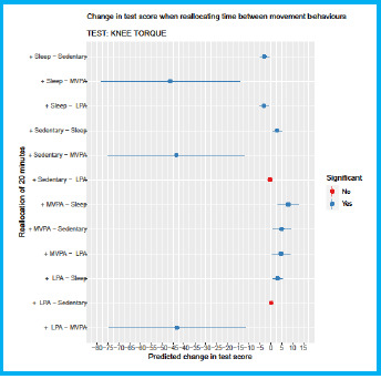
Eur J Transl Myol. 2023 Apr 27;33(2):11427.

02:00 PM SESSION IX: Non-invasive Assessments in Myology Paolo Gargiulo, Ugo Carraro, Chairs :2023Pdm3 March 31 - Abstract 68 LECTURE 3D Quantitative Muscle Color Computed Tomography (3D-QMCCT)

Paolo Gargiulo 1,2,

Abstract

In 2009 Ugo Carraro and Paolo Gargiulo introduced the term 3D Quantitative Muscle Color Computed Tomography (3D-QMCCT) as the process which, starting from computed tomography images, analyzes the distribution in Hounsfield Units (HU), that is the densitometry of a specific anatomical region, assigning a color (essentially derived from the colors of histologically labeled muscle biopsies) based on its density or morphology.1,2 3D-QMCCT has been a key tool primarily to assess, quantify, and monitor muscle atrophy in denervated tissue,3 but also to advance bone mineral density assessment and support surgical planning.4 3D-QMCCT provided the best evidence of the effectiveness of home-based Functional Electrical Stimulation of denervated muscles (hbFES) during validation by the Europenan project RISE.5 A development of 3D-QMCCT is the numerical muscle profile called Nonlinear Trimodal Regression Analysis (NTRA), which numerically summarizes the connective tissue, fat and muscle content from a CT cross section. NTRAs have significantly contributed to demonstrating the interactions between muscle tissue quality and comorbidities such as cholesterol disease and diabetes and correlations with lifestyle and mobility in the aging population.6,7 Today, with the improvement of medical imaging, human anatomy can be studied non-invasively with micrometre resolution. As the dose of X-rays has been drastically reduced in recent years, the use of CT for research purposes is now much more feasible. In conclusion, new frontiers and applications, such as virtual cardiac histology, can be explored using 3D-QMCCT and NTRA methods.

graphic file with name ejtm-33-2-11427-g042.jpg

Key Words: 3D Quantitative Muscle Color Computed Tomography (3D-QMCCT), Nonlinear Trimodal Regression Analysis (NTRA), skeletal muscle atrophy and degeneration, home-based Functional Electrical Stimulation of denervated muscles (hbFES)

References

  • 1.Gargiulo P, Vatnsdal B, Ingvarsson P, Knútsdóttir S, Gudmundsdóttir V, Yngvason S, Kern H, Carraro U, Helgason T. Computational methods to analyze tissue composition and structural changes in denervated muscle undergoing therapeutic electrical stimulation. Basic Applied Myology 19(4): 157-161, 2009. [Google Scholar]
  • 2.Gargiulo P, Kern H, Carraro U, Ingvarsson P, Knútsdóttir S, Gudmundsdóttir V, Yngvason S, Vatnsdal B, Helgason T. Quantitative color three-dimensional computer tomography imaging of human long-term denervated muscle. Neurol Res. 2010. Feb;32(1):13-9. doi: 10.1179/016164109X 12536042424171. PMID: 20092691. [DOI] [PubMed] [Google Scholar]
  • 3.Gargiulo P, Reynisson PJ, Helgason B, Kern H, Mayr W, Ingvarsson P, Helgason T, Carraro U. Muscle, tendons, and bone: structural changes during denervation and FES treatment. Neurol Res. 2011. Sep;33(7):750-8. doi: 10.1179/ 1743132811Y.0000000007. PMID: 21756556. [DOI] [PubMed] [Google Scholar]
  • 4.Esposito L, Bifulco P, Gargiulo P, Gíslason MK, Cesarelli M, Iuppariello L, Jónsson H, Cutolo A, Fraldi M. Towards a patient-specific estimation of intra-operative femoral fracture risk. Comput Methods Biomech Biomed Engin. 2018. Sep;21(12):663-672. doi: 10.1080/10255842.2018. 1508570. Epub 2018 Oct 27. PMID: 30370789. [DOI] [PubMed] [Google Scholar]
  • 5.Kern H, Carraro U, Adami N, Biral D, Hofer C, Forstner C, Mödlin M, Vogelauer M, Pond A, Boncompagni S, Paolini C, Mayr W, Protasi F, Zampieri S. Home-based functional electrical stimulation rescues permanently denervated muscles in paraplegic patients with complete lower motor neuron lesion. Neurorehabil Neural Repair. 2010. Oct;24(8):709-21. doi: 10.1177/1545968310366129. Epub 2010 May 11. PMID: 20460493. [DOI] [PubMed] [Google Scholar]
  • 6.Recenti M, Ricciardi C, Edmunds K, Jacob D, Gambacorta M, Gargiulo P. Testing soft tissue radiodensity parameters interplay with age and self-reported physical activity. Eur J Transl Myol. 2021. Jul 12;31(3):9929. doi: 10.4081/ejtm.2021.9929. PMID: 34251162; PMCID: PMC8495362. [DOI] [PMC free article] [PubMed] [Google Scholar]
  • 7.Edmunds K, Gíslason M, Sigurðsson S, Guðnason V, Harris T, Carraro U, Gargiulo P. Advanced quantitative methods in correlating sarcopenic muscle degeneration with lower extremity function biometrics and comorbidities. PLoS One. 2018. Mar 7;13(3):e0193241. doi: 10.1371/journal.pone. 0193241. PMID: 29513690; PMCID: PMC5841751 [DOI] [PMC free article] [PubMed] [Google Scholar]
Eur J Transl Myol. 2023 Apr 27;33(2):11427.

2023Pdm3 March 31 - Abstract 69 Virtual cardiac histology: a densitometric characterisation of left ventricular tissue

Riccardo Forni 1,2,, Carmine Gelormini 3, Bjarni Torfason 4, Cristiana Corsi 2, Paolo Gargiulo 1,5

Abstract

Understanding the mechanisms behind changes in cardiac tissue has always been a topic of great interest. In recent years, biochemical, molecular, and genetic aspects have been studied. In this work, we investigated tissue properties from Computed Tomography images. We designed a densitometric profile and assessed its relations with age, gender, and pathological conditions such as ventricular septal defect/rupture and hypertrophic cardiomyopathy. We developed a novel workflow to segment cardiac tissue to extract 3D samples from regions of clinical interest on the left ventricle. The densitometric profile evaluates the average density, variability, and complexity of the Hounsfield distribution. To assess these relationships, we implemented a linear mixed model, taking into account the non-independence of the data due to repeated measurements of the same subjects within different regions. The results showed that gender is a discriminating factor in average cardiac tissue density, while there is no real relationship between age and absorption. The study, including the diseases, showed that the mean free wall density of the left ventricle changes in the presence of hypertrophic cardiomyopathy. At the same time, there were no statistically significant changes between healthy subjects and patients with post-infarction septal rupture. Moreover, we employed machine learning technology to evaluate the densitometric profile's predictive power, finding peak amplitude and peak position as the most promising features. Finally, this work shows a novel methodology to study cardiac tissue non-invasive, potentially becoming a valid clinical assessment to support an early-stage diagnosis.

graphic file with name ejtm-33-2-11427-g043.jpg

Key Words: Virtual histology, cardiac tissue composition, densitometric profile, hypertrophic cardiomyopathy, ventricular septal rupture

References

  • 1.Mazonakis M, Damilakis J. Computed tomography: What and how does it measure? Eur J Radiol. 2016. Aug;85(8):1499-504. doi: 10.1016/j.ejrad.2016.03. 002. Epub 2016 Mar 10. PMID: 26995675. [DOI] [PubMed] [Google Scholar]
  • 2.Maron BJ, Maron MS. Hypertrophic cardiomyopathy. Lancet. 2013. Jan 19;381(9862):242-55. doi: 10.1016/S0140-6736(12) 60397-3. Epub 2012 Aug 6. PMID: 22874472. [DOI] [PubMed] [Google Scholar]
  • 3.Olivetti G, Melissari M, Capasso JM, Anversa P. Cardiomyopathy of the aging human heart. Myocyte loss and reactive cellular hypertrophy. Circ Res. 1991. Jun;68(6):1560-8. doi: 10.1161/01.res.68.6.1560. PMID: 2036710. [DOI] [PubMed] [Google Scholar]
  • 4.Recenti M, Ricciardi C, Edmunds KJ, Gislason MK, Sigurdsson S, Carraro U, Gargiulo P. Healthy Aging Within an Image: Using Muscle Radiodensitometry and Lifestyle Factors to Predict Diabetes and Hypertension. IEEE J Biomed Health Inform. 2021. Jun;25(6):2103-2112. doi: 10.1109/JBHI.2020. 3044158. Epub 2021 Jun 3. PMID: 33306475. [DOI] [PubMed] [Google Scholar]
  • 5.Bates D, Mächler M, Bolker B, Walker S. Fitting linear mixed-effects models using lme4. arXiv preprint 2014; arXiv:1406.5823. [Google Scholar]
  • 6.Recenti M, Ricciardi C, Edmunds K, Jacob D, Gambacorta M, Gargiulo P. Testing soft tissue radiodensity parameters interplay with age and self-reported physical activity. Eur J Transl Myol. 2021. Jul 12;31(3):9929. doi: 10.4081/ejtm.2021.9929. PMID: 34251162; PMCID: PMC8495362. [DOI] [PMC free article] [PubMed] [Google Scholar]
Eur J Transl Myol. 2023 Apr 27;33(2):11427.

2023Pdm3 March 31 - Abstract 70 Assessing early-stage Parkinson’s Disease using a moving platform (BioVRSea)

Deborah Jacob 1,, Romain Aubonnet 1, Marco Recenti 1, Sigrun Anna Audardottir 1, Thorbjorg Ida Ivarsdottir 1, Berangere Burgunder 1, Itziar Mengual i Escalona 1, Andrea Colacino 2, Anna Bjornsdottir 3, Hannes Petersen 4, Paolo Gargiulo 1

Abstract

Parkinson’s Disease is among the most prevalent neurological diseases in the world today. Typically characterised by impairments in motor function, there remains no known cure for the disease. Treatments generally take the form of medication and/or surgical intervention in combination with physical therapy. Deficits in postural control are commonly seen in Parkinson’s sufferers. Our work using the unique BioVRSea setup aims to assess early-stage Parkinson’s using a combination of neurphysiological (Electromyography and Heart Rate) and centre of pressure (or sway) measurements. 11 early-stage Parkinson’s subjects and 46 healthy over-50s took part in the experiment. Significant differences were found between the two groups in electromyographic and centre of pressure measurements. Correlation analysis indicated opposite correlations in skewness in the right soleus muscle. Finally, machine learning was able to predict with a maximum of 94.6% accuracy whether a subject belonged to the healthy or Parkinson’s group based on their measurements from the experiment. Our results are a first step in a prototype of the quantitative evaluation of early-stage Parkinson’s.

graphic file with name ejtm-33-2-11427-g044.jpg

Key Words: Parkinson’s Disease, biomarkers, BioVRSea, electromyography, centre of pressure, postural control

References

  • 1.Doná F, Aquino CC, Gazzola JM, Borges V, Silva SM, Ganança FF, Caovilla HH, Ferraz HB. Changes in postural control in patients with Parkinson's disease: a posturographic study. Physiotherapy. 2016. Sep;102(3):272-9. doi: 10.1016/j.physio.2015.08.009. Epub 2015 Oct 3. PMID: 26582134 [DOI] [PubMed] [Google Scholar]
  • 2.Recenti M, Ricciardi C, Aubonnet R, Picone I, Jacob D, Svansson HÁR, Agnarsdóttir S, Karlsson GH, Baeringsdóttir V, Petersen H, Gargiulo P. Toward Predicting Motion Sickness Using Virtual Reality and a Moving Platform Assessing Brain, Muscles, and Heart Signals. Front Bioeng Biotechnol. 2021. Apr 1;9:635661. doi: 10.3389/fbioe.2021.635661. PMID: 33869153; PMCID: PMC8047066.. [DOI] [PMC free article] [PubMed] [Google Scholar]
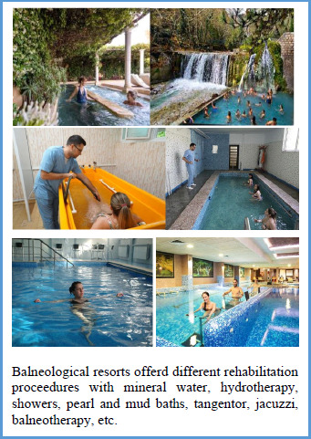
  • 3.Jacob D, Unnsteinsdóttir Kristensen IS, Aubonnet R, Recenti M, Donisi L, Ricciardi C, Svansson HÁR, Agnarsdóttir S, Colacino A, Jónsdóttir MK, Kristjánsdóttir H, Sigurjónsdóttir HÁ, Cesarelli M, Eggertsdóttir Claessen LÓ, Hassan M, Petersen H, Gargiulo P. Towards defining biomarkers to evaluate concussions using virtual reality and a moving platform (BioVRSea). Sci Rep. 2022. May 30;12(1):8996. doi: 10.1038/s41598-022-12822-0. PMID: 35637235; PMCID: PMC9151646. [DOI] [PMC free article] [PubMed] [Google Scholar]
  • 4.Busa MA, van Emmerik REA. Multiscale entropy: A tool for understanding the complexity of postural control. J Sport Health Sci. 2016. Mar;5(1):44-51. doi: 10.1016/j.jshs.2016.01.018. Epub 2016 Jan 21. PMID: 30356502; PMCID: PMC6188573. [DOI] [PMC free article] [PubMed] [Google Scholar]
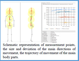
  • 5.Fadil R, Huether A, Brunnemer R, Blaber AP, Lou JS, Tavakolian K. Early Detection of Parkinson's Disease Using Center of Pressure Data and Machine Learning. Annu Int Conf IEEE Eng Med Biol Soc. 2021. Nov;2021:2433-2436. doi: 10.1109/EMBC46164.2021.9630451. PMID: 34891772. [DOI] [PubMed] [Google Scholar]
  • 6.Recenti M, Ricciardi C, Edmunds K, Jacob D, Gambacorta M, Gargiulo P. Testing soft tissue radiodensity parameters interplay with age and self-reported physical activity. Eur J Transl Myol. 2021. Jul 12;31(3):9929. doi: 10.4081/ejtm.2021.9929. PMID: 34251162; PMCID: PMC8495362. [DOI] [PMC free article] [PubMed] [Google Scholar]
Eur J Transl Myol. 2023 Apr 27;33(2):11427.

2023Pdm3 March 31 - Abstract 71 An in silico 3d approach to evaluate bone remodelling after total hip arthroplasty: a six years longitudinal study

Valentina Betti 1,, Halldór Jónsson Jr 2, Luca Cristofolini 1, Magnús Kjartan Gíslason 3, Paolo Gargiulo 3,4

Abstract

Nowadays, research on bone remodelling after a total hip arthroplasty (THA) is mainly performed using DXA-scans, which gives a quantitative assessment but it can lose some pivotal features of how bone remodels since it is a 3d mechanism. The aim of this work was to enhance the understanding of the volumetric bone mineral density (vBMD) evolution after THA by developing a protocol to (i) three-dimensionally localise the changes in the proximal femur, and (ii) assess such changes in the long term. Twelve patients that underwent unilateral THA (with cemented/uncemented prostheses) were recruited. Three calibrated CT scans of the proximal femur were collected at different time points: 24 hours, 1 year and 6 years after surgery. The 3D models of the proximal femur were extracted, and an algorithm was developed to split them into the Gruen zones. The bone density gain and loss for both the whole proximal femur and those specific regions was calculated by comparing density values between the three sets of scans. Results showed lower amount of trabecular bone observed in the cemented cohort compared to the uncemented group, and some differences in terms of vBMD evolution were noticed in the whole femur and specifically in some Gruen zones. A high inter-patient variability was observed, ranging from some cases that showed a physiological bone remodelling, to some pathological conditions in which an unexpected increase/decrease of vBMD (e.g. +340% after one year) was noticed. In conclusion, the presented analysis is an useful tool (i) to understand the vBMD evolution in THA patients in the long-term, and (ii) for the follow-up of patients where a failure of the implant is to be expected.

graphic file with name ejtm-33-2-11427-g045.jpg

Key Words: Bone remodeling, total hip arthroplasty, Gruen zones, longitudinal study

References

  • 1.Gislason MK, Lupidio F, Jónsson H Jr, Cristofolini L, Esposito L, Bifulco P, Fraldi M, Gargiulo P. Three dimensional bone mineral density changes in the femur over 1 year in primary total hip arthroplasty patients. Clin Biomech (Bristol, Avon). 2020. Aug;78:105092. doi: 10.1016/j.clinbiomech.2020.105092. Epub 2020 Jun 11. PMID: 32590143. [DOI] [PubMed] [Google Scholar]
  • 2.Gargiulo P, Edmunds KJ, Gíslason MK, Latour C, Hermannsson Þ, Esposito L, Bifulco P, Cesarelli M, Fraldi M, Cristofolini L, Jónsson H Jr. Patient-specific mobility assessment to monitor recovery after total hip arthroplasty. Proc Inst Mech Eng H. 2018. Oct;232(10):1048-1059. doi: 10.1177/0954411918797971. Epub 2018 Sep 7. PMID: 30191747. [DOI] [PubMed] [Google Scholar]
  • 3.Pitto RP, Bhargava A, Pandit S, Walker C, Munro JT. Quantitative CT-assisted osteodensitometry of femoral adaptive bone remodelling after uncemented total hip arthroplasty. Int Orthop. 2008. Oct;32(5):589-95. doi: 10.1007/s00264-007-0389-7. Epub 2007 Jun 20. PMID: 17579860; PMCID: PMC2551722. [DOI] [PMC free article] [PubMed] [Google Scholar]
  • 4.Recenti M, Ricciardi C, Edmunds K, Jacob D, Gambacorta M, Gargiulo P. Testing soft tissue radiodensity parameters interplay with age and self-reported physical activity. Eur J Transl Myol. 2021. Jul 12;31(3):9929. doi: 10.4081/ejtm.2021.9929. PMID: 34251162; PMCID: PMC8495362. [DOI] [PMC free article] [PubMed] [Google Scholar]
Eur J Transl Myol. 2023 Apr 27;33(2):11427.

2023Pdm3 March 31 - Abstract 72 Novel strategies for cartilage assessment, interplay between bone and muscles

Federica Kiyomi Ciliberti 1,, Romain Aubonnet 1, Jorgelina Ramos 1, Marco Recenti 1, Deborah Jacob 1, Lorena Guerrini 1, Magnus K Gíslason 1, Olafur Sigurjonsson 1, Mariella Tsirilaki 2, Halldór Jónsson 2,3, Paolo Gargiulo 1,2

Abstract

Assessment of human joint cartilage is a crucial tool to detect and diagnose pathological conditions. This exploratory study develop a workflow for 3D modeling of cartilage, muscle and bone based on multimodal imaging. We present a novel methodology to evaluate knee condition using features extracted from magnetic resonance imaging (MRI) and computed tomography (CT) data. New evaluation metrics were created and a unique set of data was gathered from healthy control subjects and patients with clinically evaluated degeneration or trauma. We developed patient specific 3D models of the tibial, femoral, and patellar bones and cartilages. We compare the sensitivity of different metrics to classify the cartilage condition and evaluate degeneration based on bones and muscle densitometry. The present work demonstrates the potential for improving sensitivity in cartilage assessment.

graphic file with name ejtm-33-2-11427-g046.jpg

Key Words: Image segmentation, knee cartilage, classification, osteoarthritis, 3D models

References

  • 1.Zhang Y, Jordan JM. Epidemiology of osteoarthritis. Clin Geriatr Med. 2010. Aug;26(3):355-69. doi: 10.1016/j.cger.2010.03.001. Erratum in: Clin Geriatr Med. 2013. May;29(2):ix. PMID: 20699159; PMCID: PMC2920533. [DOI] [PMC free article] [PubMed] [Google Scholar]
  • 2.Roemer FW, Demehri S, Omoumi P, Link TM, Kijowski R, Saarakkala S, Crema MD, Guermazi A. State of the Art: Imaging of Osteoarthritis-Revisited 2020. Radiology. 2020. Jul;296(1):5-21. doi: 10.1148/radiol.2020192498. Epub 2020 May 19. PMID: 32427556. [DOI] [PubMed] [Google Scholar]
  • 3.Ciliberti FK, Guerrini L, Gunnarsson AE, Recenti M, Jacob D, Cangiano V, Tesfahunegn YA, Islind AS, Tortorella F, Tsirilaki M, Jónsson H Jr, Gargiulo P, Aubonnet R. CT- and MRI-Based 3D Reconstruction of Knee Joint to Assess Cartilage and Bone. Diagnostics (Basel). 2022. Jan 22;12(2):279. doi: 10.3390/diagnostics12020279. PMID: 35204370; PMCID: PMC8870751 [DOI] [PMC free article] [PubMed] [Google Scholar]
  • 4.Baker JF, Von Feldt J, Mostoufi-Moab S, Noaiseh G, Taratuta E, Kim W, Leonard MB. Deficits in muscle mass, muscle density, and modified associations with fat in rheumatoid arthritis. Arthritis Care Res (Hoboken). 2014. Nov;66(11):1612-8. doi: 10.1002/acr.22328. PMID: 24664868; PMCID: PMC4551488 [DOI] [PMC free article] [PubMed] [Google Scholar]
  • 5.Cai G, Otahal P, Cicuttini F, Wu F, Munugoda IP, Jones G, Aitken D. The association of subchondral and systemic bone mineral density with osteoarthritis-related joint replacements in older adults. Osteoarthritis Cartilage. 2020. Apr;28(4):438-445. doi: 10.1016/j.joca.2020.02.832. Epub 2020 Feb 28. PMID: 32119971. [DOI] [PubMed] [Google Scholar]
  • 6.Recenti M, Ricciardi C, Edmunds K, Jacob D, Gambacorta M, Gargiulo P. Testing soft tissue radiodensity parameters interplay with age and self-reported physical activity. Eur J Transl Myol. 2021. Jul 12;31(3):9929. doi: 10.4081/ejtm.2021.9929. PMID: 34251162; PMCID: PMC8495362. [DOI] [PMC free article] [PubMed] [Google Scholar]
Eur J Transl Myol. 2023 Apr 27;33(2):11427.

2023Pdm3 March 31 - Abstract 73 Interplay between the age and the asymmetry of NTRA in elderly people

Carlo Ricciardi 1,, Alfonso Maria Ponsiglione 1, Marco Recenti 2, Francesco Amato 1, Paolo Gargiulo 2,3

Abstract

The 11 nonlinear trimodal regression analysis (NTRA) parameters represent the radiodensitometric distributions (RDD) of muscle, fat and connective tissues extracted from a CT scan of the mid-femur. Previous studies showed how these parameters are correlated with physiological variables (1) and can be used for building predictive models of cardiovascular diseases and for assessing the impact of lifestyle factors in aging subjects. The aim of this study is to assess the role of age in the asymmetry of the 11 NTRA parameters. The dataset consists of 3162 Icelandic subjects, aged between 65 and 95 years. A statistical approach was employed to investigate the interplay between the age and the NTRA asymmetry. A paired Student’s t test was performed to compare right- and left-femur NTRA parameters. Then, 11 asymmetry indicators were calculated as the absolute difference between right- and left-femur values of each NTRA parameters. Finally, an analysis of variance (ANOVA) and a regression analysis were carried out to study the relationships between subjects’ age and the degree of asymmetry. Results demonstrate a statistically significant difference between the RDDs of the right- and lef-femur for 7 out of 11 NTRA parameters. Then, the variation of the asymmetry indicators with respect to age is shown and the three most significant age-related asymmetry indicators were identified, namely the amplitude of the RDD of the fat (Nf), the width of the RDD of the muscle (σm), and the width of the RDD of the connective tissue (σt). It is finally observed that the age-related curves of the identified indicators follow a second-order polynomial law showing an increasing trend of the asymmetry indicators with age (determination coefficients, R2, equal to 0.50 for Nf, 0.76 for σm, and 0.75 for σt). In conclusion, the work shows that the NTRA are sensitive to the asymmetry, the degree of asymmetry increases with age, and σm is among the most sensitive age-related asymmetry indicator.

graphic file with name ejtm-33-2-11427-g047.jpg

Key Words: Aging, medical imaging, biomedical data analysis

References

  • 1.Edmunds K, Gíslason M, Sigurðsson S, Guðnason V, Harris T, Carraro U, Gargiulo P. Advanced quantitative methods in correlating sarcopenic muscle degeneration with lower extremity function biometrics and comorbidities. PLoS One. 2018. Mar 7;13(3):e0193241. doi: 10.1371/journal.pone. 0193241. PMID: 29513690; PMCID: PMC5841751 [DOI] [PMC free article] [PubMed] [Google Scholar]
  • 2.Ricciardi C, Edmunds KJ, Recenti M, Sigurdsson S, Gudnason V, Carraro U, Gargiulo P. Assessing cardiovascular risks from a mid-thigh CT image: a tree-based machine learning approach using radiodensitometric distributions. Sci Rep. 2020. Feb 18;10(1):2863. doi: 10.1038/s41598-020-59873-9. PMID: 32071412; PMCID: PMC7029006. [DOI] [PMC free article] [PubMed] [Google Scholar]
  • 3.Recenti M, Ricciardi C, Edmunds KJ, Gislason MK, Sigurdsson S, Carraro U, Gargiulo P. Healthy Aging Within an Image: Using Muscle Radiodensitometry and Lifestyle Factors to Predict Diabetes and Hypertension. IEEE J Biomed Health Inform. 2021. Jun;25(6):2103-2112. doi: 10.1109/JBHI.2020. 3044158. Epub 2021 Jun 3. PMID: 33306475. [DOI] [PubMed] [Google Scholar]
  • 4.Recenti M, Ricciardi C, Edmunds K, Jacob D, Gambacorta M, Gargiulo P. Testing soft tissue radiodensity parameters interplay with age and self-reported physical activity. Eur J Transl Myol. 2021. Jul 12;31(3):9929. doi: 10.4081/ejtm.2021.9929. PMID: 34251162; PMCID: PMC8495362. [DOI] [PMC free article] [PubMed] [Google Scholar]
Eur J Transl Myol. 2023 Apr 27;33(2):11427.

2023Pdm3 March 31 - Abstract 74 Time shift of peak activation levels in quadriceps and hamstrings after ACL reconstruction during single leg jump

Einar Einarsson 1,2, Kristín Briem 3, Rodney Whiteley 2, Magnus K Gislason 1,

Abstract

Recurrance of injuries are prevalent in athletes that have undergone ACL reconstruction which partly can be attributed to weakening and the reduction of neuromuscular control of the hamstrings and the quadriceps.1,2 When using autograft for the ACL reconstruction, the tissue is mainly harvested either from the hamstring tendon or from the patellar tendon which can have an effect of the overall biomechanics and load transfer integrity of the knee joint. In the presented study a 30s continuous single leg jump was carried out by subjects from three cohorts: a healthy control, patients that were recovering after ACL reconstruction using both hamstring and patellar tendon autograft. EMG measurements were taken of six muscles during the jump sequence: 1) Vastus Lateralis, 2) Vastus Medialis, 3) Biceps Femoris 4) Tensor Fascia Latae, 5) Semitendinousus and 6) Gluteus Medius. An acceleorometer located at the inferior lumbar region was synchronized with the EMG measurements to capture the events of push off and landing. The jump cycle was normalized from push off to the next push off as can be seen in the figure and the EMG signals rectified and processed using a bandpass filter and RMS and finally fitted to each normalized jump cycle. The jump sequence was divided into three parts containing equal number of jumps. The average EMG values were analyzed between the first third and the final third of the jump sequence. The results demonstrated that there is a temporal shift in the peak activation levels of the quadriceps muscles. Significant shift in peak activation is seen in Control group for both quadriceps and hamstring, and in Injured HS group for quadriceps muscle and Healthy BTB for lateral hamstring muscle and lower peak activation levels of the vastus lateralis (p<0.001) and vastus medialis (p<0.001) and lateral hamstrings (p<0.001) in the injured compared to healthy legs.

graphic file with name ejtm-33-2-11427-g048.jpg

Key Words: ACL reconstruction, biomechanics, EMG, single leg jump

References

  • 1.Einarsson E, Thomson A, Sas B, Hansen C, Gislason M, Whiteley R. Lower medial hamstring activity after ACL reconstruction during running: a cross-sectional study. BMJ Open Sport Exerc Med. 2021. Mar 11;7(1):e000875. doi: 10.1136/bmjsem-2020-000875. PMID: 33782638; PMCID: PMC7957131. [DOI] [PMC free article] [PubMed] [Google Scholar]
  • 2.Recenti M, Ricciardi C, Edmunds K, Jacob D, Gambacorta M, Gargiulo P. Testing soft tissue radiodensity parameters interplay with age and self-reported physical activity. Eur J Transl Myol. 2021. Jul 12;31(3):9929. doi: 10.4081/ejtm.2021.9929. PMID: 34251162; PMCID: PMC8495362. [DOI] [PMC free article] [PubMed] [Google Scholar]
Eur J Transl Myol. 2023 Apr 27;33(2):11427.

2023Pdm3 March 31 - Abstract 75 Exploring myofibril alignment in muscular tissues using circular statistics

Ettore Rocchi 1, Sara Peluso 1, Stefano Amatori 2,, Davide Sisti 2

Abstract

An interesting problem in analysing the image of muscle tissue is represented by the attempt to evaluate the linearity of myofibrillar structures and their possible deviation from a straight line. By indicating with αi the angles between two successive sarcomeres along the same myofibril, Cisterna et al. (2021) proposed an index that can be thought as the mean of the angles (taken in absolute value). In this contribution, we propose an alternative approach to the same problem. Our proposal, based on circular statistics, taking a cue from the same angular data αi proposed by Cisterna et al.; circular statistics has been used earlier to study muscle cell alignment. Ideally, for each angle αi, it is possible to construct a unit vector having the base at the centre of a goniometric circumference and the vertex on the circumference from which to calculate the resulting vector, which, divided by n (that is the number of angles or the number of sarcomeres minus 1), gives the resulting mean vector. From here, two parameters can be obtained: (a) the direction of the mean vector (if it is equal to 0, it means that the displacements to the right balance those to the left; or, as an extreme case, that the sarcomeres are perfectly aligned); b) the length of the mean vector r, that can range between 0 (representing perfect isotropy – or a circular uniform distribution – i.e., the maximum possible misalignment) and 1 (representing perfect anisotropy, i.e., the maximum possible alignment). It is also possible to perform a statistical test using the mean vector length r as test statistics: Rayleigh test is the best known in circular statistics; its null hypothesis is the uniform circular distribution of the angles, and the alternative hypothesis is a generic anisotropy. The method we propose, unlike the one indicated by Cisterna et al., uses circular analysis techniques instead of linear analysis methods, which makes it more elegant and gives greater substance to statistical analysis (but in contrast, it also has greater computational complexity).2 In conclusion, our method has potential use in several sarcomere-related conditions by providing a quantitative definition of myofibril linearity in skeletal muscle.

graphic file with name ejtm-33-2-11427-g049.jpg

Key Words: Circular statistics, myofibrillar structures, sarcomeres

References

  • 1.Cisterna B, Malatesta M, Zancanaro C, Boschi F. A computational approach to quantitatively define sarcomere dimensions and arrangement in skeletal muscle. Comput Methods Programs Biomed. 2021;211:106437. doi: 10.1016/j.cmpb.2021. 106437. [DOI] [PubMed] [Google Scholar]
  • 2.Rocchi E, Peluso S, Amatori S, Sisti D. New indexes for myofibril linearity in muscle image analysis. Eur J Transl Myol. 2022. Oct 4;32(4):10736. doi: 10.4081/ejtm.2022.10736. PMID: 36193819; PMCID: PMC9830398. [DOI] [PMC free article] [PubMed] [Google Scholar]
Eur J Transl Myol. 2023 Apr 27;33(2):11427.

04:30 PM SESSION X: Muscle Rehabilitation in Dentistry and beyond Riccardo Rosati, Elena P. Ivanova, Chairs :Riccardo Rosati explains sEMG of masseter muscle :2023Pdm3 March 31 - Abstract 76 Standardised protocols for sEMG of the masticatory muscles in oral rehabilitation

Gaia Pellegrini 1,

Abstract

On-site presentation of unpublished results. The author denied also Zoom circulation, recording and post-meeting dissemination by YOUTUBE

Eur J Transl Myol. 2023 Apr 27;33(2):11427.

2023Pdm3 March 31 - Abstract 77 Innovative methods of full dental rehabilitation

Elena Ivanova 1,, Frank Saxler 2, Andrey Lobanov 1, Sergey Andronov 1

Abstract

Dentistry in the twenty first century has been marked by multidisciplinary approach to diagnosis and treatment. Functional diagnosis, functional treatment, functional occlusion of patients with cranio-mandibular disorders depend on relation with functional diagnosis and treatment of full body. Innovative technologies of functional diagnosis and full dental rehabilitation calculating by 4D analysis developed on understanding dental and medical symptoms. Dental bruxism, abrasion shipping, parafunction, fracture of teeth, fracture of implant, fracture of prosthodontic, hyperemia of uninfected pulp of sensitive teeth, cranio-mandibular dysfunction CMD, TMJ Symptoms, structure loss with deflective contact, projection pain in teeth define function in cranio-mandibular system. Functional determinants of occlusion and individual tooth library include occlusion plane, vertical dimension, condyle inclination, mandibula position, inclination based on bones of the skull. So, we calculate complete digital concept of 4D Analysis connected with STL data, CAD device, 4D and Ray imaging of functional determinants, dynamic 4D moving of bone, patient adapting based to Fibonacci number, X-Ray and CAD with STL life on the screen. Digital way of examination and treatment options advantages changing occlusal plane, mandibular position, functional training device, resetting of bio processes and bone structure of the skull, directly changing of mobility of individual force, compliance of the patient mobility, rehabilitation functional determinants with new calculated functional occlusion after digital set up. Functional occlusion calculates with 4D Analysis service for prosthodontics, implantology and orthodontics. In every case we get the CNC file for high milling your functional occlusion and devices.

graphic file with name ejtm-33-2-11427-g050.jpg

Key Words: Functional diagnosis, functional treatment, functional occlusion, 4D analysis, cranio-mandibular disorders, multidisciplinary approach

References

  • 1.Baldini A, Nota A, Tripodi D, Longoni S, Cozza P. Evaluation of the correlation between dental occlusion and posture using a force platform. Clinics (Sao Paulo). 2013. Jan;68(1):45-9. doi: 10.6061/clinics/2013(01)oa07. PMID: 23420156; PMCID: PMC3552467.. [DOI] [PMC free article] [PubMed] [Google Scholar]
  • 2.Cuccia AM. Interrelationships between dental occlusion and plantar arch. J Bodyw Mov Ther. 2011;15(2):242–50. doi: 10.1016/j.jbmt.2010.10.007 PMID: 21419367 [DOI] [PubMed] [Google Scholar]
  • 3.Saccucci M, Tettamanti L, Mummolo S, Polimeni A, Festa F, Tecco S. Scoliosis and dental occlusion: a review of the literature. Scoliosis. 2011. Jul 29;6:15. doi: 10.1186/1748-7161-6-15. PMID: 21801357; PMCID: PMC3162939. [DOI] [PMC free article] [PubMed] [Google Scholar]
  • 4.Ohlendorf D, Seebach K, Hoerzer S, Nigg S, Kopp S. The effects of a temporarily manipulated dental occlusion on the position of the spine: a comparison during standing and walking. Spine J. 2014. Oct 1;14(10):2384-91. doi: 10.1016/j.spinee.2014.01.045. Epub 2014 Jan 31. PMID: 24486478. [DOI] [PubMed] [Google Scholar]
Eur J Transl Myol. 2023 Apr 27;33(2):11427.

2023Pdm3 March 31 - Abstract 78 Electromyographic analysis of masticatory muscles before and after rapid palatal expansion

Francesca Ferrante 1,, Maria Francesca Sfondrini 1, Andrea Scribante 1, Riccardo Rosati 1

Abstract

Maxillary contraction (MC) is one of the most pervasive disorders of the craniofacial region and Rapid Palatal Expansion (RPE) is an orthopedic procedure widely accepted and routinely used in growing patients to correct this condition, but its effects on the neuromuscular system are not clear yet. Surface electromyography (sEMG) is a valid non-invasive tool for assesing the activity of the main masticatory muscles. The present prospective clinical study aims to evaluate the sEMG activity of superficial masseters (MM) and anterior temporalis (TA) in patients with MC candidates to RPE, before treatment (t0), at the end of expansion (t1) and after 6 months (t2). sEMG activity were assessed bilaterally during maximum voluntary clenching (MCV).

21 patients were selected: 11 completed the protocol while 10 performed only the first two measurements. Preliminary results did not show statistically significant differences of the analyzed electromyographic indices, before and after RPE. Although there was no statistical significance, the main indices showed a reduction at t1, compared to t0. However, the ATTIV index at t1 is significantly different between patients with an initial Maxillary Transversal Diameter (MTD) shorter than 31 mm compared to those with an initial MTD longer than 31 mm. The first group showed a negative index value, contrary to the second group, which means a major TA recruitment. The greater involvement of TA during MCV, found in subjects with a more severe initial MC, could indicate a greater occlusal instability after the expansion. Therefore, the RPE appears not to affect neuromuscular activity significantly, but further studies are needed to either confirm or deny this hypothesis.

graphic file with name ejtm-33-2-11427-g051.jpg

Key Words: Surface electromyography, rapid palatal expansion, masticatory muscles

References

  • 1.McNamara JA. Maxillary transverse deficiency. Am J Orthod Dentofacial Orthop 2000. May;117(5):567-70. doi: 10.1016/s0889-5406(00)70202-2. [DOI] [PubMed] [Google Scholar]
  • 2.Ferrario, et al. An electromyographic investigation of masticatory muscles symmetry in normo-occlusion subjects. Journal of Oral Rehabilitation 2000; 27:33–40. doi: 10.1046/j.1365-2842.2000.00490.x. [DOI] [PubMed] [Google Scholar]
  • 3.Botelho AL, et al. Standardization of the Electromyographic Signal Through the Maximum Isometric Voluntary Contraction, CRANIO 29:1, 23-31 2011. doi: 10.1179/crn.2011.005. [DOI] [PubMed] [Google Scholar]
  • 4.Di Palma E, et al. Longitudinal effects of rapid maxillary expansion on masticatory muscles activity. J Clin Exp Dent 2017. May 1; 9(5):e635-40. doi: 10.4317/jced.53544. [DOI] [PMC free article] [PubMed] [Google Scholar]
  • 5.Michelotti A, et al. Evaluation of masticatory muscle activity in patients with unilateral posterior crossbite before and after rapid maxillary expansion. Eur J Orthod 2019. Jan 23; 41(1):46-53. doi: 10.1093/ejo/cjy019. [DOI] [PubMed] [Google Scholar]
Eur J Transl Myol. 2023 Apr 27;33(2):11427.

2023Pdm3 March 31 - Abstract 79 Masticatory muscles pain management

Roberto Rongo 1,

Abstract

Temporomandibular disorders (TMD) are a set of diseases or disorders that are related to alterations in structure, function, or physiology of the masticatory system that involve the temporomandibular joints (TMJs), the masticatory muscles and all associated tissues. The signs and symptoms associated with these disorders are diverse, and may include difficulties with chewing, speaking and other orofacial functions. They also are frequently associated with acute or persistent pain, and the patients often suffer from other painful disorders (comorbidities). The chronic forms of TMD pain may lead to absence from or impairment of work or social interactions, resulting in an overall reduction in the quality of life. The biopsychosocial model has been used to describe the complex nature of TMD along with its etiology. This model describes a dynamic relationship between physiological, psychological, and social factors and thus underlines the importance of the patient’s functional status. Diagnostic criteria for temporomandibular disorders are set of instruments useful for the diagnosis of TMD and included two axes: one for physical diagnoses (Axis I) and one for a biobehavioral assessment of pain-related disabilities (Axis II). TMD muscle pain included myalgia, myalgia with referral, and headache attributed to TMD. These disorders are characterized by dull pain, of moderate intensity that often increase after function. Management of these disorders in mainly linked to behavioural therapy, physiotherapy and the use of an occlusal splint.

Key Words: Temporomandibular disorders, Temporomandibular joint, myalgia, physiotherapy

References

  • 1.Nilsson IM, Ekberg E, Michelotti A, Al-Khotani A, Alstergren P, Conti PCR, Durham J, Goulet JP, Hirsch C, Kalaykova S, Kapos FP, King CD, Komiyama O, Koutris M, List T, Lobbezoo F, Ohrbach R, Palermo TM, Peck CC, Penlington C, Restrepo C, Rodrigues MJ, Sharma S, Svensson P, Visscher C, Wahlund K, Rongo R; International Network for Orofacial Pain and Related Disorders (INfORM). Diagnostic criteria for temporomandibular disorders-INfORM recommendations: Comprehensive and short-form adaptations for children. J Oral Rehabil. 2023. Feb;50(2):99-112. [DOI] [PubMed] [Google Scholar]
  • 2.Rongo R, Ekberg E, Nilsson IM, Al-Khotani A, Alstergren P, Rodrigues Conti PC, Durham J, Goulet JP, Hirsch C, Kalaykova SI, Kapos FP, King CD, Komiyama O, Koutris M, List T, Lobbezoo F, Ohrbach R, Palermo TM, Peck CC, Penlington C, Restrepo C, Rodrigues MJ, Sharma S, Svensson P, Visscher CM, Wahlund K, Michelotti A. Diagnostic criteria for temporomandibular disorders in children and adolescents: An international Delphi study-Part 2-Development of Axis II. J Oral Rehabil. 2022. May;49(5):541-552. [DOI] [PubMed] [Google Scholar]
  • 3.Rongo R, Ekberg E, Nilsson IM, Al-Khotani A, Alstergren P, Conti PCR, Durham J, Goulet JP, Hirsch C, Kalaykova SI, Kapos FP, Komiyama O, Koutris M, List T, Lobbezoo F, Ohrbach R, Peck CC, Restrepo C, Rodrigues MJ, Sharma S, Svensson P, Visscher CM, Wahlund K, Michelotti A. Diagnostic criteria for temporomandibular disorders (DC/TMD) for children and adolescents: An international Delphi study- Part 1-Development of Axis I. J Oral Rehabil. 2021. Jul;48(7):836-845. [DOI] [PMC free article] [PubMed] [Google Scholar]
  • 4.Michelotti A, Rongo R, D'Antò V, Bucci R. Occlusion, orthodontics, and temporomandibular disorders: Cutting edge of the current evidence. J World Fed Orthod. 2020. Oct;9(3S):S15-S18. [DOI] [PubMed] [Google Scholar]
  • 5.Paduano S MD, DDS Bucci R DDS, PhD Rongo R DDS, PhD Silva R DDS, Michelotti A DDS. Prevalence of temporomandibular disorders and oral parafunctions in adolescents from public schools in Southern Italy. Cranio. 2020. Nov;38(6):370-375. [DOI] [PubMed] [Google Scholar]
Eur J Transl Myol. 2023 Apr 27;33(2):11427.

2023Pdm3 March 31 - Abstract 80 Definite Orthodontic treatment for patients with Temporomandibular Joint problems and/or TMD

Mauricio Gonzalez Balut 1,

Abstract

The recent way of life and social stress can activate latent Temporomandibular Joint problems which is a severe physiological disease that affects and deteriorates the quality of life of many patients and has been increasing among our patients due to the different stressful situations. For more than half a century there has been controversy among general dentists, prosthodontists, orthognathic surgeons, and orthodontists on how to diagnose and treat TMD problems. This means that we have many different concepts in dentistry regarding how TMD problems must be diagnosed and approached. Some TMJ concepts and treatments are obsolete and only give a temporary solution to this very complex craniomandibular syndrome. Some invasive surgical treatments are performed, but many of them give a temporary or provisional solution to this complicated disease. An evidence-based “definite and non-invasive therapy” with orthodontic treatment has been performed for the last 15 years. A definitive treatment for the patients suffering from this severe disease is provided to avoid the long-term use of "an occlusal splint” as a lifelong therapy device”. The prolotherapy and infiltration of autogenous stem cells are used to regenerate the damaged tissues of the TMJ and avoid its continued degeneration.

Key Words: Definite orthodontic treatment, patients with temporomandibular joint problems, TMD

Eur J Transl Myol. 2023 Apr 27;33(2):11427.

2023Pdm3 March 31 - Abstract 81 Chewing hard food and its importance for general health

Bazar Amarsaikhan 1,, Nomingerel Sukhbaatar 1

Abstract

Anthropological research has shown that the Japanese and the Mongolian populations share morphological and genetic similarities, although their dietary lives are not the same. The purpose of this study was to evaluate the relationship between environmental factors such as dietary life and stomatognathic function with a dynamic analysis of physiological tooth displacement. Ten clinically healthy subjects were recruited (mean age 24.8 +/- 1.0 years). The subjects were divided into two groups 1) Mongolian group: five Mongolians grown with a more or less natural texture diet and 2) Japanese group: five Japanese grown with a relatively soft diet. The displacement of the upper left first molar was measured during function using a three-dimensional tooth displacement transducer Type M-3 developed by Miura. The tooth displacement in the Japanese group occurred mostly in an apicopalatal direction but intruded basically parallel to the tooth axis in the Mongolian group. The stress-strain curve revealed that elastic socket deformation and viscous elements were more pronounced in the Japanese group. It was concluded that environmental factors such as dietary life could influence tooth displacement during function.

graphic file with name ejtm-33-2-11427-g052.jpg

Key Words: Dentistry, chewing hard food, general health

References

  • 1.Nishino M, Amarsaikhan B, Furumoto N, Hirao S, Bando H, Nakagawa A, Nomingerel S, Bolorchimeg B, Fujimoto M. Dental Caries in Children Under Five Years of Age in Mongolia. Int J Environ Res Public Health. 2020. Jul 1;17(13):4741. doi: 10.3390/ijerph17134741. PMID: 32630306; PMCID: PMC7370011. [DOI] [PMC free article] [PubMed] [Google Scholar]
  • 2.Hasegawa Y, Batbayar N, Sukhbaatar N, Bazar A, Kageyama I. Relationship between index of orthodontic treatment need dental health component (IOTN-DHC) and caries prevalence in school-age children. Int J Dent Hyg. 2022. Dec 7. doi: 10.1111/idh.12647. Epub ahead of print. PMID: 36479593. [DOI] [PubMed] [Google Scholar]
Eur J Transl Myol. 2023 Apr 27;33(2):11427.

2023Pdm3 March 31 - Abstract 82 Peculiarities of the chewing muscles electrophysiological activity in mouth breathing individuals

Avtandil Bakradze 1,

Abstract

Various factors, that play significant role in development of facial, bony structures, formation of normal growth and functionally correct dentition, have become the subject of increasing interest recently. According to the latest studies, neuromuscular balance is considered to be the main prerequisite for preventing relapse after orthodontic treatment and also the way to optimize its outcome. One of the main factors for normal growth of the jaws is not only masticatory muscles proper, coordinated work, but also its contractility (and therefore excitability) - physiological characteristics. It should be noted, that as a result of decreased muscle activity, significant changes in the location, size and growth of the jaws can develop, which can be manifested by the vertical growth of the face. The study was conducted on the group of 65 male and female volunteers. Several breathing tests were carried out to determine the breathing type. Cephalometric analysis was performed for all individuals to conclude the position, growth type and size of the jaws. In order to reduce the dentoalveolar proprioceptive signal, all subjects had been indicated to clench on the cotton rolls and the data was obtained during maximal voluntary contraction. In mouth breathing individuals, mean electromyographic activity of both masseter and temporalis muscles registered during maximal contraction, turned out to be inhomogeneous and asymmetric, occlusal anomalies had been noted, most often - skeletal second class.

Key Words: Electromyography, mouth breathing, skeletal growth, masticatory muscles, neuromuscular balance

References

  • 1.Ferrario VF, Alessandro Miani, Chiarella Sforza, Antonio D’Addona. Electromyographic activityof human mastica- tory muscles in normal young people. Statistical evaluation of reference values for clinical application, J Oral Rehabil. 1993,May;20(3):271-80. [DOI] [PubMed] [Google Scholar]
  • 2.Souki BQ, Pimenta GB, Souki MQ, et al. Prevalence of malocclusion among mouth breathing children: do expectations meet reality? Int J Pediatr Otorhinolaryngol. 2009;73:767–773. [DOI] [PubMed] [Google Scholar]
  • 3.Valera FC, Travitzki LV, Mattar SE, et al. Muscular, functional and orthodontic changes in preschool children with enlarged adenoids and tonsils. J Pediatr Otorhinolaringol. 2003;67:761–770. [DOI] [PubMed] [Google Scholar]
  • 4.Vig KW. Nasal obstruction and facial growth: the strength of evidence for clinical assumptions. Am J Orthod Dentofacial Orthop. 1998;113:603–611. [DOI] [PubMed] [Google Scholar]
Eur J Transl Myol. 2023 Apr 27;33(2):11427.

2023Pdm3 March 31 - Abstract 83 Craniomandibular dysfunctions – modern diagnostic methods

Mariana Dimova-Gabrovska 1,

Abstract

To examine the uses of thermodiagnostics as part of the diagnostic protocol for patients with craniomandibular disorders (CMD) is the aim of this study. The investigation is based on 68 patients, divided into two groups: main group constituting 34 patients with CMD (22 women and 12 men, at 40.1± 13.6 years of age) and control group comprising 34 patients (20 women and 14 men, at 45.7 ± 13,5 years of age). Thermo-visual diagnostics was carried out twice at an interval of 20 days. Over that period of time only the patients with CMD received preprosthetic muscular preparation of the masticatory system. The analysis of thermal images from both tests in the control group indicated a high degree of thermal symmetry with the facial median line. Thermal images from patients of the main group in the first test revealed a significant percentage (91,4%) of high thermal intensities above the pathological threshold. In the second test, however, the positive thermographic findings decreased significantly in number, area and intensity. The results obtained demonstrate the benefit of infrared thermography for the visualization of basic muscular structures of the masticatory system with regard to norm and pathology. This method provides a clear image, documentary and diagnostic aid in the diagnosis for patients with CMD.

Key Words: Craniomandibular disorders, diagnostic protocol, thermodiagnostics, thermal images, masticatory system

References

  • 1.Gratt BM, Graff-Radford SB, Shetty V, Solberg WK, Sickles EA. A 6-year clinical assessment of electronic facial thermography. Dentomaxillofac Radiol. 1996. Nov;25(5):247-55. doi: 10.1259/dmfr.25.5.9161178. PMID: 9161178. [DOI] [PubMed] [Google Scholar]
  • 2.Rossbach A, Tschernitschek H, Wichmann M, Landeberg Y. Der Einsatz der Thermographie als erweiterte diagnostische Möglichkeit im funktionsgestörten Kauorgan. [The use of thermography as an extended diagnostic option in the dysfunctional masticatory organ. German dentist Journal.] Dtsch. Zahnärztl. Z. 1993. 48: 561. [Google Scholar]
  • 3.Tschernitschek H, Bremer B. Die Kopf- und Halsregion im lateralen Thermographiebild [The head and neck region in the lateral thermographic image. Quintessence]. Quintessenz. 1995; 46: 1365. [Google Scholar]
  • 4.Dimova-Gabrovska M. Algorithm for Computerized Analysis of Static. Dynamic and Functional Occlusion in Patients with Bruxism and Bruxomania. Comptes rendus de l’Académie bulgare des Sciences. 2019;72(2): 259-266. [Google Scholar]
  • 5.Dimova-Gabrovska M. Thermographic Assessment of Structural Analysis in Patients with Temporomandibular Disorders. Comptes rendus de l’Académie bulgare des Sciences. 2018;71(5):712-716. [Google Scholar]
Eur J Transl Myol. 2023 Apr 27;33(2):11427.

2023Pdm3 March 31 - Abstract 84 Improvement of gait, balance and coordination after application of Taopatch® device

Marieta Karadjova 1,2,3,, Tanya Dimitrova 2, Dyliana Dobreva 3, Julia Petrova 1,2, Giuseppe Messina 4,5

Abstract

Multiple sclerosis (MS) is a chronic autoimmune disease of the central nervous system (CNS) characterized by inflammation, demyelination, gliosis, and neuronal loss. Neurological symptoms vary, depending on lesion location - vertigo, gait imbalance, weakness, tremor, spasticity, fatigue, loss of sensation, paresthesias, dysesthesias. The most disabling symptoms are imbalance, muscle weakness and fatigue. Taopatch® is a patented wearable nanotechnology device that combines acupuncture with light therapy. It contains layers of nanocrystals, which capture body’s heat and convert it into impulses of very weak light, and then emitted onto the specific points on the body. A recent study of Lomeo et al. in subjects with MS investigated the effect of Taopatch® and showed that the use of these devices can improve the proprioception, balance and movements of the affected limbs. The aim of the study was to evaluate properly the early effects of Taopatch® device on MS patients by applying clinical scales and tests for determining disability and the patient’s subjective perspective of their condition severity. Twelve patients with MS were enrolled in the study – 4 males at average age of 38.3 ± 4.7 years and 8 females at average age of 44.5 ±10.5 years. The study protocol included Expanded Disability Status Scale (EDSS), Tandem gait - 10 m., Timed up and go, 9-Hole Peg Test (9-HPT test) at baseline and after the application of Taopatch® - one device on C7 and one on xyphoid process for 40 minutes. During Taopatch® wearing, every participant performed kinesitherapy and for proper hydration consumed 500 ml of water. The study showed no improvement in the EDSS score. There was a statistically significant reduction in tandem gait (from 40.6s. to 27.9s.) and in 9-HPT test in both dominant (from 39.5s. to 35.4s.) and non-dominant (from 37.6s. to 31.4s.) hands. We observed also improvement of coordination, stability and the quality of movements. Timed up and go test (TUG) was also reduced, but the change wasn’t significant. In conclusion, the early effect (in 40 minutes) of Taopatch® device application showed immediate improvement of gate speed, stability and coordination with reduced tremor. Most probably the photons of light, which are very similar to the biophotons help the patients’ cells to communicate with each other and give additional pathway of proprioception. It could allow the CNS to communicate with the rest of a body in a way faster and more efficient, bypassing the plaques of demyelination.

Key Words: multiple sclerosis, light therapy, tandem gait, 9-HPT test, Taopatch®

References

  • 1.Carbonari B, Balducci F, Cesaretti G, et al. Performance, balance and posture variations with occlusal splint and Taopatch® devices. J Sport Med Phys Fitness, 2021; 61(2):317-323. [DOI] [PubMed] [Google Scholar]
  • 2.Lomeo A, Cacciaguerra G, Garsia D, Scolaro A. Utilisation des dispositifs nanotechnologiques dans les pathologies degeneratives cerebrales. Etude prospective sur 28 patients de sclerose en plaques. Hegel. 2019:2:114 [Google Scholar]
  • 3.Messina G, Amato A, Alioto A, Stallone R, Rizzo F, Ragonese P, Fischetti F, Genua D, Francavilla V, Iovane A, Proia P. A new road to improve vitamin D and balance through Taopatch® and proprioceptive protocol in Multiple Sclerosis patients. Eur J Transl Myol. 2022. Sep 16;32(4):10774. doi: 10.4081/ejtm.2022.10774. PMID: 36112067; PMCID: PMC9830399. [DOI] [PMC free article] [PubMed] [Google Scholar]
Eur J Transl Myol. 2023 Apr 27;33(2):11427.

2023Pdm3 March 31 - Abstract 85 Intra-articular ultrasound-guided injection with Hyaluronic Acid and corticosteroid in retrodiscal tissue for TMD

Francesco Mantia 1,, Eleonora Maria Iovane 3, Angelo Iovane 1, Giuseppe Messina 1,2,

Abstract

Temporomandibular disorders (TMD) are heterogeneous pathological conditions characterized by similar signs and symptoms. The main representative symptom is a dull pain near the ear that increase with the movement of jaw. Sometimes pain is associated with headache. In literature different methods of classification of TMD are described. The DC/TMD criteria are still fundamental for the diagnosis and classification of TMD.

28 patients (16 females and 10 males with a mean age of 39± 8.9 years) undergone a specialist gnathological evaluation and MRI of the temporo-mandibular joint (TMJ) to value retrodiscal tissue of TMJ, were examined by Visual Analogical Scale (VAS) for ante-auricular pain and by the presence of other symptoms such as: tinnitus, vertigo, headache, joint click. Every evaluation were performed at basal time (T0) and at 7 (T1), 30 (T2) and 90 (T3) days from treatment. After a preliminary clinical and ultrasound evaluation, patients were undergone ultrasound-guided bilateral infiltration of low molecular weight (LMW) hyaluronic acid and corticosteroid (1 cc methylprednisolone aeacetate) (T0). An individualized interocclusal devices was used to perform ultrasound evaluation and ultrasound-guided infiltration; the device allows the mouth to be blocked in its maximum opening to facilitate the localization of the glenoid fossa, which is empty, as the condyle is moved towards the articular tubercle. 80% of the patients treated reported an immediately disappearance of joint click. Data analysis revealed a statistically significant (p <0.05) reduction from baseline for all three subsequent measurements, but no significant change between measurements at 30, 60 and 90 days. Therefore it is evident that a plateau is reached after the 30th day. Clinical evaluations were accompanied by ultrasound exam which didn’t show the presence of local complications. Ultrasound-guided infiltration seems a safe and efficacious treatment of TMD; the effects showed a rapid e significant reduction of pain. A longer follow-up and more cases are necessary to confirm the positive effects of this treatment.

graphic file with name ejtm-33-2-11427-g053.jpg

Key Words: TMJ, hyaluronic acid, auricular pain, TMD

References

  • 1.Kapos FP, Exposto FG, Oyarzo JF, Durham J. Temporomandibular disorders: a review of current concepts in aetiology, diagnosis and management. Oral Surg. 2020. Nov;13(4):321-334. [DOI] [PMC free article] [PubMed] [Google Scholar]
  • 2.Reiter S, Goldsmith C, Emodi-Perlman A, Friedman-Rubin P, Winocur E. Masticatory muscle disorders diagnostic criteria: the American Academy of Orofacial Pain versus the research diagnostic criteria/temporomandibular disorders (RDC/TMD). J Oral Rehabil. 2012. Dec;39(12):941-7. [DOI] [PubMed] [Google Scholar]
  • 3.Li DTS, Leung YY. Temporomandibular Disorders: Current Concepts and Controversies in Diagnosis and Management. Diagnostics (Basel). 2021. Mar 6;11(3):459. [DOI] [PMC free article] [PubMed] [Google Scholar]
  • 4.De Nordenflycht D, Tesch RS. Advantages of ultrasound guidance for TMJ arthrocentesis and intra-articular injection: A narrative review. Dent Med Probl. 2022. Oct-Dec;59(4):647-656. [DOI] [PubMed] [Google Scholar]
  • 5.Tanaka E., Detamore M. S., Mercuri L. G. "Degenerative disorders of the temporomandibular joint: etiology, diagnosis, and treatment. Journal of dental research 87.4 (2008): 296-307. [DOI] [PubMed] [Google Scholar]
Eur J Transl Myol. 2023 Apr 27;33(2):11427.

2023Pdm3 March 31 - Abstract 86 Effects of Platelet-Rich-Plasma injection in association with therapeutic exercise in the management of Medial Epicondylitis

Francesco Mantia 1, Eleonora Maria Iovane 2, Micol Terrasi 3, Carlotta Mantia 3, Angelo Iovane 1, Giuseppe Messina 1,4,

Abstract

Medial epicondylitis (ME) is an inflammatory-degenerative tendinopathy that occurs typically in people playing sports like throwing sports, bowling, badminton, tennis and golf. Platelet-Rich-Plasma (PRP) is currently the most exploited strategy in the clinical practice to provide a regenerative stimulus for tendon healing. This paper values the effects of the ultrasound-guided injections with autologous PRP in association with a specific physiokinesitherapy program in the management of ME. We examined 18 patients (11 males and 7 females, with a mean age of 41 ± 8.4 years) with a diagnosis of ME, practicing sport activities. In according to imaging (US and MRI) we identified two groups: group A (n =12) included patients with ME without tendon lesion; group B (n=6) included ME with a focal tendon lesion. Both groups have undergone injection of PRP (T0). Patients were evaluated by the numeral rating score (NRS), hand grip strength (HGS), DASH and US examination at baseline, after 3 weeks (T1) and after 9 weeks from the treatment (T2). HGS wasn’t performed at T1 to not overstress the tendon structures. At T1 patients started to receive a 3-week physiokinesitherapy treatment for 3 days a week. Both groups achieve significant improvements in NRS, DASH and HGST after 3 and 9 weeks from the infiltrative treatment. These improvements were similar in the two groups. Regarding the US evaluation at T1 it showed: the persistence of the typical signs of chronic tendinopathy, the reduction of the phenomena of neo-microangiogenesis, and in the group A the disappearance of the hypoechoic areas replaced by diffuse hyperechogenicity. At T2 the US exam showed the persistence of microangiogenesis phenomena in once patient out of 18. All patients resumed regular sporting activity. We recommend that autologous PRP injections should be considered in association with physiokinesitherapy for treating patients with newly diagnosed ME.

graphic file with name ejtm-33-2-11427-g054.jpg

Key Words: Medial Epicondylitys, platelet-rich-plasma, ultrasound-guided, non-operative, injection

References

  • 1.Maffulli N, Longo UG, Loppini M, Denaro V. Current treatment options for tendinopathy. Expert Opin Pharmacother. 2010. Sep;11(13):2177-86. doi: 10.1517/14656566.2010.495715. PMID: 20569088. [DOI] [PubMed] [Google Scholar]
  • 2.Karjalainen TV, Silagy M, O'Bryan E, Johnston RV, Cyril S, Buchbinder R. Autologous blood and platelet-rich plasma injection therapy for lateral elbow pain. Cochrane Database Syst Rev. 2021. Sep 30;9(9):CD010951. doi: 10.1002/14651858. CD010951.pub2. PMID: 34590307; PMCID: PMC8481072. [DOI] [PMC free article] [PubMed] [Google Scholar]
  • 3.Cavallo C, Roffi A, Grigolo B, Mariani E, Pratelli L, Merli G, Kon E, Marcacci M, Filardo G. Platelet-Rich Plasma: The Choice of Activation Method Affects the Release of Bioactive Molecules. Biomed Res Int. 2016;2016:6591717. doi: 10.1155/2016/6591717. Epub 2016 Sep 8. PMID: 27672658; PMCID: PMC5031826 [DOI] [PMC free article] [PubMed] [Google Scholar]
  • 4.la Fleur P, Argáez C. Platelet-Rich Plasma Injections for Wound Healing and Tissue Rejuvenation: A Review of Clinical Effectiveness, Cost-Effectiveness and Guidelines [Internet]. Ottawa (ON): Canadian Agency for Drugs and Technologies in Health; 2017. Jun 13. PMID: 30188645. [PubMed] [Google Scholar]
  • 5.Woodley BL, Newsham-West RJ, Baxter GD. Chronic tendinopathy: effectiveness of eccentric exercise. Br J Sports Med. 2007. Apr;41(4):188-98; discussion 199. doi: 10.1136/bjsm.2006.029769. Epub 2006 Oct 24. PMID: 17062655; PMCID: PMC2658941. [DOI] [PMC free article] [PubMed] [Google Scholar]
Eur J Transl Myol. 2023 Apr 27;33(2):11427.

2023Pdm3 March 31 - Abstract 87 LECTURE Mechanisms underlying exercise-dependant remodelling of the sarcotubular system: the role of temperature and pH

Feliciano Protasi 1,2,

Abstract

Skeletal muscle function is regulated by intracellular Ca2+ levels. Two main mechanisms control movements of Ca2+ ions from intracellular stores (i.e. the sarcoplasmic reticulum, SR) and from extracellular space: i) excitation-contraction (EC) coupling; and ii) store-operated Ca2+ entry (SOCE). SOCE is a mechanism that allows recovery of external Ca2+ when intracellular stores (i.e. the sarcoplasmic reticulum, SR) are depleted. We recently discovered that SOCE is mediated by specialized intracellular junctions named Calcium Entry Units (CEUs), which increase in number and size during exercise. CEUs are formed by two elements: i) SR stacks containing STIM1; and ii) I-band extensions of the transverse tubule (TT) containing Orai1 (1-3). The mechanisms underlying exercise-dependent formation of CEUs remain to be elucidated. First, we verified that functional CEUs can assemble ex-vivo in absence of blood supply and innervation, subjecting isolated extensor digitorum longus (EDL) muscles from wild type (WT) mice to an ex-vivo exercise protocol. Then, we evaluated if temperature and pH, parameters that are influenced by exercise, may promote the assembly of CEUs. Results collected indicate that higher temperature (36°C vs. 25°C) and lower pH (7.2 vs. 7.4) promote the formation of CEUs increasing the percentage of fibers containing SR stacks, the n. of SR stacks/area, and the elongation of TTs at the I band. Functionally, assembly of CEUs at higher temperature (36°C) or at lower pH (7.2) correlates with increased fatigue resistance of EDL muscles in presence of extracellular Ca2+.

Key Words: Exercise, excitation-contraction (EC) coupling, skeletal Muscle, Store-Operated Ca2+ Entry (SOCE)

References

  • 1.Boncompagni S., Michelucci A., Pietrangelo L., Dirksen R. T., Protasi F. 2017. Exercise-dependent formation of new junctions that promote STIM1-Orai1 assembly in skeletal muscle. Scientific Reports. 7(1): 14286. PMID: 29079778 [DOI] [PMC free article] [PubMed] [Google Scholar]
  • 2.Michelucci A., Boncompagni S., Pietrangelo L., García-Castañeda M., Takano T., Malik S., Dirksen R.T., Protasi F. 2019. Transverse tubule remodeling enhances Orai1-dependent Ca2+ entry in skeletal muscle. eLife. 8: e47576. PMID: 31657717 [DOI] [PMC free article] [PubMed] [Google Scholar]
  • 3.Protasi F, Pietrangelo L., Boncompagni S. 2020. Calcium entry units (CEUs): perspectives in skeletal muscle function and disease. J Muscle Res Cell Motil. 42(2): 233-249. PMID: 32812118 [DOI] [PMC free article] [PubMed] [Google Scholar]
Eur J Transl Myol. 2023 Apr 27;33(2):11427.

09:00 AM Session XI: LBI workshop on muscle rehabilitation - from mouse to elderly Sandra Zampieri, Feliciano Protasi, Chairs From left: Stefan Loefler, Vincent Grote, Nejc Sarabon, Sandra Zampieri, Antonio Musarò, Feliciano Protasi: 2023Pdm3 April 1 - Abstract 88 Mimicking disuse and rehabilitation in a mouse model

Laura Pietrangelo, Alice Brasile, Barbara Girolami, Feliciano Protasi 1,2,

Abstract

Proper skeletal muscle function is controlled by intracellular Ca2+ levels and by efficient production of energy (ATP). In the last 15 years our laboratory collected the following results:

  1. Short-term (7-15 days) denervation of muscle fibers causes misplacement and damage of membrane structures involved in EC coupling (calcium release units, CRUs) and of the mitochondrial network;1

  2. Sedentary ageing causes partial disarray/damage of CRUs and of calcium entry units (CEUs, structures involved in SOCE) and loss/misplacement of mitochondria;2,3

  3. Re-innervation and regular exercise promote rescue/maintenance of the proper architecture of CRUs, CEUs, and mitochondria in both denervation and ageing.1,3

All these structural changes were accompanied by related functional changes, i.e. loss/decay in function caused by denervation and ageing, and improved function following re-innervation and exercise. These data suggested that integrity and proper disposition of intracellular organelles deputed to Ca2+ handling and aerobic generation of ATP is challenged by inactivity (or reduced activity) and improved following recover of movements.4 To definitely test this hypothesis, we generated a model of short-term inactivity, and randomly assigned mice to two experimental groups: i) immobilized adult mice, in which inactivity is obtained for 6 days by unilateral immobilization of a hind limb; ii) rehabilitation group, in which mice underwent 15 days of treadmill running after the immobilization procedure. The results collected in this study indicate that: i) even short-term inactivity caused significant alterations to the membrane system and organelles previously discussed; and ii) 2 weeks rehabilitation rescued proper intracellular organization of CRUs, CEUs, and mitochondria.

Key Words: Mitochondria, excitation-contraction coupling, store operated calcium entry, immobilization, exercise

References

  • 1.Pietrangelo L, Michelucci A, Ambrogini P, Sartini S, Guarnier FA, Fusella A, Zamparo I, Mammucari C, Protasi F, Boncompagni S. Muscle activity prevents the uncoupling of mitochondria from Ca2+ Release Units induced by ageing and disuse. Arch Biochem Biophys. 2019. Mar 15;663:22-33. doi: 10.1016/j.abb.2018.12.017. Epub 2018 Dec 20. PMID: 30578752; PMCID: PMC6377823. [DOI] [PMC free article] [PubMed] [Google Scholar]
  • 2.Zampieri S, Pietrangelo L, Loefler S, Fruhmann H, Vogelauer M, Burggraf S, Pond A, Grim-Stieger M, Cvecka J, Sedliak M, Tirpáková V, Mayr W, Sarabon N, Rossini K, Barberi L, De Rossi M, Romanello V, Boncompagni S, Musarò A, Sandri M, Protasi F, Carraro U, Kern H. Lifelong physical exercise delays age-associated skeletal muscle decline. J Gerontol A Biol Sci Med Sci. 2015. Feb;70(2):163-73. doi: 10.1093/gerona/glu006. Epub 2014 Feb 18. PMID: 24550352. [DOI] [PubMed] [Google Scholar]
  • 3.Boncompagni S, Pecorai C, Michelucci A, Pietrangelo L, Protasi F. Long-Term Exercise Reduces Formation of Tubular Aggregates and Promotes Maintenance of Ca2+ Entry Units in Aged Muscle. Front Physiol. 2021. Jan 5;11:601057. doi: 10.3389/fphys.2020.601057. PMID: 33469430; PMCID: PMC7813885. [DOI] [PMC free article] [PubMed] [Google Scholar]
  • 4.Protasi F, Pietrangelo L, Boncompagni S. Improper Remodeling of Organelles Deputed to Ca2+ Handling and Aerobic ATP Production Underlies Muscle Dysfunction in Ageing. Int J Mol Sci. 2021. Jun 8;22(12):6195. doi: 10.3390/ijms22126195. PMID: 34201319; PMCID: PMC8228829. [DOI] [PMC free article] [PubMed] [Google Scholar]
Eur J Transl Myol. 2023 Apr 27;33(2):11427.

2023Pdm3 April 1 - Abstract 89 Molecular biological basis and effects of immobility and training in young and aging

Irene Casola 1, Gabriella Dobrowolny 1, Gaia Laurenzi 1, Barbara Girolami 2, Stefan Löfler 3, Helmut Kern 3, Sandra Zampieri 4, Feliciano Protasi 2, Antonio Musarò 1,

Abstract

The progressive age-related decline in skeletal muscle mass and strength (sarcopenia), responsible for impaired mobility and disability in elderly is the result of multiple molecular and cellular changes occurring during the aging process, including an imbalance between protein synthesis and degradation, changes in metabolic/hormonal status and in circulating levels of inflammatory mediators.1,2 Loss of motor units, decrease in structural and functional integrity of neuromuscular junctions, decline in mitochondrial function, and altered autophagic mechanism represent other pathogenic mechanisms of muscle aging.3 Thus, factors/conditions that increase muscle mass and promote anabolic pathways might be of therapeutic benefit to counteract sarcopenia.4 Current data, also from our lab, point out that the development of muscle wasting is a multifactorial process and believed to be the result of both intrinsic factors, involving changes in molecular and cellular levels, and extrinsic ones, such as nutrition and exercise. Many factors, including motor-unit remodeling, decreased hormone levels with consequent negative effect on protein synthesis, stress oxidative damage, alteration in satellite cells activity may all contribute to decrease in muscle mass and functional performance.5-7 Despite numerous theories and intensive research, the principal molecular mechanisms underlying the process of muscle wasting are still unknown. Here we provide insights into the cellular and molecular mechanisms that control muscle atrophy, adaptation/response of muscle to immobilization/disuse and exercise/training. We explored whether and how long-term physical exercise modulates relevant biomarkers of muscle function, atrophy, and remodeling. To reach this objective we took advantage of using in vivo experimental models of disuse and training and human samples from regularly active, sedentary/regularly active and athlete individuals. The molecular data suggest that regular activity and lifelong physical activity induce molecular adaptations of muscle, counteracting muscle atrophy and improving functional outcomes with positive influence on quality of life.

graphic file with name ejtm-33-2-11427-g055.jpg

Key Words: Aging, sarcopenia, muscle wasting, physical exercise, molecular mechanisms

References

  • 1.Larsson L, Degens H, Li M, Salviati L, Lee YI, Thompson W, Kirkland JL, Sandri M. Sarcopenia: Aging-Related Loss of Muscle Mass and Function. Physiol Rev. 2019. Jan 1;99(1):427-511. [DOI] [PMC free article] [PubMed] [Google Scholar]
  • 2.Barberi L, Scicchitano BM, Musaro A. Molecular and Cellular Mechanisms of Muscle Aging and Sarcopenia and Effects of Electrical Stimulation in Seniors. Eur J Transl Myol. 2015. Aug 25;25(4):231-6. [DOI] [PMC free article] [PubMed] [Google Scholar]
  • 3.Franco-Romero A, Sandri M. Role of autophagy in muscle disease. Mol Aspects Med. 2021. Dec; 82:101041. [DOI] [PubMed] [Google Scholar]
  • 4.Musarò A. To the heart of the problem. mIGF-1: local effort for global impact. Aging (Albany NY). 2012. [DOI] [PMC free article] [PubMed] [Google Scholar]
  • 5.Musarò A, Scicchitano BM. Counteracting sarcopenia: the role of IGF-1 isoforms. Aging (Albany NY). 2019. Jun 13;11(11):3410-3411. [DOI] [PMC free article] [PubMed] [Google Scholar]
  • 6.Ascenzi F, Barberi L, Dobrowolny G, Villa Nova Bacurau A, Nicoletti C, Rizzuto E, Rosenthal N, Scicchitano BM, Musarò A. Effects of IGF-1 isoforms on muscle growth and sarcopenia. Aging Cell. 2019. Jun;18(3): e12954. [DOI] [PMC free article] [PubMed] [Google Scholar]
  • 7.Scicchitano BM, Pelosi L, Sica G, Musarò A. The physiopathologic role of oxidative stress in skeletal muscle. Mech Ageing Dev. 2018. Mar; 170:37-44. [DOI] [PubMed] [Google Scholar]
Eur J Transl Myol. 2023 Apr 27;33(2):11427.

2023Pdm3 April 1 - Abstract 90 C-Terminal Agrin Fragment as a biomarker of muscle wasting and weakness in aging and disuse

Barbara Ravara 1, Elena Monti 2,3, Fabio Sarto 4, Martina Maino 1, Feliciano Protasi 5, Barbara Girolami 5, Antonio Musarò 6, Stefan Löfler 7, Helmut Kern 7, Marco V Narici 4,8, Sandra Zampieri 1,4,7,8,

Abstract

In the last decades the extended human longevity resulted in increasing numbers of senior individuals in the general population. Ageing is accompanied by a progressive decline in muscle mass and function. It is associated with increased risk of adverse outcomes including knee or hip injuries and degeneration, falls, or bone fractures, possibly determining the onset of a clinical syndrome termed sarcopenia. Sarcopenia is not only caused by ageing (primary sarcopenia) but may be linked to the presence of several co-morbidities (secondary sarcopenia), such as disuse/inactivity, advanced organ failure, or inadequate intake of energy/proteins. Denervation and NMJ impairment have been proposed as key determinants of age-related muscle wasting and weakness. Thus, screening, monitoring and prevention of those conditions inducing muscle dysfunction is essential to improve the quality of life, potentially reducing ageing and sarcopenia-related social and economic costs. To this aim, the reliability and accessibility of non-invasive blood derived biomarkers is being evaluated. C-terminal Agrin Fragment (CAF), a circulating C-22 kDa peptide resulting from the proteolytic cleavage of agrin, a protein responsible for the neuromuscular junction (NMJ) assembly and maintenance, has been widely investigated by us and numerous other groups as circulating NMJ-related biomarker of muscle dysfunction. Essentially, serum CAF concentration was observed to increase with age and in sarcopenic individuals when compared to age-matched, medically stable peers. Serum CAF was reported also to raise following chronic inactivity or disuse and seemed to be lowered or maintained by exercise training. Finally, CAF was found to correlate with appendicular lean mass, handgrip and gait speed. Therefore, CAF seems to be a specific biomarker for screening and monitoring muscle wasting and weakness. It may be a useful tool for tailored approaches of muscle rehabilitation and for prevention or rescue of muscle dysfunction. When findings will be confirmed on larger cohort of subjects, future guidelines may be implemented for introducing CAF as a biomarker in clinical settings.

Key Words: C-terminal Agrin Fragment, aging, sarcopenia, muscle wasting, muscle weakness

References

  • 1.Hettwer S, Dahinden P, Kucsera S, Farina C, Ahmed S, Fariello R, Drey M, Sieber CC, Vrijbloed JW. Elevated levels of a C-terminal agrin fragment identifies a new subset of sarcopenia patients. Exp Gerontol. 2013. Jan;48(1):69-75. doi: 10.1016/j.exger.2012.03.002. Epub 2012 Mar 11. PMID: 22433628. [DOI] [PubMed] [Google Scholar]
  • 2.Gonzalez-Freire M, de Cabo R, Studenski SA, Ferrucci L. The Neuromuscular Junction: Aging at the Crossroad between Nerves and Muscle. Front Aging Neurosci. 2014. Aug 11;6:208. doi: 10.3389/fnagi.2014.00208. PMID: 25157231; PMCID: PMC4127816. [DOI] [PMC free article] [PubMed] [Google Scholar]
  • 3.Cruz-Jentoft AJ, Bahat G, Bauer J, Boirie Y, Bruyère O, Cederholm T, Cooper C, Landi F, Rolland Y, Sayer AA, Schneider SM, Sieber CC, Topinkova E, Vandewoude M, Visser M, Zamboni M; Writing Group for the European Working Group on Sarcopenia in Older People 2 (EWGSOP2) the Extended Group for EWGSOP2. Sarcopenia: revised European consensus on definition and diagnosis. Age Ageing. 2019. Jan 1;48(1):16-31. doi: 10.1093/ageing/afy169. Erratum in: Age Ageing. 2019. Jul 1;48(4): 601. PMID: 30312372; PMCID: PMC6322506. [DOI] [PMC free article] [PubMed] [Google Scholar]
  • 4.Stephan A, Mateos JM, Kozlov SV, Cinelli P, Kistler AD, Hettwer S, Rülicke T, Streit P, Kunz B, Sonderegger P. Neurotrypsin cleaves agrin locally at the synapse. FASEB J. 2008. Jun;22(6):1861-73. doi: 10.1096/fj.07-100008. Epub 2008 Jan 29. PMID: 18230682. [DOI] [PubMed] [Google Scholar]
  • 5.Monti E, Sarto F, Sartori R, Zanchettin G, Lofler S, Kern H, Narici M, Zampieri S. C-terminal agrin fragment as a biomarker of muscle wasting and weakness: a narrative review. Journal of Cachexia, Sarcopenia and Muscle, 2023 in press. DOI: 10.1002/jcsm.13189. [DOI] [PMC free article] [PubMed] [Google Scholar]
Eur J Transl Myol. 2023 Apr 27;33(2):11427.

2023Pdm3 April 1 - Abstract 91 Exercise intervention in elderly: a novel system within the Centre of Active Aging in Bratislava

Jan Cvecka 1,

Abstract

On-site presentation of unpublished results. The author denied Zoom circulation, recording and post-meeting dissemination by YOUTUBE

Eur J Transl Myol. 2023 Apr 27;33(2):11427.

2023Pdm3 April 1 - Abstract 92 Relationship between 24-h movement behaviors and physical performance among older adults: insight into cross-sectional findings from CAA study

Nejc Sarabon 1,2,3,5,, Kaja Kastelic 3, Stefan Löfler 4,5,6

Abstract

Studies showed that engaging in physical activities play a very important role in preserving physical abilities among older adults. While engaging in physical exercise has been consistently shown to have favourable impact, less is known about the impact of free-living physical activity, sedentary behaviour and/or sleep duration (i.e., 24-h movement behaviours) on physical performance. Objectives: The objective of this study is to provide an insight into cross-sectional findings on the relationship between 24-h movement behaviours and physical performance among older adults that participated in Centre of Active Aging (CAA) study. We recruited 103 older adults (62 female, 41 males; 68 ± 5 years old; BMI: 28 ± 4; EQ-5D index score: 96 ± 5; SF-12 PCS score: 89 ± 13; SF-12 MCS score: 82 ± 10) for baseline assessment. Participants were asked to wear activPAL accelerometer for 7 full days (24-h per day), and they underwent a battery test that included several tests of physical performance: handgrip, knee extension strength, 30 s chair stand, 5-x chair rise, 30 s arm curl, 2 min step test, sit and reach test, back scratch test, up and go test, 10 m preferred speed walking test, 10 m fast speed walking test, single leg stance test – open eyes, and single leg stance test – closed eyes. A series of compositional isotemporal substitution analyses based on a linear regression model was used to examine associations between 24-h movement behaviours (consisting of time spent in moderate-vigorous physical activity (MVPA), light physical activity (LPA), sedentary behaviour (SB), and sleep and physical performance tests. All models were adjusted for sex, age, and BMI. Our sample spent on average 7.5 h/day 8.9 h/day 7.2 h/day, and 20 min/day in sleep, SB, LPA, and MVPA, respectively. The 24-h movement behaviours were shown to be significantly associated with 9/13 physical performance test scores. Reallocating time to MVPA from sleep, SB, or LPA showed favourable associations with 8/13 test scores, whereas reallocating time from MVPA to other behaviours showed unfavourable associations. For example, reallocation of 20 minutes from MVPA to LPA was associated with a mean reduction of generated knee torque by 43 Nm (95% CI: 75 to 12) (Figure). Also, reallocating time to LPA from sleep showed favourable associations with 3/13 test scores. In conclusion, our cross-sectional results are showing towards the importance of engaging in free-living MVPA and LPA on physical performance among older adults. Based on a study sample, replacing SB with LPA do not suggest beneficial impact on physical performance. Our results corroborate with previous findings from studies that used compositional data analysis, showing favourable relationship between MVPA and physical performance (Pina et al., 2021), frailty (Kikuchi et al., 2021; Moradell et al., 2021) and bone health (Rodríguez-Gómez et al., 2018) among older adults.

graphic file with name ejtm-33-2-11427-g056.jpg

Key Words: compositional data analysis, time-use epidemiology, physical activity, motor abilities

References

  • 1.Kikuchi H., Inoue S., Amagasa S., Fukushima N., Machida M., Murayama H., Fujiwara T., Chastin S., Owen N., Shobugawa Y. (2021). Associations of older adults' physical activity and bout-specific sedentary time with frailty status: Compositional analyses from the NEIGE study. Experimental Gerontology, 143, 111149. Doi: 10.1016/j.exger.2020.111149 [DOI] [PubMed] [Google Scholar]
  • 2.Moradell A. Rodríguez-Gómez I. Fernández-García Á. I., Navarrete-Villanueva D. Marín-Puyalto J. Pérez-Gómez J. Villa-Vicente J. G. González-Gross M. Ara I. Casajús J. A. Gómez-Cabello A. Vicente-Rodríguez G. (2021). Associations between Daily Movement Distribution, Bone Structure, Falls, and Fractures in Older Adults: A Compositional Data Analysis Study. International Journal of Environmental Research and Public Health, 18(7), 3757. https://www.mdpi.com/1660-4601/18/7/3757 [DOI] [PMC free article] [PubMed] [Google Scholar]
  • 3.Pina I., Mendham A. E., Tomaz S. A., Goedecke J. H., Micklesfield L. K., Brooks N. E., Gallagher I. J., Crockett R., Dudchenko P., Hunter A. M. (2021). Intensity Matters for Musculoskeletal Health: A Cross-Sectional Study on Movement Behaviors of Older Adults from High-Income Scottish and Low-Income South African Communities. International Journal of Environmental Research and Public Health, 18(8). Doi: 10.3390/ijerph18084310 [DOI] [PMC free article] [PubMed] [Google Scholar]
  • 4.Rodríguez-Gómez I., Mañas A., Losa-Reyna J., Rodríguez-Mañas L., Chastin S. F. M., Alegre L. M., García-García F. J., Ara I. (2018). Associations between sedentary time, physical activity and bone health among older people using compositional data analysis. PloS One, 13(10), e0206013. Doi: 10.1371/journal.pone.0206013. [DOI] [PMC free article] [PubMed] [Google Scholar]
Eur J Transl Myol. 2023 Apr 27;33(2):11427.

2023Pdm3 April 1 - Abstract 93 AMB-REMOB – results of an early outpatient rehabilitation program

Stefan Löfler 1,, Špela Matko 1, Ferdinand Prüfer 1, Jan Cvecka 2, Matej Vajda 2, Helmut Kern 1

Abstract

Aquatic exercise has been explored in a number of studies as a potential tool for early rehabilitation following total knee and total hip arthroplasty surgeries. A systematic review by Niu et al. (2020) reported that aquatic exercise was effective in improving range of motion, reducing pain, and enhancing physical function in patients undergoing total knee or hip arthroplasty. Similar findings were reported in a systematic review by Wallis et al. (2018), which concluded that aquatic exercise could improve physical function and reduce pain in patients undergoing total knee or hip arthroplasty. A randomized controlled trial by Wang et al. (2020) investigated the effects of a 6-week aquatic exercise program on patients with total knee arthroplasty. The authors reported that the aquatic exercise group had significantly improved knee function and quality of life compared to the control group. A similar study by Jang et al. (2021) investigated the effects of a 4-week aquatic exercise program on patients with total hip arthroplasty. The authors reported that the aquatic exercise group had significantly improved physical function and pain compared to the control group. Aquatic exercise has also been compared to land-based exercise in some studies. A randomized controlled trial by Cheung et al. (2016) compared aquatic exercise to land-based exercise in patients with total knee arthroplasty. The authors reported that both groups had significant improvements in physical function and pain, but the aquatic exercise group had greater improvements in knee flexion range of motion. A systematic review by Wallis et al. (2018) also reported that aquatic exercise was more effective than land-based exercise in improving physical function in patients undergoing total knee or hip arthroplasty. In the INTERREG SK-AT cross border project AMB-REMOB we investigaged the effects of early (10 days after operation) vs. late (6 weeks) after operation) therapy onset of the use of a 4 week program including underwater therapy, underwater pressure jet massage and electrical stimulation. In addition, we compared the results with a group of inpatients at the time points 6 and 10 weeks after surgery. In line with the current scientific literature our study shows that aquatic exercise can be an effective tool for early rehabilitation following total knee and total hip arthroplasty surgeries.

Key Words: Rehabilitation, total knee arthroplasty, total hip arthroplasty, aquatic exercise, outpatient

References

  • 1.Niu X., Zhang Y., Li C., Guo X. (2020). The effectiveness of aquatic exercise for patients with total knee or hip arthroplasty: a systematic review and meta-analysis. Clinical Rehabilitation, 34(6), 685-695. [Google Scholar]
  • 2.Wallis J. A., Taylor N. F. Prendergast L. A. (2018). Therapeutic aquatic exercise in the treatment of low back pain: a systematic review. Clinical Rehabilitation, 32(2), 139-150. [DOI] [PubMed] [Google Scholar]
  • 3.Wang H., Zhang Y., Li X. (2020). Effects of aquatic exercise on knee function and quality of life in patients with total knee arthroplasty: a randomized controlled trial. Journal of Physical Therapy Science, 32(9), 611-615.33132517 [Google Scholar]
  • 4.Jang E. H., Koo H. M. Jung D. Y. (2021). The effects of aquatic exercise on physical function and pain in patients with total hip arthroplasty. Journal of Physical Therapy Science, 33(1), 18-22. [Google Scholar]
  • 5.Cheung C., Wyman J. F., Resnick B., Savik K., Halcon L. (2016). Does aquatic exercise improve pain and disability associated with osteoarthritis? A meta-analysis. Journal of Geriatric Physical Therapy, 39(2), 38-52.25695471 [Google Scholar]
Eur J Transl Myol. 2023 Apr 27;33(2):11427.

2023Pdm3 April 1 - Abstract 94 Outcomes of early rehabilitation in elderly patients

Vincent Grote 1,, Michael J Fischer 1,2

Abstract

In recent years, the prevalence of chronic diseases has increased, especially in the aging population. Rehabilitation plays a key role in alleviating and even preventing the physical limitations associated with aging and living with chronic diseases. Unfortunately, many older people who could benefit from inpatient follow-up rehabilitation (WHO Phase II) do not have access to it because of their fragile health status after hospitalization. To bridge the gap between primary/acute care and rehabilitation, the concept of transitional rehabilitative care was introduced. This has allowed patients to spend fewer days in the hospital, avoiding long hospital stays and the need for long-term care. To evaluate the effectiveness of this transitional care, a pilot project was conducted in 2022 with 114 patients. Of these, 50 patients participated in at least three measurement time points (with a mean length of stay of 7 ± 3 weeks), and 36 patients were followed up 24 weeks after admission (t4). The sample had a mean age of 82 ± 7 years, with 88% of patients being female. The results of the study showed significant improvements in care needs (Barthel Index, HAQ), quality of life (pain, EQ-VAS), and physical mobility (Timed Up-and-Go Test, 10m gait speed) from the beginning (t1) to the end (t3) of the stay. This was especially true for the first three weeks of treatment (t1-t2). However, at follow-up 24 weeks after admission (t4), there was a marked deterioration in quality of life. Nevertheless, after six months, more than 80% of geriatric patients received both private (5 ± 7 hours per week) and/or professional (13 ± 12 hours per week) care at home, and only 15% of patients were in a long-term care facility. Qualified transitional care and rehabilitation is essential to enable elderly, multimorbid patients to return to independent living at home. The results of this study provide evidence for this and will be used to develop an evidence-based treatment pathway for early rehabilitation of geriatric patients.

Key Words: Chronic diseases, ageing, transitional care, geriatrics, rehabilitation

References

  • 1.Grote V, Unger A, Böttcher E, Muntean M, Puff H, Marktl W, Mur E, Kullich W, Holasek S, Hofmann P, Lackner HK, Goswami N, Moser M. General and Disease-Specific Health Indicator Changes Associated with Inpatient Rehabilitation. J Am Med Dir Assoc. 2020. Dec;21(12):2017.e10-2017.e27. doi: 10.1016/j.jamda.2020.05.034. Epub 2020 Jul 28. PMID: 32736990. [DOI] [PubMed] [Google Scholar]
  • 2.Grote V, Böttcher E, Unger A, Hofmann P, Moser M, Fischer MJ. Observational study of an inpatient program for musculoskeletal disorders: The effects of gender and physical activity. Medicine (Baltimore). 2021. Oct 29;100(43):e27594. doi: 10.1097/MD.0000000000027594. PMID: 34713836; PMCID: PMC8556041. [DOI] [PMC free article] [PubMed] [Google Scholar]
  • 3.Tschuggnall M, Grote V, Pirchl M, Holzner B, Rumpold G, Fischer MJ. Machine learning approaches to predict rehabilitation success based on clinical and patient-reported outcome measures. Informatics in Medicine Unlocked 2021, 24, 100598, doi:10.1016/j.imu.2021.100598. [Google Scholar]
  • 4.Wagner B, Zdravkovic A, Pirchl M, Puhan MA, Zwick RH, Grote V, Crevenna R, Fischer MJ. Performance Score (T2D)-A New Perspective in the Assessment of Six-Minute Walking Tests in Pulmonary Rehabilitation. Diagnostics (Basel). 2022. Oct 3;12(10):2402. doi: 10.3390/diagnostics 12102402. PMID: 36292092; PMCID: PMC9601141. [DOI] [PMC free article] [PubMed] [Google Scholar]
  • 5.Zdravkovic A, Grote V, Pirchl M, Stockinger M, Crevenna R, Fischer MJ. Comparison of patient- and clinician-reported outcome measures in lower back rehabilitation: introducing a new integrated performance measure (t2D). Qual Life Res. 2022. Jan;31(1):303-315. doi: 10.1007/s11136-021-02905-2. Epub 2021 Jun 15. PMID: 34129172; PMCID: PMC8800917. [DOI] [PMC free article] [PubMed] [Google Scholar]
Eur J Transl Myol. 2023 Apr 27;33(2):11427.

03:20 AM Session XIIa: European Medical Thermalism and the World Federation Hydrotherapy (FEMTEC) Stefano Masiero, Ugo Carraro, Chairs From left: Stefano Masiero, Ugo Carraro, Helmut Kern :2023Pdm3 April 1 - Abstract 95 LECTURE Acute therapy (0-5 days) and outpatient remobilization (10-40 days) program after total knee arthroplasty

Helmut Kern 1,

Abstract

Total knee arthroplasty (TKA) is a commonly performed procedure to relieve pain and improve function in patients with advanced osteoarthritis. Appropriate management in the acute and early postoperative period is crucial for optimal outcomes. Recent literature highlights the importance of acute and early therapies after TKA, including pain management, early mobilization, and physiotherapy. Effective pain management strategies are essential for early recovery after TKA. Various analgesic techniques have been studied, including opioids, nonsteroidal anti-inflammatory drugs (NSAIDs), and regional anesthesia. A meta-analysis by Smith et al. (2020) showed that multimodal analgesia, including opioids, NSAIDs, and regional anesthesia, resulted in better pain control and reduced opioid use compared to opioid monotherapy alone. Early mobilization after TKA is also important for preventing complications such as deep vein thrombosis and pulmonary embolism, reducing length of hospital stay, and improving functional outcomes. A systematic review by Ficklscherer et al. (2020) found that early mobilization, defined as the initiation of physical therapy within 24 hours of surgery, was associated with improved functional outcomes, reduced length of stay, and lower rates of complications. Physiotherapy plays a critical role in the postoperative management of TKA patients. A randomized controlled trial by Artz et al. (2021) found that a six-week structured physiotherapy program following TKA resulted in significant improvements in physical function, pain, and quality of life compared to usual care. In summary, recent literature highlights the importance of acute and early therapies after TKA, including multimodal analgesia, early mobilization, and structured physiotherapy programs. These interventions can lead to better pain control, reduced opioid use, improved functional outcomes, and shorter hospital stays. This talk provides practical descriptions starting on the day of operation until the first 10 weeks.

Key Words: Acute therapy, total knee arthroplasty, early rehabilitation, mobilization, physiotherapy

References

  • 1.Smith M. T., et al. (2020). Multimodal analgesia after total knee arthroplasty: A systematic review and meta-analysis. British Journal of Anaesthesia, 125(5), e420-e434.32863017 [Google Scholar]
  • 2.Ficklscherer A., et al. (2020). Early mobilization after total knee arthroplasty: A systematic review and meta-analysis of randomized controlled trials. Journal of Arthroplasty, 35(3), 749-756. [Google Scholar]
  • 3.Artz N.et al. (2021). Six-week postoperative structured physiotherapy intervention following total knee arthroplasty: Randomised controlled trial. British Journal of Surgery, 108(2), 165-174. [Google Scholar]
Eur J Transl Myol. 2023 Apr 27;33(2):11427.

2023Pdm3 April 1 - Abstract 96 Case Study: The use of Balneotherapy in a comprehensive integrative medicine treatment plan for Fibromyalgia

Marcus Coplin 1,

Abstract

The successful treatment of Fibromyalgia is multifaceted and requires a patient specific approach. The addition of Balneotherapy into the treatment plan for Fibromyalgia represents a multi-system intervention that can be incorporated alongside conventional and integrative therapies. The rationale for Balneotherapy will be reviewed along with the synergistic applications between Balneotherapy and integrative treatment plans.1-5

A case review will be presented highlighting the use of a Health Resort intervention focusing on Balneotherapy in the on-going management of a patient with Fibromyalgia, and case management concepts will be presented for discussion.

graphic file with name ejtm-33-2-11427-g057.jpg

Key Words: Balneotherapy, fibromyalgia, health resort medicine, integrative medicine, physical medicine

References

  • 1.Evcik D, Kizilay B, Gökçen E. The effects of balneotherapy on fibromyalgia patients. Rheumatol Int. 2002. Jun;22(2):56-9. doi: 10.1007/s00296-002-0189-8. Epub 2002 Mar 29. PMID: 12070676. [DOI] [PubMed] [Google Scholar]
  • 2.Ardiç F, Ozgen M, Aybek H, Rota S, Cubukçu D, Gökgöz A. Effects of balneotherapy on serum IL-1, PGE2 and LTB4 levels in fibromyalgia patients. Rheumatol Int. 2007. Mar;27(5):441-6. doi: 10.1007/s00296-006-0237-x. PMID: 17033835. [DOI] [PubMed] [Google Scholar]
  • 3.Fraioli A, Grassi M, Mennuni G, Geraci A, Petraccia L, Fontana M, Conte S, Serio A. Clinical researches on the efficacy of spa therapy in fibromyalgia. A systematic review. Ann Ist Super Sanita. 2013;49(2):219-29. doi: 10.4415/ANN_13_02_13. PMID: 23771266. [DOI] [PubMed] [Google Scholar]
  • 4.Cao CF, Ma KL, Li QL, Luan FJ, Wang QB, Zhang MH, Viswanath O, Myrcik D, Varrassi G, Wang HQ. Balneotherapy for Fibromyalgia Syndrome: A Systematic Review and Meta-Analysis. J Clin Med. 2021. Apr 3;10(7):1493. doi: 10.3390/jcm10071493. PMID: 33916744; PMCID: PMC8038322. [DOI] [PMC free article] [PubMed] [Google Scholar]
  • 5.Clauw DJ. Fibromyalgia: a clinical review. JAMA. 2014. Apr 16;311(15):1547-55. doi: 10.1001/jama.2014.3266. PMID: 24737367. [DOI] [PubMed] [Google Scholar]
Eur J Transl Myol. 2023 Apr 27;33(2):11427.

2023Pdm3 April 1 - Abstract 97 Event-related potential analysis in healthy and spinal cord injured individuals: some results

Thordur Helgason 1,

Abstract

On-site presentation of unpublished results. The author denied Zoom circulation, recording and post-meeting dissemination by YOUTUBE

Eur J Transl Myol. 2023 Apr 27;33(2):11427.

2023Pdm3 April 1 - Abstract 98 World Thermal Clusters

Umberto Solimene 1,

Abstract

On-site presentation of unpublished results. The author denied Zoom circulation, recording and post-meeting dissemination by YOUTUBE

Eur J Transl Myol. 2023 Apr 27;33(2):11427.

2023Pdm3 April 1 – Abstract 99 Health Resort Medicine and rehabilitation in the Euganean Hills Thermae: Building the future

Maria Chiara Maccarone 1,, Ugo Carraro 2,3, Stefano Masiero 1,2,4

Abstract

In recent years rapid and epoch-making changes have taken place in all the medicine fields. Rehabilitation has been one of the areas most affected by these changes, accelerated also by the COVID-19 pandemic. In this context, it is essential to determine new rehabilitation settings, which can represent appropriate alternatives to the hospital setting, closer to the community. Health resort facilities, thanks to the presence of a multidisciplinary staff and the possibility of exploiting synergies between traditional spa treatments and rehabilitative interventions (physical energies, respiratory, balance and muscle strengthening exercises, water massage, therapeutic massage, etc.),1 can be appropriate rehabilitative settings for patients suffering from musculoskeletal, cardiorespiratory and neurological disabilities.1 Aquatic exercise has several well-known therapeutic effects due to the physical properties of the immersion in water. To these, the anti-inflammatory, myorelaxant and antalgic effects of the thermal mineral-rich waters must be added. Rehabilitation programs conducted in the Health Resort settings also have a positive impact on quality of life, as the environment itself promotes patients' sociality and general well-being. Innovative rehabilitative approaches can be proposed in the Health Resort setting due to its many properties. Recently, research projects that broaden the horizons of Health Resort Medicine are being conducted at the Euganean Hills Thermae with the scientific support of the University of Padua. Innovative rehabilitation protocols have been proposed for the management of patients with musculoskeletal disabilities resulting from orthopedic surgery, for patients suffering of movement disorders secondary to neurodegenerative diseases and for subjects with Long Covid outcomes.2-5 All protocols allow integration between traditional spa treatments (Figure) and advanced technologies, including Telerehabilitation, and demonstrated positive effects in ameliorating patients motor function and quality of life. The increasing development of innovative rehabilitation protocols in the Health Resort setting may have significant social, health and economic consequences. Therefore, in the future comprehensive scientific research should be conducted in order to confirm their efficacy and safety.

graphic file with name ejtm-33-2-11427-g058.jpg

Key Words: Balneotherapy, aquatic exercise, telerehabilitation, long covid, COVID-19 pandemic syndrome

References

  • 1.Tognolo L, Coraci D, Fioravanti A, Tenti S, Scanu A, Magro G, Maccarone MC, Masiero S. Clinical Impact of Balneotherapy and Therapeutic Exercise in Rheumatic Diseases: A Lexical Analysis and Scoping Review. Applied Sciences. 2022; 12(15):7379. [Google Scholar]
  • 2.Lobanov AA, Irina A Grishechkina, Andronov SV, Gleb N Barashkov, Andrey I Popov, Anatoliy D Fesyun, Elena P Ivanova, Maccarone MC, Stefano Masiero. Can aquatic exercises contribute to the improvement of the gait stereotype function in patients with Long COVID outcomes? Eur J Transl Myol. 2022. Jul 14;32(3):10698. doi: 10.4081/ejtm.2022.10698. PMID: 35833897; PMCID: PMC9580543. [DOI] [PMC free article] [PubMed] [Google Scholar]
  • 3.Masiero S, Maccarone MC. Health resort therapy interventions in the COVID-19 pandemic era: what next? Int J Biometeorol. 2021. Nov;65(11):1995-1997. doi: 10.1007/s00484-021-02134-9. Epub 2021 Apr 21. PMID: 33880643; PMCID: PMC8057917. [DOI] [PMC free article] [PubMed] [Google Scholar]
  • 4.Maccarone MC, Masiero S. Can spa rehabilitative interventions play a role for patients suffering from neurodegenerative disorders at the early stages? A scoping review. Int J Biometeorol. 2022. Dec;66(12):2369-2377. doi: 10.1007/s00484-022-02369-0. Epub 2022 Sep 21. PMID: 36129582; PMCID: PMC9684286 [DOI] [PMC free article] [PubMed] [Google Scholar]
  • 5.Maccarone MC, Magro G, Albertin C, Barbetta G, Barone S, Castaldelli C, Manica P, Marcoli S, Mediati M, Minuto D, Poli P, Sigurtà C, Raffaetà G, Masiero S. Short-time effects of spa rehabilitation on pain, mood and quality of life among patients with degenerative or post-surgery musculoskeletal disorders. Int J Biometeorol. 2023. Jan;67(1):29-36. doi: 10.1007/s00484-022-02381-4. Epub 2022 Oct 8. PMID: 36207541; PMCID: PMC9546417. [DOI] [PMC free article] [PubMed] [Google Scholar]
Eur J Transl Myol. 2023 Apr 27;33(2):11427.

2023Pdm3 April 1 - Abstract 100 Technological transition of different rehabilitation approaches: challenges and answers

Daniele Coraci 1, Lucrezia Tognolo 1, Maria Chiara Maccarone 1, Ugo Carraro 2,3,4, Stefano Masiero 1,2

Abstract

Medicine, in its history, has shown important innovations fundamental for increasing life duration and for implementing quality of life. In the last decades, Medicine has seen a constant transformation, with a larger and larger dialogue between physicians and high-tech tools. Rehabilitation is an emblematic field where this technological innovation is regularly present. Indeed, in rehabilitation, we have a lot of new tools useful for evaluation and treatment. However, one of the main issues only poorly analyzed in research is the implications for society and policies of the technological transition. In fact, the simple adaptation of an industrial device (for example a robot) for medical application does not necessarily imply a real clinical and efficient applicability for patients. A complex relationship of multiple systems on multiple levels is required: different networks (research, industrial, cultural, financial, user-related) mutually interact and they interact with the sociotechnical progress. Concerning the technologies used in rehabilitation, this complex interaction should be assessed to understand how single innovations can transform the medical and social systems and how the worldwide recent challenges (pandemic, war, economic/financial crisis) can modulate the future technological transformation of Medicine. We analyzed the literature data about three common technology-based rehabilitation approaches: robotics, virtual reality and telerehabilitation (including rehabilitation in additional settings, like the thermae). The analysis was performed in order to assess the consequences on the medical and social systems of the approaches. The analysis revealed a continuous high interest in the approaches but confirms the rare estimation of their consequences from a multi-system point of view. In conclusion, rehabilitation should have a key role in the future development of Medicine, but we need a large implementation of our vision. Future research should focus on the factors determining the applicability and the success of the different rehabilitation approaches to get a real and efficient technological transition.

graphic file with name ejtm-33-2-11427-g059.jpg

Key Words: Rehabilitaation, robotics, virtual reality, tele-rehabilitation, technology transition

References

  • 1.McLeish C, Johnstone P, Schot J. The changing landscape of deep transitions: Sociotechnical imprinting and chemical warfare. Environmental Innovation and Societal Transitions 2022;43:146-159. [Google Scholar]
  • 2.Coraci D, Capobianco SV, Romano M, Calvaruso S, Vecchio M, Giovannini S, Loreti C, Fusco A, Masiero S, Santilli V, Padua L. Neuropathic Pain and Ultrasonography: A Multiperspective Literature Evaluation. Diagnostics (Basel). 2021. Sep 17;11(9):1705. doi: 10.3390/diagnostics11091705. PMID: 34574046; PMCID: PMC8470278. [DOI] [PMC free article] [PubMed] [Google Scholar]
Eur J Transl Myol. 2023 Apr 27;33(2):11427.

03:20 AM Session XIIb: European Medical Thermalism and the World Federation Hydrotherapy FEMTEC Stefano Masiero, Elena P. Ivanova, Chairs :2023Pdm3 April 1 - Abstract 101 Balneotherapy - prospects for the development of health tourism in Bulgaria

Anna Mihaylova 1,, Boris Djurdjev 2, Antonya Yaneva 3, Dobromira Shopova 4, Maria Semerdzhieva 1, Desislava Bakova 1

Abstract

European countries are among the most popular health tourism destinations worldwide. Currently, the European health market is undergoing intense processes of transformation and the identification of new trends for this type of tourism in conditions of great competition. Their research is essential in order to draw attention to balneo resorts and to support Bulgarian health (medical) tourism. Bulgaria has significant potential to develop health tourism with an emphasis on balneology (Medical SPA and Wellness SPA). The results of the systematic review in the scientific databases PubMed, Scopus, Medline, etc., indicate that among the European countries with the largest number of balneo resorts and developed rehabilitation tourism are Italy (180), Germany (131) and Spain (106). These countries also have the largest number of patients who used the services of spa and wellness resorts. There are 47 balneological resorts in Bulgaria with over 225 mineral springs of different composition and temperature. The healing properties of the waters have a scientifically proven effect in the treatment of a wide range of diseases. Currently, the prices of the medical services offered in our country are extremely attractive, and our country has the potential to receive patients from other countries. The climate, mineral springs, nature and cultural attractions allow combining rest and prevention with treatment and rehabilitation of a number of socially significant diseases. Promoting health SPA treatments as preventive medicine and enhancing specific rehabilitation training programs are the future perspective of developing health tourism in Bulgaria.

graphic file with name ejtm-33-2-11427-g060.jpg

Key Words: Balneotherapy, health tourism, spa and wellness, rehabilitation, hydrotherapy

References

Eur J Transl Myol. 2023 Apr 27;33(2):11427.

2023Pdm3 April 1 - Abstract 102 Modern methods of neuro-rehabilitation

Andrey Rachin 1,

Abstract

On-site presentation of unpublished results. The author denied Zoom circulation, recording and post-meeting dissemination by YOUTUBE

Eur J Transl Myol. 2023 Apr 27;33(2):11427.

2023Pdm3 April 1 - Abstract 103 Correction of increased meteosensitivity of obese patients when using Terrainkur, taking into account the bioclimatic characteristics during stay in health resorts

Maxim Yu Yakovlev 1,, Anatoliy D Fesyun 1, Irina A Grishechkina 1, Igor P Bobrovnitkyi 1, Andrey A Parfenov 1, Elena A Valtseva 1, Michail V Nikitin 1, Sergey A Pavlovskyi 1

Abstract

Obesity (overweight) is a common multifactorial multisystem disease associated with a high incidence of cardiovascular diseases.1,2 Graduated physical exercise is the main therapeutic intervention.3 The combination of exercise with climatotherapy (terrainkur) cumulates the therapeutic effect. However, external meteorological conditions and meteotropic reactions that occur in the patient must be taken into account, which can neutralise the achieved therapeutic effect.4 The aim of the work is to improve the effectiveness and safety of the use of the health resort in the treatment of overweight patients by using digital bioclimatic prediction technology and microclimatic zoning. Evaluation of the bioclimatic potential of Krasnodar territory and microzonation of the area where “Vulan” sanatorium and resort complex is located. Verbal and communicative examination of 735 patients in “Vulan” sanatorium and resort complex using "Method of evaluation of human meteosensitivity" (Patent No. 2736612 C1 of 19.11.2020) was carried out. Based on data from weather stations in Gelendzhik, Novorossiysk, Krasnodar and Vulan sanatorium and resort complex, a risk model for the development of hypertensive crisis was made for the first time for this area. It has been established that the macroclimatic characteristics of Krasnodar territory vary over a wide range throughout the year, which is due to the physical and geographical features of the region, namely a sharp transition from a continental dry climate in Taman and on the border with Stavropol territory to a moderately continental one in the Kuban lowlands, from a cold climate in the highlands to a subtropical one on the Black Sea coast (Figure).

Based on the meteorological data obtained, a risk model for the development of hypertensive crisis was built. A binary logistic regression formula was obtained, which allows predicting days with an increased risk of hypertensive crisis development and giving individual recommendations to obese patients on physical training and on the selection of a terrainkur route based on the results of the microclimatic zoning. Analysis of subjective evaluation of meteosensitivity showed that overweight patients had meteopathic reactions of the following nature in 95,5% of cases: headache (59,49%), increased blood pressure (47,03%), weakness, decreased activity (42,49%), drowsiness (41,36%), joint pain (40,51%). An association between the severity of meteopathy and the presence of more comorbidities was found (χ2=20.83, p < 0.01). The methods for the correction of increased meteosensitivity using terrainkur, taking into account the bioclimatic characteristics of the weather conditions during the period of stay in health resort in obese patients enable the correction of the emerging weather-related reactions and consequently maintain and enhance the achievable therapeutic effects.

graphic file with name ejtm-33-2-11427-g061.jpg

Key Words: Obesity, cardiovascular diseases, meteopathic reactions, weather-related diseases

References

  • 1.Forouzanfar M.H., Afshin A., Alexander L.T., et al. Global, regional, and national comparative risk assessment of 79 behavioural, environmental and occupational, and metabolic risks or clusters of risks, 1990-2015: a systematic analysis for the Global Burden of Disease Study 2015. Lancet. 2016; 388(10053): 1659-1724. [DOI] [PMC free article] [PubMed] [Google Scholar]
  • 2.Nguyen NT, Magno CP, Lane K, et al. Association of Hypertension, Diabetes, Dyslipidemia, and Metabolic Syndrome with Obesity: Findings from the National Health and Nutrition Examination Survey, 1999 to 2004. J. Am. Coll. Surg. 2008; 6(207): 928-34 [DOI] [PubMed] [Google Scholar]
  • 3.Bessesen D., Kushner R. Evaluation & Management of Obesity. Рhiladelphia: Hanley & Belfus. 2002, 6. [Google Scholar]
  • 4.Barashkov G.N., Sergeev V.N. Physical training by walking and health path - modern approaches and opportunities at the resort. Vestnik fizioterapii i kurortologii (Bulletin of physiotherapy and balneology). 2020; 26(1): 47-54. (In Russian). [Google Scholar]
Eur J Transl Myol. 2023 Apr 27;33(2):11427.

2023Pdm3 April 1 - Abstract 104 Video analysis of patients’ gait during Terrainkur in obese people

Kirill V Terentev 1, Andrey A Lobanov 2, Sergey V Andronov 2, Andrey I Popov 2, Irina A Grishechkina 2, Michail A Eremushkin 2, Rinat R Bogdanov 1

Abstract

Overweight and obesity is a multifactorial, multisystem disease that the World Health Organization (WHO) declared a global epidemic back in 1997.1 In Russia, at least 30% of the Russian working-age population is overweight.2 The use of physical activity as an essential (basic) part of treating obesity and maintaining the body weight achieved during the body weight treatment can provide a durable and long-lasting treatment result, as well as significant changes in body composition (body fat / lean body mass ratio).3,4 The combination of physical activity (Terrainkur) and climatotherapy results in the cumulation and mutual potentiation of the therapeutic effect. One of the problems in performing physical activity in obese individuals is the difficulty experienced by this category of patients in performing physical activity as a result of impaired walking patterns due to imbalances in the muscular circuits, including the pelvic region, cervical region, which reduces endurance and commitment to physical activity.5,6 The study revealed that the exposure group (Terrainkur) demonstrated lower values of "total fat", "metabolic age", " basic calorie intake" compared to baseline results and the control group; the exposure group (Terrainkur) revealed a decrease in the deviation of the centre of body axis projection from the proper axis, percentage of nonconformity with the proper fluctuations of ankle underextension. These changes contributed to the elimination of restrictions during the terrainkur and, as a result, increased compliance of the patients with the terrainkur exercises.

graphic file with name ejtm-33-2-11427-g062.jpg

Key Words: Overweight, habilect, walking, gait, software

References

  • 1.Guénard F., Houde A., Bouchard L., Tchernof A., Deshaies Y., Biron S., Lescelleur O., Biertho L., Marceau S., Pérusse L., Vohl M.C. Association of LIPA gene polymorphisms with obesity-related metabolic complications among severely obese patients. Obesity (Silver Spring). 2012; 20(10): 2075-82. [DOI] [PubMed] [Google Scholar]
  • 2.Mel'nichenko G.A., Romantsova T.I. Ozhirenie: epidemiologiya, klassifikatsiya, patogenez, klinicheskaya simptomatika i diagnostika [Obesity: epidemiology, classification, pathogenesis, clinical symptoms and diagnosis]; MIA, Moscow, Russian Federation, 2004. 456 p. [In Russian]. [Google Scholar]
  • 3.Wimalawansa S.J. Controlling Obesity and Its Complications by Elimination of Causes and Adopting Healthy Habits: «Cause-Driven» Approach. Advances in Medical Sciences. 2014; 3(1): P. 1-15. [Google Scholar]
  • 4.Salimgareeva I.A., Ahmadullin R.V., Gil'mutdinova L.T., Gabdullin N.T. Dietoterapiya ozhireniya v usloviyah sanatoriya [Diet therapy of obesity in a sanatorium]. Actual problems of restorative medicine, balneology and physiotherapy, Proceedings of the International Congress. "All-Russian forum "Zdravnitsa 2003", Moscow, Russian Federation, 2003. P. 172. [Google Scholar]
  • 5.Platonov V.N. Sohranenie i ukreplenie zdorov'ya lyudej – prioritetnoe napravlenie sovremennogo zdravoohraneniya [Preservation and strengthening of people's health is a priority direction of modern health care]. Sportivnaya medicina 2006; 2: P. 3–14. [Google Scholar]
  • 6.Barieva Yu.B., Botvineva L.A., Kajsinova A.S., Samsonova N.A. Rol' fizicheskih nagruzok i pit'evyh mineral'nyh vod v profilaktike i lechenii abdominal'nogo ozhireniya - osnovy metabolicheskogo sindroma [The role of physical activity and drinking mineral water in the prevention and treatment of abdominal obesity - the basis of the metabolic syndrome]. Fizioterapiya, bal'neologiya i reabilitaciya 2017; 16(5): P. 228-233. [Google Scholar]
Eur J Transl Myol. 2023 Apr 27;33(2):11427.

2023Pdm3 April 1 - Abstract 105 Comparative characteristics of long-term outcomes of rehabilitation programmes in patients with post-COVID syndrome

Irina A Grishechkina 1,, Andrey A Lobanov 2, Sergey V Andronov 2, Elena P Ivanova 1, Natalia P Sanina 3

Abstract

Post-COVID-19 syndrome (Long-COVID-19 syndrome) occurs in persons with a history of probable or confirmed SARS-CoV-2 infection, usually within 3 months of the onset of COVID-19 and characterized by the presence of symptoms for at least 2 months and failure to explain them with an alternative diagnosis.1 In order to develop rehabilitation programmes for this condition, there have 19 syndrome, death or disability, and the need for other types of care. The patients in the comparison groups were more and more likely to seek therapeutic care for emerging post-COVID syndrome symptoms (χ2=6.635, p=0.001; χ2=13.463, p=0.001; χ2=10.949, p=0.001) and were also more likely to be hospitalized (χ2=5.357, p=0.021; χ2=0.125, p=0.724; χ2=0.856, p=0.355) than those in the main group. The relative risk (RR) of hospital admissions in the observed cohort was 0.143(CI: 0.019; 1.078); 0.580 (CI: 0.056; 6.022); 0.340(CI: 0.040; 2.860). The reduction of the relative risk of hospital admissions when analysing the results of the methods under study compared to alternative rehabilitation methods for patients with post-COVID-19 syndrome was 85.7%; 42.0% and 66.0%, respectively. The best outcomes in patients with 6 months post-COVID were observed after application of rehabilitation programme designed with respiratory rehabilitation principles in mind.

graphic file with name ejtm-33-2-11427-g063.jpg

Key Words: Post-COVID-19 syndrome, rehabilitation programs, treatment strategies, exercise therapy, relative risk

References

  • 1.WHO: Clinical case definition post-COVID-19 by Delphi consensus. 06.10.2021. Available online: URL https://apps.who.int/iris/bitstream/handle/10665/345824/WHO-2019-nCoV-Post-COVID-19-condition-Clinical-case-definition-2021.1-rus.pdf (accessed on 29 May 2022). [Google Scholar]
  • 2.Halpin SJ, McIvor C, Whyatt G, Adams A, Harvey O, McLean L, Walshaw C, Kemp S, Corrado J, Singh R, Collins T, O'Connor RJ, Sivan M. Post discharge symptoms and rehabilitation needs in survivors of COVID-19 infection: A cross-sectional evaluation. J of Medical Virology. 2021;93(2):1013–1022. doi:10.1002/jmv. [DOI] [PubMed] [Google Scholar]
  • 3.Rutsch M, Frommhold J, Buhr-Schinner H, Djeiranachvili L, Gross Th, Schüller Per O, Katalinic A, Deck R. Study protocol medical rehabilitation after COVID-19 disease: an observational study with a comparison group with obstructive airway disease / Re_Co. BMC Health Serv Res. 2021;21: 373. doi:10.1186/s12913-021-06378-4. [DOI] [PMC free article] [PubMed] [Google Scholar]
  • 4.Lobanov AA, Irina A Grishechkina, Andronov SV, Gleb N Barashkov, Andrey I Popov, Anatoliy D Fesyun, Elena P Ivanova, Maccarone MC, Stefano Masiero. Can aquatic exercises contribute to the improvement of the gait stereotype function in patients with Long COVID outcomes? Eur J Transl Myol. 2022. Jul 14;32(3):10698. doi: 10.4081/ejtm.2022.10698. [DOI] [PMC free article] [PubMed] [Google Scholar]
  • 5.Lobanov AA, Grishechkina IA, Fesyun AD, Rachin AP, Yakovlev MYu, Andronov SV, Barashkov GN, Lebedeva OD, Popov AI, Styazhkina EM, Ansokova MA. Vasil’eva VAInvestigation of the Effectiveness and Safety of the Rehabilitation Program for Patients with Long Covid Using Aquatic Training. Bulletin of Rehabilitation Medicine. 2022; 21(3): 45-57. 10.38025/2078-1962-2022-21-3-45-57. [DOI] [Google Scholar]
Eur J Transl Myol. 2023 Apr 27;33(2):11427.

2023Pdm3 April 1 - Abstract 106 Final remarks

Stefano Masiero 1,
Eur J Transl Myol. 2023 Apr 27;33(2):11427.

2023Pdm3 April 1 - Abstract 107 Adijo, Adiós, Arrivederci, Auf Wiedersehen, Aurevoir, Goodbye to the 2024 Padua Days on Muscle and Mobility Medicine (2024 Pdm3), Padua, Italy - February 27 to March 02, 2024

Ugo Carraro 1,2,3,
Eur J Transl Myol. 2023 Apr 27;33(2):11427.

Index: Chairs, Lecturers, Speakers, Authors


  • Agostini Rachele, 47

  • Albertin Giovanna, 28, 57, 59

  • Alcázar Caminero Julian, 13

  • Amarsaikhan Bazar, 73

  • Amato Francesco, 68

  • Amatori Stefano, 69

  • Ambrosi Alessandro, 31

  • Andronov Sergey V., 71 90, 91

  • Angelini Corrado, 37, 41

  • Annibalini Giosuè, 44

  • Argenton Francesco, 40

  • Armani A., 24

  • Astolfi Laura, 57, 59

  • Aubonnet Romain, 65, 67

  • Audardottir Sigrun Anna, 65

  • Azaam Ahmad M., 27

  • Bakova Desislava, 88

  • Bakradze Avtandil, 73

  • Balut Mauricio Gonzalez, 73

  • Barberi Laura, 44

  • Barbieri Elena, 44

  • Barone Mara, 23

  • Barton Elisabeth R., 32, 34

  • Battistelli Michela, 47

  • Beltrà Marc, 20

  • Benetollo Alberto, 39, 40

  • Bensalah Mona, 35

  • Berardo Alice, 61

  • Bergonzoni Matteo, 43

  • Bersch Ines, 1, 25, 25

  • Betti Valentina, 66

  • Biasutto Lucia, 39

  • Bigot Anne, 35

  • Biolo Gianni, 14, 54

  • Biz Carlo, 62

  • Bjornsdottir Anna, 65

  • Blaauw Bert, 24, 39

  • Bobrovnitkyi Igor P., 89

  • Bocconcelli Matteo, 44

  • Bogdanov Rinat R., 90

  • Bonaldi Lorenza, 61

  • Borgatti Monica, 43

  • Bottinelli Roberto, 12, 13, 53

  • Bottino Cinzia, 20

  • Bouchè Marina, 33

  • Boulinguiez Alexis, 35

  • Bourg Nathalie, 47

  • Brandt Zachary, 34

  • Brasile Alice, 78

  • Briem Kristín, 68

  • Brocca Lorenza, 53

  • Brotto Davide, 57

  • Burgunder Berangere, 65

  • Butler-Browne Gillian, 35

  • Caccin Paola, 39, 40

  • Calandra Patrizia, 46

  • Capri Miriam, 42

  • Cardozo Christopher, 49

  • Caretti Giuseppina, 20

  • Caria Filomena, 45

  • Carotti Marcello, 39, 40

  • Carraro Ugo, 1, 15, 17, 28, 29, 63, 84, 86, 92

  • Casola Irene, 24, 79

  • Catalucci D., 24

  • Cazzador Diego, 57

  • Ceccaroli Paola, 47

  • Cenacchi Giovanna, 42, 43

  • Chiappa Michela, 23

  • Ciana Paolo, 23

  • Ciliberti Federica Kiyomi, 67

  • Ciurca Erika, 42

  • Ciurli Beatrice, 41

  • Colacino Andrea, 65

  • Coplin Marcus, 85

  • Coraci Daniele, 87

  • Corsi Cristiana, 64

  • Cortese Francesca, 41

  • Costa Roberta, 42, 43

  • Cotti Piccinelli Stefano, 45

  • Cristofolini Luca, 66

  • Cvecka Jan, 81, 82

  • Dalla Barba Francesco, 39, 40

  • De Caro Raffaele, 4, 56, 58, 59, 62

  • De Mario Agnese, 52

  • De Vito Giuseppe, 15

  • Deibener-Kaminsky Joëlle, 33

  • Deidda Giancarlo, 46

  • Dellavia Claudia, 55, 55

  • di Blasio Giorgia, 46

  • di Girolamo Filippo Giorgio, 54

  • Di Mauro V., 24

  • Di Pasquale Fiasca Valerio Maria, 57

  • di Patrizio Soldateschi Emanuele, 19

  • Dimitrova Tanya, 75

  • Dimova-Gabrovska Mariana, 75

  • Djurdjev Boris, 88

  • Dobreva Dyliana, 75

  • Dobrowolny Gabriella, 24, 79

  • Dokshokova L., 24

  • Dowling Paul, 32, 35

  • Einarsson Einar, 69

  • Eremushkin Michail A., 90

  • Fan Chenglei, 56, 62

  • Fantoni Ilaria, 62

  • Favaro G., 24

  • Fazzina Martina, 43

  • Fede Caterina, 59, 59, 60, 62

  • Feillet François, 33

  • Ferrante Francesca, 71

  • Ferrari Francesco, 19

  • Fesyun Anatoliy D., 89

  • Filosto Massimiliano, 41, 45,

  • Fiotti Nicola, 54

  • Fischer Michael J., 83

  • Fontanella Chiara Giulia, 61

  • Forni Riccardo, 64,

  • Forti Mara, 23

  • Frabetti Flavia, 43

  • Franchi Martino, 14

  • Franco-Romero A., 24

  • Galvani Christel, 18, 48

  • Ganassi, Massimo, 36

  • Gargan Stephen, 32

  • Gargioli Cesare, 46

  • Gargiulo Paolo, 1, 63, 64, 65, 66, 67, 68

  • Garibaldi Matteo, 41

  • Gelormini Carmine, 64

  • Geremia Alessia, 48

  • Giacomelli Luca, 47

  • Giacomello Emiliana, 54

  • Giorgini Ludovica, 46

  • Girolami Barbara, 78, 79, 80

  • Gislason Magnus K., 66, 67, 68

  • Giuriati Walter, 15

  • Gorgey Ashraf S., 27

  • Grassi Bruno, 13, 53

  • Grishechkina Irina A., 89, 90, 91

  • Grote Vincent, 83

  • Grumati Paolo, 24

  • Guerrini Lorena, 67

  • Guescini Michele, 24, 47

  • Guha Shalini, 21

  • Harlow Lauren, 49

  • Helgason Thordur, 86

  • Henley-BeasleySpencer C., 34

  • Hepple Russell T., 12

  • Hockerman Gregory H., 21

  • Hong Ai Vu 47

  • Hood David, 51

  • Hoppeler Hans, 23

  • Iovane Angelo, 76, 77

  • Iovane Eleonora Maria, 76, 77

  • Israeli David, 47

  • Ivanova Elena P., 70, 71, 88, 91

  • Ivarsdottir Thorbjorg Ida, 65

  • Jaber Abbass, 47

  • Jacob Deborah, 65, 67

  • Jarvis Jonathan C., 49

  • Jaussaud Roland, 33

  • Jónsson Halldór Jr., 66

  • Kaminsk Pierre V, 33

  • Karadjova Marieta, 75

  • Katelic Kaja, 81

  • Kern Helmut, 1, 25, 28, 79, 80, 82, 84,

  • Khalil Refka E., 27

  • Klein Arianna, 24

  • Koran Jennifer, 21

  • Labella Beatrice, 45

  • Lacau-St Guily Jean, 35

  • Lanzuolo Chiara, 19

  • Lattanzi Giovanna, 37

  • Laurenzi Gaia, 79

  • LaVigne Emily, 21

  • LeBrasseur Nathan, 1,50, 50

  • Leeuwenburgh Christiaan, 1, 50, 52

  • Lo Verso F., 24

  • Lobanov Andrey A., 71, 90, 91

  • Löfler Stefan, 79, 80, 81, 82

  • Lømo Terje, 24

  • Longo Federica, 42

  • Lucini Federica, 19

  • Luvisetto Siro, 46

  • Maccarone Maria Chiara, 15, 86, 87

  • Macchi Veronica, 58

  • Maggi Adriana, 25

  • Maino Martina, 73, 80

  • Maiullari Fabio, 46

  • Maiullari Silvia, 46

  • Mammucari Cristina, 52

  • Mancino Fabio, 46

  • Manni Isabella, 46

  • Mantia Carlotta, 77

  • Mantia Francesco, 76, 77

  • Martini Alessandro, 56, 57

  • Marzorati Mauro, 13

  • Masiero Stefano, 15, 84, 86, 87, 88, 92

  • Matko Špela, 82

  • Mayr Winfried, 25, 28

  • Mazzaro Antonio, 24, 24

  • McClure Natalie, 21

  • Mele Giada, 46

  • Mengual i Escalona Itziar, 65

  • Merola Giovanni, 42

  • Messina Giuseppe, 68, 69, 75, 76, 77

  • Mihaylova Anna, 88

  • Missaglia Sara, 18, 44, 48

  • Mongillo Marco, 24, 24

  • Monti Elena, 15, 80,

  • Mordente Alvaro, 18, 48

  • Moretti Fabiola, 46

  • Morsiani Cristina, 42

  • Mouly Vincent, 35

  • Muraine Laura, 35

  • Musarò Antonio, 27, 44, 79, 80

  • Mutarelli Margherita, 19

  • Narici Marco V., 1, 12, 14, 15, 80,

  • Negro F., 15

  • Negroni Elisa, 35

  • Nevi Lorenzo, 20

  • Nikitin Michail V., 89

  • Njå Arild, 24

  • Nogara Leonardo, 39

  • Noguera Nelida Inés, 41

  • Ohlendieck Kay, 32, 35

  • Pacilio Serafina, 43

  • Padovani Alessandro, 45

  • Parfenov Andrey A., 89

  • Parrasia Sofia, 39

  • Pavesi Giulio, 20

  • Pavlovskyi Sergey A., 89

  • Pecci Claudio, 18, 48

  • Pegoraro Giovanni, 31

  • Pellegrini Gaia, 70

  • Pellegrino Maria Antonietta, 53

  • Peluso Sara, 69

  • Penna Fabio, 20

  • Pennisi Elena Maria, 41

  • Perie Sophie, 35

  • Perrin Philippe, 33

  • Petersen Hannes, 65

  • Petrelli Lucia, 60, 62

  • Petrini Cristiano, 19

  • Petrova Julia, 75

  • Peultier-Celli Laetitia, 33

  • Piazza Ilaria, 52

  • Piccirillo Rosanna, 21, 23

  • Pietrangelo Laura, 78

  • Pirri Carmelo, 58, 60, 61, 62

  • Pisot Rado, 54

  • Poli Loris, 45

  • Polidori Emanuela, 47

  • Pond Amber, 21, 21

  • Ponsiglione Alfonso Maria, 68

  • Pontecorvi Alfredo, 46

  • Popov Andrey I., 90

  • Porcelli Simone, 13, 53

  • Porzionato Andrea, 28

  • Prando V., 24

  • Proietti Luca, 46

  • Protasi Feliciano, 78, 78, 78, 79, 80

  • Pruefer Ferdinand, 82

  • Rachin Andrey, 89

  • Ramos Jorgelina, 67

  • Ravara Barbara, 15, 80

  • Re Cecconi Andrea, 23

  • Recenti Marco, 65, 67

  • Reggiani Carlo, 12

  • Rende Mariaconcetta, 37

  • Ricci Enzo, 46

  • Ricci Giulia, 38, 41

  • Ricciardi Carlo, 68

  • Richard Isabelle, 47

  • Rinaldi Rita, 45

  • Risi Barbara, 45

  • Rizzi Nicoletta, 23

  • Rizzi Roberto, 46

  • Rizzuto Rosario, 52

  • Rocchi Ettore, 69

  • Rodia Maria Teresa, 43

  • Ronfini Marco, 24, 24

  • Rongo Roberto, 72

  • Rosati Riccardo, 1, ,55, 55, 71

  • Rossi Maira, 53

  • Rosti Valentina, 19

  • Ruggieri Pietro, 62

  • Saini Sunil K, 52

  • Saltarelli Roberta 44

  • Salviato Elisa, 19

  • Sandonà Dorianna, 39, 40

  • Sandri Marco, 19, 19, 24,

  • Sanina Natalia, 91

  • Santarelli Philina, 19

  • Santi Spartaco, 43

  • Sarabon Nejc, 1, 81

  • Sarto Fabio, 15, 80

  • Saxler Frank, 71

  • Sazzi Cristiana, 53

  • Scano Martina, 39, 40

  • Scionti Isabella, 20

  • Scribante Andrea, 71

  • Semerdzhieva Maria, 88

  • Sfondrini Maria Francesca, 71

  • Shopova Dobromira, 88

  • Siciliano Gabriele, 37, 41

  • Sigurjonsson Olafur, 67

  • Silvestrini Andrea, 18,48

  • Sirago G., 15

  • Sisti Davide, 69

  • Smeriglio Piera, 19, 20

  • Smigliani Ariela, 41

  • Solimene Umberto, 86

  • Sorarù Gianni, 24, 24,

  • Stecco Carla, 56, 56, 58, 59, 59, 60, 61,62

  • Sukhbaatar Nomingerel, 73

  • Sutherland Hazel, 49

  • Swandulla Dieter, 32

  • Sweeney H. Lee, 32, 32, 34, 37

  • Tavian Daniela, 18, 37, 41, 48

  • Terentev Kirill V., 90

  • Terrasi Micol, 77

  • Teveroni Emanuela, 46

  • Tognolo Lucrezia, 87

  • Tommasini Ester, 18, 48

  • Torfason Bjarni, 64

  • Torri Francesca, 37, 41

  • Trollet Capucine, 34, 35

  • Tsirilaki Mariella, 67

  • Ummu Guven, 20

  • Vadi Gabriele, 37

  • Vago Paola, 18, 48

  • Vajda Matej, 82

  • Valli Giacomo, 15, 44, 47

  • Valtseva Elena A., 89

  • van der Maarel Silvère, 21

  • Viggars Mark, 49

  • Villani Katelyn, 34

  • Vita Veronica, 24

  • Wei-LaPierre Lan, 34

  • Whiteley Rodney, 68

  • Yakovlev Maxim Yu., 89

  • Yaneva Antonya, 88

  • Zacchini Claudia, 43

  • Zaglia Tania, 24, 24

  • Zammit Peter S., 36

  • Zampieri Sandra, 1,15,78, 79, 80

  • Zanoletti Elisabetta, 57

  • Zaza Alessandra, 41

  • Zentilin Lorena, 23

  • Zhong Renjia, 34

  • Zuccarelli Lucrezia, 13, 53


Articles from European Journal of Translational Myology are provided here courtesy of PAGEPress

RESOURCES