Abstract
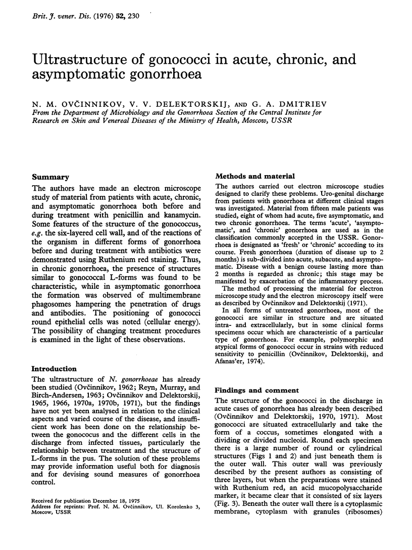
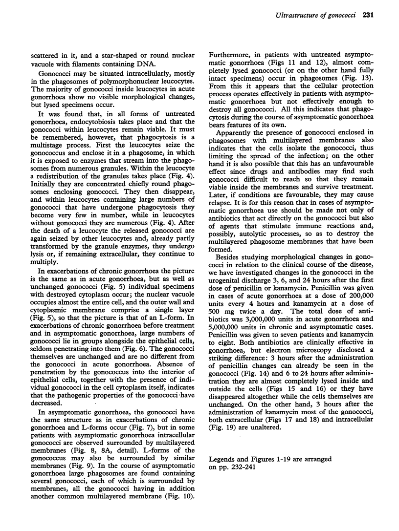
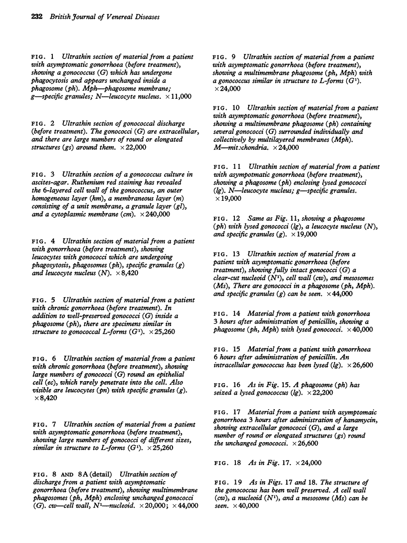
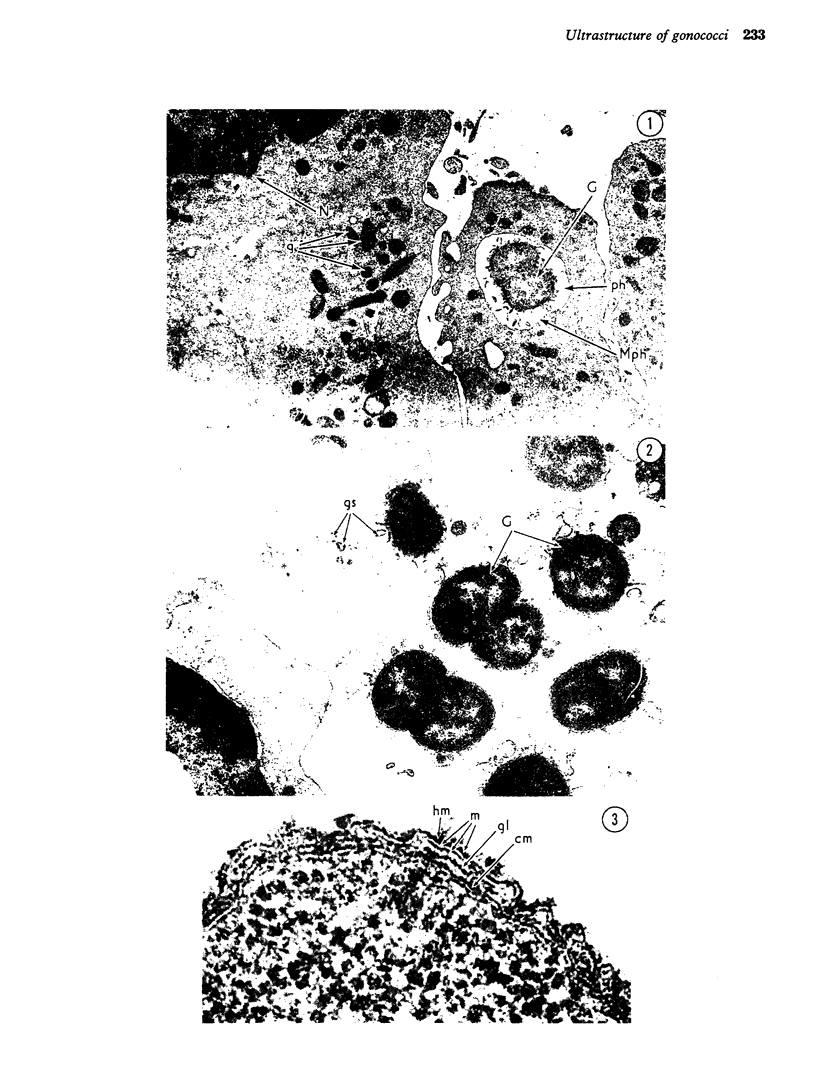
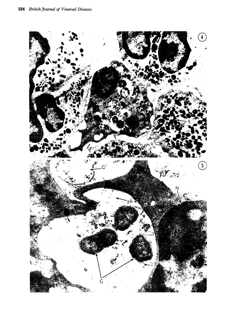
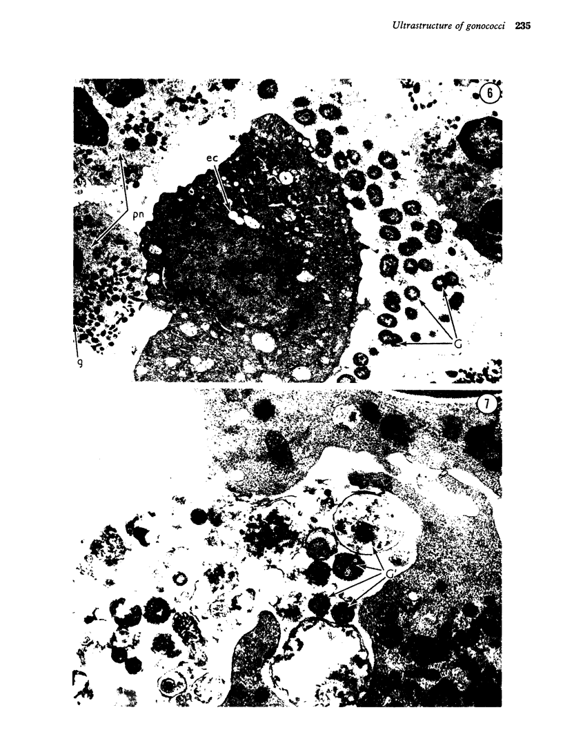
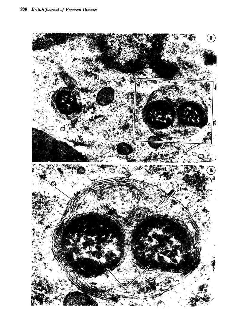
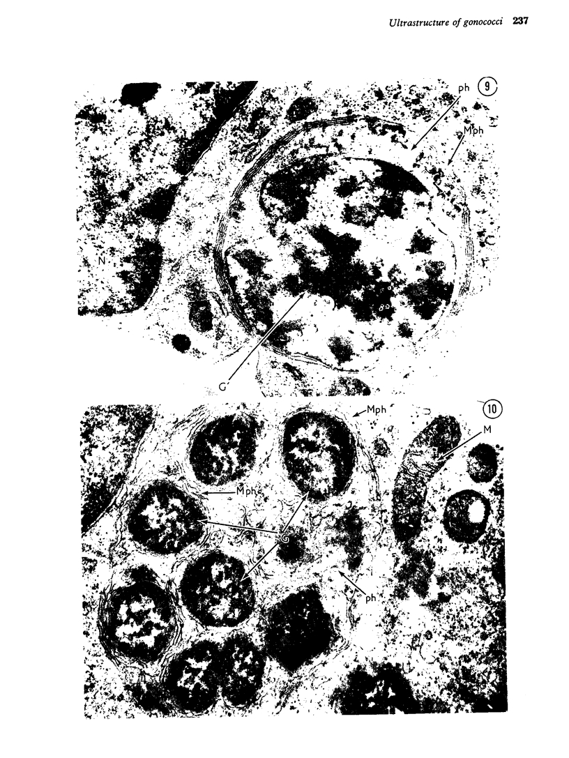
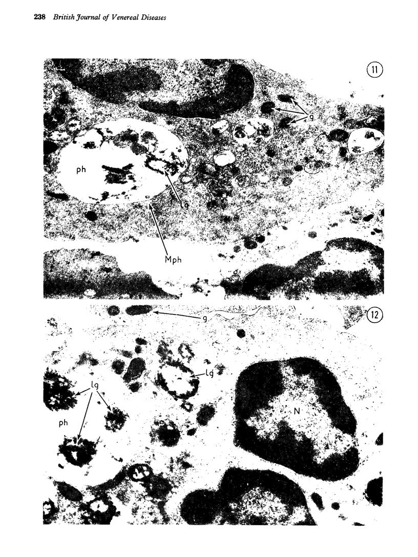
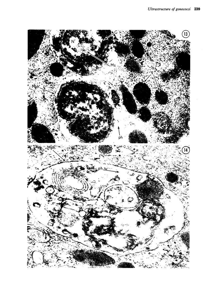
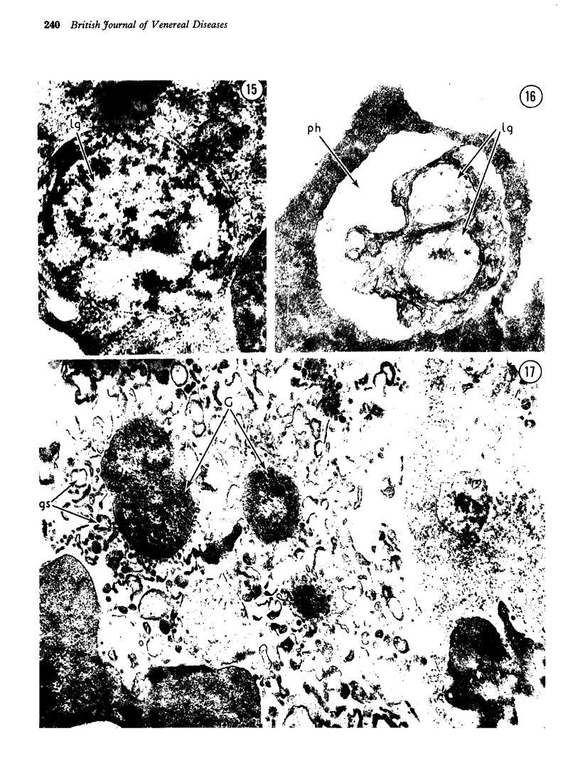
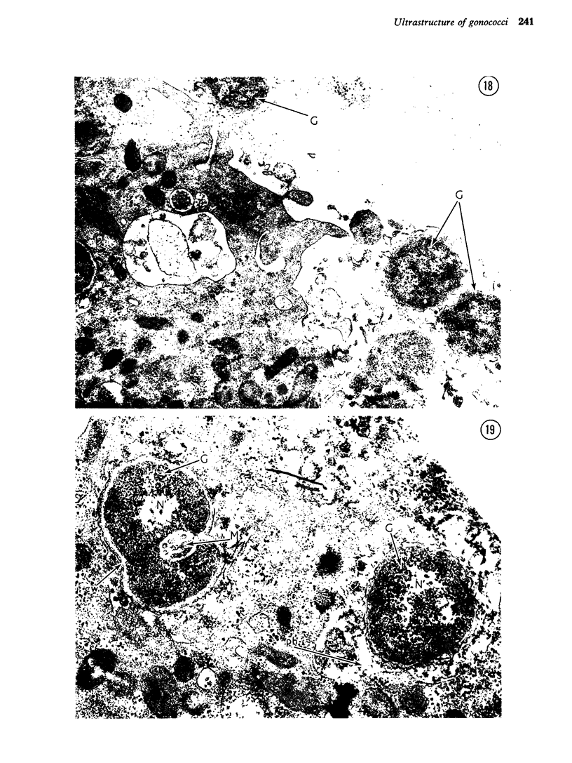
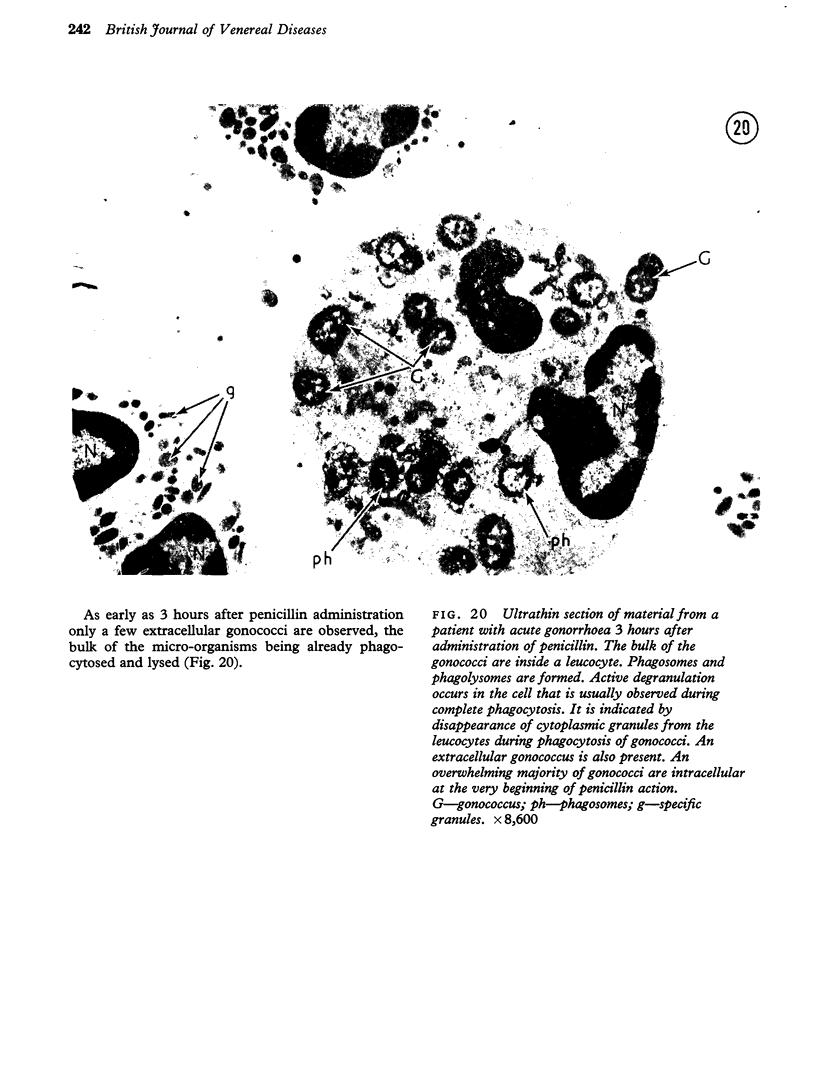
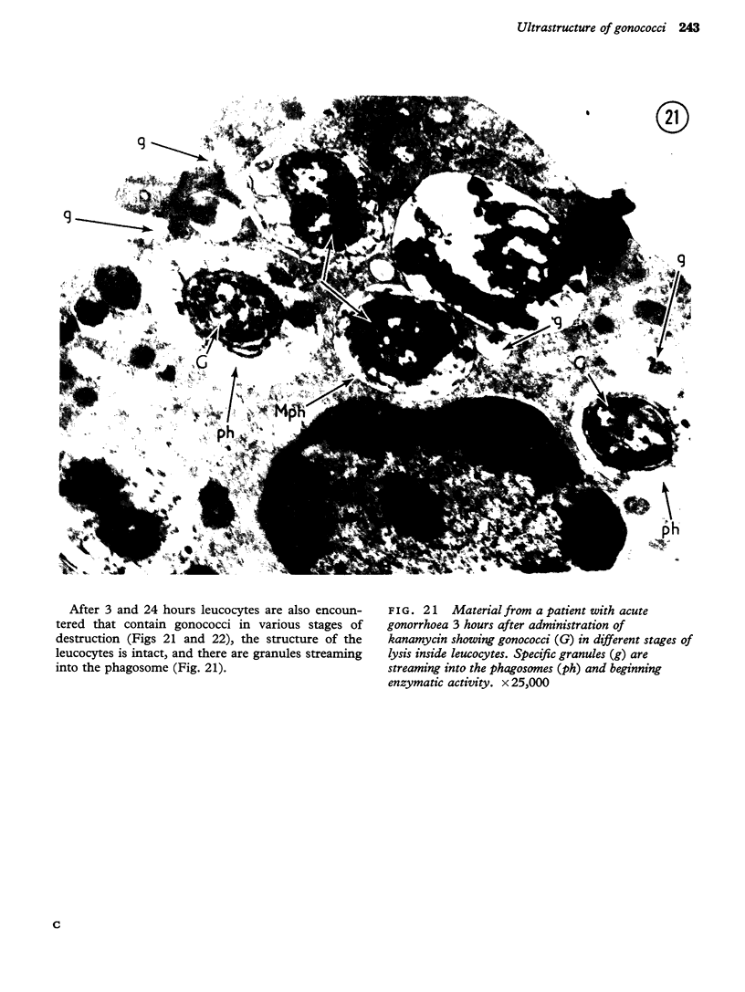
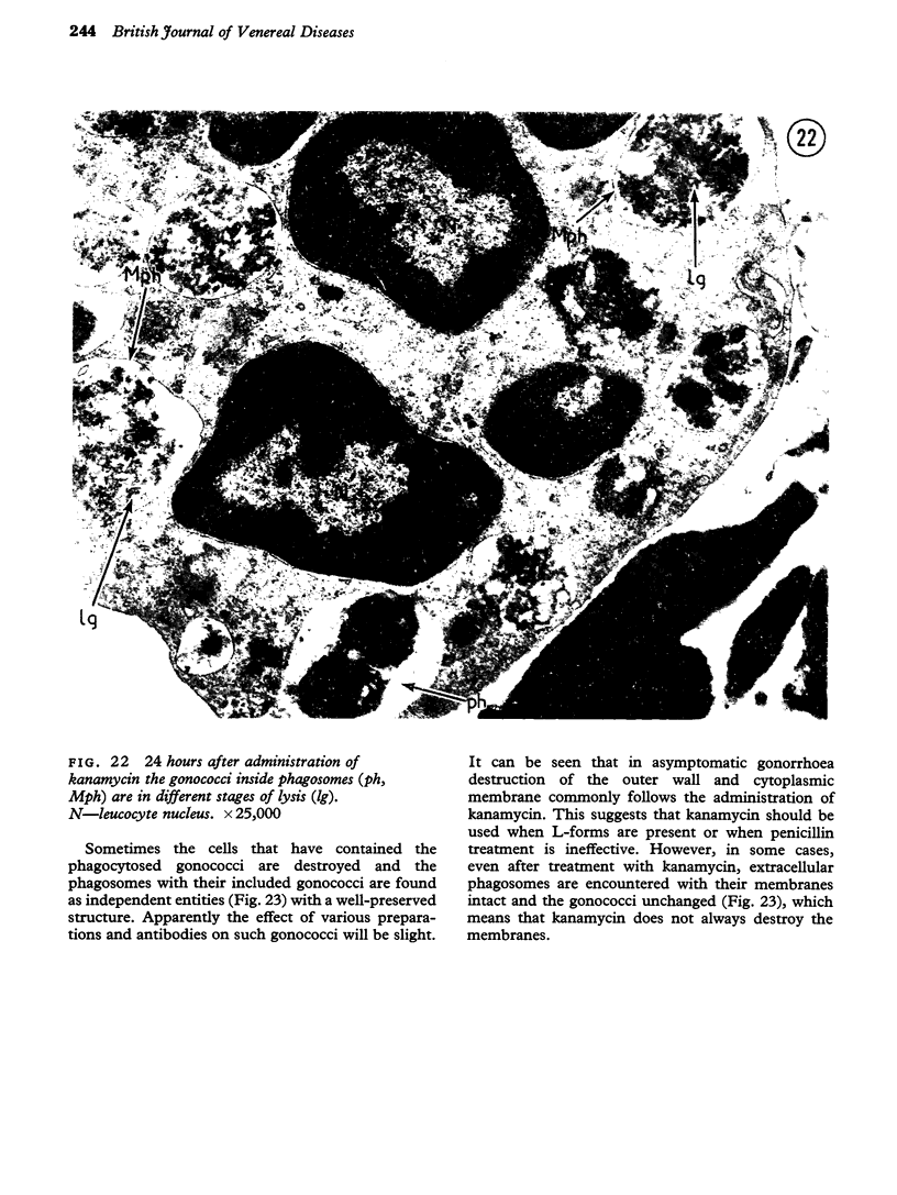
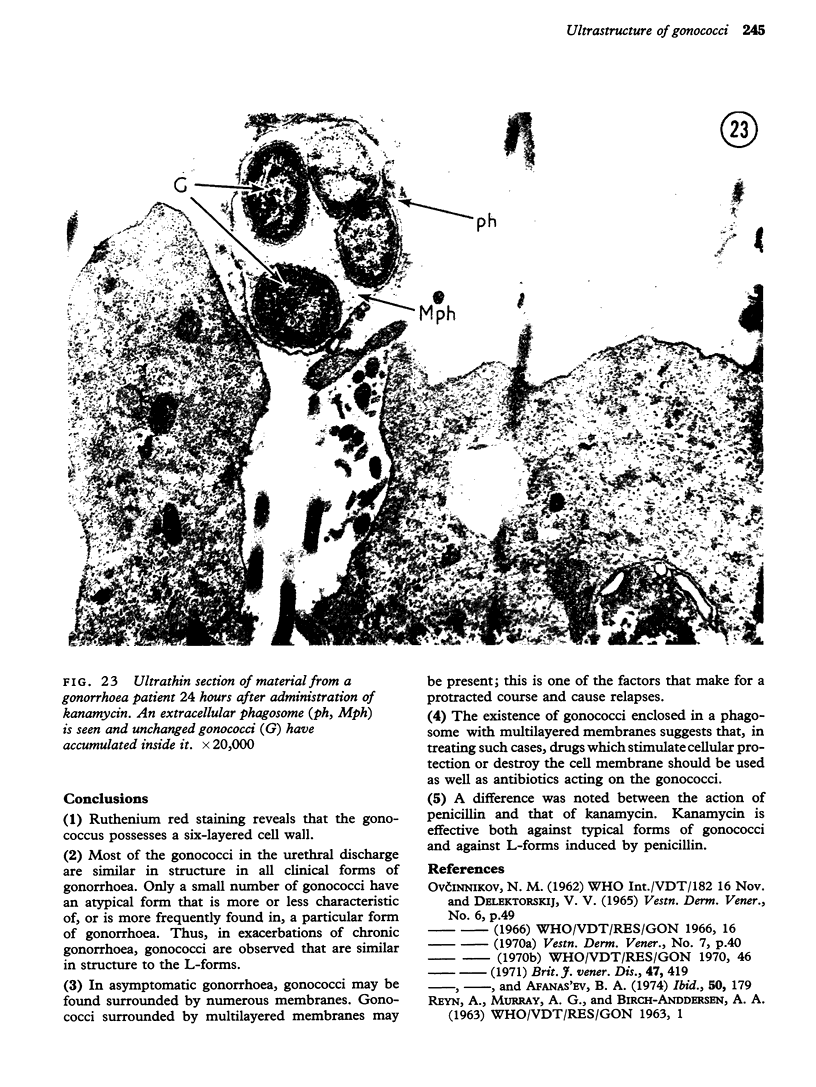
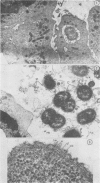
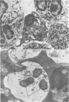
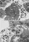
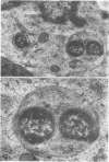
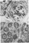
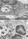
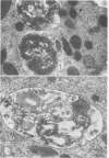
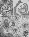
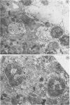
The authors have made an electron microscope study of material from patients with acute, chronic, and asymptomatic gonorrhoea both before and during treatment with penicillin and kanamycin. Some features of the structure of the gonococcus, e.g. the six-layered cell wall, and of the reactions of the organism in different forms of gonorrhoeae before and during treatment with antibiotics were demonstrated using Ruthenium red staining. Thus, in chronic gonorrhoea, the presence of structures similar to gonococcal L-forms was found to be characteristic, while in asymptomatic gonorrhoea the formation was observed of multimembrane phagosomes hampering the penetration of drugs and antibodies. The positioning of gonococci round epithelial cells was noted (cellular energy). The possibility of changing treatment procedures is examined in the light of these observations.
Full text
PDF