Abstract
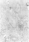
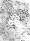
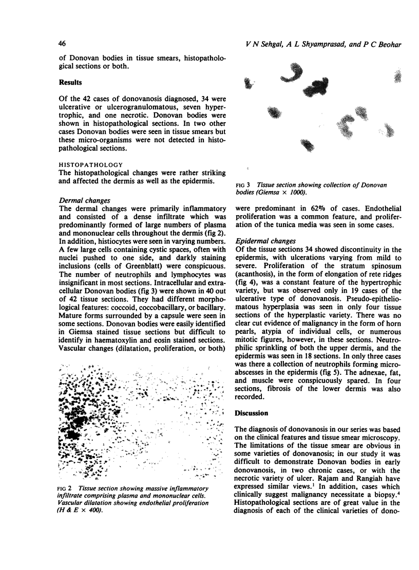
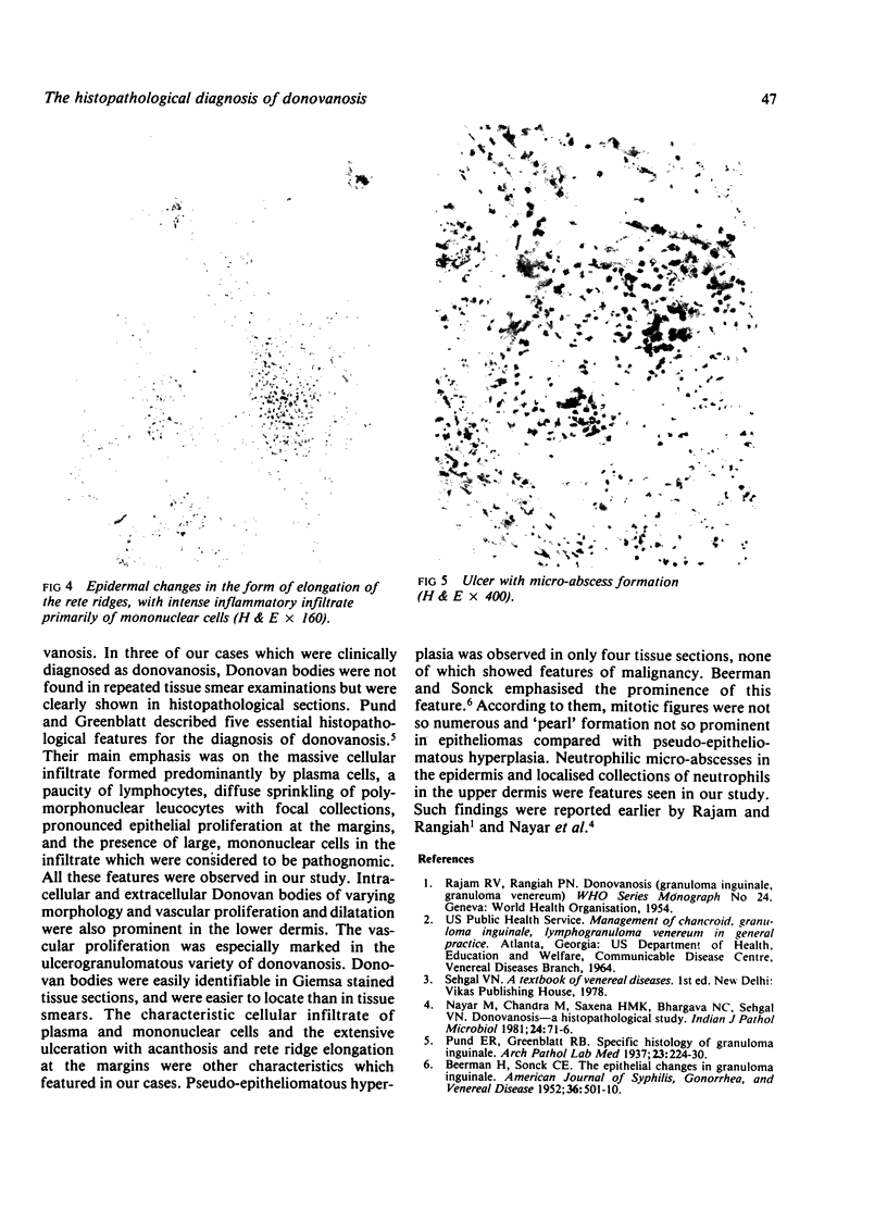
The role of histopathology in the diagnosis of donovanosis was assessed in 42 patients. There was heavy infiltration of the dermis with plasma and mononuclear cells but with few lymphocytes and neutrophils. The epidermis contained focal collections of polymorphoneuclear leucocytes. Endothelial proliferation and dilation of dermal blood vessels was striking. Intracellular and extracellular Donovan bodies were shown in Giemsa stained sections from 40 patients. Pseudoepitheliomatous hyperplasia was found in biopsy specimens from a few patients.
Full text
PDF
Images in this article
Selected References
These references are in PubMed. This may not be the complete list of references from this article.
- BEERMAN H., SONCK C. E. The epithelial changes in granuloma inguinale. Am J Syph Gonorrhea Vener Dis. 1952 Nov;36(6):501–510. [PubMed] [Google Scholar]
- Nayar M., Chandra M., Saxena H. M., Bhargava N. C., Sehgal V. N. Donovanosis - a histopathological study. Indian J Pathol Microbiol. 1981 Apr;24(2):71–76c. [PubMed] [Google Scholar]


