Abstract
Evolutionarily conserved genes often play critical roles in organismal physiology. Here, we describe multiple roles of a previously uncharacterized Class III metallophosphodiesterase in Drosophila, an ortholog of the MPPED1 and MPPED2 proteins expressed in the mammalian brain. dMpped, the product of CG16717, hydrolyzed phosphodiester substrates including cAMP and cGMP in a metal-dependent manner. dMpped is expressed during development and in the adult fly. RNA-seq analysis of dMppedKO flies revealed misregulation of innate immune pathways. dMppedKO flies showed a reduced lifespan, which could be restored in Dredd hypomorphs, indicating that excessive production of antimicrobial peptides contributed to reduced longevity. Elevated levels of cAMP and cGMP in the brain of dMppedKO flies was restored on neuronal expression of dMpped, with a concomitant reduction in levels of antimicrobial peptides and restoration of normal life span. We observed that dMpped is expressed in the antennal lobe in the fly brain. dMppedKO flies showed defective specific attractant perception and desiccation sensitivity, correlated with the overexpression of Obp28 and Obp59 in knock-out flies. Importantly, neuronal expression of mammalian MPPED2 restored lifespan in dMppedKO flies. This is the first description of the pleiotropic roles of an evolutionarily conserved metallophosphodiesterase that may moonlight in diverse signaling pathways in an organism.
Author summary
The MPPED2 gene maps to a human genetic locus associated with the WAGR syndrome, manifesting as Wilms Tumor, genitourinary abnormalities, and mental retardation. The mammalian protein is expressed in the brain, but the function of this protein in neurons is unknown. Here we have used Drosophila to identify various functions of the fly ortholog of MPPED2. We find that this gene product is expressed in a variety of tissues and can cleave phosphodiester bonds present in cyclic nucleotides. The protein is important for optimum life span in the fly, mediated by regulating the expression of immune genes. Longevity could be rescued by neuronal expression of the mammalian ortholog. The gene product is expressed in neurons in the antennal lobe of the fly and modulates responsiveness to odorants. Therefore, this evolutionarily conserved protein has multiple roles in the physiology of an organism, either by interacting with other proteins, or cleaving natural phosphodiester bonds, and further studies in mammals are warranted.
Introduction
Evolutionarily conserved proteins usually play a role in fundamental biological processes in an organism [1,2]. Sequence conservation at the level of amino acids, especially at the catalytic site of an enzyme, implies strong structural conservation and perhaps common activities in organisms separated by large tracts of time. If a gene linked to a genetic disease in humans has a counterpart in lower organisms more amenable to genetic manipulation, important insights into the gene’s function can be gained by either deletion or overexpression of the ortholog in a simpler organism. Such studies pave the way toward defining approaches that can be utilized in mammalian model systems later.
WAGR syndrome (Wilms’ tumor, aniridia, genitourinary anomalies, mental retardation) [3] is associated with interstitial deletions in a region in chromosome 11p13 [4,5]. This locus contains several genes such as WT1, BDNF [6], PAX6 [7] (all important during development), FSHB [8], RCN1, and MPPED2 (239FB) [9,10]. MPPED2 mRNA is predominantly expressed in the fetal brain [11], and our earlier biochemical characterization revealed that this protein belonged to the large family of metallophosphoesterases with promiscuous utilization of substrates [9,12]. We could identify orthologs in all mammals and other vertebrates, including Caenorhabditis elegans and Drosophila melanogaster [9,12]. Indeed, a closely related structural ortholog is found in Mycobacterium tuberculosis [13]. There are two variants of the MPPED family in higher organisms, MPPED1, and MPPED2 (239AB) [14], and these two mammalian orthologs show more than 80% similarity at the amino acid level, are expressed in the brain and share similar biochemical properties [9]. Recent studies have shown that MPPED2 is one of the risk loci for migraine [15], and is associated with altered systemic inflammation and increased organ dysfunction in trauma patients [16]. Further, MPPED2 is downregulated in glioblastoma [17] and thyroid neoplasia [18], acts as a tumor suppressor in breast cancer [19] and oral squamous cell carcinoma [20], and the MPPED2 gene is differentially methylated in colorectal cancer [21] and in individuals with gender incongruence [22]. These reports suggest diverse roles for MPPED2 in multiple tissue types.
The metallophosphoesterases represent a large and diverse group of proteins with similar structural folds that harbor two essential metal ions at the catalytic site [12]. Rat Mpped1 and Mpped2 hydrolyze phosphodiester bonds [9]. The substrates for many of these enzymes remain unknown, and while Mpped1 and Mpped2 can hydrolyze 3’,5’-cAMP, the product of this reaction is 3’AMP [9,23], in contrast to well-characterized mammalian cyclic nucleotide phosphodiesterases that produce 5’ AMP as the hydrolysis product [24]. Interestingly, 2’3’-cAMP is the preferred substrate for this group of enzymes, forming 3’ AMP and 2’ AMP as products [25], but the biological relevance of this reaction is unknown. MPPED1 and MPPED2 are also able to hydrolyze several colorigenic substrates, and such assays revealed that these enzymes could not hydrolyze monoesters but only diester-containing molecules such as bis-p-nitrophenyl phosphate (bis-pNPP), p-nitrophenyl phenylphosphonate (p-NPP) and TMP p-nitrophenyl ester (TMPP) [9].
D. melanogaster harbors a single ortholog of MPPED1/MPPED2, which we have named dMpped [9]. In FlyBase, this gene is annotated as Fbgn0036028 or CG16717. There is no information available on the role of this gene product to date. To decipher the role of dMpped in the fly, we have biochemically characterized this protein and found that it is a phosphodiesterase that can hydrolyze cAMP and cGMP. The major sites of expression of dMpped are in the adult brain, testis, and ovaries. Mutant flies harboring a deletion of this gene have a dramatically reduced lifespan, and aberrant responses to odorants. RNA sequencing (RNA-seq) analysis identified several misregulated pathways, including immune genes and odorant-binding proteins. Importantly, lifespan could be restored in the mutant fly by neuronal expression of the mammalian MPPED2 ortholog. Since we could detect expression of the mammalian proteins in cultured neurons, our findings will aid in identifying the pleiotropic role(s) of the mammalian proteins.
Results
dMpped is a metallophosphodiesterase expressed during development and in multiple adult tissues
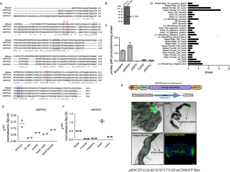
CG16717 is located on chromosome 3 at cytogenetic location 67C4. The CG16717 gene has two exons that are spliced to yield a single transcript, encoding for a protein of 300 amino acids. The entire protein coding sequence is present in exon 2 (http://flybase.org/reports/FBgn0036028.html). Since CG16717 is the only ortholog of the MPPED1/MPPED2 family of proteins in Drosophila, we will henceforth refer to it as dMpped (Drosophila metallophosphoesterase domain containing). Sequence alignment of dMpped, hMPPED1 (human MPPED1), hMPPED2 (human MPPED2) and rMpped2 (rat MPPED2) reveals that dMpped shares ∼50% sequence similarity to the mammalian orthologs (Fig 1A). All the critical residues required to classify dMpped as a metallophosphoesterase are conserved including the metal binding residue aspartate at amino acid position 49 (D49) and histidine at position 51 (H51; H67 in MPPED2) [23] (Fig 1A).
Fig 1. dMpped is a metallophosphoesterase and its expression is enriched in neurons.
(A) Sequence alignment of dMpped with human MPPED1, human MPPED2 and rat MPPED2. Red boxes highlight the conserved residues characteristic of metallophosphoesterases. * indicates residues D49 and H52 mentioned in the text. Highlighted in the blue box is the histidine residue that distinguishes MPPED1 from MPPED2. (B) A Coomassie R stained gel showing purified dMpped protein used for biochemical assays. (C) The catalytic activity of dMpped with the indicated colorigenic substrates (10 mM), in the presence of Mn2+ (5 mM) with dMpped (500 ng protein). Values represent the mean ± S.E. of duplicate determinations of experiments performed using two independent protein preparations. BispNPP, bis(p-nitrophenyl) phosphate; pNPPP, p-nitrophenyl phenylphosphonate; pNPP, p-nitrophenyl phosphate; TmPP, thymidine 5’-monophosphate-p-nitrophenyl ester; pNPPC, p-nitrophenylphosphoryl-choline. (D) Expression of CG16717 (dMpped) obtained from an analysis of RNAseq data from the modEncode data hosted in Flybase (www.flybase.org). Transcript levels are seen at low levels in many tissues, including the CNS and larvae (E) Expression pattern of dMpped across developmental stages of the fly. Embryos (2 h after egg laying; 50), third instar larvae (wandering; 10), pupae (21 h after pupae formation; 10), male and female flies (3 days old; 10 each) were collected and RT-qPCR was performed. dMpped transcript levels have been normalized to RpL32 transcript levels. The graph represents mean from 2 sets of samples collected independently. (F) Expression pattern of dMpped in different adult tissues. Testis and ovaries were collected from 50 male and female flies, respectively. The intestine (gut) was dissected from ∼ 50 flies in total, and 100 flies were used for muscles and brains. The graph represents mean from 2 sets of samples collected independently. (G) Confocal images of the ovary, testis, posterior midgut (R3-R4) and brain in pBAC(IT.GAL4)CG16717/UAS-mCD8GFP. pBac(IT.GAL4) enhancer trap element is positioned within the CG16717 gene. Ovaries show expression in the oviduct, the testis in secondary cells, and scattered enterocytes are GFP-positive in the posterior midgut. In the brain, strong expression is seen in the antennal lobe, marked with white asterix and in the optic lobe. The scale bar indicates 100 μm. 3D renderings of the brain sections of female brains are shown in S1 Movie, and enlarged images are shown in S1 Fig.
We expressed and purified dMpped to determine its biochemical properties. The purified protein migrated as a protein of 37 kDa corresponding to the predicted molecular weight (Fig 1B). Purified dMpped was tested for phosphoesterase activity against a panel of colorigenic substrates, and like its mammalian orthologs rMPPED1 and rMPPED2 [9], dMpped, in the presence of Mn2+ as the metal cofactor, showed phosphodiesterase activity and no detectable phosphomonoesterase activity (Fig 1C). dMpped hydrolyzed TmPP poorly, but not pNPPC, indicating some degree of specificity towards the kind of phosphodiester substrates it utilizes [9]. The enzyme could use several divalent cations, including Ni2+ (S1A Fig). In contrast, MPPED2 showed little detectable activity with Ni2+ [9]. The Km for Mn2+ and pNPPP was 1.8 mM and ∼10 mM, respectively, comparable to that of MPPED2 [9] (S1B and S1C Fig). dMpped was active against cyclic nucleotides and hydrolyzed 2’3’cAMP more efficiently than either 3’5’ cAMP or 3’5’ cGMP (S1D Fig).
modEncode data indicated that dMpped was expressed at all stages of development and in various tissues (Fig 1D). To explore the expression profile of dMpped we performed RT-qPCR. We observed that dMpped is expressed through development from embryos to adult flies (Fig 1E) and in the testis and ovary (Fig 1F). We noted high expression in the brain (Fig 1F), reminiscent of the reported expression of mammalian MPPED1 and MPPED2 transcripts in the brain. Expression was similar during aging (S2A Fig).
We utilized genetic approaches to monitor the expression of the dMpped gene, using IT-gal41111-G4 flies [26]. These flies have the GAL4 coding sequence inserted within the only intron of the dMpped gene (Fig 1G). Therefore, transcription is expected to be under the control of the same genetic elements and enhancers as dMpped. We crossed these flies to UAS-mCD8-GFP flies, adult tissues were dissected and imaged. In agreement with the RT-qPCR data, GFP expression was detected in the adult ovaries in the oviduct, accessory glands of the testis, and low levels in enterocytes in the intestine (Fig 1G).
We have used female flies throughout this study in order to avoid differences in behavioral responses that we described later. Expression in female brains was localized to specific neurons in the brain (S2B Fig; [27]). These neurons could represent olfactory sensory neurons, neurons involved in thermosensation and hygrosensation [28] and/or projection neurons which project from the antennae to the antennal lobe, where they innervate specific glomeruli. Female flies demonstrated significant expression in the optic lobes (S2C Fig).
Generation of of dMppedKO flies and RNAseq analysis
To determine the functional role of dMpped in Drosophila, we generated a null allele (dMppedKO) using ends-out homologous recombination [29–31] (S3A Fig). The gene deletion was verified by genomic PCR and RT-PCR (S3B and S3C Fig). In addition, we generated an antibody to dMpped, and western blotting confirmed the absence of protein in the heads of dMppedKO flies (S3D Fig). To rule out off-site target effects on adjacent genes, we monitored the expression levels of α-tubulin and furry and observed that they were not affected in dMppedKO flies (S3E Fig).
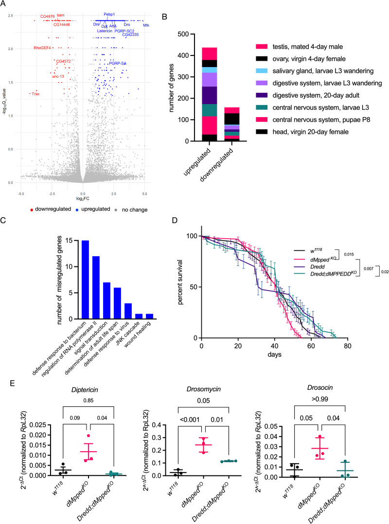
dMppedKO flies were homozygous viable and showed no obvious developmental or phenotypic differences compared to wildtype flies. Since the role of dMpped in flies is unknown, we adopted an unbiased approach by identifying misregulated genes in dMppedKO flies by RNAseq [32]. RNAseq analysis of 3-day old, virgin female flies revealed several differentially regulated genes in dMppedKO flies (Fig 2A), with 592 up-regulated genes and 260 down-regulated genes at q-value < 0.01. Most of the genes were of unknown function, but it was interesting to note that several misregulated genes were co-expressed with dMpped in various tissues (Fig 2B).
Fig 2. RNA-seq analysis and misregulation of genes associated with defense pathways.
(A) A volcano plot showing protein-coding genes altered in 35-day old dMppedKOflies. The Log2 fold change (Log2FC) values are plotted on the x-axis and the negative log (base 10) of the Q value (-Log10Q_value) on the y-axis. Blue (upregulated) and red (downregulated) dots represent genes with Q-values <0.05 and Log2 fold change values of more or less than one-fold. (B) The tissue distribution of differentially regulated genes. Misregulated genes (852, with 592 up-regulated and 260 down-regulated) were analysed by DGET (http://www.flyrnai.org/tools/dget/web/) based on published analysis of the Drosophila transcriptome [88]. (C) KEGG analysis of genes with Log2FC >2. Apart from several genes of unknown function, many misregulated genes were associated with defense response and immunity. (D) dMppedKO flies were crossed with Dredd hypomorph flies (P[39]DreddEP1412 w1118) and life span monitored in virgin female flies. Flies (w1118, n = 76; dMppedKO n = 66; Dredd, n = 40; Dredd;dMppedKO, n = 59) were from at least three independent experiments. Log-rank test was used to compare survival across genotypes. Values shown at each time point are the mean ± SD, and the line represents the average across all experiments. p values are shown and compare the average life span across all three replicates of each genotype. (E) RT-qPCR of AMPs from RNA prepared from whole flies. Reducing Dredd activity in dMppedKO flies reduced expression the AMPs, indicating that dMppedKO flies showed misregulation of the IMD pathway. Each data point represents RNA prepared from 10 female flies, and values shown are the mean ± SD of three independent experiments. Data were analyzed by ANOVA and corrected for multiple comparisons using Tukey’s test. p values are indicated across genotypes.
A large set of genes important in defense response to bacteria (Diptericin, Dpt; Drosomycin, Drs; and Drosocin were upregulated in dMppedKO flies (Fig 2A and 2C). Inflammation is a hallmark of faster aging and shorter lifespan [33–35]. Interestingly, dMpped is one of the major genes upregulated in S2 cells following infection with Drosophila C virus [36]. Thus, these findings appear to place dMpped as a link between innate immune pathways and life span in the fly.
Indeed, the life span of dMppedKO flies was reduced when compared to w1118 flies (Fig 2D). Changes in levels of genes in the IMD and Toll pathways could account for the upregulation of antimicrobial peptides (AMPs). IMD associates with the Fas-associated death domain protein (FADD), that recruits the caspase-8 homolog death related ced-3/Nedd2-like caspase, DREDD. DREDD cleaves IMD, allowing interaction with Drosophila inhibitor of apoptosis-2 (dIAP-2), that ubiquitinates and stabilizes IMD. Recruitment of transforming growth factor β (TGF-β)-activating kinase 1 (TAK1) mediates phosphorylation of the IκB kinase (IKK) and Jun nuclear kinase (JNK). IΚK phosphorylates the N-terminal domain of Relish (Rel, the NFκB ortholog in flies), which, following DREDD-mediated cleavage of the C-terminus of Rel, allows nuclear migration of Rel and the induction of AMP transcription. To determine if the overexpression of AMPs contributed to life span reduction, we cross dMppedKO with Dredd hypomorph flies. Life span was restored in dMppedKO flies (Fig 2D), and a reduction in transcript levels of AMPs was seen (Fig 2E). Thus, overexpression of AMPs in the dMppedKO flies could contribute to the reduced life span in flies, as suggested in earlier studies [37,38].
Neuronal expression of dMpped regulates fly lifespan
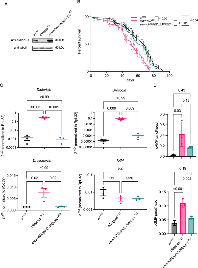
Given that dMpped is expressed at high levels in the brain, and neuronal regulation of lifespan is well studied [37,39–41], we used a pan-neuronal driver, elav-GAL4, to express dMpped in dMppedKO flies, after backcrossing flies used in experiments for 9 generations against w1118 flies. This was done to ensure no other mutations were present in the dMppedKO flies in subsequent experiments that describe phenotypes related to life span with comparisons made to w1118 flies. We confirmed expression of dMpped by western blotting of protein extracts prepared from the brain (Figs 3A and S4). Further, restoration of dMpped expression in neurons was sufficient to rescue the reduced lifespan of dMppedKO flies (Fig 4B).
Fig 3. Neuronal expression of dMpped determines fly lifespan, AMP expression and cyclic nucleotide levels in the brain.
(A) Western blot to confirm that dMppedKO is a protein null allele, and expression of dMpped in the brain of flies where dMpped expression is driven in the neurons. The blot was re-probed with an anti-tubulin antibody to normalize protein loading. (B) Survival curves of backcrossed flies of indicated genotypes. Flies (w1118, n = 66; dMppedKO, n = 66; elav-Gal4>dMpped; dMppedKO, n = 30) were from at least three independent experiments. Log-rank test was used to compare across genotypes. Values shown at each time point are the mean ± SD, and the line represents the average across all experiments. p values are shown and compare the average life span across all three replicates of each genotype. (C) RT-qPCR to confirm upregulation of AMPs in dMppedKO flies. Levels were restored following neuronal expression of dMpped in dMppedKO flies. Each data point represents RNA prepared from 10 female flies, and histograms correspond to the mean value ± SD of three independent experiments. Data were analyzed by ANOVA and corrected for multiple comparisons using Tukey’s test. p values are indicated across genotypes. (D) Cyclic AMP and Cyclic GMP levels were measured in the heads of flies of the indicated genotypes. Levels of cAMP and cGMP were significantly higher in dMppedKO flies, correlated with reduced phosphodiesterase activity in flies deleted for dMpped. Extracts were prepared from 10 female fly heads, and histograms correspond to the mean value ± SD of three independent experiments. Data were analyzed by ANOVA and corrected for multiple comparisons using Tukey’s test. p values are indicated across genotypes.
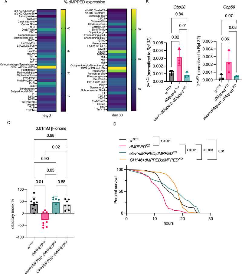
Fig 4. dMpped regulates expression of Obps and affects odorant-driven behavior and tolerance to desiccation.
(A) Expression of dMpped in specific neurons in the fly brain. Data was extracted from single cell RNAseq analysis [47] and shown as a heat map indicating expression levels in flies of 3 and 30 days of age. The highest expression is seen in OPNs in agreement with Fig 1G. (B) RT-qPCR of Obp28 and Obp59 indicating misregulation in dMppedKO flies with levels restored to that seen in control flies following neuronal expression of dMpped in dMppedKO flies. At least 10 flies were used for each experiment and data shown is across 3 independent experiments. Data were analyzed by ANOVA and corrected for multiple comparisons using Tukey’s test. Values are mean ± SD and p values across genotypes are shown. (C) Response of flies to varying concentrations of β-ionone, an attractant that binds to Obp28. dMppedKO flies show no preference in the Y-maze test at low concentrations (0.01mM) of β-ionone, and the behavioral response was restored in dMppedKO flies with the expression of dMpped restored pan neuronally or specifically in olfactory projection neurons. Flies (w1118, n = 104; dMppedKO, n = 89; elav-Gal4>dMpped; dMppedKO, n = 44; GH>dMPPED;dMPPEDKO, n = 64) were from at least six independent experiments. Data were analyzed by ANOVA and corrected for multiple comparisons using Tukey’s test. Values are mean ± SD and p values are shown. (D) Flies were subjected to desiccation and the time taken for to die was monitored. Restoration of expression of dMpped in dMppedKO flies restored desiccation resistance to that seen in wildtype flies. Flies per genotype are pooled from at least three independent experiments. Log-rank test used for comparing wild type (w1118, n = 92); dMppedKO (n = 95) flies; elav-Gal4>dMpped; dMppedKO (n = 49) and GH146>dMPPED;dMPPEDKO, n = 50. p values across genotypes are shown in the graph.
We then measured levels of AMPs in the head of wildtype, dMppedKO and neuronally rescued flies and found that the elevated levels of Diptericin, Drosocin and Drosomycin were restored to that seen in wildtype flies (Fig 3C) and were correlated with an increased life-span in elav>dMpped;dMppedKO flies (Fig 3B). Levels of Turandot M (TotM) transcript, which is a target of the Jak/Stat pathway and also induced in response to bacterial infection [42], were unchanged in dMppedKO flies, indicating that dMpped specifically modulated the IMD and Toll pathways.
Metallophosphoesterases are promiscuous in their substrate utilization, but the diesterases specifically cleave only molecules with diester bonds. Since dMpped could hydrolyze cAMP and cGMP (S1D Fig), we measured both cAMP and cGMP levels in the brains of wildtype, dMppedKO, and dMppedKO flies expressing dMpped in neurons. We detected higher levels of both cyclic nucleotides in the brains of dMppedKO flies (Fig 3D), which was restored on neuronal expression of dMpped. Interestingly, cyclic nucleotide levels are associated with life span in Drosophila. Dunce encodes a cAMP phosphodiesterase, and knock-out female flies (which should harbor elevated cAMP levels) show a reduction in life span [43], in agreement with our results. Cyclic nucleotides can bring about their action by activating cAMP or cGMP-dependent kinases. Interestingly, reduced levels of the cGMP-dependent protein kinase (PKG) in flies increase life span [44]. Further, suppression of neuronal cGMP levels in C. elegans results in extended life-span in a FOXO-dependent manner [45]. Therefore, we propose that neuronally elevated cGMP in dMppedKO flies may also contribute to life span regulation in the fly.
Attractant perception is compromised in dMppedKO flies
Among the genes significantly upregulated in dMppedKO flies in the RNAseq analysis were olfactory binding proteins (Obps; S5 Fig), a diverse group of proteins that show odorant binding in vitro but also have a broader role in insects than previously envisaged [46]. There are 52 Obps localized mainly to the sensilla found in insect antennae and are thought to transport hydrophobic odorants across the aqueous sensillar lymph to olfactory receptors. We noted that from single cell RNAseq analysis of the Drosophila brain [47], dMpped was expressed at high levels in the olfactory anterodorsal, lateral, and ventral lineage projection neurons (Fig 4A) in agreement with our expression data in the antennal lobe (Fig 1F). Furthermore, an increase in levels of dMPPED transcripts are seen in OPNs on day 30 and in peptidergic neurons (Fig 4A). Peptidergic neurons control a number of processes including metabolism, circadian timing, and cues that regulate food search, aggression and mating [48]. Therefore, increased expression of dMPPED in these neurons could regulate these functions in aging flies.
We focused on the implications of the overexpression of two Obps in dMppedKO flies, Obp28 and Obp59. Obp28a binds the floral odor attractant β-ionone with micromolar affinity, and deletion of the Obp28a gene resulted in reduced olfactory preference at low concentrations of β-ionone [49]. We first validated the overexpression of Obp28 in dMppedKO flies by RT-qPCR (Fig 4B) and saw that expression of dMpped in the neurons of dMppedKO flies could restore transcript levels of Obp28 to those seen in control flies. We then tested the ability of flies to move towards β-ionone in a Y-shaped olfactometer [49] at two different concentrations of β-ionone. While attraction to the odor was similar in control and dMppedKO flies at the higher (0.05 μM) concentration (S5B Fig), we observed that at low concentrations of β-ionone (0.01 μM) dMppedKO flies showed no preference for a movement toward the odorant-containing arm. These results paradoxically mimic those seen in Obp28a-deleted flies, suggesting that at low concentrations of β-ionone, Obp28a-mediated responses depend on a balanced expression of Obp28. Alternatively, one could speculate that the interaction of Obp28 with the receptor could depend on the presence of neuronally expressed dMpped, either by direct interaction with the Obp or its receptor, and this interaction could be altered in the absence of dMPPED.
To confirm that the behavior towards odorants in dMppedKO flies was explicitly in response to β-ionone and mediated by a unique set of neurons, we tested the behavior of dMppedKO flies to ammonia and acetophenone. High ammonia concentrations are repellant to wild-type flies (S5C Fig). Ammonia at a lower concentration is a strong attractant for flies, and attraction behavior is mediated by olfactory sensory neurons that express ionotropic receptor IR92a [50] and do not require an Obp. Acetophenone is a repellant and induces behavioral responses mediated via Obp56f, Obp56h, and Obp83a [51], none of which were misregulated in dMppedKO flies (S5A Fig). We observed that dMppedKO flies responded similarly to control flies with these odorants at concentrations tested in earlier studies [50,51] (S5C and S5D Fig).
We then focused on Obp59a, upregulated ∼ 4.5-fold in the RNAseq analysis (S5A Fig). Loss of Obp59a leads to an increase in desiccation resistance [52]. We validated the upregulation of Obp59 by RT-qPCR in dMppedKO flies, and expression was restored to control levels on expression of dMpped in the neurons of dMppedKO flies (Fig 4B). In agreement with the results seen in Obp59 knockout flies, we saw increased sensitivity to desiccation in dMppedKO flies, which was complemented by reducing levels of Obp59a following neuronal expression of dMpped (Fig 4D).
To rescue the expression of dMPPED in olfactory neurons, we used the GH146-Gal4 driver line, which labels a broad set of second-order olfactory projection neurons with little background expression [53]. We tested olfaction at low concentrations of β-ionone (0.01 μM) in dMppedKO flies expressing dMPPED in olfactory neurons using the GH146-Gal4 line. We found that the expression of dMPPED in olfactory projection neurons allowed them to sense 0.01 μM β-ionone (Fig 4C). Further, these complemented flies were not as sensitive to desiccation as the dMppedKO flies (Fig 4D). Therefore, expression of dMPPED in olfactory neurons is required to allow specific odorant perception and optimum response to desiccation stress.
In conclusion, the expression of dMpped in possibly very specific neurons alters odorant perception and hygrosensation in flies, emphasizing the pleiotropic roles of dMpped in the physiology of the fly.
The mammalian ortholog of dMpped restores life span in dMppedKO flies
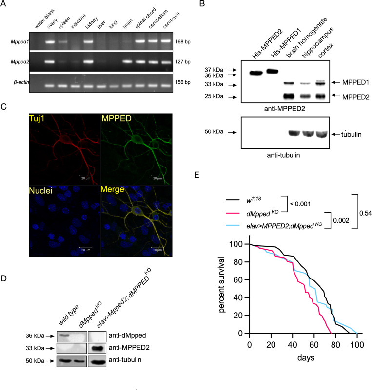
The MPPEDs are highly evolutionarily conserved proteins across metazoans and the sequence similarity between the fly and the mammalian MPPED proteins is ∼50% (Fig 1A). The reduced lifespan of dMppedKO flies offered an excellent model to test possible functional conservation between dMpped and its mammalian ortholog, MPPED2. We first monitored the expression of Mpped1 and Mpped2 in mouse tissues by PCR and observed the expression of both these genes in the ovary, kidney, and various brain regions (Fig 5A). Interestingly, Mpped2 was also expressed in the heart. To confirm protein expression in the brain, we performed western blot analysis with a monoclonal antibody raised to Mpped2 and showed that this antibody could recognize both Mpped1 and Mpped2 (Fig 5B). We could detect robust protein expression (s) in brain homogenates and higher levels in the cortex compared to the hippocampus (Figs 5B and S5). Two bands of molecular weight 33 kDa and 25 kDa could be observed. A perusal of the Allen Brain Atlas (http://mouse.brain-map.org) indicated that expression of Mpped1 was highest in the isocortex, olfactory areas, hippocampal formation, and cortical subplate. No data was provided for Mpped2. However, based on the molecular weight predicted of Mpped1 and Mpped2, either Mpped2 is the major protein expressed in the brain, or post-translational proteolysis of Mpped1, initiation of translation of rodent Mpped1 at Met25 or Met31 (Fig 1A), or post-translational proteolysis of both proteins cannot be ruled out.
Fig 5. Mammalian ortholog of dMpped regulates lifespan in flies.
(A) Expression of mMPPED1 and mMPPED2 in mouse tissues. RNA was prepared from the indicated tissues, and expression of mMPPED1, and mMPPED2 monitored following reverse transcription and PCR using specific primers. Expression of β-actin was used to check equivalent cDNA synthesis across tissues. (B) Western blot analysis with purified mMPPED1, mMPPED2 and lysates prepared from total mouse brain or homogenates prepared from the hippocampus and cortex using a monoclonal antibody raised to rat MPPED2. Blots were probed with the MPPED monoclonal antibody and protein loaded in each lane normalized to tubulin. Recombinant, purified, histidine-tagged mMPPED1 and mMPPED2 were used as controls and migrate higher than the endogenous protein present in brain lysates due the hexahistidine tag at the N-terminus of the proteins that facilitated purification. Data is representative of experiments performed with two independently prepared homogenates. The two bands seen in brain homogenates migrate at sizes predicted for mMPPED1 and mMPPED2. (C) Immunocytochemical analysis to show expression of mMPPED in neurons prepared from the cortex of 1-day old mice. (D) Western blot performed homogenates prepared from the brain of flies of the indicated genotypes. Blots were probed either with the monoclonal antibody raised to mammalian Mpped2 or the polyclonal antibody raised to dMpped. Protein loading was normalized by using an antibody to tubulin. The data shown is representative of experiments performed with independently prepared homogenates at least twice. (E) Flies (w1118, n = 66; dMppedKO, n = 66; elav-Gal4>MPPED2; dMppedKO, n = 34) were from three independent experiments. Log-rank test was used for comparing across genotypes. p-values across genotypes are shown in the Figure.
We prepared neuronal cultures from the mouse cortex, and immunofluorescence indicated co-expression of Tuj1(expressed only in the neurons) and Mpped1 and/or Mpped2 (Fig 5C). This is the first demonstration of the expression of these proteins in mammalian neurons.
To determine whether mammalian Mpped2 could restore life span in flies, we expressed rat MPPED2 neuronally in dMppedKO flies and confirmed expression in the fly brain by western blot analysis (Fig 5D). Interestingly, lifespan in flies expressing MPPED2 was restored to wildtype levels in dMppedKO flies (Fig 5E). Therefore, mammalian MPPED2 has functional conservation with dMpped, suggesting that the fly could be used to delineate further the mechanisms by which MPPED2 and/or MPPED1 regulate diverse pathways in different mammalian tissues.
Discussion
Here, we present the first characterization of the fly ortholog of mammalian MPPED1/MPPED2 proteins and show how its neuronal expression regulates lifespan, odorant perception and hygrosensation in the adult fly. While several genes that regulate lifespan in the fly are also critical for normal development, dMpped is not essential during development but plays a role in aging in the adult fly. dMpped can hydrolyze 2’3’-cAMP to 3’AMP like the mammalian orthologs (S1D Fig). CNPase hydrolyzes 2’3’-cAMP in mammals, and accumulation of this cyclic nucleotide leads to an increased susceptibility to brain injury and neurological disease [54]. Whether an additional role of dMpped relates to its catalytic activity and whether 2’3’ cAMP plays a role in the fly brain remains to be investigated.
The importance of cAMP in dopamine-mediated signaling in the mushroom body [55], and odor-induced cAMP production in olfactory sensory neurons in the antenna plays a central role of olfactory conditioning in Drosophila [56]. Single-cell RNAseq data showed low expression of dMpped in dopaminergic neurons (Fig 4A) but robust expression in olfactory projection neurons. Projection neurons respond to a broader range of odors than their corresponding olfactory receptor neurons. It is conceivable that the elevated levels of cAMP/cGMP in these neurons in dMppedKO flies could modify output and memory in an odor-specific manner.
There is increasing evidence that the aging immune system can lead to low-grade inflammation, which is associated with increased mortality. We have shown an unexpected role of a metallophosphoesterase in controlling lifespan by modulating the levels of AMPs during aging. The higher levels of AMPs detected in whole dMppedKO flies could originate from the fat body following innervation by neurons in which dMpped is expressed. In the Malpighian tubules, increased cGMP levels can lead to the translocation of the transcription factor Relish to the nucleus and activation of the IMD pathway [57]. Therefore, elevated levels of cAMP and cGMP in the heads of dMppedKO flies could lead to activation of IMD in a cell non-autonomous manner in the fat body fragments present near the brain.
Is there a link between odorant-binding proteins and AMP production? Growing evidence suggests that Obps are expressed in several tissues, including hemocytes from the mosquito and fat bodies [58]. We have observed that specific Obps are expressed in hemocytes prepared from Drosophila after sterile injury [59]. A recent study has shown that an Obp28a mutant Drosophila line perished faster following sterile thoracic injury, with lower melanin deposition at the wound site [60]. Further, expression of Obp28a was significantly reduced in larvae reared axenically, while conventionally reared flies with endogenous microbiota showed increased expression of lozenge (a gene that controls the lineage specification of prohemocytes into crystal cells that release melanin at the site of injury)[61]. Crystal cells are important in Drosophila innate immunity and stress responses, even to gaseous chemicals such as CO2 [62]. Therefore, the production of AMPs may be linked to Obp expression in dMppedKO flies, which in turn impacts on the reduced lifespan seen in these flies.
An additional role of odor perception and immunity lies in the finding that upon olfactory stimulation, specific olfactory neurons induce the secretion of GABA from a subset of neurosecretory cells. GABA then binds to metabotropic GABAB receptors expressed on blood progenitors signal causing high cytosolic Ca2+, required for progenitor maintenance [63]. Since dMpped is expressed in olfactory neurons, the activation of these neurons could be altered in its absence, which in turn may alter hematopoiesis.
Expression of Obps is affected both by age and nutrient availability in flies [64]. An Obp83b knock-out strain is long-lived, and longevity in these flies appears to be largely controlled by a diet-independent pathway. Further, female flies were more resistant to hyperoxia and resistant to starvation. In dMPPEDKO flies, Obp83b shows a trend of upregulation (∼ 4-fold, p value 0.05), and this increase in expression could reduce the lifespan, based on studies with Obp83 knock-out flies. Links between odorant perception and longevity need to be explored further, and the evolutionary conservation of the MPPED family suggests that such associations may also emerge in mammals.
Given the moonlighting activities of the metallophosphoesterase family of proteins [12,65], dMpped could serve as a scaffolding protein. From the analysis of the Drosophila interactome described by high throughput approaches [66], only one gene product, Sh3px1, was reported to interact with dMpped with some confidence. This protein is the single fly ortholog of the human sorting nexin 9 family known to function in vesicular sorting [67,68]. SH3PX1 has been found to regulate the formation of lamellipodia, tubules, and long protrusions in S2 cells [69]. In the fly, SH3PX1 localizes to neuromuscular junctions where it regulates synaptic ultrastructure [70]. Neurotransmitter release was significantly diminished in SH3PX1 mutants and functional interactions with Nwk, a conserved F-BAR protein that attenuates synaptic growth and promotes synaptic function in Drosophila, were observed [71]. Interaction with Nwk was via the SH3 domain of SH3PX1, which recognizes proline-rich sequences. The co-expression of dMpped and SH3PX1 in neuronal tissue may allow for functional interaction that could modulate Nwk action, since the SH3 domain in SH3PX1 could interact with the PXXP motif in dMpped (residues 4–7; Fig 1A) and sequester it from Nwk.
Recently, a role for Sh3px1 in regulating the innate immune response was described that was mediated by its interaction with the autophagy protein, Atg8a and Tyk/Tab2 [72]. Tak1 and Tab2, a co-activator of Tak1, interact with Atg8 and are selectively targeted for autophagy to regulate the IMD pathway. The predicted interaction of dMpped and Sh3px1 could indicate links between the regulation of the IMD pathway, autophagy machinery and dMpped.
dMpped shows a marginally higher sequence identity to hMPPED1 than hMPPED2 (48.7% to 47.6% respectively). Are there proteins that have been shown to interact with MPPED1/MPPED2 which may have implications as interacting partners of dMpped? In an extensive screen across the human genome involving affinity purification and mass spectrometry of interacting proteins (https://bioplex.hms.harvard.edu), the only common proteins that interacted with both MPPED1 and MPPED2 were transcription factors NR2F1 and NR2F6. These nuclear receptor factors are not conserved in Drosophila, which has far fewer nuclear-receptor genes than any other model organism [73]. However, a close homolog of these proteins in mammals is NR2F3, or COUP-TF1, which has the ortholog seven-up (svp) in the fly [73]. svp has crucial roles in neuronal development during embryogenesis and in the development of photoreceptor cells [74]. COUP-TF1 is also required for neuronal development and axon guidance [75]. Interactions, either direct or genetic, between dMpped and svp would be an interesting line of study in the future.
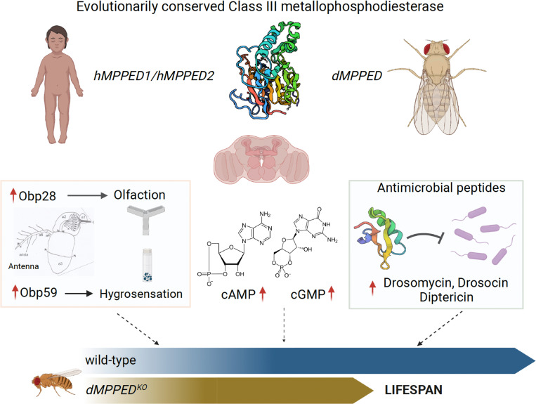
In summary, we have identified a new player in regulating several physiological responses, which may impinge on longevity in flies (Fig 6). The presence of orthologs of dMpped in higher animals suggests that the role of this protein in mammals is worthy of study. Our analysis reveals that this protein could serve as a focal point for interaction and cross-talk with several pathways, either through direct interaction or by modulating the activity of a few proteins, which could then impact more globally.
Fig 6. Schematic showing pleiotropic roles of dMPPED.
Class III metallophosphoesterases are evolutionarily conserved and members of the MPPED family are neuronally expressed in the brain of mammals and flies. dMPPEDKO flies show misregulation of Obps, elevated levels of cAMP and cGMP and higher expression of AMPs. All these changes can influence longevity, with dMPPEDKO flies showing a decreased life span. The Figure was created with BioRender.com.
Materials and methods
Cloning and mutagenesis of dMpped
The full-length coding region of dMpped was amplified from cDNA prepared from whole flies using dMpped_MfeI_Fwd and dMpped_XhoI_Rvs primers (S1 Table), digested with XhoI and cloned into EcoRV and XhoI digested pBKSII vector to generate pBKS-dMpped. The clone was verified by sequencing (Macrogen, South Korea). The MfeI-XhoI fragment from pBKS-dMpped was cloned into EcoRI and XhoI digested pPROExHT-B vector to obtain pPRO-dMpped. The same fragment was cloned into EcoRI and XhoI digested pUAST-attB vector to obtain pUAST-attB-dMpped.
Expression and purification of dMpped
The pPRO clones of dMpped were transformed into E. coli BL21DE3 and proteins were expressed as described earlier [9]. Gel filtration of purified protein was carried out in buffer containing 50 mM Tris/HCl, 5 mM 2-mercaptoethanol, 50 mM NaCl, and 10% glycerol at pH 8.8 and 4°C at a flow rate of 200 ul/min using a Superose 12 column and an AKTA fast protein liquid chromatography system (GE Healthcare). The protein eluates were stored in aliquots at -70°C until further use.
Biochemical assays
Enzyme assays for various activities (phosphatase, phosphodiesterase, nuclease and phospholipase) were performed in a triple buffer system (MES, HEPES, diethanolamine, 50 mM (pH 9.0)), 5 mM 2-mercaptoethanol, and 10 mM NaCl in the presence of 10 mM concentrations of the specified substrate and 5mM Mn2+ as the metal cofactor. Assays were stopped by adding 10 ml of 200 mM NaOH, and absorbance was monitored at 405 nm. The amount of p-nitrophenol formed was estimated based on its molar extinction coefficient of 18,450 M-1 cm-1.
Hydrolysis of cyclic nucleotides was measured using the malachite green assay [76]. Purified dMpped (5 μg) was incubated with either 2’3’ cAMP (1mM), 3’5’ cAMP (5 mM) or 3’5’ cGMP (1 mM) in 50 mM of the triple buffer system (MES, HEPES and diethanolamine) [77] at pH 9.0, containing 10 mM NaCl, 5 mM β-mercaptoethanol, 5 mM MnCl2, and 0.1U calf intestinal alkaline phosphatase in a final volume of 50 μl. The reaction was carried out at 37°C for 15 min and terminated by the addition of 100 μl malachite green solution. Absorption at 620 nm was recorded and the amount of inorganic phosphate released (following the action of alkaline phosphatase on the products 5’/2’/3’ AMP/GMP formed during the reaction) was interpolated from a standard curve, generated by using known amounts of inorganic phosphate.
Fly culture
Unless mentioned otherwise, flies (Drosophila melanogaster) were reared reared using standard fly medium comprising of 8% cornmeal, 4% sucrose, 2% dextrose, 1.5% yeast extract, 0.8% agar, supplemented with 0.4% propionic acid, 0.06% orthophosphoric acid and 0.07% benzoic acid. Cultures were maintained at 25°C and 50% relative humidity under 12h light/12h dark cycles. The wild type strain used was w1118. All flies used in this study are listed in S2 Table.
Virgin female flies (0–3 day old) were collected and kept in groups of 10 flies per vial. The number of dead flies was recorded every 3 days, when flies were transferred to fresh media vials, for lifespan analyses. All experiments described here were performed at 25°C.
Dissection and imaging
Adult tissues were dissected in cold PBS and fixed in 4% paraformaldehyde (PFA) for 20 minutes at room temperature. Samples were then washed thrice with PBS containing 0.1% Triton X-100 for 5 min each and stained with Hoechst nucleic acid stain for 20 minutes. Samples were mounted on a glass slide using Antifade solution and coverslips were sealed using nail-polish. Images were acquired using a Leica TCS SP8 confocal microscope.
Generation of dMppedKO flies
A loss-of-function mutant was generated using ends-out homologous recombination [29]. For this, 4.5kb genomic region immediately upstream of the dMpped coding sequence and 4.5kb genomic region immediately downstream were amplified using fly genomic DNA as template and ExTaq polymerase (Takara). The two amplicons were sequentially cloned into the 5’ and 3’ multiple cloning sites of the pGX-attP vector [78], respectively, thus obtaining pGX-attP-dMpped. This construct was microinjected into w1118 embryos to obtain P ‘donor’ flies [29]. These flies were used in a series of crosses and the progeny screened for loss of dMpped as described previously [78]. The dMpped knock-out thus obtained was further verified by genomic and RT-PCR.
The dMpped mutant, the driver elav-gal4 and UAS-dMpped were backcrossed nine times into w1118 to homogenize the genetic background.
Lifespan
Virgin female flies (0–3 day old) were collected and kept in groups of 10 flies per vial. Flies were maintained at 25°C in an incubator set on a 12 h light/12 h dark cycle. The number of dead flies was recorded every 3 days, when flies were transferred to fresh media vials.
Genomic and quantitative real-time PCR
For genomic DNA isolation, 10 flies were homogenized in 100 μL of buffer A (100 mM Tris-HCl (pH 7.5), 100 mM EDTA, 100 mM NaCl, 0.5% SDS). An additional 100 μL of buffer A was added and the samples were incubated at 65 oC for 30 min. 400 μL of buffer B (1 part 5 M potassium acetate and 2.5 parts of 6 M lithium chloride) was added, and the mix was incubated on ice for 10 min. Following this, the debris was removed by centrifugation and the genomic DNA in the supernatant was precipitated using 300 μL of 2-propanol, washed with 70% ethanol, air dried, and resuspended in 20 μL of TE (10 mM Tris-HCl (pH 7.5), 1 mM EDTA) and genomic DNA was quantified by measuring the absorbance at 260 nm on a NanoDrop spectrophotometer (Thermo Scientific). Genomic PCR was performed using 50 ng of genomic DNA, 2.5 pmoles of gene-specific forward and reverse primers, 0.2 mM dNTPs, and 1 U of Taq DNA polymerase in a 20 μL reaction containing 1X standard Taq buffer.
RNA was isolated from flies of indicated age using the TRI reagent (Sigma). Real time quantitative PCR (RT-qPCR) was performed using the VeriQuest SYBR Green qPCR master mix with ROX (Affymetrix) on an ABI 7000 real time PCR machine (Applied Biosystems). Transcript levels of all the genes tested were normalized to transcript levels of ribosomal protein-49 (RpL32) using the ΔCt method wherein Ct stands for Cycle Threshold. Transcript levels have been plotted as 2-ΔCt wherein ΔCt = Ctgene—CtRpL32.
Western blot
Purified dMpped protein was injected into rabbits to raise polyclonal antibodies to the protein. Polyclonal antibody against MPPED2 was available in the laboratory [9]. Fly brains were homogenized in 20 μL of homogenization buffer (50 mM Tris-Cl (pH 7.5), 2 mM EDTA, 1 mM DTT, 100 mM NaCl, and 1x Roche protease inhibitor mix). Samples were centrifuged at 13,000 g for 10 min, Laemmli sample buffer was added to the supernatant and boiled for 5 min. Protein samples were resolved on a 12% SDS-polyacrylamide gel and transferred onto a PVDF membrane (Immobilion P, Millipore). The PVDF membrane was rinsed with TBST (10 mM Tris- HCl (pH 7.5), 100 mM NaCl, 0.1% Tween 20) and blocked for 1 h using 5% BSA made in TBST. Polyclonal anti-dMpped IgG (1 μg/mL) or anti-MPPED2 (culture supernatant from hybridoma at 1: 500 dilution) was added into the blocking solution, and the blot was incubated overnight at 4°C. The membrane was washed and then incubated with TBST containing 0.2% BSA and horse radish peroxidase-conjugated anti-rabbit secondary antibody for 1h at room temperature. Bound antibody was detected by Immobilon Western chemiluminescent HRP substrate (Millipore) on the FluorChem Q MultiImage III system (Alpha Innotech). Anti-tubulin antibody (12G10; Developmental Studies Hybridoma Bank) (1:1000) was used to detect tubulin, serving as the loading control for individual lanes.
RNA-Sequencing
10 virgin wild type and dMppedKO female flies (35 days old) were anaesthetized and collected. The collection and RNA extraction was done in triplicates. RNA extraction was performed with the help of RNeasy kit (Qiagen) as per manufacturer’s protocol, DNased, and quantified using Nanodrop. 1.5 μg of RNA was subjected to RNA-Sequencing (Genotypic Technology Pvt. Ltd, Bangalore, India). RNA QC was confirmed by Bioanalyzer. The RNA library was prepared as per NEBNext Ultra directional RNA library prep kit. Illumina HiSeq paired end sequencing was performed.
The quality of RNA-Seq reads in the Fastq files of each sample was checked using the FastQC program (v.0.11.4) [79]. The quality of raw reads was measured using quality scores (Phred scores), GC content, per base N content, sequence length distributions, duplication levels, overrepresented sequences, and K-mer content as parameters. Trimmomatic (v.0.36) was used to remove adaptors, and low-quality sequences to rid the raw reads of any artefacts [80]. After filtering, the paired-end reads from each sample were mapped to the reference genome of Drosophila melanogaster (Dmel_Release_6) using HISAT2 program (v.2.0.5) [81,82]. The index for the reference genome required by HISAT2 to identify the genomic positions of each read was provided by downloading the prebuilt index for D. melanogaster from the HISAT2 site (http://ccb.jhu.edu/software/hisat2/manual.shtml).
Transcript assembly and relative abundances of isoforms were determined using StringTie (v.2.1.1) [83]. The merged transcripts were fed back into StringTie to re-estimate the transcript abundances using the merged structures. The read counts from this were normalized against gene length to obtain FPKM (Fragment Per Kilobase of exon model per million mapped reads) values using Ballgown [84] and TPM (Transcript Per Million) values were obtained. All FPK values in a particular sample were added and divided by 1,000,000 to get a “per million” scaling factor. Each FPK value was then divided by the “per million” scaling factor to obtain the Transcript Per Million (TPM) values. Genes and transcripts that were differentially expressed between the two genotypes were determined using DESeq2 (V1.26.0) [85]. The resulting P values were adjusted using Benjamini and Hochberg’s statistical test to control the false discovery rate (FDR), and the volcano plot was generated. Bioinformatic analysis was performed by DeepSeeq Bioinformatics, Bengaluru, India.
Y-maze olfaction assay
A Y-maze was used to carry out olfaction assays (https://www.jove.com/v/20142). In brief, the Y connector was fitted on one side with a straight tip and two tapered tips were placed on the other two connecting arms. Three vials to house flies were connected to the straight and tapered tips. Between 10–15 female flies were starved for 2–3 hours in empty vials containing filter papers soaked with water. 40 μl of the volatile to be tested was spotted onto filter paper and loaded in one of the arms with the tapered tips. In the other arm, solvent alone was loaded. The volatiles tested were ammonia, acetophenone, and ß-ionone at concentrations indicated in the Figures. Ammonia and acetophenone were dissolved in water, while ß-ionone was dissolved in 7% ethanol. Following starvation, the flies were cold anesthetized and loaded into the tube with a straight tip. The Y-maze with flies was kept in the dark at 25°C for 24 hours. After 24 hours, the number of flies present in each arm was counted, and the olfactory index was calculated. The olfactory index percentage = (flies in arm containing β-ionone-flies in arm containing solvent/total number of flies) x 100.
Desiccation survival analysis
For desiccation survival analysis vials were made by adding 4.5 g of Drierite to 50-ml glass vials. Foam stoppers were then placed to cover the Drierite and prevent direct access of flies to Drierite [86]. 20 female flies were anesthetized with CO2 and placed in the vials, which were sealed with Parafilm. The number of dead flies were counted at regular intervals until all the flies were dead. The experiment was repeated at least 3 times.
Expression and purification of recombinant proteins
RNA was isolated from one day old mouse pup brains and subjected to first strand DNA synthesis using reverse transcriptase (Fermentas). The mouse MPPED2 full length coding region was amplified from the cDNA by PCR using mouse MPPED2 fwd NcoI and mouse MPPED2 rvs XhoI (5’ TGCTCGAGTTTRTAGACYKTCCCTCACATTCCAA 3’). The PCR product (∼956 bp) was digested with NcoI and XhoI and ligated into NcoI and XhoI digested pPRO-ExHTC vector (Invitrogen Life Technologies, USA), and the clone verified by sequencing (Macrogen, South Korea). Wildtype mouse MPPED2 full length protein was expressed and purified as described earlier [9].
Preparation of homogenates and RNA from mouse tissues
Brains from day 1 old pups were homogenized in homogenization buffer (50 mM Tris-Cl (pH 7.5), 2 mM EDTA, 1mM DTT, 100 mM sodium chloride, and 1x Roche protease inhibitor cocktail) using a tissue homogenizer. Samples were centrifuged at 17,000g for 30 min at 4°C to remove insoluble material. Aliquots were made and stored at -70°C. Total protein concentration was determined using Bradford’s Method [87]. Recombinant protein (20 ng) or protein from homogenates (50 μg) were subjected to SDS gel electrophoresis and western blot analysis using a monoclonal antibody generated in the laboratory earlier [9].
RNA was prepared from the cortex of adult mice as described for the preparation of RNA from flies.
Primary neuronal culture
For primary neuronal cultures, glial feeder layers were prepared 15 days before the culture. 4 day old mouse pups were decapitated and cerebral cortices were removed and kept in Calcium-Magnesium Free phosphate-buffered saline (CMF-PBS). Meninges were removed with the help of fine forceps to minimize non-glial cells contamination. Cortices were minced into small pieces in CMF-PBS with the help of scissors and allowed to settle down. CMF-PBS was removed and tissue was re-suspended in Trypsin-DNase solution and incubated for 5 min at 37°C. Trypsin-DNase solution was removed and tissue was re-suspended in CMF-PBS containing DNase and incubated for 5 min at 37°C. PBS-DNase solution was removed and tissue was gently triturated in CMF-PBS and then centrifuged at 2000 rpm for 5 min. The pellet of tissue was suspended and gently triturated in serum containing media. This was followed by centrifugation at 2000 rpm for 5 min and the media was removed. Serum containing media (Basal Medium Eagle’s, 5% fetal bovine serum, 10% horse serum, 30% glucose, 1X Pen-Strep and 1X glutamax) was added and trituration performed to obtain a single cell suspension. The suspension was plated on dishes as required. Glial cells were grown to confluency and fed at regular intervals with serum free media for 10 hours. The conditioned media was used for maintaining the primary neuronal cultures.
One day old mouse pups were decapitated and the cortex dissected, minced into small pieces in CMF-PBS and allowed to settle. Tissue was processed as described above for glial cells except that trituration was performed in neuronal plating media (Basal Medium Eagle’s, 5% fetal bovine serum, 10% horse serum, 30% glucose, 1x Pen-Strep and 1x Glutamax). Re-suspended tissue was then centrifuged at 2000 rpm for 5 min and media was removed. Trituration was repeated to generate a single cell suspension. The single cell suspension was plated on coverslips in neuronal plating media, and 10 hours later, when ∼ 90% of the cells had adhered to the coverslip, media was replaced with serum free glial conditioned media. The serum free glial conditioned media was replaced every third day. To stop proliferation of glial cells, 5 μM of Ara-C was added to the media from day 4 onwards till the cultures were harvested.
Immunofluorescence imaging
Cells grown on coverslip were fixed with the addition of 4% paraformaldehyde (PFA) at room temperature for 20 minutes. Cells were rinsed with PBS and permeabilized by the addition of 0.1% Triton-X100 for 10 minutes at room temperature. Blocking solution (5% BSA in PBS) was added for 1 hour at room temperature. Primary antibody (either anti MPPED2-2B1 mAb (1 μg/ml) or anti-Tuj1 (1:2500, Covance) were diluted to the desired concentration, added to the cells and incubation continued overnight at 4°C. Cells were washed thrice with PBS, and incubated with Alexa Flour-conjugated secondary antibodies (1:200, Invitrogen) for 1 hour at room temperature. Cells were washed thrice with PBS, nuclei stained with Hoechst dye and mounted with anti-fade on glass slides. Coverslips were sealed with nail polish, and confocal imaging performed with a Zeiss LSM 880 confocal microscope with Airyscan detector. Normal mouse IgG, normal rabbit IgG and normal sheep IgG (Sigma) were used as non-specific staining controls.
Data analysis
All data was analyzed using GraphPad Prism 9. Statistical analysis was performed across groups of flies (20–50 in each group). If two groups were present, a parametric, unpaired t-test was used to compare the groups. If more than two groups were present, one-way ANOVA was used and corrected for multiple comparisons using Tukey’s test. Other details are indicated in the Legends and p values are shown in the graphs or mentioned in the Legends.
Supporting information
(DOCX)
(DOCX)
(PDF)
(PDF)
(PDF)
(PDF)
(PDF)
(PDF)
(AVI)
Acknowledgments
We thank Dr. Raghu Padinjat, National Centre for Biological Sciences (NCBS), for advising on genetic analysis in the initial stages of the study. We also acknowledge the efforts of Dr. Richa Tyagi and Dr. Upendra Nongthomba in early work. Microinjection of flies was performed in the Fly Facility at NCBS.
Data Availability
All original data from the RNA-seq has been deposited in ArrayExpress with accession E-MTAB-9081 (https://www.ebi.ac.uk/biostudies/arrayexpress/studies/E-MTAB-9081).
Funding Statement
Financial Disclosure Statement Funding from the DBT-IISc Partnership Program Phase-II BT/PR27952/INF/22/212/2018/21.01.2019 is acknowledged (https://dbtindia.gov.in). SSV is a JC Bose National Fellow (SB/S2/JCB-18/2013; https://www.serbonline.in/SERB/jcbose_fellowship) and a Margdarshi Fellow supported by the Wellcome Trust DBT India Alliance (IA/M/16/1/502606; https://www.indiaalliance.org/about-us/about-ia). The funders had no role in study design, data collection and analysis, decision to publish, or preparation of the manuscript.
References
- 1.Curran SP, Ruvkun G. Lifespan regulation by evolutionarily conserved genes essential for viability. PLoS Genet. 2007;3(4):e56. doi: 10.1371/journal.pgen.0030056 ; PubMed Central PMCID: PMC1847696. [DOI] [PMC free article] [PubMed] [Google Scholar]
- 2.Bitto A, Wang AM, Bennett CF, Kaeberlein M. Biochemical Genetic Pathways that Modulate Aging in Multiple Species. Cold Spring Harb Perspect Med. 2015;5(11). doi: 10.1101/cshperspect.a025114 ; PubMed Central PMCID: PMC4632857. [DOI] [PMC free article] [PubMed] [Google Scholar]
- 3.Fischbach BV, Trout KL, Lewis J, Luis CA, Sika M. WAGR syndrome: a clinical review of 54 cases. Pediatrics. 2005;116(4):984–8. doi: 10.1542/peds.2004-0467 . [DOI] [PubMed] [Google Scholar]
- 4.Gessler M, Thomas GH, Couillin P, Junien C, McGillivray BC, Hayden M, et al. A deletion map of the WAGR region on chromosome 11. Am J Hum Genet. 1989;44(4):486–95. ; PubMed Central PMCID: PMC1715590. [PMC free article] [PubMed] [Google Scholar]
- 5.Hanson IM, Seawright A, van Heyningen V. The human BDNF gene maps between FSHB and HVBS1 at the boundary of 11p13-p14. Genomics. 1992;13(4):1331–3. doi: 10.1016/0888-7543(92)90060-6 . [DOI] [PubMed] [Google Scholar]
- 6.Han JC, Liu QR, Jones M, Levinn RL, Menzie CM, Jefferson-George KS, et al. Brain-derived neurotrophic factor and obesity in the WAGR syndrome. N Engl J Med. 2008;359(9):918–27. doi: 10.1056/NEJMoa0801119 ; PubMed Central PMCID: PMC2553704. [DOI] [PMC free article] [PubMed] [Google Scholar]
- 7.Crolla JA, van Heyningen V. Frequent chromosome aberrations revealed by molecular cytogenetic studies in patients with aniridia. Am J Hum Genet. 2002;71(5):1138–49. doi: 10.1086/344396 ; PubMed Central PMCID: PMC385089. [DOI] [PMC free article] [PubMed] [Google Scholar]
- 8.Glaser T, Lewis WH, Bruns GA, Watkins PC, Rogler CE, Shows TB, et al. The beta-subunit of follicle-stimulating hormone is deleted in patients with aniridia and Wilms’ tumour, allowing a further definition of the WAGR locus. Nature. 1986;321(6073):882–7. doi: 10.1038/321882a0 . [DOI] [PubMed] [Google Scholar]
- 9.Tyagi R, Shenoy AR, Visweswariah SS. Characterization of an evolutionarily conserved metallophosphoesterase that is expressed in the fetal brain and associated with the WAGR syndrome. J Biol Chem. 2009;284(8):5217–28. Epub 2008/11/14. doi: 10.1074/jbc.M805996200 . [DOI] [PubMed] [Google Scholar]
- 10.Xu S, Han JC, Morales A, Menzie CM, Williams K, Fan YS. Characterization of 11p14-p12 deletion in WAGR syndrome by array CGH for identifying genes contributing to mental retardation and autism. Cytogenet Genome Res. 2008;122(2):181–7. doi: 10.1159/000172086 . [DOI] [PubMed] [Google Scholar]
- 11.Schwartz F, Neve R, Eisenman R, Gessler M, Bruns G. A WAGR region gene between PAX-6 and FSHB expressed in fetal brain. Hum Genet. 1994;94(6):658–64. doi: 10.1007/BF00206960 . [DOI] [PubMed] [Google Scholar]
- 12.Matange N, Podobnik M, Visweswariah SS. Metallophosphoesterases: structural fidelity with functional promiscuity. Biochem J. 2015;467(2):201–16. Epub 2015/04/04. doi: 10.1042/BJ20150028 . [DOI] [PubMed] [Google Scholar]
- 13.Shenoy AR, Sreenath N, Podobnik M, Kovacevic M, Visweswariah SS. The Rv0805 gene from Mycobacterium tuberculosis encodes a 3’,5’-cyclic nucleotide phosphodiesterase: biochemical and mutational analysis. Biochemistry. 2005;44(48):15695–704. doi: 10.1021/bi0512391 [DOI] [PubMed] [Google Scholar]
- 14.Schwartz F, Ota T. The 239AB gene on chromosome 22: a novel member of an ancient gene family. Gene. 1997;194(1):57–62. doi: 10.1016/s0378-1119(97)00149-2 . [DOI] [PubMed] [Google Scholar]
- 15.Hautakangas H, Winsvold BS, Ruotsalainen SE, Bjornsdottir G, Harder AVE, Kogelman LJA, et al. Genome-wide analysis of 102,084 migraine cases identifies 123 risk loci and subtype-specific risk alleles. Nat Genet. 2022;54(2):152–60. Epub 20220203. doi: 10.1038/s41588-021-00990-0 ; PubMed Central PMCID: PMC8837554. [DOI] [PMC free article] [PubMed] [Google Scholar]
- 16.Schimunek L, Namas RA, Yin J, Barclay D, Liu D, El-Dehaibi F, et al. MPPED2 Polymorphism Is Associated With Altered Systemic Inflammation and Adverse Trauma Outcomes. Front Genet. 2019;10:1115. Epub 20191108. doi: 10.3389/fgene.2019.01115 ; PubMed Central PMCID: PMC6857553. [DOI] [PMC free article] [PubMed] [Google Scholar]
- 17.Pellecchia S, De Martino M, Esposito F, Quintavalle C, Fusco A, Pallante P. MPPED2 is downregulated in glioblastoma, and its restoration inhibits proliferation and increases the sensitivity to temozolomide of glioblastoma cells. Cell Cycle. 2021;20(7):716–29. Epub 20210318. doi: 10.1080/15384101.2021.1901042 ; PubMed Central PMCID: PMC8078659. [DOI] [PMC free article] [PubMed] [Google Scholar]
- 18.Sepe R, Pellecchia S, Serra P, D’Angelo D, Federico A, Raia M, et al. The Long Non-Coding RNA RP5-1024C24.1 and Its Associated-Gene MPPED2 Are Down-Regulated in Human Thyroid Neoplasias and Act as Tumour Suppressors. Cancers (Basel). 2018;10(5). Epub 20180518. doi: 10.3390/cancers10050146 ; PubMed Central PMCID: PMC5977119. [DOI] [PMC free article] [PubMed] [Google Scholar]
- 19.Pellecchia S, Sepe R, Federico A, Cuomo M, Credendino SC, Pisapia P, et al. The Metallophosphoesterase-Domain-Containing Protein 2 (MPPED2) Gene Acts as Tumor Suppressor in Breast Cancer. Cancers (Basel). 2019;11(6). Epub 20190608. doi: 10.3390/cancers11060797 ; PubMed Central PMCID: PMC6627064. [DOI] [PMC free article] [PubMed] [Google Scholar]
- 20.Li S, Liu X, Zhou Y, Acharya A, Savkovic V, Xu C, et al. Shared genetic and epigenetic mechanisms between chronic periodontitis and oral squamous cell carcinoma. Oral Oncol. 2018;86:216–24. Epub 20181003. doi: 10.1016/j.oraloncology.2018.09.029 . [DOI] [PubMed] [Google Scholar]
- 21.Gu S, Lin S, Ye D, Qian S, Jiang D, Zhang X, et al. Genome-wide methylation profiling identified novel differentially hypermethylated biomarker MPPED2 in colorectal cancer. Clin Epigenetics. 2019;11(1):41. Epub 20190307. doi: 10.1186/s13148-019-0628-y ; PubMed Central PMCID: PMC6407227. [DOI] [PMC free article] [PubMed] [Google Scholar]
- 22.Ramirez K, Fernandez R, Collet S, Kiyar M, Delgado-Zayas E, Gomez-Gil E, et al. Epigenetics Is Implicated in the Basis of Gender Incongruence: An Epigenome-Wide Association Analysis. Front Neurosci. 2021;15:701017. Epub 20210819. doi: 10.3389/fnins.2021.701017 ; PubMed Central PMCID: PMC8418298. [DOI] [PMC free article] [PubMed] [Google Scholar]
- 23.Dermol U, Janardan V, Tyagi R, Visweswariah SS, Podobnik M. Unique utilization of a phosphoprotein phosphatase fold by a mammalian phosphodiesterase associated with WAGR syndrome. J Mol Biol. 2011;412(3):481–94. Epub 2011/08/10. doi: 10.1016/j.jmb.2011.07.060 . [DOI] [PubMed] [Google Scholar]
- 24.Francis SH, Blount MA, Corbin JD. Mammalian cyclic nucleotide phosphodiesterases: molecular mechanisms and physiological functions. Physiol Rev. 2011;91(2):651–90. doi: 10.1152/physrev.00030.2010 . [DOI] [PubMed] [Google Scholar]
- 25.Keppetipola N, Shuman S. A phosphate-binding histidine of binuclear metallophosphodiesterase enzymes is a determinant of 2’,3’-cyclic nucleotide phosphodiesterase activity. J Biol Chem. 2008;283(45):30942–9. doi: 10.1074/jbc.M805064200 ; PubMed Central PMCID: PMC2576524. [DOI] [PMC free article] [PubMed] [Google Scholar]
- 26.Gohl DM, Silies MA, Gao XJ, Bhalerao S, Luongo FJ, Lin CC, et al. A versatile in vivo system for directed dissection of gene expression patterns. Nat Methods. 2011;8(3):231–7. doi: 10.1038/nmeth.1561 ; PubMed Central PMCID: PMC3079545. [DOI] [PMC free article] [PubMed] [Google Scholar]
- 27.Masse NY, Turner GC, Jefferis GS. Olfactory information processing in Drosophila. Curr Biol. 2009;19(16):R700–13. doi: 10.1016/j.cub.2009.06.026 . [DOI] [PubMed] [Google Scholar]
- 28.Marin EC, Buld L, Theiss M, Sarkissian T, Roberts RJV, Turnbull R, et al. Connectomics Analysis Reveals First-, Second-, and Third-Order Thermosensory and Hygrosensory Neurons in the Adult Drosophila Brain. Curr Biol. 2020;30(16):3167–82 e4. Epub 20200702. doi: 10.1016/j.cub.2020.06.028 ; PubMed Central PMCID: PMC7443704. [DOI] [PMC free article] [PubMed] [Google Scholar]
- 29.Gong WJ, Golic KG. Ends-out, or replacement, gene targeting in Drosophila. Proc Natl Acad Sci U S A. 2003;100(5):2556–61. doi: 10.1073/pnas.0535280100 ; PubMed Central PMCID: PMC151379. [DOI] [PMC free article] [PubMed] [Google Scholar]
- 30.Huang J, Zhou W, Dong W, Watson AM, Hong Y. From the Cover: Directed, efficient, and versatile modifications of the Drosophila genome by genomic engineering. Proc Natl Acad Sci U S A. 2009;106(20):8284–9. doi: 10.1073/pnas.0900641106 ; PubMed Central PMCID: PMC2688891. [DOI] [PMC free article] [PubMed] [Google Scholar]
- 31.Huang J, Zhou W, Watson AM, Jan YN, Hong Y. Efficient ends-out gene targeting in Drosophila. Genetics. 2008;180(1):703–7. doi: 10.1534/genetics.108.090563 ; PubMed Central PMCID: PMC2535722. [DOI] [PMC free article] [PubMed] [Google Scholar]
- 32.Visweswariah SS. RNA-seq of wild type and mutant Drosophila flies with a deletion of the gene CG16717 (dMPPED). 2021. doi: https://www.ebi.ac.uk/biostudies/arrayexpress/studies/E-MTAB-9081.
- 33.Xia J, Gravato-Nobre M, Ligoxygakis P. Convergence of longevity and immunity: lessons from animal models. Biogerontology. 2019;20(3):271–8. doi: 10.1007/s10522-019-09801-w ; PubMed Central PMCID: PMC6535424. [DOI] [PMC free article] [PubMed] [Google Scholar]
- 34.Eleftherianos I, Castillo JC. Molecular mechanisms of aging and immune system regulation in Drosophila. Int J Mol Sci. 2012;13(8):9826–44. doi: 10.3390/ijms13089826 ; PubMed Central PMCID: PMC3431831. [DOI] [PMC free article] [PubMed] [Google Scholar]
- 35.Remolina SC, Chang PL, Leips J, Nuzhdin SV, Hughes KA. Genomic basis of aging and life-history evolution in Drosophila melanogaster. Evolution. 2012;66(11):3390–403. doi: 10.1111/j.1558-5646.2012.01710.x ; PubMed Central PMCID: PMC4539122. [DOI] [PMC free article] [PubMed] [Google Scholar]
- 36.Zhu F, Ding H, Zhu B. Transcriptional profiling of Drosophila S2 cells in early response to Drosophila C virus. Virol J. 2013;10:210. doi: 10.1186/1743-422X-10-210 ; PubMed Central PMCID: PMC3704779. [DOI] [PMC free article] [PubMed] [Google Scholar]
- 37.Badinloo M, Nguyen E, Suh W, Alzahrani F, Castellanos J, Klichko VI, et al. Overexpression of antimicrobial peptides contributes to aging through cytotoxic effects in Drosophila tissues. Arch Insect Biochem Physiol. 2018;98(4):e21464. Epub 20180410. doi: 10.1002/arch.21464 ; PubMed Central PMCID: PMC6039247. [DOI] [PMC free article] [PubMed] [Google Scholar]
- 38.Lin YR, Parikh H, Park Y. Stress resistance and lifespan enhanced by downregulation of antimicrobial peptide genes in the Imd pathway. Aging (Albany NY). 2018;10(4):622–31. doi: 10.18632/aging.101417 ; PubMed Central PMCID: PMC5940113. [DOI] [PMC free article] [PubMed] [Google Scholar]
- 39.Alcedo J, Flatt T, Pasyukova EG. The role of the nervous system in aging and longevity. Front Genet. 2013;4:124. doi: 10.3389/fgene.2013.00124 ; PubMed Central PMCID: PMC3694294. [DOI] [PMC free article] [PubMed] [Google Scholar]
- 40.Boulianne GL. Neuronal regulation of lifespan: clues from flies and worms. Mech Ageing Dev. 2001;122(9):883–94. doi: 10.1016/s0047-6374(01)00245-7 . [DOI] [PubMed] [Google Scholar]
- 41.Kounatidis I, Chtarbanova S. Role of Glial Immunity in Lifespan Determination: A Drosophila Perspective. Front Immunol. 2018;9:1362. doi: 10.3389/fimmu.2018.01362 ; PubMed Central PMCID: PMC6004738. [DOI] [PMC free article] [PubMed] [Google Scholar]
- 42.Agaisse H, Perrimon N. The roles of JAK/STAT signaling in Drosophila immune responses. Immunol Rev. 2004;198:72–82. doi: 10.1111/j.0105-2896.2004.0133.x . [DOI] [PubMed] [Google Scholar]
- 43.Bellen HJ, Kiger JA Jr, Sexual hyperactivity and reduced longevity of dunce females of Drosophila melanogaster. Genetics. 1987;115(1):153–60. doi: 10.1093/genetics/115.1.153 ; PubMed Central PMCID: PMC1203051. [DOI] [PMC free article] [PubMed] [Google Scholar]
- 44.Kelly SP, Dawson-Scully K. Natural polymorphism in protein kinase G modulates functional senescence in D rosophila melanogaster. J Exp Biol. 2019;222(Pt 7). Epub 20190409. doi: 10.1242/jeb.199364 . [DOI] [PubMed] [Google Scholar]
- 45.Hahm JH, Kim S, Paik YK. Endogenous cGMP regulates adult longevity via the insulin signaling pathway in Caenorhabditis elegans. Aging Cell. 2009;8(4):473–83. Epub 20090531. doi: 10.1111/j.1474-9726.2009.00495.x . [DOI] [PubMed] [Google Scholar]
- 46.Sun JS, Xiao S, Carlson JR. The diverse small proteins called odorant-binding proteins. Open Biol. 2018;8(12):180208. doi: 10.1098/rsob.180208 ; PubMed Central PMCID: PMC6303780. [DOI] [PMC free article] [PubMed] [Google Scholar]
- 47.Davie K, Janssens J, Koldere D, De Waegeneer M, Pech U, Kreft L, et al. A Single-Cell Transcriptome Atlas of the Aging Drosophila Brain. Cell. 2018;174(4):982–98 e20. Epub 20180618. doi: 10.1016/j.cell.2018.05.057 ; PubMed Central PMCID: PMC6086935. [DOI] [PMC free article] [PubMed] [Google Scholar]
- 48.Nassel DR, Zandawala M. Endocrine cybernetics: neuropeptides as molecular switches in behavioural decisions. Open Biol. 2022;12(7):220174. Epub 20220727. doi: 10.1098/rsob.220174 ; PubMed Central PMCID: PMC9326288. [DOI] [PMC free article] [PubMed] [Google Scholar]
- 49.Gonzalez D, Rihani K, Neiers F, Poirier N, Fraichard S, Gotthard G, et al. The Drosophila odorant-binding protein 28a is involved in the detection of the floral odour ss-ionone. Cell Mol Life Sci. 2020;77(13):2565–77. Epub 20190928. doi: 10.1007/s00018-019-03300-4 . [DOI] [PMC free article] [PubMed] [Google Scholar]
- 50.Min S, Ai M, Shin SA, Suh GS. Dedicated olfactory neurons mediating attraction behavior to ammonia and amines in Drosophila. Proc Natl Acad Sci U S A. 2013;110(14):E1321–9. Epub 20130318. doi: 10.1073/pnas.1215680110 ; PubMed Central PMCID: PMC3619346. [DOI] [PMC free article] [PubMed] [Google Scholar]
- 51.Swarup S, Williams TI, Anholt RR. Functional dissection of Odorant binding protein genes in Drosophila melanogaster. Genes Brain Behav. 2011;10(6):648–57. Epub 20110614. doi: 10.1111/j.1601-183X.2011.00704.x ; PubMed Central PMCID: PMC3150612. [DOI] [PMC free article] [PubMed] [Google Scholar]
- 52.Sun JS, Larter NK, Chahda JS, Rioux D, Gumaste A, Carlson JR. Humidity response depends on the small soluble protein Obp59a in Drosophila. Elife. 2018;7. Epub 20180919. doi: 10.7554/eLife.39249 ; PubMed Central PMCID: PMC6191283. [DOI] [PMC free article] [PubMed] [Google Scholar]
- 53.Heimbeck G, Bugnon V, Gendre N, Keller A, Stocker RF. A central neural circuit for experience-independent olfactory and courtship behavior in Drosophila melanogaster. Proc Natl Acad Sci U S A. 2001;98(26):15336–41. Epub 20011211. doi: 10.1073/pnas.011314898 ; PubMed Central PMCID: PMC65030. [DOI] [PMC free article] [PubMed] [Google Scholar]
- 54.Jackson EK. Discovery and Roles of 2’,3’-cAMP in Biological Systems: Springer; 2015. [DOI] [PubMed] [Google Scholar]
- 55.Blum AL, Li W, Cressy M, Dubnau J. Short- and long-term memory in Drosophila require cAMP signaling in distinct neuron types. Curr Biol. 2009;19(16):1341–50. Epub 20090730. doi: 10.1016/j.cub.2009.07.016 ; PubMed Central PMCID: PMC2752374. [DOI] [PMC free article] [PubMed] [Google Scholar]
- 56.Miazzi F, Hansson BS, Wicher D. Odor-induced cAMP production in Drosophila melanogaster olfactory sensory neurons. J Exp Biol. 2016;219(Pt 12):1798–803. Epub 20160404. doi: 10.1242/jeb.137901 . [DOI] [PubMed] [Google Scholar]
- 57.Davies SA, Cabrero P, Overend G, Aitchison L, Sebastian S, Terhzaz S, et al. Cell signalling mechanisms for insect stress tolerance. J Exp Biol. 2014;217(Pt 1):119–28. doi: 10.1242/jeb.090571 . [DOI] [PubMed] [Google Scholar]
- 58.Thomas T, De TD, Sharma P, Lata S, Saraswat P, Pandey KC, et al. Hemocytome: deep sequencing analysis of mosquito blood cells in Indian malarial vector Anopheles stephensi. Gene. 2016;585(2):177–90. Epub 20160223. doi: 10.1016/j.gene.2016.02.031 . [DOI] [PubMed] [Google Scholar]
- 59.Chakrabarti S, Visweswariah SS. Intramacrophage ROS Primes the Innate Immune System via JAK/STAT and Toll Activation. Cell Rep. 2020;33(6):108368. doi: 10.1016/j.celrep.2020.108368 ; PubMed Central PMCID: PMC7662148. [DOI] [PMC free article] [PubMed] [Google Scholar]
- 60.Benoit JB, Vigneron A, Broderick NA, Wu Y, Sun JS, Carlson JR, et al. Symbiont-induced odorant binding proteins mediate insect host hematopoiesis. Elife. 2017;6. Epub 20170112. doi: 10.7554/eLife.19535 ; PubMed Central PMCID: PMC5231409. [DOI] [PMC free article] [PubMed] [Google Scholar]
- 61.Koranteng F, Cha N, Shin M, Shim J. The Role of Lozenge in Drosophila Hematopoiesis. Mol Cells. 2020;43(2):114–20. doi: 10.14348/molcells.2019.0249 ; PubMed Central PMCID: PMC7057836. [DOI] [PMC free article] [PubMed] [Google Scholar]
- 62.Cho B, Spratford CM, Yoon S, Cha N, Banerjee U, Shim J. Systemic control of immune cell development by integrated carbon dioxide and hypoxia chemosensation in Drosophila. Nat Commun. 2018;9(1):2679. Epub 20180711. doi: 10.1038/s41467-018-04990-3 ; PubMed Central PMCID: PMC6041325. [DOI] [PMC free article] [PubMed] [Google Scholar]
- 63.Shim J, Mukherjee T, Mondal BC, Liu T, Young GC, Wijewarnasuriya DP, et al. Olfactory control of blood progenitor maintenance. Cell. 2013;155(5):1141–53. doi: 10.1016/j.cell.2013.10.032 ; PubMed Central PMCID: PMC3865989. [DOI] [PMC free article] [PubMed] [Google Scholar]
- 64.Libert S, Zwiener J, Chu X, Vanvoorhies W, Roman G, Pletcher SD. Regulation of Drosophila life span by olfaction and food-derived odors. Science. 2007;315(5815):1133–7. Epub 20070201. doi: 10.1126/science.1136610 . [DOI] [PubMed] [Google Scholar]
- 65.Matange N. Revisiting bacterial cyclic nucleotide phosphodiesterases: cyclic AMP hydrolysis and beyond. FEMS Microbiol Lett. 2015;362(22). Epub 2015/10/02. doi: 10.1093/femsle/fnv183 . [DOI] [PubMed] [Google Scholar]
- 66.Giot L, Bader JS, Brouwer C, Chaudhuri A, Kuang B, Li Y, et al. A protein interaction map of Drosophila melanogaster. Science. 2003;302(5651):1727–36. doi: 10.1126/science.1090289 . [DOI] [PubMed] [Google Scholar]
- 67.Worby CA, Simonson-Leff N, Clemens JC, Huddler D Jr., Muda M, Dixon JE. Drosophila Ack targets its substrate, the sorting nexin DSH3PX1, to a protein complex involved in axonal guidance. J Biol Chem. 2002;277(11):9422–8. doi: 10.1074/jbc.M110172200 . [DOI] [PubMed] [Google Scholar]
- 68.Worby CA, Simonson-Leff N, Clemens JC, Kruger RP, Muda M, Dixon JE. The sorting nexin, DSH3PX1, connects the axonal guidance receptor, Dscam, to the actin cytoskeleton. J Biol Chem. 2001;276(45):41782–9. doi: 10.1074/jbc.M107080200 . [DOI] [PubMed] [Google Scholar]
- 69.Hicks L, Liu G, Ukken FP, Lu S, Bollinger KE, O’Connor-Giles K, et al. Depletion or over-expression of Sh3px1 results in dramatic changes in cell morphology. Biol Open. 2015;4(11):1448–61. doi: 10.1242/bio.013755 ; PubMed Central PMCID: PMC4728355. [DOI] [PMC free article] [PubMed] [Google Scholar]
- 70.Wasserman SS, Shteiman-Kotler A, Harris K, Iliadi KG, Persaud A, Zhong Y, et al. Regulation of SH3PX1 by dNedd4-long at the Drosophila neuromuscular junction. J Biol Chem. 2019;294(5):1739–52. doi: 10.1074/jbc.RA118.005161 ; PubMed Central PMCID: PMC6364787. [DOI] [PMC free article] [PubMed] [Google Scholar]
- 71.Ukken FP, Bruckner JJ, Weir KL, Hope SJ, Sison SL, Birschbach RM, et al. BAR-SH3 sorting nexins are conserved interacting proteins of Nervous wreck that organize synapses and promote neurotransmission. J Cell Sci. 2016;129(1):166–77. doi: 10.1242/jcs.178699 ; PubMed Central PMCID: PMC4732300. [DOI] [PMC free article] [PubMed] [Google Scholar]
- 72.Tsapras P, Petridi S, Chan S, Geborys M, Jacomin AC, Sagona AP, et al. Selective autophagy controls innate immune response through a TAK1/TAB2/SH3PX1 axis. Cell Rep. 2022;38(4):110286. doi: 10.1016/j.celrep.2021.110286 . [DOI] [PubMed] [Google Scholar]
- 73.King-Jones K, Thummel CS. Nuclear receptors—a perspective from Drosophila. Nat Rev Genet. 2005;6(4):311–23. doi: 10.1038/nrg1581 . [DOI] [PubMed] [Google Scholar]
- 74.Begemann G, Michon AM, vd Voorn L, Wepf R, Mlodzik M. The Drosophila orphan nuclear receptor seven-up requires the Ras pathway for its function in photoreceptor determination. Development. 1995;121(1):225–35. doi: 10.1242/dev.121.1.225 . [DOI] [PubMed] [Google Scholar]
- 75.Zhou C, Tsai SY, Tsai MJ. COUP-TFI: an intrinsic factor for early regionalization of the neocortex. Genes Dev. 2001;15(16):2054–9. doi: 10.1101/gad.913601 ; PubMed Central PMCID: PMC312763. [DOI] [PMC free article] [PubMed] [Google Scholar]
- 76.Baykov AA, Evtushenko OA, Avaeva SM. A malachite green procedure for orthophosphate determination and its use in alkaline phosphatase-based enzyme immunoassay. Anal Biochem. 1988;171(2):266–70. doi: 10.1016/0003-2697(88)90484-8 . [DOI] [PubMed] [Google Scholar]
- 77.Newman J. Novel buffer systems for macromolecular crystallization. Acta Crystallogr D Biol Crystallogr. 2004;60(Pt 3):610–2. Epub 20040225. doi: 10.1107/S0907444903029640 . [DOI] [PubMed] [Google Scholar]
- 78.Huang J, Zhou W, Dong W, Watson AM, Hong Y. Directed, efficient, and versatile modifications of the Drosophila genome by genomic engineering. Proc Natl Acad Sci U S A. 2009;106(20):8284–9. Epub 05/08. doi: 10.1073/pnas.0900641106 [DOI] [PMC free article] [PubMed] [Google Scholar]
- 79.Andrews S. FastQC: A quality control tool for high throughput sequence data. 2010:Babraham Institute, Cambridge, United Kingdom-Babraham Institute, Cambridge, United Kingdom. [Google Scholar]
- 80.Bolger AM, Lohse M, Usadel B. Trimmomatic: a flexible trimmer for Illumina sequence data. Bioinformatics (Oxford, England). 2014;30(15):2114–20. Epub 04/01. doi: 10.1093/bioinformatics/btu170 [DOI] [PMC free article] [PubMed] [Google Scholar]
- 81.Kim D, Langmead B, Salzberg SL. HISAT: a fast spliced aligner with low memory requirements. Nat Methods. 2015;12(4):357–60. Epub 03/09. doi: 10.1038/nmeth.3317 [DOI] [PMC free article] [PubMed] [Google Scholar]
- 82.Pertea M, Kim D, Pertea GM, Leek JT, Salzberg SL. Transcript-level expression analysis of RNA-seq experiments with HISAT, StringTie and Ballgown. Nature Protocols. 2016;11(9):1650–67. doi: 10.1038/nprot.2016.095 [DOI] [PMC free article] [PubMed] [Google Scholar]
- 83.Pertea M, Pertea GM, Antonescu CM, Chang T-C, Mendell JT, Salzberg SL. StringTie enables improved reconstruction of a transcriptome from RNA-seq reads. Nature Biotech. 2015;33(3):290–5. doi: 10.1038/nbt.3122 [DOI] [PMC free article] [PubMed] [Google Scholar]
- 84.Frazee AC, Pertea G, Jaffe AE, Langmead B, Salzberg SL, Leek JT. Ballgown bridges the gap between transcriptome assembly and expression analysis. Nature Biotech. 2015;33(3):243–6. doi: 10.1038/nbt.3172 [DOI] [PMC free article] [PubMed] [Google Scholar]
- 85.Love MI, Huber W, Anders S. Moderated estimation of fold change and dispersion for RNA-seq data with DESeq2. Genome Biol. 2014;15(12):550-. doi: 10.1186/s13059-014-0550-8 [DOI] [PMC free article] [PubMed] [Google Scholar]
- 86.Kellermann V, Hoffmann AA, Overgaard J, Loeschcke V, Sgro CM. Plasticity for desiccation tolerance across Drosophila species is affected by phylogeny and climate in complex ways. Proc Biol Sci. 2018;285(1874). doi: 10.1098/rspb.2018.0048 ; PubMed Central PMCID: PMC5879635. [DOI] [PMC free article] [PubMed] [Google Scholar]
- 87.Bradford MM. A rapid and sensitive method for the quantitation of microgram quantities of protein utilizing the principle of protein-dye binding. Anal Biochem. 1976;72:248–54. doi: 10.1006/abio.1976.9999. PubMed PMID: 942051. [DOI] [PubMed] [Google Scholar]
- 88.Brown JB, Boley N, Eisman R, May GE, Stoiber MH, Duff MO, et al. Diversity and dynamics of the Drosophila transcriptome. Nature. 2014;512(7515):393–9. doi: 10.1038/nature12962 ; PubMed Central PMCID: PMC4152413. [DOI] [PMC free article] [PubMed] [Google Scholar]


