Abstract
The Influenza A virus is a continuous threat to public health that causes yearly epidemics with the ever-present threat of the virus becoming the next pandemic. Due to increasing levels of resistance, several of our previously used antivirals have been rendered useless. There is a strong need for new antivirals that are less likely to be susceptible to mutations. One strategy to achieve this goal is structure-based drug development. By understanding the minute details of protein structure, we can develop antivirals that target the most conserved, crucial regions to yield the highest chances of long-lasting success. One promising IAV target is the virulence protein non-structural protein 1 (NS1). NS1 contributes to pathogenicity through interactions with numerous host proteins, and many of the resulting complexes have been shown to be crucial for virulence. In this review, we cover the NS1-host protein complexes that have been structurally characterized to date. By bringing these structures together in one place, we aim to highlight the strength of this field for drug discovery along with the gaps that remain to be filled.
Keywords: influenza, NS1, non-structural protein 1, structure, structural virology
1. Introduction
The Influenza A virus is a significant public health concern that causes recurrent, seasonal epidemics in human populations with occasional global pandemics resulting in substantial levels of morbidity and mortality. These seasonal epidemics result in over 200,000 hospitalizations [1] and approximately 35,000 deaths [2] in the United States alone. The ability of influenza A viruses to adapt to various hosts and to undergo genetic reassortment (i.e., mixing of gene segments from unique viruses), ensures constant generation of unique strains. Each of these strains have varying degrees of pathogenicity, transmissibility, and pandemic potential. The most recent reassortment occurred in 2009 when a novel influenza virus reassortant circulating in pigs began infecting humans resulting in a pandemic strain commonly known as “swine flu”. This newly emergent 2009 H1N1 pandemic strain rendered vaccines ineffective [3], however treatment with the antiviral Oseltamivir (Tamiflu) was associated with a decrease in morbidity and mortality in infected patients [4]. This demonstrates the importance of both prevention (vaccines) and treatment (antivirals) as strategies to combat influenza viruses in the future. Moving forward, this will require a comprehensive understanding of the molecular determinants of pathogenesis and replication of the virus.
Influenza A virus (IAV) is a negative-sense, single-stranded RNA virus in the family Orthomyxoviridae. IAV’s genome consists of eight segments (PB1, PB2, PA, HA, NA, NP, M, NS) that were initially assumed to encode ten proteins. The Polymerase Basic 1 (PB1), Polymerase Basic 2 (PB2), and Polymerase Acidic (PA) proteins comprise the tri-partite RNA-dependent RNA polymerase (RdRp) complex responsible for transcription and replication of the IAV genome [5]. Hemagglutinin (HA) and neuraminidase (NA) are the IAV surface proteins involved in virus entry (HA) and release (NA) [6,7,8,9]. Antigenic differences in HA and NA are used to classify influenza viruses into distinct subtypes (i.e., H1N1, H7N9, H5N1, etc.) [10]. To date, there are 16 distinct HA subtypes and 9 distinct NA subtypes [11]. If you include the influenza-like viruses found in bats that are phylogenetically close to IAVs but cannot reassort with them [12,13], the total number of HA and NA subtypes increases to 18 and 11, respectively. The nucleoprotein (NP) is a single-stranded RNA binding protein that encapsidates the viral genome to form the ribonucleoprotein (RNP) particle [14]. The M and NS gene segments generate viral mRNAs that undergo alternative splicing, resulting in each segment typically encoding two distinct proteins [15]. The M gene segment encodes the matrix 1 (M1) and matrix 2 (M2) proteins, both of which are structural. The M1 protein is the most abundant protein within the virion particle and participates in nuclear RNA export, virion particle assembly, and virus disassembly [16]. M2 proteins are homotetrameric, type III integral membrane proteins that function as proton channels and are essential to influenza replication [17,18]. The NS gene segment encodes non-structural protein 1 (NS1) and the nuclear export protein (NEP). NS1 is known to play critical roles during viral infection, such as suppressing the innate immune response and acting as a virulence-modulator [19,20]. NEP was initially thought to have no structural function within the virion, resulting in its original designation as non-structural protein 2 (NS2). However, it was later renamed to NEP after small quantities of the protein were found to be present in the virion where it interacted with M1 [21,22,23]. NEP is a multifunctional protein implicated in the export of vRNP complexes from the host nucleus [24,25,26] as well as regulation of the RNA-dependent RNA polymerase (RdRp), which has been linked to the zoonosis of H5N1 influenza strains [27,28,29].
Interestingly, additional accessory proteins have been found to be expressed using a variety of mechanisms, such as alternative splicing and ribosomal frameshift. These proteins include PA-X [30], PA-N155 [31], PA-N182 [31], PB1-F2 [32], PB1-N40 [33], PB2-S1 [34], M42 [35], and NS3 [36], which brings the total number of potential proteins expressed by the influenza genome up to eighteen. This greatly expands the number of proteins potentially expressed by influenza that are not directly involved in the replication process. The most well-characterized protein not directly involved in IAV replication is NS1. Only 10–15% of NS1 mRNA is spliced to generate NEP [37,38], resulting in significant accumulation of NS1 during infection. In the absence of NS1, viral replication is significantly attenuated due to the rapid and uncontrolled induction of the cellular type I interferon response (IFN-α/β) in response to the infection [39]. NS1 accomplishes its functions by interacting with cellular proteins in both the nucleus (e.g., CPSF30 [40,41,42]) and the cytoplasm (e.g., RIG-I [43,44,45,46], PKR [47,48], PI3K [49,50,51,52,53]). Because these interactions occur in different subcellular compartments, the regulation of NS1 translocation is critical to NS1 function [20]. Most of these interactions contribute to the central role of NS1 during IAV infection: the antagonism of the type-I interferon response [19,20,54,55,56]. NS1’s potent antagonism of the host innate immune response to infection means that it can play a key role in IAV replication [39,57,58], pathogenicity [58,59,60,61,62], and host range [36,63,64,65]. Because of this critical role in the viral lifecycle, numerous studies have highlighted NS1 as a high-value target for the development of anti-influenza compounds [66,67,68,69,70,71,72,73].
Despite NS1 being a high-valued target for the development of antivirals, there are currently no antiviral treatments that target NS1 at any stage of clinical trial [74,75]. We posit that the lag in development of antivirals targeting NS1 would be alleviated if the structural aspects of each interaction and the functional consequences of abrogating them on viral replication and pathogenesis were more understood. There have been numerous interactome studies focusing on identifying interactions between NS1 and host cell proteins [76,77,78,79]. Validating each of these many interactions is a large undertaking, making progress on this front slower than desired. The purpose of this review is to provide a brief summary of each interaction that has been verified as interacting directly with NS1 and provide structural analysis when available.
2. NS1 Structure
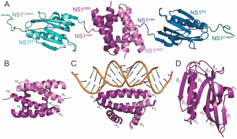
The NS1 protein of influenza A is a highly multifunctional protein that ranges in sequence length from 215 to 237 amino acids (~26kDa) depending on the strain from which it is derived [80,81]. NS1 is divided into four discrete structural regions (Figure 1A). Its N-terminal RNA binding domain (NS1RBD) is composed of amino acid residues 1-73 and is primarily responsible for NS1’s RNA binding activity [82,83,84,85]. The effector domain (NS1ED), consisting of amino acid residues 86-205, interacts with numerous host factors involved in the innate immune response to infection. Between the NS1RBD and NS1ED is a short, flexible linker (~residues 74-85). The functional importance for this linker region is poorly understood; however, a naturally occurring deletion of residues 80-84 has been observed in some highly pathogenic H5N1 strains of influenza [86]. Finally, NS1 contains an unstructured C-terminal tail (~residues 206-237) of varying length depending on strain (NS1C-term). Numerous host interactions have been reported to be mediated by the NS1 C-terminus [87,88,89], some of which affect pathogenicity [90,91]. The three-dimensional structures of individually expressed NS1RBD and NS1ED have been extensively studied [92,93,94,95,96,97,98,99,100,101,102,103]. However, little is known regarding the interdomain contacts and quaternary structure of NS1. This is mainly due to the propensity of full-length NS1 to aggregate at relatively low concentrations in solution [103]. Indeed, the few published full-length NS1 structures include mutations to mitigate its aggregation, allowing for its amenability to traditional structural techniques [104,105].
Figure 1.
Crystal structures of NS1 and its domains. (A) Full-length NS1 derived from the A/Vietnam/1203/2004 (H5N1) strain of influenza. Two mutations (R38A and K41A) were needed to make the full-length protein amenable for crystallographic studies. The four discrete structural regions of NS1 are labeled as NS1RBD, NS1inter, NS1ED, and NS1C-term and color coded in violet, blue violet, blue, and green, respectively. Each monomer of full-length NS1 is color coded for each region using different shades of the same color. (B) The NS1RBD derived from the A/Brevig Mission/1/1918 (H1N1) strain of influenza. (C) The NS1RBD derived from the A/Puerto Rico/8/1934 (H1N1) strain of influenza bound to dsRNA. (D) The NS1ED derived from the A/Brevig Mission/1/1918 (H1N1) strain of influenza. The secondary structural elements of each independently folding domain of NS1 are labeled.
3. NS1RBD
The three-dimensional structure of the NS1RBD has been solved using X-ray crystallography [98] and nuclear magnetic resonance (NMR) [82,95]. It is a symmetric homodimer (Figure 1B) consisting of six α-helices (three from each monomeric unit), two of which (α2 and α2′) form a “track” that interacts with the major groove of dsRNAs [106,107]. Basic residues along this RNA-binding track are heavily involved in the interaction, as can been seen in the high-resolution structure of the NS1RBD bound to dsRNA (Figure 1C) [106,108]. Specifically, Arg-38 and Lys-41 are required for this interaction, and recombinant IAVs harboring mutations at these positions are severely attenuated [83]. NS1RBD sequence and structure is very well-conserved among different strains of IAV [82,95,99,106]; however, subtle differences between strains have recently been identified. For example, a recent study showed that a potential salt bridge present in the NS1RBD of the A/Brevig Mission/1/1918 (H1N1) strain of IAV (1918H1N1) facilitates a strain-dependent interaction with the second caspase activation and recruitment domain (CARD2) of the retinoic acid inducible gene-I (RIG-I) [95,109].
4. Interdomain Linker
The interdomain linker is a short, flexible linker (~residues 74-85) that connects the two independently folding domains of NS1 (NS1RBD and NS1ED) [105]. Although the amino acid sequence of the linker is conserved among influenza A viruses, its impact on overall NS1 function is not known [110]. There have been several studies that have begun to characterize its role in NS1 function and in the context of influenza infection. For example, it has been shown that a naturally occurring deletion in the interdomain linker (residues 80-84) increases viral replication and pathogenicity [86]. This deletion is found in some highly pathogenic H5N1 influenza strains [86]. It has also been shown that mutating the interdomain linker of NS1 inhibited its ability to suppress the innate immune response, decreased overall viral replication, and abrogated NS1’s ability to activate the PI3K/Akt anti-apoptotic pathway [110]. Finally, mutations in the interdomain linker of NS1 were identified as possible enhancers of zoonotic transmission [111,112]. Follow-up studies are needed to fully characterize the role that the interdomain linker plays in the regulation of NS1 function, influenza pathogenesis, and zoonotic transmission.
5. NS1ED
The NS1ED is significantly less well-conserved across strains of IAV than the NS1RBD. This is likely due to the numerous strain-dependent host interactions that it participates in during IAV infection [76]. Like the NS1RBD, structures of the NS1ED have been solved using both X-ray crystallography [92,94,96,97,100,101,102] and NMR [103]. The structure of the monomeric unit of the NS1ED consists of three α-helices and seven β-strands (Figure 1D). The core of the protein is arranged as a long central α-helix surrounded by a twisted antiparallel β-sheet. The independently expressed NS1ED dimerizes rather weakly compared to the NS1RBD, and the importance of its dimerization is still unclear [103,113].
6. NS1C-term
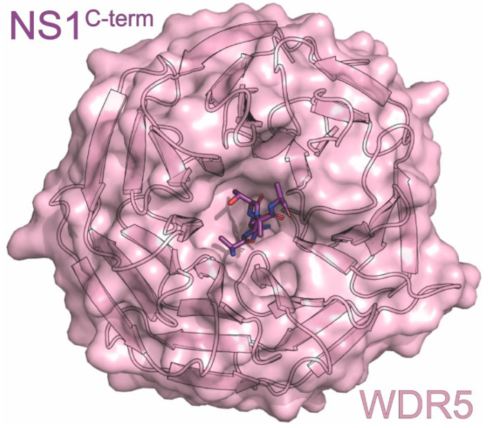
Although many constructs used for solving NS1ED structures have included residues from NS1’s C-terminal tail (~residues 206-230), no electron density for this region has been observed [92,96,114]. It is possible, however, that this region assumes some structure upon interaction with host binding partners. Studies utilizing short peptides derived from NS1’s C-terminus have allowed the identification of binding partners such as the chromatin-associated factor WDR5 [115] (Figure 2) [116]. Further studies are necessary to determine the contribution of this region to overall NS1 structure and function.
Figure 2.
Crystal structure of the WDR5 in complex with NS1. The structure consists of WDR5 (pink) in complex with the carboxyl terminus of NS1 (purple) derived from the A/Udorn/307/1972 (H3N2) strain of influenza. The PDB ID for this structure is 4O45.
7. CHD1
CHD1 is a chromatin remodeling protein that can be found in the nuclei of host cells [117,118,119]. It consists of two chromodomains, an SWI/SNF domain for its chromatin remodeling functions and a C-terminal DNA binding domain [115,120]. CHD1 recruits post-transcriptional initiation and splicing factors to the DNA through its recognition of trimethylated histone H3 lysine 4 (H3K4), a sign of actively transcribed chromatin [115,121]. Although many histone binding proteins cradle the free N-terminal amine of histone H3 in a deep, negatively charged pocket, CHD1 has a shallow open pocket [115]. This shallow binding pocket allows the C-terminus of NS1 to imperfectly mimic the histone tail and bind to CHD1. Unlike most other proteins that bind histones, however, its interaction with NS1 specifically affects CHD1’s binding pool of actively transcribed DNA.
The C-terminal tail of NS1 is an unstructured region with binding sites for many different types of host proteins [90,94,122]. NS1 introduces an additional binding site to this region through a sequence that mimics the histone H3 tail. The “226ARSK229” sequence is chemically analogous to histone H3’s N-terminal “1ARTK4”, even retaining its ability to be post-translationally modified by lysine methyltransferases Set1 and Set7/9 or acetyltransferase TIP60 [87]. One key difference is that the methylation that would mark a histone as transcriptionally active is unable to be removed from NS1 by demethylase LSD1, allowing it to continuously be recognized by CHD1 [115].
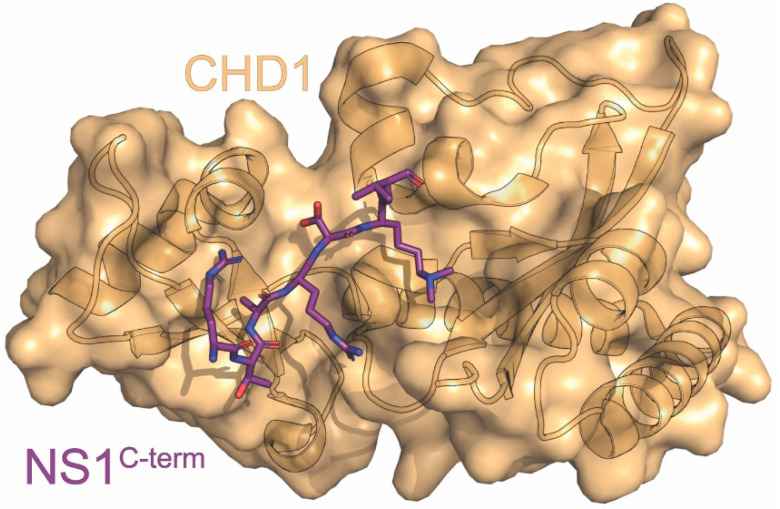
CHD1 was found to bind to the di- or tri-methylated NS1 C-terminal tail through immunoprecipitation and western blot with purified NS1 peptide, ITC, and an X-ray crystal structure of CHD1’s chromodomains complexed with NS1 C-terminal tail peptide (Figure 3) [87,115]. The two chromodomains bind to the NS1 “226ARSK229” sequence through two types of interactions: 1) cation-π interactions between the methylated K229 of NS1 and W322 and W325 from CHD1 chromodomain 1, reinforced by E272 from chromodomain 1 interacting with the lysine through its negative charge and 2) a series of hydrogen bonds either directly or through ordered solvent [115]. We note that dimethylated NS1 was preferred to trimethylated, due to the third methyl being solvent exposed [120]. The crystal structure obtained was almost identical to chromodomain 1 complexed with trimethylated histone H3, with the main difference being A226 of NS1’s 60º rotation relative to A1 of histone H3. This rotation allows an additional salt bridge between R224 of NS1 and D408 and D425 of CHD1 [115].
Figure 3.
Crystal structure of CHD1 in complex with NS1C-term. The structure consists of CHD1 (orange) in complex with the carboxyl terminus of NS1 (purple) derived from the A/Hong Kong/5/1983 (H3N2) strain of influenza. The PDB ID for this structure is 4O42.
NS1’s C-terminal tail contains an imperfect histone mimic due to the lack of a free N-terminal amine that is normally required for interactions with histone binding factors, but this serves as an advantage for IAV. Its ability to bind to CHD1’s shallow binding pocket allows NS1 to target its effects to actively transcribed chromatin, aiding its efforts to curtail virus-induced production of antiviral genes. Additionally, it is unable to be demethylated, allowing it to escape from normal host regulation of this process. The mechanistic underpinnings of NS1’s hijacking of CHD1 still need to be elucidated, but this interaction adds another host to NS1’s toolkit for downregulation of host genes that could be a novel target for antiviral drugs.
8. CrkII
The Crk family is a set of important adaptor proteins that are required for many essential cellular signaling processes. They contain Src-homology domains (SH) that bind to proline-rich regions on their ligands. CrkL and CrkII have one SH2 and two SH3 domains, while CrkI, an alternative splicing product of CrkII, has one of each [123,124]. The SH2 and SH3 domains have binding pools that largely overlap with a few exceptions, and the Crk proteins exert their function by bringing together proteins that interact with these domains [125,126]. A subset of IAV strains is able to prevent these linkages through host–pathogen interactions involving their NS1 proteins and the Crk family of proteins. For these strains, the NS1 C-terminal tail encodes a proline-rich motif (PRM) that can bind to the Crk family of proteins. The PPLPPK motif at residues 212–217 of NS1 is a perfect class II SH3-binding motif that is highly conserved in avian strains but is only present in a handful of human strains, notably including the 1918H1N1 pandemic strain [125,127]. P212, P215, and K217 were found to be especially important, with a T215P mutation proving sufficient to confer SH3 binding to the normally binding-incompetent WSN strain [126,127]. Most PRMs bind to SH3 domains with low specificity, but NS1 binds to CrkII’s SH3 in a highly specific manner, leading Shen et al. to investigate the structural mechanisms behind the interaction [116,127].
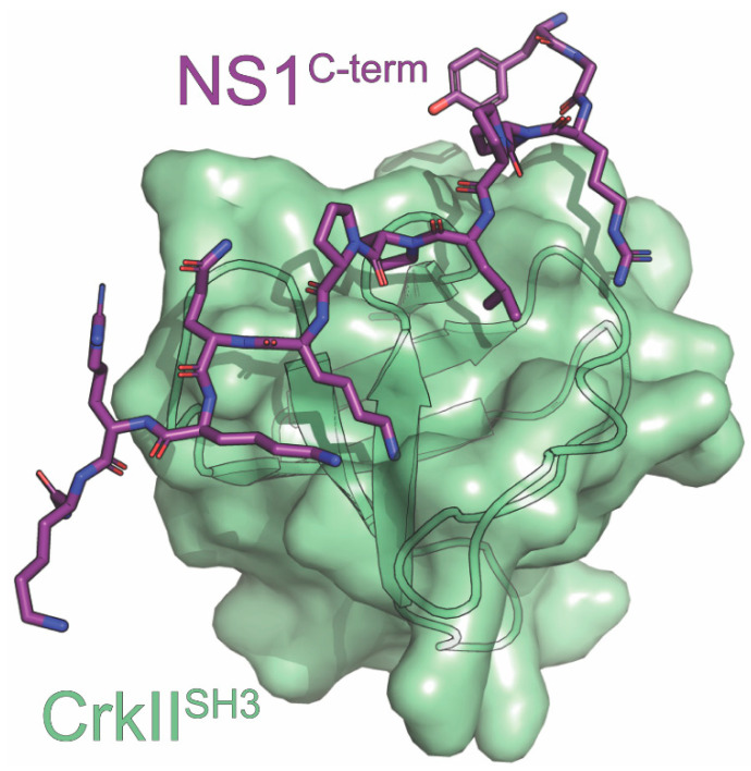
Shen et al. structurally defined the interaction of the PRM of NS1 derived from the 1918H1N1 strain of influenza (1918 NS1PRM) and the N-terminal SH3 domain of CrkII (nSH3CrkII) [116]. The methods used in this study included X-ray crystallography, NMR relaxation dispersion, and fluorescence spectroscopy (Figure 4) [116]. Using these methods, they revealed a bipartite binding interface that was responsible for NS1’s increased selectivity. The N-terminal region of the 1918 NS1PRM interacts with CrkII’s SH3 domain via hydrophobic interactions mediated by the PPLP motif. This observation is consistent with other SH3-PRM complexes. The C-terminal region of the 1918 NS1PRM forms two sets of electrostatic interactions with a negatively charged surface of the nSH3CrkII domain. A salt-bridge network forms between the ε-amino group of K217 of the 1918 NS1PRM and D147, E149, and D150 of nSH3CrkII. This network has been observed in CrkII complexes with Abl kinase and C3G, but it is not common. Additionally, a bidentate hydrogen bond forms between E166 of nSH3CrkII and the backbone amides of Q218 and K219 of the 1918 NS1PRM. No other SH3 domains besides nSH3CrkII have been observed to form both sets of interactions with its binding partner, which would explain 1918 NS1PRM’s high specificity for CrkII.
Figure 4.
Crystal structure of CrkIISH3 in complex with NS1. The structure consists of CrkIISH3 (light green) in complex with the carboxyl terminus of NS1 (purple) derived from the A/Brevig Mission /01/1918 (H1N1) strain of influenza. The PDB ID for this structure is 5UL6.
Shen et al. also compared the binding kinetics between nSH3CrkII and the 1918 NS1PRM to a previously identified nSH3CrkII interaction partner, the PRM encoded by c-Jun-N-terminal kinase 1 (JNKPRM) [116]. The Kd of the 1918 NS1PRM-nSH3CrkII complex was measured to be 6nM, which is 3300-fold lower than the Kd measured for the JNKPRM- nSH3CrkII complex. For reference, the Kd values for SH3-PRM complex typically range between 1 and 10µM [128]. Additionally, the kon for NS1 binding to nSH3CrkII is 100-fold faster, while the koff for the 1918 NS1PRM-nSH3CrkII complex is 35-fold slower compared to the kon and koff for the JNKPRM- nSH3CrkII complex. Taken together, the significantly increased kon, decreased koff, and decreased Kd observed for the 1918 NS1PRM-nSH3CrkII complex offers a clear mechanistic explanation for NS1’s ability to competitively inhibit the interaction between JNKPRM and nSH3CrkII.
Since the Crk family binds to numerous proteins that facilitate various processes in the host cell, NS1’s displacement of these proteins influences several cellular processes. For example, NS1 suppresses the activation of the host antiviral immune response by outcompeting the interactions between the Crk family and kinases such as JNK1 and Abl [129,130,131]. The interaction between NS1 and JNK1 is also thought to suppress apoptosis that is normally triggered upon viral infection. However, it has been suggested that the CrkII modulation of the apoptotic pathway may not be as important as its counterparts in this process [125,129]. Furthermore, Ylosmaki et al. showed that NS1 binding to Crk proteins can translocate them into the nucleus, leading to a change in nuclear protein phosphorylation. This could also lead to changes in apoptosis since CrkII has been reported to bind to nuclear cell cycle regulator Wee1 along with activating caspases [126,132]. Finally, Crk binding is involved in PI3K signaling. During infection, NS1 hyperactivates PI3K signaling by binding to both p85β of PI3K and the Crk family. The interaction between NS1 and PI3K is further discussed later in this review [127,133]. In summary, the heightened selectivity of interactions between NS1 and the Crk family of proteins, especially CrkII, facilitates control of many important cellular processes by the IAV that prove beneficial for viral fitness.
9. Scribble
Proper cell polarity is essential for tissue development, organization, and function. It ensures that macromolecules, including cellular junctions, are located in their proper positions. This process is controlled by the actions of three competing protein modules: Crumbs, PAR, and Scribble [134]. The Scribble module consists of the leucine-rich repeats and the PDZ domain (LAP) protein Scribble bound to Dlg and Lgl, and the three are normally located together in the cytoplasm near the plasma membrane [135,136,137]. While previously thought to only be scaffolding proteins, PDZ proteins have since been shown to have more involvement in cellular processes, such as Scribble’s proapoptotic functions [138,139]. Scribble enacts its effects via interactions with different proteins through its various domains, including its four PDZ domains, which multiple viruses exploit [136,140].
There are several NS1 proteins that encode a PDZ-binding motif (PBM) at their extreme C-termini. We note that PBMs are not encoded universally by all NS1 proteins. Of the NS1 proteins that do, there are differences in their sequences when comparing avian and human strains [141]. About 90% of avian isolates have ESEV or ESKV at the C-terminus, while human strains usually have an RSKV. These follow Scribble’s class I PDZ recognition sequence motif of X-T/S-X-V/LCOOH. However, due to their varying sequences, they have different effects on pathogenicity and virulence. Using peptides that encode these NS1 PBMs, the interaction between the PBM of NS1 and Scribble has been studied via pulldowns, co-IP, in vitro binding assays with recombinant purified protein, ITC, and X-ray crystallography [88,89,137,142,143]. The avian sequences ESEV and ESKV have been found to interact with Scribble’s PDZ domains, while the human isolate sequences lack this ability [88,89,137,142,143]. To confirm that the PBMs were primarily responsible for these interactions, chimeras were made in which the human and avian PBMs were interchanged between the two NS1 proteins. This interchange of PBMs lead to reversal of the previously observed interactions between the human and avian strains with Scribble. However, it was shown that avian NS1 with a mutated EAEA non-functional PBM was still able to bind more tightly than human NS1, suggesting that other regions could also be involved [89,137]. It is still unclear which PDZ domain of Scribble interacts with the PBM encoded by NS1. Specifically, in vitro binding assays showed that full-length NS1 was unable to bind to individual PDZ domains of Scribble. However, it was determined that a tandem construct encoding PDZ1 and PDZ2 was necessary and sufficient for the interaction. Finally, all four individual PDZ domains of scribble were able to bind 10-mer peptides encoding the extreme NS1C-term, and these complexes were used to solve the high-resolution crystal structures (Figure 5) [137,143].
Figure 5.
Crystal structures of Scribble in complex with NS1. (A) The first PDZ domain (PDZ1) of scribble (red) in complex with the PDZ binding motif (PBM) of NS1 derived from the A/Vietnam/1194/2004 (H5N1) (purple). (B) PDZ1 (orange) in complex with the PBM of NS1 derived from the A/Chicken/Hubei/327/2004 (H5N1) strain of influenza (purple). (C) The third PDZ domain (PDZ3) of scribble (yellow) in complex with the PBM of NS1 derived from the A/Chicken/ Hubei/327/2004 (H5N1) strain of influenza (purple). The PBM of NS1 proteins derived from the A/Vietnam/1194/2004 (H5N1) consist of ESEV residues at their carboxyl terminus (NS1ESEV), while the PBM of NS1 proteins derived from the A/Chicken/Hubei/327/2004 (H5N1) strain of influenza consist of ESKV residues at their carboxyl terminus (NS1ESKV). The PDB IDs for these structures are 7QTO, 7QTP, and 7QTU for PDZ1:NS1ESEV, PDZ1:NS1ESKV, and PDZ3:NS1ESKV, respectively.
The ITC and X-ray crystallography experiments performed by Javorsky et al. revealed several important interactions that were also confirmed via mutational analysis [143]. The PBM of NS1 was found in the canonical ligand-binding pocket of the PDZ domain, and the structures overlaid with Scribble complexed with an endogenous ligand generated RMSD values ranging from 0.57 to 0.75Å. Their structures of PDZ1 complexed with either ESEV or ESKV (Figure 5A,B) and PDZ3 (Figure 5C) with ESKV identified a number of hydrogen bonds and van der Waals contacts that can be disrupted to abolish binding. The conserved Histidine found on the α2 helix of each PDZ domain is critical to the interaction, as mutating it to an alanine abolished binding in each case except for PDZ2, which enhanced binding. Both PBMs encoding ESEV and ESKV interacted with all four PDZ domains with affinities in the low micromolar. The relative binding affinities for the PBM encoding ESEV to each PDZ domain were PDZ4 > PDZ3 > PDZ2 > PDZ1. For the PBM encoding ESKV, the relative affinities were PDZ2 > PDZ3 >PDZ1 with no interaction detected for PDZ4. Compared to the endogenous ligand β-PIX, the NS1 PBM had lower affinity for PDZ1, higher for PDZ2, and higher for PDZ4 since β-PIX is also unable to bind this domain. This provides support for a mechanism by which NS1 outcompetes Scribble’s endogenous ligands to prevent its functions.
Scribble function has been shown to be disrupted by NS1 proteins derived from avian IAVs by blocking its interaction with natural ligands as well as causing mislocalization of Scribble. Specifically, Scribble is typically found in the cytoplasm adjacent to the plasma membrane, but it is found in perinuclear puncta with NS1 and Dlg1 during infection with avian IAVs [88,137]. This mislocalization results in disruption of tight junctions that allows for increased viral dissemination within the host and decreased apoptosis that is crucial for early survival of the virus [88,137]. However, the effect on NS1’s interferon antagonism is unclear in this context. One study showed that avian NS1’s reduction in interferon-induced STAT phosphorylation was hindered by a non-functional PBM, while another showed that mutants and wild-type PBMs had no difference in their blocking of IFNβ induction [89,142]. Another aspect that is not fully understood is the PBM’s synergistic upregulation of Akt phosphorylation with the PI3K binding loop [142]. What is clear, however, is that disruption of the normal function of Scribble by NS1 is able to regulate many different aspects of cellular function to benefit viral replication.
10. MORC3
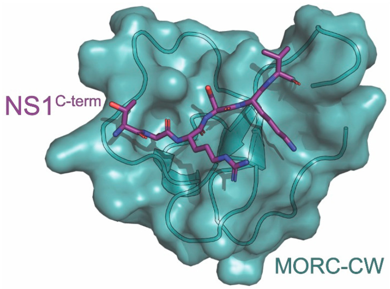
Microrchidia 3 (MORC3, also known as NXP2, ZCW5, ZCWCC3, or KIAA0136) is a human ATPase involved in the host antiviral response, transcription regulation, and chromatin remodeling [144]. Pertinent to this review, MORC3 participates in the antiviral response through its recruitment to promyelocytic leukemia nuclear bodies (PML-NBs). It consists of a GHKL (gyrase, Hsp90, histidine kinase, and MutL)-type ATPase domain, a CW-type zinc finger domain, and a coiled-coil region [145]. Upon localization to PML-NBs, it recruits and activates p53 to induce cellular senescence among other antiviral defenses [146,147]. In its native state, MORC3 function is inhibited by its own CW domain (MORC3-CW) that self-associates with its neighboring ATPase domain to prevent DNA binding, which is required for catalysis. MORC3 becomes activated when its CW domain binds to the tail of histone H3, preferably histone H3 proteins that are tri-methylated at the 4th lysine residue (H3K4me) [148]. Many viruses have developed ways to overcome this regulation through histone mimicry. One such histone mimic includes the C-terminal tail of NS1 derived from a number of IAV strains [87].
The interaction between the NS1C-term and MORC3-CW has been characterized by Zhang et al. using NMR spectroscopy and X-ray crystallography (Figure 6) [149]. NMR chemical shift perturbation analysis revealed direct binding between the MORC3-CW and residues 222-230 of the NS1C-term. It was also found that using an NS1C-term peptide trimethylated at K229 (NS1K229me3) increased its binding affinity with MORC3-CW. To solve the X-ray crystal structure of the complex, the NS1C-term (residues 225-230) was fused to MORC3 (residues 407-455) using a GGSG linker. NMR analysis using heteronuclear single quantum coherence (HSQC) spectra determined that the fusion protein consisted of separated domains. The X-ray crystal structure of the MORC3-CW:NS1C-term complex was then solved at 1.41 Å resolution. The structure revealed that the MORC3-CW folds into a compact globular structure of a double-stranded antiparallel β-sheet with helical turns stabilized by its zinc-binding cluster [149]. It was also noted that residues R227-V230 of the NS1C-term form a third antiparallel β-sheet that is paired with MORC3-CW’s. This third antiparallel sheet includes characteristic β-sheet hydrogen bonds between R227 and K229 of NS1 and W410 and Q412 of MORC3-CW. K229 of NS1C-term is caged between W410 and W419 of MORC3-CW while A226 of NS1C-term settles in a well-defined pocket of MORC3-CW. A comparison of the MORC3-CW:NS1C-term and MORC3-CW:H3K4me3 complexes yields an RMSD of 0.2 Å, indicating that the NS1C-term is an authentic mimic of H3K4me3.
Figure 6.
Crystal structure of MORC3-CW in complex with NS1. The structure consists of MORC3-CW (teal) in complex with the carboxyl terminus of NS1 (purple) derived from the A/Wyoming/03/2003 (H3N2) strain of influenza. The PDB ID for this structure is 6O5W.
MORC3-CW interacted with the NS1C-term (residues 222-230) with a KD of 157µM, while using NS1K229me3 as the target ligand increased the binding affinity (KD = 14 µM) [149]. This increased binding affinity is comparable to the binding affinity of MORC3-CW to histone H3 and the ATPase domain (7 µM and 9 µM, respectively). However, it is still weaker when compared to MORC3-CW’s binding affinity to H3K4me3 (0.6µM). This suggests a mechanism whereby NS1 outcompetes the release of MORC3’s autoinhibition by preventing the interaction between MORC3-CW and the adjacent catalytic domain. Mutating W410 or W419 of MORC3-CW, the residues involved in caging K229 of the NS1C-term, to alanine abrogated its binding to the NS1C-term. Furthermore, mutating E431 of MORC3-CW to alanine diminished its binding to NS1C-term but had no effect on its interaction with H3K4me3. E431 was selected for study due to its projected proximity to residue 224 of the NS1C-term, which was not included in the chimera.
MORC3 has previously been shown to interact with two of the influenza RNA-dependent RNA polymerase (RdRP) subunits (PB1 and PA) as well as with viral RNPs [150,151]. Different studies have shown both positive and negative effects of MORC3 on the virus that were studied through the lens of this polymerase association. Ver et al. demonstrated that MORC3 silencing with shRNA resulted in a significant reduction in viral transcription and viral titers [151]. On the other hand, Bortz et al’s knockdown of MORC3 with siRNA found that viral polymerase activity was significantly increased with a mild increase in viral titers [152]. Zhang et al showed that overexpression of MORC3 resulted in a slight increase in virus infectivity that depended on functional MORC3-CW and ATPase domains, supporting the former study [149]. This dependence is thought to be due to its interaction with the NS1C-term. However, further studies will be necessary to fully understand the effect of MORC3 on the viral lifecycle.
11. CPSF30
One of NS1’s most studied interactions is that with the host cleavage and polyadenylation specificity factor 30kDa subunit (CPSF30 or CPSF4). CPSF30 is found in the nucleus, where it is involved in mRNA processing and maturation. CPSF, as its name suggests, is involved in both cleavage and polyadenylation of pre-mRNAs through binding the RNA, recruiting other host factors, and catalyzing the cleavage step [153]. NS1 binding to the second and third zinc finger (F2F3) of CPSF30 prevents these functions and results in a global downregulation of host genes. These include interferon and interferon-stimulated genes that ultimately contribute to NS1’s function as a virulence factor for influenza infection [41]. Importantly, this downregulation does not affect viral genes, as their mRNAs are polyadenylated through stuttering of the viral polymerase [154]. Abolishing the interaction between NS1 and F2F3 results in a 10-fold decrease in viral replication and an increase in IFNβ mRNA production [68]. This result alone highlights this interaction as a potential target for novel antiviral development, a process which is aided by the presence of a high-resolution complex structure.
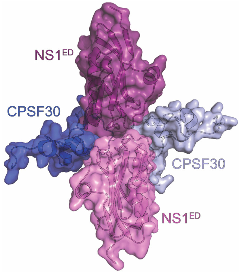
To this end, a crystal structure was solved for F2F3 of CPSF30 bound to the NS1ED (residues 85-215) at 1.95 Å resolution [40]. The overall structure is a tetramer, with two F2F3 molecules wrapped around two NS1EDs (Figure 7). The NS1EDs in the structure interact with each other in a head-to-head orientation which differs from dimer interfaces previously observed for the NS1ED. The F2F3 binding pocket of the NS1ED is largely hydrophobic and very well-conserved among human IAVs. The NS1ED residues critical to the interaction, according to the structure, are K110, I117, I119, Q121, V180, G183, G184, and W187. These residues interact with the aromatic side chains of Y97, F98, and F102 of the corresponding F2F3 protein. Additionally, F103 and M106 were found to be important for stabilization of the complex despite them being outside of the binding pocket. Specifically, the side chain of M106 is positioned in the middle of the tetramer and interacts with the M106 of the other NS1 and residues in both F2F3 molecules. F103’s aromatic side chain of the NS1ED interacts with the hydrophobic residues L72, Y88, and P111 from the adjacent F2F3 molecule. The structure highlights several important regions and residues that could be targeted to hinder NS1’s interaction with CPSF30.
Figure 7.
Crystal structure of CPSF30 in complex with NS1. The tetrameric complex is composed of two NS1EDs derived from the A/Udorn/307/1972 (H3N2) strain of influenza (violet and light violet) and two F2F3 domains of CPSF30 (blue and light blue). The PDB ID for this structure is 2RHK.
There have been many in vitro and in vivo studies that have identified residues in NS1 that modulate this interaction in several different strains and hosts. In the seasonal H3N2 strain, the I64T mutation in the NS1RBD was found to decrease binding to CPSF30 and increase interferon levels. As the NS1RBD was not included in the crystal structure, the structural mechanism of how this mutation affects the interaction between the NS1ED and F2F3 remains unclear [155]. M106 and F103 have also been shown in numerous studies to be critical to the interaction, but the effects of mutation vary depending on strain and host species. For example, the L103F and I106M mutations in the H7N9 strain restored CPSF30 binding and enhanced virulence. For the 2009 pandemic H1N1 strain, however, restoring the interaction between the NS1ED and CPSF30 impaired virulence [62,156,157,158,159,160].
Loss of binding is commonly seen in avian-to-human transmission, such as with H5N1 and H9N2, suggesting a role in adaptation to mammalian hosts. The H5N1 strain HN01, which lacks CPSF30 binding, is able to recover binding and subsequently decrease interferon activity in human cells with the mutations K55E, K66E, and C133F [161]. Similarly, the HK/97 H9N2 gained binding to CPSF30 and inhibition of host gene expression with the mutations L103F, I106M, P114S, G125D, and N139D [162]. In the swine H5N1 SW/FJ/03, a naturally occurring deletion from 191 to 195 decreased binding to CPSF30 and attenuated virulence in chickens [163]. Some of the strains that lack binding to CPSF30 have evolved to compensate in different ways. The H5N1 strain HK97 lacks binding due to mutations in the stabilizing residues F103 and M106, while descendant strains have lost this defect. HK97 is still able to infect, however, due to compensatory stabilization by its viral polymerase [158,160]. The mouse-adapted H1N1 strain PR8 (PR8H1N1) has evolved a different mechanism to make up for a hydrophilic residue at position 103. Since it cannot globally downregulate host genes, it suppresses IRF-3 activation to suppress production of IFN-β mRNA at an earlier step [159]. This has been selected against during replication in humans, suggesting that CPSF30 binding is important for circulating human strains. Collectively, these results highlight the critical role of NS1’s interaction with CPSF30, along with sites that could be targeted by novel antivirals to decrease virulence.
12. NXF1-NXT1
NXF1-NXT1 (also known as TAP-p15) is a heterodimeric mRNA export receptor that binds to both mRNA and nucleoporins. This function allows it to shuttle mRNAs through the dense phenylalanine-glycine (FG) repeats of the nuclear pore complex (NPC) into the cytoplasm [164,165,166]. The retinoic acid early inducible 1 protein (Rae1) also participates in the complex and is thought to mediate NXF1’s interaction with mRNAs [167,168,169]. Rae1 facilitates the interaction between NXF1 and mRNAs by recruiting both the nuclear pore complex protein Nup98 and E1B-AP5 into the complex. Both Rae1 and Nup98 are induced by interferons upon viral infection, increasing nuclear export of mRNAs of interferon-stimulated genes such as IFIT2, IRF-1, MHC1, and ICAM-1 [169,170,171,172]. This enhancement makes this pathway a popular target of viral proteins evolved to inhibit the induction of the immune system [171,173,174]. While the influenza A virus already targets mRNA export through the processing machinery (CPSF30), it has also developed a two-pronged approach via interaction with NXF1-NXT1 of the nuclear export machinery.
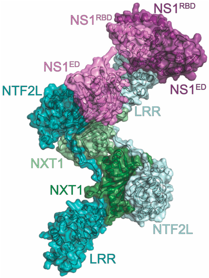
The crystal structure of NS1 complexed with NXF1-NXT1 (resolution at 3.8 Å) revealed a 2:2:2 orientation (Figure 8) [172]. To avoid aggregation and structural polymorphism of NS1, the linker between the NS1RBD and NS1ED was shortened, and two mutations were engineered into NS1 (R38A/K41A). The NXF1 construct used for crystallization only included the leucine-rich repeat (LRR) and nuclear transport factor 2-like (NTF2L) domains. The tetramer, consisting of two NXT1s, each interacting with an LRR and NTF2L from each NXF1, is capped on one end by NS1 dimerized through its NS1RBDs. One NS1ED interacts with NTF2L from one NXF1 (interface I), while the other NS1ED interacts with the LRR from the other NXF1 (interface II). The other end of the NXF1-NXT1 tetramer is unoccupied, potentially leaving room for another NS1 dimer. Two highly conserved NS1 residues were identified to be critical for complex formation. F103, located in the α1-β2 loop of the NS1ED, inserts into a hydrophobic pocket on NXF1-NTF2L consisting of L383, L386, L491, P521, and L527 to form interface I. F138, located in the β4-β5 loop of the NS1ED, binds NXF1-LRR residues K213, M216, Y220, N263, and I264 to form interface II. Additionally, NS1 was found in the same binding site as a nucleoporin FG peptide. It was confirmed through binding analyses that NS1 displaces Nup98 binding to the complex [172,175].
Figure 8.
Crystal structure of the NXF1:NXT1 heterodimer in complex with NS1. The complex consists of two copies each of NS1 (violet and light violet), NXF1 (teal and light teal), and NXT1 (green and light green). The NS1 protein is derived from the A/Texas/36/1991 (H1N1) strain of influenza. The nucleoporin-binding domain (NTF2L) and leucine-rich repeat domains (LRR) of NXF1 are labeled. The PDB ID for this structure is 6E5U.
Both IAV infection and ectopic expression of NS1 inhibit the nuclear export of mRNA. This inhibition can be blocked with expression of NXF1, NXT1, Rae1, or Nup98, as well as engineering specific mutations into NS1 (F103A or F138A) [169,172]. These mutations result in a greatly attenuated virus with reduced viral protein levels and replication. Conversely, infections in cells with low levels of Rae1 or Nup98 are more virulent and cytotoxic [169]. The mRNA export pathway has an important role in regulating innate and adaptive immunity, which IAV NS1 blocks to replicate more efficiently. The crystal structure reveals that NS1 enhances IAV replication efficiency by outcompeting Nup98 binding to the complex, thereby blocking nucleoporin binding. These data identify two residues and NS1 regions that could be targeted in the development of anti-influenza therapeutics.
13. PDlim2
NS1 proteins derived from some strains of IAV encode a PDZ binding motif (PBM) at their extreme C-terminus. This region is responsible for interactions with many different PDZ-domain containing proteins within the cell, including Scribble, which is discussed above. The sequence of this PBM differs depending on the origin of the strain. For example, most NS1 proteins derived from avian strains, including the highly pathogenic H5N1 strain, encode the PBM ESEV. In contrast, most human strains encode the PBM RSKV or RSEV at this position [141]. This leads to strain-dependent differences in their target proteins that could be responsible for observed changes in pathogenicity upon swapping these sequences. One such interaction is with PDlim2 (aka Mystique or SLIM), a PDZ scaffolding protein involved in the regulation of the cytoskeleton and transcription factors. PDlim2 was found to bind specifically to NS1 derived from the avian H5N1 strain but not to the NS1 derived from an H1N1 strain [176,177]. Structural studies were therefore performed to determine if this difference in binding explains the differences in pathogenesis observed between these two strains.
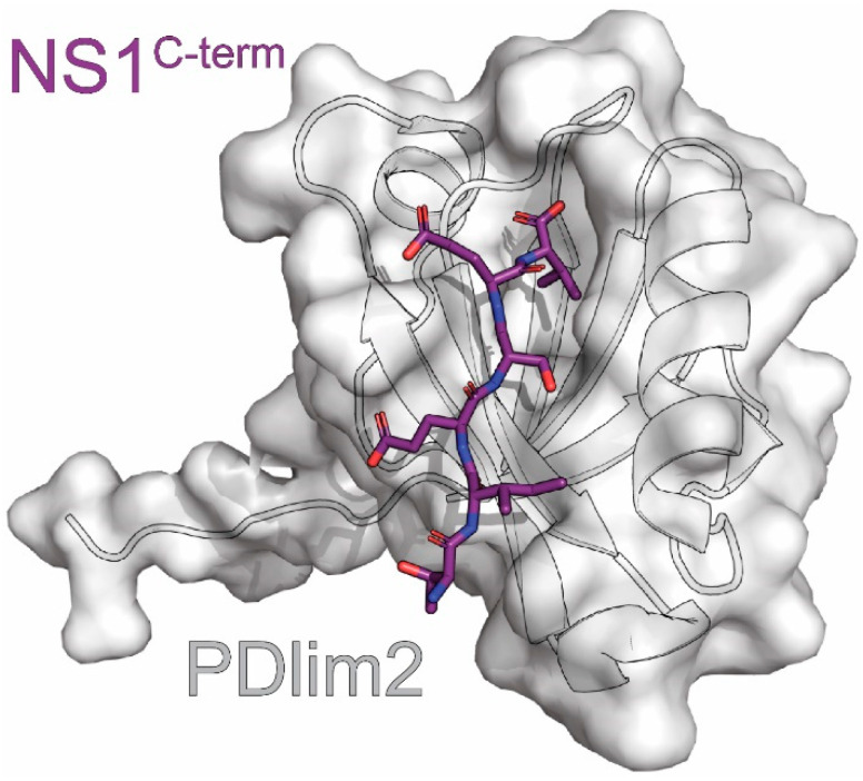
Yu et al. solved a crystal structure of the PDZ domain of PDlim2 fused to the C-terminal hexapeptide of the NS1C-term derived from the avian H5N1 strain at 2.2Å resolution. (Figure 9) [176,178]. They observed an archetypical PDZ domain with five β-strands and three α-helices, with the helices capping the open ends of the β-sheet barrel. The interaction site was occupied by the NS1C-term hexapeptide from the neighboring fusion protein in the crystal lattice. Residues 0 to -5 of the NS1C-term hexapeptide formed a β-strand that paired with PDlim2 β2 in an anti-parallel manner. A more granular analysis of the structure showed the following: 1) the NS1 residue in the -2 position formed a hydrogen bond with H62 from the α3 helix of PDlim2, 2) glutamines encoded by NS1 in the -1 and -3 positions were stabilized by salt bridges involving R16 and K31 of PDlim2, and 3) the valine encoded by NS1 in the 0 position was nestled in a hydrophobic pocket of PDlim2 consisting of F15 and I17 from the β2 strand, I69 from the α3 helix, and residue W13. These interactions are consistent with previous PDZ-ligand structures from both fusions and co-crystallizations [178,179].
Figure 9.
Crystal structure of PDlim2 in complex with NS1. The complex consists of the PBM of NS1 derived from the A/Chicken/Henan/12/2004 (H5N1) strain of influenza (purple) and the PDZ domain of PDlim2 (grey). The PDB ID for this structure is 3PDV.
The interactions between NS1 residues in the -1 and -3 positions and PDlim2 residues Arg16 and Lys31 have proven to be particularly important. The -3 position determines differences in PDlim2 binding between strains HN12 (E-3) and PR8H1N1 (R-3). Mutating only this position to the other strain’s residue reversed binding between the two both in vitro and in vivo [176]. Similarly, mutating the PDlim2 sites in this interaction greatly affected binding. Both R16A and K31S showed reduced binding, while a double mutant completely abolished it [176]. R16Q was unable to bind even in the presence of K31, indicating a steric requirement. The importance of the salt bridge to this interaction, as shown with mutational analysis and natural variation between strains, makes it a promising candidate for inhibiting NS1 function.
Although we now have knowledge of how to interrupt this interaction, its biological significance is still not understood. To date, two separate PDlim2 functions have been interrogated to no effect. Although PDlim2 functions as an E3 ligase for the p65 subunit of NF-κB and NS1 is known to prevent NF-κB activation, a reporter assay showed no difference between HN12-NS1 WT or ∆PBM in the absence or presence of PDlim2 [176,180,181]. Similarly, HN12-NS1 WT and ∆PBM both resulted in similar levels of STAT1 phosphorylation despite PDlim2’s role in phosphorylation and degradation of STAT1 and STAT4 [176,182]. It is possible that the latter’s lack of effect could be due to the different strain used compared to previous studies and differences between infection and transfection. Additional research is needed to investigate the interaction’s effects on PDlim2’s other functions, such as cell migration or its other roles in the immune system, including through the STAT2 and STAT3 pathways [183,184,185]. Further research is needed to determine the importance of the PDlim2-NS1 interaction, but the information gathered so far will serve as useful tools in this endeavor.
14. PI3K
Phosphatidylinositol-3-kinase (PI3K) is targeted by many viruses due to its pivotal role in host survival through various processes, such as apoptosis regulation, metabolism, and immunity [186,187]. When activated, its catalytic subunit p110 converts PIP2 to PIP3, which acts as a secondary messenger to stimulate various pathways such as Akt [188,189]. During homeostasis, p110 is inhibited by PI3K’s regulatory subunit p85 [189,190]. When adaptor proteins bind to the SH2 domain of p85, PI3K is recruited to the plasma membrane where p110 initiates catalysis of PIP2. NS1 has been shown to activate PI3K by mimicking this process and binding to the inter-SH2 domain of p85 [50,188,191,192]. It is this activation of PI3K by NS1 that enhances virulence through delaying apoptosis and/or changing the distribution of PI3K [20,50,191]. However, Jackson et al. posits that NS1’s apoptotic effects are not due to its interaction with PI3K [51]. Additionally, one study linked the interaction to changes in the conductance of lung epithelia [193]. Many studies have been performed to address how this interaction occurs at the residue level and how it contributes to viral pathogenesis.
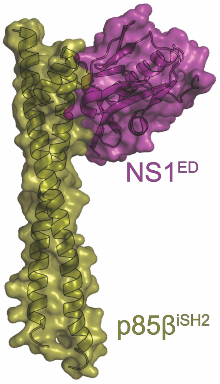
The NS1ED binds to the β isoform of PI3K’s p85 regulatory subunit (p85β) [188,191]. This interaction has been structurally analyzed via X-ray crystallography, revealing many details that will provide insight for further studies (Figure 10). To date, multiple structures have been solved using NS1EDs derived from multiple strains of IAV and the inter SH2 (iSH2) domain of p85β. Specifically, Hale et al. used the NS1ED derived from the PR8 strain of influenza (PR8H1N1 NS1ED) and bovine p85β, while Cho et al. used the NS1EDs derived from the 1918H1N1and Udorn strains of influenza (1918H1N1 NS1ED and UdornH3N2 NS1ED, respectively) and human p85β [49,194]. They revealed a “golf club-shaped” complex formed from the largely hydrophobic interaction between the NS1ED and one end of the iSH2 coiled-coil, notably at the opposite end from p110’s binding site. Both groups’ structures illustrated the importance of the conserved NS1 residue Y89, as it formed a crucial hydrogen bond with D575 of iSH2 at the center of the complex [49,194]. Previous studies have shown that Y89 is necessary for binding. Specifically, the Y89F mutation in NS1 is unable to pulldown p85β, reduces phosphorylated Akt, and has small plaques and low titers compared to wild-type NS1 [51,188,191,195]. The structure also explained why NS1 has evolved to bind p85β over the more ubiquitous p85α [49,196]. In the complex structure, p85β Val579 lies at the interface, burying 70Å2 upon complex formation [49]. In p85α, position 579 encodes a methionine, which sterically hinders the interaction. Superimposing the NS1ED:p85β structure onto that of p85α:p110 reveals that the NS1ED sterically prevents p85 SH2 inhibition of p110, explaining the mechanics behind IAV-induced PI3K activation [49]. These structures support and elaborate upon previous work in the field, giving a mechanistic explanation of the interaction that could be used to target this NS1 function for future development of IAV antivirals.
Figure 10.
Crystal structure of PI3K in complex with NS1. The complex consists of the NS1ED derived from the A/Brevig Mission/1/1918 (H1N1) strain of influenza (purple) and the p85biSH2 subunit of PI3K (gold). The PDB ID for this structure is 6U28.
Further analysis of the binding characteristics of the 1918H1N1 NS1ED and UdornH3N2 NS1ED with p85β suggests that differences in binding kinetics may contribute to the observed differences in the pathogenicity of each strain [194,197]. In solution, the 1918H1N1 NS1ED transiently samples both a binding competent state and binding incompetent state on a sub-millisecond timescale [194]. However, the UdornH3N2 NS1ED is much less dynamic, with its native conformation resembling the binding competent state [194]. Although the KD of the UdornH3N2 NS1ED is six times lower relative to the 1918H1N1 NS1ED, the 1918H1N1 NS1ED hijacks PI3K much more efficiently due to differences in their kon and koff rates [194]. The rapidity with which the 1918H1N1 hijacks PI3K could be important for outrunning the innate immune system, contributing to its increased pathogenicity.
Although structural studies with purified domains have garnered important information about complex formation, this interaction is more complicated in the context of the cellular environment. Since p110 is tethered to p85, p110 could be present in the NS1:p85 interaction. A heterotrimer was able to form in vitro and in vivo that proved active during a PI3K assay [192,196]. This is compatible with the X-ray crystal structure, as NS1 and p110 bind at opposite ends of p85. Additionally, NS1 proteins from some strains of influenza can form a heterotrimer with p85β and Crk proteins, discussed previously, in a manner that further enhances PI3K activation [133,198]. SH3 binding competent NS1 proteins were able to displace endogenous p85β-CrkL complexes and form a heterotrimer with NS1 as the bridge. NS1 proteins that could not bind Crk were still observed forming a trimer with p85 as the bridge when p85 was overexpressed [133,198].
Whether via apoptosis inhibition or some other mechanism, NS1-induced PI3K activation is critical for influenza virus replication. This is unsurprising given the vast number of processes PI3K is involved in [188,191]. Further work needs to be undertaken to settle the debate of which PI3K process’s disruption proves most advantageous for the virus. The complex structures should be helpful in this endeavor and will serve as jumping off points for other regions of interest that could be targeted to inhibit NS1.
15. RIG-I
NS1’s most well-known function is antagonism of the retinoic acid-inducible gene I (RIG-I) pathway to inhibit the innate immune response. RIG-I is the main cytoplasmic sensor of RNA virus infections, and, as such, it is a crucial part of the immune system’s response to IAV [43,199]. RIG-I consists of two N-terminal caspase activation and recruitment domains (CARDs), a central helicase domain, and a regulatory C-terminal domain. In a normal cellular state, the CARDs are bound to the regulatory domain [200]. Upon binding to 5′-triphosphorylated RNAs, as would be present during IAV infection, a conformational change occurs that exposes the CARDs to ubiquitination by TRIM25 [46,200,201,202]. RIG-I then translocates to the mitochondria where a CARD-CARD interaction occurs with MAVS, leading to the recruitment of kinases IKK and TBK1 to phosphorylate the transcription factor IRF3, which finally moves to the nucleus to promote transcription of type I interferons [203,204,205]. Since RIG-I is the initiator of this long pathway, many viruses have evolved methods to target it to prevent activation of the innate immune response [206,207]. IAV antagonizes the interferon pathway through direct binding of NS1 to RIG-I [44,62,208].
An interaction between purified CARD2 of RIG-I and NS1RBD was directly observed using NMR chemical shift perturbation (CSP) [95]. Interestingly, the binding interface was determined to be localized to the α1 and α3 helices, a functionally novel site on the NS1RBD opposite from the RNA binding interface [95]. Additionally, the interaction was determined to be strain specific when it was observed that perturbations were only observed when using the NS1RBD derived from the 1918H1N1 strain but not from the UdornH3N2 strain. Comparing the solution structures of the two RBDs attributed this to a change in orientation of the α3 helix. Specifically, this change in orientation was due to a salt bridge present in the 1918H1N1 NS1RBD between residues E72 and R21 that was absent in the UdornH3N2 NS1RBD. Mutating the Arg at position 21 to a Glu (as encoded by the UdornH3N2 strain) led to the abrogation of the interaction between the NS1RBD and CARD2 of RIG-I [95]. Overall, the structural confirmation of NS1-RIG-I binding was an exciting finding that was made even more significant by the discovery of a novel binding interface.
Several NS1 residues have been observed to be important for their interactions with RIG-I. Firstly, the aforementioned salt bridge between R21′ and E72 was seen in the solution structure to be crucial for proper placement of the 1918H1N1NS1RBD α3 helix to facilitate binding to CARD2. Mutational analysis of the 1918H1N1and UdornH3N2 NS1RBD demonstrated that R21 is necessary, but not sufficient, for CARD2 binding, as 1918H1N1 R21Q binding was greatly inhibited but not completely removed while UdornH3N2 Q21R was still unable to interact [109]. Viral studies in A549 and HEK293T cells revealed that R21Q exhibited reduced interferon antagonism due to a lessened inhibition of CARD ubiquitination by TRIM25 [109]. Secondly, two NS1 mutations important for host adaptation and virulence in the H5N1 strain HK97 were shown to affect RIG-I binding [62]. F103L and M106I were investigated in the WSN background using a bacterial reverse two-hybrid system. While F103 and M106 could not interact with any of the RIG-I subunits, different combinations of the two mutations restored binding to up to all three subunits: the double mutant bound to the regulatory domain and CARD, F103L bound CARD and helicase, and M106I bound all three [62]. All three of the above mutations are promising avenues to explore to better understand the NS1-RIG-I interaction.
NS1’s crucial function of RIG-I antagonism allows for IAV to gain a foothold in cells through interrupting not only the innate immune response but also the inflammasome, through RIG-I’s binding to ASC and caspase 1 [209]. This is at least partly achieved through preventing ubiquitination of the CARDs by TRIM25, perhaps by physically blocking access to the CARDS by TRIM25 and/or free-floating ubiquitin chains [95,109]. However, there have also been reports of direct NS1 binding to the other RIG-I subunits, the regulatory domain and helicase, along with a potential complex of NS1, RIG-I, and MAVS, implying that there is more to this mechanism that is yet to be discovered [44,62,208]. Whatever the mechanism may be, the importance of the interaction for IAV virulence has been proven numerous times. Transfection of RIG-I into infected cells significantly decreases viral titers, while ∆NS1 infections produce drastically more interferon and are cleared much faster, an effect which is reversible through RIG-I silencing [43,45,46]. Given its importance, more structural studies should be performed to determine the extent to which the other subunits are involved in order to obtain the full picture of this mechanism. The NS1-RIG-I interaction is an enticing antiviral target, but there are still gaps in our knowledge that could be further illuminated.
16. TRIM25
As discussed above, NS1’s ability to antagonize the innate immune response through inhibition of the RIG-I pathway is among its most important contributions to IAV virulence. It therefore stands to reason that to completely suppress this pathway, multiple points would be targeted. One other target in this system is tripartite motif-containing protein 25 (TRIM25). The TRIM family of proteins are RING-type E3 ligases that are involved in pathways leading to type-I interferon and inflammatory cytokine production. In fact, TRIM25 is specifically required to activate the RIG-I pathway. When the cytosolic sensor RIG-I binds to viral RNA, a conformational change exposes the CARD domains, allowing TRIM25 to bind to CARD1 and ubiquitinate K172 in CARD2 [201,202]. The ubiquitinated CARDs allow for interaction with MAVS, triggering the downstream effects of the pathway [203,204]. TRIM25 consists of a catalytic RING domain, one or two B-box domains, and a coiled-coil (CC) domain [210]. A C-terminal PRYSPRY motif mediates the interaction with CARD1 [202,211]. Co-IP, confocal microscopy, and bacterial real-time hybrid simulation (RTHS) experiments identified that IAV NS1 directly binds to TRIM25 to antagonize interferon production. However, there was some debate over how this binding occurred, along with its precise effects, which structural studies sought to address [208,212,213,214].
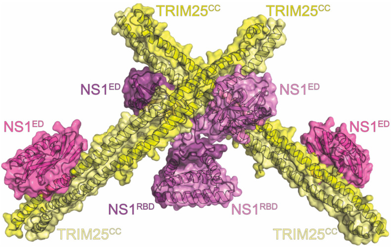
Crystal structures of purified TRIM25-CC complexed with both full-length and the NS1ED were obtained at 4.26Å and 2.8Å resolution, respectively. We note that the NS1ED alone was sufficient for binding and that each purified NS1ED bound to a TRIM25-CC dimer at two separate interfaces [114]. However, the second interface was not present with full-length NS1(Figure 11). This was additionally confirmed via mutational analysis [114]. The remaining interface consists of a linker between TRIM25-CC helices α2 and α3 making contacts with a highly conserved NS1 motif that includes a short α-helix comprising residues 95-99. NS1 L95 was especially important in the interaction, as it contacted a hydrophobic pocket of TRIM25 formed by residues from both chains of the CC, including V223, F274, I277, I324, and V327. L95 also participates in a hydrogen bond with TRIM25-CC E326. Other interactions can be observed between Y89ED, D222CC, and D229CC, as well as E101ED and Q212CC [114].
Figure 11.
Crystal structure of TRIM25 in complex with NS1. The complex consists of the NS1 homodimer (light violet and violet), two copies of the coiled-coil region of TRIM25 (TRIM25CC) colored light yellow and yellow, and two additional copies of the NS1ED (mauve). The NS1 protein in the structure is derived from the A/Puerto Rico/8/1934 (H1N1) strain of influenza. The PDB ID for this structure is 5NT1.
The complex structures cleared up some misconceptions about the mechanics of this interaction. While it was previously thought that NS1 binding to the TRIM25-CC would prevent TRIM25 dimerization, which is needed for activity, the complex showed that the dimer itself was involved in the interaction [114,212]. The dimeric TRIM25-CC is bound by two NS1EDs symmetrically at each end. This arrangement, along with NS1 being an obligate homodimer, was hypothesized to mediate higher order oligomer chains of NS1 and TRIM25, leading to more efficient sequestration [114]. This hypothesis is supported by the finding that mutating crucial NS1 residues L95, S99, and Y89 identified in the structure severely impacted the interaction while only slightly affecting RIG-I ubiquitination and interferon production. It was posited that even lower than expected binding affinity for each individual interaction can be overcome by oligomerization [114]. Previously investigated NS1 residues were not seen to be involved in the binding interface. E96/E97, which, when mutated, lost TRIM25 interference and virulence in mice, actually faced away from the interface and are likely needed for NS1 structural integrity rather than binding [114,212]. R38/K41 are known to be involved in RNA binding and RBD dimerization, meaning that, in this context, they likely interfere with higher-order oligomerization rather than direct TRIM25 binding as previously suggested [114,212].
Superposition of the structures with that of TRIM25CC-PRYSPRY revealed a different mechanism for NS1’s antagonism. Both PRYSPRY and NS1 ED contacted the linker region between α2 and α3, leading to steric hindrance in the presence of both [114]. Therefore, NS1 is likely to inhibit TRIM25 by preventing PRYSPRY and RING from being in the correct orientation to allow ubiquitination. The interaction was found to be host-species-specific, as NS1s from various strains have adapted to interact more efficiently with their host’s TRIM25 homologue. This adaptation demonstrates how NS1 can help drive host adaptation [213]. By adding another protein into its long list of interactions, NS1 achieves greater coverage of the RIG-I pathway to further antagonize the innate immune system and interferon production.
17. Conclusions
The ever-evolving nature of the Influenza A virus has rendered many of our defenses ineffective, making the need for new, innovative antivirals that much more pressing. Taking a structural approach to the development of these new antivirals arms us with the knowledge of how exactly we are targeting the virus, allowing us to make the most informed decisions possible. By knowing the structural underpinnings of crucial complexes for IAV pathogenesis, we can design drugs that target the most important interfaces to have the greatest effects. If we combine that knowledge with genetic data, we can narrow our targets to the most conserved regions of the viral protein that are the least likely to mutate. This process starts with the work covered in this review, but there is still much to be done, as has been highlighted. Some of the complexes discussed are still not fully mapped out, while many other complexes remain structurally unstudied (Table 1). Every piece of data drives us closer to being able to make the most effective IAV antivirals possible, making this field of study important and worthwhile.
Table 1.
Verified interactions between cellular host proteins and NS1 with the techniques used listed. Abbreviations used in the table are as follows: X-ray = X-ray crystallography, NMR = Nuclear magnetic resonance. Y2H = Yeast two hybrid, SEC-MALS = Size exclusion chromatography with multi-angle light scattering, 2DE = 2D gel electrophoresis, MS = Mass spectrometry, MD = Molecular dynamics simulations, FRET = Fluorescence resonance energy transfer, FA = Fluorescence anisotropy, BLI = Bio-layer interferometry, M2H = Mammalian two hybrid, CD = Circular dichroism, and IP = Immunoprecipitation. Certain techniques listed (e.g., IP) can be performed with either whole cell lysate or purified protein. For these types of experiments, an asterisk (*) denotes that these experiments were performed with purified proteins. Interactions that are defined with single amino acid resolution are denoted by grey shading of the cells.
| Name | Host Protein Function | Results of Interaction | Methods | PDB ID | Figure | Citations |
|---|---|---|---|---|---|---|
| CHD1 | chromatin remodeling | downregulation of host genes | IP*, ITC, X-ray | 4NW2 4O42 | 3 | Marazzi (2012) [87] Qin (2014) [115] |
| Ci/Gli1 | transcriptional mediator in Hedgehog pathway | modulates Hedgehog signaling | FRET-FLIM | Smelkinson (2017) [215] | ||
| CPSF30 | mRNA processing | downregulation of host genes | co-IP, Confocal, Pulldown, X-ray, Y2H | 2RHK | 7 | Das (2008) [40] Nemeroff (1998) [41] Dankar (2013) [62] Twu (2006) [68] Kuo (2018) [77] DeDiego (2016) [155] Kuo (2009) [158] Kochs (2007) [159] Twu (2007) [160] Li (2018) [161] Rodriguez (2018) [162] Zhu (2008) [163] |
| CrkII | cellular signaling through binding to JNK1 | inhibits innate immune response | Confocal, NMR Relaxation Dispersion, Pulldown, X-ray | 5UL6 | 4 | Shen (2017) [116] Ylösmäki (2016) [126] |
| CrkL | PI3K signaling | inhibits apoptosis | Binding assays *, co-IP | Hrincius (2010) [125] Heikinnen (2008) [127] Ylösmäki (2015) [133] Miyazaki (2013) [216] |
||
| Dlg | tight junction formation | disrupts tight junction formation to enhance viral spread | co-IP, Confocal, Pulldown * | Golebiewski (2011) [88] Thomas (2011) [89] Liu (2010) [137] |
||
| eIF4E | translation initiation | stimulates translation of viral mRNA | FA | Cruz (2022) [217] | ||
| eIF4G1 | translation initiation | enhances viral translation | co-IP, Pulldown * | Burgui (2003) [218] Aragon (2000) [219] |
||
| hPAF1C | transcription elongation | downregulation of host genes | IP * | Marazzi (2012) [87] | ||
| IFI35 | RIG-I regulation through degradation | inhibits type I interferon production to inhibit innate immune system | co-IP, Confocal, Pulldown * | Yang (2021) [220] | ||
| MAGI-1 | tight junction formation | disables tight junction formation to enhance viral spread | co-IP, Confocal, Pulldown * | Thomas (2011) [89] Liu (2010) [137] Kumar (2012) [221] |
||
| MORC3 | ATPase with antiviral activity | prevents MORC3 histone binding and autoinhibition | NMR, X-ray | 6O5W | 6 | Zhang (2019) [149] |
| NS1-BP | inhibits splicing, inhibits some NS1 activities | regulates cell viability through blocking NS1 stimulation of ERK pathway | IP *, Pulldown, Y2H | Miyazaki (2013) [216] Wolff (1998) [222] |
||
| Nucleolin and Fibrillarin | rRNA production | represses ribosome production | co-IP *, Confocal, Pulldown * | Yan (2017) [223] Melen (2012) [224] Murayama (2007) [225] |
||
| NXF1-NXT1 | RNA nuclear export | inhibits host mRNA translation | IP, Pulldown *, X-ray | 6E5U | 8 | Satterly (2007) [169] Zhang (2019) [172] |
| PABP1 | translation initiation | enhances viral translation | co-IP, EMSA, FA, FRET, Pulldown * | Burgui (2003) [218] de Rozieres (2020) [226] Arias-Mireles (2018) [227] |
||
| PABPII | mRNA processing | downregulation of host genes | IP * | Chen (1999) [228] | ||
| PACT | stimulates RIG-I-induced interferon production | inhibits interferon production | co-IP, Pulldown *, SILAC | Li (2006) [47] Tawaratsumida (2014) [229] |
||
| PDlim2 | scaffold protein that recruits NF-kB to ubiquitin ligase | unknown | BiFC, Pulldown *, M2H, X-ray | 3PDV | 9 | Yu (2011) [176] |
| PI3K | apoptosis regulation | inhibits apoptosis | BLI, co-IP, Confocal, ITC, MD, NMR, Pulldown, X-ray | 6U28 6OX7 | 10 | Hale (2010) [49] Ehrhardt (2007) [50] Ylösmäki (2015) [133] Hale (2006) [188] Shin (2007) [191] Hale (2008) [192] Cho (2020) [194] Li (2008) [196] Dubrow (2021) [197] Dubrow (2020) [198] |
| PKR | antiviral activity through translational shutdown | continued viral mRNA translation | co-IP, FRET, Pulldown *, Y2H | Li (2006) [47] Min (2007) [48] Schierhorn (2017) [230] Tan (1998) [231] |
||
| PSD-95 | regulates NO production | reduces NO production | IP *, Pulldown *, Y2H | Zhang (2011) [232] | ||
| RIG-I | interferon induction | inhibits interferon production | co-IP, Confocal, Crosslinking IP, NMR, Pulldown, Y2H, | Mibayashi (2007) [44] Dankar (2013) [62] Jureka (2015) [95] Jureka (2020) [109] Pothlichet (2013) [209] |
||
| RIL and Src Kinase | kinase cascade for cell signaling | alters cell signaling | Protein arrays, Pulldown * | Bavagnoli (2011) [233] | ||
| RuvBL2 | ATPase and helicase with multiple regulatory functions | inhibits infection-induced apoptosis by maintaining RuvBL2 protein abundance | 2DE-MS, co-IP, Pulldown * | Wang (2021) [79] | ||
| Scribble | cell polarity regulator, tight junction formation | disrupts tight junction formation to enhance viral spread | CD, co-IP, Confocal, ITC, Pulldown *, X-ray | 7QTO 7QTP 7QTU | 5 | Golebiewski (2011) [88] Thomas (2011) [89] Liu (2010) [137] Javorsky (2022) [143] |
| Staufen | RNA translocation and regulation | inhibits viral mRNA degradation | Confocal, IP *, Tethering assay, Y2H | Cho (2013) [234] Lee (2011) [235] Falcon (1999) [236] |
||
| TRIM25 | RIG-I activation | inhibits innate immune response | BLI, co-IP *, Confocal, NMR, SECMALS, X-ray | 5NT1 5NT2 |
11 | Koliopoulos (2018) [114] Gack (2009) [212] Rajsbaum (2012) [213] |
| WDR5 | coactivator involved in gene regulation | downregulation of host genes | ITC, X-ray | 4O45 | 1 | Qin (2014) [115] |
| XRN1 | mRNA decay factor in P-bodies | inhibits degradation of viral mRNAs | co-IP, Confocal, Pulldown * | Liu (2021) [237] | ||
| YAP/TAZ | Hippo pathway activation | downregulates cytokines and the innate immune system | co-IP with exogenous Protein | Zhang (2022) [238] |
Author Contributions
Writing—original draft presentation: M.E.B., A.B.K., A.S.J. and C.M.P.; Writing—review and editing: M.E.B. and C.M.P.; Funding acquisition: C.M.P. All authors have read and agreed to the published version of the manuscript.
Institutional Review Board Statement
Not applicable.
Informed Consent Statement
Not applicable.
Data Availability Statement
Not applicable.
Conflicts of Interest
The authors declare no conflict of interest.
Funding Statement
This work was funded by NIH NIAID grant AI134693.
Footnotes
Disclaimer/Publisher’s Note: The statements, opinions and data contained in all publications are solely those of the individual author(s) and contributor(s) and not of MDPI and/or the editor(s). MDPI and/or the editor(s) disclaim responsibility for any injury to people or property resulting from any ideas, methods, instructions or products referred to in the content.
References
- 1.Thompson W.W., Comanor L., Shay D.K. Epidemiology of seasonal influenza: Use of surveillance data and statistical models to estimate the burden of disease. J. Infect. Dis. 2006;194((Suppl. S2)):S82–S91. doi: 10.1086/507558. [DOI] [PubMed] [Google Scholar]
- 2.Molinari N.A., Ortega-Sanchez I.R., Messonnier M.L., Thompson W.W., Wortley P.M., Weintraub E., Bridges C.B. The annual impact of seasonal influenza in the US: Measuring disease burden and costs. Vaccine. 2007;25:5086–5096. doi: 10.1016/j.vaccine.2007.03.046. [DOI] [PubMed] [Google Scholar]
- 3.Yang Y., Sugimoto J.D., Halloran M.E., Basta N.E., Chao D.L., Matrajt L., Potter G., Kenah E., Longini I.M., Jr. The transmissibility and control of pandemic influenza A (H1N1) virus. Science. 2009;326:729–733. doi: 10.1126/science.1177373. [DOI] [PMC free article] [PubMed] [Google Scholar]
- 4.Muthuri S.G., Venkatesan S., Myles P.R., Leonardi-Bee J., Al Khuwaitir T.S., Al Mamun A., Anovadiya A.P., Azziz-Baumgartner E., Baez C., Bassetti M., et al. Effectiveness of neuraminidase inhibitors in reducing mortality in patients admitted to hospital with influenza A H1N1pdm09 virus infection: A meta-analysis of individual participant data. Lancet Respir. Med. 2014;2:395–404. doi: 10.1016/S2213-2600(14)70041-4. [DOI] [PMC free article] [PubMed] [Google Scholar]
- 5.Zhou Z., Liu T., Zhang J., Zhan P., Liu X. Influenza A virus polymerase: An attractive target for next-generation anti-influenza therapeutics. Drug Discov. Today. 2018;23:503–518. doi: 10.1016/j.drudis.2018.01.028. [DOI] [PubMed] [Google Scholar]
- 6.Gamblin S.J., Skehel J.J. Influenza hemagglutinin and neuraminidase membrane glycoproteins. J. Biol. Chem. 2010;285:28403–28409. doi: 10.1074/jbc.R110.129809. [DOI] [PMC free article] [PubMed] [Google Scholar]
- 7.Wu N.C., Wilson I.A. Influenza Hemagglutinin Structures and Antibody Recognition. Cold Spring Harb. Perspect. Med. 2020;10:a038778. doi: 10.1101/cshperspect.a038778. [DOI] [PMC free article] [PubMed] [Google Scholar]
- 8.Giurgea L.T., Morens D.M., Taubenberger J.K., Memoli M.J. Influenza Neuraminidase: A Neglected Protein and Its Potential for a Better Influenza Vaccine. Vaccines. 2020;8:409. doi: 10.3390/vaccines8030409. [DOI] [PMC free article] [PubMed] [Google Scholar]
- 9.McKimm-Breschkin J.L. Influenza neuraminidase inhibitors: Antiviral action and mechanisms of resistance. Influenza Other Respir. Viruses. 2013;7((Suppl. 1)):25–36. doi: 10.1111/irv.12047. [DOI] [PMC free article] [PubMed] [Google Scholar]
- 10.A revision of the system of nomenclature for influenza viruses: A WHO memorandum. Bull. World Health Organ. 1980;58:585–591. [PMC free article] [PubMed] [Google Scholar]
- 11.Krammer F., Smith G.J.D., Fouchier R.A.M., Peiris M., Kedzierska K., Doherty P.C., Palese P., Shaw M.L., Treanor J., Webster R.G., et al. Influenza. Nat. Rev. Dis. Primers. 2018;4:3. doi: 10.1038/s41572-018-0002-y. [DOI] [PMC free article] [PubMed] [Google Scholar]
- 12.Ma W., Garcia-Sastre A., Schwemmle M. Expected and Unexpected Features of the Newly Discovered Bat Influenza A-like Viruses. PLoS Pathog. 2015;11:e10048192015. doi: 10.1371/journal.ppat.1004819. [DOI] [PMC free article] [PubMed] [Google Scholar]
- 13.Tong S., Li Y., Rivailler P., Conrardy C., Castillo D.A., Chen L.M., Recuenco S., Ellison J.A., Davis C.T., York I.A., et al. A distinct lineage of influenza A virus from bats. Proc. Natl. Acad. Sci. USA. 2012;109:4269–4274. doi: 10.1073/pnas.1116200109. [DOI] [PMC free article] [PubMed] [Google Scholar]
- 14.Portela A., Digard P. The influenza virus nucleoprotein: A multifunctional RNA-binding protein pivotal to virus replication. J. Gen. Virol. 2002;83:723–734. doi: 10.1099/0022-1317-83-4-723. [DOI] [PubMed] [Google Scholar]
- 15.Esparza M., Bhat P., Fontoura B.M. Viral-host interactions during splicing and nuclear export of influenza virus mRNAs. Curr. Opin. Virol. 2022;55:101254. doi: 10.1016/j.coviro.2022.101254. [DOI] [PMC free article] [PubMed] [Google Scholar]
- 16.Peukes J., Xiong X., Briggs J.A.G. New structural insights into the multifunctional influenza A matrix protein 1. FEBS Lett. 2021;595:2535–2543. doi: 10.1002/1873-3468.14194. [DOI] [PMC free article] [PubMed] [Google Scholar]
- 17.Pinto L.H., Lamb R.A. The M2 proton channels of influenza A and B viruses. J. Biol. Chem. 2006;281:8997–9000. doi: 10.1074/jbc.R500020200. [DOI] [PubMed] [Google Scholar]
- 18.Jalily P.H., Duncan M.C., Fedida D., Wang J., Tietjen I. Put a cork in it: Plugging the M2 viral ion channel to sink influenza. Antiviral Res. 2020;178:104780. doi: 10.1016/j.antiviral.2020.104780. [DOI] [PMC free article] [PubMed] [Google Scholar]
- 19.Krug R.M. Functions of the influenza A virus NS1 protein in antiviral defense. Curr. Opin. Virol. 2015;12:1–6. doi: 10.1016/j.coviro.2015.01.007. [DOI] [PMC free article] [PubMed] [Google Scholar]
- 20.Ayllon J., Garcia-Sastre A. The NS1 protein: A multitasking virulence factor. Curr. Top. Microbiol. Immunol. 2015;386:73–107. doi: 10.1007/82_2014_400. [DOI] [PubMed] [Google Scholar]
- 21.Richardson J.C., Akkina R.K. NS2 protein of influenza virus is found in purified virus and phosphorylated in infected cells. Arch. Virol. 1991;116:69–80. doi: 10.1007/BF01319232. [DOI] [PubMed] [Google Scholar]
- 22.Ward A.C., Castelli L.A., Lucantoni A.C., White J.F., Azad A.A., Macreadie I.G. Expression and analysis of the NS2 protein of influenza A virus. Arch. Virol. 1995;140:2067–2073. doi: 10.1007/BF01322693. [DOI] [PubMed] [Google Scholar]
- 23.Yasuda J., Nakada S., Kato A., Toyoda T., Ishihama A. Molecular assembly of influenza virus: Association of the NS2 protein with virion matrix. Virology. 1993;196:249–255. doi: 10.1006/viro.1993.1473. [DOI] [PubMed] [Google Scholar]
- 24.O’Neill R.E., Talon J., Palese P. The influenza virus NEP (NS2 protein) mediates the nuclear export of viral ribonucleoproteins. EMBO J. 1998;17:288–296. doi: 10.1093/emboj/17.1.288. [DOI] [PMC free article] [PubMed] [Google Scholar]
- 25.Akarsu H., Burmeister W.P., Petosa C., Petit I., Müller C.W., Ruigrok R.W.H., Baudin F. Crystal structure of the M1 protein-binding domain of the influenza A virus nuclear export protein (NEP/NS2) EMBO J. 2003;22:4646–4655. doi: 10.1093/emboj/cdg449. [DOI] [PMC free article] [PubMed] [Google Scholar]
- 26.Huang S., Chen J., Chen Q., Wang H., Yao Y., Chen J., Chen Z. A Second CRM1-Dependent Nuclear Export Signal in the Influenza A Virus NS2 Protein Contributes to the Nuclear Export of Viral Ribonucleoproteins. J. Virol. 2013;87:767–778. doi: 10.1128/JVI.06519-11. [DOI] [PMC free article] [PubMed] [Google Scholar]
- 27.Robb N.C., Smith M., Vreede F.T., Fodor E. NS2/NEP protein regulates transcription and replication of the influenza virus RNA genome. J. General. Virol. 2009;90:1398–1407. doi: 10.1099/vir.0.009639-0. [DOI] [PubMed] [Google Scholar]
- 28.Reuther P., Giese S., Götz V., Kilb N., Mänz B., Brunotte L., Schwemmle M. Adaptive mutations in the nuclear export protein of human-derived H5N1 strains facilitate a polymerase activity-enhancing conformation. J. Virol. 2014;88:263–271. doi: 10.1128/JVI.01495-13. [DOI] [PMC free article] [PubMed] [Google Scholar]
- 29.Mänz B., Schwemmle M., Brunotte L. Adaptation of Avian Influenza A Virus Polymerase in Mammals To Overcome the Host Species Barrier. J. Virol. 2013;87:7200–7209. doi: 10.1128/JVI.00980-13. [DOI] [PMC free article] [PubMed] [Google Scholar]
- 30.Jagger B.W., Wise H.M., Kash J.C., Walters K.A., Wills N.M., Xiao Y.L., Dunfee R.L., Schwartzman L.M., Ozinsky A., Bell G.L., et al. An overlapping protein-coding region in influenza A virus segment 3 modulates the host response. Science. 2012;337:199–204. doi: 10.1126/science.1222213. [DOI] [PMC free article] [PubMed] [Google Scholar]
- 31.Muramoto Y., Noda T., Kawakami E., Akkina R., Kawaoka Y. Identification of novel influenza A virus proteins translated from PA mRNA. J. Virol. 2013;87:2455–2462. doi: 10.1128/JVI.02656-12. [DOI] [PMC free article] [PubMed] [Google Scholar]
- 32.Chen W., Calvo P.A., Malide D., Gibbs J., Schubert U., Bacik I., Basta S., O’Neill R., Schickli J., Palese P., et al. A novel influenza A virus mitochondrial protein that induces cell death. Nat. Med. 2001;7:1306–1312. doi: 10.1038/nm1201-1306. [DOI] [PubMed] [Google Scholar]
- 33.Wise H.M., Foeglein A., Sun J., Dalton R.M., Patel S., Howard W., Anderson E.C., Barclay W.S., Digard P. A complicated message: Identification of a novel PB1-related protein translated from influenza A virus segment 2 mRNA. J. Virol. 2009;83:8021–8031. doi: 10.1128/JVI.00826-09. [DOI] [PMC free article] [PubMed] [Google Scholar]
- 34.Yamayoshi S., Watanabe M., Goto H., Kawaoka Y. Identification of a Novel Viral Protein Expressed from the PB2 Segment of Influenza A Virus. J. Virol. 2016;90:444–456. doi: 10.1128/JVI.02175-15. [DOI] [PMC free article] [PubMed] [Google Scholar]
- 35.Wise H.M., Hutchinson E.C., Jagger B.W., Stuart A.D., Kang Z.H., Robb N., Schwartzman L.M., Kash J.C., Fodor E., Firth A.E., et al. Identification of a novel splice variant form of the influenza A virus M2 ion channel with an antigenically distinct ectodomain. PLoS Pathog. 2012;8:e1002998. doi: 10.1371/journal.ppat.1002998. [DOI] [PMC free article] [PubMed] [Google Scholar]
- 36.Selman M., Dankar S.K., Forbes N.E., Jia J.J., Brown E.G. Adaptive mutation in influenza A virus non-structural gene is linked to host switching and induces a novel protein by alternative splicing. Emerg. Microbes Infect. 2012;1:e42. doi: 10.1038/emi.2012.38. [DOI] [PMC free article] [PubMed] [Google Scholar]
- 37.Robb N.C., Jackson D., Vreede F.T., Fodor E. Splicing of influenza A virus NS1 mRNA is independent of the viral NS1 protein. J. Gen. Virol. 2010;91:2331–2340. doi: 10.1099/vir.0.022004-0. [DOI] [PubMed] [Google Scholar]
- 38.Chua M.A., Schmid S., Perez J.T., Langlois R.A., Tenoever B.R. Influenza A virus utilizes suboptimal splicing to coordinate the timing of infection. Cell Rep. 2013;3:23–29. doi: 10.1016/j.celrep.2012.12.010. [DOI] [PMC free article] [PubMed] [Google Scholar]
- 39.Garcia-Sastre A., Egorov A., Matassov D., Brandt S., Levy D.E., Durbin J.E., Palese P., Muster T. Influenza A virus lacking the NS1 gene replicates in interferon-deficient systems. Virology. 1998;252:324–330. doi: 10.1006/viro.1998.9508. [DOI] [PubMed] [Google Scholar]
- 40.Das K., Ma L.C., Xiao R., Radvansky B., Aramini J., Zhao L., Marklund J., Kuo R.L., Twu K.Y., Arnold E., et al. Structural basis for suppression of a host antiviral response by influenza A virus. Proc. Natl. Acad. Sci. USA. 2008;105:13093–13098. doi: 10.1073/pnas.0805213105. [DOI] [PMC free article] [PubMed] [Google Scholar]
- 41.Nemeroff M.E., Barabino S.M., Li Y., Keller W., Krug R.M. Influenza virus NS1 protein interacts with the cellular 30 kDa subunit of CPSF and inhibits 3′end formation of cellular pre-mRNAs. Mol. Cell. 1998;1:991–1000. doi: 10.1016/S1097-2765(00)80099-4. [DOI] [PubMed] [Google Scholar]
- 42.Noah D.L., Twu K.Y., Krug R.M. Cellular antiviral responses against influenza A virus are countered at the posttranscriptional level by the viral NS1A protein via its binding to a cellular protein required for the 3′ end processing of cellular pre-mRNAS. Virology. 2003;307:386–395. doi: 10.1016/S0042-6822(02)00127-7. [DOI] [PubMed] [Google Scholar]
- 43.Guo Z., Chen L.M., Zeng H., Gomez J.A., Plowden J., Fujita T., Katz J.M., Donis R.O., Sambhara S. NS1 protein of influenza A virus inhibits the function of intracytoplasmic pathogen sensor, RIG-I. Am. J. Respir. Cell Mol. Biol. 2007;36:263–269. doi: 10.1165/rcmb.2006-0283RC. [DOI] [PubMed] [Google Scholar]
- 44.Mibayashi M., Martínez-Sobrido L., Loo Y.M., Cárdenas W.B., Gale M., Jr., García-Sastre A. Inhibition of retinoic acid-inducible gene I-mediated induction of beta interferon by the NS1 protein of influenza A virus. J. Virol. 2007;81:514–524. doi: 10.1128/JVI.01265-06. [DOI] [PMC free article] [PubMed] [Google Scholar]
- 45.Opitz B., Rejaibi A., Dauber B., Eckhard J., Vinzing M., Schmeck B., Hippenstiel S., Suttorp N., Wolff T. IFNbeta induction by influenza A virus is mediated by RIG-I which is regulated by the viral NS1 protein. Cell. Microbiol. 2007;9:930–938. doi: 10.1111/j.1462-5822.2006.00841.x. [DOI] [PubMed] [Google Scholar]
- 46.Pichlmair A., Schulz O., Tan C.P., Näslund T.I., Liljeström P., Weber F., Reis e Sousa C. RIG-I-mediated antiviral responses to single-stranded RNA bearing 5′-phosphates. Science. 2006;314:997–1001. doi: 10.1126/science.1132998. [DOI] [PubMed] [Google Scholar]
- 47.Li S., Min J.Y., Krug R.M., Sen G.C. Binding of the influenza A virus NS1 protein to PKR mediates the inhibition of its activation by either PACT or double-stranded RNA. Virology. 2006;349:13–21. doi: 10.1016/j.virol.2006.01.005. [DOI] [PubMed] [Google Scholar]
- 48.Min J.Y., Li S., Sen G.C., Krug R.M. A site on the influenza A virus NS1 protein mediates both inhibition of PKR activation and temporal regulation of viral RNA synthesis. Virology. 2007;363:236–243. doi: 10.1016/j.virol.2007.01.038. [DOI] [PubMed] [Google Scholar]
- 49.Hale B.G., Kerry P.S., Jackson D., Precious B.L., Gray A., Killip M.J., Randall R.E., Russell R.J. Structural insights into phosphoinositide 3-kinase activation by the influenza A virus NS1 protein. Proc. Natl. Acad. Sci. USA. 2010;107:1954–1959. doi: 10.1073/pnas.0910715107. [DOI] [PMC free article] [PubMed] [Google Scholar]
- 50.Ehrhardt C., Wolff T., Pleschka S., Planz O., Beermann W., Bode J.G., Schmolke M., Ludwig S. Influenza A virus NS1 protein activates the PI3K/Akt pathway to mediate antiapoptotic signaling responses. J. Virol. 2007;81:3058–3067. doi: 10.1128/JVI.02082-06. [DOI] [PMC free article] [PubMed] [Google Scholar]
- 51.Jackson D., Killip M.J., Galloway C.S., Russell R.J., Randall R.E. Loss of function of the influenza A virus NS1 protein promotes apoptosis but this is not due to a failure to activate phosphatidylinositol 3-kinase (PI3K) Virology. 2010;396:94–105. doi: 10.1016/j.virol.2009.10.004. [DOI] [PubMed] [Google Scholar]
- 52.Li W., Wang G., Zhang H., Shen Y., Dai J., Wu L., Zhou J., Jiang Z., Li K. Inability of NS1 protein from an H5N1 influenza virus to activate PI3K/Akt signaling pathway correlates to the enhanced virus replication upon PI3K inhibition. Vet. Res. 2012;43:36. doi: 10.1186/1297-9716-43-36. [DOI] [PMC free article] [PubMed] [Google Scholar]
- 53.Lu X., Masic A., Li Y., Shin Y., Liu Q., Zhou Y. The PI3K/Akt pathway inhibits influenza A virus-induced Bax-mediated apoptosis by negatively regulating the JNK pathway via ASK1. J. Gen. Virol. 2010;91:1439–1449. doi: 10.1099/vir.0.018465-0. [DOI] [PubMed] [Google Scholar]
- 54.Hale B.G., Randall R.E., Ortin J., Jackson D. The multifunctional NS1 protein of influenza A viruses. J. Gen. Virol. 2008;89:2359–2376. doi: 10.1099/vir.0.2008/004606-0. [DOI] [PubMed] [Google Scholar]
- 55.Marc D. Influenza virus non-structural protein NS1: Interferon antagonism and beyond. J. Gen. Virol. 2014;95:2594–2611. doi: 10.1099/vir.0.069542-0. [DOI] [PubMed] [Google Scholar]
- 56.Khaperskyy D.A., McCormick C. Timing Is Everything: Coordinated Control of Host Shutoff by Influenza A Virus NS1 and PA-X Proteins. J. Virol. 2015;89:6528–6531. doi: 10.1128/JVI.00386-15. [DOI] [PMC free article] [PubMed] [Google Scholar]
- 57.Egorov A., Brandt S., Sereinig S., Romanova J., Ferko B., Katinger D., Grassauer A., Alexandrova G., Katinger H., Muster T. Transfectant influenza A viruses with long deletions in the NS1 protein grow efficiently in Vero cells. J. Virol. 1998;72:6437–6441. doi: 10.1128/JVI.72.8.6437-6441.1998. [DOI] [PMC free article] [PubMed] [Google Scholar]
- 58.Dankar S.K., Wang S., Ping J., Forbes N.E., Keleta L., Li Y., Brown E.G. Influenza A virus NS1 gene mutations F103L and M106I increase replication and virulence. Virol. J. 2011;8:13. doi: 10.1186/1743-422X-8-13. [DOI] [PMC free article] [PubMed] [Google Scholar]
- 59.Cauthen A.N., Swayne D.E., Sekellick M.J., Marcus P.I., Suarez D.L. Amelioration of influenza virus pathogenesis in chickens attributed to the enhanced interferon-inducing capacity of a virus with a truncated NS1 gene. J. Virol. 2007;81:1838–1847. doi: 10.1128/JVI.01667-06. [DOI] [PMC free article] [PubMed] [Google Scholar]
- 60.Basler C.F., Reid A.H., Dybing J.K., Janczewski T.A., Fanning T.G., Zheng H., Salvatore M., Perdue M.L., Swayne D.E., Garcia-Sastre A., et al. Sequence of the 1918 pandemic influenza virus nonstructural gene (NS) segment and characterization of recombinant viruses bearing the 1918 NS genes. Proc. Natl. Acad. Sci. USA. 2001;98:2746–2751. doi: 10.1073/pnas.031575198. [DOI] [PMC free article] [PubMed] [Google Scholar]
- 61.Kim I.H., Kwon H.J., Lee S.H., Kim D.Y., Kim J.H. Effects of different NS genes of avian influenza viruses and amino acid changes on pathogenicity of recombinant A/Puerto Rico/8/34 viruses. Vet. Microbiol. 2015;175:17–25. doi: 10.1016/j.vetmic.2014.11.010. [DOI] [PubMed] [Google Scholar]
- 62.Dankar S.K., Miranda E., Forbes N.E., Pelchat M., Tavassoli A., Selman M., Ping J., Jia J., Brown E.G. Influenza A/Hong Kong/156/1997(H5N1) virus NS1 gene mutations F103L and M106I both increase IFN antagonism, virulence and cytoplasmic localization but differ in binding to RIG-I and CPSF30. Virol. J. 2013;10:243. doi: 10.1186/1743-422X-10-243. [DOI] [PMC free article] [PubMed] [Google Scholar]
- 63.Chauche C., Nogales A., Zhu H., Goldfarb D., Ahmad Shanizza A.I., Gu Q., Parrish C.R., Martinez-Sobrido L., Marshall J.F., Murcia P.R. Mammalian Adaptation of an Avian Influenza A Virus Involves Stepwise Changes in NS1. J. Virol. 2018;92:e01875-17. doi: 10.1128/JVI.01875-17. [DOI] [PMC free article] [PubMed] [Google Scholar]
- 64.Nogales A., Martinez-Sobrido L., Chiem K., Topham D.J., DeDiego M.L. Functional Evolution of the 2009 Pandemic H1N1 Influenza Virus NS1 and PA in Humans. J. Virol. 2018;92:10-1128. doi: 10.1128/JVI.01206-18. [DOI] [PMC free article] [PubMed] [Google Scholar]
- 65.Ma W., Brenner D., Wang Z., Dauber B., Ehrhardt C., Hogner K., Herold S., Ludwig S., Wolff T., Yu K., et al. The NS segment of an H5N1 highly pathogenic avian influenza virus (HPAIV) is sufficient to alter replication efficiency, cell tropism, and host range of an H7N1 HPAIV. J. Virol. 2010;84:2122–2133. doi: 10.1128/JVI.01668-09. [DOI] [PMC free article] [PubMed] [Google Scholar]
- 66.Engel D.A. The influenza virus NS1 protein as a therapeutic target. Antiviral Res. 2013;99:409–416. doi: 10.1016/j.antiviral.2013.06.005. [DOI] [PMC free article] [PubMed] [Google Scholar]
- 67.Darapaneni V., Prabhaker V.K., Kukol A. Large-scale analysis of influenza A virus sequences reveals potential drug target sites of non-structural proteins. J. Gen. Virol. 2009;90:2124–2133. doi: 10.1099/vir.0.011270-0. [DOI] [PubMed] [Google Scholar]
- 68.Twu K.Y., Noah D.L., Rao P., Kuo R.L., Krug R.M. The CPSF30 binding site on the NS1A protein of influenza A virus is a potential antiviral target. J. Virol. 2006;80:3957–3965. doi: 10.1128/JVI.80.8.3957-3965.2006. [DOI] [PMC free article] [PubMed] [Google Scholar]
- 69.Rosario-Ferreira N., Preto A.J., Melo R., Moreira I.S., Brito R.M.M. The Central Role of Non-Structural Protein 1 (NS1) in Influenza Biology and Infection. Int. J. Mol. Sci. 2020;21:1511. doi: 10.3390/ijms21041511. [DOI] [PMC free article] [PubMed] [Google Scholar]
- 70.Kim H.J., Jeong M.S., Jang S.B. Structure and Activities of the NS1 Influenza Protein and Progress in the Development of Small-Molecule Drugs. Int. J. Mol. Sci. 2021;22:4242. doi: 10.3390/ijms22084242. [DOI] [PMC free article] [PubMed] [Google Scholar]
- 71.Trigueiro-Louro J., Santos L.A., Almeida F., Correia V., Brito R.M.M., Rebelo-de-Andrade H. NS1 protein as a novel anti-influenza target: Map-and-mutate antiviral rationale reveals new putative druggable hot spots with an important role on viral replication. Virology. 2022;565:106–116. doi: 10.1016/j.virol.2021.11.001. [DOI] [PubMed] [Google Scholar]
- 72.Naceri S., Marc D., Blot R., Flatters D., Camproux A.C. Druggable Pockets at the RNA Interface Region of Influenza A Virus NS1 Protein Are Conserved across Sequence Variants from Distinct Subtypes. Biomolecules. 2022;13:64. doi: 10.3390/biom13010064. [DOI] [PMC free article] [PubMed] [Google Scholar]
- 73.Abi Hussein H., Geneix C., Cauvin C., Marc D., Flatters D., Camproux A.C. Molecular Dynamics Simulations of Influenza A Virus NS1 Reveal a Remarkably Stable RNA-Binding Domain Harboring Promising Druggable Pockets. Viruses. 2020;12:837. doi: 10.3390/v12050537. [DOI] [PMC free article] [PubMed] [Google Scholar]
- 74.Wathen M.W., Barro M., Bright R.A. Antivirals in seasonal and pandemic influenza--future perspectives. Influenza Other Respir. Viruses. 2013;7((Suppl. S1)):76–80. doi: 10.1111/irv.12049. [DOI] [PMC free article] [PubMed] [Google Scholar]
- 75.Koszalka P., Tilmanis D., Hurt A.C. Influenza antivirals currently in late-phase clinical trial. Influenza Other Respir. Viruses. 2017;11:240–246. doi: 10.1111/irv.12446. [DOI] [PMC free article] [PubMed] [Google Scholar]
- 76.de Chassey B., Aublin-Gex A., Ruggieri A., Meyniel-Schicklin L., Pradezynski F., Davoust N., Chantier T., Tafforeau L., Mangeot P.E., Ciancia C., et al. The interactomes of influenza virus NS1 and NS2 proteins identify new host factors and provide insights for ADAR1 playing a supportive role in virus replication. PLoS Pathog. 2013;9:e1003440. doi: 10.1371/journal.ppat.1003440. [DOI] [PMC free article] [PubMed] [Google Scholar]
- 77.Kuo R.L., Chen C.J., Tam E.H., Huang C.G., Li L.H., Li Z.H., Su P.C., Liu H.P., Wu C.C. Interactome Analysis of NS1 Protein Encoded by Influenza A H7N9 Virus Reveals an Inhibitory Role of NS1 in Host mRNA Maturation. J. Proteome Res. 2018;17:1474–1484. doi: 10.1021/acs.jproteome.7b00815. [DOI] [PubMed] [Google Scholar]
- 78.Kuo R.L., Li Z.H., Li L.H., Lee K.M., Tam E.H., Liu H.M., Liu H.P., Shih S.R., Wu C.C. Interactome Analysis of the NS1 Protein Encoded by Influenza A H1N1 Virus Reveals a Positive Regulatory Role of Host Protein PRP19 in Viral Replication. J. Proteome Res. 2016;15:1639–1648. doi: 10.1021/acs.jproteome.6b00103. [DOI] [PubMed] [Google Scholar]
- 79.Wang Q., Zhang Q., Zheng M., Wen J., Li Q., Zhao G. Viral-Host Interactome Analysis Reveals Chicken STAU2 Interacts With Non-structural Protein 1 and Promotes the Replication of H5N1 Avian Influenza Virus. Front. Immunol. 2021;12:590679. doi: 10.3389/fimmu.2021.590679. [DOI] [PMC free article] [PubMed] [Google Scholar]
- 80.Krug R.M., García-Sastre A. Textbook of Influenza. 2013. [(accessed on 1 August 2023)]. The NS1 Protein: A Master Regulator of Host and Viral Functions; pp. 114–132. Available online: https://onlinelibrary.wiley.com/doi/abs/10.1002/9781118636817.ch7. [Google Scholar]
- 81.Palese P., Shaw M.L. Orthomyxoviridae: The Viruses and Their Replication. In: Knipe D.M., Katze M.G., editors. Fields Viology. 5th ed. Lippincott, Williams, & Wilkins; Philadelphia, PA, USA: 2006. pp. 1647–1689. [Google Scholar]
- 82.Chien C.Y., Tejero R., Huang Y., Zimmerman D.E., Rios C.B., Krug R.M., Montelione G.T. A novel RNA-binding motif in influenza A virus non-structural protein 1. Nat. Struct. Biol. 1997;4:891–895. doi: 10.1038/nsb1197-891. [DOI] [PubMed] [Google Scholar]
- 83.Wang W., Riedel K., Lynch P., Chien C.Y., Montelione G.T., Krug R.M. RNA binding by the novel helical domain of the influenza virus NS1 protein requires its dimer structure and a small number of specific basic amino acids. RNA. 1999;5:195–205. doi: 10.1017/S1355838299981621. [DOI] [PMC free article] [PubMed] [Google Scholar]
- 84.Hatada E., Saito S., Okishio N., Fukuda R. Binding of the influenza virus NS1 protein to model genome RNAs. Pt 5J. Gen. Virol. 1997;78:1059–1063. doi: 10.1099/0022-1317-78-5-1059. [DOI] [PubMed] [Google Scholar]
- 85.Hatada E., Takizawa T., Fukuda R. Specific binding of influenza A virus NS1 protein to the virus minus-sense RNA in vitro. Pt 1J. Gen. Virol. 1992;73:17–25. doi: 10.1099/0022-1317-73-1-17. [DOI] [PubMed] [Google Scholar]
- 86.Trapp S., Soubieux D., Marty H., Esnault E., Hoffmann T.W., Chandenier M., Lion A., Kut E., Quere P., Larcher T., et al. Shortening the unstructured, interdomain region of the non-structural protein NS1 of an avian H1N1 influenza virus increases its replication and pathogenicity in chickens. J. Gen. Virol. 2014;95:1233–1243. doi: 10.1099/vir.0.063776-0. [DOI] [PubMed] [Google Scholar]
- 87.Marazzi I., Ho J.S., Kim J., Manicassamy B., Dewell S., Albrecht R.A., Seibert C.W., Schaefer U., Jeffrey K.L., Prinjha R.K., et al. Suppression of the antiviral response by an influenza histone mimic. Nature. 2012;483:428–433. doi: 10.1038/nature10892. [DOI] [PMC free article] [PubMed] [Google Scholar]
- 88.Golebiewski L., Liu H., Javier R.T., Rice A.P. The avian influenza virus NS1 ESEV PDZ binding motif associates with Dlg1 and Scribble to disrupt cellular tight junctions. J. Virol. 2011;85:10639–10648. doi: 10.1128/JVI.05070-11. [DOI] [PMC free article] [PubMed] [Google Scholar]
- 89.Thomas M., Kranjec C., Nagasaka K., Matlashewski G., Banks L. Analysis of the PDZ binding specificities of Influenza A Virus NS1 proteins. Virol. J. 2011;8:25. doi: 10.1186/1743-422X-8-25. [DOI] [PMC free article] [PubMed] [Google Scholar]
- 90.Jackson D., Hossain M.J., Hickman D., Perez D.R., Lamb R.A. A new influenza virus virulence determinant: The NS1 protein four C-terminal residues modulate pathogenicity. Proc. Natl. Acad. Sci. USA. 2008;105:4381. doi: 10.1073/pnas.0800482105. [DOI] [PMC free article] [PubMed] [Google Scholar]
- 91.Soubies S.M., Volmer C., Croville G., Loupias J., Peralta B., Costes P., Lacroux C., Guérin J.-L., Volmer R. Species-Specific Contribution of the Four C-Terminal Amino Acids of Influenza A Virus NS1 Protein to Virulence. J. Virol. 2010;84:6733. doi: 10.1128/JVI.02427-09. [DOI] [PMC free article] [PubMed] [Google Scholar]
- 92.Bornholdt Z.A., Prasad B.V. X-ray structure of influenza virus NS1 effector domain. Nat. Struct. Mol. Biol. 2006;13:559–560. doi: 10.1038/nsmb1099. [DOI] [PubMed] [Google Scholar]
- 93.Bullough P.A., Hughson F.M., Skehel J.J., Wiley D.C. Structure of influenza haemagglutinin at the pH of membrane fusion. Nature. 1994;371:37–43. doi: 10.1038/371037a0. [DOI] [PubMed] [Google Scholar]
- 94.Hale B.G., Barclay W.S., Randall R.E., Russell R.J. Structure of an avian influenza A virus NS1 protein effector domain. Virology. 2008;378:1–5. doi: 10.1016/j.virol.2008.05.026. [DOI] [PubMed] [Google Scholar]
- 95.Jureka A.S., Kleinpeter A.B., Cornilescu G., Cornilescu C.C., Petit C.M. Structural Basis for a Novel Interaction between the NS1 Protein Derived from the 1918 Influenza Virus and RIG-I. Structure. 2015;23:2001–2010. doi: 10.1016/j.str.2015.08.007. [DOI] [PMC free article] [PubMed] [Google Scholar]
- 96.Kerry P.S., Ayllon J., Taylor M.A., Hass C., Lewis A., García-Sastre A., Randall R.E., Hale B.G., Russell R.J. A Transient Homotypic Interaction Model for the Influenza A Virus NS1 Protein Effector Domain. PLoS ONE. 2011;6:e17946. doi: 10.1371/journal.pone.0017946. [DOI] [PMC free article] [PubMed] [Google Scholar]
- 97.Kerry P.S., Long E., Taylor M.A., Russell R.J. Conservation of a crystallographic interface suggests a role for beta-sheet augmentation in influenza virus NS1 multifunctionality. Acta Crystallo. Sect. F Struct. Biol. Cryst. Commun. 2011;67:858–861. doi: 10.1107/S1744309111019312. [DOI] [PMC free article] [PubMed] [Google Scholar]
- 98.Liu J., Lynch P.A., Chien C.Y., Montelione G.T., Krug R.M., Berman H.M. Crystal structure of the unique RNA-binding domain of the influenza virus NS1 protein. Nat. Struct. Biol. 1997;4:896–899. doi: 10.1038/nsb1197-896. [DOI] [PubMed] [Google Scholar]
- 99.Turkington H.L., Juozapaitis M., Kerry P.S., Aydillo T., Ayllon J., García-Sastre A., Schwemmle M., Hale B.G. Novel Bat Influenza Virus NS1 Proteins Bind Double-Stranded RNA and Antagonize Host Innate Immunity. J. Virol. 2015;89:10696. doi: 10.1128/JVI.01430-15. [DOI] [PMC free article] [PubMed] [Google Scholar]
- 100.Xia S., Monzingo A.F., Robertus J.D. Structure of NS1A effector domain from the influenza A/Udorn/72 virus. Acta Crystallo. Sect. D Biol. Crystallogr. 2009;65:11–17. doi: 10.1107/S0907444908032186. [DOI] [PMC free article] [PubMed] [Google Scholar]
- 101.Xia S., Robertus J.D. X-ray structures of NS1 effector domain mutants. Arch. Biochem. Biophys. 2010;494:198–204. doi: 10.1016/j.abb.2009.12.008. [DOI] [PMC free article] [PubMed] [Google Scholar]
- 102.Kleinpeter A.B., Jureka A.S., Falahat S.M., Green T.J., Petit C.M. Structural analyses reveal the mechanism of inhibition of influenza virus NS1 by two antiviral compounds. J. Biol. Chem. 2018;293:14659–14668. doi: 10.1074/jbc.RA118.004012. [DOI] [PMC free article] [PubMed] [Google Scholar]
- 103.Aramini J.M., Ma L.C., Zhou L., Schauder C.M., Hamilton K., Amer B.R., Mack T.R., Lee H.W., Ciccosanti C.T., Zhao L., et al. Dimer interface of the effector domain of non-structural protein 1 from influenza A virus: An interface with multiple functions. J. Biol. Chem. 2011;286:26050–26060. doi: 10.1074/jbc.M111.248765. [DOI] [PMC free article] [PubMed] [Google Scholar]
- 104.Carrillo B., Choi J.-M., Bornholdt Z.A., Sankaran B., Rice A.P., Prasad B.V.V. The influenza A virus protein NS1 displays structural polymorphism. J. Virol. 2014;88:4113–4122. doi: 10.1128/JVI.03692-13. [DOI] [PMC free article] [PubMed] [Google Scholar]
- 105.Bornholdt Z.A., Prasad B.V. X-ray structure of NS1 from a highly pathogenic H5N1 influenza virus. Nature. 2008;456:985–988. doi: 10.1038/nature07444. [DOI] [PMC free article] [PubMed] [Google Scholar]
- 106.Cheng A., Wong S.M., Yuan Y.A. Structural basis for dsRNA recognition by NS1 protein of influenza A virus. Cell Res. 2009;19:187–195. doi: 10.1038/cr.2008.288. [DOI] [PubMed] [Google Scholar]
- 107.Chien C.Y., Xu Y., Xiao R., Aramini J.M., Sahasrabudhe P.V., Krug R.M., Montelione G.T. Biophysical characterization of the complex between double-stranded RNA and the N-terminal domain of the NS1 protein from influenza A virus: Evidence for a novel RNA-binding mode. Biochemistry. 2004;43:1950–1962. doi: 10.1021/bi030176o. [DOI] [PubMed] [Google Scholar]
- 108.Wacquiez A., Coste F., Kut E., Gaudon V., Trapp S., Castaing B., Marc D. Structure and Sequence Determinants Governing the Interactions of RNAs with Influenza A Virus Non-Structural Protein NS1. Viruses. 2020;12:947. doi: 10.3390/v12090947. [DOI] [PMC free article] [PubMed] [Google Scholar]
- 109.Jureka A.S., Kleinpeter A.B., Tipper J.L., Harrod K.S., Petit C.M. The influenza NS1 protein modulates RIG-I activation via a strain-specific direct interaction with the second CARD of RIG-I. J. Biol. Chem. 2020;295:1153–1164. doi: 10.1016/S0021-9258(17)49923-6. [DOI] [PMC free article] [PubMed] [Google Scholar]
- 110.Li W., Noah J.W., Noah D.L. Alanine substitutions within a linker region of the influenza A virus non-structural protein 1 alter its subcellular localization and attenuate virus replication. J. Gen. Virol. 2011;92:1832–1842. doi: 10.1099/vir.0.031336-0. [DOI] [PMC free article] [PubMed] [Google Scholar]
- 111.Neumann G., Macken C.A., Kawaoka Y. Identification of amino acid changes that may have been critical for the genesis of A(H7N9) influenza viruses. J. Virol. 2014;88:4877–4896. doi: 10.1128/JVI.00107-14. [DOI] [PMC free article] [PubMed] [Google Scholar]
- 112.Kanrai P., Mostafa A., Madhugiri R., Lechner M., Wilk E., Schughart K., Ylosmaki L., Saksela K., Ziebuhr J., Pleschka S. Identification of specific residues in avian influenza A virus NS1 that enhance viral replication and pathogenicity in mammalian systems. J. Gen. Virol. 2016;97:2135–2148. doi: 10.1099/jgv.0.000542. [DOI] [PubMed] [Google Scholar]
- 113.Aramini J.M., Hamilton K., Ma L.C., Swapna G.V.T., Leonard P.G., Ladbury J.E., Krug R.M., Montelione G.T. (19)F NMR reveals multiple conformations at the dimer interface of the nonstructural protein 1 effector domain from influenza A virus. Structure. 2014;22:515–525. doi: 10.1016/j.str.2014.01.010. [DOI] [PMC free article] [PubMed] [Google Scholar]
- 114.Koliopoulos M.G., Lethier M., van der Veen A.G., Haubrich K., Hennig J., Kowalinski E., Stevens R.V., Martin S.R., Reis e Sousa C., Cusack S., et al. Molecular mechanism of influenza A NS1-mediated TRIM25 recognition and inhibition. Nat. Commun. 2018;9:1820. doi: 10.1038/s41467-018-04214-8. [DOI] [PMC free article] [PubMed] [Google Scholar]
- 115.Qin S., Liu Y., Tempel W., Eram M.S., Bian C., Liu K., Senisterra G., Crombet L., Vedadi M., Min J. Structural basis for histone mimicry and hijacking of host proteins by influenza virus protein NS1. Nat. Commun. 2014;5:3952. doi: 10.1038/ncomms4952. [DOI] [PubMed] [Google Scholar]
- 116.Shen Q., Zeng D., Zhao B., Bhatt V.S., Li P., Cho J.H. The Molecular Mechanisms Underlying the Hijack of Host Proteins by the 1918 Spanish Influenza Virus. ACS Chem. Biol. 2017;12:1199–1203. doi: 10.1021/acschembio.7b00168. [DOI] [PubMed] [Google Scholar]
- 117.Lusser A., Urwin D.L., Kadonaga J.T. Distinct activities of CHD1 and ACF in ATP-dependent chromatin assembly. Nat. Struct. Mol. Biol. 2005;12:160–166. doi: 10.1038/nsmb884. [DOI] [PubMed] [Google Scholar]
- 118.Konev A.Y., Tribus M., Park S.Y., Podhraski V., Lim C.Y., Emelyanov A.V., Vershilova E., Pirrotta V., Kadonaga J.T., Lusser A., et al. CHD1 motor protein is required for deposition of histone variant H3.3 into chromatin in vivo. Science. 2007;317:1087–1090. doi: 10.1126/science.1145339. [DOI] [PMC free article] [PubMed] [Google Scholar]
- 119.Tran H.G., Steger D.J., Iyer V.R., Johnson A.D. The chromo domain protein Chd1p from budding yeast is an ATP-dependent chromatin-modifying factor. EMBO J. 2000;19:2323–2331. doi: 10.1093/emboj/19.10.2323. [DOI] [PMC free article] [PubMed] [Google Scholar]
- 120.Sundaramoorthy R., Hughes A.L., El-Mkami H., Norman D.G., Ferreira H., Owen-Hughes T. Structure of the chromatin remodelling enzyme Chd1 bound to a ubiquitinylated nucleosome. eLife. 2018;7:e35720. doi: 10.7554/eLife.35720. [DOI] [PMC free article] [PubMed] [Google Scholar]
- 121.Sims R.J., Millhouse S., Chen C.-F., Lewis B.A., Erdjument-Bromage H., Tempst P., Manley J.L., Reinberg D. Recognition of Trimethylated Histone H3 Lysine 4 Facilitates the Recruitment of Transcription Postinitiation Factors and Pre-mRNA Splicing. Mol. Cell. 2007;28:665–676. doi: 10.1016/j.molcel.2007.11.010. [DOI] [PMC free article] [PubMed] [Google Scholar]
- 122.Tu J., Guo J., Zhang A., Zhang W., Zhao Z., Zhou H., Liu C., Chen H., Jin M. Effects of the C-terminal truncation in NS1 protein of the 2009 pandemic H1N1 influenza virus on host gene expression. PLoS ONE. 2011;6:e26175. doi: 10.1371/journal.pone.0026175. [DOI] [PMC free article] [PubMed] [Google Scholar]
- 123.Matsuda M., Tanaka S., Nagata S., Kojima A., Kurata T., Shibuya M. Two species of human CRK cDNA encode proteins with distinct biological activities. Mol. Cell. Biol. 1992;12:3482–3489. doi: 10.1128/mcb.12.8.3482-3489.1992. [DOI] [PMC free article] [PubMed] [Google Scholar]
- 124.ten Hoeve J., Morris C., Heisterkamp N., Groffen J. Isolation and chromosomal localization of CRKL, a human crk-like gene. Oncogene. 1993;8:2469–2474. [PubMed] [Google Scholar]
- 125.Hrincius E.R., Wixler V., Wolff T., Wagner R., Ludwig S., Ehrhardt C. CRK adaptor protein expression is required for efficient replication of avian influenza A viruses and controls JNK-mediated apoptotic responses. Cell. Microbiol. 2010;12:831–843. doi: 10.1111/j.1462-5822.2010.01436.x. [DOI] [PubMed] [Google Scholar]
- 126.Ylösmäki L., Fagerlund R., Kuisma I., Julkunen I., Saksela K. Nuclear Translocation of Crk Adaptor Proteins by the Influenza A Virus NS1 Protein. Viruses. 2016;8:101. doi: 10.3390/v8040101. [DOI] [PMC free article] [PubMed] [Google Scholar]
- 127.Heikkinen L.S., Kazlauskas A., Melén K., Wagner R., Ziegler T., Julkunen I., Saksela K. Avian and 1918 Spanish Influenza A Virus NS1 Proteins Bind to Crk/CrkL Src Homology 3 Domains to Activate Host Cell Signaling. J. Biol. Chem. 2008;283:5719–5727. doi: 10.1074/jbc.M707195200. [DOI] [PubMed] [Google Scholar]
- 128.Li S.S. Specificity and versatility of SH3 and other proline-recognition domains: Structural basis and implications for cellular signal transduction. Biochem. J. 2005;390:641–653. doi: 10.1042/BJ20050411. [DOI] [PMC free article] [PubMed] [Google Scholar]
- 129.Dolfi F., Garcia-Guzman M., Ojaniemi M., Nakamura H., Matsuda M., Vuori K. The adaptor protein Crk connects multiple cellular stimuli to the JNK signaling pathway. Proc. Natl. Acad. Sci. USA. 1998;95:15394–15399. doi: 10.1073/pnas.95.26.15394. [DOI] [PMC free article] [PubMed] [Google Scholar]
- 130.Ren R., Ye Z.S., Baltimore D. Abl protein-tyrosine kinase selects the Crk adapter as a substrate using SH3-binding sites. Genes. Dev. 1994;8:783–795. doi: 10.1101/gad.8.7.783. [DOI] [PubMed] [Google Scholar]
- 131.Bhatt V.S., Zeng D., Krieger I., Sacchettini J.C., Cho J.H. Binding Mechanism of the N-Terminal SH3 Domain of CrkII and Proline-Rich Motifs in cAbl. Biophys. J. 2016;110:2630–2641. doi: 10.1016/j.bpj.2016.05.008. [DOI] [PMC free article] [PubMed] [Google Scholar]
- 132.Smith J.J., Richardson D.A., Kopf J., Yoshida M., Hollingsworth R.E., Kornbluth S. Apoptotic Regulation by the Crk Adapter Protein Mediated by Interactions with Wee1 and Crm1/Exportin. Mol. Cell. Biol. 2002;22:1412–1423. doi: 10.1128/MCB.22.5.1412-1423.2002. [DOI] [PMC free article] [PubMed] [Google Scholar]
- 133.Ylösmäki L., Schmotz C., Ylösmäki E., Saksela K. Reorganization of the host cell Crk(L)–PI3 kinase signaling complex by the influenza A virus NS1 protein. Virology. 2015;484:146–152. doi: 10.1016/j.virol.2015.06.009. [DOI] [PubMed] [Google Scholar]
- 134.Assémat E., Bazellières E., Pallesi-Pocachard E., Le Bivic A., Massey-Harroche D. Polarity complex proteins. Biochim. Biophys. Acta (BBA) Biomembr. 2008;1778:614–630. doi: 10.1016/j.bbamem.2007.08.029. [DOI] [PubMed] [Google Scholar]
- 135.Massimi P., Narayan N., Thomas M., Gammoh N., Strand S., Strand D., Banks L. Regulation of the hDlg/hScrib/Hugl-1 tumour suppressor complex. Exp. Cell Res. 2008;314:3306–3317. doi: 10.1016/j.yexcr.2008.08.016. [DOI] [PubMed] [Google Scholar]
- 136.Stephens R., Lim K., Portela M., Kvansakul M., Humbert P.O., Richardson H.E. The Scribble Cell Polarity Module in the Regulation of Cell Signaling in Tissue Development and Tumorigenesis. J. Mol. Biol. 2018;430:3585–3612. doi: 10.1016/j.jmb.2018.01.011. [DOI] [PubMed] [Google Scholar]
- 137.Liu H., Golebiewski L., Dow E.C., Krug R.M., Javier R.T., Rice A.P. The ESEV PDZ-binding motif of the avian influenza A virus NS1 protein protects infected cells from apoptosis by directly targeting Scribble. J. Virol. 2010;84:11164–11174. doi: 10.1128/JVI.01278-10. [DOI] [PMC free article] [PubMed] [Google Scholar]
- 138.Zhan L., Rosenberg A., Bergami K.C., Yu M., Xuan Z., Jaffe A.B., Allred C., Muthuswamy S.K. Deregulation of scribble promotes mammary tumorigenesis and reveals a role for cell polarity in carcinoma. Cell. 2008;135:865–878. doi: 10.1016/j.cell.2008.09.045. [DOI] [PMC free article] [PubMed] [Google Scholar]
- 139.Lee H.-J., Zheng J.J. PDZ domains and their binding partners: Structure, specificity, and modification. Cell Commun. Signal. 2010;8:8. doi: 10.1186/1478-811X-8-8. [DOI] [PMC free article] [PubMed] [Google Scholar]
- 140.Javier R.T., Rice A.P. Emerging Theme: Cellular PDZ Proteins as Common Targets of Pathogenic Viruses. J. Virol. 2011;85:11544–11556. doi: 10.1128/JVI.05410-11. [DOI] [PMC free article] [PubMed] [Google Scholar]
- 141.Obenauer J.C., Denson J., Mehta P.K., Su X., Mukatira S., Finkelstein D.B., Xu X., Wang J., Ma J., Fan Y., et al. Large-Scale Sequence Analysis of Avian Influenza Isolates. Science. 2006;311:1576–1580. doi: 10.1126/science.1121586. [DOI] [PubMed] [Google Scholar]
- 142.Fan S., Macken C.A., Li C., Ozawa M., Goto H., Iswahyudi N.F.N., Nidom C.A., Chen H., Neumann G., Kawaoka Y. Synergistic Effect of the PDZ and p85β-Binding Domains of the NS1 Protein on Virulence of an Avian H5N1 Influenza A Virus. J. Virol. 2013;87:4861–4871. doi: 10.1128/JVI.02608-12. [DOI] [PMC free article] [PubMed] [Google Scholar]
- 143.Javorsky A., Humbert P.O., Kvansakul M. Structural Basis of the Avian Influenza NS1 Protein Interactions with the Cell Polarity Regulator Scribble. Viruses. 2022;14:583. doi: 10.3390/v14030583. [DOI] [PMC free article] [PubMed] [Google Scholar]
- 144.Li D.-Q., Nair S.S., Kumar R. The MORC family. Epigenetics. 2013;8:685–693. doi: 10.4161/epi.24976. [DOI] [PMC free article] [PubMed] [Google Scholar]
- 145.Kimura Y., Sakai F., Nakano O., Kisaki O., Sugimoto H., Sawamura T., Sadano H., Osumi T. The Newly Identified Human Nuclear Protein NXP-2 Possesses Three Distinct Domains, the Nuclear Matrix-binding, RNA-binding, and Coiled-coil Domains. J. Biol. Chem. 2002;277:20611–20617. doi: 10.1074/jbc.M201440200. [DOI] [PubMed] [Google Scholar]
- 146.Takahashi K., Yoshida N., Murakami N., Kawata K., Ishizaki H., Tanaka-Okamoto M., Miyoshi J., Zinn A.R., Shime H., Inoue N. Dynamic Regulation of p53 Subnuclear Localization and Senescence by MORC3. Mol. Biol. Cell. 2007;18:1701–1709. doi: 10.1091/mbc.e06-08-0747. [DOI] [PMC free article] [PubMed] [Google Scholar]
- 147.Geoffroy M.-C., Chelbi-Alix M.K. Role of Promyelocytic Leukemia Protein in Host Antiviral Defense. J. Interferon Cytokine Res. 2011;31:145–158. doi: 10.1089/jir.2010.0111. [DOI] [PubMed] [Google Scholar]
- 148.Zhang Y., Klein B.J., Cox K.L., Bertulat B., Tencer A.H., Holden M.R., Wright G.M., Black J., Cardoso M.C., Poirier M.G., et al. Mechanism for autoinhibition and activation of the MORC3 ATPase. Proc. Natl. Acad. Sci. USA. 2019;116:6111–6119. doi: 10.1073/pnas.1819524116. [DOI] [PMC free article] [PubMed] [Google Scholar]
- 149.Zhang Y., Ahn J., Green K.J., Vann K.R., Black J., Brooke C.B., Kutateladze T.G. MORC3 Is a Target of the Influenza A Viral Protein NS1. Structure. 2019;27:1029–1033.e1023. doi: 10.1016/j.str.2019.03.015. [DOI] [PMC free article] [PubMed] [Google Scholar]
- 150.Jorba N., Juarez S., Torreira E., Gastaminza P., Zamarreño N., Albar J.P., Ortín J. Analysis of the interaction of influenza virus polymerase complex with human cell factors. Proteomics. 2008;8:2077–2088. doi: 10.1002/pmic.200700508. [DOI] [PubMed] [Google Scholar]
- 151.Ver L.S., Marcos-Villar L., Landeras-Bueno S., Nieto A., Ortín J. The Cellular Factor NXP2/MORC3 Is a Positive Regulator of Influenza Virus Multiplication. J. Virol. 2015;89:10023–10030. doi: 10.1128/JVI.01530-15. [DOI] [PMC free article] [PubMed] [Google Scholar]
- 152.Bortz E., Westera L., Maamary J., Steel J., Albrecht R.A., Manicassamy B., Chase G., Martínez-Sobrido L., Schwemmle M., García-Sastre A. Host- and Strain-Specific Regulation of Influenza Virus Polymerase Activity by Interacting Cellular Proteins. mBio. 2011;2:10-1128. doi: 10.1128/mBio.00151-11. [DOI] [PMC free article] [PubMed] [Google Scholar]
- 153.Chan S., Choi E.A., Shi Y. Pre-mRNA 3′-end processing complex assembly and function. Wiley Interdiscip. Rev. RNA. 2011;2:321–335. doi: 10.1002/wrna.54. [DOI] [PMC free article] [PubMed] [Google Scholar]
- 154.Poon L.L., Pritlove D.C., Fodor E., Brownlee G.G. Direct evidence that the poly(A) tail of influenza A virus mRNA is synthesized by reiterative copying of a U track in the virion RNA template. J. Virol. 1999;73:3473–3476. doi: 10.1128/JVI.73.4.3473-3476.1999. [DOI] [PMC free article] [PubMed] [Google Scholar]
- 155.DeDiego M.L., Nogales A., Lambert-Emo K., Martinez-Sobrido L., Topham D.J. NS1 Protein Mutation I64T Affects Interferon Responses and Virulence of Circulating H3N2 Human Influenza A Viruses. J. Virol. 2016;90:9693–9711. doi: 10.1128/JVI.01039-16. [DOI] [PMC free article] [PubMed] [Google Scholar]
- 156.Ayllon J., Domingues P., Rajsbaum R., Miorin L., Schmolke M., Hale B.G., García-Sastre A. A single amino acid substitution in the novel H7N9 influenza A virus NS1 protein increases CPSF30 binding and virulence. J. Virol. 2014;88:12146–12151. doi: 10.1128/JVI.01567-14. [DOI] [PMC free article] [PubMed] [Google Scholar]
- 157.Spesock A., Malur M., Hossain M.J., Chen L.-M., Njaa Bradley L., Davis Charles T., Lipatov Aleksandr S., York Ian A., Krug Robert M., Donis Ruben O. The Virulence of 1997 H5N1 Influenza Viruses in the Mouse Model Is Increased by Correcting a Defect in Their NS1 Proteins. J. Virol. 2011;85:7048–7058. doi: 10.1128/JVI.00417-11. [DOI] [PMC free article] [PubMed] [Google Scholar]
- 158.Kuo R.-L., Krug R.M. Influenza A Virus Polymerase Is an Integral Component of the CPSF30-NS1A Protein Complex in Infected Cells. J. Virol. 2009;83:1611–1616. doi: 10.1128/JVI.01491-08. [DOI] [PMC free article] [PubMed] [Google Scholar]
- 159.Kochs G., García-Sastre A., Martínez-Sobrido L. Multiple anti-interferon actions of the influenza A virus NS1 protein. J. Virol. 2007;81:7011–7021. doi: 10.1128/JVI.02581-06. [DOI] [PMC free article] [PubMed] [Google Scholar]
- 160.Twu K.Y., Kuo R.L., Marklund J., Krug R.M. The H5N1 influenza virus NS genes selected after 1998 enhance virus replication in mammalian cells. J. Virol. 2007;81:8112–8121. doi: 10.1128/JVI.00006-07. [DOI] [PMC free article] [PubMed] [Google Scholar]
- 161.Li J., Zhang K., Chen Q., Zhang X., Sun Y., Bi Y., Zhang S., Gu J., Li J., Liu D., et al. Three amino acid substitutions in the NS1 protein change the virus replication of H5N1 influenza virus in human cells. Virology. 2018;519:64–73. doi: 10.1016/j.virol.2018.04.004. [DOI] [PubMed] [Google Scholar]
- 162.Rodriguez L., Nogales A., Iqbal M., Perez D.R., Martinez-Sobrido L. Identification of Amino Acid Residues Responsible for Inhibition of Host Gene Expression by Influenza A H9N2 NS1 Targeting of CPSF30. Front. Microbiol. 2018;9:2546. doi: 10.3389/fmicb.2018.02546. [DOI] [PMC free article] [PubMed] [Google Scholar]
- 163.Zhu Q., Yang H., Chen W., Cao W., Zhong G., Jiao P., Deng G., Yu K., Yang C., Bu Z., et al. A naturally occurring deletion in its NS gene contributes to the attenuation of an H5N1 swine influenza virus in chickens. J. Virol. 2008;82:220–228. doi: 10.1128/JVI.00978-07. [DOI] [PMC free article] [PubMed] [Google Scholar]
- 164.Stutz F., Izaurralde E. The interplay of nuclear mRNP assembly, mRNA surveillance and export. Trends Cell Biol. 2003;13:319–327. doi: 10.1016/S0962-8924(03)00106-5. [DOI] [PubMed] [Google Scholar]
- 165.Carmody S.R., Wente S.R. mRNA nuclear export at a glance. J. Cell Sci. 2009;122:1933–1937. doi: 10.1242/jcs.041236. [DOI] [PMC free article] [PubMed] [Google Scholar]
- 166.Braun I.C., Herold A., Rode M., Conti E., Izaurralde E. Overexpression of TAP/p15 Heterodimers Bypasses Nuclear Retention and Stimulates Nuclear mRNA Export. J. Biol. Chem. 2001;276:20536–20543. doi: 10.1074/jbc.M100400200. [DOI] [PubMed] [Google Scholar]
- 167.Blevins M.B., Smith A.M., Phillips E.M., Powers M.A. Complex Formation among the RNA Export Proteins Nup98, Rae1/Gle2, and TAP*. J. Biol. Chem. 2003;278:20979–20988. doi: 10.1074/jbc.M302061200. [DOI] [PubMed] [Google Scholar]
- 168.Bachi A., Braun I.C., Rodrigues J.P., Panté N., Ribbeck K., von Kobbe C., Kutay U., Wilm M., Görlich D., Carmo-Fonseca M., et al. The C-terminal domain of TAP interacts with the nuclear pore complex and promotes export of specific CTE-bearing RNA substrates. RNA. 2000;6:136–158. doi: 10.1017/S1355838200991994. [DOI] [PMC free article] [PubMed] [Google Scholar]
- 169.Satterly N., Tsai P.L., van Deursen J., Nussenzveig D.R., Wang Y., Faria P.A., Levay A., Levy D.E., Fontoura B.M. Influenza virus targets the mRNA export machinery and the nuclear pore complex. Proc. Natl. Acad. Sci. USA. 2007;104:1853–1858. doi: 10.1073/pnas.0610977104. [DOI] [PMC free article] [PubMed] [Google Scholar]
- 170.Enninga J., Levy D.E., Blobel G., Fontoura B.M.A. Role of Nucleoporin Induction in Releasing an mRNA Nuclear Export Block. Science. 2002;295:1523–1525. doi: 10.1126/science.1067861. [DOI] [PubMed] [Google Scholar]
- 171.Faria P.A., Chakraborty P., Levay A., Barber G.N., Ezelle H.J., Enninga J., Arana C., van Deursen J., Fontoura B.M.A. VSV Disrupts the Rae1/mrnp41 mRNA Nuclear Export Pathway. Mol. Cell. 2005;17:93–102. doi: 10.1016/j.molcel.2004.11.023. [DOI] [PubMed] [Google Scholar]
- 172.Zhang K., Xie Y., Muñoz-Moreno R., Wang J., Zhang L., Esparza M., García-Sastre A., Fontoura B.M.A., Ren Y. Structural basis for influenza virus NS1 protein block of mRNA nuclear export. Nat. Microbiol. 2019;4:1671–1679. doi: 10.1038/s41564-019-0482-x. [DOI] [PMC free article] [PubMed] [Google Scholar]
- 173.Gong D., Kim Y.H., Xiao Y., Du Y., Xie Y., Lee K.K., Feng J., Farhat N., Zhao D., Shu S., et al. A Herpesvirus Protein Selectively Inhibits Cellular mRNA Nuclear Export. Cell Host Microbe. 2016;20:642–653. doi: 10.1016/j.chom.2016.10.004. [DOI] [PMC free article] [PubMed] [Google Scholar]
- 174.Kuss S.K., Mata M.A., Zhang L., Fontoura B.M. Nuclear imprisonment: Viral strategies to arrest host mRNA nuclear export. Viruses. 2013;5:1824–1849. doi: 10.3390/v5071824. [DOI] [PMC free article] [PubMed] [Google Scholar]
- 175.Fribourg S., Braun I.C., Izaurralde E., Conti E. Structural basis for the recognition of a nucleoporin FG repeat by the NTF2-like domain of the TAP/p15 mRNA nuclear export factor. Mol. Cell. 2001;8:645–656. doi: 10.1016/S1097-2765(01)00348-3. [DOI] [PubMed] [Google Scholar]
- 176.Yu J., Li X., Wang Y., Li B., Li H., Li Y., Zhou W., Zhang C., Wang Y., Rao Z., et al. PDlim2 selectively interacts with the PDZ binding motif of highly pathogenic avian H5N1 influenza A virus NS1. PLoS ONE. 2011;6:e19511. doi: 10.1371/journal.pone.0019511. [DOI] [PMC free article] [PubMed] [Google Scholar]
- 177.Guo Z.S., Qu Z. PDLIM2: Signaling pathways and functions in cancer suppression and host immunity. Biochim. Biophys. Acta (BBA) Rev. Cancer. 2021;1876:188630. doi: 10.1016/j.bbcan.2021.188630. [DOI] [PMC free article] [PubMed] [Google Scholar]
- 178.Karthikeyan S., Leung T., Ladias J.A. Structural basis of the Na+/H+ exchanger regulatory factor PDZ1 interaction with the carboxyl-terminal region of the cystic fibrosis transmembrane conductance regulator. J. Biol. Chem. 2001;276:19683–19686. doi: 10.1074/jbc.C100154200. [DOI] [PubMed] [Google Scholar]
- 179.Zhang J., Yan X., Shi C., Yang X., Guo Y., Tian C., Long J., Shen Y. Structural basis of beta-catenin recognition by Tax-interacting protein-1. J. Mol. Biol. 2008;384:255–263. doi: 10.1016/j.jmb.2008.09.034. [DOI] [PubMed] [Google Scholar]
- 180.Tanaka T., Grusby M.J., Kaisho T. PDLIM2-mediated termination of transcription factor NF-kappaB activation by intranuclear sequestration and degradation of the p65 subunit. Nat. Immunol. 2007;8:584–591. doi: 10.1038/ni1464. [DOI] [PubMed] [Google Scholar]
- 181.Wang X., Li M., Zheng H., Muster T., Palese P., Beg A.A., García-Sastre A. Influenza A virus NS1 protein prevents activation of NF-kappaB and induction of alpha/beta interferon. J. Virol. 2000;74:11566–11573. doi: 10.1128/JVI.74.24.11566-11573.2000. [DOI] [PMC free article] [PubMed] [Google Scholar]
- 182.Tanaka T., Soriano M.A., Grusby M.J. SLIM is a nuclear ubiquitin E3 ligase that negatively regulates STAT signaling. Immun. 2005;22:729–736. doi: 10.1016/j.immuni.2005.04.008. [DOI] [PubMed] [Google Scholar]
- 183.Loughran G., Healy N.C., Kiely P.A., Huigsloot M., Kedersha N.L., O’Connor R. Mystique is a new insulin-like growth factor-I-regulated PDZ-LIM domain protein that promotes cell attachment and migration and suppresses Anchorage-independent growth. Mol. Biol. Cell. 2005;16:1811–1822. doi: 10.1091/mbc.e04-12-1052. [DOI] [PMC free article] [PubMed] [Google Scholar]
- 184.Joyce M.A., Berry-Wynne K.M., dos Santos T., Addison W.R., McFarlane N., Hobman T., Tyrrell D.L. HCV and flaviviruses hijack cellular mechanisms for nuclear STAT2 degradation: Up-regulation of PDLIM2 suppresses the innate immune response. PLoS Pathog. 2019;15:e1007949. doi: 10.1371/journal.ppat.1007949. [DOI] [PMC free article] [PubMed] [Google Scholar]
- 185.Qu Z., Fu J., Ma H., Zhou J., Jin M., Mapara M.Y., Grusby M.J., Xiao G. PDLIM2 restricts Th1 and Th17 differentiation and prevents autoimmune disease. Cell Biosci. 2012;2:23. doi: 10.1186/2045-3701-2-23. [DOI] [PMC free article] [PubMed] [Google Scholar]
- 186.Cooray S. The pivotal role of phosphatidylinositol 3-kinase–Akt signal transduction in virus survival. J. General. Virol. 2004;85:1065–1076. doi: 10.1099/vir.0.19771-0. [DOI] [PubMed] [Google Scholar]
- 187.Buchkovich N.J., Yu Y., Zampieri C.A., Alwine J.C. The TORrid affairs of viruses: Effects of mammalian DNA viruses on the PI3K–Akt–mTOR signalling pathway. Nat. Rev. Microbiol. 2008;6:266–275. doi: 10.1038/nrmicro1855. [DOI] [PMC free article] [PubMed] [Google Scholar]
- 188.Hale B.G., Jackson D., Chen Y.H., Lamb R.A., Randall R.E. Influenza A virus NS1 protein binds p85beta and activates phosphatidylinositol-3-kinase signaling. Proc. Natl. Acad. Sci. USA. 2006;103:14194–14199. doi: 10.1073/pnas.0606109103. [DOI] [PMC free article] [PubMed] [Google Scholar]
- 189.Sirico M., D’Angelo A., Gianni C., Casadei C., Merloni F., De Giorgi U. Current State and Future Challenges for PI3K Inhibitors in Cancer Therapy. Cancers. 2023;15:703. doi: 10.3390/cancers15030703. [DOI] [PMC free article] [PubMed] [Google Scholar]
- 190.Carpenter C.L., Duckworth B.C., Auger K.R., Cohen B., Schaffhausen B.S., Cantley L.C. Purification and characterization of phosphoinositide 3-kinase from rat liver. J. Biol. Chem. 1990;265:19704–19711. doi: 10.1016/S0021-9258(17)45429-9. [DOI] [PubMed] [Google Scholar]
- 191.Shin Y.K., Li Y., Liu Q., Anderson D.H., Babiuk L.A., Zhou Y. SH3 binding motif 1 in influenza A virus NS1 protein is essential for PI3K/Akt signaling pathway activation. J. Virol. 2007;81:12730–12739. doi: 10.1128/JVI.01427-07. [DOI] [PMC free article] [PubMed] [Google Scholar]
- 192.Hale B.G., Batty I.H., Downes C.P., Randall R.E. Binding of influenza A virus NS1 protein to the inter-SH2 domain of p85 suggests a novel mechanism for phosphoinositide 3-kinase activation. J. Biol. Chem. 2008;283:1372–1380. doi: 10.1074/jbc.M708862200. [DOI] [PubMed] [Google Scholar]
- 193.Gallacher M., Brown S.G., Hale B.G., Fearns R., Olver R.E., Randall R.E., Wilson S.M. Cation currents in human airway epithelial cells induced by infection with influenza A virus. J. Physiol. 2009;587:3159–3173. doi: 10.1113/jphysiol.2009.171223. [DOI] [PMC free article] [PubMed] [Google Scholar]
- 194.Cho J.H., Zhao B., Shi J., Savage N., Shen Q., Byrnes J., Yang L., Hwang W., Li P. Molecular recognition of a host protein by NS1 of pandemic and seasonal influenza A viruses. Proc. Natl. Acad. Sci. USA. 2020;117:6550–6558. doi: 10.1073/pnas.1920582117. [DOI] [PMC free article] [PubMed] [Google Scholar]
- 195.Ayllon J., Hale B.G., García-Sastre A. Strain-specific contribution of NS1-activated phosphoinositide 3-kinase signaling to influenza A virus replication and virulence. J. Virol. 2012;86:5366–5370. doi: 10.1128/JVI.06722-11. [DOI] [PMC free article] [PubMed] [Google Scholar]
- 196.Li Y., Anderson D.H., Liu Q., Zhou Y. Mechanism of influenza A virus NS1 protein interaction with the p85beta, but not the p85alpha, subunit of phosphatidylinositol 3-kinase (PI3K) and up-regulation of PI3K activity. J. Biol. Chem. 2008;283:23397–23409. doi: 10.1074/jbc.M802737200. [DOI] [PubMed] [Google Scholar]
- 197.Dubrow A., Kim I., Topo E., Cho J.H. Understanding the Binding Transition State After the Conformational Selection Step: The Second Half of the Molecular Recognition Process Between NS1 of the 1918 Influenza Virus and Host p85β. Front. Mol. Biosci. 2021;8:716477. doi: 10.3389/fmolb.2021.716477. [DOI] [PMC free article] [PubMed] [Google Scholar]
- 198.Dubrow A., Lin S., Savage N., Shen Q., Cho J.H. Molecular Basis of the Ternary Interaction between NS1 of the 1918 Influenza A Virus, PI3K, and CRK. Viruses. 2020;12:338. doi: 10.3390/v12030338. [DOI] [PMC free article] [PubMed] [Google Scholar]
- 199.Yoneyama M., Onomoto K., Jogi M., Akaboshi T., Fujita T. Viral RNA detection by RIG-I-like receptors. Curr. Opin. Immunol. 2015;32:48–53. doi: 10.1016/j.coi.2014.12.012. [DOI] [PubMed] [Google Scholar]
- 200.Saito T., Hirai R., Loo Y.M., Owen D., Johnson C.L., Sinha S.C., Akira S., Fujita T., Gale M., Jr. Regulation of innate antiviral defenses through a shared repressor domain in RIG-I and LGP2. Proc. Natl. Acad. Sci. USA. 2007;104:582–587. doi: 10.1073/pnas.0606699104. [DOI] [PMC free article] [PubMed] [Google Scholar]
- 201.Beckham S.A., Brouwer J., Roth A., Wang D., Sadler A.J., John M., Jahn-Hofmann K., Williams B.R., Wilce J.A., Wilce M.C. Conformational rearrangements of RIG-I receptor on formation of a multiprotein:dsRNA assembly. Nucleic Acids Res. 2013;41:3436–3445. doi: 10.1093/nar/gks1477. [DOI] [PMC free article] [PubMed] [Google Scholar]
- 202.Gack M.U., Shin Y.C., Joo C.H., Urano T., Liang C., Sun L., Takeuchi O., Akira S., Chen Z., Inoue S., et al. TRIM25 RING-finger E3 ubiquitin ligase is essential for RIG-I-mediated antiviral activity. Nature. 2007;446:916–920. doi: 10.1038/nature05732. [DOI] [PubMed] [Google Scholar]
- 203.Jiang X., Kinch L.N., Brautigam C.A., Chen X., Du F., Grishin N.V., Chen Z.J. Ubiquitin-induced oligomerization of the RNA sensors RIG-I and MDA5 activates antiviral innate immune response. Immunity. 2012;36:959–973. doi: 10.1016/j.immuni.2012.03.022. [DOI] [PMC free article] [PubMed] [Google Scholar]
- 204.Fang R., Jiang Q., Zhou X., Wang C., Guan Y., Tao J., Xi J., Feng J.M., Jiang Z. MAVS activates TBK1 and IKKε through TRAFs in NEMO dependent and independent manner. PLoS Pathog. 2017;13:e1006720. doi: 10.1371/journal.ppat.1006720. [DOI] [PMC free article] [PubMed] [Google Scholar]
- 205.Honda K., Taniguchi T. IRFs: Master regulators of signalling by Toll-like receptors and cytosolic pattern-recognition receptors. Nat. Rev. Immunol. 2006;6:644–658. doi: 10.1038/nri1900. [DOI] [PubMed] [Google Scholar]
- 206.Fensterl V., Grotheer D., Berk I., Schlemminger S., Vallbracht A., Dotzauer A. Hepatitis A Virus Suppresses RIG-I-Mediated IRF-3 Activation To Block Induction of Beta Interferon. J. Virol. 2005;79:10968–10977. doi: 10.1128/JVI.79.17.10968-10977.2005. [DOI] [PMC free article] [PubMed] [Google Scholar]
- 207.Haller O., Kochs G., Weber F. The interferon response circuit: Induction and suppression by pathogenic viruses. Virology. 2006;344:119–130. doi: 10.1016/j.virol.2005.09.024. [DOI] [PMC free article] [PubMed] [Google Scholar]
- 208.Miranda E., Forafonov F., Tavassoli A. Deciphering interactions used by the influenza virus NS1 protein to silence the host antiviral sensor protein RIG-I using a bacterial reverse two-hybrid system. Mol. Biosyst. 2011;7:1042–1045. doi: 10.1039/c0mb00318b. [DOI] [PubMed] [Google Scholar]
- 209.Pothlichet J., Meunier I., Davis B.K., Ting J.P., Skamene E., von Messling V., Vidal S.M. Type I IFN triggers RIG-I/TLR3/NLRP3-dependent inflammasome activation in influenza A virus infected cells. PLoS Pathog. 2013;9:e1003256. doi: 10.1371/journal.ppat.1003256. [DOI] [PMC free article] [PubMed] [Google Scholar]
- 210.Esposito D., Koliopoulos M.G., Rittinger K. Structural determinants of TRIM protein function. Biochem. Soc. Trans. 2017;45:183–191. doi: 10.1042/BST20160325. [DOI] [PubMed] [Google Scholar]
- 211.D’Cruz A.A., Kershaw N.J., Chiang J.J., Wang M.K., Nicola N.A., Babon J.J., Gack M.U., Nicholson S.E. Crystal structure of the TRIM25 B30.2 (PRYSPRY) domain: A key component of antiviral signalling. Biochem. J. 2013;456:231–240. doi: 10.1042/BJ20121425. [DOI] [PMC free article] [PubMed] [Google Scholar]
- 212.Gack M.U., Albrecht R.A., Urano T., Inn K.S., Huang I.C., Carnero E., Farzan M., Inoue S., Jung J.U., García-Sastre A. Influenza A virus NS1 targets the ubiquitin ligase TRIM25 to evade recognition by the host viral RNA sensor RIG-I. Cell Host Microbe. 2009;5:439–449. doi: 10.1016/j.chom.2009.04.006. [DOI] [PMC free article] [PubMed] [Google Scholar]
- 213.Rajsbaum R., Albrecht R.A., Wang M.K., Maharaj N.P., Versteeg G.A., Nistal-Villán E., García-Sastre A., Gack M.U. Species-specific inhibition of RIG-I ubiquitination and IFN induction by the influenza A virus NS1 protein. PLoS Pathog. 2012;8:e10030592012. doi: 10.1371/journal.ppat.1003059. [DOI] [PMC free article] [PubMed] [Google Scholar]
- 214.Meyerson N.R., Zhou L., Guo Y.R., Zhao C., Tao Y.J., Krug R.M., Sawyer S.L. Nuclear TRIM25 Specifically Targets Influenza Virus Ribonucleoproteins to Block the Onset of RNA Chain Elongation. Cell Host Microbe. 2017;22:627–638.e627. doi: 10.1016/j.chom.2017.10.003. [DOI] [PMC free article] [PubMed] [Google Scholar]
- 215.Smelkinson M.G., Guichard A., Teijaro J.R., Malur M., Loureiro M.E., Jain P., Ganesan S., Zuniga E.I., Krug R.M., Oldstone M.B., et al. Influenza NS1 directly modulates Hedgehog signaling during infection. PLoS Pathog. 2017;13:e1006588. doi: 10.1371/journal.ppat.1006588. [DOI] [PMC free article] [PubMed] [Google Scholar]
- 216.Miyazaki M., Nishihara H., Hasegawa H., Tashiro M., Wang L., Kimura T., Tanino M., Tsuda M., Tanaka S. NS1-binding protein abrogates the elevation of cell viability by the influenza A virus NS1 protein in association with CRKL. Biochem. Biophys. Res. Commun. 2013;441:953–957. doi: 10.1016/j.bbrc.2013.11.011. [DOI] [PubMed] [Google Scholar]
- 217.Cruz A., Joseph S. Interaction of the Influenza A Virus NS1 Protein with the 5′-m7G-mRNA.eIF4E.eIF4G1 Complex. Biochemistry. 2022;61:1485–1494. doi: 10.1021/acs.biochem.2c00019. [DOI] [PMC free article] [PubMed] [Google Scholar]
- 218.Burgui I., Aragon T., Ortin J., Nieto A. PABP1 and eIF4GI associate with influenza virus NS1 protein in viral mRNA translation initiation complexes. J. Gen. Virol. 2003;84:3263–3274. doi: 10.1099/vir.0.19487-0. [DOI] [PubMed] [Google Scholar]
- 219.Aragon T., de la Luna S., Novoa I., Carrasco L., Ortin J., Nieto A. Eukaryotic translation initiation factor 4GI is a cellular target for NS1 protein, a translational activator of influenza virus. Mol. Cell. Biol. 2000;20:6259–6268. doi: 10.1128/MCB.20.17.6259-6268.2000. [DOI] [PMC free article] [PubMed] [Google Scholar]
- 220.Yang H., Winkler W., Wu X. Interferon Inducer IFI35 regulates RIG-I-mediated innate antiviral response through mutual antagonism with Influenza protein NS1. J. Virol. 2021;95 doi: 10.1128/JVI.00283-21. [DOI] [PMC free article] [PubMed] [Google Scholar]
- 221.Kumar M., Liu H., Rice A.P. Regulation of interferon-beta by MAGI-1 and its interaction with influenza A virus NS1 protein with ESEV PBM. PLoS ONE. 2012;7:e41251. doi: 10.1371/journal.pone.0041251. [DOI] [PMC free article] [PubMed] [Google Scholar]
- 222.Wolff T., O’Neill R.E., Palese P. NS1-Binding protein (NS1-BP): A novel human protein that interacts with the influenza A virus nonstructural NS1 protein is relocalized in the nuclei of infected cells. J. Virol. 1998;72:7170–7180. doi: 10.1128/JVI.72.9.7170-7180.1998. [DOI] [PMC free article] [PubMed] [Google Scholar]
- 223.Yan Y., Du Y., Zheng H., Wang G., Li R., Chen J., Li K. NS1 of H7N9 Influenza A Virus Induces NO-Mediated Cellular Senescence in Neuro2a Cells. Cell Physiol. Biochem. 2017;43:1369–1380. doi: 10.1159/000481848. [DOI] [PubMed] [Google Scholar]
- 224.Melen K., Tynell J., Fagerlund R., Roussel P., Hernandez-Verdun D., Julkunen I. Influenza A H3N2 subtype virus NS1 protein targets into the nucleus and binds primarily via its C-terminal NLS2/NoLS to nucleolin and fibrillarin. Virol. J. 2012;9:167. doi: 10.1186/1743-422X-9-167. [DOI] [PMC free article] [PubMed] [Google Scholar]
- 225.Murayama R., Harada Y., Shibata T., Kuroda K., Hayakawa S., Shimizu K., Tanaka T. Influenza A virus non-structural protein 1 (NS1) interacts with cellular multifunctional protein nucleolin during infection. Biochem. Biophys. Res. Commun. 2007;362:880–885. doi: 10.1016/j.bbrc.2007.08.091. [DOI] [PubMed] [Google Scholar]
- 226.de Rozieres C.M., Joseph S. Influenza A Virus NS1 Protein Binds as a Dimer to RNA-Free PABP1 but Not to the PABP1.Poly(A) RNA Complex. Biochemistry. 2020;59:4439–4448. doi: 10.1021/acs.biochem.0c00666. [DOI] [PMC free article] [PubMed] [Google Scholar]
- 227.Arias-Mireles B.H., de Rozieres C.M., Ly K., Joseph S. RNA Modulates the Interaction between Influenza A Virus NS1 and Human PABP1. Biochemistry. 2018;57:3590–3598. doi: 10.1021/acs.biochem.8b00218. [DOI] [PMC free article] [PubMed] [Google Scholar]
- 228.Chen Z., Li Y., Krug R.M. Influenza A virus NS1 protein targets poly(A)-binding protein II of the cellular 3′-end processing machinery. EMBO J. 1999;18:2273–2283. doi: 10.1093/emboj/18.8.2273. [DOI] [PMC free article] [PubMed] [Google Scholar]
- 229.Tawaratsumida K., Phan V., Hrincius E.R., High A.A., Webby R., Redecke V., Hacker H. Quantitative proteomic analysis of the influenza A virus nonstructural proteins NS1 and NS2 during natural cell infection identifies PACT as an NS1 target protein and antiviral host factor. J. Virol. 2014;88:9038–9048. doi: 10.1128/JVI.00830-14. [DOI] [PMC free article] [PubMed] [Google Scholar]
- 230.Schierhorn K.L., Jolmes F., Bespalowa J., Saenger S., Peteranderl C., Dzieciolowski J., Mielke M., Budt M., Pleschka S., Herrmann A., et al. Influenza A Virus Virulence Depends on Two Amino Acids in the N-Terminal Domain of Its NS1 Protein To Facilitate Inhibition of the RNA-Dependent Protein Kinase PKR. J. Virol. 2017;91 doi: 10.1128/JVI.00198-17. [DOI] [PMC free article] [PubMed] [Google Scholar]
- 231.Tan S.L., Katze M.G. Biochemical and genetic evidence for complex formation between the influenza A virus NS1 protein and the interferon-induced PKR protein kinase. J. Interferon Cytokine Res. 1998;18:757–766. doi: 10.1089/jir.1998.18.757. [DOI] [PubMed] [Google Scholar]
- 232.Zhang H., Li W., Wang G., Su Y., Zhang C., Chen X., Xu Y., Li K. The distinct binding properties between avian/human influenza A virus NS1 and Postsynaptic density protein-95 (PSD-95), and inhibition of nitric oxide production. Virol. J. 2011;8:298. doi: 10.1186/1743-422X-8-298. [DOI] [PMC free article] [PubMed] [Google Scholar]
- 233.Bavagnoli L., Dundon W.G., Garbelli A., Zecchin B., Milani A., Parakkal G., Baldanti F., Paolucci S., Volmer R., Tu Y., et al. The PDZ-ligand and Src-homology type 3 domains of epidemic avian influenza virus NS1 protein modulate human Src kinase activity during viral infection. PLoS ONE. 2011;6:e27789. doi: 10.1371/journal.pone.0027789. [DOI] [PMC free article] [PubMed] [Google Scholar]
- 234.Cho H., Ahn S.H., Kim K.M., Kim Y.K. Non-structural protein 1 of influenza viruses inhibits rapid mRNAdegradation mediated by double-stranded RNA-binding protein, staufen1. FEBS Lett. 2013;587:2118–2124. doi: 10.1016/j.febslet.2013.05.029. [DOI] [PubMed] [Google Scholar]
- 235.Lee J.H., Oh J.Y., Pascua P.N., Kim E.G., Choi Y.K., Kim H.K. Impairment of the Staufen1-NS1 interaction reduces influenza viral replication. Biochem. Biophys. Res. Commun. 2011;414:153–158. doi: 10.1016/j.bbrc.2011.09.042. [DOI] [PubMed] [Google Scholar]
- 236.Falcon A.M., Fortes P., Marion R.M., Beloso A., Ortin J. Interaction of influenza virus NS1 protein and the human homologue of Staufen in vivo and in vitro. Nucleic Acids Res. 1999;27:2241–2247. doi: 10.1093/nar/27.11.2241. [DOI] [PMC free article] [PubMed] [Google Scholar]
- 237.Liu Y.C., Mok B.W., Wang P., Kuo R.L., Chen H., Shih S.R. Cellular 5′-3′ mRNA Exoribonuclease XRN1 Inhibits Interferon Beta Activation and Facilitates Influenza A Virus Replication. mBio. 2021;12:e0094521. doi: 10.1128/mBio.00945-21. [DOI] [PMC free article] [PubMed] [Google Scholar]
- 238.Zhang Q., Zhang X., Lei X., Wang H., Jiang J., Wang Y., Bi K., Diao H. Influenza A virus NS1 protein hijacks YAP/TAZ to suppress TLR3-mediated innate immune response. PLoS Pathog. 2022;18:e1010505. doi: 10.1371/journal.ppat.1010505. [DOI] [PMC free article] [PubMed] [Google Scholar]
Associated Data
This section collects any data citations, data availability statements, or supplementary materials included in this article.
Data Availability Statement
Not applicable.


