Persoonia : Molecular Phylogeny and Evolution of Fungi logoLink to Persoonia : Molecular Phylogeny and Evolution of Fungi
. 2023 Jun 29;50:158–310. doi: 10.3767/persoonia.2023.50.05

Fungal Planet description sheets: 1478–1549

PW Crous 1,2, ER Osieck 3, RG Shivas 4, YP Tan 5, SL Bishop-Hurley 5, F Esteve-Raventós 6, E Larsson 7, JJ Luangsa-ard 8, F Pancorbo 9, S Balashov 10, IG Baseia 11, T Boekhout 12, S Chandranayaka 13, DA Cowan 14, RHSF Cruz 15, P Czachura 16, S De la Peña-Lastra 17, F Dovana 18, B Drury 19, J Fell 20, A Flakus 16, R Fotedar 21, Ž Jurjević 10, A Kolecka 1, J Mack 22, G Maggs-Kölling 23,24, S Mahadevakumar 25,26, A Mateos 27, S Mongkolsamrit 8, W Noisripoom 8, M Plaza 28, DP Overy 22, M Piątek 16, M Sandoval-Denis 1, J Vauras 29, MJ Wingfield 2, SE Abell 30, A Ahmadpour 31, A Akulov 32, F Alavi 31, Z Alavi 31, A Altés 6, P Alvarado 33, G Anand 34, N Ashtekar 34, B Assyov 35, G Banc-Prandi 36, KD Barbosa 37, GG Barreto 38, J-M Bellanger 39, JL Bezerra 40, DJ Bhat 12, P Bilański 41, T Bose 2, F Bozok 42, J Chaves 43,44, DH Costa-Rezende 38, C Danteswari 45, V Darmostuk 16, G Delgado 46, S Denman 47, A Eichmeier 48, J Etayo 49, G Eyssartier 50, S Faulwetter 51, KGG Ganga 52, Y Ghosta 53, J Goh 54, JS Góis 37, D Gramaje 55, L Granit 56, M Groenewald 1, G Gulden 57, LFP Gusmão 38, A Hammerbacher 58, Z Heidarian 31, N Hywel-Jones 59, R Jankowiak 41, M Kaliyaperumal 60, O Kaygusuz 61, K Kezo 60, A Khonsanit 8, S Kumar 25, CH Kuo 62, T Læssøe 63, KPD Latha 52, M Loizides 64, SM Luo 65, JG Maciá-Vicente 66,67, P Manimohan 52, PAS Marbach 68, P Marinho 69, TS Marney 5, G Marques 70, MP Martín 71, AN Miller 72, F Mondello 73, G Moreno 6, KT Mufeeda 25, HY Mun 54, T Nau 74, T Nkomo 2, A Okrasińska 75, JPAF Oliveira 76, RL Oliveira 37, DA Ortiz 77, J Pawłowska 75, MÀ Pérez-De-Gregorio 78, AR Podile 45, A Portugal 79,80, N Privitera 81, KC Rajeshkumar 34, I Rauf 82, B Rian 57, A Rigueiro-Rodríguez 17, GF Rivas-Torres 43,83,77, P Rodriguez-Flakus 16, M Romero-Gordillo 84, I Saar 85, M Saba 82, CD Santos 86, PVSRN Sarma 45, JL Siquier 87, S Sleiman 88, M Spetik 48, KR Sridhar 89, M Stryjak-Bogacka 16, K Szczepańska 90, H Taşkın 91, DS Tennakoon 92, D Thanakitpipattana 8, J Trovão 79, İ Türkekul 93, AL van Iperen 1, P van ’t Hof 43,77, G Vasquez 94, CM Visagie 2, BD Wingfield 2, PTW Wong 65, WX Yang 95, M Yarar 96, O Yarden 56, N Yilmaz 2, N Zhang 95, YN Zhu 95, JZ Groenewald 1
PMCID: PMC10983837  PMID: 38567263

Abstract

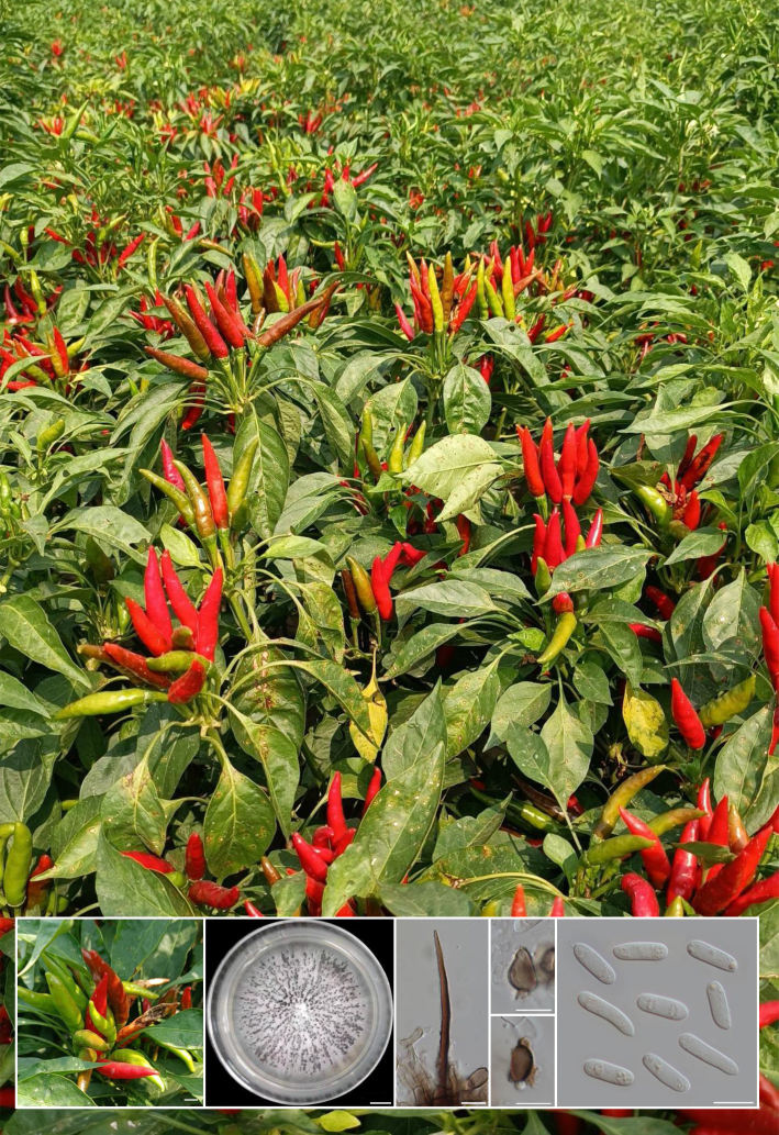
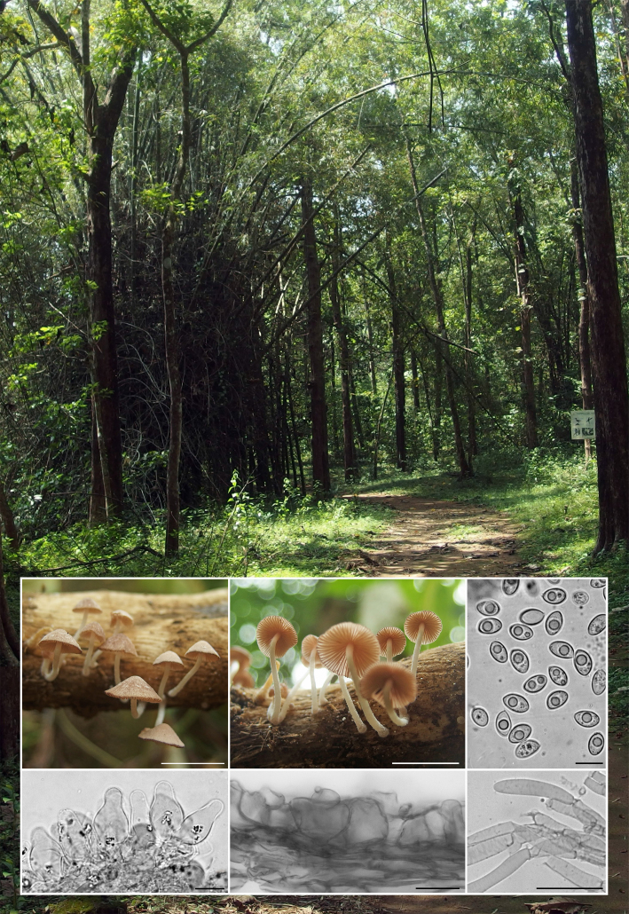
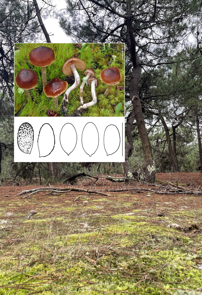
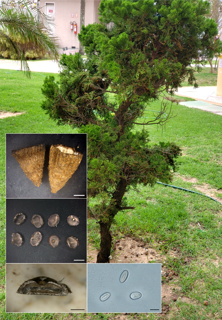
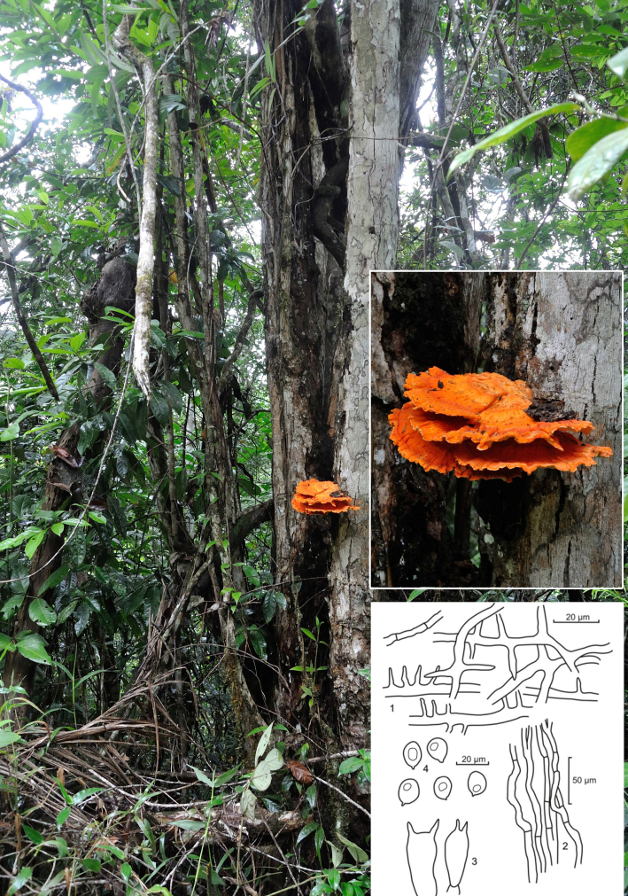
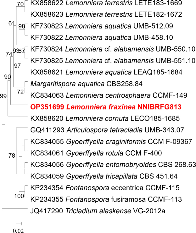
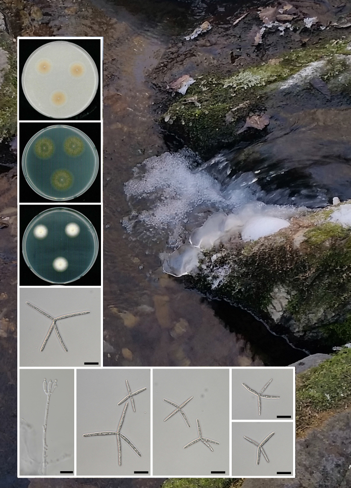
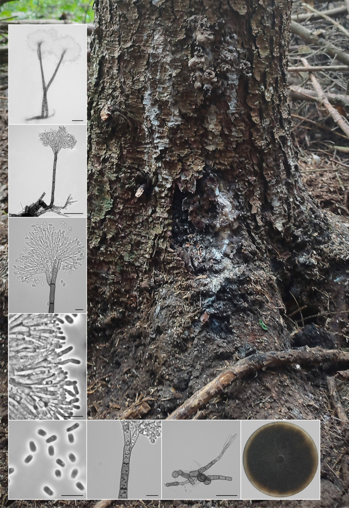
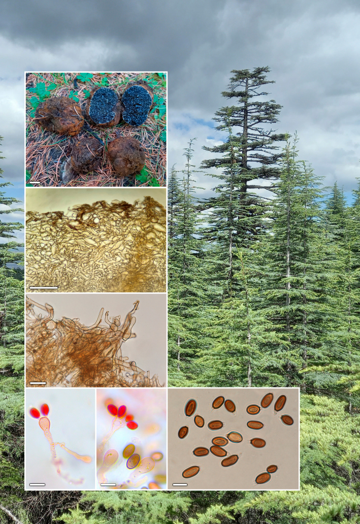
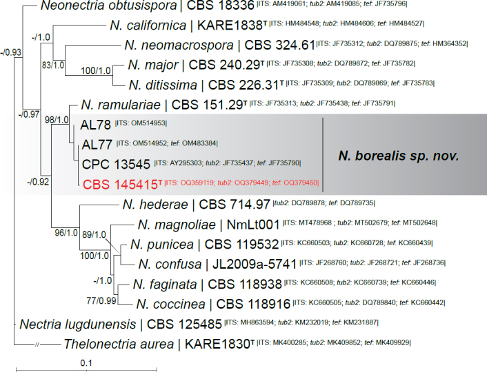
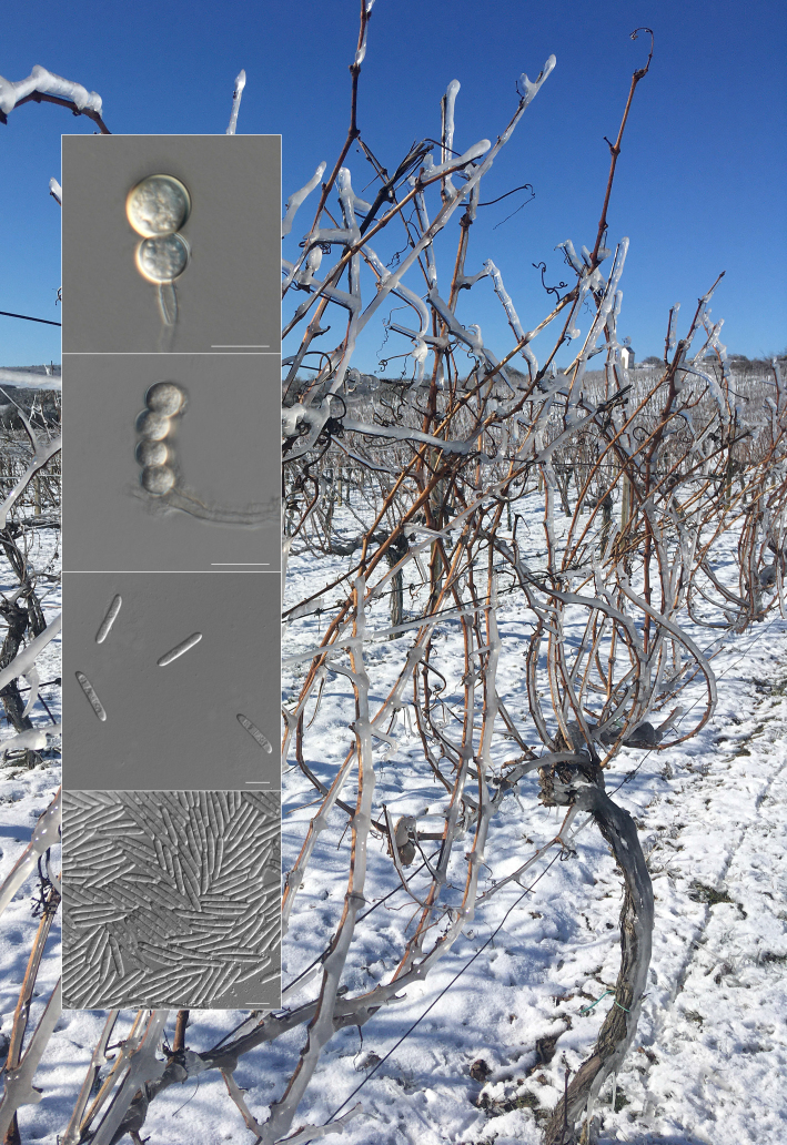
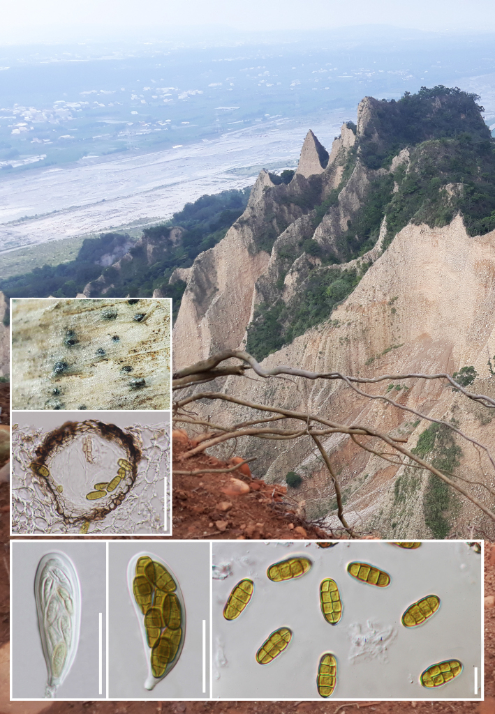
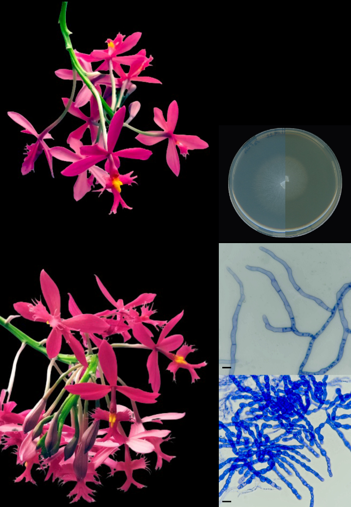
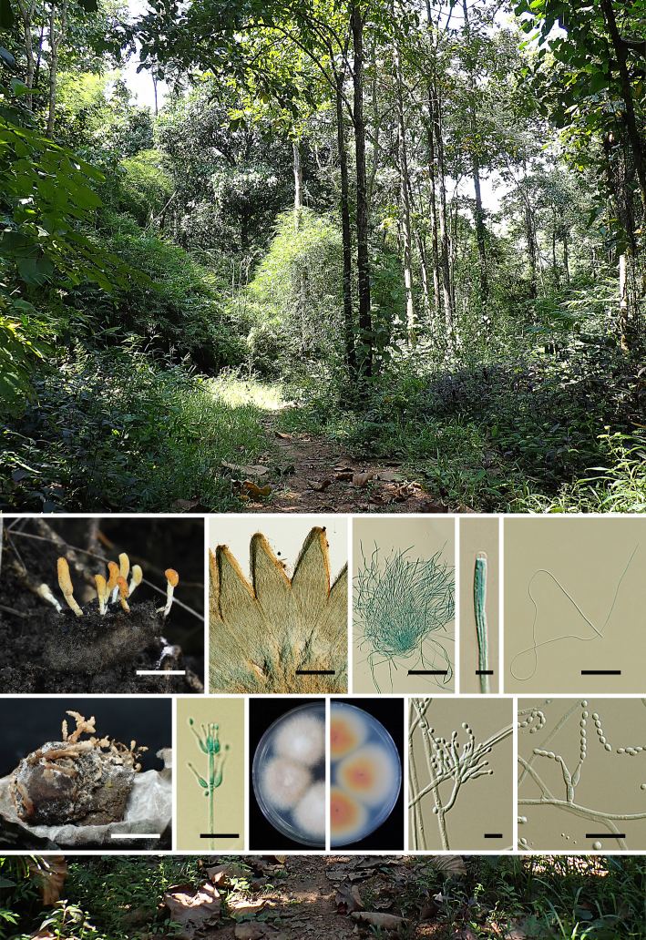
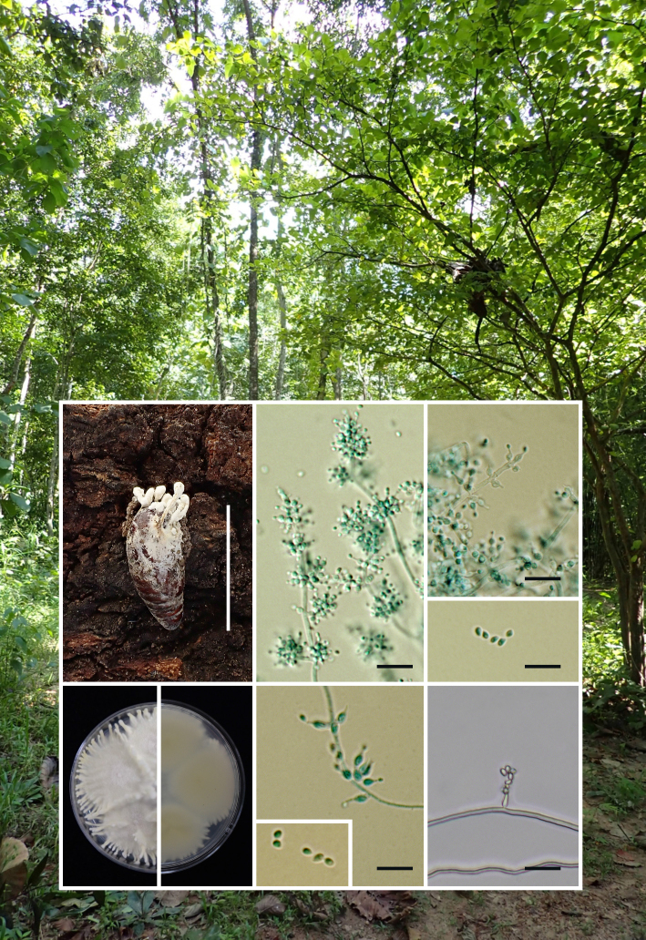
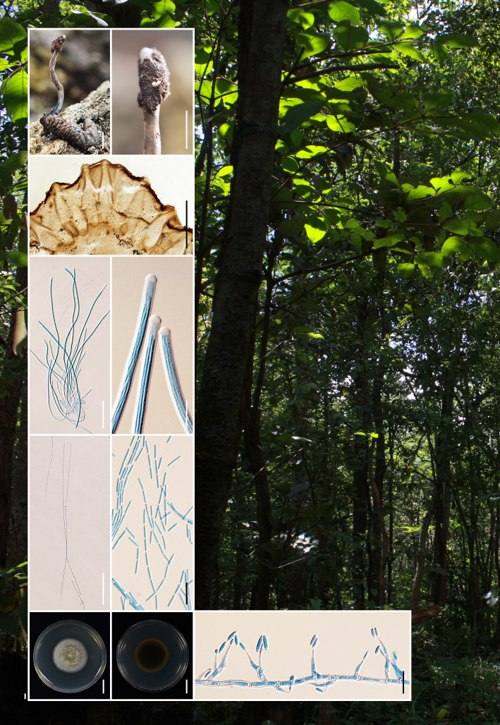
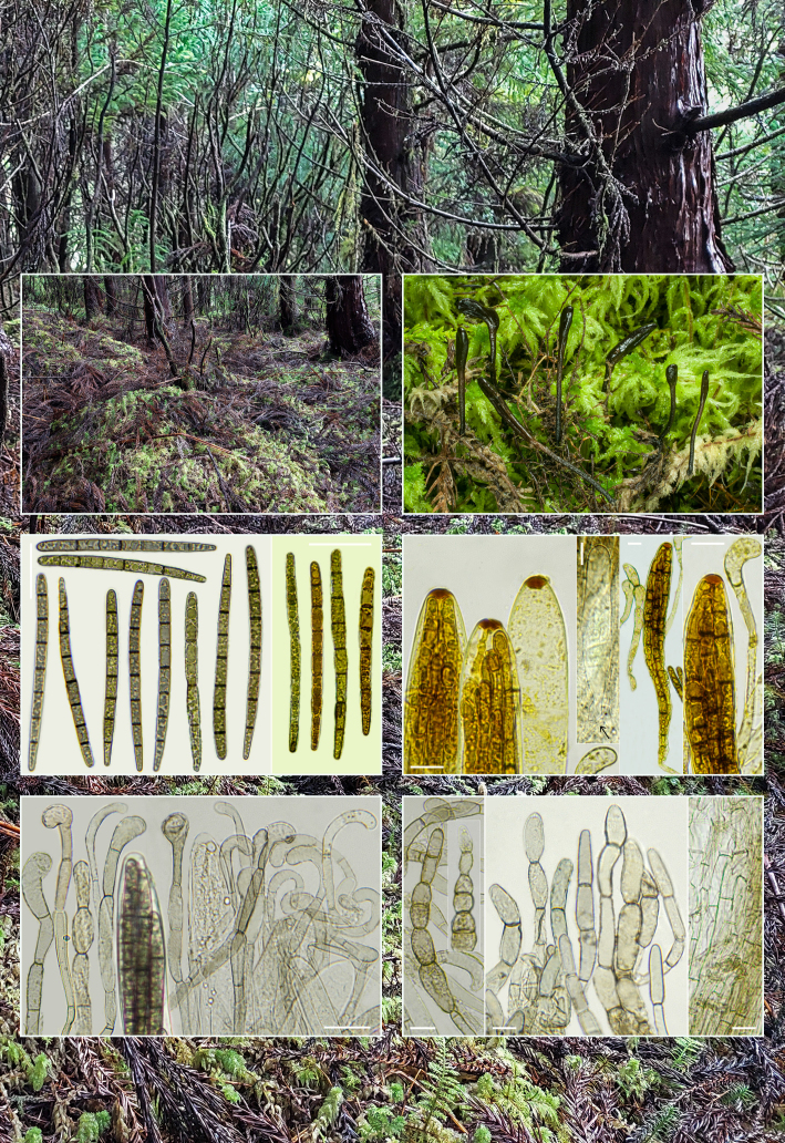
Novel species of fungi described in this study include those from various countries as follows: Australia, Aschersonia mackerrasiae on whitefly, Cladosporium corticola on bark of Melaleuca quinquenervia, Penicillium nudgee from soil under Melaleuca quinquenervia, Pseudocercospora blackwoodiae on leaf spot of Persoonia falcata, and Pseudocercospora dalyelliae on leaf spot of Senna alata. Bolivia, Aspicilia lutzoniana on fully submersed siliceous schist in high-mountain streams, and Niesslia parviseta on the lower part and apothecial discs of Erioderma barbellatum on a twig. Brazil, Cyathus bonsai on decaying wood, Geastrum albofibrosum from moist soil with leaf litter, Laetiporus pratigiensis on a trunk of a living unknown hardwood tree species, and Scytalidium synnematicum on dead twigs of unidentified plant. Bulgaria, Amanita abscondita on sandy soil in a plantation of Quercus suber. Canada, Penicillium acericola on dead bark of Acer saccharum, and Penicillium corticola on dead bark of Acer saccharum. China, Colletotrichum qingyuanense on fruit lesion of Capsicum annuum. Denmark, Helminthosphaeria leptospora on corticioid Neohypochnicium cremicolor. Ecuador (Galapagos), Phaeosphaeria scalesiae on Scalesia sp. Finland, Inocybe jacobssonii on calcareous soils in dry forests and park habitats. France, Cortinarius rufomyrrheus on sandy soil under Pinus pinaster, and Periconia neominutissima on leaves of Poaceae. India, Coprinopsis fragilis on decaying bark of logs, Filoboletus keralensis on unidentified woody substrate, Penicillium sankaranii from soil, Physisporinus tamilnaduensis on the trunk of Azadirachta indica, and Poronia nagaraholensis on elephant dung. Iran, Neosetophoma fici on infected leaves of Ficus elastica. Israel, Cnidariophoma eilatica (incl. Cnidariophoma gen. nov.) from Stylophora pistillata. Italy, Lyophyllum obscurum on acidic soil. Namibia, Aureobasidium faidherbiae on dead leaf of Faidherbia albida, and Aureobasidium welwitschiae on dead leaves of Welwitschia mirabilis. Netherlands, Gaeumannomycella caricigena on dead culms of Carex elongata, Houtenomyces caricicola (incl. Houtenomyces gen. nov.) on culms of Carex disticha, Neodacampia ulmea (incl. Neodacampia gen. nov.) on branch of Ulmus laevis, Niesslia phragmiticola on dead standing culms of Phragmites australis, Pseudopyricularia caricicola on culms of Carex disticha, and Rhodoveronaea nieuwwulvenica on dead bamboo sticks. Norway, Arrhenia similis half-buried and moss-covered pieces of rotting wood in grass-grown path. Pakistan, Mallocybe ahmadii on soil. Poland, Beskidomyces laricis (incl. Beskidomyces gen. nov.) from resin of Larix decidua ssp. polonica, Lapidomyces epipinicola from sooty mould community on Pinus nigra, and Leptographium granulatum from a gallery of Dendroctonus micans on Picea abies. Portugal, Geoglossum azoricum on mossy areas of laurel forest areas planted with Cryptomeria japonica, and Lunasporangiospora lusitanica from a biofilm covering a biodeteriorated limestone wall. Qatar, Alternaria halotolerans from hypersaline sea water, and Alternaria qatarensis from water sample collected from hypersaline lagoon. South Africa, Alfaria thamnochorti on culm of Thamnochortus fraternus, Knufia aloeicola on Aloe gariepensis, Muriseptatomyces restionacearum (incl. Muriseptatomyces gen. nov.) on culms of Restionaceae, Neocladosporium arctotis on nest of cases of bag worm moths (Lepidoptera, Psychidae) on Arctotis auriculata, Neodevriesia scadoxi on leaves of Scadoxus puniceus, Paraloratospora schoenoplecti on stems of Schoenoplectus lacustris, Tulasnella epidendrea from the roots of Epidendrum × obrienianum, and Xenoidriella cinnamomi (incl. Xenoidriella gen. nov.) on leaf of Cinnamomum camphora. South Korea, Lemonniera fraxinea on decaying leaves of Fraxinus sp. from pond. Spain, Atheniella lauri on the bark of fallen trees of Laurus nobilis, Halocryptovalsa endophytica from surface-sterilised, asymptomatic roots of Salicornia patula, Inocybe amygdaliolens on soil in mixed forest, Inocybe pityusarum on calcareous soil in mixed forest, Inocybe roseobulbipes on acidic soils, Neonectria borealis from roots of Vitis berlandieri × Vitis rupestris, Sympoventuria eucalyptorum on leaves of Eucalyptus sp., and Tuber conchae from soil. Sweden, Inocybe bidumensis on calcareous soil. Thailand, Cordyceps sandindaengensis on Lepidoptera pupa, buried in soil, Ophiocordyceps kuchinaraiensis on Coleoptera larva, buried in soil, and Samsoniella winandae on Lepidoptera pupa, buried in soil. Taiwan region (China), Neophaeosphaeria livistonae on dead leaf of Livistona rotundifolia. Türkiye, Melanogaster anatolicus on clay loamy soils. UK, Basingstokeomyces allii (incl. Basingstokeomyces gen. nov.) on leaves of Allium schoenoprasum. Ukraine, Xenosphaeropsis corni on recently dead stem of Cornus alba. USA, Nothotrichosporon aquaticum (incl. Nothotrichosporon gen. nov.) from water, and Periconia philadelphiana from swab of coil surface. Morphological and culture characteristics for these new taxa are supported by DNA barcodes.

Citation: Crous PW, Osieck ER, Shivas RG, et al. 2023. Fungal Planet description sheets: 1478–1549. Persoonia 50: 158– 310. https://doi.org/10.3767/persoonia.2023.50.05.

Keywords: ITS nrDNA barcodes, LSU, new taxa, systematics

Acknowledgments

The work of P.W. Crous and colleagues benefitted from funding by the European Union’s Horizon 2020 research and innovation program (RISE) under the Marie Skłodowska-Curie grant agreement No. 101008129, project acronym ‘Mycobiomics’, and the Dutch NWO Roadmap grant agreement No. 2020/ENW/00901156, project ‘Netherlands Infrastructure for Ecosystem and Biodiversity Analysis – Authoritative and Rapid Identification System for Essential biodiversity information’ (acronym NIEBAARISE). G. Gulden, B. Rian and I. Saar thank K. Bendiksen at the fungarium and G. Marthinsen at NorBol, both Natural History Museum, University of Oslo for valuable help with the collections, and the sequencing of our finds of A. similis from 2022. Sincere thanks to A. Voitk for assistance with the colour plate and review of the manuscript. I. Saar was supported by the Estonian Research Council (grant PRG1170). P. Rodriguez-Flakus and co-authors are greatly indebted to their colleagues and all staff of the Herbario Nacional de Bolivia, Instituto de Ecología, Universidad Mayor de SanAndrés, La Paz, for their generous long-term cooperation. Their research was financially supported by the National Science Centre (NCN) in Poland (grants numbers 2018/02/X/NZ8/02362 and 2021/43/B/NZ8/02902). Y.P. Tan and colleagues thank M.K. Schutze (Department of Agriculture and Fisheries, Queensland, Australia) for determining the identity of the insect hosts for Aschersonia mackerrasiae. The Australian Biological Resources Study funded the project that led to the discovery of Aschersonia mackerrasiae. K.G.G. Ganga acknowledges support from the University Grants Commission (UGC), India, in the form of a UGC research fellowship (Ref No. 20/12/2015(ii) EU-V), and the authorities of the University of Calicut for providing facilities to conduct this study. S. Mahadevakumar acknowledges the Director, KSCSTE - Kerala Forest Research Institute and Head of Office, Botanical Survey of India, Andaman and Nicobar Regional Centre, Port Blair for the necessary support and M. Madappa, Department of Studies in Botany, University of Mysore for technical assistance. A.R. Podile thanks the Department of Science and Technology, Govt. of India for the JC Bose Fellowship (Grant No. JCB/2017/000053) & MoE and IOE-Directorate-UOH for project (Grant No.UOH-IOE-RC3-21-065). Financial support was provided to R. de L. Oliveira and K.D. Barbosa by the Coordenação deAperfeiçoamento de Pessoal de Nível Superior - Brazil (CAPES) – Finance code 001, and to I.G. Baseia and M.P. Martín by the National Council for Scientific and Technological Development (CNPq) under CNPq-Universal 2016 (409960/2016-0) and CNPq-visiting researcher (407474/2013-7). E. Larsson acknowledges the Swedish Taxonomy Initiative, SLU Artdatabanken, Uppsala, Sweden. H.Y. Mun and J. Goh were supported by a grant from the Nakdonggang National Institute of Biological Resources (NNIBR), funded by the Ministry of Environment (MOE) of the Republic of Korea (NNIBR202301106). J. Trovão and colleagues were financed by FEDER - Fundo Europeu de Desenvolvimento Regional funds through the COMPETE 2020 - Operational Programme for Competitiveness and Internationalisation (POCI), and by Portuguese funds through FCT-Fundação para a Ciência e a Tecnologia in the framework of the project POCI-01-0145-FEDER-PTDC/EPH-PAT/3345/2014. Their research was carried out at the R & D Unit Centre for Functional Ecology – Science for People and the Planet (CFE), with reference UIDB/04004/2020, financed by FCT/MCTES through national funds (PIDDAC). João Trovão was supported by POCH - Programa Operacional Capital Humano (co-funding by the European Social Fund and national funding by MCTES), through a ‘FCT-Fundação para a Ciência e Tecnologia’ PhD research grant (SFRH/BD/132523/2017). O. Kaygusuz and colleagues thank the Research Fund of the Isparta University of Applied Sciences for their financial support under the project number 2021-ILK1-0155. They also thank N. Sánchez Biezma of the Department of Drawing and Scientific Photography at the Alcalá University for his help in the digital preparation of the photographs. The research of M. Spetik and co-authors was supported by project No. IGA-ZF/2021-SI1003. V. Darmostuk and colleagues acknowledge our colleagues and all staff of the Herbario Nacional de Bolivia, Instituto de Ecología, Universidad Mayor de San Andrés, La Paz, for their generous long-term cooperation. They would also like to thank the SERNAP (http://sernap.gob.bo), and all protected areas staff, for providing permits for scientific studies, as well as their assistance and logistical support during the field works. This research was financially supported by the National Science Centre (NCN) in Poland (grant number DEC-2013/11/D/NZ8/03274). M. Kaliyaperumal and co-authors thank the Centre of Advanced Studies in Botany, University of Madras for the laboratory facilities. M. Kaliyaperumal thanks the Extramural Research-SERB, DST (EMR/2016/003078), Government of India, for financial assistance. M. Kaliyaperumal and K. Kezo thanks RUSA 2.0 (Theme-1, Group-1/2021/49) for providing a grant. M. Shivannegowda and colleagues thank C.R. Santhosh, Department of Studies in Microbiology, University of Mysore, Manasagangotri, Mysuru for technical support. They also thank K.R. Sridhar, Mangalore University, Karnataka, India and S.S.N. Maharachchikumbura, University of Electronic Science and Technology of China, Chengdu for their support and helping with technical inputs. The study of G.G. Barreto and co-authors was financed in part by the Coordenação de Aperfeiçoamento de Pessoal de Nível Superior - Brazil (CAPES - Finance Code 001), and the Conselho Nacional de Desenvolvimento Científico e Tecnológico (CNPq - Proc. 131503/2019-7; Proc. 312984/2018-9); the authors also thank to Programa de Pós-Graduação em Botânica – PPGBOT. L.F.P. Gusmão thanks to Conselho Nacional de Desenvolvimento Científico e Tecnológico (CNPq) for a research grant. T. Nkomo and colleagues thank the National Research Foundation of SouthAfrica for funding this study, with additional funding from the Forestry and Agricultural Biotechnology Institute and the University of Pretoria. G. Delgado is grateful to W. Colbert and S. Ward (Eurofins Built Environment) for continual encouragement and provision of laboratory facilities. J.G. Maciá-Vicente acknowledges support from the Landes-Offensive zur Entwicklung Wissenschaftlich-ökonomischer Exzellenz (LOEWE) of the state of Hesse within the framework of the Cluster for Integrative Fungal Research (IPF) of Goethe University Frankfurt. F. Esteve-Raventós and colleagues acknowledge P. Juste and J.C. Campos for the loan of some collections for study and N. Subervielle and L. Hugot (Conservatoire Botanique National de Corse, Office de l’Environnement de la Corse, Corti) for their assistance. They also acknowledge the Balearic Mycology Group (FCB) for their permanent help in the search for collections in the Balearic Islands, and Y. Turégano for obtaining some of the sequences presented here, and L. Parra for his suggestions and help on nomenclatural issues. S. Mongkolsamrit and colleagues were financially supported by the Platform Technology Management Section, National Centre for Genetic Engineering and Biotechnology (BIOTEC), Project Grant No. P19-50231. S. De la Peña-Lastra and colleagues thank the Atlantic Islands National Maritime-Terrestrial Park authorities and guards. A. Mateos and co-authors would like to thank Secretaria Regional doAmbiente eAlterações Climáticas Açores for the permission granted for the sampling (Licença nº 16/2021/DRAAC). To the ECOTOX group for co-funding the trip. J. Mack & D.P. Overy were funded byAgriculture &Agri-Food Canada (Project ID#002272: Fungal and Bacterial Biosystematics-bridging taxonomy and “omics” technology in agricultural research and regulation) and are grateful for molecular sequencing support from the Molecular Technologies Laboratory (MTL) at the Ottawa Research & Development Centre of Agriculture & Agri-Food Canada. The study of P. Czachura was funded by the National Science Centre, Poland, under the project 2019/35/N/NZ9/04173. The study of M. Piątek and co-authors was funded by the National Science Centre, Poland, under the project 2017/27/B/NZ9/02902. O. Yarden and L. Granit were funded by the Israel Science Foundation (grant number 888/19). H. Taşkın and colleagues received support from the Bulgarian Academy of Sciences and the Scientific and Technological Research Council of Türkiye (Bilateral grant agreement between BAS and TÜBİTAK, project number 118Z640). The authors would also like to thank S. Şahin (İzmir, Türkiye) for conveying one of the localities of A. abscondita. Andrew Miller would like to thank the Roy J. Carver Biotechnology Center at the University of Illinois for Sanger sequencing. E.R. Osieck thanks Staatsbosbeheer for permission to collect fungi in Nieuw Wulven, in the Netherlands. P. van ‘t Hof and co-authors thank the Galapagos Genetic Barcode project supported by UK Research and Innovation, Global Challenges Research Fund, Newton Fund, University of Exeter, Galapagos Science Center, Universidad San Francisco de Quito, Galapagos Conservation Trust, and Biosecurity Agency of Galapagos (ABG).

REFERENCES

  1. Abdel-Wahab MA, Dayarathne MC, Suetrong SG. et al. 2017. New saprobic marine fungi and a new combination. Botanica Marina 60: 469–488. [Google Scholar]
  2. Adamčíková K, Juhásová G, Kobza M. 2011. The first report of Libertella spp. on Fagaceae in Slovakia. Mycoscience 52: 268–270. [Google Scholar]
  3. Ahmadi N, Arzanlou M, Narmani A. 2021. Molecular phylogeny and morphology differentiate a new Neosetophoma species from Iran. Nova Hedwigia 112: 383–397. [Google Scholar]
  4. Alvarado P, Cabero J, Moreno-Mateos D. et al. 2021. Phylogenetic relationships among false truffle genera of Paxillaceae – Alpova, Melanogaster, Neoalpova, and Paralpova, gen. nov. Mycologia 113: 828–841. [DOI] [PubMed] [Google Scholar]
  5. Alvarado P, Moreno G, Manjón JL. 2012. Comparison between Tuber gennadii and T. oligospermum lineages reveals the existence of the new species T. cistophilum (Tuberaceae, Pezizales). Mycologia 104: 894–910. [DOI] [PubMed] [Google Scholar]
  6. Antonín V, Noordeloos ME. 2004. A monograph of the genera Hemimycena, Delicatula, Fayodia, Gamundia, Myxomphalia, Resinomycena, Rickenella, and Xeromphalina (Tribus Mycenae sensu Singer, Mycena excluded) in Europe. IHW-Verlag. [Google Scholar]
  7. Aptroot A. 2006. Mycosphaerella and its anamorphs: 2. Conspectus of Mycosphaerella. CBS Biodiversity Series 5: 1–231. Centraalbureau voor Schimmelcultures, Utrecht, The Netherlands. [Google Scholar]
  8. Arauzo S, Iglesias P. 2014. La familia Geoglossaceae ss. str. en la península Ibérica y la Macaronesia. Errotari 11: 166–259. [Google Scholar]
  9. ARSEF dataset. 2023. U.S. Department of Agriculture, Agricultural Research Service, Biological Integrated Pest Management Research Unit (2016). ARS Collection of Entomopathogenic Fungal Cultures (ARSEF). U.S. Department of Agriculture, Agricultural Research Service. 10.15482/USDA.ADC/1326695 (accessed 2023-02-17). [DOI] [Google Scholar]
  10. Arzanlou M, Groenewald JZ, Fullerton RA. et al. 2008. Multiple gene genealogies and phenotypic characters differentiate several novel species of Mycosphaerella and related anamorphs on banana. Persoonia: 20: 19–37. [DOI] [PMC free article] [PubMed] [Google Scholar]
  11. Arzanlou M, Groenewald JZ, Gams W. et al. 2007. Phylogenetic and morphotaxonomic revision of Ramichloridium and allied Genera. Studies in Mycology 58: 57–93. [DOI] [PMC free article] [PubMed] [Google Scholar]
  12. Arzanlou M, Khodaei S. 2012. Aureobasidium iranianum, a new species on bamboo from Iran. Mycosphere 3: 404–408. [Google Scholar]
  13. Bandini D, Oertel B, Eberhardt U. 2021. A fresh outlook on the smooth-spored species of Inocybe: type studies and 18 new species. Mycological Progress 20: 1019–1114. [Google Scholar]
  14. Bandini D, Oertel B, Eberhardt U. 2022a. More smooth-spored species of Inocybe (Agaricales, Basidiomycota): type studies and 12 new species from Europe. Persoonia 48: 91–149. [DOI] [PMC free article] [PubMed] [Google Scholar]
  15. Bandini D, Oertel B, Eberhardt U. 2022b. Noch mehr Risspilze (3): Einundzwanzig neue Arten der Familie Inocybaceae. Mycologia Bavarica 22: 31–138. [Google Scholar]
  16. Banik MT, Lindner DL, Ortiz-Santana B. et al. 2012. A new species of Laetiporus (Basidiomycota, Polyporales) from the Caribbean basin. Kurtziana 37: 15–21. [Google Scholar]
  17. Barr ME. 1992. Additions to and notes on the Phaeosphaeriaceae (Pleosporales, Loculoascomycetes). Mycotaxon 43: 371–400. [Google Scholar]
  18. Beker HJ, Eberhardt U, Vesterholt J. 2016. Hebeloma (Fr.) P. Kumm. Fungi Europaei 14: 1–1218. Ed. Tecnografica. [Google Scholar]
  19. Bellanger J-M, Bidaud A, Moreau P-A. 2021. Cortinarius subturibulosus. « Illumina »-tion d’un champion de l’adaptation. Journal des JEC 23: 3–15. [Google Scholar]
  20. Bellanger J-M, Moreau P-A, Corriol G. et al. 2015. Plunging hands into the mushroom jar: a phylogenetic framework for Lyophyllaceae (Agaricales, Basidiomycota). Genetica 143: 169–194. [DOI] [PubMed] [Google Scholar]
  21. Bensch K, Braun U, Groenewald JZ. et al. 2012. The genus Cladosporium. Studies in Mycology 72: 1–401. [DOI] [PMC free article] [PubMed] [Google Scholar]
  22. Bensch K, Groenewald JZ, Braun U. et al. 2015. Common but different: The expanding realm of Cladosporium. Studies in Mycology 82: 23–74. [DOI] [PMC free article] [PubMed] [Google Scholar]
  23. Bills GF, Menéndez VG, Platas G. 2012. Kabatiella bupleuri sp. nov. (Dothideales), a pleomorphic epiphyte and endophyte of the Mediterranean plant Bupleurum gibraltarium (Apiaceae). Mycologia 104: 962–973. [DOI] [PubMed] [Google Scholar]
  24. Boscaiu M, Ballesteros G, Naranjo MA. et al. 2011. Responses to salt stress in Juncus acutus and J. maritimus during seed germination and vegetative plant growth. Plant Biosystems 145: 770–777. [Google Scholar]
  25. Branco S, Ree RH. 2010. Serpentine soils do not limit mycorrhizal fungal diversity. PLoS ONE 5: e11757. [DOI] [PMC free article] [PubMed] [Google Scholar]
  26. Braun U, Cunnington J, Priest MJ. et al. 2005. Annotated checklist of Ramularia species in Australia. Australasian Plant Pathology 34: 1–7. [Google Scholar]
  27. Bresadola G. 1902. Mycetes Lusitanici novi. Atti dell’ Imperiale Regia Accademia di Scienze. Lettere ed Arti Degli Agiati di Rovereto 3: 127–133. [Google Scholar]
  28. Brodie HJ. 1975. The Bird’s Nest Fungi. University of Toronto Press, Toronto, Canada. [Google Scholar]
  29. Cabral A, Groenewald JZ, Rego C. et al. 2012. Cylindrocarpon root rot: multi-gene analysis reveals novel species within the Ilyonectria radicicola species complex. Mycological Progress 11: 655–688. [Google Scholar]
  30. Cailleux A. 1981. Code des couleurs des sols. Boubée, Paris. [Google Scholar]
  31. Calonge FD, Pasaban PM. 1993. Nuevos datos sobre los hongos hipogeos de España V. Registro de nueve citas nuevas. Boletín de la Sociedad Micológica de Madrid 18: 41–58. [Google Scholar]
  32. Calvelo S, Liberatore S. 2002. Catálogo de los líquenes de la Argentina. Kurtziana 29: 7–170. [Google Scholar]
  33. Câmara MP, Ramaley AW, Castlebury LA. 2003. Neophaeosphaeria and Phaeosphaeriopsis, segregates of Paraphaeosphaeria. Mycological Research 107: 516–522. [DOI] [PubMed] [Google Scholar]
  34. Capote N, Del Río MÁ, Herencia JF. et al. 2022. Molecular and pathogenic characterization of Cylindrocarpon-like anamorphs causing root and basal rot of almonds. Plants 11: 984. [DOI] [PMC free article] [PubMed] [Google Scholar]
  35. Chalabuda TV. 1950. Species novae e genere Penicillium Link. Notulae systematicae e Sectione Cryptogamica Instituti Botanici nomine VL Komarovii Academiae Scientiarum URSS Botanicheskie materialy 6: 161–169. [Google Scholar]
  36. Chatin A. 1896. Truffes (Terfaz) de Grèce, Terfezia gennadii. Bulletin de la Société Botanique de France 43: 611–617. [Google Scholar]
  37. Chen W-H, Han Y-F, Liang J-D. et al. 2020. Morphological and phylogenetic characterisations reveal three new species of Samsoniella (Cordycipitaceae, Hypocreales) from Guizhou, China. MycoKeys 74: 1–15. [DOI] [PMC free article] [PubMed] [Google Scholar]
  38. Chernomor O, Von Haeseler A, Quang Minh B. 2016. Terrace aware data structure for phylogenomic inference from supermatrices. Systematic Biology 65: 997–1008. [DOI] [PMC free article] [PubMed] [Google Scholar]
  39. Chien CY. 1972. Mortierella umbellata, a new species from Georgia. Mycologia 34: 99–102. [Google Scholar]
  40. Clémençon H. 1982. Types studies and typifications in Lvophyllum (Agaricales). I. Staining species. Mycotaxon 15: 67–94. [Google Scholar]
  41. Consiglio G. 1998. Lyophyllum aemiliae, Rivista di Micologia 41: 99–104. [Google Scholar]
  42. Consiglio G, Contu M. 2002. Il Genere Lyophyllum P. Karst. Emend. Huhnér, in Italia. Rivista di Micologia II: 99–181. [Google Scholar]
  43. Consiglio G, Papetti C. 2001. Funghi d’Italia, ed. AMB 2: 613–619. [Google Scholar]
  44. Consiglio G, Papetti C. 2009. Funghi d’Italia, ed. AMB 3: 1090–1101. [Google Scholar]
  45. Cripps CL, Larsson E, Vauras J. 2020. Nodulose-spored Inocybe species from the Rocky Mountain alpine zone: molecularly linked to European type specimens. Mycologia 112: 133–153. [DOI] [PubMed] [Google Scholar]
  46. Crous PW, Begoude BAD, Boers J. et al. 2022a. New and interesting fungi. 5. Fungal Systematics and Evolution 10: 19–90. [DOI] [PMC free article] [PubMed] [Google Scholar]
  47. Crous PW, Boers J, Holdom D. et al. 2022b. Fungal Planet description sheets: 1383-1435. Persoonia 48: 261–371. [DOI] [PMC free article] [PubMed] [Google Scholar]
  48. Crous PW, Cowan DA, Maggs-Kölling G. et al. 2020a. Fungal Planet description sheets: 1112-1181. Persoonia 45: 251–409. [DOI] [PMC free article] [PubMed] [Google Scholar]
  49. Crous PW, Cowan DA, Maggs-Kölling G. et al. 2021a. Fungal Planet description sheets: 1182-1283. Persoonia 46: 313–528. [DOI] [PMC free article] [PubMed] [Google Scholar]
  50. Crous PW, Hernández-Restrepo M, Schumacher RK. et al. 2021b. New and interesting fungi. 4. Fungal Systematics and Evolution 7: 255–343. [DOI] [PMC free article] [PubMed] [Google Scholar]
  51. Crous PW, Mohammed C, Glen M. et al. 2007. Eucalyptus microfungi known from culture. 3. Eucasphaeria and Sympoventuria genera nova, and new species of Furcaspora, Harknessia, Heteroconium and Phacidiella. Fungal Diversity 25: 19–36. [Google Scholar]
  52. Crous PW, Osieck ER, Jurjević Ž. et al. 2021c. Fungal Planet description sheets: 1284-1382. Persoonia 47: 178–374. [DOI] [PMC free article] [PubMed] [Google Scholar]
  53. Crous PW, Schumacher RK, Akulov A. et al. 2019a. New and Interesting Fungi. 2. Fungal Systematics and Evolution 3: 57–134. [DOI] [PMC free article] [PubMed] [Google Scholar]
  54. Crous PW, Shivas RG, Quaedvlieg W. et al. 2014. Fungal Planet description sheets: 214-280. Persoonia 32: 184–306. [DOI] [PMC free article] [PubMed] [Google Scholar]
  55. Crous PW, Wingfield MJ, Burgess TI. et al. 2016. Fungal Planet description sheets: 469-557 Persoonia 37: 218–403. [DOI] [PMC free article] [PubMed] [Google Scholar]
  56. Crous PW, Wingfield MJ, Burgess TI. et al. 2017a. Fungal Planet description sheets: 558-624. Persoonia 38: 240–384. [DOI] [PMC free article] [PubMed] [Google Scholar]
  57. Crous PW, Wingfield MJ, Burgess TI. et al. 2017b. Fungal Planet description sheets: 625-715. Persoonia 39: 270–467. [DOI] [PMC free article] [PubMed] [Google Scholar]
  58. Crous PW, Wingfield MJ, Guarro J. et al. 2013. Fungal Planet description sheets: 154-213. Persoonia 31: 188–296. [DOI] [PMC free article] [PubMed] [Google Scholar]
  59. Crous PW, Wingfield MJ, Lombard L. et al. 2019b. Fungal Planet description sheets: 951-1041. Persoonia 43: 223–425. [DOI] [PMC free article] [PubMed] [Google Scholar]
  60. Crous PW, Wingfield MJ, Schumacher RK. et al. 2020b. New and interesting fungi 3. Fungal Systematics and Evolution 6: 157–231. [DOI] [PMC free article] [PubMed] [Google Scholar]
  61. Cruz RHSF, Baseia IG. 2014. Four new Cyathus species (Nidulariaceae, Basidiomycota, Fungi) from the semi-arid region of Brazil. Journal of the Torrey Botanical Society 141: 173–180. [Google Scholar]
  62. Dähncke RM, Contu M, Vizzini A. 2009. Some rare, critical, interesting taxa of the genus Lyophyllum s.l. (Basidiomycota, Agaricomycetes) from La Palma (Canary Islands, Spain). Österreichische Zeitschrift Pilzkunde 18: 129–139. [Google Scholar]
  63. Dähncke RM, Contu M, Vizzini A. 2011. Two new species of Lyophyllum s.l. (Basidiomycota, Agaricomycetes) from La Palma (Canary Islands, Spain). Mycotaxon 115: 63–71. [Google Scholar]
  64. Dai SJ, Dai YC. 2018. Morphological characters and molecular data reveal a new species of Physisporinus (Basidiomycota) from Southeast Asia. Mycosystema 37: 145–150. [Google Scholar]
  65. Damm U, Sato T, Alizadeh A. et al. 2018. The Colletotrichum dracaenophilum, C. magnum and C. orchidearum species complexes. Studies in Mycology 90: 71–118. [DOI] [PMC free article] [PubMed] [Google Scholar]
  66. Dayarathne MC, Phookamsak R, Hyde KD. et al. 2016. Halodiatrype, a novel diatrypaceous genus from mangroves with H. salinicola and H. avicenniae spp. nov. Mycosphere 7: 612–627. [Google Scholar]
  67. Dayarathne MC, Wanasinghe D N, Devadatha B. et al. 2020. Modern taxonomic approaches to identifying diatrypaceous fungi from marine habitats, with a novel genus Halocryptovalsa Dayarathne & K.D. Hyde, gen. nov. Cryptogamie, Mycologie 41: 21–67. [Google Scholar]
  68. De Beer W, Procter M, Wingfield MJ. et al. 2022. Generic boundaries in the Ophiostomatales reconsidered and revised. Studies in Mycology 101: 57–120. [DOI] [PMC free article] [PubMed] [Google Scholar]
  69. De Wildeman E. 1894. Notes mycologiques. Fascicle 3. Annual Society Belge Microscopy XVIII: 135–161. [Google Scholar]
  70. Dereeper A, Guignon V, Blanc G. et al. 2008. Phylogeny.fr: robust phylogenetic analysis for the non-specialist. NucleicAcids Research 36: W465–W469. [DOI] [PMC free article] [PubMed] [Google Scholar]
  71. Diederich P, Garnier-Delcourt M, Lücking R. et al. 2022. Flora of lichenicolous fungi. Vol. 1, Basidiomycota. National Museum of Natural History, Luxembourg. [Google Scholar]
  72. Dong W, Wang B, Hyde KD. et al. 2020. Freshwater Dothideomycetes. Fungal Diversity 105: 319–575. [Google Scholar]
  73. Edgar RC. 2004. MUSCLE: a multiple sequence alignment method with reduced time and space complexity. BMC Bioinformatics 5: 113. [DOI] [PMC free article] [PubMed] [Google Scholar]
  74. Egidi E, De Hoog GS, Isola D. et al. 2014. Phylogeny and taxonomy of meristematic rock-inhabiting black fungi in the Dothideomycetes based on multi-locus phylogenies. Fungal Diversity 65: 127–165. [Google Scholar]
  75. Ellis JB, Everhart BM. 1888. New species of fungi from various localities. Journal of Mycology 4: 73–82. [Google Scholar]
  76. Ellis MB. 1971. Dematiaceous Hyphomycetes. Commonwealth Mycological Institute, Kew, Surrey, England. [Google Scholar]
  77. Ertz D, Diederich P, Lawrey JD. et al. 2015. Phylogenetic insights resolve Dacampiaceae (Pleosporales) as polyphyletic: Didymocyrtis (Pleosporales, Phaeosphaeriaceae) with Phoma-like anamorphs resurrected and segregated from Polycoccum (Trypetheliales, Polycoccaceae fam. nov.). Fungal Diversity 74: 53–89. [Google Scholar]
  78. Esteve-Raventós F, Pancorbo F, Larsson E. et al. 2022. Inocybe vaurasii (Agaricales, Inocybaceae), a new species of the I. xanthomelas group and similar European species with asteriform spores. Phytotaxa 566: 171–188. [Google Scholar]
  79. Etayo J. 2017. Hongos Liquenícolas de Ecuador. Opera Lilloana 50: 1–535. [Google Scholar]
  80. Etayo J, Flakus A, Kukwa M. 2013. Niesslia echinoides (Niessliaceae, Ascomycota), a new lichenicolous fungus on Erioderma from Bolivia. The Lichenologist 45: 21–24. [Google Scholar]
  81. Fang F, Chen JJ, Ji XH. et al. 2017. Phylogeny and diversity of the morphologically similar polypore genera Rigidoporus, Physisporinus, Oxyporus, and Leucophellinus. Mycologia 109: 749–765. [DOI] [PubMed] [Google Scholar]
  82. Favre J. 1955. Les champignons supérieurs de la zone alpine du Parc National Suisse. Ergebnisse der Wissensnschaftlichen Untersuchungen des Schweizerrischen National Parks 5: 1–212. [Google Scholar]
  83. Felsenstein J. 1985. Confidence limits on phylogenies: An approach using the bootstrap. Evolution 39: 783–791. [DOI] [PubMed] [Google Scholar]
  84. Feuerer T, Ahti T, Vitikainen O. 1998. Lichenological investigations in Bolivia. In: Marcelli MP, Seaward MRD. (eds), Lichenology in LatinAmerica: History, current knowledge and applications. CETESB, Sao Paulo: 71–86. [Google Scholar]
  85. Finy P, Papp V, Knapp DG. et al. 2021. Geastrum dolomiticum, a new earthstar species from Central Europe. Plant Systematics and Evolution 307: 43. [Google Scholar]
  86. Flakus A, Sipman HJM, Bach K. et al. 2013. Contribution to the knowledge of the lichen biota of Bolivia. 5. Polish Botanical Journal 58: 697–733. [Google Scholar]
  87. Fletcher A, Purvis OW, Coppins BJ. 2009. Aspicilia A. Massal. In: Smith CW, Aptroot A, Coppins BJ. et al. (eds), The Lichens of Great Britain and Ireland. The British Lichen Society, London: 181–188. [Google Scholar]
  88. Fotedar R, Kolecka A, Boekhout T. et al. 2018. Fungal diversity of the hypersaline Inland Sea in Qatar. Botanica Marina 61: 595–609. [Google Scholar]
  89. Fryday AM, Wheeler TB, Etayo J. 2021. A new species of Aspicilia (Megasporaceae), with a new lichenicolous Sagediopsis (Adelococcaceae), from the Falkland Islands. The Lichenologist 53: 307–315. [Google Scholar]
  90. Fuckel L. 1860. Enumeratio Fungorum Nassoviae. Jahrbücher des Nassauischen Vereins für Naturkunde 15: 1–123. [Google Scholar]
  91. Galloway DJ, Quilhot W. 1998. Checklist of Chilean lichen-forming and lichenicolous fungi. Gayana Botanica 55: 111–185. [Google Scholar]
  92. Gams W. 1977. A key to the species of Mortierella. Persoonia 9: 381–391. [Google Scholar]
  93. Gams W, Stielow B, Gräfenhan T. et al. 2019. The ascomycete genus Niesslia and associated monocillium-like anamorphs. Mycological Progress 18: 1–72. [Google Scholar]
  94. Ge Y, Liu Z, Zeng H. et al. 2021. Updated description of Atheniella (Mycenaceae, Agaricales), including three new species with brightly coloured pilei from Yunnan Province, southwest China. MycoKeys 81: 139–164. [DOI] [PMC free article] [PubMed] [Google Scholar]
  95. Gibbons AT, Idnurm A, Seiter M. et al. 2019. Amblypygid-fungal interactions: The whip spider exoskeleton as a substrate for fungal growth. Fungal Biology 123: 497–506. [DOI] [PubMed] [Google Scholar]
  96. Giraldo A, Crous PW. 2019. Inside Plectosphaerellaceae. Studies in Mycology 92: 227–286. [DOI] [PMC free article] [PubMed] [Google Scholar]
  97. Goh YK, Goh TK, Marzuki NF. et al. 2015. Scytalidium parasiticum sp. nov., a new species parasitizing on Ganoderma boninense isolated from oil palm in Peninsular Malaysia. Mycobiology 43: 107–117. [DOI] [PMC free article] [PubMed] [Google Scholar]
  98. Góis JS, Cruz RHSF, Baseia IG. 2021. Taxonomic review and updates of the genus Cyathus (Agaricales, Basidiomycota) from Brazil. The Journal of the Torrey Botanical Society 148: 155–196. [Google Scholar]
  99. Gouy M, Guindon S, Gascuel O. 2010. SeaView version 4: A multiplatform graphical user interface for sequence alignment and phylogenetic tree building. Molecular Biology and Evolution 27: 221–224. [DOI] [PubMed] [Google Scholar]
  100. Guindon S, Dufayard JF, Lefort V. et al. 2010. New algorithms and methods to estimate maximum-likelihood phylogenies: assessing the performance of PhyML 3.0. Systematic Biology 59: 307–321 [DOI] [PubMed] [Google Scholar]
  101. Haji Moniri M, Gromakova AB, Lőkös L. et al. 2017. New members of the Megasporaceae (Pertusariales, lichen-forming Ascomycota): Megaspora iranica spec. nova and Oxneriaria gen. nova. Acta Botanica Hungarica 59: 343–370. [Google Scholar]
  102. Haridas S, Albert R, Binder M. et al. 2020. 101 Dothideomycetes genomes: A test case for predicting lifestyles and emergence of pathogens. Studies in Mycology 96: 141–153. [DOI] [PMC free article] [PubMed] [Google Scholar]
  103. Hawksworth DL. 1975. Notes on British lichenicolous fungi, I. Kew Bulletin 30: 183–203. [Google Scholar]
  104. Hernández-Restrepo M, Groenewald JZ, Crous PW. 2016a. Taxonomic and phylogenetic re-evaluation of Microdochium, Monographella and Idriella. Persoonia 36: 57–82. [DOI] [PMC free article] [PubMed] [Google Scholar]
  105. Hernández-Restrepo M, Groenewald JZ, Elliott ML. et al. 2016b. Take-all or nothing. Studies in Mycology 83: 19–48. [DOI] [PMC free article] [PubMed] [Google Scholar]
  106. Hirooka Y, Ichihara Y, Masuya H. et al. 2012. Seed rot, a new disease of beech tree caused by Neonectria ramulariae (anamorph: Cylindrocarpon obtusiusculum). Journal of Phytopathology 160: 504–506. [Google Scholar]
  107. Hoang DT, Chernomor O, Von Haeseler A. et al. 2018. UFBoot2: Improving the ultrafast bootstrap approximation. Molecular Biology and Evolution 35: 518–522. [DOI] [PMC free article] [PubMed] [Google Scholar]
  108. Horner HT, Tiffany LH, Knaphus G. 1995. Oak-leaf-litter rhizomorphs from Iowa and Texas: Calcium oxalate producers. Mycologia 87: 34–40. [Google Scholar]
  109. Houbraken J, Kocsube S, Visagie CM. et al. 2020. Classification ofAspergillus, Penicillium, Talaromyces and related genera (Eurotiales): an overview of families, genera, subgenera, sections, series and species. Studies in Mycology 95: 5–169. [DOI] [PMC free article] [PubMed] [Google Scholar]
  110. Houbraken J, Samson RA. 2011. Phylogeny of Penicillium and the segregation of Trichocomaceae into three families. Studies in Mycology 70: 1–51 [DOI] [PMC free article] [PubMed] [Google Scholar]
  111. Houbraken J, Visagie CM, Meijer M. et al. 2014. A taxonomic and phylogenetic revision of Penicillium section Aspergilloides. Studies in Mycology 78: 373–451. [DOI] [PMC free article] [PubMed] [Google Scholar]
  112. Hu JH, Yu WJ, Deng LS. et al. 2023. The detection of major clades and new species of Mallocybe (Inocybaceae, Agaricales) from China with elongate cheilocystidia. Mycological Progress 22: 15. [Google Scholar]
  113. Huelsenbeck JP, Ronquist F. 2001. MrBayes: Bayesian inference of phylogenetic trees. Bioinformatics 17: 754–755. [DOI] [PubMed] [Google Scholar]
  114. Hustad VP. 2015. A circumscription of the earth tongue fungi class Geoglossomycetes. Doctoral dissertation, University of Illinois at Urbana-Champaign. [Google Scholar]
  115. Hyde KD, Chaiwan N, Norphanphoun C. et al. 2018. Mycosphere notes 169–224. Mycosphere 9: 271–430. [Google Scholar]
  116. Hyde KD, Dong Y, Phookamsak R. et al. 2020. Fungal diversity notes 1151–1276: taxonomic and phylogenetic contributions on genera and species of fungal taxa. Fungal Diversity 16: 1–273. [Google Scholar]
  117. Hyde KD, Hongsanan S, Jeewon R. et al. 2016. Fungal diversity notes 367–490: taxonomic and phylogenetic contributions to fungal taxa. Fungal Diversity 80: 1–270. [Google Scholar]
  118. Inderbitzin P, Kohlmeyer J, Volkmann-Kohlmeyer B. et al. 2002. Decorospora, a new genus for the marine ascomycete Pleospora gaudefroyi. Mycologia 94: 651–659. [DOI] [PubMed] [Google Scholar]
  119. Ingold GT. 1942. Aquatic hyphomycetes of decaying alder leaves. Transactions of British Mycological Society 25: 339–417. [Google Scholar]
  120. Isola D, Prigione VP, Zucconi L. et al. 2022. Knufia obscura sp. nov. and Knufia victoriae sp. nov., two new species from extreme environments. International Journal of Systematic and Evolutionary Microbiology 72: 10. [DOI] [PubMed] [Google Scholar]
  121. Jacobsson S, Larsson E. 2012. Inocybe (Fr.) Fr. In: Knudsen H, Vesterholt J. (eds), Funga Nordica (Agaricoid, boletoid, clavarioid, cyphelloid and gastroid genera. Nordsvamp, Copenhagen: ): 981–1021. [Google Scholar]
  122. Jaklitsch W, Baral H-O, Lücking R. et al. 2016. Syllabus of plant families. Part 1/2 Ascomycota. Borntraeger, Stuttgart. [Google Scholar]
  123. Justo A, Morgenstern I, Hallen-Adams HE. et al. 2010. Convergent evolution of sequestrate forms in Amanita under Mediterranean climate conditions. Mycologia 102: 675–688. [DOI] [PubMed] [Google Scholar]
  124. Kalyaanamoorthy S, Minh BQ, Wong TKF. et al. 2017. ModelFinder: Fast model selection for accurate phylogenetic estimates. Nature Methods 14: 587–589. [DOI] [PMC free article] [PubMed] [Google Scholar]
  125. Kang HJ, Sigler L, Lee J. et al. 2010. Xylogone ganodermophthora sp. nov., an ascomycetous pathogen causing yellow rot on cultivated mushroom Ganoderma lucidum in Korea. Mycologia 102: 1167–1184. [DOI] [PubMed] [Google Scholar]
  126. Katoh K, Standley DM. 2013. MAFFT Multiple SequenceAlignment Software version 7: improvements in performance and usability. Molecular Biology and Evolution 30: 772–780. [DOI] [PMC free article] [PubMed] [Google Scholar]
  127. Kaygusuz O, Knudsen H, Bandini D. et al. 2022: Inocybe viscida (Inocybaceae: Agaricomycetes) a new species from Mediterranean forests of Turkey. Turkish Journal of Botany 46: 517–527. [Google Scholar]
  128. Kearse M, Moir R, Wilson A. et al. 2012. Geneious Basic: an integrated and extendable desktop software platform for the organization and analysis of sequence data. Bioinformatics 28: 1647–1649. [DOI] [PMC free article] [PubMed] [Google Scholar]
  129. Kimura M. 1980. A simple method for estimating evolutionary rate of base substitutions through comparative studies of nucleotide sequences. Journal of Molecular Evolution 16: 111–120. [DOI] [PubMed] [Google Scholar]
  130. Kizlik S, Trescol F. 1991. Cortinarius subturibulosus sp. nov. Documents mycologiques XXI: 41–42. [Google Scholar]
  131. Klaysuban A, Sakayaroj J, Jones EBG. 2014. An additional marine fungal lineage in the Diatrypaceae, Xylariales: Pedumispora rhizophorae. Botanica Marina 57: 413–420. [Google Scholar]
  132. Kokkonen K. 2020. Diversity of boreal small species of Cortinarius subgenus Telamonia with Salix. Karstenia 58: 60–117. [Google Scholar]
  133. Kong HZ, Qi ZT. 1988. Three new species of Penicillium. Mycosystema 1: 107–114. [Google Scholar]
  134. Kornerup A, Wanscher JH. 1967. Methuen Handbook of Colour. 2nd edn. Methuen & Co Ltd, London, England. [Google Scholar]
  135. Kornerup A, Wanscher JH. 1978. Methuen handbook of colour. 3rd ed. Eyre Methuen, London. [Google Scholar]
  136. Kornerup A, Wanscher JH. 1981. Metheun’s Handbook of colours, 3rd ed. Methuen and Co. Ltd., London. [Google Scholar]
  137. Kosecka M, Kukwa M, Jabłońska A. et al. 2022. Phylogeny and ecology of Trebouxia photobionts from Bolivian lichens. Frontiers in Microbiology 13: 779784. [DOI] [PMC free article] [PubMed] [Google Scholar]
  138. Kozlov AM, Darriba D, Flouri T. et al. 2019. RAxML-NG: a fast, scalable and user-friendly tool for maximum likelihood phylogenetic inference. Bioinformatics 35: 4453–4455. [DOI] [PMC free article] [PubMed] [Google Scholar]
  139. Kuhar F, Castiglia V, Papinutti L. 2013. Geastrum species of the La Rioja province. Mycotaxon 122: 145–156. [Google Scholar]
  140. Kühner R. 1955. Compléments à la “FloreAnalytique”. V. Inocybe léiosporés cystidiés. Bulletin de la Société des Naturalistes d’Oyonnax, supplement Mémoire hors série No. 2, 9: 3–95. [Google Scholar]
  141. Kühner R. 1988. Diagnoses de quelques nouveaux Inocybes récoltés en zone alpine de la Vanoise (Alpes françaises). Documents Mycologiques 19: 1–27. [Google Scholar]
  142. Kumar S, Stecher G, Li M. et al. 2018. MEGA X: Molecular Evolutionary Genetics Analysis across computing platforms. Molecular Biology and Evolution 35: 1547–1549. [DOI] [PMC free article] [PubMed] [Google Scholar]
  143. Kumar S, Stecher G, Tamura K. 2016. MEGA7: Molecular Evolutionary Genetics Analysis Version 7.0 for Bigger Datasets. Molecular Biology and Evolution 33: 1870–1874. [DOI] [PMC free article] [PubMed] [Google Scholar]
  144. Kuo M. 2017. Inocybe unicolor. Retrieved from the MushroomExpert.Com Web site: http://www.mushroomexpert.com/inocybe_unicolor.html.
  145. Kuyper TW. 1986. A revision of the genus Inocybe in Europe I. Subgenus Inosperma and the smooth-spored species of subgenus Inocybe. Persoonia supplement 3: 1–247. [Google Scholar]
  146. Kytövuori I, Nummela-Salo U. et al. 2005: Helttasienten ja tattien levinneisyystaulukko. Distribution table of agarics and boletes in Finland. In: Salo P, Niemelä T. et al. (eds), Suomen helttasienten ja tattien ekologia, levinneisyys ja uhanalaisuus. Suomen ympäristökeskus, Helsinki. Suomen ympäristö: 769: 109–224. [Google Scholar]
  147. La Spina L. 2021. Funghi di Sicilia, Tomo IV, ed. T. Italgrafica: 2193–2194. [Google Scholar]
  148. Landry J, Labbé R. 2023. Les champignons du Québec - Base de données de Mycoquébec. https://www.mycoquebec.org.
  149. Lanfear R, Calcott B, Ho SYW. et al. 2012. PartitionFinder: Combined Selection of Partitioning Schemes and Substitution Models for Phylogenetic Analyses. Molecular Biology and Evolution 29: 1695–1701. [DOI] [PubMed] [Google Scholar]
  150. Lanfear R, Frandsen PB, Wright AM. et al. 2017. PartitionFinder 2: new methods for selecting partitioned models of evolution for molecular and morphological phylogenetic analyses. Molecular Biology and Evolution 34: 772–773. [DOI] [PubMed] [Google Scholar]
  151. Larkin MA, Blackshields G, Brown NP. et al. 2007. Clustal W and Clustal X version 2.0. Bioinformatics 23: 2947–2948. [DOI] [PubMed] [Google Scholar]
  152. Larsson E, Vauras J, Cripps CL. 2014. Inocybe leiocephala, a species with an intercontinental distribution range – disentangling the I. leiocephala – subbrunnea – catalaunica morphological species complex. Karsternia 54: 15–39. [Google Scholar]
  153. Lee BG, Shin HT, Hur JS. 2022. A new lichen-forming fungus, Aspicilia humida, from a forested wetland in South Korea, with a taxonomic key for aspicilioid species of Korea. Mycobiology 50: 20–29. [DOI] [PMC free article] [PubMed] [Google Scholar]
  154. Liimatainen K, Niskanen T, Dima B. et al. 2020. Mission impossible completed: unlocking the nomenclature of the largest and most complicated subgenus of Cortinarius, Telamonia. Fungal Diversity 104: 291–331. [Google Scholar]
  155. Linde CC, May TW, Phillips RD. et al. 2017. New species of Tulasnella associated with terrestrial orchids in Australia. IMA Fungus 8: 28–47. [DOI] [PMC free article] [PubMed] [Google Scholar]
  156. Liu F, Ma ZY, Hou LW. et al. 2022. Updating species diversity of Colletotrichum, with a phylogenomic overview. Studies in Mycology 101: 1–56. [DOI] [PMC free article] [PubMed] [Google Scholar]
  157. Liu JK, Hyde KD, Jones EG. et al. 2015. Fungal diversity notes 1–110: taxonomic and phylogenetic contributions to fungal species. Fungal Diversity 72: 1–97. [Google Scholar]
  158. Liu XZ, Wang QM, Göker M. et al. 2015. Towards an integrated phylogenetic classification of the Tremellomycetes. Studies in Mycology 81: 85–147. [DOI] [PMC free article] [PubMed] [Google Scholar]
  159. Lloyd CG. 1907. New notes on the Geasters. Mycological Writings 2: 309–317. [Google Scholar]
  160. Loizides M, Bellanger J-M, Yiangou Y. et al. 2018. Preliminary phylogenetic investigations into the genusAmanita (Agaricales) in Cyprus, with a review of previous records and poisoning incidents. Documents Mycologiques 37: 201–218. [Google Scholar]
  161. Lombard L, Houbraken J, Decock C. et al. 2016. Generic hyper-diversity in Stachybotriaceae. Persoonia 36: 156–246. [DOI] [PMC free article] [PubMed] [Google Scholar]
  162. Luangsa-ard JJ, Ridkaew R, Mongkolsamt S. et al. 2010. Ophiocordyceps barnesii and its relationship to other melolonthid pathogens with dark stromata. Fungal Biology 739–745. [DOI] [PubMed] [Google Scholar]
  163. Luangsa-ard JJ, Tasanathai K, Mongkolsamrit S. et al. 2007. Atlas of inverte-brate-pathogenic fungi of Thailand, vol 1. BIOTEC, National Science and Technology Development Agency, Pathumthani. [Google Scholar]
  164. Ludwig E. 2001. Pilzkompendium 1: 306. IHW-Verlag. [Google Scholar]
  165. Ludwig E. 2017. Pilzkompendium 4 (parts 1 & 2). Fungicon, Berlin. [Google Scholar]
  166. Lumbsch HT, Feige GB, Schmitz KE. 1994. Systematic studies in the Pertusariales I. Megasporaceae, a new family of lichenizedAscomycetes. The Journal of the Hattori Botanical Laboratory 75: 295–304. [Google Scholar]
  167. Luo ZL, Hyde KD, Liu JK. et al. 2019. Freshwater Sordariomycetes. Fungal Diversity 99: 451–660. [Google Scholar]
  168. Maas Geesteranus RA. 1992. Mycenas of the Northern Hemisphere. II. Conspectus of the Mycenas of the Northern Hemisphere. North-Holland, Amsterdam. [Google Scholar]
  169. Maciá-Vicente JG, Jansson HB, Abdullah SK. et al. 2008. Fungal root endophytes from natural vegetation in Mediterranean environments with special reference to Fusarium spp. FEMS Microbiology Ecology 64: 90–105. [DOI] [PubMed] [Google Scholar]
  170. Maciá-Vicente JG, Nau T, Piepenbring M. 2016. Low diversity and abundance of root endophytes prevail throughout the life cycle of an annual halophyte. Mycological Progress 15: 1303–1311. [Google Scholar]
  171. Magdama F, Sosa D, Espinoza F. et al. 2020. Guayaquilia gen. nov., typified by Idriella cubensis. Mycotaxon 135: 501–512. [Google Scholar]
  172. Malençon G, Bertault R. 1970. Flore des Champignons supérieurs du Maroc, Vol. 1. Institut Scientifique Chérifien et Faculté des Sciences, Rabat. [Google Scholar]
  173. Malloch D, Sigler L, Hambleton S. et al. 2016. Fungi associated with hibernating bats in New Brunswick caves: the genus Leuconeurospora. Botany 94: 1171–1181. [Google Scholar]
  174. Marin-Felix Y, Hernández-Restrepo M, Iturrieta-González I. et al. 2019. Genera of phytopathogenic fungi: GOPHY 3. Studies in Mycology 94: 1–24. [DOI] [PMC free article] [PubMed] [Google Scholar]
  175. Matheny BP, Swenie RA. 2018. The Inocybe geophylla group in North America: a revision of the lilac species surrounding I. lilacina. Mycologia 110: 618–634. [DOI] [PubMed] [Google Scholar]
  176. Mattirolo O. 1900. Gli ipogei di Sardegna e di Sicilia. Malpighia 14:1–74 [Google Scholar]
  177. Melis M, Contu M. 2000. Una nuova specie di Lyophyllum sect. Lyophyllum dalla Sardegna meridionale: L. maleolens spec. nov. Micologia e Vegetazione Mediterranea 15: 101–105. [Google Scholar]
  178. Métrod G. 1956. Les inocybes leiosporés a cystides courtes. Bulletin trimestrale de la Société Mycologique de France 72: 122–131. [Google Scholar]
  179. Miller AN, Huhndorf SM, Fournier J. 2014. Phylogenetic relationships of five uncommon species of Lasiosphaeria and three new species in the Helminthosphaeriaceae (Sordariomycetes). Mycologia 106: 505–524. [DOI] [PubMed] [Google Scholar]
  180. Miller MA, Pfeiffer W, Schwartz T. 2010. Creating the CIPRES science gateway for inference of large phylogenetic trees. In: Proceedings of the Gateway Computing Environments Workshop (GCE), 14 Nov. 2010, New Orleans, LA: 1–8. [Google Scholar]
  181. Miller OK, Horak E. 1992. Observations on the genus Torrendia and a new species from Australia. Mycologia 84: 64–71. [Google Scholar]
  182. Minh BQ, Schmidt HA, Chernomor O. et al. 2020. IQ-TREE 2: New models and efficient methods for phylogenetic inference in the genomic era. Molecular Biology and Evolution 37: 1530–1534. [DOI] [PMC free article] [PubMed] [Google Scholar]
  183. Mongkolsamrit S, Khonsanit A, Noisripoom W. et al. 2014. Aschersonia narathiwatensis sp. nov. from southern Thailand. Mycotaxon 139: 33–40. [Google Scholar]
  184. Mongkolsamrit S, Noisripoom W, Thanakitpipattana D. et al. 2018. Disentangling cryptic species with isaria-like morphs in Cordycipitaceae. Mycologia 110: 230–257. [DOI] [PubMed] [Google Scholar]
  185. Montecchi A, Sarasini M. 2000. Funghi ipogei d’Europa. Trento, Italy: AMB Fondazione Centro Studi Micologici. [Google Scholar]
  186. Moreau PA, Rochet J, Richard F. et al. 2011. Taxonomy of Alnus-associated hypogeous species of Alpova and Melanogaster (Basidiomycota, Paxillaceae) in Europe. Cryptogamie, Mycologie 32: 33–62. [Google Scholar]
  187. Munsell Color 1994. Soil Color Charts (revised edition). Macbeth Division of Kollmorgen Instruments Corporation, New Windsor, New York, USA. [Google Scholar]
  188. Neville P, Pourmat S. 2004. Amaniteae: Amanita, Limacella & Torrendia. Fungi Europaei 9. Candusso, Italy. [Google Scholar]
  189. Nguyen L-T, Schmidt HA, Von Haeseler A. et al. 2015. IQ-TREE: A fast and effective stochastic algorithm for estimating maximum-likelihood phylogenies. Molecular Biology and Evolution 32: 268–274. [DOI] [PMC free article] [PubMed] [Google Scholar]
  190. Nordin A, Savic S, Tibell L. 2010. Phylogeny and taxonomy of Aspicilia and Megasporaceae. Mycologia 102: 1339–1349. [DOI] [PubMed] [Google Scholar]
  191. Núñez M, Ryvarden L. 2001. East Asian polypores 2. Polyporaceae s. lato. Synopsis Fungorum 14: 170–522. [Google Scholar]
  192. Nylander JAA. 2004. MrModeltest v2. Program distributed by the author. Evolutionary Biology Centre, Uppsala University, Uppsala, Sweden. [Google Scholar]
  193. Nylander W. 1861. Additamentum ad lichenographiam Andium Boliviensium. Annales des Sciences Naturelles, la Botanique 15: 365–382. [Google Scholar]
  194. Oppicelli N. 2020. Funghi in Italia, ed. Erredì Grafiche: 360–361. [Google Scholar]
  195. Örstadius L, Ryberg M, Larsson E. 2015. Molecular phylogenetics and taxonomy in Psathyrellaceae (Agaricales) with focus on psathyrelloid species: introduction of three new genera and 18 new species. Mycological Progress 14: 1–42. [Google Scholar]
  196. Owe-Larsson B, Nordin A, Tibell L. 2007. Aspicilia. In: Nash III TH, Ryan BD, Diederich P. et al. (eds), Lichen Flora of the Greater Sonoran Desert Region, Vol. 3: 61–108. Lichens Unlimited, Arizona State University, Tempe. [Google Scholar]
  197. Persoon CH. 1794. Neuer Versuch einer systematischen Einteilung der Schwämme. Neues Magazin für die Botanik 1: 63–128. [Google Scholar]
  198. Peterson SW, Manitchotpisit P, Leathers TD. 2013. Aureobasidium thailandense sp. nov. isolated from leaves and wooden surfaces. International Journal of Systematic and Evolutionary Microbiology 63: 790–795. [DOI] [PubMed] [Google Scholar]
  199. Pitt JI. 1980. The Genus Penicillium and Its Teleomorphic States Eupenicillium and Talaromyces. Academic Press, London. [Google Scholar]
  200. Pordel A, Khodaparast S, Mckenzie E. et al. 2017. Two new species of Pseudopyricularia from Iran. Mycological Progress 16: 729–736. [Google Scholar]
  201. Quaedvlieg W, Binder M, Groenewald JZ. et al. 2014. Introducing the Consolidated Species Concept to resolve species in the Teratosphaeriaceae. Persoonia 33: 1–40. [DOI] [PMC free article] [PubMed] [Google Scholar]
  202. Rambaut A. 2016. FigTree v. 1.4.0. http://tree.bio.ed.ac.uk/software/figtree/.
  203. Ramirez C, Martinez T. 1980. Some new species of Penicillium recovered from the atmosphere in Madrid and from other substrata. Mycopathology 72181–191 [DOI] [PubMed] [Google Scholar]
  204. Raper KB, Thom C. 1949. Manual of the Penicillia. The Williams & Wilkins Company, Baltimore, United States. [Google Scholar]
  205. Rashmi M, Kushveer JS, Sarma VV. 2019. A worldwide list of endophytic fungi with notes on ecology and diversity. Mycosphere 10: 798–1079. [Google Scholar]
  206. Rayner RW. 1970. A mycological colour chart. Commonwealth Mycological Institute & British Mycological Society, Kew, Richmond. [Google Scholar]
  207. Réblová M. 2009. Teleomorph of Rhodoveronaea (Sordariomycetidae) discovered and re-evolution of Pleurophragmium. Fungal Diversity 36: 129–139. [Google Scholar]
  208. Redhead SA. 2012. Nomenclatural novelties. Index Fungorum 14: 1. [Google Scholar]
  209. Rincón A, Santamaría BP, Ocaña L. et al. 2014. Structure and phylogenetic diversity of post-fire ectomycorrhizal communities of maritime pine. Mycorrhiza 24: 131–141. [DOI] [PubMed] [Google Scholar]
  210. Roberts P. 1999. Rhizoctonia-forming fungi: a taxonomic guid. Royal Botanic Gardens, Kew, Surrey, UK. [Google Scholar]
  211. Robich G. 2003. Mycena d’Europa. Associazione Micologica Bresadola, Trento, Italy. [Google Scholar]
  212. Rodriguez-Flakus P, Kukwa M, Etayo J. et al. 2016. Preliminary catalogue of lichens and lichenicolous fungi from Bolivia. https://bio.botany.pl/lichens-bolivia.
  213. Romagnesi H. 1987. Sur la tribu des Lyophylleae Kühner (Agaricales, Tricholomataceae). Beiträge zur Kenntnis der Pilze Mitteleuropas 3: 117–123. [Google Scholar]
  214. Ronquist F, Huelsenbeck JP. 2003. MrBayes 3: Bayesian phylogenetic inference under mixed models. Bioinformatics 19: 1572–1574. [DOI] [PubMed] [Google Scholar]
  215. Ronquist F, Teslenko M, Van der Mark P. et al. 2012. MrBayes 3.2: efficient Bayesian phylogenetic inference and model choice across a large model space. Systematic Biology 61: 539–542. [DOI] [PMC free article] [PubMed] [Google Scholar]
  216. Rossman AY. 2021. In memoriam: Marie Leonore (“Lennie”) Farr, 6 September 1927–13 May 2014 First Woman President of MSA. Mycologia 113: 509–511. [Google Scholar]
  217. Rossman AY, Allen WC, Braun U. et al. 2016. Overlooked competing asexual and sexually typified generic names ofAscomycota with recommendations for their use or protection. IMA Fungus 7: 289–308. [DOI] [PMC free article] [PubMed] [Google Scholar]
  218. Roux C, Masson D, Bricaud O. et al. 2011. Flore et végétation des lichens et champignons lichénicoles de quatre réserves naturelles des Pyrénées-Orientales (France). Bulletin de la Société Linnéenne de Provence 14: 3–151. [Google Scholar]
  219. Roy M, Rochet J, Manzi S. et al. 2013. What determines Alnus-associated ectomycorrhizal community diversity and specificity? A comparison of host and habitat effects at a regional scale. New Phytologist 198: 1228–1238. [DOI] [PubMed] [Google Scholar]
  220. Royal Horticultural Society. 2015. Colour Chart, 6th ed.; Royal Horticultural Society: London, UK. [Google Scholar]
  221. Ryberg M, Nilsson RH, Kristiansson E. et al. 2008. Mining metadata from unidentified ITS sequences in GenBank: a case study in Inocybe (Basidiomycota). BMC Evolutionary Biology 8: 50. [DOI] [PMC free article] [PubMed] [Google Scholar]
  222. Saitou N, Nei M. 1987. The neighbor-joining method:A new method for reconstructing phylogenetic trees. Molecular Biology and Evolution 4: 406–425. [DOI] [PubMed] [Google Scholar]
  223. Samson RA. 1974. Paecilomyces and some allied hyphomycetes. Study in Mycology 6: 1–119. [Google Scholar]
  224. Samuels GJ, Candoussau F, Magni J-F. 1997. Fungicolous pyrenomycetes 1. Helminthosphaeria and the new family Helminthosphaeriaceae. Mycologia 89: 141–155. [Google Scholar]
  225. Sánchez-Gavilán I, Ramírez E, de la Fuente V. 2021. Bioactive compounds in Salicornia patula Duval-Jouve: A Mediterranean edible euhalophyte. Foods 10: 410. [DOI] [PMC free article] [PubMed] [Google Scholar]
  226. Séguy E. 1936. Encyclopedie Pratique du Naturaliste, 30. Paul Lechevalier, Paris. [Google Scholar]
  227. Sigler L, Carmichael JW. 1976. Taxonomy of Malbranchea and some other hyphomycetes with arthroconidia. Mycotaxon 4: 349–488. [Google Scholar]
  228. Sigler L, Lumley TC, Currah RS. 2000. New species and records of saprophytic ascomycetes (Myxotrichaceae) from decaying logs in the boreal forest. Mycoscience 41: 495–502. [Google Scholar]
  229. Simmons EG. 2007. Alternaria. An identification manual. CBS Biodiversity Series 6. Westerdijk Fungal Biodiversity Institute, Utrecht, The Netherlands. [Google Scholar]
  230. Smith AH. 1947. North American species of Mycena. University of Michigan Press (and Oxford University Press). [Google Scholar]
  231. Soares F, Trovão J, Portugal A. 2022. Phototrophic and fungal communities inhabiting the Roman cryptoporticus of the national museum Machado de Castro (UNESCO site, Coimbra, Portugal). World Journal of Microbiology and Biotechnology 38: 157. [DOI] [PubMed] [Google Scholar]
  232. Sogonov MV, Schroers HJ, Gams W. et al. 2005. The hyphomycete Teberdinia hygrophila gen. nov., sp. nov. and related anamorphs of Pseudeurotium species. Mycologia 97: 695–709. [DOI] [PubMed] [Google Scholar]
  233. Sohrabi M, Leavitt SD, Rico VJ. et al. 2013. Teuvoa, a new lichen genus in Megasporaceae (Ascomycota: Pertusariales), including Teuvoa junipericola sp. nov. The Lichenologist 45: 347–360. [Google Scholar]
  234. Song J, Cui BK. 2017. Phylogeny, divergence time and historical biogeography of Laetiporus (Basidiomycota, Polyporales). BMC Evolutionary Biology 17: 102. [DOI] [PMC free article] [PubMed] [Google Scholar]
  235. Stamatakis A. 2014. RAxML v. 8: A tool for phylogenetic analysis and postanalysis of large phylogenies. Bioinformatics 30: 1312–1313. [DOI] [PMC free article] [PubMed] [Google Scholar]
  236. Suárez JP, Weiß M, Abele A. et al. 2006. Diverse tulasnelloid fungi form mycorrhizas with epiphytic orchids in an Andean cloud forest. Mycological Research 110: 1257–1270. [DOI] [PubMed] [Google Scholar]
  237. Suárez-Santiago VN, Ortega A, Peintner U. et al. 2009. Study on Cortinarius subgenus Telamonia section Hydrocybe in Europe, with especial emphasis on Mediterranean taxa. Mycological Research 113: 1070–1090. [DOI] [PubMed] [Google Scholar]
  238. Sunhede S. 1989. Geastraceae (Basidiomycotina). Morphology, ecology and systematics with special emphasis on the North European species. Synopsis Fungorum 1: 1–534. [Google Scholar]
  239. Sutton BC. 1980. The Coelomycetes. Fungi imperfecti with pycnidia, acervuli and stromata. Commonwealth Mycological Institute, Kew. [Google Scholar]
  240. Swofford DL. 2003. PAUP* 4.0: phylogenetic analysis using parsimony (*and other methods). Sinauer Associates, Sunderland, Massachusetts. [Google Scholar]
  241. Tamura K, Peterson D, Peterson N. et al. 2011. MEGA5: molecular evolutionary genetics analysis using maximum likelihood, evolutionary distance, and maximum parsimony methods. Molecular Biology and Evolution 28: 2731–2739. [DOI] [PMC free article] [PubMed] [Google Scholar]
  242. Tamura K, Stecher G, Kumar S. 2021. MEGA 11: Molecular Evolutionary GeneticsAnalysis Version 11. Molecular Biology and Evolution 38: 3022–3027. [DOI] [PMC free article] [PubMed] [Google Scholar]
  243. Tan YP, Bishop-Hurley SL, Shivas RG. et al. 2022. Fungal Planet description sheets: 1436-1477. Persoonia 49: 261–350. [DOI] [PMC free article] [PubMed] [Google Scholar]
  244. Tanaka K, Hirayama K, Yonezawa H. et al. 2015. Revision of the Massarineae (Pleosporales, Dothideomycetes). Studies in Mycology 82: 75–136. [DOI] [PMC free article] [PubMed] [Google Scholar]
  245. Tanney JB, Seifert KA. 2020. Mollisiaceae: An overlooked lineage of diverse endophytes. Studies in Mycology 95: 293–380. [DOI] [PMC free article] [PubMed] [Google Scholar]
  246. Thanakitpipattana D, Tasanathai K, Mongkolsamrit S. et al. 2019. Fungal pathogens occurring on Orthopterida in Thailand. Persoonia 44: 140–160. [DOI] [PMC free article] [PubMed] [Google Scholar]
  247. Thomson JW. 1984. AmericanArctic Lichens. 2. The Microlichens. University of Wisconsin Press, Madison. [Google Scholar]
  248. Thüs H, Schultz M. 2009. Fungi 1. Lichens. In: Büdel B, Gärtner G, Krienitz L. et al. (eds), Freshwater Flora of Central Europe. Heidelberg: Spektrum Akademischer Verlag: 211–229. [Google Scholar]
  249. Trierveiler-Pereira L, Gomes-Silva AC, Baseia IG. 2009. Notes on gasteroid fungi in the Brazilian Amazon rainforest. Mycotaxon 110: 73–80. [Google Scholar]
  250. Trifinopoulos J, Nguyen L-T, von Haeseler A. et al. 2016. W-IQ-TREE: a fast online phylogenetic tool for maximum likelihood analysis. Nucleic Acids Research 44: W232–W235. [DOI] [PMC free article] [PubMed] [Google Scholar]
  251. Tulasne LR, Tulasne C. 1844. Recherches sur l’organisation et le mode de frutification des champignons de la tribu des Nidulariées, suivies d’um essai monographique. Annales des Sciences Naturalles series 3: 41–107. [Google Scholar]
  252. Van Ryckegem G. 2005. Fungi on common reed (Phragmites australis). Fungal diversity, community structure and decompositions processes. PhD thesis, Universiteit Gent, Belgium. [Google Scholar]
  253. Van Waveren EK. 1985. The Dutch, French and British species of Psathyrella. Persoonia supplement 2: 3–300. [Google Scholar]
  254. Vandepol N, Liber J, Desirò A. et al. 2020. Resolving the Mortierellaceae phylogeny through synthesis of multi-gene phylogenetics and phylogenomics. Fungal Diversity 104: 267–289. [DOI] [PMC free article] [PubMed] [Google Scholar]
  255. Vauras J, Larsson E. 2016. Inocybe caprimulgi and I. lacunarum, two new nodulose-spored species from Fennoscandia. Karstenia 55: 1–18. [Google Scholar]
  256. Vellinga EC. 1988. Glossary. In: Bas C, Kuyper TW, Noordeloos ME. et al. (eds), Flora Agaricina Neerlandica vol. 1: 54–64. Balkema, Rotterdam, The Netherlands. [Google Scholar]
  257. Vesterholt J, Ludwig E. 2012. Lyophyllum. In: Knudsen H, Vesterholt J. (eds), Funga Nordica, 2nd edition. Nordsvamp, Copenhagen, 2 vols. [Google Scholar]
  258. Visagie CM, Renaud JB, Burgess KMN. et al. 2016b. Fifteen new species of Penicillium. Persoonia 36: 247–280. [DOI] [PMC free article] [PubMed] [Google Scholar]
  259. Visagie CM, Seifert KA, Houbraken J. et al. 2016a. A phylogenetic revision of Penicillium sect. Exilicaulis, including nine new species from fynbos in South Africa. IMA Fungus 7: 75–117. [DOI] [PMC free article] [PubMed] [Google Scholar]
  260. Visagie CM, Yilmaz N. 2022. Along the footpath of Penicillium discovery: Six new species from the Woodville Big Tree Forest Trail. Mycologia 28: 1–20. [DOI] [PubMed] [Google Scholar]
  261. Vittadini C. 1842. Monographia Lycoperdineorum. Ex Officina Regia, Italy. [Google Scholar]
  262. Voitk A, Saar I, Lücking R. et al. 2020. Surprising morphological, ecological and ITS sequence diversity in the Arrhenia acerosa complex (Basidiomycota: Agaricales: Hygrophoraceae). Sydowia 73: 133–162. [Google Scholar]
  263. Von Brackel W. 2014. Kommentierter Katalog der flechtenbewohnenden Pilze Bayerns. Bibliotheca Lichenologica 109: 1–476. [Google Scholar]
  264. Wächter D, Melzer A. 2020. Proposal for a subdivision of the family Psathyrellaceae based on a taxon-rich phylogenetic analysis with iterative multigene guide tree. Mycological Progress 19: 1151–1265. [Google Scholar]
  265. Wang B, Yu Y, Wang L. 2014. Penicillium fusisporum and P. zhuangii, two new monoverticillate species with apical-swelling stipes of section Aspergilloides isolated from plant leaves in China. PLoS One 9: e101454. [DOI] [PMC free article] [PubMed] [Google Scholar]
  266. Wang XC, Chen K, Zeng ZQ. et al. 2017. Phylogeny and morphological analyses of Penicillium section Sclerotiora (Fungi) lead to the discovery of five new species. Scientific Reports 7: 8233. [DOI] [PMC free article] [PubMed] [Google Scholar]
  267. Wang YB, Wang Y, Fan Q. et al. 2020. Multigene phylogeny of the family Cordycipitaceae (Hypocreales): New taxa and the new systematic position of the Chinese cordycipitoid fungus Paecilomyces hepiali. Fungal Diversity 103: 1–46. [Google Scholar]
  268. Wang Z, Wang Y, Dong Q. et al. 2022. Morphological and phylogenetic characterization reveals five new species of Samsoniella (Cordycipitaceae, Hypocreales). Journal of Fungi 8: 747. [DOI] [PMC free article] [PubMed] [Google Scholar]
  269. Watling R, Işıloğlu M. 1991. Torrendia pulchella Bres. A new and interesting record from Türkiye. Turkish Journal of Botany 15: 297–299. [Google Scholar]
  270. Wei TP, Zhang H, Zeng XY. et al. 2022. Re-evaluation of Sympoventuriaceae. Persoonia 48: 219–260. [DOI] [PMC free article] [PubMed] [Google Scholar]
  271. Wingfield MJ, Harrington TC, Crous PW. 1994. Three new Leptographium species associated with conifer roots in the United States. Canadian Journal of Botany 72: 227–238. [Google Scholar]
  272. Woudenberg JH, Groenewald JZ, Binder M. et al. 2013. Alternaria redefined. Studies in Mycology 75: 171–212. [DOI] [PMC free article] [PubMed] [Google Scholar]
  273. Xiao C, Rogers J. 2004. A postharvest fruit rot in d’Anjou pears caused by Sphaeropsis pyriputrescens sp. nov. Plant Disease 88: 114–118. [DOI] [PubMed] [Google Scholar]
  274. Yang E-F, Phookamsak R, Jiang H-B. et al. 2022. Taxonomic reappraisal of Periconiaceae with the description of three new Periconia species from China. Journal of Fungi 8: 243. [DOI] [PMC free article] [PubMed] [Google Scholar]
  275. Yarden. O. 2014. Fungal association with sessile marine invertebrates. Frontiers in Microbiology 5: 228. [DOI] [PMC free article] [PubMed] [Google Scholar]
  276. Zakeri Z, Divakar PK, Otte V. 2017. Taxonomy and phylogeny of Aspiciliella, a resurrected genus of Megasporaceae, including the new species A. portosantana. Herzogia 30: 166–176. [Google Scholar]
  277. Zamora JC, Calonge FD, Martín MP. 2015. Integrative taxonomy reveals an unexpected diversity in Geastrum section Geastrum (Geastrales, Basidiomycota). Persoonia 34: 130–165. [DOI] [PMC free article] [PubMed] [Google Scholar]
  278. Zeller SM. 1939. New and Noteworthy Gasteromycetes. Mycologia 31: 1–32. [Google Scholar]
  279. Zeller SM, Dodge CW. 1937. Melanogaster. Annals of the Missouri Botanical Garden 23: 639–655. [Google Scholar]
  280. Zhang Z, Dong C, Chen W. et al. 2020. The enigmatic Thelebolaceae (Thelebolales, Leotiomycetes): one new genus Solomyces and five new species. Frontiers in Microbiology 11: 572596. [DOI] [PMC free article] [PubMed] [Google Scholar]
  281. Zhang ZY, Han YF, Chen WH. et al. 2023. Additions to Thelebolales (Leotiomycetes, Ascomycota): Pseudogeomyces lindneri gen. et sp. nov. and Pseudogymnoascus campensis sp. nov. MycoKeys 95: 47–60. [DOI] [PMC free article] [PubMed] [Google Scholar]
  282. Zhang ZY, Shao QY, Li X. et al. 2021. Culturable fungi from urban soils in China I: description of 10 new taxa. Microbiology Spectrum 9: e00867–21. [DOI] [PMC free article] [PubMed] [Google Scholar]
  283. Zhao P, Crous PW, Hou LW. et al. 2021. Fungi of quarantine concern for China I: Dothideomycetes. Persoonia 47: 45–105. [DOI] [PMC free article] [PubMed] [Google Scholar]
  284. Zhao RL, Jeewon R, Desjardin DE. et al. 2007. Ribosomal DNA phylogenies of Cyathus: Is the current infrageneric classification appropriate? Mycologia 99: 385–395. [DOI] [PubMed] [Google Scholar]
  285. Zhu H, Pan M, Wijayawardene NN. et al. 2021. The hidden diversity of Diatrypaceous fungi in China. Frontiers in Microbiology 12: 646262. [DOI] [PMC free article] [PubMed] [Google Scholar]
  286. Zhurbenko MP, Pino Bodas R. 2017. A revision of lichenicolous fungi growing on Cladonia, mainly from the Northern Hemisphere, with a worldwide key to the known species. Opuscula Philolichenum 16: 188–266. [Google Scholar]
Persoonia. 2023 Jun 29;50:162–163.

Fungal Planet 1478 – 29 June 2023

Rhodoveronaea nieuwwulvenica Crous & Osieck, sp. nov.

PW Crous 1, JZ Groenewald 1, ER Osieck 2

Etymology. Name refers to the location where it was collected, Nieuw Wulven.

Classification — Rhamphoriaceae, Rhamphoriales, Sordariomycetes.

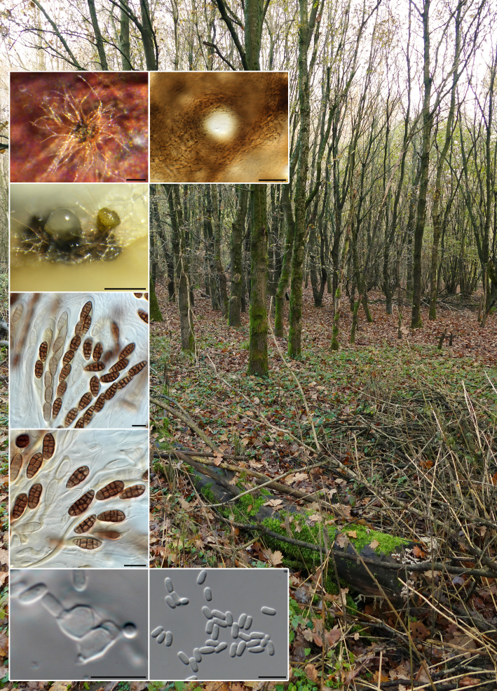
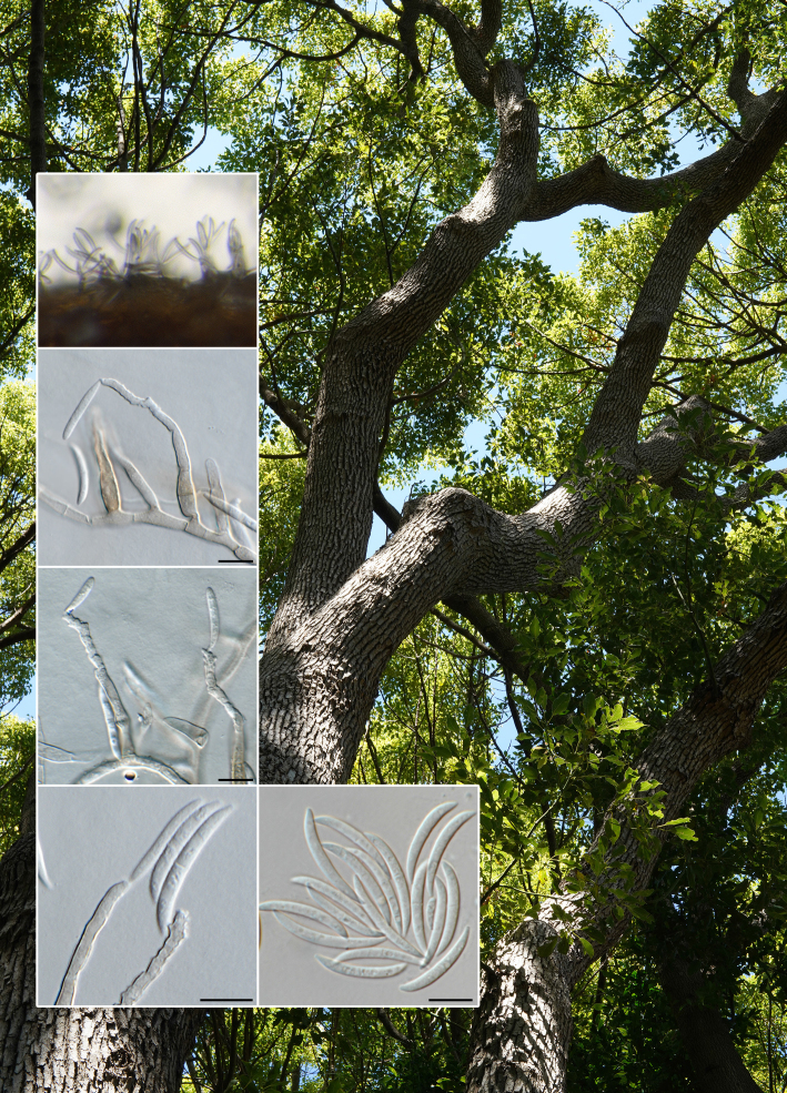
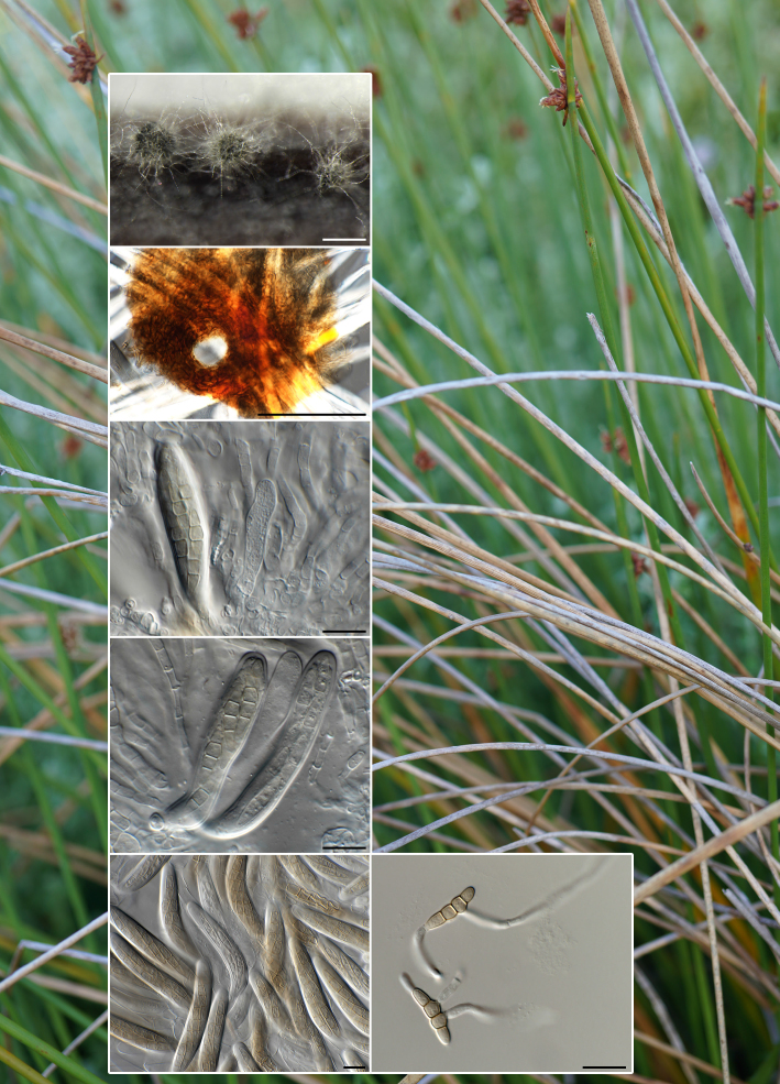
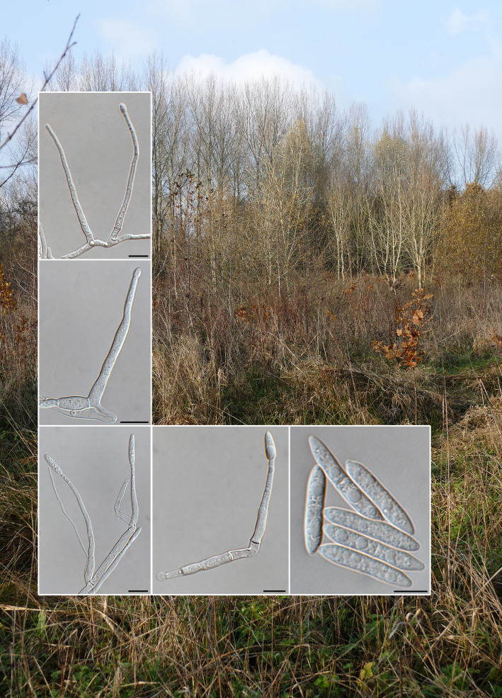
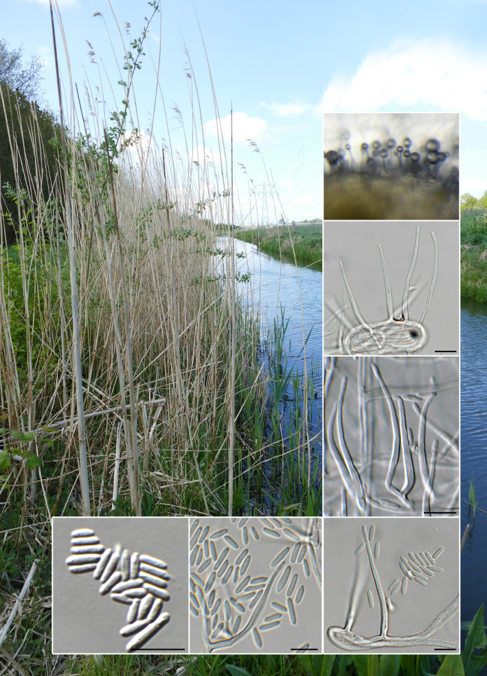
Mycelium consisting of hyaline to pale brown, smooth, branched, septate, 1.5–2 µm diam hyphae. Conidiophores solitary, erect, subcylindrical, flexuous, arising from superficial hyphae, 20–60 × 4–5 µm, thick-walled, red-brown, 2–3-septate, finely roughened. Conidiogenous cells terminal, integrated, subcylindrical, 20–50 × 4–5 µm, medium brown, finely verruculose, forming a rachis with subdenticulate loci, 0.5 µm diam, not darkened, slightly thickened. Conidia solitary, subcylindrical to narrowly fusoid-ellipsoid, apex subobtuse, base bluntly rounded, hilum 0.5–1 µm diam, medium brown, guttulate, smooth-walled, 3-septate, (8–)11–13(–14) × (3–)4(–4.5) µm.

Culture characteristics — Colonies erumpent, folded, with sparse aerial mycelium and smooth, lobate margin, reaching 5 mm diam after 2 wk at 25 °C. On MEA, PDA and OA surface and reverse umber.

Typus. NETHERLANDS, Utrecht Province, Nieuw Wulven, near Houten, 1.5 m a.s.l., N52°03′ E05°10′, on dead bamboo stick, 11 Feb. 2022, E.R. Osieck, HPC 3837 = WI-48/#4404 (holotype CBS H-25172; culture ex-type CPC 43109 = CBS 149447; ITS, LSU, rpb2 (first part) and tef1 (second part) sequences GenBank OQ628466.1, OQ629048.1, OQ627935.1 and OQ627955.1; MycoBank MB 848053).

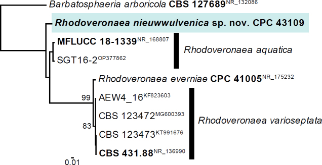
Notes — Rhodoveronaea was introduced by Arzanlou et al. (2007) for R. varioseptata (conidia 0–2(–3)-septate, (8–)11–13(–15) × (2–)3–4(–6) μm), which was later linked to a sexual morph by Réblová (2009). Although R. nieuwwulvenica has conidia of similar dimensions, they tend to be primarily 3-septate. Rhodoveronaea aquatica has larger conidia (1–3-septate, 23–27 × 9–11 μm; Lou et al. 2019), while R. everniae (conidia 0–3-septate, (6.5–)9–11(–12) × (3–)4(–4.5) μm; Crous et al. 2021c) again has smaller conidia. Phylogenetically, R. nieuwwulvenica is distinct from all presently known species.

Based on a megablast search of NCBIs GenBank nucleotide database, the closest hits using the ITS sequence of CPC 43109 had highest similarity to Rhodoveronaea aquatica (voucher MFLU 18-1593, GenBank NR_168807.1; Identities = 451/476 (95 %), four gaps (0 %)), Rhodoveronaea varioseptata (strain CBS 123473, GenBank KT991676.1; Identities = 401/451 (89 %), nine gaps (1 %)) and Rhodoveronaea everniae (strain CBS 148309, GenBank NR_175232.1; Identities = 449/512 (88 %), 12 gaps (2 %)). Closest hits using the LSU sequence of CPC 43109 are Rhodoveronaea aquatica (strain MFLUCC 18-1339, GenBank NG_068647.1; Identities = 778/783 (99 %), no gaps), Rhodoveronaea varioseptata (strain CBS 431.88, GenBank NG_057786.1; Identities = 792/819 (97 %), no gaps) and Rhodoveronaea everniae (strain CPC 41005, GenBank OK663776.1; Identities = 754/781 (97 %), no gaps). Closest hits using the rpb2 (first part) sequence of CPC 43109 had highest similarity to Rhodoveronaea aquatica (strain GZCC 20-0447, GenBank OP473107.1; Identities = 809/865 (94 %), no gaps), Rhodoveronaea varioseptata (strain CBS 123472, GenBank JX066701.1; Identities = 726/886 (82 %), eight gaps (0 %)) and Barbatosphaeria arboricola (strain CBS 114120, GenBank KM492900.1; Identities = 717/889 (81 %), ten gaps (1 %)). Closest hits using the tef1 (second part) sequence of CPC 43109 had highest similarity to Rhodoveronaea aquatica (strain GZCC 20-0447, GenBank OP473041.1; Identities = 829/865 (96 %), no gaps), Xylolentia sp. JY-2022a (strain GZCC 20-0426, GenBank OP473039.1; Identities = 806/867 (93 %), four gaps (0 %)) and Stilbochaeta novae-guineensis (strain CBS 147515, GenBank OL654060.1; Identities = 814/882 (92 %), four gaps (0 %)).

graphic file with name per-2024-50-5-F1a.jpg

Consensus phylogram (50 % majority rule) obtained from the maximum likelihood analysis with IQ-TREE v. 2.1.3 (Kalyaanamoorthy et al. 2017, Minh et al. 2020) of the Rhodoveronaea ITS nucleotide alignment. Bootstrap support values (> 74 % are shown) from 1000 non-parametric bootstrap replicates are shown at the nodes. Culture collection numbers and GenBank accession numbers (superscript) are indicated for all species. The tree was rooted to Barbatosphaeria arboricola (CBS 127689; GenBank NR_132086) and the novelty described here is highlighted with a coloured block and bold font. Sequences from material with a type status are indicated in bold font. Alignment statistics: 9 strains including the outgroup; 526 characters including alignment gaps analysed: 103 distinct patterns, 63 parsimony-informative, 62 singleton sites, 401 constant sites. The best-fit model identified for the entire alignment in IQ-TREE using the TESTNEW option was: TIM2e+G4. The scale bar shows the expected number of nucleotide substitutions per site. The alignment and tree were deposited at figshare.com (doi: 10.6084/m9.figshare.22249690).

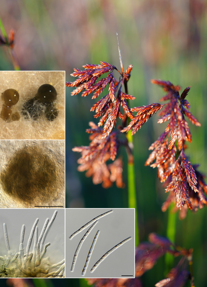
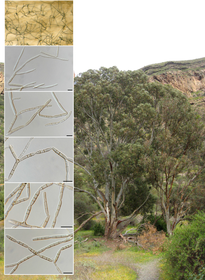
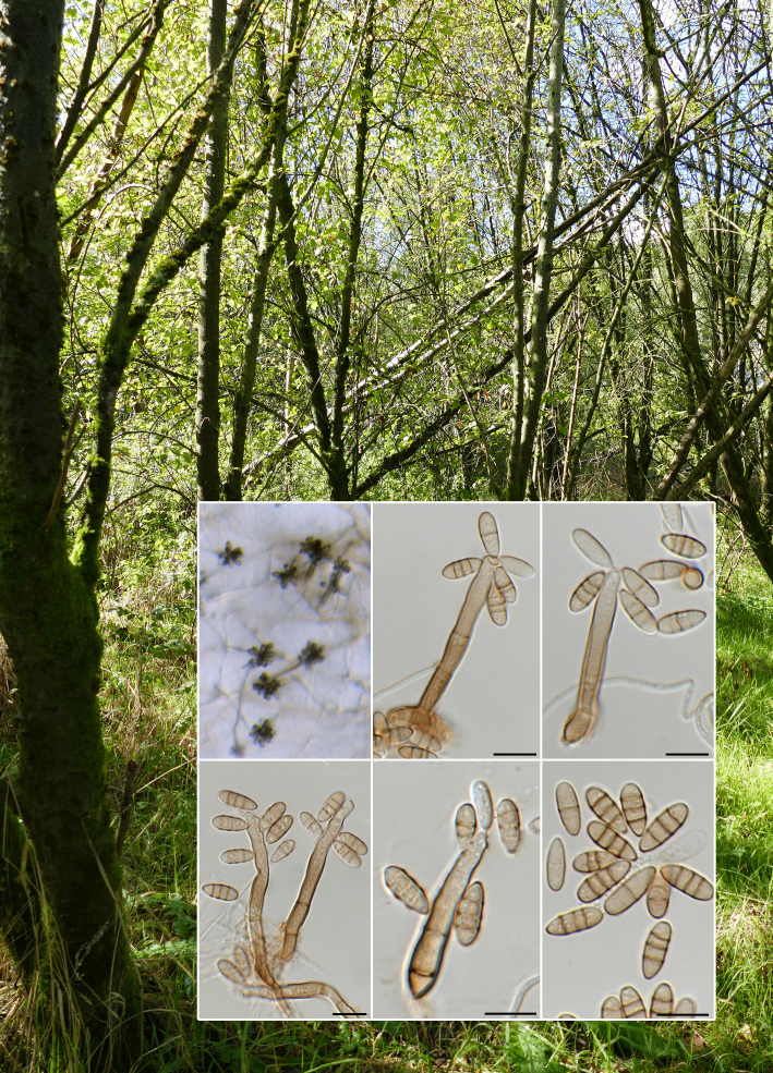
Colour illustrations. Nieuw Wulven, near Houten in Utrecht Province, the Netherlands. Conidiophores and conidiogenous cells giving rise to conidia on SNA; conidia. Scale bars = 10 µm.

Persoonia. 2023 Jun 29;50:164–165.

Fungal Planet 1479 – 29 June 2023

Knufia aloeicola Crous, sp. nov.

PW Crous 1, JZ Groenewald 1, MJ Wingfield 2

Etymology. Name refers to the host genus Aloe from which it was isolated.

Classification — Trichomeriaceae, Chaetothyriales, Chaetothyriomycetidae, Eurotiomycetes.

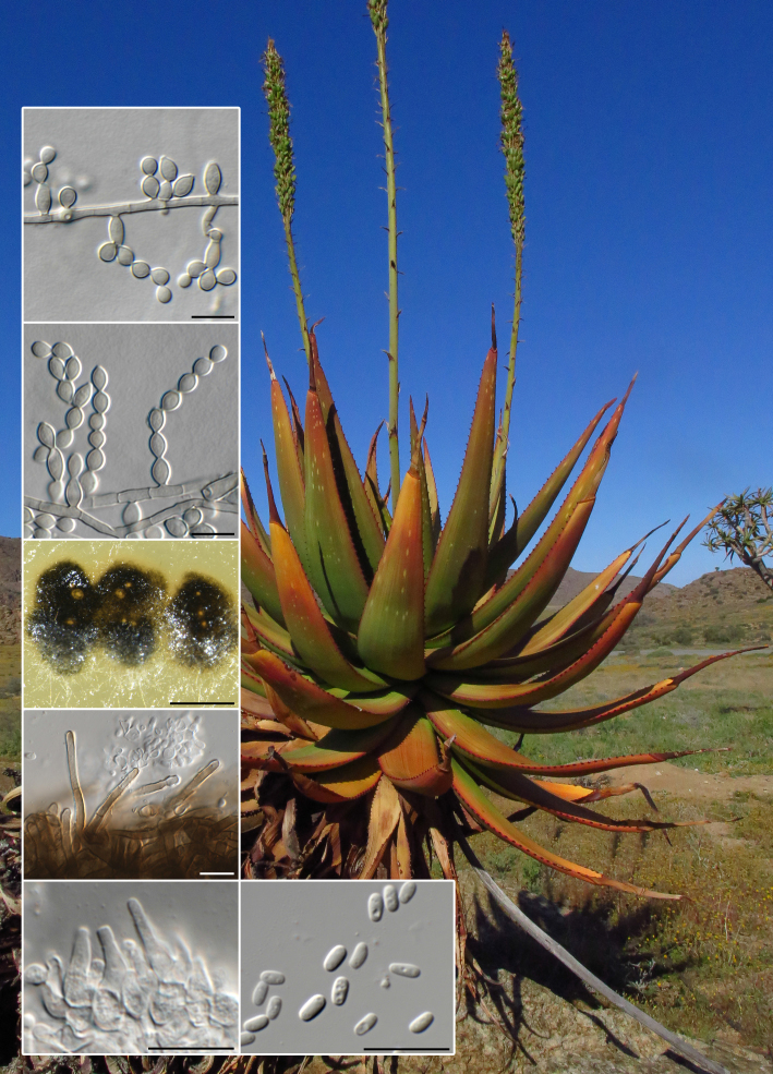
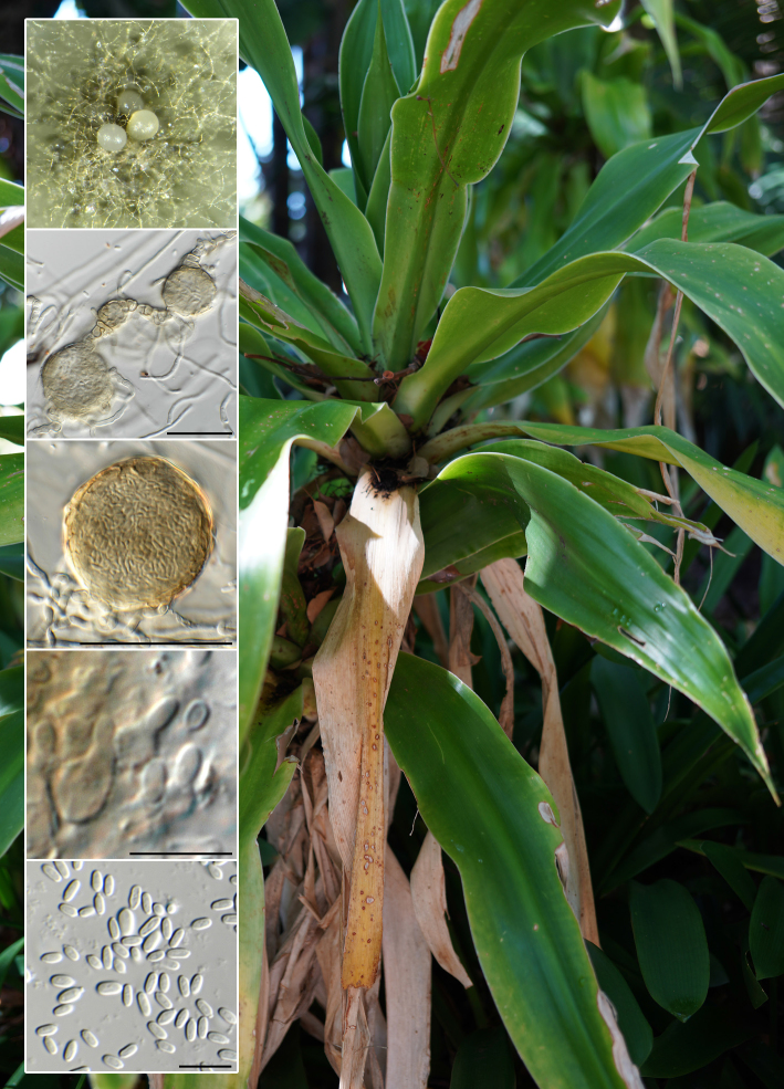
Mycelium consisting of medium brown, smooth, septate, branched, 1.5–2 µm diam hyphae. Conidiophores solitary, erect, medium brown, smooth, subcylindrical, 1–2-septate, 10–20 × 2.5–3.5 µm, giving rise to conidial chains or reduced to a conidiogenous cell with 1–2 sympodial loci, giving rise to branched or unbranched conidial chains. Conidiogenous cells medium brown, fusoid-ellipsoid, smooth, 5–10 × 3–4 µm. Conidia guttulate, in branched or unbranched chains, broadly ellipsoid to subglobose, aseptate, medium brown, smooth, 3.5–6 × 3–3.5 µm, with median apical and basal locus, 1 µm diam, not thickened; conidia remaining attached in chains, breaking into shorter chains with age. Synasexual morph coelomycetous: conidiomata developing in concentric circles, eustromatic, pycnidial, immersed on OA, subglobose with one to several ostioles oozing a pale creamy conidial mass; conidiomata surrounded by brown setae up to 20 µm tall, 2.5–3 µm diam with obtuse ends. Conidiophores lining the inner cavity, hyaline, smooth, reduced to conidiogenous cells or a supporting cell, subcylindrical. Conidiogenous cells hyaline, smooth, ampulliform to subcylindrical, phialidic with periclinal thickening, 5–7 × 4–5 µm. Conidia solitary, aseptate, hyaline, smooth, guttulate, subcylindrical to ellipsoid with obtusely rounded ends, 4–6 × 2–2.5 µm.

Culture characteristics — Colonies flat, spreading, with moderate aerial mycelium and smooth, lobate margin, reaching 15–30 mm diam after 2 wk at 25 °C. On malt extract agar (MEA) surface sienna, reverse sienna to umber; on potato dextrose agar (PDA) surface and reverse olivaceous grey; on oatmeal agar (OA) surface olivaceous grey in centre, pale luteous in outer region.

Typus. SOUTH AFRICA, Northern Cape Province, Springbok, on Aloe gariepensis (Aloaceae), 28 Aug. 2021, M.J. Wingfield, HPC 3769 (holotype CBS H-24971; culture ex-type CPC 42456 = CBS 149069; ITS and LSU sequences GenBank OQ628467.1 and OQ629049.1; MycoBank MB 848054).

Additional isolate examined. SOUTH AFRICA, Northern Cape Province, Springbok, on leaves of A. gariepensis, Nov. 2018, M.J. Wingfield, HPC 3769 = CBS H-24963, culture CPC 42454 = CBS 149044, ITS and LSU sequences GenBank OQ628468.1 and OQ629050.1.

Notes — Knufia includes melanised ascomycetes that are commonly found in extreme environments, ranging from rock-inhabiting, lichenicolous fungi, opportunistic human pathogens, insect associates and plant pathogens (Isola et al. 2022). Knufia aloeicola is related to ‘Phialocephala’ fluminis and Knufia hypolithi (Crous et al. 2021a), but is phylogenetically and morphologically distinct. Furthermore, ‘Phialocephala’ fluminis is better accommodated in Knufia (also see Tanney & Seifert 2020), and hence a new combination is proposed below.

Knufia fluminis (Shearer et al.) Crous, comb. nov. — MycoBank MB 848055

Basionym. Phialocephala fluminis Shearer et al., Mycologia 68: 186. 1976.

Material examined. USA, Illinois, Macon County, Decatur, Sangamon River, submerged balsa wood, 15 Apr. 1975, C.A. Shearer & J.L. Crane (holotype ILLS 36160, culture ex-type ATCC 32105 = CBS 351.85).

Based on a megablast search of NCBIs GenBank nucleotide database, the closest hits using the ITS sequence of CPC 42454 had highest similarity to Phialocephala fluminis (strain CBS 351.85, GenBank MH861889.1; Identities = 549/559 (98 %), one gap (0 %)), Knufia hypolithi (strain CBS 146991, GenBank NR_173045.1; Identities = 545/568 (96 %), ten gaps (1 %)) and Knufia tsunedae (strain FMR 10621, GenBank NR_132842.1; Identities = 483/518 (93 %), 14 gaps (2 %)). The ITS sequence of CPC 42454 is identical to that of CPC 42456 (558/558 nucleotides). Closest hits using the LSU sequence of CPC 42454 are Phialocephala fluminis (strain CBS 351.85, GenBank MH873578.1; Identities = 749/754 (99 %), two gaps (0 %)), Knufia hypolithi (strain CBS 146991, GenBank NG_076736.1; Identities = 743/754 (99 %), no gaps) and Knufia karalitana (strain CCFEE 5656, GenBank NG_067535.1; Identities = 742/755 (98 %), two gaps (0 %)). The LSU sequence of CPC 42454 is identical to that of CPC 42456 (752/752 nucleotides).

Colour illustrations. Aloe gariepensis growing near Springbok, South Africa. Branched conidial chains of hyphomycetous morph; coelomycetous synasexual morph; setae; conidiogenous cells giving rise to conidia; conidia. Scale bars = 250 µm (conidiomata), 10 µm (all others).

graphic file with name per-2024-50-5-F2.jpg

Knufia aloeicola

Supplementary material

FP1479

Phylogenetic tree.

per-2024-50-5-SF1.jpg (1.5MB, jpg)
Persoonia. 2023 Jun 29;50:166–167.

Fungal Planet 1480 – 29 June 2023

Neodacampia Crous & Osieck, gen. nov.

PW Crous 1, JZ Groenewald 1, ER Osieck 2

Etymology. Name refers to its morphological similarity to Dacampia.

Classification — Phaeosphaeriaceae, Pleosporales, Pleosporomycetidae, Dothideomycetes.

Pseudothecia immersed, becoming erumpent, papillate, associated with red discolouration of wood, globose, brown; wall of 6–10 layers of brown textura angularis. Pseudoparaphyses hyphae-like, hyaline, smooth, branched, septate, intermingled among asci. Asci subcylindrical, bitunicate with apical chamber, 8-septate, stipitate with basal stalk. Ascospores overlapping, biseriate, fusoid-ellipsoid, brown, guttulate, smooth, muriformly septate, 3(–6) horizontal septa, constricted at primary median septum, with numerous vertical or oblique septa; mucoid sheath absent.

Type species. Neodacampia ulmea Crous & Osieck

MycoBank MB 848056

graphic file with name per-2024-50-5-F3.jpg

Neodacampia ulmea

Persoonia. 2023 Jun 29;50:167.

Fungal Planet 1480 – 29 June 2023

Neodacampia ulmea Crous & Osieck, sp. nov.


Etymology. Name refers to the host genus Ulmus from which it was isolated.

Pseudothecia immersed in host tissue, becoming erumpent, papillate, associated with red discolouration of wood, globose, brown, 300–350 µm diam; wall of 6–10 layers of brown textura angularis. Pseudoparaphyses hyphae-like, hyaline, smooth, branched, septate, 2–2.5 µm diam, intermingled among asci. Asci subcylindrical, bitunicate with apical chamber, 8-septate, stipitate with basal stalk, 100–140 × 10–13 µm. Ascospores overlapping, biseriate, fusoid-ellipsoid, brown, guttulate, smooth, muriformly septate, 3(–6) horizontal septa, constricted at primary median septum, less at secondary septa, with 0–2 vertical or oblique septa (also occurring in end cells), 2nd cell often enlarged, end cells obtuse; mucoid sheath absent, (14–)15–17(–18) × (6.5–)7(–7.5) µm. In culture forming spermatogonia similar to ascomata in anatomy; spermatia hyaline, smooth, aseptate, guttulate, 3.5–5 × 2 µm.

Culture characteristics — Colonies flat, spreading, with sparse to moderate aerial mycelium and smooth, lobate margin, reaching 40–45 mm diam after 2 wk at 25 °C. On malt extract agar (MEA) surface olivaceous grey, reverse smoke grey in centre, olivaceous grey in outer region; on potato dextrose agar (PDA) surface and reverse amber in centre, red in outer region; on oatmeal agar (OA) surface umber.

Typus. NETHERLANDS, Utrecht Province, Nieuw Wulven, near Houten, 1.5 m a.s.l., N52°02′56″ E05°09′58″, on branch of Ulmus laevis (Ulmaceae), 31 Dec. 2021, E.R. Osieck, HPC 3813 = WI-43/#4367 (XVIII-26) (holotype CBS H-25165; culture ex-type CPC 42680 = CBS 149452; ITS, LSU, SSU, rpb2 (first part) and tef1 (second part) sequences GenBank OQ628469.1, OQ629051.1, OQ628447.1, OQ627936.1 and OQ627956.1; MycoBank MB 848057).

Notes — Neodacampia resembles the lichenicolous Dacampia (Dacampiaceae; Ertz et al. 2015), but is more closely related to the asexual Banksiophoma (Phaeosphaeriaceae; Crous et al. 2017a). Neodacampia, which is monotypic based on N. ulmea, is associated with a red discolouration of wood on which solitary pseudothecia occur, characterised by pseudoparaphyses, and muriformly septate ascospores that lack a sheath, and for which no asexual morph was observed in culture. The asexual morph, if found, would likely be phoma-like, given its close association to Banksiophoma. The taxonomic placement of this wood saprobe in Phaeosphaeriaceae was quite unexpected since members of this family occur mostly on monocotyledons (Jaklitsch et al. 2016).

Based on a megablast search of NCBIs GenBank nucleotide database, the closest hits using the ITS sequence had highest similarity to ‘Phaeosphaeria’ sp. (strain G330, GenBank KR094442.1; Identities = 495/503 (98 %), no gaps), Septoriella hibernica (strain CBS 145371, GenBank NR_164460.1; Identities = 415/455 (91 %), 12 gaps (2 %)) and Septoriella allojunci (strain MFLUCC 15-0701, GenBank KU058718.1; Identities = 415/454 (91 %), 15 gaps (3 %)). Closest hits using the LSU sequence are Didymocyrtis brachylaenae (strain CPC 32651, GenBank NG_067827.1; Identities = 826/836 (99 %), one gap (0 %)), Diederichomyces cladoniicola (strain UTHSC DI16-313, GenBank LN907456.1; Identities = 825/836 (99 %), one gap (0 %)) and Neosulcatispora agaves (strain CPC 26407, GenBank KT950867.1; Identities = 824/836 (99 %), one gap (0 %)). Closest hits using the SSU sequence are Coniothyrium concentricum (strain CBS 589.79, GenBank EU754053.1; Identities = 1006/1006 (100 %), no gaps), Parastagonospora uniseptata (strain MFLUCC 13-0387, GenBank NG_063666.1; Identities = 1007/1008 (99 %), one gap (0 %)) and Wojnowicia dactylidicola (strain MFLUCC 13-0738, GenBank NG_063564.1; Identities = 1007/1008 (99 %), one gap (0 %)). Closest hits using the rpb2 (first part) sequence had highest similarity to Banksiophoma australiensis (strain CBS 142163, GenBank KY979846.1; Identities = 668/794 (84 %), no gaps), Brunneomurispora lonicerae (strain KUMCC 18-0157, GenBank MK359079.1; Identities = 714/856 (83 %), four gaps (0 %)) and Septoriella callistemonis (strain CPC 38761, GenBank MZ078194.1; Identities = 721/874 (82 %), 14 gaps (1 %)). Closest hits using the tef1 (second part) sequence had highest similarity to Didymocyrtis cladoniicola (strain UTHSC DI16-313, GenBank LT797117.1; Identities = 787/819 (96 %), no gaps), Phaeosphaeria sinensis (strain MFLUCC 18-1552, GenBank MK360072.1; Identities = 778/810 (96 %), no gaps) and Paraphoma radicina (strain 17chuncheon01-04, GenBank MT946675.1; Identities = 765/798 (96 %), no gaps).

Colour illustrations. Ulmus laevis at Nieuw Wulven, near Houten in Utrecht Province, the Netherlands. Ascoma on wood, with red discolouration; ostiole; conidiomata in culture; asci and ascospores; conidiogenous cells giving rise to conidia; conidia. Scale bars = 300 µm (ascomata and conidiomata), 10 µm (all others).

Supplementary material

FP1480

Phylogenetic tree.

per-2024-50-5-SF2.jpg (4.4MB, jpg)
Persoonia. 2023 Jun 29;50:168–169.

Fungal Planet 1481 – 29 June 2023

Nothotrichosporon Crous, M. Groenew. & Jurjević, gen. nov.

PW Crous 1, M Groenewald 1, JZ Groenewald 1, Ž Jurjević 2, S Balashov 2

Etymology. Name refers to the fact that it is distinct from Trichosporon.

Classification — Trichosporonaceae, Trichosporonales, Tremellomycetes.

Mycelium consisting of hyaline, smooth, thin-walled, sparsely septate, rarely branched hyphae, disarticulating into subcylindrical arthroconidia, and budding to form ellipsoid conidia. Arthroconidia hyaline, smooth, guttulate, thin-walled, subcylindrical, ends become obtuse. Secondary conidia hyaline, smooth, guttulate, thin-walled, ellipsoid.

Type species. Nothotrichosporon aquaticum Crous, M. Groenew. & Jurjević

MycoBank MB 848058

graphic file with name per-2024-50-5-F4.jpg

Nothotrichosporon aquaticum

Persoonia. 2023 Jun 29;50:169.

Fungal Planet 1481 – 29 June 2023

Nothotrichosporon aquaticum Crous, M. Groenew. & Jurjević, sp. nov.


Etymology. Name refers to the fact that it was isolated from water.

Mycelium consisting of hyaline, smooth, thin-walled, sparsely septate, rarely branched hyphae, 4–8 µm diam, disarticulating into subcylindrical arthroconidia, and budding to form ellipsoid conidia. Arthroconidia hyaline, smooth, guttulate, thin-walled, subcylindrical, ends become obtuse, 35–55 × 12–17 µm. Secondary conidia hyaline, smooth, guttulate, thin-walled, ellipsoid, (22–)28–32(–35) × (17–)19–20(–22) µm.

All physiological growth tests were done at 24 °C for a period of 10 d. Assimilation tests were performed using the ID 32 C identification system for yeasts (bioMérieux SA).

Fermentation — Glucose is fermented.

Carbon assimilation — D-glucose, D-galactose (w), cycloheximide, sucrose, lactic acid, D-cellobiose, D-maltose, D-tre-halose, 2-keto-D-gluconate, a-methyl-D-glucoside (delayed), D-mannitol, myo-inositol, D-sorbitol, D-xylose, D-ribose, glycerol, meso-erythritol, sodium glucoronate, D-melezitose, potassium gluconate, L-sorbose (delayed), are assimilated. N-acetyl-glucosamine, L-arabinose, D-raffinose, D-lactose, L-rhamnose, D-melibiose, D-glucosamine, are not assimilated.

Nitrogen assimilation — ethylamine, L-lysine, cadaverine are assimilated. Nitrate, nitrite, D-glucosamine HCl, tryptophane, pepton are not assimilated.

Other tests — Growth in 0.01 % cycloheximide is positive. Growth at 15 °C, 24 °C, 30 °C is positive. Growth at 37 °C is negative. Growth in 50 % glucose is negative.

Culture characteristics — Colonies flat, spreading, sectoring, folded, with sparse aerial mycelium and smooth, lobate margin, reaching 30 mm diam after 2 wk at 25 °C. On malt extract agar (MEA), potato dextrose agar (PDA) and oatmeal agar (OA) surface and reverse dirty white.

Typus. USA, Louisiana, Abbeville, pen water, Oct. 2021, Ž. Jurjević 5667 (holotype CBS H-25224; culture ex-type CPC 42870 = CBS 18113; ITS and LSU sequences GenBank OQ628470.1 and OQ629052.1; MycoBank MB 848059).

Notes — Effuseotrichosporon was described for a singlespecies lineage formed by Trichosporon vanderwaltii in Trichosporonaceae, and characterised by true hyphae that disarticulate into arthroconidia. The present collection is related to Effuseotrichosporon based on ITS blast searches, although phylogenetically distinct, and therefore accommodated in the new genus, Nothotrichosporon.

Based on a megablast search of NCBIs GenBank nucleotide database, the closest hits using the ITS sequence had highest similarity to Effuseotrichosporon vanderwaltii (strain CBS 12124, GenBank NR_153975.1; Identities = 459/485 (95 %), 12 gaps (2 %)), Prillingera fragicola (strain JCM 1530, GenBank NR_136945.1; Identities = 423/450 (94 %), 11 gaps (2 %)) and Apiotrichum gamsii (strain CBS 8245, GenBank NR_073247.1; Identities = 452/483 (94 %), six gaps (1 %)). Closest hits using the LSU sequence are Vanrija humicola (strain CBS 571, GenBank KY110010.1; Identities = 824/858 (96 %), six gaps (0 %)), Apiotrichum scarabaeorum (strain CBS 5601, GenBank NG_057009.1; Identities = 810/845 (96 %), one gap (0 %)) and Aegeritella tuberculata (strain WA51216, GenBank KT380846.1; Identities = 772/802 (96 %), three gaps (0 %)).

Colour illustrations. Collection site near Abbeville, Louisiana, USA. Colony on synthetic nutrient-poor agar; hyphae giving rise to arthroconidia and secondary conidia. Scale bars = 10 µm.

Supplementary material

FP1481

Phylogenetic tree.

per-2024-50-5-SF3.jpg (2.3MB, jpg)
Persoonia. 2023 Jun 29;50:170–171.

Fungal Planet 1482 – 29 June 2023

Xenoidriella Crous, gen. nov.

PW Crous 1, JZ Groenewald 1

Etymology. Name refers to its morphological similarity to Idriella.

Classification — Microdochiaceae, Xylariales, Xylariomycetidae, Sordariomycetes.

Mycelium consisting of pale brown, smooth, branched, septate hyphae. Conidiophores solitary, arising from superficial hyphae, medium brown, thick-walled, smooth, subcylindrical, flexuous, 1–6-septate. Conidiogenous cells terminal, integrated, brown, smooth, forming a rachis of denticulate loci, not thickened nor darkened. Conidia solitary, hyaline, smooth, guttulate, fusoid, medianly 1-septate, tapering towards subacutely rounded apex and truncate hilum.

Type species. Xenoidriella cinnamomi Crous

MycoBank MB 848060.

graphic file with name per-2024-50-5-F5.jpg

Xenoidriella cinnamomi

Persoonia. 2023 Jun 29;50:171.

Fungal Planet 1482 – 29 June 2023

Xenoidriella cinnamomi Crous, sp. nov.


Etymology. Name refers to the host genus Cinnamomum from which it was isolated.

Mycelium consisting of pale brown, smooth, branched, septate, 2–3.5 µm diam hyphae. Conidiophores solitary, arising from superficial hyphae, medium brown, thick-walled, smooth, subcylindrical, flexuous, 1–6-septate, 40–120 × 4–6 µm. Conidiogenous cells terminal, integrated, 20–40 × 3–4 µm, brown, smooth, forming a rachis of denticulate loci, 0.5–1 × 1 µm, not thickened nor darkened. Conidia solitary, hyaline, smooth, guttulate, fusoid, medianly 1-septate, tapering towards subacutely rounded apex and truncate hilum, 1 µm diam, (20–)23–27(–30) × (2.5–)3 µm.

Culture characteristics — Colonies erumpent, spreading, with sparse to moderate aerial mycelium and smooth, lobate margin, reaching 35 mm diam after 2 wk at 25 °C. On malt extract agar (MEA), potato dextrose agar (PDA) and oatmeal agar (OA) surface and reverse umber.

Typus. SOUTH AFRICA, Western Cape Province, Cape Town, on leaf of Cinnamomum camphora (Lauraceae), 27 Feb. 2022, P.W. Crous, HPC 3839 (holotype CBS H-25173; culture ex-type CPC 43130 = CBS 149458; ITS, LSU, SSU, rpb2 (first part) and tef1 (second part) sequences GenBank OQ628471.1, OQ629053.1, OQ628448.1, OQ627937.1 and OQ627957.1; MycoBank MB 848061).

Notes — Xenoidriella cinnamomi is related to Neoidriella desertorum CBS 985.72 (Hernández-Restrepo et al. 2016a), Guayaquilia cubensis and the appendaged coelomycete Ciliosporella italica. Morphologically X. cinnamomi is distinct from N. desertorum in having 1-septate conidia, and lacking chlamydospores. Furthermore, it is distinct from Guayaquilia, which is characterised by having macronematous, tree-like conidiophores, (0–)1-septate navicular conidia, and thick-walled, 1-septate, brown chlamydospores (Magdama et al. 2020).

Closest hits using the ITS sequence of CPC 43130 are Monographella sp. (strain HGGJ-16, GenBank KR708990.1; Identities = 365/400 (91 %), seven gaps (1 %)), Guayaquilia cubensis (strain SG1001, GenBank OP380877.1; Identities = 378/420 (90 %), 13 gaps (3 %)) and Ciliosporella italica (strain MFLUCC 16-1146, GenBank NR_169717.1; Identities = 465/519 (90 %), 26 gaps (5 %)). Closest hits using the LSU sequence of CPC 43130 are Ciliosporella italica (strain MFLUCC 16-1146, GenBank NG_073833.1; Identities = 763/789 (97 %), three gaps (0 %)), Selenodriella fertilis (strain CBS 148328, GenBank ON400824.1; Identities = 758/786 (96 %), one gap (0 %)) and Idriella cubensis (strain MUCL 39017, GenBank KC775708.1; Identities = 758/790 (96 %), five gaps (0 %)). Closest hits using the SSU sequence of CPC 43130 are Pirozynskiomyces sinensis (no strain number supplied, GenBank KY994108.1; Identities = 932/956 (97 %), no gaps), Arthrinium japonicum (strain IFO 30500, GenBank AB220211.1; Identities = 932/956 (97 %), no gaps) and Xenoanthostomella calami (as Xenoanthostomella sp. SK-2022a, strain MFLUCC 14-0617B, GenBank ON650709.1; Identities = 932/957 (97 %), two gaps (0 %)). Closest hits using the rpb2 (first part) sequence of CPC 43130 had highest similarity to Biscogniauxia arima (voucher 122, GenBank GQ304736.1; Identities = 641/818 (78 %), six gaps (0 %)), Peroneutypa scoparia (strain MFLUCC 11-0478, GenBank KU940179.1; Identities = 641/823 (78 %), 19 gaps (2 %)) and Kretzschmaria pavimentosa (voucher 109, GenBank GQ844787.1; Identities = 641/827 (78 %), ten gaps (1 %)). Distant hits obtained using the tef1 (second part) sequence of CPC 43130 had highest similarity to Microdochium trichocladiopsis (strain MPI-CAGE-CH-0230, GenBank XM_046152772.1; Identities = 736/802 (92 %), no gaps), Microdochium chrysanthemoides (strain LC5466, GenBank KX855236.1; Identities = 731/802 (91 %), no gaps) and Xenoanthostomella chromolaenae (strain MFLUCC 17-1484, GenBank MN648732.1; Identities = 701/773 (91 %), no gaps).

Colour illustrations. Cinnamomum camphora growing near Cape Town, SouthAfrica. Conidiophores and conidiogenous cells giving rise to conidia on pine needle agar; conidiogenous cells giving rise to conidia; conidia. Scale bars = 10 µm.

Supplementary material

FP1482

Phylogenetic tree.

per-2024-50-5-SF4.jpg (1.8MB, jpg)
Persoonia. 2023 Jun 29;50:172–173.

Fungal Planet 1483 – 29 June 2023

Paraloratospora schoenoplecti Crous, sp. nov.

PW Crous 1, JZ Groenewald 1

Etymology. Name refers to the host genus Schoenoplectus from which it was isolated.

Classification — Phaeosphaeriaceae, Pleosporales, Pleosporomycetidae, Dothideomycetes.

Ascomata immersed in bleached white areas on culms, becoming erumpent, globose, brown, 200–300 µm diam with central ostiole, 15–20 µm diam; wall of 3–5 layers of brown textura angularis. Pseudoparaphyses intermingled among asci, hyphae-like, hyaline, smooth, branched, septate, 2.5–3 µm diam. Asci fusoid-ellipsoid, bitunicate, 8-spored with ocular chamber, stipitate, fasciculate, 70–80 × 11–13 µm. Ascospores tri- to multiseriate, fusoid, medium brown, smooth, guttulate, 3-septate, slightly constricted at septa, second cell from the apex slightly swollen, (22–)25–28(–30) × (3–)3.5(–5) µm. Ascospores shot onto agar brown, germinating from multiple cells, in irregular direction, constricted at septa, 4–5 µm diam. Cultures homothallic, also forming sexual morph in culture.

Culture characteristics — Colonies flat to erumpent, spreading, with moderate aerial mycelium and feathery, lobate margin, reaching 40 mm diam after 2 wk at 25 °C. On malt extract agar (MEA) surface and reverse olivaceous grey; on potato dextrose agar (PDA) surface pale olivaceous grey, reverse olivaceous grey; on oatmeal agar (OA) surface saffron to pale luteous.

Typus. SOUTH AFRICA, Western Cape Province, Cape Town, on stems of Schoenoplectus lacustris (Cyperaceae), 6 Mar. 2022, P.W. Crous, HPC 3844 (holotype CBS H-25175; culture ex-type CPC 43149 = CBS 149459; ITS, LSU, rpb2 (first part), tef1 (first part) and tub2 sequences GenBank OQ628472.1, OQ629054.1, OQ627938.1, OQ627947.1 and OQ627960.1; MycoBank MB 848062).

Notes — Paraloratospora schoenoplecti clusters with Pa. gahniae, but has longer ascospores, and is phylogenetically distinct. Paraloratospora, based on Pa. camporesii, is characterised as being phaeosphaeria-like and having hyaline to pale brown, fusoid to ellipsoidal, septate ascospores, the second cell from the apex swollen, smooth-walled, with or without a mucilaginous sheath (Hyde et al. 2020). Paraloratospora is presently known to accommodate two species, Pa. camporesii (ascospores 3-septate, 20–24 × 3–5 µm) and Pa. gahniae (ascospores 3(–4)-septate, (18–)20–22(–25) × (4.5–)5 μm; Crous et al. 2017b). Paraloratospora represents a distinct clade in the Phaeosphaeriaceae, and several other taxa accommodated in Phaeosphaeria will have to be allocated to it.

Based on a megablast search of NCBIs GenBank nucleotide database, the closest hits using the ITS sequence had highest similarity to Paraloratospora gahniae (strain CPC 32454, GenBank NR_156675.1; Identities = 545/562 (97 %), four gaps (0 %)), Phaeosphaeria norfolcia (strain CBS 593.86, GenBank MH861997.1; Identities = 487/527 (92 %), two gaps (0 %)) and Phaeosphaeria caricicola (strain CBS 603.86, GenBank KF251182.1; Identities = 515/566 (91 %), 11 gaps (1 %)). Closest hits using the LSU sequence are Paraloratospora gahniae (strain CPC 32454, GenBank NG_059852.1; Identities = 806/806 (100 %), no gaps), Phaeosphaeria caricicola (strain CBS 133078, GenBank MH877523.1; Identities = 804/806 (99 %), no gaps) and Phaeosphaeria glyceriae-plicatae (strain CBS 101261, GenBank MH874330.1; Identities = 804/806 (99 %), no gaps). Closest hits using the rpb2 (first part) sequence had highest similarity to Paraloratospora gahniae (strain CPC 32454, GenBank MG386148.1; Identities = 739/768 (96 %), no gaps), Septoriella callistemonis (strain CPC 38761, GenBank MZ078194.1; Identities = 653/820 (80 %), eight gaps (0 %)) and Paraphoma fimeti (strain UTHSC DI16-296, GenBank LT797032.1; Identities = 653/821 (80 %), eight gaps (0 %)). The best hit using the tef1 (first part) sequence had highest similarity to Paraloratospora gahniae (strain CPC 32454 (GenBank MG386157.1; Identities = 245/290 (84 %), three gaps (1 %)). Closest hits using the tub2 sequence had highest similarity to Paraloratospora gahniae (strain CPC 32454, GenBank MG386170.1; Identities = 286/298 (96 %), one gap (0 %)), Dothidotthia robiniae (strain MFLUCC 16-1185, GenBank MK933788.1; Identities = 266/301 (88 %), 14 gaps (4 %)) and Phaeosphaeria caricicola (strain CBS 603.86, GenBank KF252676.1; Identities = 264/300 (88 %), five gaps (1 %)).

Colour illustrations. Schoenoplectus lacustris growing near Cape Town, South Africa. Ascomata forming on pine needle agar; ascoma with ostiole; asci with ascospores and pseudoparaphyses; germinating ascospores. Scale bars = 150 µm (ascoma), 10 µm (all others).

graphic file with name per-2024-50-5-F6.jpg

Paraloratospora schoenoplecti

Supplementary material

See the supplementary material FP1480 for a phylogenetic tree containing this novel species.

FP1480

Phylogenetic tree.

per-2024-50-5-SF2.jpg (4.4MB, jpg)
Persoonia. 2023 Jun 29;50:174–175.

Fungal Planet 1484 – 29 June 2023

Alfaria thamnochorti Crous, sp. nov.

PW Crous 1, JZ Groenewald 1

Etymology. Name refers to the host genus Thamnochortus from which it was isolated.

Classification — Stachybotryaceae, Hypocreales, Hypocreomycetidae, Sordariomycetes.

Conidiomata sporodochial, globose, brown, 80–100 µm diam; wall of 3–4 layers of pale brown textura intricata to angularis. Conidiogenous cells lining inner cavity, subcylindrical, pale brown, smooth, 10–25 × 2–3 µm, proliferating inconspicuously percurrently at apex. Conidia cylindrical, guttulate, straight to flexuous, smooth, pale to dark brown (depending on agar medium), apex obtuse, base truncate, aseptate, (25–)30–45(–50) × 2(–3) µm.

Culture characteristics — Colonies flat, spreading, with moderate aerial mycelium and feathery, lobate margin, reaching 30 mm diam after 2 wk at 25 °C. On malt extract agar (MEA) surface saffron, reverse sienna; on potato dextrose agar (PDA) surface saffron, reverse ochreous; on oatmeal agar (OA) surface saffron.

Typus. SOUTH AFRICA, Western Cape Province, Cape Town, on culm of Thamnochortus fraternus (Restionaceae), 6 Mar. 2022, P.W. Crous, HPC 3848 (holotype CBS H-25176; culture ex-type CPC 43155 = CBS 149460; ITS, LSU, rpb2 (first part), tef1 (first part) and tub2 sequences GenBank OQ628473.1, OQ629055.1, OQ627939.1, OQ627948.1 and OQ627961.1; MycoBank MB 848063).

Notes — Alfaria was introduced by Crous et al. (2014) for a sexual genus associated with leaf apical necrosis of Cyperus esculentus, and later shown to have microthyrium-like asexual morphs. Alfaria thamnochorti is placed basal to a lineage containing amongst others A. terrestris (conidia fusoid, 6–8 × 2–3 μm), A. ossiformis (conidia ossiform to ellipsoid, (5–)6–7 × 2–3 μm; Lombard et al. 2016), and A. junci (conidia subcylindrical, (10–)11–12(–13) × 2(–2.5) μm; Crous et al. 2021c), but is distinct in having much larger conidia.

Based on a megablast search of NCBIs GenBank nucleotide database, the closest hits using the ITS sequence had highest similarity to Alfaria terrestris (strain F9(NSA3&NLC2), GenBank MW301193.1; Identities = 505/519 (97 %), six gaps (1 %)), Alfaria ossiformis (strain CBS 324.54, GenBank NR_145068.1; Identities = 502/516 (97 %), two gaps (0 %)) and Myrothecium gramineum (strain CBS 324.54, GenBank AY254151.1; Identities = 502/516 (97 %), two gaps (0 %)). Closest hits using the LSU sequence are Alfaria terrestris (strain CBS 168.97, GenBank KU845996.1; Identities = 811/813 (99 %), no gaps), Alfaria dandenongensis (strain CBS 143399, GenBank NG_069537.1; Identities = 810/813 (99 %), no gaps) and Amerosporium atrum (strain CBS 151.69, GenBank MH877704.1; Identities = 810/813 (99 %), no gaps). Closest hits using the rpb2 (first part) sequence had highest similarity to Alfaria dandenongensis (strain CBS 143399, GenBank MG386146.1; Identities = 771/818 (94 %), no gaps), Alfaria ossiformis (strain CBS 324.54, GenBank KU846002.1; Identities = 677/724 (94 %), no gaps) and Alfaria humicola (strain ZSY10, GenBank MH818829.1; Identities = 673/724 (93 %), no gaps). Closest hits using the tef1 (first part) sequence had highest similarity to Alfaria terrestris (strain CBS 127305, GenBank KU846012.1; Identities = 298/345 (86 %), 12 gaps (3 %)), Alfaria ossiformis (strain CBS 324.54, GenBank KU846009.1; Identities = 333/396 (84 %), 16 gaps (4 %)) and Alfaria acaciae (strain CBS 143504, GenBank MH108012.1; Identities = 322/387 (83 %), 31 gaps (8 %)). The best hit using the tub2 sequence had highest similarity to Alfaria tabebuiae (strain CBS 145066, GenBank MK047579.1; Identities = 471/551 (85 %), 25 gaps (4 %)).

Colour illustrations. Culm of Thamnochortus fraternus growing in Cape Town, South Africa. Conidiomata forming on oatmeal agar; conidiogenous cells giving rise to conidia; conidia. Scale bars = 50 µm (conidiomata), 10 µm (all others).

graphic file with name per-2024-50-5-F7.jpg

Alfaria thamnochorti

Supplementary material

FP1484

Phylogenetic tree.

per-2024-50-5-SF5.jpg (1.4MB, jpg)
Persoonia. 2023 Jun 29;50:176–177.

Fungal Planet 1485 – 29 June 2023

Neodevriesia scadoxi Crous, sp. nov.

PW Crous 1, JZ Groenewald 1

Etymology. Name refers to the host genus Scadoxus from which it was isolated.

Classification — Neodevriesiaceae, Mycosphaerellales, Dothideomycetidae, Dothideomycetes.

Conidiomata pycnidial, solitary or gregarious, globose, 30–90 µm diam with central ostiole; wall of 2–3 layers of brown textura angularis. Conidiophores reduced to conidiogenous cells lining inner cavity, ampulliform, hyaline, smooth, phialidic, 4–5 × 3–4 µm. Conidia solitary, hyaline, smooth, subcylindrical with obtuse ends, aseptate, rarely 1-septate in older conidia, 3– 5 × 1.5–2 µm.

Culture characteristics — Colonies erumpent, spreading, surface folded, with sparse aerial mycelium and smooth, lobate margin, reaching 6 mm diam after 2 wk at 25 °C. On malt extract agar (MEA), potato dextrose agar (PDA) and oatmeal agar (OA) surface and reverse fuscous black.

Typus. SOUTH AFRICA, Western Cape Province, Cape Town, on leaves of Scadoxus puniceus (Amaryllidaceae), 27 Feb. 2022, P.W. Crous, HPC 3851 (holotype CBS H-25177; culture ex-type CPC 43161 = CBS 149461; ITS, LSU, rpb2 (first part) and tub2 sequences GenBank OQ628474.1, OQ629056.1, OQ627940.1 and OQ627962.1; MycoBank MB 848064).

Notes — Neodevriesia is a cladosporium-like genus including species that are foliicolous, saprobic or plant pathogenic (Quaedvlieg et al. 2014). Neodevriesia scadoxi is allied to other species of Neodevriesia, and probably represents a synasexual morph, given the fact that it has a phoma-like morphology.

Based on a megablast search of NCBIs GenBank nucleotide database, the closest hits using the ITS sequence had highest similarity to Recurvomyces sp. (strain CR18, GenBank KY111910.1; Identities = 473/476 (99 %), one gap (0 %)), Neodevriesia bulbillosa (strain CBS 118285, GenBank NR_144953.1; Identities = 484/500 (97 %), seven gaps (1 %)) and Neodevriesia lagerstroemiae (strain CBS 125422, GenBank MH863701.1; Identities = 480/522 (92 %), 18 gaps (3 %)). Closest hits using the LSU sequence are Neodevriesia bulbillosa (strain CBS 118285, GenBank KF310029.1; Identities = 742/747 (99 %), one gap (0 %)), Neodevriesia strelitziicola (strain CPC 37387, GenBank MN567651.1; Identities = 788/807 (98 %), four gaps (0 %)) and Neodevriesia kalakoutskii (strain VKM F-4872, GenBank MZ025963.1; Identities = 800/820 (98 %), four gaps (0 %)). The best hit using the rpb2 (first part) sequence had highest similarity to Capnodiales sp. (strain CBS 118346, GenBank GU371752.1; Identities = 665/738 (90 %), no gaps). Closest hits using the tub2 sequence had highest similarity to Neodevriesia bulbillosa (strain TRN81, GenBank KF546782.1; Identities = 388/398 (97 %), two gaps (0 %)), Petrophila incerta (strain TRN139b, GenBank KF546769.1; Identities = 300/371 (81 %), 16 gaps (4 %)) and Teratosphaeria ohnowa (strain CBS 112896, GenBank KF442464.1; Identities = 284/352 (81 %), 20 gaps (5 %)).

Colour illustrations. Scadoxus puniceus growing at Cape Town, South Africa. Conidiomata forming on synthetic-nutrient-poor agar; conidiogenous cells giving rise to conidia; conidia. Scale bars = 90 µm (conidiomata), 10 µm (all others).

graphic file with name per-2024-50-5-F8.jpg

Neodevriesia scadoxi

Supplementary material

FP1485

Phylogenetic tree.

per-2024-50-5-SF6.jpg (2.8MB, jpg)
Persoonia. 2023 Jun 29;50:178–179.

Fungal Planet 1486 – 29 June 2023

Neocladosporium arctotis Crous, sp. nov.

PW Crous 1, JZ Groenewald 1, MJ Wingfield 2

Etymology. Name refers to the host genus Arctotis from which it was isolated.

Classification — Cladosporiaceae, Cladosporiales, Dothideomycetidae, Dothideomycetes.

Ascomata (in vivo) pseudothecial, brown, erumpent, globose, slightly papillate, 60–90 µm diam with central ostiole, 10–15 µm diam; wall of 3–4 layers of brown textura angularis. Pseudothecia linked via a network of superficial brown hyphae on host tissue. Asci fasciculate, stipitate, 8-spored, bitunicate, broadly ellipsoid, with ocular chamber, 37–45 × 13–17 µm. Ascospores multiseriate, guttulate with angular inclusions, hyaline, smooth, thick-walled, with thin sheath covering spore, not constricted at submedian septum (apical cell 1 µm longer than basal cell), (13–)15–16 × 5(–6) µm.

Culture characteristics — Colonies erumpent, spreading, with moderate aerial mycelium and smooth, lobate margin, reaching 30 mm diam after 2 wk at 25 °C. On malt extract agar (MEA) surface olivaceous grey, reverse iron grey; on potato dextrose agar (PDA) surface and reverse olivaceous grey; on oatmeal agar (OA) surface pale olivaceous grey (cultures sterile).

Typus. SOUTHAFRICA, Northern Cape Province, Springbok, on nest of cases of bag worm moths (Lepidoptera, Psychidae) on Arctotis auriculata (Asteraceae), 2 Sept. 2021, M.J. Wingfield, HPC 3750 (holotype CBS H-25162; culture ex-type CPC 42433 = CBS 149508; ITS and LSU sequences GenBank OQ628475.1 and OQ629057.1; MycoBank MB 848065).

Notes — The mycosphaerella-like morphology, and ascospores with angular inclusions, directly point to the Davidiella sexual morphs of cladosporium-like taxa (Aptroot 2006, Bensch et al. 2012). This is the first sexual morph reported for a species of Neocladosporium, being closely related to the asexual species N. syringae and N. osteospermi (Crous et al. 2020a, b).

Based on a megablast search of NCBIs GenBank nucleotide database, the closest hits using the ITS sequence had highest similarity to Neocladosporium syringae (strain CPC 35750, GenBank NR_170057.1; Identities = 520/540 (96 %), two gaps (0 %)), Neocladosporium osteospermi (strain CBS 146813, GenBank NR_171766.1; Identities = 525/551 (95 %), 12 gaps (2 %)) and Davidiellomyces australiensis (strain CBS 142165, GenBank NR_154036.1; Identities = 501/543 (92 %), 16 gaps (2 %)). Closest hits using the LSU sequence are Neocladosporium syringae (strain CPC 35750, GenBank NG_074421.1; Identities = 813/817 (99 %), one gap (0 %)), Neocladosporium osteospermi (strain CBS 146813, GenBank NG_074494.1; Identities = 800/805 (99 %), no gaps) and Neocladosporium leucadendri (strain CBS 131317, GenBank NG_057949.1; Identities = 836/844 (99 %), no gaps).

Colour illustrations. Arctotis auriculata growing at Springbok, South Africa. Ascomata in host tissue; ascoma; asci and ascospores; ascospores with angular inclusions. Scale bars = 90 µm (ascomata), 45 µm (ascoma), 10 µm (all others).

graphic file with name per-2024-50-5-F9.jpg

Neocladosporium arctotis

Supplementary material

FP1486

Phylogenetic tree.

per-2024-50-5-SF7.jpg (3.1MB, jpg)
Persoonia. 2023 Jun 29;50:180–181.

Fungal Planet 1487 – 29 June 2023

Xenosphaeropsis corni Crous & Akulov, sp. nov.

PW Crous 1, JZ Groenewald 1, A Akulov 2

Etymology. Name refers to the host genus Cornus from which it was isolated.

Classification — Phacidiaceae, Phacidiales, Leotiomycetes.

Conidiomata on dead stems immersed, subepidermal, dark brown, pycnidial, globose, 250–300 µm diam with central ostiole; wall of 6–8 layers of brown textura angularis. Description on OA: Conidiophores subcylindrical, hyaline, smooth, branched at base or reduced to conidiogenous cells, 15–22 × 5–8 µm. Conidiogenous cells ampulliform to subcylindrical, hyaline, smooth, proliferating percurrently at apex, 10–17 × 5–8 µm. Conidia solitary, aseptate, median to golden brown, finely roughened, prominently thick-walled, ellipsoid, apex subobtuse, base truncate, 2 µm diam, at times with marginal frill, (10–)12–13(–15) × 7–8(–9) µm.

Culture characteristics — Colonies flat, spreading, growing in concentric circles, with moderate aerial mycelium and smooth, lobate margin, reaching 50 mm diam after 2 wk at 25 °C. On malt extract agar (MEA) surface and reverse saffron; on potato dextrose agar (PDA) surface ochreous, reverse umber; on oatmeal agar (OA) surface saffron.

Typus. UKRAINE, Kharkiv region, artificial decorative growth near the main building of V.N. Karazin National University of Kharkiv, on recently dead stem of Cornus alba (Cornaceae), 22 Aug. 2021, A. Akulov, HPC 3747, CWU (Myc) AS 8240 (holotype CBS H-25160; culture ex-type CPC 42402 = CBS 149511; ITS, LSU, rpb2 (first part), tef1 (first part) and tub2 sequences GenBank OQ628476.1, OQ629058.1, OQ627941.1, OQ627949.1 and OQ627963.1; MycoBank MB 848066).

Notes — Xenosphaeropsis corni is related to X. pyriputrescens (Phacidiaceae), the causal agent of stem-end rot, ca-lyx-end rot and wound-associated rot on Pyrus communis fruit in the USA (Xiao & Rogers 2004). It is distinct in that X. pyriputrescens has larger conidia that are clavate, ovoid, subglobose to ellipsoidal, 10–19.5 × 7.5–13.5 μm (Zhao et al. 2021).

Based on a megablast search of NCBIs GenBank nucleotide database, the closest hits using the ITS sequence had highest similarity to Allantophomopsis cytisporea (strainA_DA_21_15, GenBank MK790134.1; Identities = 471/480 (98 %), one gap (0 %)), Allantophomopsis sp. 1(strain CBS 322.36, GenBank KJ663839.2; Identities = 490/501 (98 %), two gaps (0 %)) and Allantophomopsis lunata (strain CBS 137781, GenBank NR_132922.1; Identities = 489/501 (98 %), two gaps (0 %)). Closest hits using the LSU sequence are Allantophomopsis sp. 2 (strain CPC 24280, GenBank KR873265.1; Identities = 826/833 (99 %), no gaps), Bacilliformis hyalinus (voucher MFLU 18-2671, GenBank NG_068618.1; Identities = 840/848 (99 %), one gap (0 %)) and Allantophomopsiella pseudotsugae (strain CBS 437.71, GenBank MH871973.1; Identities = 839/847 (99 %), no gaps). Closest hits using the rpb2 (first part) sequence had highest similarity to Xenosphaeropsis pyriputrescens (strain CBS 115176, GenBank MW735656.1; Identities = 813/848 (96 %), one gap (0 %)), Allantophomopsis sp. 1(strain CBS 322.36, GenBank KY676741.1; Identities = 602/668 (90 %), no gaps) and Allantophomopsis cytisporea (strainATCC 66955, GenBank CP103035.1; Identities = 747/831 (90 %), one gap (0 %)). Closest hits using the tef1 (first part) sequence had highest similarity to Xenosphaeropsis pyriputrescens (strain CBS 115176, GenBank MZ073952.1; Identities = 209/221 (95 %), three gaps (1 %)), Allantophomopsis cytisporea (strain ATCC 66955, GenBank CP103023.1; Identities = 182/188 (97 %), two gaps (1 %)) and Dothiora oleae (strain SAG 68856-SF, GenBank KY613610.1; Identities = 174/187 (93 %), one gap (0 %)). Closest hits using the tub2 sequence had highest similarity to Xenosphaeropsis pyriputrescens (strain CBS 115176, GenBank MZ073942.1; Identities = 492/527 (93 %), three gaps (0 %)), Allantophomopsis cytisporea (strain ATCC 66955, GenBank CP103031.1; Identities = 453/526 (86 %), ten gaps (1 %)), and Allantophomopsis lycopodina (strain ATCC 66958, GenBank CP103007.1; Identities = 448/513 (87 %), four gaps (0 %)).

Colour illustrations. The photo depicts the main building of V.N. Karazin National University of Kharkiv, near which the type specimen was collected. Conidiomata on host tissue; conidiomata on oatmeal agar; conidiogenous cells giving rise to conidia; conidia. Scale bars: conidiomata = 300 µm, all others = 10 µm.

graphic file with name per-2024-50-5-F10.jpg

Xenosphaeropsis corni

Supplementary material

FP1487

Phylogenetic tree.

per-2024-50-5-SF8.jpg (1.6MB, jpg)
Persoonia. 2023 Jun 29;50:182–183.

Fungal Planet 1488 – 29 June 2023

Muriseptatomyces Crous, gen. nov.

PW Crous 1, JZ Groenewald 1

Etymology. Name refers to its muriformly septate ascospores.

Classification — Lindgomycetaceae, Pleosporales, Pleosporomycetidae, Dothideomycetes.

Ascomata immersed, becoming erumpent, subglobose, brown with long neck, and central ostiole, surrounded by brown, septate setae; wall of 3–6 layers of brown textura angularis. Hamathecium composed of numerous filiform, septate, branched, hyphae-like pseudoparaphyses. Asci 8-spored, bitunicate, fissitunicate, cylindrical-clavate, pedicellate. Ascospores 1–2-seriate, fusoid with subobtuse ends, golden brown, muriformly septate, constricted at septa, third cell from the apex swollen, straight to slightly curved, verruculose, guttulate, thick-walled, surrounded by a gelatinous sheath.

Type species. Muriseptatomyces restionacearum Crous

MycoBank MB 848067.

graphic file with name per-2024-50-5-F11.jpg

Muriseptatomyces restionacearum

Persoonia. 2023 Jun 29;50:183.

Fungal Planet 1488 – 29 June 2023

Muriseptatomyces restionacearum Crous, sp. nov.


Etymology. Name refers to the Restionaceae.

Saprobic on dead culms. Ascomata immersed, 200–250 µm diam, becoming erumpent, subglobose, brown with long neck up to 300 µm long, and central ostiole, surrounded by brown, septate setae; wall of 3–6 layers of brown textura angularis. Hamathecium composed of numerous filiform, septate, branched, hyphae-like pseudoparaphyses, 2–3 µm diam. Asci 115–160 × 21– 25 µm, 8-spored, bitunicate, fissitunicate, cylindrical-clavate, pedicellate. Ascospores (32–)34–36(–40) × (7–)9–10 µm, 1–2-seriate, fusoid with subobtuse ends, golden brown, 5–7(–8)-septate, with several oblique septa, constricted at septa, third cell from the apex swollen, straight to slightly curved, verruculose, guttulate, thick-walled, surrounded by a gelatinous sheath.

Culture characteristics — Colonies flat, spreading, with moderate aerial mycelium and smooth, lobate margin, reaching 20 mm diam after 2 wk at 25 °C. On malt extract agar (MEA), potato dextrose agar (PDA) and oatmeal agar (OA) surface and reverse olivaceous grey.

Typus. SOUTH AFRICA, Western Cape Province, Cape Town, Green Point Park, on culms of Restionaceae, 6 Mar. 2022, P.W. Crous, HPC 3843 (holotype CBS H-25174; culture ex-type CPC 43141 = CBS 149513; ITS, LSU, SSU and tef1 (second part) sequences GenBank OQ628477.1, OQ629059.1, OQ628449.1 and OQ627958.1; MycoBank MB 848068).

Notes — Muriseptatomyces restionacearum is related to species of Hongkongmyces (based on H. pedis; Lindgomycetaceae), characterised by pseudothecia with bitunicate asci, and overlapping biseriate ascospores, broad-fusiform, sometimes tapering towards the ends, hyaline, 1-septate, surrounded with mucilaginous sheath (Dong et al. 2020), and the hyphomycete Mycofalcella iqbalii (CBS 400.93). However, Mycofalcella is based on M. calcarata (Tricladiaceae), so the culture CBS 400.93 appears to represent a different genus, allied to the M. restionacearum. Muriseptatomyces is characterised by having pseudothecia with long necks surrounded by setae, hyphae-like pseudoparaphyses, and muriformly septate ascospores, third cell from the apex swollen, ascospores surrounded by a sheath.

Based on a megablast search of NCBIs GenBank nucleotide database, the closest hits using the ITS sequence had highest similarity to Hongkongmyces kokensis (strain LR4, GenBank MZ538507.1; Identities = 351/377 (93 %), nine gaps (2 %)), Hongkongmyces thailandica (voucher MFLU 17-0004, GenBank NR_156319.1; Identities = 427/461 (93 %), eight gaps (1 %)) and Clohesyomyces aquaticus (strain MFLUCC 18-1037, GenBank MT627725.1; Identities = 324/352 (92 %), ten gaps (2 %)). Closest hits using the LSU sequence are Hongkongmyces brunneosporus (strain DLUCC 1425, GenBank MW004644.1; Identities = 838/846 (99 %), no gaps), Clohesyomyces symbioticus (voucherARIZ DM0177, GenBank OK135171.1; Identities = 824/833 (99 %), one gap (0 %)) and Hongkongmyces aquaticus (strain MFLUCC 18-1150, GenBank MN913694.1; Identities = 835/846 (99 %), no gaps). Closest hits using the SSU sequence are Lepidosphaeria nicotiae (strain SRS-172-F-2019, GenBank MT328170.1; Identities = 994/995 (99 %), no gaps), Lindgomyces angustiascus (strain ILL A640-1a, GenBank NG_065002.1; Identities = 994/995 (99 %), no gaps), and Lindgomyces lemonweirensis (strain ILL 40793, GenBank NG_064975.1; Identities = 994/995 (99 %), no gaps). Closest hits using the tef1 (second part) sequence had highest similarity to Hongkongmyces brunneosporus (as Hongkongmyces sp. DB-2020a, strain MFLUCC 17-1317, GenBank MW018839.1; Identities = 896/928 (97 %), no gaps), Aquimassariosphaeria kunmingensis (strain KUMCC 18-1019, GenBank MT954409.1; Identities = 799/837 (95 %), two gaps (0 %)) and Hongkongmyces thailandica (strain MFLUCC 16-0406, GenBank KY771327.1; Identities = 862/904 (95 %), one gap (0 %)).

Colour illustrations. Culms of Restionaceae growing near Cape Town, South Africa. Ascomata developing in culture; asci and ascospores; ascospores with mucoid sheath. Scale bars: ascomata = 250 µm, all others = 10 µm.

Supplementary material

FP1488

Phylogenetic tree.

per-2024-50-5-SF9.jpg (2.8MB, jpg)
Persoonia. 2023 Jun 29;50:184–185.

Fungal Planet 1489 – 29 June 2023

Periconia neominutissima Crous, sp. nov.

PW Crous 1, JZ Groenewald 1

Etymology. Name refers to its morphological similarity to Periconia minutissima.

Classification — Periconiaceae, Pleosporales, Pleosporomycetidae, Dothideomycetes.

Conidiophores arising from superficial to immersed mycelium, subcylindrical, flexuous, solitary or in clusters of 2–3, base swollen, 12–15 µm diam, with brown rhizoids; stalk up to 700 µm tall, thick-walled, medium to dark brown, smooth, up to 18-septate, 7–12 µm diam above basal cell, 5–8 µm diam below apical conidiogenous region; lateral branches: primary branches subcylindrical, 0–3-septate, 14–40 × 6–7 µm; secondary branches subcylindrical, 0–1-septate, 12–25 × 6–7 µm. Conidiogenous cells in whorls of 1–3, ampulliform to obovoid, pale to red brown, 6–8 × 4–5 µm. Conidia in short, branched, compact chains, spherical, medium to red brown, verruculose, thick-walled, (4–)5(–6) µm diam.

Culture characteristics — Colonies erumpent, spreading, with moderate to abundant aerial mycelium, covering dish after 2 wk at 25 °C. On malt extract agar (MEA) surface smoke grey, reverse luteous; on potato dextrose agar (PDA) surface and reverse olivaceous grey; on oatmeal agar (OA) surface smoke grey.

Typus. FRANCE, Normandy, Sinte Marguerite sur mer, on Poaceae, 24 Aug. 2021, P.W. Crous, HPC 3745 (holotype CBS H-25159; culture ex-type CPC 42368 = CBS 149514; ITS, LSU and tef1 (first part) sequences GenBank OQ628478.1, OQ629060.1 and OQ627950.1; MycoBank MB 848069).

Notes — Periconia neominutissima is morphologically similar to P. minutissima in having spherical, verruculose conidia, 4–6(–7) µm diam (Ellis 1971). However, conidia are medium to red-brown, thus different to the straw-coloured to pale brown conidia of P. minutissima, and distinct from the reference strain of P. minutissima (MUT 2887; Yang et al. 2022).

Based on a megablast search of NCBIs GenBank nucleotide database, the closest hits using the ITS sequence had highest similarity to Periconia sp. (strain 10b, GenBank KX394553.1; Identities = 485/486 (99 %), no gaps), Periconia minutissima (strain MUT<ITA> 2887, GenBank MG813227.1; Identities = 443/453 (98 %), no gaps) and Periconia macrospinosa (strain UFMG PEZ8, GenBank KY364630.1; Identities = 472/486 (97 %), no gaps). Closest hits using the LSU sequence are Periconia thysanolaenae (strain KUMCC 20-0262, GenBank NG_081511.1; Identities = 840/844 (99 %), one gap (0 %)), Periconia neobrittanica (strain CPC 37903, GenBank NG_068342.1; Identities = 834/843 (99 %), no gaps) and Periconia banksiae (strain CBS 129526, GenBank NG_064279.1; Identities = 834/843 (99 %), no gaps). Closest hits using the tef1 (first part) sequence had highest similarity to Periconia macrospinosa (voucher CRB-JDA126, GenBank MT793777.1; Identities = 123/130 (95 %), no gaps) and Periconia cyperacearum (strain CPC 32138, GenBank MH327882.1; Identities = 239/297 (80 %), 26 gaps (8 %)).

Colour illustrations. Collection site at Sinte Marguerite sur mer, Normandy, France. Conidiophores; conidiophores and conidiogenous cells giving rise to conidia on synthetic nutrient-poor agar; conidiophores with rhizoids; conidia. Scale bars = 20 µm (conidiophores), 10 µm (all others).

graphic file with name per-2024-50-5-F12.jpg

Periconia neominutissima

Supplementary material

FP1489

Phylogenetic tree.

per-2024-50-5-SF10.jpg (2.9MB, jpg)
Persoonia. 2023 Jun 29;50:186–187.

Fungal Planet 1490 – 29 June 2023

Basingstokeomyces Crous & Denman, gen. nov.

PW Crous 1, JZ Groenewald 1, S Denman 2

Etymology. Name refers to Basingstoke, UK where it was isolated.

Classification — Vandijckellaceae, Helotiales, Leotiomycetes.

Mycelium consisting of hyaline, smooth, branched, septate, hyphae. Conidiophores erect, penicillate, flame-like, hyaline, becoming bright yellow on synthetic nutrient-poor agar; conidiophores in clusters, subcylindrical, septate, with terminal clusters of 2–4 cylindrical, smooth, hyaline, aseptate primary branches with several flat-tipped apical loci that give rise to secondary and tertiary cylindrical branches. Conidiogenous cells terminal, subcylindrical, with several flat-tipped apical loci, unthickened and not darkened. Conidia occurring in branched chains, subcylindrical, aseptate, hyaline, smooth, guttulate, ends obtuse, aggregated in mucoid packets.

Type species. Basingstokeomyces allii Crous & Denman

MycoBank MB 848070

graphic file with name per-2024-50-5-F13.jpg

Basingstokeomyces allii

Persoonia. 2023 Jun 29;50:187.

Fungal Planet 1490 – 29 June 2023

Basingstokeomyces allii Crous & Denman, sp. nov.


Etymology. Name refers to the host genus Allium from which it was isolated

Mycelium consisting of hyaline, smooth, branched, septate, 2–2.5 µm diam hyphae. Conidiophores erect, penicillate, flame-like, hyaline, becoming bright yellow on synthetic nutrient-poor agar; conidiophores in clusters, subcylindrical, 1–3-septate, 30–50 × 3–3.5 µm, with terminal clusters of 2–4 cylindrical, smooth, hyaline, aseptate primary branches with several flat-tipped apical loci that give rise to secondary and tertiary cylindrical branches, 5–6 × 1.5–2 µm. Conidiogenous cells terminal, subcylindrical, 18–30 × 2.5–3 µm, with several flat-tipped apical loci, unthickened and not darkened, 1–1.5 µm diam. Conidia occurring in branched chains, subcylindrical, aseptate, hyaline, smooth, guttulate, ends obtuse, (4–)5–6(–8) × 1.5–2 µm, aggregated in mucoid packets.

Culture characteristics — Colonies flat, spreading, with sparse aerial mycelium and feathery, lobate margin, reaching 15 mm diam after 2 wk at 25 °C. On malt extract agar (MEA) surface and reverse ochreous; on potato dextrose agar (PDA) surface and reverse dirty white; on oatmeal agar (OA) surface dirty white.

Typus. UK, England, Basingstoke, on Allium schoenoprasum (Amaryllidaceae), May 2022, P.W. Crous (holotype CBS H-25210; culture ex-type CPC 44071 = CBS 149671; ITS, LSU and rpb2 (first part) sequences GenBank OQ628479.1, OQ629061.1 and OQ627942.1; MycoBank MB 848071).

Notes — Basingstokeomyces is related to Vandijckella johannae, a hyphomycete isolated from soil in the Netherlands, characterised by simple conidiophores, mostly reduced to monophialides borne singly and laterally on aerial hyphae, and chains of aseptate conidia (Crous et al. 2017b). Basingstokeomyces is distinct in that it has penicillate conidiophores, and conidiogenous cells with flat-tipped apical loci, unthickened and not darkened, giving rise to chains of aseptate conidia.

Based on a megablast search of NCBIs GenBank nucleotide database, the closest hits using the ITS sequence had highest similarity to Vandijckella johannae (strain CBS 143182, GenBank NR_172156.1; Identities = 477/485 (98 %), one gap (0 %)) and Mycoarthris corallina (no strain number listed, GenBank AH009124.2; Identities = 442/450 (98 %), one gap (0 %)). Closest hits using the LSU sequence are Vandijckella johannae (strain CBS 143182, GenBank NG_075222.1; Identities = 836/840 (99 %), no gaps), Polyphilus sieberi (strain DSM 106515, GenBank NG_067556.1; Identities = 833/840 (99 %), no gaps) and Polyphilus frankenii (strain DSM 106520, GenBank NG_067557.1; Identities = 831/840 (99 %), no gaps). No significant hits were obtained using the rpb2 (first part) sequence.

Colour illustrations. Allium schoenoprasum growing at Basingstoke, England, UK. Conidiophores and conidiogenous cells giving rise to conidia on synthetic nutrient-poor agar; penicillate conidiophores with branched apical apparatus; catenulate conidia. Scale bars = 10 µm.

Supplementary material

FP1490

Phylogenetic tree.

per-2024-50-5-SF11.jpg (2.7MB, jpg)
Persoonia. 2023 Jun 29;50:188–189.

Fungal Planet 1491 – 29 June 2023

Cnidariophoma Crous & Yarden, gen. nov.

PW Crous 1, JZ Groenewald 1, O Yarden 2, L Granit 2, G Banc-Prandi 3

Etymology. Name refers to the coral phylum Cnidaria, a substrate from which it was isolated and its phoma-like morphology.

Classification — Pleosporaceae, Pleosporales, Pleosporomycetidae, Dothideomycetes.

Conidiomata pycnidial, eustromatic, pale to medium brown, covered in brown setae, with one to several ostioles; wall of 3–4 layers of textura prismatica to angularis. Conidiophores lining inner cavity, hyaline, smooth, reduced to conidiogenous cells with supporting cell, ampulliform, phialidic with periclinal thickening prominent cylindrical collarette, extending past conidia in some cases. Conidia solitary, aseptate, hyaline, smooth, guttulate, subcylindrical with obtuse ends.

Type species. Cnidariophoma eilatica Crous & Yarden

MycoBank MB 848072

graphic file with name per-2024-50-5-F14.jpg

Cnidariophoma eilatica

Persoonia. 2023 Jun 29;50:189.

Fungal Planet 1491 – 29 June 2023

Cnidariophoma eilatica Crous & Yarden, sp. nov.


Etymology. Name refers to Eilat, the location where it was collected.

Conidiomata pycnidial, eustromatic, pale to medium brown, 70–150 µm diam, covered in brown setae, with one to several ostioles, 20–30 µm diam; wall of 3–4 layers of textura prismatica to angularis. Conidiophores lining inner cavity, hyaline, smooth, reduced to conidiogenous cells with supporting cell, ampulliform, phialidic with periclinal thickening prominent cylindrical collarette, 1–5 µm tall, extending past conidia in some cases, 5–10 × 3–4 µm. Conidia solitary, aseptate, hyaline, smooth, guttulate, subcylindrical with obtuse ends, (3–)4–5 × 2–3 µm.

Culture characteristics — Colonies flat, spreading, with moderate aerial mycelium and smooth, lobate margin, reaching 20–25 mm diam after 2 wk at 25 °C. On malt extract agar (MEA) surface olivaceous grey, reverse iron grey; on potato dextrose agar (PDA) surface and reverse iron grey; on oatmeal agar (OA) surface olivaceous grey.

Typus. ISRAEL, Eilat, N29.5° E34.9°, from Stylophora pistillata (Pocilloporidae), a coral in the Red Sea, May 2022, O. Yarden (holotype CBS H-25212; culture ex-type CPC 44117 = CBS 149672; ITS, LSU, actA, rpb2 (first part) and tub2 sequences GenBank OQ628480.1, OQ629062.1, OQ627931.1, OQ627943.1 and OQ627964.1; MycoBank MB 848073).

Notes — Many members of the phylum Cnidaria have been shown to harbour a diverse variety of fungi (Yarden 2014). Cnidariophoma eilatica is related to Decorospora gaudefroyi, a marine ascomycete characterised by having ascospores that are surrounded by a mucoid sheath, and occurring in temperate waters of Europe, North America and Argentina on driftwood, pilings and a variety of marsh plants (Inderbitzin et al. 2002, Haridas et al. 2020). Phylogenetically and morphologically, it appears to represent a distinct phoma-like genus. Of interest is that an additional new species, Microascus rothbergiorum, was recently described from the same coral species (Crous et al. 2022b).

Based on a megablast search of NCBIs GenBank nucleotide database, the closest hits using the ITS sequence had highest similarity to Dendryphion penicillatum (strain 48/3.6.1, GenBank DQ865101.1; Identities = 521/582 (90 %), 19 gaps (3 %)), Tamaricicola muriformis (strain IT_9172, GenBank KU900317.1; Identities = 513/581 (88 %), 17 gaps (2 %)) and Comoclathris spartii (strain TGF26-MRL, GenBank KU714703.1; Identities = 429/472 (91 %), 10 gaps (2 %)). Closest hits using the LSU sequence are Sphaerellopsis macroconidiale (strain CBS 233.51, GenBank KP170726.1; Identities = 789/805 (98 %), no gaps), Tamaricicola muriformis (strain IT_9175, GenBank KU729856.1; Identities = 800/818 (98 %), three gaps (0 %)) and Alloleptosphaeria clematidis (strain MFLUCC 17-2071, GenBank MT214557.1; Identities = 791/810 (98 %), no gaps). Distant hits using the actA sequence had highest similarity to Exserohilum longirostratum (strain CBS 128055, GenBank LT837620.1; Identities = 569/635 (90 %), 14 gaps (2 %)), Setosphaeria rostrata (strain CBS 128062, GenBank LT837683.1; Identities = 567/633 (90 %), 12 gaps (1 %)) and Setosphaeria holmii (strain BRIP 10724, GenBank LT837654.1; Identities = 567/634 (89 %), 13 gaps (2 %)). Distant hits using the rpb2 (first part) sequence had highest similarity to Comoclathris incompta (strain CBS 467.76, GenBank KC584504.1; Identities = 716/813 (88 %), four gaps (0 %)), Comoclathris lonicerae (voucher MFLU 18-1236, GenBank OL771441.1; Identities = 689/811 (85 %), no gaps) and Clathrospora elynae (strain CBS 161.51, GenBank KC584495.1; Identities = 691/814 (85 %), six gaps (0 %)). Distant hits using the tub2 sequence had highest similarity to Libertasomyces platani (strain CPC 29609, GenBank KY173604.1; Identities = 387/466 (83 %), 23 gaps (4 %)), Libertasomyces quercus (strain CBS 134.97, GenBank KY929212.1; Identities = 385/466 (83 %), 23 gaps (4 %)) and Fenestella crataegi (strain C314, GenBank MK357599.1; Identities = 388/470 (83 %), 28 gaps (5 %)).

Colour illustrations. The coral Stylophora pistillata from the Red Sea.. Conidiomata forming on synthetic-nutrient-poor agar; conidioma with setate; ostioles; conidiogenous cells giving rise to conidia; conidia. Scale bars = 150 µm (conidiomata), 20 µm (ostioles), 10 µm (all others).

Supplementary material

FP1491

Phylogenetic tree.

per-2024-50-5-SF12.jpg (2.8MB, jpg)
Persoonia. 2023 Jun 29;50:190–191.

Fungal Planet 1492 – 29 June 2023

Sympoventuria eucalyptorum Crous, sp. nov.

PW Crous 1, AL van Iperen 1, JZ Groenewald 1

Etymology. Name refers to the host genus Eucalyptus from which it was isolated.

Classification — Sympoventuriaceae, Venturiales, Pleosporomycetidae, Dothideomycetes.

Mycelium consisting of pale olivaceous, smooth, branched, septate, 1.5–2 µm diam hyphae. Conidiophores solitary, arising from superficial hyphae, reduced to conidiogenous cells, forming branched, sympodially proliferating chains of conidia. Conidiogenous cells subcylindrical, pale brown, smooth, 3–10 × 2–3 µm, proliferating sympodially at apex, with unthickened, not darkened, flat-tipped apical loci. Conidia in branched chains, subcylindrical, smooth, pale brown, (1–)3-septate; 1-septate conidia 16–17 × 2.5–3 µm, 3-septate conidia (18–)22–24(–28) × 3(–3.5) µm.

Culture characteristics — Colonies erumpent, spreading, with moderate aerial mycelium and smooth, lobate margin, reaching 15 mm diam after 2 wk at 25 °C. On malt extract agar (MEA) surface isabelline, reverse brown vinaceous; on potato dextrose agar (PDA) surface and reverse umber; on oatmeal agar (OA) surface sienna.

Typus. SPAIN, Gran Canaria, Caldera, on leaves of Eucalyptus sp. (Myrtaceae), Apr. 2022, A.L. van Iperen, HPC 3902 (holotype CBS H-25205; culture ex-type CPC 43332 = CBS 149673; ITS, LSU and tef1 (first part) sequences GenBank OQ628481.1, OQ629063.1 and OQ627951.1; MycoBank MB 848074).

Notes — Sympoventuria is presently known from four species occurring on Myrtaceae leaf litter, three of which occur on Eucalyptus (Wei et al. 2022). Sympoventuria eucalyptorum is closely related to S. capensis (conidia (1–)3(–5)-septate; 10–65 × 2.5–5 μm; Crous et al. 2007), but distinct.

Based on a megablast search of NCBIs GenBank nucleotide database, the closest hits using the ITS sequence had highest similarity to Sympoventuria capensis (strain CBS 120136, GenBank NR_121323.1; Identities = 519/535 (97 %), two gaps (0 %)), Sympoventuria melaleucae (strain CBS 143407, GenBank NR_156668.1; Identities = 496/540 (92 %), 10 gaps (1 %)) and Helicoma olivaceum (strain CBS 728.83, GenBank DQ351725.1; Identities = 498/548 (91 %), 17 gaps (3 %)). Closest hits using the LSU sequence are Sympoventuria capensis (strain CBS 120136, GenBank NG_057984.1; Identities = 785/785 (100 %), no gaps), Sympoventuria melaleucae (strain CBS 143407, GenBank NG_058520.1; Identities = 817/822 (99 %), no gaps) and Sympoventuria africana (strain CBS 121639, GenBank NG_073684.1; Identities = 773/780 (99 %), no gaps). No significant hits were obtained using the tef1 (first part) sequence.

Colour illustrations. Eucalyptus sp. growing at Gran Canaria, Spain. Conidiophores and conidiogenous cells giving rise to conidia on synthetic nutrient-poor agar; catenulate conidia. Scale bars = 10 µm

graphic file with name per-2024-50-5-F15.jpg

Sympoventuria eucalyptorum

Supplementary material

FP1492

Phylogenetic tree.

per-2024-50-5-SF13.jpg (1.3MB, jpg)
Persoonia. 2023 Jun 29;50:192–193.

Fungal Planet 1493 – 29 June 2023

Pseudopyricularia caricicola Crous & Osieck, sp. nov.

PW Crous 1, JZ Groenewald 1, ER Osieck 2

Etymology. Name refers to the host genus Carex from which it was isolated.

Classification — Pyriculariaceae, Magnaporthales, Sordariomycetidae, Sordariomycetes.

Conidiophores solitary, erect, straight or slightly curved, unbranched, medium brown, smooth, 1–2-septate, 120–180 × 6–7 µm. Conidiogenous cells integrated, terminal, medium brown, smooth, 90–120 × 6–7 µm, forming a rachis with several protruding denticles, 1–2 × 1 µm. Conidia solitary, obclavate, pale brown, finely roughened, guttulate, 2-septate, hila truncate, 2 µm diam, slightly protruding, unthickened, not darkened, (33–)38–42 × (8–)9(–10) µm.

Culture characteristics — Colonies flat, spreading, with sparse to moderate aerial mycelium and feathery, lobate margin, reaching 50 mm diam after 2 wk at 25 °C. On malt extract agar (MEA), potato dextrose agar (PDA) and oatmeal agar (OA) surface and reverse saffron, with patches of umber.

Typus. NETHERLANDS, Utrecht Province, Nieuw Wulven, near Houten, 1.5 m a.s.l., N52°02′47″ E05°10′34″, on culms of Carex disticha (Cyperaceae), 26 Apr. 2022, E.R. Osieck, HPC 3954 = WI-52/#4464 (XVIII-58) (holotype CBS H-25209; culture ex-type CPC 44063 = CBS 149674; ITS, LSU and actA sequences GenBank OQ628482.1, OQ629064.1 and OQ627932.1; MycoBank MB 848075).

Notes — Pseudopyricularia caricicola is closely related to P. festucae (conidia (25–)30–38(–40) × (6–)7 μm), on Festuca californica in California; Crous et al. 2021b) and P. iraniana (conidia (20–)22–30 × 5–8 μm, on Juncus sp. in Iran; Pordel et al. 2017), but has larger conidia.

Based on a megablast search of NCBIs GenBank nucleotide database, the closest hits using the ITS sequence had highest similarity to Pseudopyricularia festucae (strain CPC 37915, GenBank MW883447.1; Identities = 498/511 (97 %), two gaps (0 %)), Pseudopyricularia iraniana (strain IRAN 2761C, GenBank NR_158928.1; Identities = 486/501 (97 %), two gaps (0 %)) and Pseudopyricularia higginsii (as Dactylaria higginsii, strain CBS 121934, GenBank KM009164.1; Identities = 476/503 (95 %), five gaps (0 %)). Closest hits using the LSU sequence are Pseudopyricularia festucae (strain CPC 37915, GenBank MW883838.1; Identities = 853/858 (99 %), no gaps), Pyricularia caricis (strain JAC12652, GenBank MK431456.1; Identities = 852/858 (99 %), no gaps) and Pseudopyricularia iraniana (strain IRAN 2761C, GenBank NG_060183.1; Identities = 848/854 (99 %), one gap (0 %)). Closest hits using the actA sequence had highest similarity to Pseudopyricularia bothriochloae (strain CBS 136427, GenBank KY905700.1; Identities = 639/688 (93 %), eight gaps (1 %)), Pyricularia oryzae (strain LpKY97, GenBank CP050925.1; Identities = 583/697 (84 %), 27 gaps (3 %)) and Xenopyricularia junci (strain CPC 40968, GenBank OK651127.1; Identities = 494/556 (89 %), 14 gaps (2 %)).

Colour illustrations. Carex disticha growing at Nieuw Wulven, near Houten in Utrecht Province, the Netherlands. Conidiophores and conidiogenous cells giving rise to conidia on synthetic nutrient-poor agar; denticulate conidiogenous cells giving rise to conidia; conidia. Scale bars = 10 µm.

graphic file with name per-2024-50-5-F16.jpg

Pseudopyricularia caricicola

Supplementary material

FP1493

Phylogenetic tree.

per-2024-50-5-SF14.jpg (1.7MB, jpg)
Persoonia. 2023 Jun 29;50:194–195.

Fungal Planet 1494 – 29 June 2023

Houtenomyces Crous & Osieck, gen. nov.

PW Crous 1, JZ Groenewald 1, ER Osieck 2

Etymology. Name refers to Houten, the town close to where it was collected.

Classification — Plectosphaerellaceae, Glomerellales, Hypocreomycetidae, Sordariomycetes.

Mycelium consisting of hyaline, smooth, branched, septate hyphae. Conidiophores solitary, unbranched, erect, septate, subcylindrical, hyaline, smooth. Conidiogenous cells integrated, terminal, hyaline, smooth, phialidic with periclinal thickening. Conidia solitary, aggregating in mucoid mass, hyaline, smooth, guttulate, 1-septate, subcylindrical, tapering at ends, apex subobtuse, hilum protruding, truncate.

Type species. Houtenomyces caricicola Crous & Osieck

MycoBank MB 848076

graphic file with name per-2024-50-5-F17.jpg

Houtenomyces caricicola

Persoonia. 2023 Jun 29;50:195.

Fungal Planet 1494 – 29 June 2023

Houtenomyces caricicola Crous & Osieck, sp. nov.


Etymology. Name refers to the host genus Carex from which it was isolated.

Mycelium consisting of hyaline, smooth, branched, septate, 1.5–2 µm diam hyphae. Conidiophores solitary, unbranched, erect, 1–3-septate, subcylindrical, hyaline, smooth, 60–100 × 3–5 µm. Conidiogenous cells integrated, terminal, hyaline, smooth, phialidic with periclinal thickening, 30 –70 × 3–5 µm. Conidia solitary, aggregating in mucoid mass, hyaline, smooth, guttulate, 1-septate, subcylindrical, tapering at ends, apex subobtuse, hilum protruding, truncate, 1–1.5 µm diam, (18–)25–28(–32) × (3.5–)4–5(–6) µm.

Culture characteristics — Colonies erumpent, spreading, folded, with moderate aerial mycelium and feathery, lobate margin, reaching 30 mm diam after 2 wk at 25 °C. On malt extract agar (MEA), potato dextrose agar (PDA) and oatmeal agar (OA) surface dirty white, reverse luteous to honey.

Typus. NETHERLANDS, Utrecht Province, Nieuw Wulven, near Houten, 1.5 m a.s.l., N52°02′47″ E05°10′34″, on culms of Carex disticha (Cyperaceae), 26 Apr. 2022, E.R. Osieck, HPC 3954 = WI-52/#4464 (XVIII-58) (holotype CBS H-25208; culture ex-type CPC 44030 = CBS 149675; ITS, LSU and rpb2 (first part) sequences GenBank OQ628483.1, OQ629065.1 and OQ627944.1; MycoBank MB 848077).

Notes — Houtenomyces adds another hyphomycetous genus to the Acremonium complex. Phylogenetically it is allied to Plectosphaerella and Verticillium (Giraldo & Crous 2019), sharing features such as solitary, septate conidiophores with phialidic conidiogenous cells that give rise to 1-septate conidia that aggregate in a mucoid mass. Phylogenetically, it is distinct from other genera in this complex. Although morphologically similar to taxa in Plectosphaerellaceae, its large, 1-septate, subcylindrical conidia are characteristic.

Based on a megablast search of NCBIs GenBank nucleotide database, the closest hits using the ITS sequence had highest similarity to Gibellulopsis nigrescens (strain C3, GenBank MK361141.1; Identities = 482/532 (91 %), 15 gaps (2 %)), Gibellulopsis serrae (as Cephalosporium serrae, strain JW 38004, GenBank LR590188.1; Identities = 482/532 (91 %), 15 gaps (2 %)) and Gibellulopsis catenata (strain IHEM 06573, GenBank OW984155.1; Identities = 482/532 (91 %), 15 gaps (2 %)). Closest hits using the LSU sequence are Musicillium theobromae (strain MFLUCC 18-0109, GenBank MH260305.1; Identities = 821/840 (98 %), no gaps), Stachylidium bicolor (strain DAOM 226658, GenBank GU180651.1; Identities = 835/855 (98 %), no gaps) and Brunneochlamydosporium nepalense (strain CBS 971.72, GenBank NG_067402.1; Identities = 820/840 (98 %), three gaps (0 %)). Closest hits using the rpb2 (first part) sequence had highest similarity to Gibellulopsis nigrescens (strain CBS 100829, GenBank LR026144.1; Identities = 509/602 (85 %), no gaps), Chordomyces antarcticum (strain A140, GenBank KJ443153.1; Identities = 738/883 (84 %), two gaps (0 %)) and Plectosphaerella plurivora (strain JW 255003, GenBank LR594800.1; Identities = 666/798 (83 %), five gaps (0 %)).

Colour illustrations. Carex disticha growing at Nieuw Wulven, near Houten in Utrecht Province, the Netherlands. Conidiophores and conidiogenous cells giving rise to conidia on synthetic nutrient-poor agar; conidia. Scale bars = 10 µm.

Supplementary material

FP1494

Phylogenetic tree.

per-2024-50-5-SF15.jpg (3.6MB, jpg)
Persoonia. 2023 Jun 29;50:196–197.

Fungal Planet 1495 – 29 June 2023

Aureobasidium welwitschiae Crous, sp. nov.

PW Crous 1, JZ Groenewald 1, DA Cowan 2, G Maggs-Kölling 3

Etymology. Name refers to the host genus Welwitschia from which it was isolated.

Classification — Saccotheciaceae, Dothideales, Dothideomycetidae, Dothideomycetes.

Mycelium consisting of hyaline, smooth, branched, septate hyphae that become dark brown, thick-walled, verruculose, covered in mucilage, 7–9 µm diam, forming solitary or clusters of globose chlamydospores, 12–25 µm diam. Conidiogenous cells reduced to conidiogenous loci which are inconspicuous phialidic openings on hyphae, giving rise to conidia that aggregate in a mucoid mass. Conidia solitary, aseptate, hyaline, smooth, guttulate, fusoid-ellipsoid, straight to slightly curved, apex obtuse, base tapered, hilum subobtuse, (6–)8–12(–13) × (3.5–)4(–4.5) µm.

Culture characteristics — Colonies flat, spreading, with sparse aerial mycelium and smooth, lobate margin, reaching 50 mm diam after 2 wk at 25 °C. On malt extract agar (MEA), potato dextrose agar (PDA) surface and reverse dark mouse grey; on oatmeal agar (OA) surface umber in centre, pale luteous in outer region.

Typus. NAMIBIA, Gobabeb-Namib Research Institute, Gorob Mine, Namib-Naukluft National Park, on dead leaf tips of Welwitschia mirabilis (Welwitschiaceae), 4 Apr. 2022, P.W. Crous, HPC 3885 (holotype CBS H-25203; culture ex-type CPC 43222 = CBS 149676; ITS, LSU, actA, tef1 (second part) and tub2 sequences GenBank OQ628484.1, OQ629066.1, OQ627933.1, OQ627959.1 and OQ627965.1; MycoBank MB 848078).

Notes — Aureobasidium welwitschiae resembles A. iranianum (conidia (5–)7–9(–14) × 3–6 µm; hyaline to dark brown, smooth, aseptate, thin-walled, ellipsoidal to spherical to ovoid; Arzanlou & Khodaei 2012) and A. thailandense (conidia 3–10 × 5–12 µm; subglobose, ovoid, or pyriform; Peterson et al. 2013), but from which it is phylogenetically distinct.

Based on a megablast search of NCBIs GenBank nucleotide database, the closest hits using the ITS sequence had highest similarity to Aureobasidium iranianum (strain QCC M016/17, GenBank KY781746.1; Identities = 540/552 (98 %), three gaps (0 %)), Aureobasidium thailandense (strain PBUAP70, GenBank KP965483.1; Identities = 539/551 (98 %), three gaps (0 %)) and Kabatiella bupleuri (strain G3 IBMiP, GenBank MW450864.1; Identities = 539/551 (98 %), three gaps (0 %)). Closest hits using the LSU sequence are Selenophoma mahoniae (strain CBS 388.92, GenBank EU754213.1; Identities = 849/861 (98 %), no gaps), Kabatiella microsticta (strain CBS 114.64, GenBank MH870008.1; Identities = 854/869 (98 %), one gap (0 %)) and Aureobasidium tremulum (strain UN_1, GenBank MK503660.1; Identities = 853/869 (98 %), four gaps (0 %)). Closest hits using the actA sequence had highest similarity to Aureobasidium subglaciale (strain EXF2481, GenBank XM_013491737.1; Identities = 393/406 (97 %), no gaps), Aureobasidium melanogenum (strain CBS 110374, GenBank XM_041025991.1; Identities = 393/406 (97 %), no gaps) and Aureobasidium pullulans (strain EXF-150, GenBank XM_029905906.1; Identities = 391/406 (96 %), no gaps). Closest hits using the tef1 (second part) sequence had highest similarity to Aureobasidium melanogenum (strain CBS 110374, GenBank XM_041024707.1; Identities = 877/914 (96 %), one gap (0 %)), Aureobasidium namibiae (strain CBS 147.97, GenBank XM_013574385.1; Identities = 871/914 (95 %), one gap (0 %)) and Aureobasidium pullulans (strain R106, GenBank U19723.1; Identities = 867/914 (95 %), one gap (0 %)). Closest hits using the tub2 sequence had highest similarity to Aureobasidium pullulans (strain EXF-1702B, GenBank FJ157861.1; Identities = 312/364 (86 %), 21 gaps (5 %)), Aureobasidium melanogenum (strain GXZ-6, GenBank MT114425.1; Identities = 303/357 (85 %), 16 gaps (4 %)) and Aureobasidium namibiae (strain CBS 147.97, GenBank FJ157863.1; Identities = 308/363 (85 %), 20 gaps (5 %)).

Colour illustrations. Welwitschia mirabilis growing in the Namib Desert, Namibia. Hypha with conidiogenous locus giving rise to conidium; colony on synthetic nutrient-poor agar; conidiogenous cells giving rise to conidia; conidia. Scale bars = 10 µm.

graphic file with name per-2024-50-5-F18.jpg

Aureobasidium welwitschiae

Supplementary material

FP1495

Phylogenetic tree.

per-2024-50-5-SF16.jpg (3.1MB, jpg)
Persoonia. 2023 Jun 29;50:198–199.

Fungal Planet 1496 – 29 June 2023

Aureobasidium faidherbiae Crous, sp. nov.

PW Crous 1, JZ Groenewald 1, DA Cowan 2, G Maggs-Kölling 3

Etymology. Name refers to the host genus Faidherbia from which it was isolated.

Classification — Saccotheciaceae, Dothideales, Dothideomycetidae, Dothideomycetes.

Mycelium consisting of hyaline, smooth, branched, septate hyphae, 4–6 µm diam, frequently with nodulose swellings; hyphae turn dark brown with age, becoming thick-walled, verruculose, covered in mucilage, constricted at septa, 10–12 µm diam. Conidiogenous cells reduced to inconspicuous phialidic loci on hyphae, 1–1.5 µm diam. Conidia hyaline, smooth, aseptate, guttulate, fusoid-ellipsoid, curved to hooked, widest in middle, apex subobtuse, tapering to truncate hilum, 1–1.5 µm diam; primary conidia 15–20 × 4–5 µm, forming several flat-tipped denticulate apical loci that give rise to clusters of secondary conidia, (10–)12–15(–17) × (3–)3.5(–4) µm.

Culture characteristics — Colonies flat, spreading, with sparse aerial mycelium and smooth, lobate margin, reaching 50 mm diam after 2 wk at 25 °C. On malt extract agar (MEA), potato dextrose agar (PDA) and oatmeal agar (OA) surface and reverse leaden black.

Typus. NAMIBIA, Gobabeb-Namib Research Institute, Kuiseb River, on dead leaf of Faidherbia albida (Fabaceae), 5 Apr. 2022, P.W. Crous, HPC 3883 (holotype CBS H-25204; culture ex-type CPC 43294 = CBS 149677; ITS, LSU, rpb2 (first part) and tef1 (first part) sequences GenBank OQ628485.1, OQ629067.1, OQ627945.1 and OQ627952.1; MycoBank MB 848079).

Notes — Aureobasidium faidherbiae is related to A. thailandense (conidia 3–10 × 5–12 µm; subglobose, ovoid, or pyriform; Peterson et al. 2013), A. bupleuri (conidia 10–20 × 4–8 µm; ellipsoid, reniform or lunate, hyaline to olivaceous black; Bills et al. 2012), and A. welwitschiae (conidia (6–)8–12(–13) × (3.5–)4(–4.5) µm fusoid-ellipsoid; see elsewhere in this paper), but is phylogenetically distinct.

Based on a megablast search of NCBIs GenBank nucleotide database, the closest hits using the ITS sequence had highest similarity to Kabatiella sp. from soil in a dam in an arid region of Oman (strain SQU-KH09, GenBank KU945836.1; Identities = 527/527 (100 %), no gaps), Aureobasidium thailandense (strain PBUAP70, GenBank KP965483.1; Identities = 538/550 (98 %), two gaps (0 %)) and Aureobasidium bupleuri (strain G3 IBMiP, GenBank MW450864.1; Identities = 538/550 (98 %), two gaps (0 %)). Closest hits using the LSU sequence are Kabatiella microsticta (strain CBS 114.64, GenBank MH870008.1; Identities = 814/827 (98 %), three gaps (0 %)), Aureobasidium pullulans (strain RGA003, GenBank JX188092.1; Identities = 810/825 (98 %), no gaps) and Aureobasidium tremulum (strain UN_1, GenBank MK503660.1; Identities = 810/825 (98 %), one gap (0 %)). Closest hits using the rpb2 (first part) sequence had highest similarity to Aureobasidium melanogenum (strain P16, GenBank CP061917.1; Identities = 718/836 (86 %), two gaps (0 %)), Aureobasidium pullulans (strain EXF-150, GenBank XM_029907989.1; Identities = 712/837 (85 %), two gaps (0 %)) and Kabatiella microsticta (strain KUC3002, GenBank KX893330.1; Identities = 693/813 (85 %), three gaps (0 %)). No significant hits were obtained when the tef1 sequence was used in blastn and megablast searches.

Colour illustrations. Faidherbia albida growing along Kuiseb River in Namibia. Colonies on synthetic nutrient-poor agar; hyphae with conidiogenous loci giving rise to primary and secondary conidia; conidia become thick-walled, septate and pigmented with age. Scale bars = 10 µm.

graphic file with name per-2024-50-5-F19.jpg

Aureobasidium faidherbiae

Supplementary material

See the supplementary material FP1495 for a phylogenetic tree containing this novel species.

FP1495

Phylogenetic tree.

per-2024-50-5-SF16.jpg (3.1MB, jpg)
Persoonia. 2023 Jun 29;50:200–201.

Fungal Planet 1497 – 29 June 2023

Periconia philadelphiana Crous & Jurjević, sp. nov.

PW Crous 1, JZ Groenewald 1, Ž Jurjević 2, S Balashov 2

Etymology. Name refers to the location where it was collected, Philadelphia.

Classification — Periconiaceae, Pleosporales, Pleosporomycetidae, Dothideomycetes.

Conidiophores arising from superficial and immersed mycelium, subcylindrical, flexuous, solitary, stalk up to 500 µm tall, thick-walled, dark brown, smooth, up to 20-septate, 4–6 µm diam above basal septum, 4–5 µm diam below apical conidiogenous region. Conidiogenous cells integrated, terminal, in whorls of up to three, clavate to obovoid, pale brown, verruculose, 8–14 × 4–6 µm. Conidia in short, branched, compact chains, spherical, pale to dark brown, verruculose to warty, thick-walled, (6–)8–9(–11) µm diam; mature conidia with hilum appearing as subhyaline germ pore, 1 µm diam.

Culture characteristics — Colonies flat, spreading, with moderate aerial mycelium and smooth, lobate margin, reaching 50 mm diam after 2 wk at 25 °C. On malt extract agar (MEA) surface ochreous, reverse sienna; on potato dextrose agar (PDA) surface pale luteous, reverse umber in middle, pale luteous in outer region; on oatmeal agar (OA) surface umber in middle, pale luteous in outer region. No growth at 37 °C.

Typus. USA, PA, Philadelphia, HVAC coil, swab, Oct. 2020, Ž. Jurjević 5568 (holotype CBS H-25197; culture ex-type CPC 42854 = CBS 149681; ITS, LSU and tef1 (first part) sequences GenBank OQ628486.1, OQ629068.1 and OQ627953.1; MycoBank MB 848080).

Notes — Periconia philadelphiana is closely related, but morphologically and phylogenetically distinct from P. chimonanthi (conidia 7–8 × 6–7 μm, catenate, globose, initially hyaline greenish brown, becoming yellowish brown to brown at maturity, verruculose; Yang et al. 2022), P. homothallica, a sexual species (Tanaka et al. 2015), P. minutissima (conidia spherical, verruculose conidia, 4–6(–7) µm diam; Ellis 1971), and P. neominutissima (conidia spherical, medium to red brown, verruculose, thick-walled, (4–)5(–6) µm diam; see FP1489).

Based on a megablast search of NCBIs GenBank nucleotide database, the closest hits using the ITS sequence had highest similarity to Periconia sp. from a Cyphomyrmex wheeleri nest in the USA (strain CY191, GenBank HQ608021.1; Identities = 525/529 (99 %), no gaps), Periconia chimonanthi (strain KUMCC 20-0266, GenBank NR_176752.1; Identities = 502/529 (95 %), four gaps (0 %)) and Periconia homothallica (voucher HHUF 29105, GenBank NR_153446.1; Identities = 494/518 (95 %), five gaps (0 %)). Closest hits using the LSU sequence are Periconia banksiae (strain CBS 129526, GenBank NG_064279.1; Identities = 852/856 (99 %), no gaps), Periconia neobrittanica (strain CPC 37903, GenBank NG_068342.1; Identities = 848/856 (99 %), no gaps) and Periconia homothallica (voucher HHUF 29105, GenBank NG_059397.1; Identities = 841/849 (99 %), no gaps). No significant hits were obtained using the tef1 (first part) sequence.

Colour illustrations. Collection site at Philadelphia, USA. Conidiophores and conidiogenous cells giving rise to conidia on synthetic nutrient-poor agar; conidiogenous cells and conidia; conidia. Scale bars = 10 µm.

graphic file with name per-2024-50-5-F20.jpg

Periconia philadelphiana

Supplementary material

See the supplementary material FP1489 for a phylogenetic tree containing this novel species.

FP1489

Phylogenetic tree.

per-2024-50-5-SF10.jpg (2.9MB, jpg)
Persoonia. 2023 Jun 29;50:202–203.

Fungal Planet 1498 – 29 June 2023

Niesslia phragmiticola Crous & Osieck, sp. nov.

PW Crous 1, JZ Groenewald 1, ER Osieck 2

Etymology. Name refers to the host genus Phragmitis from which it was isolated.

Classification — Niessliaceae, Hypocreales, Hypocreomycetidae, Sordariomycetes.

Mycelium consisting of hyaline, smooth, branched, septate, 1.5–2 µm diam hyphae. Conidiophores reduced to conidiogenous cells, arising singly from superficial hyphae, erect, flexuous, subcylindrical to subulate, with apical taper, thick-walled in lower region, phialidic with cylindrical collarette, 1–2 µm long, 20–40 × 2–3 µm. Conidia solitary, aggregating in mucoid mass, subcylindrical with subobtuse apices, aseptate, hyaline, smooth, guttulate, (3.5–)5–6(–9) × 1.5(–2) µm.

Culture characteristics — Colonies erumpent, spreading, with moderate aerial mycelium and smooth, lobate margin, reaching 10 mm diam after 2 wk at 25 °C. On malt extract agar (MEA), potato dextrose agar (PDA) and oatmeal agar (OA) surface and reverse saffron.

Typus. NETHERLANDS, Utrecht Province, Nieuw Wulven, near Houten, 1.5 m a.s.l., N52°03′05″ E05°09′47″, on dead standing culms of Phragmites australis (Poaceae), 28 Jan. 2022, E.R. Osieck, HPC 3825 = WI-45/#4384 (holotype CBS H-25198; culture ex-type CPC 42923 = CBS 149682; ITS, LSU, actA, tef1 (first part) and tub2 sequences GenBank OQ628487.1, OQ629069.1, OQ627934.1, OQ627954.1 and OQ627966.1; MycoBank MB 848081).

Notes — Niesslia phragmiticola is related to N. elymi (Scotland, on Elymus arenarius; sporodochia common; phialides 25–38 μm long; conidia subcylindrical, 4–6 × 1.2–1.5 μm; Gams et al. 2019). Niesslia phragmiticola lacks sporodochia, and has somewhat larger conidia than N. elymi, but the two species are best separated based on their DNA phylogeny. This represents the second Niesslia species on Phragmites culms at this collection site (1st N. neoexosporoides; type of this species on Carex, Crous et al. 2022a). Interestingly, the genus has been rarely reported from this substrate (Van Ryckegem 2005 did not find any during intensive sampling of this substrate in Flanders, Belgium).

Based on a megablast search of NCBIs GenBank nucleotide database, the closest hits using the ITS sequence had highest similarity to Niesslia elymi (as Niesslia exosporioides, strain CBS 607.75, GenBank MG827016.1; Identities = 485/494 (98 %), two gaps (0 %)), Niesslia ilicifolia (strain CBS 459.74, GenBank MG826995.1; Identities = 459/509 (90 %), 17 gaps (3 %)) and Niesslia stellenboschiana (strain CBS 145531, GenBank NR_165230.1; Identities = 455/509 (89 %), 20 gaps (3 %)). Closest hits using the LSU sequence are Niesslia elymi (as Niesslia exosporioides, strain CBS 607.75, GenBank MG826817.1; Identities = 835/838 (99 %), no gaps), Niesslia cladoniicola (strain CBS 960.73, GenBank MG826850.1; Identities = 825/838 (98 %), no gaps) and Niesslia ilicifolia (strain CBS 459.74, GenBank MG826798.1; Identities = 822/838 (98 %), no gaps). Closest hits using the actA sequence had highest similarity to Niesslia pseudoexilis (strain CBS 148333, GenBank ON803514.1; Identities = 574/641 (90 %), 16 gaps (2 %)), Xenocylindrocladium serpens (strain CBS 128439, GenBank KM231125.1; Identities = 382/412 (93 %), no gaps) and Gliocephalotrichum longibrachium (strain CBS 126571, GenBank KM231117.1; Identities = 395/424 (93 %), one gap (0 %)). Distant hits obtained using the tef1 (first part) sequence had highest similarity to Memnoniella echinata (strain CBS 344.39, GenBank KU846225.1; Identities = 202/231 (87 %), 12 gaps (5 %)), Memnoniella pseudonilagirica (strain CBS 136405, GenBank KU846237.1; Identities = 199/229 (87 %), 12 gaps (5 %)) and Memnoniella brunneoconidiophora (strain CBS 109477, GenBank KU846218.1; Identities = 197/227 (87 %), seven gaps (3 %)). The best hit obtained using the tub2 sequence had highest similarity to Niesslia pseudoexilis (strain CBS 148333, GenBank ON803596.1; Identities = 406/503 (81 %), 30 gaps (5 %)).

Colour illustrations. Phragmites australis growing at Nieuw Wulven, near Houten in Utrecht Province, the Netherlands. Conidiophores on synthetic nutrient-poor agar; conidiogenous cells giving rise to conidia; conidia; conidiogenous cells and conidia. Scale bars = 10 µm.

graphic file with name per-2024-50-5-F21.jpg

Niesslia phragmiticola

Supplementary material

FP1498

Phylogenetic tree.

per-2024-50-5-SF17.jpg (1.8MB, jpg)
Persoonia. 2023 Jun 29;50:204–205.

Fungal Planet 1499 – 29 June 2023

Gaeumannomycella caricigena Crous & Osieck, sp. nov.

PW Crous 1, JZ Groenewald 1, ER Osieck 2

Etymology. Name refers to the host genus Carex from which it was isolated.

Classification — Magnaporthaceae, Magnaporthales, Sordariomycetidae, Sordariomycetes.

Perithecia immersed in host tissue, solitary, black, with somewhat irregular, cylindrical, black periphysate neck, up to 600 µm long, up to 80 µm diam, and globose base, 250–300 µm diam; wall of 3–6 layers of brown textura angularis. Paraphyses hyaline, septate, unbranched, 2.5–3 µm diam. Asci unitunicate, 8-spored, subclavate to subcylindrical, with basal taper and foot cell, curved with non-amyloid apical mechanism (in Melzer), 80–100 × 9–11 µm. Ascospores hyaline, smooth, guttulate, 3-septate, apex obtuse, base tapered, subobtuse, (35–)38–40(–42) × (3.5–)4 µm. Cultures sterile.

Culture characteristics — Colonies flat, spreading, with moderate aerial mycelium and smooth, even margin, reaching 50 mm diam after 2 wk at 25 °C. On malt extract agar (MEA), potato dextrose agar (PDA) and oatmeal agar (OA) surface and reverse olivaceous grey

Typus.NETHERLANDS, Overijssel Province, Reutumerveen near Reutum, N52°23′43″ E06°49′24″, on dead culms of Carex elongata (Cyperaceae), 20 Mar. 2022, E.R. Osieck, HPC 3952 = WI-50/#4449 (holotype CBS H-25207; culture ex-type CPC 44028 = CBS 149683; ITS and LSU sequences GenBank OQ628488.1 and OQ629070.1; MycoBank MB 848082).

Notes — Gaeumannomycella was introduced by Hernández-Restrepo et al. (2016) for species occurring on Cyperaceae, namely G. caricis (asexual, conidia lunate or cylindrical, hyaline, 6.5–9.5 × 1–2 μm; Hernández-Restrepo et al. 2016) and G. caricicola (sexual, ascomatal necks up to 167 µm long, asci 73 – 210 × 9 –18 μm, ascospores 0 – 3-septate, 32 – 45 × 3–3.5 μm; Crous et al. 2019a). Gaeumannomycella caricigena is phylogenetically distinct from both species, but also morphologically distinct in that it has perithecia with necks up to 600 µm long, and shorter asci (80–100 × 9–11 µm). Gaeumannomycella (confined to Cyperaceae, Carex) is close to Gaeumannomyces (mainly on Poaceae, grasses) but has much shorter ascospores.

Based on a megablast search of NCBIs GenBank nucleotide database, the closest hits using the ITS sequence had highest similarity to Gaeumannomycella caricicola (strain CBS 145041, GenBank NR_163360.1; Identities = 505/518 (97 %), three gaps (0 %)), Gaeumannomycella caricis (strain CBS 388.81, GenBank NR_146245.1; Identities = 476/504 (94 %), seven gaps (1 %)) and Nakataea oryzae (voucher DAR71531, GenBank MH516052.1; Identities = 485/523 (93 %), eight gaps (1 %)). Closest hits using the LSU sequence are Gaeumannomycella caricis (strain CBS 388.81, GenBank NG_058109.1; Identities = 737/748 (99 %), no gaps), Gaeumannomyces tritici (strain CBS 450.77, GenBank MH872854.1; Identities = 752/764 (98 %), no gaps) and Slopeiomyces cylindrosporus (strain CBS 610.75, GenBank NG_057751.1; Identities = 752/764 (98 %), no gaps).

Colour illustrations. Carex elongata growing at Reutumerveen near Reutum in Overijssel Province, the Netherlands. Perithecial necks protruding from host tissue; asci with ascospores; ascospores. Scale bars = 10 µm.

graphic file with name per-2024-50-5-F22.jpg

Gaeumannomycella caricigena

Persoonia. 2023 Jun 29;50:206–207.

Fungal Planet 1500 – 29 June 2023

Phaeosphaeria scalesiae Crous, sp. nov.

P van ‘t Hof 1, GF Rivas-Torres 2, DA Ortiz 3, J Chaves 4, PW Crous 5

Etymology. Name refers to the host genus Scalesia from which it was isolated.

Classification — Phaeosphaeriaceae, Pleosporales, Pleosporomycetidae, Dothideomycetes.

Conidiomata pycnidial, solitary, immersed to erumpent, globose, brown, with central ostiole, 150–300 µm diam; wall of 3–6 layers of brown textura angularis. Conidiophores reduced to conidiogenous cells lining inner cavity. Conidiogenous cells subhyaline, smooth, ampulliform to doliiform, 4–6 × 4–5 µm; proliferating inconspicuously at apex. Conidia solitary, pale brown, smooth, guttulate, subcylindrical, straight to curved, apex obtuse, base obclavate to subcylindrical, hilum truncate, 1.5 – 2 µm diam, (1–)3(– 5)-septate, (18 –)24 – 26(– 32) × 2.5(–3) µm.

Culture characteristics — Colonies flat, spreading, with moderate aerial mycelium and smooth, lobate margin, reaching 50 mm diam after 2 wk at 25 °C. On malt extract agar (MEA), potato dextrose agar (PDA) and oatmeal agar (OA) surface smoke grey, reverse sienna to smoke grey.

Typus. ECUADOR, Galapagos Islands, San Cristobal Island, on Scalesia sp. (Asteraceae), 20 May 2021, D.A. Ortiz, HPC 3921 = S13-AZ1 (holotype CBS H-25206; culture ex-type CPC 44012 = CBS 149684; ITS, LSU and rpb2 (first part) sequences GenBank OQ628489.1, OQ629071.1 and OQ627946.1; MycoBank MB 848083).

Notes — Phaeosphaeria scalesiae is related to P. phoenicicola (conidia 1–3-septate, (8–)12–14(–16) × (2–)2.5(–3) μm; Crous et al. 2016) based on a blast search using its ITS sequence, but morphologically distinct as it has larger conidia, and is the first species of Phaeosphaeria reported on Scalesia.

Based on a megablast search of NCBIs GenBank nucleotide database, the closest hits using the ITS sequence had highest similarity to Phaeosphaeria phoenicicola (strain CPC 28711, GenBank NR_156608.1; Identities = 484/497 (97 %), no gaps), Hendersonia culmiseda (strain CBS 100.72, GenBank MH860403.1; Identities = 484/498 (97 %), two gaps (0 %)) and Phaeosphaeria oryzae (strain MUT<ITA> 2928, GenBank MG813229.1; Identities = 474/488 (97 %), one gap (0 %)). Closest hits using the LSU sequence are Phaeosphaeria acaciae (voucher MFLU 17-0496, GenBank NG_069453.1; Identities = 819/819 (100 %), no gaps), Phaeosphaeria lunariae (strain CPC 26679, GenBank KX306791.1; Identities = 814/814 (100 %), no gaps) and Phaeosphaeria cycadis (strain KUMCC 18-0161, GenBank NG_070078.1; Identities = 810/810 (100 %), no gaps). Closest hits using the rpb2 (first part) sequence had highest similarity to Phaeosphaeria acaciae (strain KUMCC 20-0214, GenBank MW192765.1; Identities = 737/809 (91 %), two gaps (0 %)), Phaeosphaeria oryzae (strain MFLUCC 11-0170, GenBank KM434306.1; Identities = 580/638 (91 %), no gaps) and Phaeosphaeria musae (strain MFLUCC 11-0133, GenBank KM434304.1; Identities = 579/638 (91 %), no gaps).

Colour illustrations. Scalesia sp. on San Cristobal Island (Photo credit G.F. Rivas-Torres). Conidioma on pine needle agar; broken conidioma with conidia; conidiogenous cells; conidia. Scale bars = 300 µm (conidiomata), 10 µm (all others).

graphic file with name per-2024-50-5-F23.jpg

Phaeosphaeria scalesiae

Supplementary material

See the supplementary material FP1480 for a phylogenetic tree containing this novel species.

FP1480

Phylogenetic tree.

per-2024-50-5-SF2.jpg (4.4MB, jpg)
Persoonia. 2023 Jun 29;50:208–209.

Fungal Planet 1501 – 29 June 2023

Arrhenia similis Gulden, Rian & I. Saar, sp. nov.

G Gulden 1, B Rian 1, I Saar 2

Etymology. From Latin simile = likeness, referring to the similarity to other species of the complex.

Classification — Hygrophoraceae, Agaricales, Agaricomycetes.

Basidioma dimidiate, fan-shaped, laterally stipitate. Pileus 0.5–2.8 cm, young almost plane to faintly convex, gradually becoming wavy and slightly depressed towards stipe, surface smooth with faint concentric ridges, opaque, hygrophanous, margin turned in at first, even and smooth, becoming straight, crenulate, sometimes slightly ribbed, grey brown (M51, N51, P51, S75, R 75; Cailleux 1981), drying to beige (L91, M91). Lamellae adnexed to adnate, close, occasionally forked, thin, approximately 2 mm high, lamellulae from pileus margin, grey (N91, P91) almost concolourous with the pileus. Stipe 0.4–0.6 × 0.1–0.3 cm, lateral, solid, concolourous with the pileus, more or less white strigose to pubescent from base upwards. Context thin, concolourous with pileus, without noticeable smell and taste. Spore print white. Dry basidioma with a conspicuously white stipe well contrasting lamellae and pileus. Basidiospores 5.5–7.5(–7.9) × 4.0–6.2 µm, av. 6.4 × 5.0 µm, Q = 1.1–1.5, Qav. = 1.3, n = 190, ellipsoid to pip-shaped to broadly ellipsoidsubglobose with prominent apiculus, smooth, hyaline, inamyloid, acyanophilic. Spores of holotype measured in Congo red from spore print: (5.5–)5.8–7.0(–7.5) × 4.5–5.4 µm, av. 6.4 × 5.1 µm, Q = 1.2–1.4, Qav. = 1.3, n = 25. Basidia 25–32 × 6–7 µm, 4-spored, clavate, hyaline, not siderophilous. Cystidia absent. Hymenophoral trama of ± parallel 4–10 µm wide hyphae with segments of medium length, pale brown from membranal pigment, in some parts darker brown. Pileipellis of ± parallel, 5–10 µm wide hyphae with segments of medium length, brown from membranal pigment, occasionally zebra-incrusted. Stipitipellis of similar hyphae; strigosity at base of stipe of hyaline, 3–4.5 µm wide, long-celled hyphae. All hyphae thin-walled. Clamp connections at base of basidia and abundant in all tissues.

Habitat & Distribution — Solitary or in small groups, growing on half-buried pieces of rotting wood (e.g., Picea), old lichen fragments (Peltigera), grass turf, on and among bryophytes, often in or along paths, in dry grassland and woodland. Known in Europe (Norway, Russian North Karelia) and western North America (Canada: BC; USA: AL, WA).

Typus. NORWAY, Buskerud, Ringerike, near Grunntjern, N60°09′09″ E10°20′06″, in grass-grown path, on and around a wooden chip overgrown with grass turf (Deschampsia flexuosa) and bryophytes, in a hemiboreal, mixed spruce and pine forest with occasional Betula, Salix etc., on calcareous ground (Cambro-Silurian rocks), 12 Oct. 2021, B. Rian, G. Gulden & G. Strømsøe, GG 8/21 (holotype O-F-203792, isotype TUF117928; ITS sequence GenBank OQ161625; MycoBank MB 847742).

Additional materials examined. NORWAY, Akershus, Nannestad, Nordmokorset, among moss and Salix herbacea, on dead wood of Picea abies, 15 Sept. 1997, L. Ryvarden (O-F-63232, ITS sequence GenBank MT967299); Hedmark, Kongsvinger, Berger, among moss and Salix herbacea, 26 Aug. 2005, H. Eriksen, A.B. Stærnes, T. Solem, M. Gjestland & H. Karlsen (O-F-67900, ITS sequence GenBank MT967301). Five more collections were sampled in the type locality, growing in the same path: O-F-203791 (TUF117870, ITS sequence GenBank OQ161621) from 3 Oct. 2020, O-F-203795 (ITS sequence GenBank OQ161622), from 12 Oct. 2021, and O-F-204285, O-F-204286, O-F-204287 from 4 Oct. 2022. Basidiome size, shape, colours etc. of these were within the range of the type collection, but some specimens were almost sessile. Substrates: a leaf sheath of living grass (Deschampsia flexuosa), tiny sticks of wood, and bryophytes.

Notes — Arrhenia similis matches the taxon named AC-1 in the phylogeny of Voitk et al. (2020). Macroscopically it resembles A. subglobisemen most closely, but we have not found reliable macroscopic distinguishing characters to separate it consistently from several species in the A. acerosa complex. Intraspecific spore size variation within the complex is wide, but some species, like A. similis, with relatively short and broad spores are more consistent. On an average, the spores of A. similis are narrower and slightly shorter than those of the sister species A. subglobisemen and they are both shorter and wider than those of A. acerosa s. str. Interestingly, basidioma of A. acerosa (O-F-203793, O-F-203794/TUF117939; sequences GenBank OQ161623 and OQ161624) occurred in two places in the same path in between the A. similis sites, all within a stretch of 30 m.

Voitk et al. (2020) concluded that the A. acerosa complex demonstrated a preference for woody or dead herbaceous substrates. The occurrence of one specimen of A. similis on a leaf sheet of a living Deschampsia may not contradict this; the actual sheet appeared dry, without living tissue. Notably, the occurrence of A. similis on Peltigera in Russia and Canada does not imply any close relationship with Arrhenia peltigerina, see Diederich et al. (2022).

Based on a blast search of the UNITE database, the closest hits of ITS sequence from the isotype are Arrhenia subglobisemen(Gen-Bank MT967351; Identities = 599/604 (98 %), 13 gaps (2 %)) and A. latispora (GenBank MT967324; Identities = 602/610 (92 %), 51 gaps (8 %)).

Colour illustrations. Collection site of the holotype of Arrhenia similis, Norway. Lower inserts show holotype in situ; basidia and spores in Congo Red (spores rest on a common black baseline, apiculus upwards; the distance between the lower black line and upper white line is 5 µm). Scale bars = 10 mm (basidiomata), 10 µm (basidia).

graphic file with name per-2024-50-5-F24.jpg

Arrhenia similis

Supplementary material

FP1501

Phylogenetic tree.

per-2024-50-5-SF19.jpg (451.1KB, jpg)
Persoonia. 2023 Jun 29;50:210–211.

Fungal Planet 1502 – 29 June 2023

Aspicilia lutzoniana Rodr. Flakus, Szczepańska & Flakus, sp. nov.

P Rodriguez-Flakus 1, A Flakus 1, K Szczepańska 2

Etymology. Named in honour of François Lutzoni (Durham, USA), our friend and the prominent Canadian-born lichenologist, for his magnificent contribution to knowledge on the phylogenetics and evolution of lichen symbioses, including freshwater lichens.

Classification — Megasporaceae, Pertusariales, Lecanoromycetes.

Thallus lichenised, crustose, areolate to rimose, 0.1–0.9 mm thick, smooth, matt to indistinctly shiny, whitish grey to dark grey or olive brown, encrusted with crystals; photobiont Trebouxia clade I15 (acc. Kosecka et al. 2022), cells 5–17 μm diam. Prothallus usually present, pale grey to blackish, sometimes with a greenish tinge, faintly fimbriate. Areoles flat to slightly convex, angular, smooth, vanishing at the thallus margin, 0.2–1 mm diam. Cortex 10–30 µm thick, composed of several layers of thick-walled cells, cells 5–9 × 2–8 μm, the uppermost part brownish grey, sometimes covered with a thin epinecral layer. Apothecia aspicilioid, abundant (0.2–)0.4–0.8(–1.2) mm diam and 180–300 μm high, immersed, 1–3(–4) per areole, round to irregular (when more than one per areole). Disc dark grey to black, concave to rarely flat, epruinose. Proper margin indistinct. Thalline margin apparent, thin, slightly elevated, much darker than the thallus, greyish black, rarely with a thin white rim on the inside edge. Epihymenium greyish green to olive brown, 5–15 μm high, N+ caerulescens green, K+ orange brown (Caesiocinerea green pigment). Hymenium colourless, 100–200 μm tall, K/I+ dark blue. Paraphyses septate, 1–2 μm wide, anastomosing, submoniliform with 1–3 globose apical cells, 2–6 μm wide at the top. Hypothecium colourless, 50–100 μm high, I+ blue. Exciple paraplectenchymatous, I+ blue, composed of thick-walled cells, 2–9 × 2–7 μm, hyaline and 7–15 μm wide in the lower part, grey-pigmented and up to 50–60 μm wide in the upper part. Asci 8-spored, cylindrical to clavate, 85–140 × 30–45 μm, the outer coat K/I+ blue, the wall and apical dome K/I-. Ascospores hyaline, simple, ellipsoid, (16–)18–24(–25) × 12–16(–17) μm (n = 40). Pycnidia immersed, 100–130 × 80–100 μm, ostiolar region black, upper part in section with greyish pigment, N+ caerulescens green, K+ orange brown. Conidiogenous cells hyaline, 7–13 × 1.5–2 μm. Conidia hyaline, filiform, straight, (7–)8–12(–13) × 1–1.5 μm. Spot tests: thallus K-, C-, KC-, P-, I-. Secondary metabolites: unknown terpenoid detected by TLC.

Habit, Habitat & Distribution — Aspicilia lutzoniana is known from two localities where its abundant populations are growing on fully submersed siliceous schist in high-mountain streams in open Páramo Yungueño vegetation between 3734 and 3914 m a.s.l. in Bolivian Andes.

Typus. BOLIVIA, La Paz Department, Nor Yungas Province, Choquetanka stream, near Pongo village, close to the road Coroico-La Paz, S16°19′20.48″ W67°57′19.15″, 3914 m a.s.l., Páramo Yungueño, on siliceous schists in a stream, 14 Feb. 2019, leg. P. Rodriguez de Flakus 4025, A. Flakus & S. Gallegos (holotype KRAM L-73347, isotype LPB; ITS, LSU, and mtSSU sequences GenBank OQ586176, OQ586169 and OQ586385; MycoBank MB 847865).

Additional materials examined. BOLIVIA, La Paz Department, Nor Yungas Province, ibid., S16°19′27.89″ W67°57′22.68″, 3824 m a.s.l., 26 Nov. 2011, A. Flakus, 23323-2 (KRAM, LPB; ITS, LSU and mtSSU sequences GenBank OQ586177, OQ586165 and OQ586381); ibid., S16°19′20.48″ W67°57′19.15″, 3914 m a.s.l., 14 Feb. 2019, P. Rodriguez-Flakus 4021, A. Flakus & S. Gallegos (KRAM, LPB; ITS, LSU and mtSSU sequences GenBank OQ586178, OQ586167, OQ586182, OQ586385 and OQ586383), P. Rodriguez-Flakus 4024, A. Flakus & S. Gallegos (KRAM, LPB; ITS, LSU and mtSSU sequences GenBank OQ586175, OQ586168 and OQ586384); PNANMI Cotapata, Jinchumuruni stream, below Pongo village, road Coroico-La Paz, S16°19′15.72″ W67°56′32.89″, 3734 m a.s.l., Páramo Yungueño, on siliceous schist in a stream with crystalline water, 9 Dec. 2016, leg. P. Rodriguez-Flakus 3779 & A. Flakus (KRAM, LPB; ITS, LSU and mtSSU sequences GenBank OQ586174, OQ586166 and OQ586382).

Notes — Aspicilia is one of the eight genera established within Megasporaceae (Nordin et al. 2010, Sohrabi et al. 2013, Haji Moniri et al. 2017, Zakeri et al. 2017). Although cosmopolitan, the family is poorly studied in South America where it is represented by seven species (Galloway & Quilhot 1998, Calvelo & Liberatore 2002, Rodriguez-Flakus et al. 2016, Fryday et al. 2021). From Bolivia, only three of them, Aspicilia cinerea, Circinaria contorta and Megaspora verrucosa, were reported so far (Nylander 1861, Feuerer et al. 1998, Flakus et al. 2013). The newly introduced species, Aspicilia lutzoniana, is quite distinctive and differs from the other freshwater members of Megasporaceae by having a unique combination of ascospore (16–25 × 12–17 μm) and conidial (7–13 μm long) sizes, absence of secondary metabolites (except terpenoids), mainly rounded apothecia with much darker than the thallus margin (greyish black) and a separate phylogenetic position. In our phylogenetic analyses, A. lutzoniana is shown to be the most closely related to A. knudsenii known from the Sonora Desert, USA. Nevertheless, the latter species has a different, brown to olive brown thallus, larger ascospores ((18–)20–27(–30) × (10–)12–17(–18) µm) and conidia (5–12(–22) μm long), and also contains stictic and norstictic acids or rarely hyposalazinic acid instead of nostictic acid (Owe-Larsson et al. 2007). There are several species known from freshwater or humid habitats which are similar to A. lutzoniana in their morphology (e.g., A. aquatica, A. calcitrapa, A. cyanescens, A. humida, A. laevata A. malvinae, Circinaria caesiocinerea, and Oxneriaria supertegens). Within them, A. aquatica, A. laevata and O. supertegens, are known as the most typical aquatic species, and can be separated from A. lutzoniana in having the following characteristics: i) larger ascospores (22–35 × 13–20 μm) and conidia (12–16 μm long), ii) smaller ascospores (13–23 × 9–13 μm), larger conidia (18–25 μm long) and presence of stictic and norstictic acids, and iii) whitish thallus, larger irregular apothecia with thick white margin and larger conidia (31–37 μm long) (Thomson 1984, Fletcher et al. 2009, Thüs & Schultz 2009).

(Notes continued on Supplementary page)

Colour illustrations. The Choquetanka stream near Pongo village in high-mountain Páramo Yungueño vegetation, La Paz department, Bolivia (photo credit A. Flakus). A habit of thallus with ascomata; cross-section of the apothecial margin (mounted in LPCB); ascus (mounted in Lugol’s iodine solution pre-treated by KOH); ascospores (left) and conidia (right) (mounted in water). Scale bars = 500 μm (a habit), 10 µm (all others).

graphic file with name per-2024-50-5-F25.jpg

Aspicilia lutzoniana

Supplementary material

FP1502-1

Phylogenetic tree.

per-2024-50-5-SF20.jpg (927.9KB, jpg)

FP1502-2 Table. GenBank and voucher accession numbers of sequences included in the phylogenetic analyses.

Persoonia. 2023 Jun 29;50:212–213.

Fungal Planet 1503 – 29 June 2023

Alternaria halotolerans R. Fotedar, M. Sand.-Den., A. Kolecka & T. Boekhout, sp. nov.

R Fotedar 1, M Sandoval-Denis 2, A Kolecka 2, J Fell 3, T Boekhout 4

Etymology. Named after its ability to grow in hypersaline sea water with high salinity.

Classification — Pleosporaceae, Pleosporales, Pleosporomycetidae, Dothideomycetes.

On Potato carrot agar (PCA), mycelium consisting of pale brown, septate, smooth, straight to slightly curved hyphae. Primary conidiophores solitary, occasionally branched, smooth, straight to slightly curved, septate, pale brown with a hyaline tip, (12–)20.5–49(–70) × 2.5–3.5 µm, bearing up to four geniculate conidiogenous extensions with darkened conidiogenous loci. Conidia solitary or in short chains, simple or branched, pale olive brown, (narrowly) ovoid to obpyriform, primary conidia (19.5–)27–46(–65) × (5–)5.5–9(–11.5) µm, with 4–6 transverse septa and 0–3 oblique or longitudinal septa, rough-walled at the lower sections and smooth-walled at the top sections. The conidial body sometimes constricted near thickened and darkened transverse septa. Conidia can form an apical secondary conidiophore which can again form a geniculate conidiogenous extension. Sexual morph not observed.

Culture characteristics — Colonies after 7 d at 25 °C. On PCA, reaching 68 mm diam, flat, margin entire, colourless with olivaceous rings; with patches of felty, white aerial mycelium. On Synthetic nutrient poor agar (SNA), reaching 60 mm diam, flat, margin fimbriate; with colourless, woolly, white aerial mycelium at centre.

Typus. QATAR, Doha, Inland Sea, Khor Al-Adaid, E51.3325 N24.55226, from hypersaline sea water at 2.5 m depth, 20 Sept. 2014, R. Fotedar (holotype CBS H-24902 (dried culture); culture ex-type 2M108 = QCC M0010/16 = CBS 146348; ITS, LSU, gapdh and tef1 sequences GenBank KY387606, KY781812, KY387604 and KY387608; MycoBank MB 844257).

Notes — Alternaria halotolerans was isolated from seawater with a salinity of 57–58 PSU (Practical Salinity Units) and a temperature of 32–33 °C from a pelagic site located at the entrance of the 10 km long channel that allows exchange of sea water from the Arabian Gulf to the Inland Sea (Fotedar et al. 2018).

Phylogenies inferred from the combined gapdh, ITS, LSU and tef1 sequences placed Alternaria halotolerans within Alternaria section Infectoriae, representing a novel lineage related but phylogenetically distinct from Alternaria cesenica and A. pseudoventricosa, two recently described species known from Bellevalia romana (Asparagaceae) in Italy, and horse dung in Spain, respectively (Liu et al. 2015, Marin-Felix et al. 2019). Alternaria halotolerans differs from the two above-mentioned species most notably in its isolation substrate and geographic origin; however, being also distinguished from A. pseudoventricosa by its ovoid, and slightly slender conidia, on which longitudinal septa occur much more frequently. Conversely, A. cesenica is only known from its sexual morph (Liu et al. 2015), a morph not observed for A. halotolerans.

Based on a megablast search of NCBIs GenBank nucleotide database, the closest hits using the ITS sequence had highest similarity to Alternaria ventricosa (strain CBS 121546, GenBank MH863116.1; Identities = 544/544 (100 %), no gaps) and Alternaria quercicola (strain ISK1_7, GenBank MK460940.1; Identities = 544/544 (100 %), no gaps). Closest hits using the LSU sequence are Alternaria slovaca (strain CBS 567.66, GenBank NG_069726.1; Identities = 837/837 (100 %), no gaps), and A. quercicola (strain CPC 26165, GenBank KX228348.1; Identities = 837/837 (100 %), no gaps). Closest hits using the gapdh sequence had highest similarity to Alternaria caespitosa (strain CBS 177.80, GenBank KC584178.1; Identities = 521/525 (99 %), no gaps) and Alternaria peglionii (strain CBS 103.26, GenBank JQ646286.1; Identities = 514/522 (98 %), no gaps). Closest hits using the tef1 sequence had highest similarity to A. slovaca (strain CBS 567.66, GenBank KC584702.1; Identities = 231/238 (97 %), no gaps) and Alternaria hordeicola (strain CBS 121458, GenBank LR134371.1; Identities = 229/238 (96 %), no gaps).

Colour illustrations. KhorAl-Adaid, Qatar. Conidiophores; conidiogenous cells; conidia. Scale bars = 10 µm.

graphic file with name per-2024-50-5-F26.jpg

Alternaria halotolerans

Supplementary material

FP1503 & 1504

Phylogenetic tree.

per-2024-50-5-SF21.jpg (3.4MB, jpg)
Persoonia. 2023 Jun 29;50:214–215.

Fungal Planet 1504 – 29 June 2023

Alternaria qatarensis R. Fotedar, M. Sand.-Den., Jack W. Fell, A. Kolecka & T. Boekhout, sp. nov.

R Fotedar 1, M Sandoval-Denis 2, A Kolecka 2, J Fell 3, T Boekhout 4

Etymology. Name refers to the country of origin, Qatar, from where the fungus was collected.

Classification — Pleosporaceae, Pleosporales, Pleosporomycetidae, Dothideomycetes.

On Potato carrot agar (PCA), primary conidiophores solitary, smooth, straight to slightly curved, septate, pale brown with a hyaline tip, (17.5–)26.5–48.5(–58) × 3–3.5 µm, bearing 0–2 geniculate conidiogenous extensions with darkened conidiogenous loci. Conidia in chains, simple or branched, pale olive-brown, (narrowly) ovoid to obpyriform, primary conidia (27–)32.5–42.5(–47.5) × (5–)6–9.5(–12) µm, with 6–8 transverse septa and 0–3 oblique or longitudinal septa, smooth-walled. Conidia can form apical and lateral secondary conidiophores which can again form a geniculate conidiogenous extension. Sexual morph not observed.

Culture characteristics — Colonies after 7 d at 25 °C. On PCA, reaching 56–61 mm diam, flat, margin entire, white with woolly olivaceous grey aerial mycelium at centre, sparse aerial mycelium at the margin. On synthetic nutrient poor agar (SNA) reaching 53–55 mm diam, flat, margin fimbriate, colourless, aerial mycelium sparse.

Typus. QATAR, Doha, Inland Sea, Khor Al-Adaid, from water sample collected from hypersaline lagoon, E51.2884 N24.6706, depth 0.5 m, 11 Dec. 2013, R. Fotedar & J.W. Fell (holotype CBS 146387 preserved in a metabolically inactive state; culture ex-type INM7 = QCC M009 = CBS 146387 = MUCL 57539; ITS, LSU, gapdh, and tef1 sequences GenBank KY387605, KY781811, KY387603, and KY387607; MycoBank MB 819521).

Additional material examined. QATAR, Doha, Al-Khor, from water sample collected from hypersaline marine water, E51.52821 N25.68958, depth 2.5 m, 13 Feb. 2015, R. Fotedar, culture CBS 146346 = 3M142; ITS, LSU, gapdh and tef1 sequences GenBank MK480720, MK480721, OL964366, and OL989030.

Notes — Alternaria qatarensis was collected during a survey of mycobiota of the coastal marine water surrounding Qatar (Fotedar et al. 2018). Strains assigned to A. qatarensis cluster as a novel lineage, not directly related to any of the species currently known based on molecular data within the section Chalastospora of Alternaria.

Phylogenetically, the closest species to A. qatarensis in section Chalastospora is the recently described Alternaria pobletensis (Marin-Felix et al. 2019). Alternaria qatarensis differs from A. pobletensis not only in its biogeographic and ecological association (sea water in Qatar vs animal dung in Europe for A. pobletensis), but also morphologically by its shorter conidiophores (up to 58 µm vs up to 82 µm in A. pobletensis) and slender conidia (5–12 µm wide vs 5–20 µm in A. pobletensis). Conversely, relatively abundant longitudinal septa are observed in conidia of both A. pobletensis and A. qatarensis, an uncommon feature in species of section Chalastospora (Woudenberg et al. 2013, Marin-Felix et al. 2019).

Based on a megablast search of NCBIs GenBank nucleotide database, the closest hits using the ITS sequence had highest similarity to A. pobletensis (strain FMR 16448, GenBank NR_166226.1; Identities = 527/529 (99 %), two gaps), and Alternaria cetera (strainAW3013, GenBank MT802499.1; Identities = 523/524 (99 %), one gap). Closest hits using the LSU sequence are Alternaria malorum (strain CBS 126589, GenBank MH875629.1; Identities = 881/883 (99 %), no gaps) and Alternaria obclavata (strain CBS 124120, GenBank MH874877; Identities = 881/883 (99 %), no gaps). Closest hits using the gapdh sequence had highest similarity to Alternaria armoraciae (strain CBS 118702, GenBank KC584099.1; Identities = 492/501 (98 %), no gaps) and Alternaria breviramosa (strain CBS 121331, GenBank KC584148.1; Identities = 487/501 (97 %), no gaps). Closest hits using the tef1 sequence had highest similarity to A. obclavata (strain CBS 124120, GenBank KC584701.1; Identities = 225/240 (94 %), four gaps) and A. armoraciae (strain CBS 118702, GenBank KC584638.1; Identities = 221/235 (94 %), eight gaps).

Colour illustrations. KhorAl-Adaid Inland sea and desert, Qatar (Wikimedia commons, photo by Flashpacker Travelguide, licensed under the Creative Commons Attribution-Share Alike 2.0 generic license). Conidiophores; conidiogenous cells; conidia. Scale bars = 10 µm.

graphic file with name per-2024-50-5-F27.jpg

Alternaria qatarensis

Supplementary material

FP1503 & 1504

Phylogenetic tree.

per-2024-50-5-SF21.jpg (3.4MB, jpg)
Persoonia. 2023 Jun 29;50:216–217.

Fungal Planet 1505 – 29 June 2023

Amanita abscondita Assyov, Bozok, Taşkın & Yarar, sp. nov.

B Assyov 1, F Bozok 2, M Yarar 3, H Taşkın 4

Etymology. Latin, absconditus = hidden, concealed – with reference to both the recent recognition of the species and to its secotioid habit.

Classification — Amanitaceae, Agaricales, Agaricomycotina.

Basidiomata secotioid, scattered or clustered in groups of up to 10. Pileus almost spherical, ovoid or somewhat irregular, up to 2.5 cm across; margin strongly enrolled, sometimes slightly appendiculate, forming a narrow opening through which the stipe attaches to the pileal interior; surface pruinose, in very young basidiomata whitish, soon greyish, often with adhering irregular, whitish patches of general veil. Stipe cylindrical or subcylindrical, straight or more or less curved, up to 7 × 0.8 cm, extremely fragile, whitish, smooth or somewhat fibrillose, often cracking into large scales, with remnants of general veil as cup-shaped, whitish, crumbly volva at the base. Hymenium secotioid, in surface view with irregularly poroid appearance, whitish, in section loculate; chambers rounded or polyhedral, rarely elongated, up to 2 mm across. Basidiospores smooth, hyaline, inamyloid and non-dextrinoid, usually with a single, large, central guttule in KOH and NH4OH, 8.6–15.2 × 5.2–9 µm, Q = 1.45–1.96; Lav = 10.4–12.9 µm, Wav = 6.2–7.6 µm, Qav = 1.66–1.71 (n = 120, incl. apiculi), oblong or ellipsoid, occasionally pruniform; apiculus prominent, 1–1.5 µm long. Basidia broadly clavate to clavate, thin-walled, hyaline, 1–4-spored. Hymenial trama and subhymenium subcellular, composed of rounded, polygonal or irregular, thin-walled elements up to 23 × 16 µm. General veil on stipe base (volva), internal layer: sphaerocysts abundant, 31.8–91.7 × 26.9–75.7 μm, other inflated elements present but rare, undifferentiated hyphae 3–8 μm wide; on stipe base, external layer: compact, sphaerocysts present, but not abundant, undifferentiated hyphae sometimes in bundles; clamp connections present; tromboplerous hyphae not seen in either layer; on pileal surface: as on stipe base. Pileipellis up to 250 μm thick, composed of neatly packed spherical, ovoid, pyriform or cystidioid inflated elements, 9.3–27.7 × 7.7–25.8 µm, intermixed with scarce undifferentiated hyphae, 1–3 µm broad; in places bundles of inflated elements protruding through the main surface (corresponding to pileal pruinosity). Stipe context longitudinally acrophysalidic, undifferentiated hyphae 1.5–4 μm wide, hyaline, thin-walled acrophysalides narrowly clavate, 65–118.8 × 9.1–17.8 µm, hyaline, thin-walled, in ammoniacal Congo red with characteristic, fine, zebroid, epiparietal pigments; clamp connections occasionally seen; tromboplerous hyphae not encountered. Microchemical reactions K-K reaction negative in all parts.

Habit, Habitat & Distribution — Known from Bulgaria and Türkiye, where it appears scattered or in groups between November and December in stands with Mediterranean evergreen oaks.

Typus. BULGARIA, Blagoevgrad Province, Petrich Municipality, Ograzhden Mt, in the vicinity of Parvomay village, N41°25′03.8″ E23°07′47.3″, elev. c. 255 m a.s.l., in a plantation of Quercus suber, 12 Dec. 2021, B. Assyov (holotype in SOMF: SOMF 30424; isotype in Fungarium of Osmaniye Korkut Ata University: FBozok 1172; ITS sequence GenBank ON705264; MycoBank MB 845175).

Additional materials examined. TüRKIYE, İzmir Province, Karacadağ, N38°04′29.1″ E27°07′51.1″, elev. c. 315 m a.s.l., in sandy soil in a mixed forest of Pinus sp., Cistus sp., and Quercus cf. coccifera, 24 Nov. 2019, F. Bozok, H. Taşkın & M. Yarar (paratype in Fungarium of Osmaniye Korkut Ata University: FBozok 1010; ITS sequence GenBank ON705265).

Notes — Amanita abscondita is the second secotioid member of the genus in Europe, next to A. torrendii (Bresadola 1902, Miller & Horak 1992, Neville & Poumarat 2004, Justo et al. 2010), with which the new species is phylogenetically related and shares an obvious morphological similarity. The two species could be distinguished on account of their gross morphology, the colour of pilei in particular. In A. abscondita pilei tend to quickly turn greyish in age, while in A. torrendii they are white to yellowish or ochraceous tinted (Miller & Horak 1992, Neville & Poumarat 2004). Microscopically the new species is easily identified on account of its basidiospores, which are shorter, up to 15.2 μm long (vs up to 20 µm in A. torrendii, Neville & Poumarat 2004; up to 16 µm in Portuguese material, collected by Camille Torrend; Miller & Horak 1992) and on average considerably broader, 6.2–7.6 µm (vs 5.95 µm in A. torrendii; Neville & Poumarat 2004), with very different average quotient ratio, 1.66–1.71 (vs 2.06–2.38 in A. torrendii, apiculi included; Miller & Horak 1992, Neville & Poumarat 2004). Further on, A. abscondita is separated by the architecture of the general veil, which has an outer stratum composed predominantly of undifferentiated filamentous hyphae and few inflated elements, and an inner stratum of abundant sphaerocysts interspersed with undifferentiated hyphae. The volva of A. torrendii is reported to contain numerous sphaerocysts in all parts and may have a very thin filamentous outer stratum only by exception (Neville & Poumarat 2004). The phylogenetic analysis places the sequences of A. abscondita in a well-supported clade, sister to the clade of A. torrendii, the latter encompassing three Spanish sequences, as well as one accession from Cyprus. The Bulgarian and the Turkish sequences are almost identical to each other in pairwise comparison (99.81 %) and differ by a single indel. The sequences of A. abscondita differ considerably from those of A. torrendii by 11 to 14 substitutions and/or indels, resulting in 96.31 % to 97.00 % similarity. The mycorrhizal partner of A. abscondita is presently unknown, but evergreen species of oaks and representatives of the Cistaceae family were present in both localities where specimens of A. abscondita were collected.

(Notes continued on Supplementary page)

Colour illustrations. Holotype collection area at Ograzhden Mt., Bulgaria. Holotype collection SOMF 30424 (top); paratype collection FBozok 1010 (middle); basidiospores in Congo red (bottom; from holotype). Scale bar = 10 μm.

graphic file with name per-2024-50-5-F28.jpg

Amanita abscondita

Supplementary material

FP1505-1

Phylogenetic tree.

per-2024-50-5-SF22.jpg (775.3KB, jpg)
FP1505-2

Line drawing of microscopic characters.

per-2024-50-5-SF23.jpg (487.9KB, jpg)
Persoonia. 2023 Jun 29;50:218–219.

Fungal Planet 1506 – 29 June 2023

Aschersonia mackerrasiae Y.P. Tan, R.G. Shivas, Abell, Hywel-Jones & Marney, sp. nov.

YP Tan 1, TS Marney 1, N Hywel-Jones 2, SE Abell 3, RG Shivas 4

Etymology. Named after Mabel Josephine (Jo) Mackerras (née Bancroft) (1896–1971), an Australian zoologist, entomologist and parasitologist. Jo Mackerras studied fly-borne diseases in cattle and fatal epizootics in fresh-water fish; led pioneering research into malaria control; and made major contributions to the study of parasites in Australian marsupials. In her retirement, Jo Mackerras began a detailed study of Australian cockroaches, and helped establish the Marine Research Station on Heron Island.

Classification — Clavicipitaceae, Hypocreales, Sordariomycetes.

Hosts are whiteflies (Hemiptera: Aleyrodidae) attached to lower leaf surfaces in tropical and sub-tropical rainforests. Mycelium covers host, yellow to orange, with a membranous, fibrillose white to yellow hypothallus. Stromata less than 1 mm diam. Conidiomata scattered in stroma, cavities or convolutions in stroma, ostiolate, exude pale yellow conidial masses. Conidiophores reduced to conidiogenous cells. Conidiogenous cells phialides, cylindrical, 15–20 × 1–1.5 µm, narrowed at the apex, densely crowed on the hymenium. Paraphyses abundant, scattered throughout the hymenium, cylindrical to filiform, up to 120 × 1–2 µm. Conidia fusiform, 10–12.5 × 1.5–2 µm, straight, smooth-walled, hyaline.

Culture characteristics (25 °C, 7 d, in darkness) — On potato dextrose agar (PDA) 4–5 mm diam; after 4 wk colonies up to 1.5 cm diam and 2 mm high, compact, flat to pulvinate, straw-coloured with a flat pale luteous margin 2 mm wide; reverse sienna at the centre becoming paler towards the margin. Conidial masses produced in culture near the colony margin.

Typus. AUSTRALIA, Queensland, Lacey Creek, on whitefly, 7 June 2015, S.E. Abell, T.S. Marney, M.D.E. & R.G. Shivas (holotype BRIP 62731a, includes ex-type culture; LSU, rpb2 and tef1α sequences GenBank OP793808, OP797905 and OP797910; MycoBank MB 846892).

Additional materials examined. See Supplementary page.

Notes — Aschersonia species are entomopathogens of whiteflies (Aleyrodidae, Hemiptera) and scale insects (Coccidae, Hemiptera) that have been understudied and often overlooked. The most recent species to have been described was A. narathiwatensis from Thailand (Mongkolsamrit et al. 2014). The generic names Aschersonia and Hypocrella are synonyms (Rossman et al. 2016).

Aschersonia mackerrasiae was collected several times from parasitised whiteflies on leaves in tropical and sub-tropical rainforests across eastern Queensland. Aschersonia mackerrasiae differs from Hypocrella siamensis (strain BCC 8105) by sequence comparison of the rpb2 (GenBank DQ522411; Identities 798/826 (97 %); unique nucleotide at positions 17(C), 26(G), 29(G), 50(T), 65(C), 77(A), 80(C), 137(C), 167(T), 290(G), 293(T), 302(T), 329(C), 378(T), 431(T), 491(T), 527(G), 596(C), 638(C), 644(A), 677(T), 695(C), 720(A), 728(C), 737(T), 746(T), 755(C), 770(C)), and tef1 (GenBank DQ522317; Identities 837/915 (91 %), 19 gaps; unique nucleotide at positions 6(C), 36(A), 39(T), 69(T), 82(C), 93(T), 105(T), 108(A), 162(C), 180(A), 268(C), 276(T), 288(A), 303(C), 324(T), 335(G), 345(C), 348(G), 372(G), 384(T), 402(T), 478(C), 504(C), 522(T), 537(C), 562(A), 570(T), 612(T), 624(C), 627(T), 633(T), 645(A), 648(C), 660(C), 663(C), 675(C), 678(C), 693(A), 699(C), 723(T), 726(C), 729(T), 741(T), 745(C), 750(T), 759(T), 765(C), 780(T), 795(G), 810(T), 819(A), 822(C), 825(C), 834(G), 840(G), 858(T), 879(T), 882(A), 900(C)).

Based on a megablast search of NCBIs GenBank nucleotide database, the closest relevant hits using the LSU sequence are H. siamensis (strain BCC 8105, GenBank KC713635, Identities 825/825 (100 %)), A. luteola (strain SM00098.03, GenBank JN940907, Identities 833/834 (99 %)), H. discoidea (strain BCC 7865, GenBank DQ384946, Identities 833/834 (99 %)) and A. calendulina (strain SM00186.01, GenBank JN940909, Identities 832/836 (99 %), two gaps). The closest relevant hits using the rpb2 sequence are H. discoidea (strain BCC 9481, GenBank DQ452462, Identities 770/788 (98 %)), H. siamensis (strain BCC 8105, GenBank DQ522411, Identities 798/826 (97 %)) and A. paraphysata (strain BCC 1467, GenBank DQ452463, Identities 597/639 (93 %)). The closest relevant hits using the tef1α sequence are H. discoidea (strain BCC 7865, GenBank DQ384975, Identities 874/929 (94 %)), H. luteola (strain BCC 19360, GenBank GU552147, Identities 839/893 (94 %), one gap); and H. siamensis (strain BCC 8105, GenBank KC713630, Identities 813/875 (94 %)).

Colour illustrations. Aschersonia mackerrasiae. Stromata (BRIP 62731a); stroma (BRIP 62329a); paraphyses and conidia (BRIP 62329a, left and right); conidia (BRIP 62731a); colony on PDA after 4 wk (BRIP 62731a); upper surface of colonies (BRIP 62731a); lower surface of colonies (BRIP 62731a). Scale bars = 1 mm, 100 µm, 100 µm, 10 µm, 10 µm, 1 mm, 1 cm, respectively.

graphic file with name per-2024-50-5-F29.jpg

Aschersonia mackerrasiae

Supplementary material

FP1506

Phylogenetic tree.

per-2024-50-5-SF24.jpg (3.1MB, jpg)
Persoonia. 2023 Jun 29;50:220–221.

Fungal Planet 1507 – 29 June 2023

Cladosporium corticola Y.P. Tan, Bishop-Hurley & R.G. Shivas, sp. nov.

YP Tan 1, SL Bishop-Hurley 1, B Drury 2, RG Shivas 3

Etymology. From the Latin cortex meaning bark, from which the fungus was isolated. The bark was collected by Year 8 students at St Joseph’s Nudgee College as part of a mycology project organised by their science teacher Belinda Drury. The students were Nick Cheney, Jordan Cordingley, Finn Curran, Jackson Dalton, Henry Dennis, Oliver Douyere, Joshua Eckersall, Xander Eyles, Eric Fitzgerald, Toby Gall, William Gibson, Angus Glyde, Hayden Hamilton, Toby Harvey, Mark Hili, Samuel Howard, Lucas Jebreen, Dean Keys, Lachlan Mills, Paddy Williams, Lincoln Wright and Nicholas Zeitoun.

Classification — Cladosporiaceae, Cladosporiales, Dothideomycetes.

On synthetic nutrient agar (SNA): Mycelium comprised of pale brown to brown, finely verruculose or smooth, branched, septate, 2.5–4 μm diam hyphae. Conidiophores macronematous or sometimes micronematous, erect, straight or flexuous, terminal or lateral, cylindrical, often geniculate towards apex, unbranched, subcylindrical, 50–450 × 3–4 μm, not swollen or slightly swollen at the apex, septate, brown to dark brown; walls smooth or finely verrucose, thickened to 1 μm diam. Conidiogenous cells integrated, terminal and lateral, cylindrical, often geniculate, 5–35 × 3–4.5 μm, pale brown to brown, smooth; conidiogenous loci thickened, darkened and refractive. Ramoconidia subcylindrical and often geniculate, mid-brown, 10–35 × 3.5–6 μm, 0–1-septate, smooth; conidiogenous loci thickened, darkened, refractive, 1.5–2 μm diam. Conidia in branched chains of up to eight, narrowly ellipsoidal, 4–11 × 3–5 μm, aseptate, pale yellowish brown to brown, smooth.

Culture characteristics (25 °C, 7 d, in darkness) — Colonies on potato dextrose agar (PDA) olivaceous black, velutinous, 15-20 mm diam; reverse greenish black. On oatmeal agar (OA) olivaceous black, velutinous, 15 mm diam, reverse greenish black. On SNA olivaceous black, velutinous, 5 mm diam, reverse greenish black.

Typus. AUSTRALIA, Queensland, Brisbane, from bark of Melaleuca quinquenervia (Myrtaceae), 2 Nov. 2021, B. Drury & Year 8 science students at St Joseph’s Nudgee College (holotype preserved as metabolically inactive culture BRIP 74385a; culture ex-type BRIP 74385a; ITS, actA and teflα sequences GenBank OP256851, OP288997 and OP288998; MycoBank MB 845973).

Notes — In the phylogenetic analysis, C. corticola is sister to C. myrtacearum, which has been isolated from Corymbia and Eucalyptus spp. (Myrtaceae) in Australia and from Indigofera sp. in South Africa (Braun et al. 2005, Bensch et al. 2015). Cladosporium corticola shares similar morphology with C. myrtacearum but has longer and slender conidiophores (cf. 14–96 × 3–7(–9.5) μm, swollen at the base). Cladosporium corticola is distinguished from C. myrtacearum (ex-type strain CBS 126350) by sequence comparison of actA (GenBank HM148606; Identities 196/204 (96 %), one gap; unique nucleotide at positions 65(C), 78(C), 79(C), 89(A), 144(A), 156(G), 175(C)), and tef1α (GenBank HM148361; Identities 404/431 (94 %), seven gaps (1 %); unique nucleotide at positions 13(T), 23(T), 29(C), 31(T), 52(T), 135(G), 137(A), 138(A), 165(T), 167(A), 168(C), 169(A), 173(T), 174(T), 177(T), 179(G), 193(C), 324(C), 326(T), 341(G)).

Based on a megablast search of NCBIs GenBank nucleotide database, the closest relevant hits with the ITS region are C. perangustum (strain CPC 22375, GenBank MF473177, Identities 685/685 (100 %)), C. angustisporum (strain CBS 125983, GenBank MH863862, Identities 684/684 (100 %)) and C. asperulatum (strain CPC 22364, GenBank MF472934, Identities 683/683 (100 %)). The closest relevant hits with the actA sequence are C. myrtacearum (strain Clad 4, GenBank OK001228, Identities 152/152 (100 %)), C. myrtacearum (strain CBS 126349, GenBank HM148605, Identities 203/204 (99 %), no gap) and C. asperulatum (strain CPC 15614, GenBank KT600576, Identities 192/205 (94 %), two gaps). The closest relevant hits with the tef1α sequence are C. myrtacearum (strain CBS 126349, GenBank HM148360, Identities 301/317 (95 %), no gap), C. ruguloflabelliforme (strain CBS 140494, GenBank KT600557, Identities 298/318 (94 %), two gaps) and C. tenuissimum (strain DTO:324-A3, GenBank MF473723, Identities 382/431 (89 %), eight gaps (1 %)).

graphic file with name per-2024-50-5-F30a.jpg

Phylogenetic tree of selected Cladosporium cladosporioides species complex based on a maximum likelihood analysis of the ITS, actA and tef1α gene regions. Analyses were performed on the Geneious Prime ® 2022 platform (Biomatters Ltd.) using RAxML v. 8.2.11 (Stamatakis 2014) and MrBayes v. 3.2.6 (Huelsenbeck & Ronquist 2001), both based on the GTR substitution model with gamma-distribution rate variation. RAxML bootstrap (bs) support values greater than 70 % and Bayesian posterior probabilities (pp) greater than 0.8 are given at the nodes (bs/pp). GenBank accession numbers are indicated (superscript ITS/actA/tef1α). Cladosporium herbarium ex-type strain CBS 121621 was used as outgroup. Novel taxon is indicated in bold. Ex-type strains indicated with an asterisk (*). The alignment and trees are publicly available in 10.5281/zenodo.7714511.

Colour illustrations. Bark of Melaleuca quinquenervia growing in Brisbane, Australia (photo credit Belinda Drury). Colony on PDA, OA, and SNA; conidiophores on colony surface; ramoconidia and conidia; conidia. Scale bars = 1 cm (colony), 1 mm (conidiophores on colony surface), 10 µm (ramoconidia and conidia).

graphic file with name per-2024-50-5-F30.jpg

Cladosporium corticola

Persoonia. 2023 Jun 29;50:222–223.

Fungal Planet 1508 – 29 June 2023

Colletotrichum qingyuanense N. Zhang, P. Wong, W. Yang, S. Luo & Y. Zhu, sp. nov.

N Zhang 1, WX Yang 1, YN Zhu 1, PTW Wong 2, SM Luo 2

Etymology. Named after the location, Qingyuan, where the fungus was collected.

Classification — Glomerellaceae, Glomerellales, Sordariomycetes.

Sexual morph not observed. Asexual morph on potato dextrose agar (PDA). Hyphae 1–4 μm diam, hyaline, darkening with age, smooth-walled, septate, branched. Mycelium hyaline, forming grey or dark grey radial streaks with age from production of conidiomata. Conidiomata black, single or in clusters, 100–300 μm, superficial or embedded, irregular in outline, consisting of conidiophores and setae. Conidiogenous cells formed on conidiomata, cylindrical to ampulliform, straight to curved, hyaline to pale brown, 10.5–18 × 3.5–7 μm. Conidia hyaline, smooth-walled, aseptate, straight, sometimes slightly curved, cylindrical, apex rounded, base rounded to slightly tapering, 12.5–18 × 3.5–5.5 μm, mean ± SD = 14.15 ± 1.22 × 4.58 ± 0.41 μm, L/W ratio = 3.09, a few conidia turn pale brown with age, or are 1-septate. Setae straight to curved, smooth-walled or with small verruca, brown or dark brown, lighter towards apex, 33–79 μm in length, 2–5 μm diam, base cylindrical, some slightly inflated. Appressoria ellipsoidal, pyriform or irregular in outline, smooth-walled, pale to dark brown, 7.5–12.5 × 5.5–7.5 μm. Chlamydospores not observed.

Cultural characteristics — Colonies on PDA flat with entire margin, reaching 3.3–3.5 cm diam in 7 d at 25 °C; sparse aerial mycelium, forming grey to dark grey radial streaks with age from production of conidiomata.

Typus. CHINA, Hebei Province, Qingyuan, from fruit lesion of Capsicum annuum (Solanaceae), 12 Sept. 2020, N. Zhang (holotype preserved as metabolically inactive culture ACCC 35068; ex-type culture ACCC 35068; act, chs-1, gapdh, his3, ITS, and tub2 sequences GenBank ON804307, ON804308, ON804309, ON804310, ON804311, and ON804312; MycoBank MB 845568).

Notes — Brown spots were observed on the fruit of chili pepper (Capsicum annuum) in a field in Qingyuan, Hebei Province, China. The necrotic fruit lesions were suborbicular, sunken, with acervuli arranged in the middle of the lesions (see photo plate). The fruit spots were 5–10 mm diam, discrete, sometimes in clusters and coalescing. Severe infections could destroy whole fruit. This pathogen has not been previously recorded from China (Liu et al. 2022), and is here described as new.

Phylogenetic analyses show that it is closest to C. magnum (Fig. 1) but differs in mycelial growth rate (3.3–3.5 cm vs < 3.0 cm), septation (0–1 vs 1–3) and conidiogenous cell formation (formed in conidiomata vs directly from hyphae). It has a 97 % identity (244/252 bp) for the partial act gene sequence and 76 % identity (177/233 bp, with 45 bp gap) for the partial gapdh gene sequence of the ex-type strain of C. magnum, CBS 519.97. Phylogenetic analysis based on sequence data from six loci (act, chs-1, gapdh, his3, ITS and tub2) places the fungus in the Kahawae clade of the C. magnum complex (Damm et al. 2019, Liu et al. 2022), with a bootstrap support value of 99 %. Multilocus phylogenetic tree inferred from the combined act, chs-1, gapdh, his3, tub2 and ITS sequence alignment. The evolutionary analyses were conducted in IQ-TREE v. 2.1.3 (Nguyen et al. 2015) using the Maximum Likelihood method. The tree was rooted to Monilochaetes infuscans CBS 869.96. Ex-type strains are marked with an asterisk (*). TreeBASE number: S29731.

graphic file with name per-2024-50-5-F1.jpg

Rhodoveronaea nieuwwulvenica

graphic file with name per-2024-50-5-F31a.jpg

Multilocus phylogenetic tree inferred from the combined act, chs-1, gapdh, his3, tub2 and ITS sequence alignment. The evolutionary analyses were conducted in IQ-TREE v. 2.1.3 (Nguyen et al. 2015) using the Maximum Likelihood method. The tree was rooted to Monilochaetes infuscans CBS 869.96. Ex-type strains are marked with an asterisk (*). TreeBASE number: S29731.

Colour illustrations. A field of chilies (Capsicum annuum) in Hebei Province, China. Disease symptoms on chili fruit; colony of C. qingyuanense on PDA; seta; appressoria; conidia. Scale bars = 1 cm (disease symptoms and culture plate), 10 μm (all others).

graphic file with name per-2024-50-5-F31.jpg

Colletotrichum qingyuanense

Supplementary material

FP1508

Phylogenetic tree.

per-2024-50-5-SF25.jpg (293.8KB, jpg)
Persoonia. 2023 Jun 29;50:224–225.

Fungal Planet 1509 – 29 June 2023

Coprinopsis fragilis K.G.G. Ganga, Manim. & K.P.D. Latha, sp. nov.

KGG Ganga 1, P Manimohan 1, KPD Latha 1

Etymology. Name refers to its fragile basidiocarps.

Classification — Psathyrellaceae, Agaricales, Agaricomycetes.

Basidiocarps small, fragile, psathyrelloid. Pileus 3–10 × 2–4 mm at maturity, initially convex, becoming broadly conical with age; surface white when young, becoming white on velar remnants and brownish grey (6C3 Kornerup & Wanscher 1978) elsewhere with age, hygrophanous and becoming paler, initially completely covered with a fibrillose veil that later splits into finely fibrillose patches, more so on and around the centre, translucent-striate towards the margin; margin straight, crenate. Lamellae adnexed, crowded, initially white, becoming brownish orange (6C4) to light brown (6D4) at maturity, with lamellulae in two tiers; edge finely torn under a lens, concolourous with the sides. Stipe 9–25 × 1 mm, central, terete, equal, hollow; surface white all over, smooth to finely floccose; base slightly enlarged to subbulbous with short strigose hairs, often connate, inserted. Odour and taste not distinctive. Basidiospores 8–10(–11) × 5–6 × 5 – 6 µm, av. 9.32 × 5.75 × 5.4 µm, Q1 = 1.5 – 2.0, Q1avg = 1.63, Q2 = 1.5–2.0, Q2avg = 1.73, lenticular, ovoid in face view, somewhat phaseoliform to subamygdaliform in side view, pale brown to almost hyaline, slightly thick-walled, with an indistinct germ-pore. Basidia 19–23 × 10–11 µm, clavate or pedicellate-clavate, hyaline, thin-walled, 4-spored; sterigmata up to 5 µm long. Pleurocystidia absent. Lamella-edge sterile. Cheilocystidia 20–28 × 9–12 µm, abundant, utriform to subutriform or rarely clavate, often with amorphous contents that dissolve partially in 3 % aqueous KOH, hyaline, slightly thick-walled. Lamellar trama subregular with inflated hyphae; hyphae 5–12 µm wide, hyaline, thin-walled. Pileipellis a cutis overlaid with a suprapellis composed of globose to subglobose elements intermixed with scattered velar hyphae; hyphae 5–10 µm wide, pale brown, thin-walled; suprapellis elements 30–47 × 20–33 µm, hyaline to pale brown, thin-walled; velar hyphae 3–9 µm wide, short, cylindrical, branched, often with a clavate terminal cell, hyaline, thin-walled. Stipitipellis a cutis overlaid by velar hyphae; hyphae 3–8 µm, hyaline, slightly thick-walled; velar hyphae 5–10 µm wide, similar to those on the pileipellis. Clamp connections observed only at the base of cheilocystidia and on velar hyphae on the pileipellis.

Habit, Habitat & Distribution — In large groups, on decaying bark of logs. Known from two different localities in Kerala State, India.

Typus. INDIA, Kerala State, Ernakulam District, Thattekad Forest, N10°07′44.8″ E76°41′12.3″, 23 Oct. 2018, K.G. Greeshma Ganga (holotype G286 (CALI); ITS and LSU sequences GenBank OP852345 and OP852344; MycoBank MB 846455).

Additional materials examined. INDIA, Kerala State, Malappuram District, Calicut University Botanical Garden, N11°08′02.0″ E75°53′29.0″, 26 July 2016, K.G. Greeshma Ganga G45; ibid., 28 July 2016, K.G. Greeshma Ganga G46.

Notes — Coprinopsis fragilis is characterised by a brownish grey pileus with scattered patches of velar remnants; adnexed and crowded lamellae; a stipe with short strigose hairs at the base; ovoid and almost hyaline basidiospores with an indistinct germ-pore; a hymenium devoid of pleurocystidia; utriform to lageniform or clavate cheilocystidia with amorphous contents and a cutis-type pileipellis overlaid with a suprapellis composed of globose to subglobose elements intermixed with velar elements. This species shows a pileus that lacks radial sulcations, nondeliquescent lamellae, ovoid basidiospores and the absence of pleurocystidia and hence belongs to Coprinopsis section Quartoconatae (Wächter & Melzer 2020).

Coprinopsis musae, a species originally described from Denmark (Örstadius et al. 2015), seems to be close to C. fragilis in having smaller basidiocarps, hyaline to very pale brown basidiospores, the absence of pleurocystidia, a pileipellis with a suprapellis composed of narrow horizontal hyphae followed by globose to ellipsoid elements and a lignicolous habitat. Coprinopsis musae, however, has a pileus without visible velar remnants, a stipe which is devoid of short strigose hairs at the base and versiform cheilocystidia devoid of amorphous materials. Coprinopsis marcescibilis, a species reported from the British Isles, France and the Netherlands (Van Waveren 1985), has a pileipellis with a suprapellis composed of horizontal hyphae and a hymenium devoid of pleurocystidia. However, the macro- and microscopic characters of C. marcescibilis, such as a larger (15–35 mm diam), reddish brown pileus with an appendiculate margin, dark reddish brown basidiospores and cheilocystidia lacking amorphous materials, make it different from C. fragilis.

Coprinopsis pseudomarcescibilis, a Spanish species (Crous et al. 2017a), resembles C. fragilis in having white velar remnants on the pileus, the absence of pleurocystidia and a lamella-edge with cheilocystidia of almost similar morphology. However, C. pseudomarcicibilis can be distinguished from C. fragilis in having larger basidiocarps, an umbonate pileus with an appendiculate margin, a stipe base without strigose hairs, reddish brown and larger basidiospores (11–16.5(–17) × 6–8 µm), cheilocystidia lacking amorphous materials and a terrestrial habitat. In the ITS phylogram, C. fragilis nested within a clade representing the section Quartoconatae of the genus Coprinopsis, alongside the species so far treated under the section, C. musae, C. pseudomarcescibilis and C. marcescibilis, with maximum bootstrap (BS) support (100 % BS). Within this clade, C. fragilis is closely related to, but appeared as a lineage distinct from C. musae, with full bootstrap support (100 % BS).

Colour illustrations. India, Kerala State, Ernakulam District, Thattekad Forest, type locality. The upper insets show basidiocarps, basidiocarps showing lamellae, basidiospores, lower insets show cheilocystidia, pileipellis, velar hyphae on pileipellis. Scale bars = 5 mm (basidiocarps), 10 µm (basidiospores and cheilocystidia), 50 µm (all others).

graphic file with name per-2024-50-5-F32.jpg

Coprinopsis fragilis

Supplementary material

FP1509

Phylogenetic tree.

per-2024-50-5-SF26.jpg (642.1KB, jpg)
Persoonia. 2023 Jun 29;50:226–227.

Fungal Planet 1510 – 29 June 2023

Cortinarius rufomyrrheus Eyssart., Sleiman & Bellanger, sp. nov.

G Eyssartier 1, J-M Bellanger 2, S Sleiman 3

Etymology. Named after the general colour of the cap, from the Latin adjectives rufus, ‘reddish’, and myrrheus, ‘chestnut brown’.

Classification — Cortinariaceae, Agaricales, Agaricomycetes.

Cap 9–20 mm diam, convex with a small and obtuse umbo, hygrophanous, of a beautiful reddish chestnut brown or reddish fawn colour, abruptly faded at the margin, which is creamy clay, striated translucent on about half of the radius, delicately covered in young specimens by a tenuous and fibrillose whitish veil, especially at the margin, deciduous, quickly vanishing when the cap opens up. Gills emarginate, light brown-beige in young specimens then ochraceous-beige, paler at the edge. Stipe 35–60 × 2–3.5 mm, cylindrical or almost so, pearly white, pruinose at the top, covered by a whitish universal veil forming fine girdles on the whole length except above the whitish and little developed cortina. Flesh thin, pinkish ochraceous, whitish in the cortex; smell sweet but rather weak, evoking burnt meat and reminiscent of that of Entoloma nausiosme. Spores (holotype) measuring on spore-print (6.6–)7.7–8.2–8.6(–8.9) × (4.4 –)4.6 – 5 – 5.3(– 5.5) µm, Q = (1.3 –)1.5 –1.6 –1.8(–1.9), n = 60, and on hymenium (7.6–)8.2–8.8–9.3(–9.6) × (4.6–) 4.7–5.0–5.2(–5.4) µm, Q = (1.5–)1.6–1.8–1.9(–2.1), n = 51, yellowish brown under the microscope, not or very slightly and slowly dextrinoid, regularly ellipsoid, sometimes also ellipsoidelongated to almost subcylindrical especially on hymenium, lowly ornamented with medium to low and irregular warts, sometimes appearing almost smooth, more developed at the top on some spores.Additional material: FR2019054, on hymenium, (7.2–)7.7–8.3–8.9(–9.3) × (4.2–)4.3–4.7–5.2(–5.4) µm, Q = (1.5–)1.7–1.8–2, n = 31. FR2019055, on stipe, (8.0–)8.4–9.0–9.7(–10.3) × (4.6–)5.0–5.5–5.8(–6.2) µm, Q = (1.4–)1.5–1.6–1.8(–1.9), n = 52. FR2019057, on stipe, (7.7–)7.9–8.5–9.3(–9.8) × (4.8–)5.1–5.5–5.9(–6.1) µm, Q = (1.4–)1.5–1.6 (–1.7), n = 45. Basidia tetrasporic, clavate, 25–35 × 8–9 µm. Cystidia absent. Pileipellis a cutis of repent hyphae 5–10 µm wide, up to 15(–20) µm in the subcutis, coloured by a smooth epiparietal yellowish brown pigment. Clamp-connections present in all parts.

Typus. FRANCE, Loire-Atlantique, La Baule-Escoublac, Escoublac forest, on sandy soil under Pinus pinaster, 10 Nov. 2016, G. Eyssartier, GE 16.023 (holotype PC0714854, isotype in herb. GE; GenBank sequences ITS, LSU and RPB2 OM519318, OQ255806 and OM584297; Index Fungorum IF 559460).

Additional materials examined. LEBANON, Qamouaa, limestone soil under Abies cilicica, 1608 m a.s.l., 2 Dec. 2018, S. Sleiman, Q1-10 (GenBank sequences ITS and RPB2 MZ088103 and OM584294); ibid., Q1-11 (GenBank sequences ITS and RPB2 MZ088104 and OM584295); ibid., Q1-13 (GenBank sequences ITS and RPB2 MZ088106 and OM584296).

Notes — Cortinarius rufomyrrheus belongs to the diversified and still incompletely described section Castanei (= C. decipiens sensu Suárez-Santiago et al. 2009), in which it constitutes a moderately-supported clade in the ITS phylogeny, as well as a strongly-supported clade in the RPB2 phylogeny (not shown), close to, e.g., C. decipiens s.str., C. falsosus, C. pulchripes and C. castaneus (see Supplementary material). In the field, these species are still difficult to distinguish from each other, sharing brown to brownish black cap, a stipe often tinged with blue at the top and a lack of any obvious smell. Conversely, the numerous synonymies unveiled by the recent molecular revision of this lineage by Liimatainen et al. (2020) ignore divergent morphological characters between some synonymised taxa. This paradoxical situation may reflect the real evolutionary history of sect. Castanei, which would display both crypticism within species complexes and phenotypic homoplasy among other taxa. However, this might also result from the recent diversification of the lineage, which would not have allowed enough time for mutations to accumulate and polymorphisms to be fixed (incomplete lineage sorting) at the ITS locus of genetically isolated species to unambiguously distinguish them. We tend to favour the latter hypothesis because some species complexes not resolved in ITS phylogenies, are deciphered by the use of the RPB2 phylogenetic marker (Kokkonen 2020). It seems thus that in sect. Castanei – and probably other sections in subgen. Telamonia –, the ITS locus alone is not sufficiently discriminating and needs to be complemented by the analysis of another marker like RPB2. The taxonomic status of C. subodoratus for instance, treated as a late synonym of C. castaneus by Liimatainen et al. (2020) in spite of morphological and organoleptic differences, deserves closer inspection because the ITS sequence of the two species differs from each other by only two evolutionary events at this locus. The requirement for a second DNA barcode in Basidiomycota is in no means restricted to Cortinarius, as in the genus Hebeloma for instance, the use of mtSSU, RPB2 and Tef1a sequences is necessary to distinguish several species that were not resolved by the sole use of ITS (Beker et al. 2016).

Morphologically, C. rufomyrrheus is closest to Cortinarius subturibulosus (syn. C. subcoronatus, C. urdaibaiensis, C. salicinus, C. helianthemorum), which belongs to section Bombycini (Liimatainen et al. 2020, Bellanger et al. 2021). This ubiquitous species can co-occur with C. rufomyrrheus in the same habitat, raising risks of confusion. Fortunately, the two species can be distinguished by spores which are at least for some of them clearly dextrinoid, covered with dark, well-marked warts and on average of bigger size in C. subturibulosus: 9–11 × 5–5.5(–6) µm in the original description (Kizlik & Trescol 1991), ‘reaching 9 µm and up to 11 µm’ in Bellanger et al. (2021), and (7.5–)8.1–8.8–9.4(–10.7) × (4.7–)5–5.4–5.7(–6.0) µm, n = 50, in collection GE 16.022 (this study). In addition, although not constant in all collections, C. subturibulosus has been described with a clear smell of cedarwood, orange blossom or rosemary flower.

Colour illustrations. France, Loire-Atlantique, La Baule-Escoublac, Escoublac forest, on sandy soil under Pinus pinaster (typus). Basiodiomata at type location; spores from holotype. Scale bars = 1 cm (basidiomata), 10 µm (spores).

graphic file with name per-2024-50-5-F33.jpg

Cortinarius rufomyrrheus

Supplementary material

FP1510

Phylogenetic tree.

per-2024-50-5-SF27.jpg (1,017.4KB, jpg)
Persoonia. 2023 Jun 29;50:228–229.

Fungal Planet 1511 – 29 June 2023

Cyathus bonsai J.S. Góis, R. Cruz, P. Marinho & Baseia, sp. nov.

JS Góis 1, RHSF Cruz 2, P Marinho 3, IG Baseia 4

Etymology. Named in reference to the substrate where the species was growing, on plant debris in a bonsai pot.

Classification — Nidulariaceae, Agaricales, Agaricomycetes.

Peridium infundibuliform, 5.6–8.54 × 4.57–6.61 mm, not expanded at the mouth or tapering abruptly at the base. Emplacement 2.17–4.20 mm, conspicuous, dark brown (7F6–7F7 Kornerup & Wanscher 1978). Exoperidium woolly to hirsute, dark brown (6F7), with 0.38 – 0.68 mm tomentum, arranged in irregular and flexible tufts. External wall conspicuously plicated, 0.33–0.68 mm between the folds. Mouth slightly fimbriated in a continuous pattern, 0.22–0.31 mm in height, dark brown (7F7). Endoperidium grey (5D1–6D1), conspicuously plicated, 0.44–0.60 mm between the folds. Perceptible bright, contrasting with the exterior. Stipe 0.80–1.46 mm, brown. Epiphragm absent. Peridioles 2.47–3.01 × 1.93–2.24 mm diam, brownish grey (7F2–8E2), 6–10 per basidioma, angular to elliptical in shape at borders, smooth surface. Tunic present, hyaline. Double-layered cortex: exocortex and endocortex black in colour and mesocortex greyish white. Mesocortex powdery with loose hyphae. Basidiospores smooth, hyaline, 13.35–20.67 × 7.03–12.98 µm (L = 15.00 µm; W = 9.92 µm; n = 30), slightly elliptical to elongated (Q = 1.15–1.66 µm), elliptical in average (Qm = 1.35 µm). Apicule present in some spores and basidiospore wall 2.10–3.63 µm

Typus. BRAZIL, Natal, Rio Grande do Norte, on decaying wood, July 2014, I.G. Baseia (holotype UFRN-Fungos 3452; ITS sequence GenBank OP718804; MycoBank MB 846284).

Notes — According to Brodie’s (1975) morphological classification, Cyathus bonsai can be inserted in group VII (striatus), and in the molecular clades by Zhao et al. (2007) this species belongs to the striatum clade. Morphologically, this species resembles Cyathus montagnei and C. striatus. However, C. bonsai can be distinguished from those species by its larger peridioles, darker basidiomes, and the double-layered cortex, while the other two species have a single-layered cortex, a feature that is enough to separate the species (Tulasne & Tulasne 1844, Brodie 1975). In the ITS phylogeny, C. bonsai groups with C. amazonicus, C. parvocinereus and C. pyristriatus. The similarities with C. amazonicus are due to the presence of a fimbriated mouth, double-layered cortex, and peridium tomentum over 0.5 mm, but C. amazonicus differs from C. bonsai by the larger peridium tomentum (up to 1.25 mm), smaller peridioles, darker mesocortex hyphae and by having an expanded mouth (Trierveiler-Pereira et al. 2009, Góis et al. 2021). In addition to the differences cited above, it is essential to highlight that the phylogenetic branch of C. bonsai supports a genetic distance from C. amazonicus, showing a greater divergence in base pairs (86 % of similarity) when compared to the other species of the clade. Cyathus parvocinereus has a similar basidiospore size and double-layered cortex but differs in having smaller peridioles (1.5–2 mm), basidiospores globose to slightly ellipsoidal in shape, smaller basidiomata (up to 7 mm), and an inconspicuous peridium wall (Cruz & Baseia 2014). Regarding Cyathus pyristriatus, this species differs by larger peridioles (3–3.5 mm), smaller basidiospores (up to 17 µm), clavate basidiomata and woolly peridium, with lighter colours (Hyde et al. 2016).

Colour illustrations. Urban environment near to the locality where the type species was collected in Natal, Rio Grande do Norte, Brazil. Peridium; upper view of peridioles; cross-section of the peridiole showing the double-layered cortex; basidiospores. All images from the holotype, UFRN-Fungos 3452. Scale bars = 2 mm (peridium and peridioles), 1 mm (double-layered cortex), 10 µm (basidiospores).

graphic file with name per-2024-50-5-F34.jpg

Cyathus bonsai

Supplementary material

FP1511

Phylogenetic tree.

per-2024-50-5-SF28.jpg (1.8MB, jpg)
Persoonia. 2023 Jun 29;50:230–231.

Fungal Planet 1512 – 29 June 2023

Filoboletus keralensis Mahadevak., K.T. Mufeeda, Sh. Kumar., Chandran. & K.R. Sridhar, sp. nov.

S Mahadevakumar 1, KT Mufeeda 2, S Kumar 2, S Chandranayaka 3, KR Sridhar 4

Etymology. The name keralensis refers to the Kerala state, India, from which this species was collected.

Classification — Tricholomataceae, Agaricales, Agaricomycitidae, Agaricomycetes.

Pileus 1.5–3.8 cm diam, conico-companulate to convex with conical umbo, hygrophanous, translucently reticulate, finely white pruinose, pale whitish to greyish; during moist conditions it turns hyaline. Hymenophore tubular and adnate. Tubes 1.2–3.1 mm long, arranged in radial rows with angular-round pores, 0.3–0.9 mm wide. Stipe (35–)58 × (0.4–)2.3 mm, cylindrical, thickened in the base, white to hyaline and completely white pruinose. Odour indistinct. Edibility not known. Basidiospores (6.8–)7.6 × (4.1–)5.3 µm, white, smooth and ellipsoid to broadly ellipsoid. Basidia (14.5–)19.4 × (5.9–)6.8 µm, clamped, clavate with sterigmata reaching up to 5.5 µm long. Cheilocystidia (48.4–)67.2 × (7.1–)13.2 µm, forming a sterile lamellae edge, lageniform, subcylindrical or sub-clavate, largely diverticulate and or sometimes irregularly branched in the apex. Pleurocystidia not observed. Hyphae of pileipellis 4–12 µm wide, clamped and diverticulate terminal cells. Pileocystidia (23.9–)29.8 × (7.4–)11.2 µm, abundant and irregularly shaped.

Typus. INDIA, Kerala, Thrissur, Peechi-Vazhani Wildlife Sanctuary, basidiomata growing in groups on unidentified woody substrate, 31 May 2022, S. Mahadevakumar, S. Kumar & K.T. Mufeeda (holotype KFRIMH1503; GenBank sequences ITS and LSU OP429631 and OP429632; MycoBank MB 845633).

Notes — Based on a megablast search of NCBIs GenBank nucleotide database, the closest hits using the ITS sequence of KFRIMH1503 had highest similarity to Filoboletus pallescens (voucher DED8303, GenBank MH414562; Identities = 587/611 (91.40 %), six gaps (0 %)), Filoboletus pallescens (voucher BAP654, GenBank MH414563; Identities = 579/610 (91.39 %), five gaps (0 %)) and the type of Mycena manipularis (strain FSI6090902, GenBank MZ621236; Identities = 569/615 (89.15 %), ten gaps (1 %)), Favolaschia calocera (BCC36684, GenBank MN128538; Identities = 569/615 (86.75 %), 10 gaps (1 %)), Favolaschia calocera (voucher: Dia20086, GenBank MZ661792; Identities = 569/615 (86.36 %), 10 gaps (1 %)). Further, the closest hit using the LSU sequence of KFRIMH1503 had highest similarity to Favolaschia manipularis (Sqe2, GenBank MZ914395; Identities = 934/1025 (91 %), 31 gaps (3 %)), Poromycena sp. (RV.PR114, GenBank AF261421; Identities = 902/980 (92 %), 24 gaps (2 %)), Mycenoporella griseipora (strain JM95/156, GenBankAF261428; Identities = 872/938 (93 %), 18 gaps (1 %)), Filoboletus gracilis (strain JEJ.PR.253, GenBank AF261422; Identities = 901/980 (92 %), 24 gaps (2 %)) and Filoboletus manipularis (isolate PL110619, GenBank MZ827877; Identities = 868/937 (93 %), 17 gaps (1 %)).

Filoboletus keralensis has an overall morphological similarity to F. manipularis, except for its smaller basidia as well as basidiospores. In addition, F. keralensis has distinct gill-like structures underneath the pileus, while it is not so distinct in F. manipularis. Phylogenetic analysis revealed that the F. keralensis is closely related to F. pallescens (voucher BAP 654) and distinct from F. manipularis.

graphic file with name per-2024-50-5-F35a.jpg

Phylogenetic tree of Filoboletus keralensis constructed using MEGA v. X (Kumar et al. 2018) of the ITS-rDNA sequence alignment by Neighbour-Joining Method (Saitou & Nei 1987). Thepercentage of replicate trees in which the associated taxa clustered together in the bootstrap test (1000 replicates) are shown next to the branches (Felsenstein 1985). The evolutionary distances were computed using the Kimura 2-parameter method (Kimura 1980) and are in the units of the number of base substitutions per site. This analysis involved 16 nucleotide sequences and a total of 937 positions in the final dataset. The phylogenetic position of Filoboletus keralensis is indicated in bold. The alignment and trees are publicly available in 10.6084/m9.figshare.22255477.

Colour illustrations. Peechi-Vazhani Wildlife Sanctuary with waterfalls from where the Filoboletus keralensis was collected. Sporocarps of F. keralensis showing typical morphology (front view and reverse (left panel), details of basidia and basidiospores of F. keralensis. Scale bars = 3 cm (left panel), 20 µm (pileipellis and basidia), 10 µm (basidiospores).

graphic file with name per-2024-50-5-F35.jpg

Filoboletus keralensis

Persoonia. 2023 Jun 29;50:232–233.

Fungal Planet 1513 – 29 June 2023

Geastrum albofibrosum R.L. Oliveira, Dourado-Barbosa, R. Cruz, M.P. Martín & Baseia, sp. nov.

RL Oliveira 1, KD Barbosa 1, RHSF Cruz 2, MP Martín 3, IG Baseia 4

Etymology. In reference to the white colouration of the fibrous layer.

Classification — Geastraceae, Geastrales, Agaricomycetes.

Unexpanded basidiomata absent. Expanded basidiomata, 4.9–5.2 mm high (including peristome) × 8.2–8.4 mm wide. Rhizomorphs absent. Exoperidium splitting into 7–8 triangular rays, involute, non-hygroscopic, arched. Mycelial layer cotonous, heavily encrusted, white (1A1 Kornerup & Wanscher 1978). Fibrous layer coriaceous to tomentose, persistent, white to yellowish white (4A1, 4A2). Pseudoparenchymatous no-persistent, slightly rimose, detaching irregularly from the basidiomata at maturation, collar absent, olive brown (4E5). Endoperidial body globose, 2.9–3.1 mm high (including peristome) × 3.2–3.4 mm wide, surface glabra, light brown (5D4). Apophysis absent. Stalk 0.4–0.6 mm high × 0.2–0.3 mm wide, concolourous with endoperidium. Peristome, slightly mamiform, non-delimited, 0.3–0.4 mm high. Gleba pulverulent, brown (5F4). Mycelial layer composed of hyphae, 1.2–3.4 μm, thick-walled (0.4–0.8 μm), slightly encrusted, sinuous, non-branched, low evident lumen, hyaline. Fibrous layer composed of hyphae, 1.9–4.5 μm, thick-walled (0.6–1.2 μm), slightly encrusted, non-branched, lumen evident, hyaline. Pseudoparenchymatous layers composed of subglobose cells, 8.1–33.2 × 5.8–27.9, thin-walled (0.3–1.5 μm), hyaline. Eucapillitium 2.1–5.0 μm, thick walls (0.5–1.7 μm) not-encrusted, low evident lumen, not branched, light brown. Basidiospores subglobose, 3.9–5.0 × 3.4–4.8 μm (x = 4.4 ± 0.2 × 4.1 ± 0.3, Qm = 1.06, n = 30), ornamentation conspicuous under light microscope and verrucose under scanning electron microscope (SEM); warts columnar (0.5–0.9 μm high), with some confluent tips; apiculous conspicuous, yellowish to brownish.

Habit & Habitat — Basidiomata gregarious, growing in moist soil, along with leaf litter.

Typus. BRAZIL, Paraíba, Cuité, Serra de Cuité, in moist soil along with leaf litter, 11 Apr. 2018, R.L. Oliveira (holotype UFRN-Fungos 3064; ITS and LSU sequences GenBank OP856849 and OP856848; MycoBank MB 846451).

Additional material examined. BRAZIL, Paraíba, Cuité, Serra de Cuité, 11 Apr. 2018, R.L. Oliveira (UFRN-Fungos 3638).

Notes — Geastrum albofibrosum belongs to Section Geastrum subsect. Quadrifida (Horner et al. 1995, Zamora et al. 2015). This species is characterised by a pseudoparenchymatous layer that falls almost completely at maturity, extremely small basidiomata and a discreet pedicel almost imperceptible to the naked eye. Geastrum albofibrosum resembles G. austrominimum, G. calcium, G. granulosum, G. kuharii, G. quadrifidum and G. marginatum by presenting verrucous ornamentation (Zamora et al. 2015, Finy et al. 2021). Geastrum albofibrosum has an arched and non-fornicated basidiome, similar to G. quadrifidum, however G. albofibrosum has a delicate pedicel, discrepant from the most evident pedicel of G. quadrifidum (Persoon 1794, Sunhede 1989). Geastrum kuharii and G. albofibrosum are similar in having non-hygroscopic rays, however, G. albofibrosum has small basidiomes with 7–8 involute triangular rays on the exoperidium, and is easily differentiated from the former by the much more robust basidiomes and commonly having 8–11 rays in the exoperidium (Kuhar 2013). Geastrum albofibrosum and G. marginatum are similar in their fibrillar peristome; however, in G. albofibrosum is undelimited, and in G. marginatum is distinctly delimited (Vittadini 1842). Two other species morphologically similar to G. albofibrosum are G. calceum and G. austrominimum; however, G. calceum differs by not presenting the pedicel with the same colour that the endoperidium, and by presenting a well-developed apophysis when present, and G. austrominimum differs by presenting a light brown to greyish brown pedicel (Lloyd 1907, Zamora et al. 2015). Geastrum granulosum is also closely related to G. albofibrosum, as both species have aqueous rays. However, G. albofibrosum is a dry and hot climate species, while G. granulosum is typically from holarctic regions (Fuckel 1860). A similar species to G. albofibrosum is G. dolomiticum, but the fibrous layer of G. albofibrosum varies from leathery to tomentose, whereas G. dolomiticum shows the papyraceous type; in addition, G. albofibrosum has a much smaller endoperidium than the G. dolomiticum, which can reach up to 9 mm diam in G. dolomiticum (Finy et al. 2021).

Colour illustrations. Brazil, Paraíba, Cuité, Serra de Cuité, where the specimens were collected. From top to bottom: basidiospores under SEM; capillitium under SEM; close up of mature basidioma in situ; mature basidiomata in situ. Scale bars = 2 µm and 5 µm (SEM photos), 5 mm (basidioma), 10 mm (basidiomata).

graphic file with name per-2024-50-5-F36.jpg

Geastrum albofibrosum

Supplementary material

FP1513

Phylogenetic tree.

per-2024-50-5-SF29.jpg (899.9KB, jpg)
Persoonia. 2023 Jun 29;50:234–235.

Fungal Planet 1514 – 29 June 2023

Halocryptovalsa endophytica G. Delgado, T. Nau & Maciá-Vicente, sp. nov.

G Delgado 1, T Nau 2, JG Maciá-Vicente 3

Etymology. Named after the root endophytic lifestyle of the fungus.

Classification — Diatrypaceae, Xylariales, Sordariomycetes.

Root endophyte isolated on culture media from surface-sterilised roots of living plants. Mycelium composed of branched, septate, smooth, hyaline, thin-walled hyphae, 1.5–4 µm wide. Chlamydospores present, terminal or intercalary, solitary or catenate and in short chains of up to 6, cylindrical, subcylindrical or elongated, sometimes constricted at the centre, pale brown to brown or dark brown, thick-walled, smooth, aseptate, 7–17(–21) × 3–5 µm.

Culture characteristics — Colonies on malt extract agar (MEA) moderately fast growing, reaching 27–37 mm diam after 2 wk at 23–24 °C, cream, flat, slightly raised and producing chlamydospores around the centre, sometimes with a reduced amount of aerial mycelium, margin diffuse, reverse yellowish cream. On potato dextrose agar (PDA) reaching 29–35 mm diam, off-white, flat, sparsely cottony, cream-colour and slightly raised at the centre with a concentric ring of aerial mycelium, margin diffuse, reverse creamy white to dull white. Cultures sterile.

Habitat & Distribution — Root endophyte. Known so far from southeast Spain and possibly Iran.

Typus. SPAIN, Alicante, Santa Pola, N38°11′50.5″ W0°34′42.4″, 0 m a.s.l., isolated from surface-sterilised, asymptomatic roots of a wild plant of Salicornia patula (Amaranthaceae), 15 June 2013, coll. J.G. Maciá-Vicente, isol. T. Nau, P1589 (holotype permanently preserved in a metabolically inactive state CBS 149761; culture ex-type CBS 149761; ITS, LSU, SSU and tef1 sequences GenBank KU933993, OQ592846, OQ588704 and OQ595099; MycoBank MB 847915).

Additional material examined. SPAIN, Alicante, Santa Pola, N38°11′50.5″ W0°34′42.4″, 0 m a.s.l., isolated from surface-sterilised, asymptomatic roots of a wild plant of S. patula, 15 June 2013, coll. J.G. Maciá-Vicente, isol. T. Nau, P2003, ITS sequence GenBank OQ587950.

Notes — Several genera of diatrypaceous fungi have been found associated with halophytic plants or those well adapted to conditions of high salinity in marine environments (Klaysuban et al. 2014, Dayarathne et al. 2016, 2020, Abdel-Wahab et al. 2017). One of them is Halocryptovalsa, which was introduced to accommodate two saprobic diatrypaceous marine taxa (Dayarathne et al. 2020). The type species, H. avicenniae, was originally described from intertidal decayed wood of the mangrove Avicennia marina in Saudi Arabia, whereas H. salicorniae was collected on decaying stem of the salt marsh plant Salicornia sp. in Thailand. They are distinct from other diatrypaceous genera in having poorly developed stromata with perithecia that lack horizontal or vertical furrows within the relatively long, wide ascomatal necks lacking a white powdery entostroma, and are deeply immersed in the wood of the host forming small papillae that protrude above the substrate. The third species introduced here, H. endophytica, was isolated during a sampling for root endophytic fungi associated with amaranthaceous halophytes in southeast Spain (Maciá-Vicente et al. 2016). Both strains were obtained from roots of Salicornia patula, a common halophyte in saline ecosystems of the Mediterranean region (Sánchez-Gavilán et al. 2021). Phylogenetically, they clustered together with a third isolate identified as ‘Libertella sp. F6’ in a strongly supported monophyletic clade (95 BS/0.99 BPP). However, despite the identical geographical origin of the strains of H. endophytica, pairwise comparisons show some variation in their ITS sequences which can be attributed to intra-genomic variability between ITS copies. Curiously, the Libertella sp. F6 isolate, represented in GenBank by an unpublished ITS sequence, was obtained from Juncus acutus in Iran according to the available annotations. Unfortunately, it was not possible to infer which part of the host plant it was obtained from or its lifestyle and ecology, but J. acutus is a known halotolerant species with a wide ecological range which is frequent on dunes in zones of estuaries and tolerates well soils with high levels of sulphates and chlorides (Boscaiu et al. 2011). Furthermore, its ITS sequence is highly similar to that of CBS 149761, the ex-type culture of H. endophytica, and therefore this isolate could be considered conspecific with our fungus based on molecular and ecological evidence. Similarly, Maciá-Vicente et al. (2008) previously reported a root endophyte identified as Libertella sp. based on morphological features, and isolated from the halophyte Frankenia corymbosa in a coastal salt marsh of Alicante, Spain, 20 km north from the location where strains of H. endophytica were obtained. Lack of molecular data precludes comparison with H. endophytica, but this finding suggests that libertella-like fungi are widespread among different members of this ecological group of plants. Abdel-Wahab et al. (2017), on the other hand, reported a libertella-like asexual morph for H. avicenniae forming globose to subglobose, ostiolate pycnidia deeply immersed in the stromata of the sexual morph and having aseptate, filiform, straight, curved or hook-like, hyaline conidia. Libertella species have been previously regarded as the conidial morphs of diatrypaceous fungi (Sutton 1980, Adamčíková et al. 2011) but rarely mentioned in association with halophytic hosts (Rashmi et al. 2019). Our strains remained sterile in all the media used and a morphological comparison with previously described species of Halocryptovalsa or Libertella was not possible. They grouped sister to both known species of Halocryptovalsa with strong support (99 BS/1 BPP). A third strain of H. avicenniae, NFCCI-4389, isolated from A. marina in India, grouped distant from this clade but within the well-supported lineage (96 BS/1 BPP) representing the genus Halocryptovalsa.

(Notes continued on Supplementary page)

Colour illustrations. Salt marsh with Salicornia patula plants near Santa Pola, Alicante, Spain, where the species was isolated. Colonies on MEA and PDA surface view; mycelium with hyphae; chlamydospores. Scale bars = 10 µm (hyphae), 5 µm (chlamydospores).

graphic file with name per-2024-50-5-F37.jpg

Halocryptovalsa endophytica

Supplementary material

FP1514-1

Phylogenetic tree.

per-2024-50-5-SF30.jpg (711.4KB, jpg)

FP1514-2 Table. List of strains included in this study and their GenBank accession numbers.

Persoonia. 2023 Jun 29;50:236–237.

Fungal Planet 1515 – 29 June 2023

Helminthosphaeria leptospora A.N. Mill. & Læssøe, sp. nov.

AN Miller 1, T Læssøe 2

Etymology. Name refers to the long, slender ascospores.

Classification — Helminthosphaeriaceae, Sordariales, Sordariomycetidae, Sordariomycetes.

Ascomata globose to subglobose, slightly papillate, neck indistinct, 300–500 µm diam, numerous, scattered, brown when dry, pale yellowish in water, sparsely setose in lower twothirds, densely setose and black in upper third above mycelial mat, setae brown, 40–170 × 6–12 µm, walls 1.5–3 µm wide, ends acute; ascomata emerging from a basidioma of Neohypochnicium cremicolor on hardwood, probably Fagus sylvatica. Ascomatal wall of textura angularis in surface view, in longitudinal section 2-layered, 40–60 µm thick, inner layer textura prismatica, 10–20 µm thick, composed of 2–5 layers of elongate, flattened, thin-walled, hyaline to pale brown cells; outer layer textura angularis, 30–40 µm thick, composed of several layers of thick-walled, pale brown cells, walls 1.5–2 µm thick; setae projecting from outer wall cells. Ascomatal apex black, densely setose, indistinct. Paraphyses filiform, 1.5–5 µm wide, hyaline, abundant, septate, unbranched, persistent. Asci broadly clavate, 100–130 × 8.5–11 µm, stipitate, unitunicate, thin-walled, apex truncate; ring narrow, 1.5 µm wide, shallow, wedge-like, refractive, I-, not stained by blue and blue-black inks; with eight bi- to triseriate ascospores. Ascospores fusiform, with subacute ends, 29.5–45(–49.5) × 4–6.5 µm (av. 34.5 × 5; n = 32), hyaline and aseptate in the ascus before maturity, eventually brown and 1–3-septate, first-formed septum median, not constricted; with large guttules in all cells, smooth-walled, without sheath or appendages.

Habitat & Distribution — Deciduous, Fagus-dominated forest; fungicolous on Neohypochnicium cremicolor. Known only from Denmark.

Typus. DENMARK, Lolland, Favrsted Skov, 54.7256N 11.5465E, deciduous forest, on the corticioid Neohypochnicium cremicolor, 14 Oct. 2020, M.S. Christiansen & T. Læssøe, DMS-10149277 (holotype ILLS00121778 (ILLS); LSU sequence GenBank OQ473520; MycoBank MB 847668).

Notes — Helminthosphaeria leptospora is distinguished by its growth on a resupinate basidiomycete, ascomata with a densely setose apex, and long, eventually 3-septate, fusiform ascospores. Three other species in the genus occur on resupinate basidiomycetes: H. corticiorum, H. hyphodermiae, and H. odontiae (Samuels et al. 1997, Miller et al. 2014). Although H. leptospora is closely related to H. hyphodermiae, the other two species are more distantly related suggesting that this host ecology arose multiple times. The ascospores in H. hyphodermiae are ellipsoidal, aseptate and 7.2–16 µm long, whereas the ascospores in H. leptospora are long fusiform, up to 3-septate, and 29.5–49.5 µm long. A dematiaceous hyphomycete is also present in the type material but would appear not to be connected with the Helminthosphaeria (M. Reblova in litt.).

graphic file with name per-2024-50-5-F38a.jpg

Maximum Likelihood tree generated using PhyML in SeaView v. 5.0.4 (Gouy et al. 2010). Helminthosphaeria leptospora is in bold. Numbers above or below branches refer to bootstrap support values from 1000 bootstrap replicates. GenBank accession numbers for the LSU region are given after taxon names. The alignment and trees are publicly available in TreeBase (TB2:S30194).

Colour illustrations. Background photo of a deciduous forest near the type locality, Denmark (photo credit Jens H. Petersen). Ascomata; ascoma; longitudinal section through ascoma; setae; paraphyses; asci; ascal apices; ascospores. Scale bars = 500 µm (ascomata), 100 µm (ascoma, section), 20 µm (setae, paraphyses, asci, ascospores), 10 µm (ascal apices) (photos A. Miller). More images at: https://svampe.databasen.org/observations/10149277.

graphic file with name per-2024-50-5-F38.jpg

Helminthosphaeria leptospora

Persoonia. 2023 Jun 29;50:238–239.

Fungal Planet 1516 – 29 June 2023

Inocybe amygdaliolens Esteve-Rav., Pancorbo & Dovana, sp. nov.

F Esteve-Raventós 1, F Pancorbo 2, F Dovana 3

Etymology. The name derives from the Latin ‘amygdalus’, which refers to the almond tree, and ‘olens’, the present active participle of ‘oleo’, which means smell. It refers to the characteristic bitter almond smell of the species.

Classification — Inocybaceae, Agaricales, Agaricomycetes.

Basidiomata agaricoid and stipitate. Pileus 20–40(–50) mm, at first campanulate-convex, soon convex to plano-convex, broadly umbonate to subumbonate; margin straight to decurved, regular to sometimes wavy with age, fissurate at times; colour yellow ochraceous to yellowish brown or buff brown (Mu 2.5Y 7/6-8; 10Y/R 7/6-8, 6/6-8), uniform but in some cases slightly paler in the centre by the presence of an ephemeral whitish velipellis; surface initially smooth, delicately tomentose to fibrillose, often broken into appressed small squamulae towards the margin, dry, not or hardly hygrophanous, radially fibrous at the margin, not rimose. Velipellis generally absent or very ephemeral, when present as a white arachnoid patch or sparse remains, especially in young basidiomata. Lamellae crowded (L = 45–50(–60); l = (0–)1(–2), free to narrowly adnate, hardly to slightly ventricose, initially whitish, becoming pale grey to beige, often showing a rosaceous reflection, then pale brown ochraceous to yellowish brown, edge white, entire to finely crenulate. Stipe 20–50(–80) × 4–8(–10) mm, straight or often curved towards the base, cylindrical, tapering towards the apex, often claviform to rarely subbulbous; colour initially white, soon dirty white to beige, in some collections taking on a pink to flesh-coloured tinge at the upper end (Mu 5YR 7/4-6); surface densely pruinose along the entire surface without veil remains. Cortina absent. Context fibrous, whitish, with a similar colour to the stipe surface, exceptionally pinkish at the upper part of the stipe. Smell initially subspermatic herbaceous when cut, in aged basidiomata developing a typical smell of bitter almonds, especially remarkable when kept in a closed box, taste non-particular. Spores (7.6–)8.1–9.1–10.7(–12.4) × (4.9–)5.1–5.7–6.3(–6.9) µm (n = 317 / N = 4); Q: (1.34–)1.40–1.50–1.99(–2.36), smooth, ovoid in frontal view, amygdaloid in profile, with a rather faint suprahilar depression, apex mostly rounded, sometimes subacute. Basidia (20.9–)22.1–26.8–31.5(–32.8) × (6.9–)7.3–8.8–10.7(–11.3) µm; Q: (2.08–)2.51–3.00–3.47(–3.88), 4-spored, rarely 2-spored, clavate, sterigmata 3.5–5 µm long. Lamella edge practically sterile, composed of numerous protruding hyaline cheilocystidia mixed with abundant mostly hyaline clavate paracystidia. Cheilocystidia (29.6–)31.2–37.8–48.3(–53.9) × (9.6–)10.5–13.4–16.3 (–18.3) µm; Q: (2.06–)2.35–2.90–3.51(–4.16), similar in size and shape to pleurocystidia. Pleurocystidia (31.2–)33.5–41.1–48.4(–58.3) × (8.2–)10.0–12.4–14.7(–15.7) µm; (2.18–) 2.54–3.30–4.15(–5.24), (n = 152 / N = 4), broadly fusiform to subfusiform, hyaline, base often attenuate, sometimes pedicellate, mainly crystalliferous at the apex, walls (1.08–) 1.26–1.80–2.59(–3.05) µm thick, reaching –3.5 µm at the apex, yellowish in 10 % NH4OH. Stipitipellis a cutis of parallel hyphae (2.8–)3.7–5.0–6.7(–6.8) µm, bearing caulocystidia mainly in the upper part of the stipe, scarce in the lower part, (38.2–) 38.3–45.9–58.3(–62.0) × (10.8–)10.9–13.0–16.5(–17.2) µm; Q: (2.75–)2.95–3.50–4.20(–4.50) (n = 57 / N = 3), similar in shape to hymenial cystidia, mixed with clavate to subcylindrical hyaline paracystidia. Pileipellis a cutis formed by parallel cylindrical cells, 6–11 µm wide, somewhat constricted at septa, showing an encrusting and diffusely intracellular yellowish ochraceous pigmentation. Hymenophoral trama of parallel hyphae, 3–7 µm broad, hyaline. Subhymenium pseudoparenchymatic in appearance, with subglobose to polygonal cells7.5 × 6.4 µm in average. Clamp connections abundant.

Habitat & Distribution — See Supplementary page.

Typus. SPAIN, Madrid, Rozas de Puerto Real, El Tejar, N40°16′06″ W4°32′42″, 675 m a.s.l., in mixed Quercus suber, Q. ilex, Pinus pinaster and P. pinea forest, in acidic, sandy soil, 16 Nov. 2018, J.C. Campos & M. Hinojosa (holotypeAH 50978, isotype FP18111603; ITS and LSU sequences GenBank OQ379324 and OQ379363; MycoBank MB 846991).

Additional materials examined. See Supplementary page.

Notes — Colour codes are taken from Munsell (1994), and terminology follows Vellinga (1988) and Kuyper (1986). The macro and micro-morphological characters, and especially the typical bitter almond smell, place I. amygdaliolens in the Inocybe hirtella group (Bandini et al. 2022a). Apart from a different ITS/LSU sequence, two characteristics seem to be especially diagnostic in I. amygdaliolens: 1) its Mediterranean distribution, with a marked preference for acid and sandy soils; and 2) its small hymenial cystidia, with a clear claviform to fusiform tendency. This type of cystidia would place it in proximity to I. brevicystis, I. mycenoides and I. somae, which we have already discussed when dealing with the Mediterranean I. pityusarum. This last species inhabits limestone soils and exhibits wider spores and slightly larger cystidia. On the other hand, Inocybe brevicystis is a taxon of a doubtful interpretation, originally described by Métrod (1956) from humid continental forests of central Europe, with a scaly cap in the centre and no indication of the smell was made. The differences with respect to I. mycenoides and I. somae are discussed in the observations of I. pityusarum. The GenBank sequence KY349119 (as I. cf. brevicystis) corresponds to I. amygdaliolens. Inour ITS/LSU phylogenetic analysis, I. amygdaliolens grouped in a well-supported clade (ML-BS 98 %, BPP 1) with I. mycenoides, I. pityusarum and I. somae. Comparison of ITS sequences showed that the I. amygdaliolens sequence (GenBank OQ379324) has 567/576 (98 %) identity and seven gaps with the I. pityusarum holotype sequence (GenBank OQ379324), 623/641(97 %) identity and 10 gaps with I. mycenoides (GenBank OK057156) and 623/639 (97 %) identity and eight gaps with I. somae (GenBank OK057148).

Colour illustrations. Inocybe amygdaliolens habitat from the type locality in Rozas de Puerto Real, Spain. Quercus suber, Q. ilex, Pinus pinaster and P. pinea forest, in acidic, sandy soil. In situ basidiomata of the holotype (AH 50978); photos of basidiospores; pleurocystidia; cheilocystidia; caulocystidia in the upper third of the stipe. Scale bars = 10 mm (basidiomata), 50 µm (cystidia), 10 µm (spores).

graphic file with name per-2024-50-5-F39.jpg

Inocybe amygdaliolens

Supplementary material

FP1516

Phylogenetic tree.

per-2024-50-5-SF31.jpg (735.5KB, jpg)
Persoonia. 2023 Jun 29;50:240–241.

Fungal Planet 1517 – 29 June 2023

Inocybe bidumensis E. Larss. & Vauras, sp. nov.

E Larsson 1, J Vauras 2

Etymology. Refers to the Sami name for the region of the type locality, Pite lappmark.

Classification — Inocybaceae, Agaricales, Agaricomycetes.

Pileus 8–30 mm diam, conical to convex, umbonate or with an obtuse to broad umbo, later plano-convex to plane with broad umbo, as young with slightly incurved margin later plane to decurved, often with undulate margin, dry, rather uniformly coloured yellowish brown to ochraceous brown, at centre smooth to matted fibrillose, radially fibrillose to weakly rimose towards the margin, with age coarsely fibrillose, adpressed scaly to scaly towards the margin, cortina absent, velipellis thin, fugacious, white, visible in young basidiomata, soon disappearing. Lamellae moderately crowded, interspersed with lamellulae, L = 30–50, adnexed to almost free, first white with a greyish tone, later pale ochraceous brown, edge pale to concolorous. Stipe 15–45 × 3–5 mm, cylindrical with a distinct rounded prominent white basal bulb, with age often a bit curved, pruinose for the entire length, as young whitish to pale straw, later with a yellowish-brown tone, longitudinally striate pruinose, solid, darkening upon drying. Context in pileus pale ochraceous brown, in stipe pale ochraceous brown, base white. Smell and taste indistinct. Basidiospores (9.5–)10.4–10.9–11.4(–12.6) × (6.3–) 7.6–7.9–8.2(–8.8) µm, n = 120, Q = 1.20–1.37–1.45, variable, angular-nodulose, prominent rounded obtuse nodules and a small apiculus, pale ochraceous brown. Basidia 30–36–43 × 11–13–15 µm, n = 26, clavate, 4-spored, hyaline, sterigmata 6–7.5 µm. Pleurocystidia 58–77–95 × 14–17–24 µm, n = 50, lageniform to utriform to fusiform, with short pedicel, thick-walled (2–)2.5–3.5(–4) µm, thicker towards the apex, with abundant crystals, hyaline to slightly yellow in KOH solution. Cheilocystidia similar to pleurocystidia but shorter, 45–53–71 × 12–16–23 µm, n = 35, pedicellate to truncate-variable or with rounded base, thick-walled, paracystidia rather abundant. Caulocystidia over the entire length, less dense in the lower part, similar to pleurocystidia but narrower and longer at stipe apex, abundant with crystals, less so in the lower part, 40–60–90 × 11–12–18 µm, n = 57, fusiform to more cylindrical, hyaline to slightly yellow in KOH solution. Cauloparacystidia 20–30 × 8–12 µm, n = 30, clavate to pyriform, abundant. Pileipellis a cutis of cylindrical to inflated hyphae with 7–16 µm wide, encrusted, yellowish-brown pigmented. Clamp connections present.

Ecology & Distribution — Occurs in the arctic and alpine zone growing in moist areas in mosses associated with Salix reticulata, S. polaris, S. herbacea and Bistorta vivipara, and seems to be favored by calcareous ground. Known from Sweden, Norway and Svalbard. Blast search of NCBIs GenBank nucleotide database and the UNITE database recovered no additional data.

Typus. SWEDEN, Pite lappmark, Arjeplog, North-east side of mount Áhkáris. Fen on slope in the alpine zone on calcareous ground, associated with Salix herbacea, S. reticulata, Betula nana and Bistorta vivipara, 14 Aug. 2018, J. Vauras & E. Larsson, EL168-18 (holotype GB-0207649, isotype TUR-A 208248; ITS-LSU sequence GenBank OQ572784; MycoBank MB 847905).

Additional materials examined. NORWAY, Hordaland, Ulvik, Finse, Kongsnuten, Blåisen, in moist place with Salix herbacea, 11 Aug. 2005, E. Larsson, EL35-05, GB-0207652 (ITS sequence GenBank AM882989); Oppland, Dovre, Grimsdalen, Verkensetra, in moist area with Salix reticulata, Bistorta vivipara and Betula nana, 13 Aug. 2021, E. Larsson, EL44-21, GB-0207654 (ITS sequence GenBank OQ572787). – SVALBARD AND JAN MAYEN, Svalbard, Nordenskiöld Land, Bolterdalen, with Salix polaris and Bistorta vivipara, 13 Aug. 2015, E. Larsson, EL89-15, GB-0207650 (ITS-LSU sequence GenBank OQ572786). – SWEDEN, Lule lappmark, Jokkmokk, Padjelanta, Unna Duvgge, in alpine zone, moist area with Salix herbacea and Bistorta vivipara, 15 Aug. 2016, E. Larsson, EL177-16, GB-0207653 (ITS-LSU sequence GenBank OQ572785); Lule lappmark, Jokkmokk, Padjelanta, Sårjåsjaure, in alpine zone, moist area with Salix reticulata, S. herbacea and Bistorta vivipara, 17 Aug. 2016, E. Larsson, EL203-16, GB-0207651 (ITS-LSU sequence GenBank OQ572788).

Notes — Inocybe bidumensis is characterised by the uniformly coloured ochraceous brown radially fibrose to scaly pileus with obtuse to broad umbo, pale to yellowish brown often curved stipe with a prominent white basal bulb, long thick-walled pleuro- and caulocystidia, and angular-nodulose spores with rounded obtuse nodules. Like I. subrimosa, a rather common species in parks and grazed forest meadows in the Nordic countries, I. bidumensis develops darkening of the stipe upon drying (Esteve-Raventós et al. 2022). In alpine habitats there are several similar species that can cause confusion; I. hirculus differs by having somewhat slender basidiomata and a less distinct bulbous stipe base; I. alpinomarginata shares with I. bidumensis the greyish tone in young lamellae and uniformly coloured ochraceous brown radially fibrose to scaly pileus, but has a less coloured stipe and less bulbous stipe base (Cripps et al. 2020). Other nodulose-spored species in the alpine zone include I. substellata, that differs by having a greyish yellow-brown colour of the pileus, often with abundant white velipellis; I. obtusiuscula that differs by the acute umbonate pileus, slender basidiomata and less distinct bulbous stipe base, and I. caprimulgi that has a more strongly rimose pileus, a marginate bulbous stipe base and different ecology (Kühner 1988, Vauras & Larsson 2016). Inocybe oreina has on average larger spores and a distinct marginate basal bulb (Favre 1955). The sequence difference in the ITS region between I. bidumensis and its sister species I. subrimosa are 11 substitutions, two 5 bp, one 2 bp and 5 single bp insertion/deletion events.

Colour illustrations. Inocybe bidumensis habitat in the alpine zone, Pite lappmark, Arjeplog, Áhkáris. Basidiomata of the holotype (GB-0207649); photos of pleuro-, caulo-, cheilocystidia and basidiospores; drawing of pleurocystidia (left), basidiospores, caulocystidia (right). Scale bars = 10 µm (spores and drawing), 20 µm (pleuro, caulo- and cheilocystidia).

graphic file with name per-2024-50-5-F40.jpg

Inocybe bidumensis

Supplementary material

FP1517

Phylogenetic tree.

per-2024-50-5-SF33.jpg (221.4KB, jpg)
Persoonia. 2023 Jun 29;50:242–243.

Fungal Planet 1518 – 29 June 2023

Inocybe jacobssonii Vauras & E. Larss., sp. nov.

E Larsson 1, J Vauras 2

Etymology. In honour of Stig Jacobsson for his contributions to the knowledge of Inocybaceae in Fennoscandia.

Classification — Inocybaceae, Agaricales, Agaricomycetes.

Pileus 15–45 mm diam, as young hemisphaerical to campanulate, later becoming convex to expanded, at first without, later with more or less pronounced umbo, margin at first inflexed, later deflexed to reflexed, colour ochraceous to brown around centre, outwards gradually paler, at margin creme to whitish, surface at first sericeous-smooth, later towards the margin seri-ceous-fibrillose, subviscid when moist. Cortina whitish in young basidiomata, rather abundant, but soon disappearing. Lamellae moderately crowded, interspersed with lamellulae, to 7 mm broad, ventricose, mainly narrowly adnate, first whitish to pale greyish, then grey, brown-grey, later grey-brown, edge fimbriate, pale. Stipe 25–60 × 3–8 mm, equal, slightly clavate to subbulbous, solid, rather fragile, white, later also ocher tinged, at apex white pruinose, downwards smooth to slightly fibrillose. Context whitish, pale greyish, pale yellowish to pale brownish. Smell spermatic. Basidiospores (7.4–)7.7–8.4–9.2(–9.9) × (4.6–) 4.8–5.4–5.9(–6.4) μm, n = 140, Q = (1.3–)1.4–1.75(–1.85), Q mean = 1.57, smooth, ellipsoid to subamygdaliform, with obtuse apex and small apiculus, rather pale yellowish brown. Basidia (23–)24–30(–31) × 8–10(–11) μm, n = 40, subclavate to clavate, 4-spored, hyaline. Pleurocystidia (46–)49–57–66(–72) × (10–)14–17–20(–25) μm, Q mean = 3.4, n = 140, mainly fusiform, often with a pedicel, crystalliferous at apex, thick-walled, wall to 3 μm thick, yellowish. Cheilocystidia (35–)38–49–59(–62) × (12–)13–16–20 μm, n = 40, similar to pleurocystidia, mainly fusiform but more variable and on average shorter. Paracystidia 15–26 × 8–12 μm, n = 40, oval, pyriform to clavate, hyaline. Caulocystidia at apex similar to pleurocystidia but on average longer, abundant, with crystals, (42–)48–61–85(–92) × (12–)13–17–23(–28) μm, n = 70, less so further down, fusiform to more cylindrical, cauloparacystidia (16–)20–34(–36) × 8–14(–18) μm, n = 40, mainly clavate. Clamp connections frequent.

Ecology & Distribution — Has preference for dry forests and park habitats on calcareous soils, where it often occurs in larger groups. It is found mainly with Pinus sylvestris, but also in deciduous forests with Tilia cordata, Quercus robur and Corylus avellana. It is known from the hemiboreal and nemoral zones in Europe. Blast search of NCBIs GenBank nucleotide database and the UNITE database gave matches of ITS data of from Epipogon root community studies in Italy suggesting the species to have broad host preferences.

Typus. FINLAND, Regio aboënsis, Turku, the cemetery Turun hautausmaa, on lawn at margin of park forest with Pinus sylvestris, 35 m a.s.l., 1 Oct. 2022, J. Vauras, 33634F (holotype TUR-A 215830, isotype GB-0207655; ITS-LSU sequence GenBank OQ520012; MycoBank MB 847861).

Additional materials examined. ESTONIA, Läänemaa, Taebla, Palivere, forest with Picea abies, Pinus sylvestris, Corylus avellana and Salix caprea, 27 Sept. 2008, J. Vauras, 26717 (TUR-A 182548, GB). – FINLAND, Regio aboënsis, Halikko, Märynummi, margin of lawn, near Pinus sylvestris, 22 Sept. 2017, J. Vauras, 32379 (TUR-A 204942, GB, ITS-LSU sequence GenBank OQ520016); Regio aboënsis, Parainen, Ersby, margin of an old abandoned limestone quarry in forest with Pinus sylvestris, Picea abies and Salix, 24 Sept. 1986, J. Vauras, 2527 (TUR-A 147834, WTU, ITS and LSU sequences GenBank KY990556 and KY990512); ibid., forest with Pinus sylvestris, 6 Oct. 2022, J. Vauras, 33657 (TUR-A 215831, GB, ITS-LSU sequence GenBank OQ520015); Parainen, Tennby, mixed forest with Betula pendula, Corylus avellana, Pinus sylvestris and Picea abies, 28 Sept. 2021, J. Vauras, 33226 (TUR-A 209514, GB); Nylandia, Hanko, Tvärminne, at the old brick plant, under Pinus sylvestris, 11 Oct. 2011, J. Vauras, 28850 (TUR-A 190868, GB, ITS-LSU sequence GenBank OQ520014). – SLOVAKIA, Banskobystricky Kraj, Muránska Planina National Park, Murán, Velka Lúka, in mixed coniferous forest, 10 Oct. 2008, E. Larsson, EL260-08, GB-0207656 (ITS-LSU GenBank OQ520022). – SWEDEN, Bohuslän, Håby, Lammö, in deciduous forest under Quercus, Corylus and Tilia, 29 Sept. 2014, E. Larsson, EL200-14, GB-0207658 (ITS-LSU GenBank OQ520018). Gotland, Ardre, Mullvalds strandskog, under Pinus sylvestris on sandy soil, 18 Oct. 2021, E. Larsson, EL190-21, GB-0207657 (ITS-LSU GenBank OQ520020).

Notes — Inocybe jacobssonii belongs in the I. geophylla group, that is identified to host a high phylogenetic diversity (Ryberg et al. 2008). Several new species have recently been described within the group (Matheny & Swenie 2018, Crous et al. 2020a, Bandini et al. 2021, 2022b, Kaygusuz et al. 2022, Tan et al. 2022). Inocybe jacobssonii is a medium-sized species, characterised by a pileus that has a distinct ochre to darker brown disc and is outwards remarkably pale when older. Both in morphology and molecular data it is close to I. udicola, but differs by having slightly smaller spores and average Q value, on average larger pleurocystidia and different ecology (Tan et al. 2022). A similar species in the hemiboreal zone is I. bellidiana that has a paler pileus and spores with bulgy dorsal sides and grows in parks mainly with Quercus robur.Also, I. orionis associated with Fagus sylvatica, Carpinus betulus and Quercus robur has rather similar colours as I. jacobssonii, but has narrower spores and a larger Q value and smaller pleurocystidia (Bandini et al. 2021). In the Nordic countries I. jacobssonii has earlier been identified as I. posterula (Jacobsson & Larsson 2012, Ludwig 2017) and I. xanthodisca (Kytövuori et al. 2005). The sequence difference in the ITS region between I. jacobssonii and its sister species I. udicola are 13 substitutions, two 3 bp, one 2 bp and 7 single bp insertion/deletion events.

Colour illustrations. Inocybe jacobssonii habitat from the type locality in Turun hautausmaa, Finland: lawn at a cemetery park forest in the hemiboreal zone, growing associated with Pinus sylvestris. In situ basidiomata of the holotype (TUR-A 215830); photos of cheilocystidia and basidiospores; drawing of pleurocystidia (left), basidiospores, caulocystidia (right). Scale bars = 20 μm (cheilocystidia ans pleurocystidia), 10 μm (spores and drawing).

graphic file with name per-2024-50-5-F41.jpg

Inocybe jacobssonii

Supplementary material

FP1518

Phylogenetic tree.

per-2024-50-5-SF34.jpg (211.2KB, jpg)
Persoonia. 2023 Jun 29;50:244–245.

Fungal Planet 1519 – 29 June 2023

Inocybe pityusarum Esteve-Rav., Pancorbo, Siquier & Dovana, sp. nov.

F Esteve-Raventós 1, F Pancorbo 2, F Dovana 3, JL Siquier 4

Etymology. The name is derived from the Greek ‘Pityoessai’, which means abundant in pines. In fact, the smaller islands of the Balearic Islands are Ibiza and Formentera, which are called the Pitiusas Islands.

Classification — Inocybaceae, Agaricales, Agaricomycetes.

Basidiomata agaricoid and stipitate. Pileus 15–25 mm, at first campanulate-convex, soon convex to plano-convex, broadly umbonate to subumbonate; margin straight, regular to sometimes wavy with age, fissurate at times, often covered with appendiculate traces of the velipellis; colour tawny brownish to orange ochraceous (Mu 7.5YR 6/4-6; 5/8) or yellow brown (Mu 10YR 5/6-8), sometimes slightly paler in the centre due to the whitish velipellis; surface delicately tomentose to fibrillose, often lacerated into appressed small squamulae (in one collection of old basidiomata, AH 40235, the surface can be covered with erect recurved scales), dry, not hygrophanous, radially fibrous at the margin. Velipellis present, generally rather persistent as white arachnoid remains. Lamellae moderately crowded to subdistant (L = 35–45; l = (0–)1–2(–3), free to narrowly adnate, rather ventricose, initially whitish, becoming pale grey to beige, then pale brown ochraceous, edge white, entire to finely crenulate. Stipe 20–30(–50) × 3–5 mm, straight to curved towards the base, cylindrical, tapering towards the apex, sometimes claviform to subbulbous; colour initially white, soon dirty white to beige, in some collections (AH 50987, AH 51001) taking on a slight orange to fulvous ochraceous tinge (not reddish nor pinkish) at the upper part (Mu 10YR 6/6, 7/6); surface densely pruinose along the entire surface. Cortina absent. Context fibrous, whitish, with a similar colour to the stipe surface. Smell subspermatic herbaceous when cut in young basidiomata, also typically of bitter almonds in more aged specimens, clearly cyanic when kept in a closed box for 24 h, taste not recorded. Spores (8.7–) 9.1–9.8–11.0(–11.6) × (5.3–)5.8–6.5–7.2(–7.8) µm, Q: (1.2–) 1.4–1.5–1.6(–1.8) (n = 397 / N = 4), smooth, ovoid in frontal view, amygdaloid in profile, with a rather faint suprahilar depression, apex mostly rounded, sometimes subacute. Basidia (21.9–)25.9–29.5–33.1(–34.9) × (7.6–)7.9–9.5–10.6(–11.3) µm; Q: (2.5–)2.6–3.1–3.5(–4.2), 4-spored, rarely 2-spored, clavate, sterigmata 3–7 µm long. Lamella edge practically sterile, composed of numerous protruding hyaline cheilocystidia mixed with abundant mostly hyaline clavate paracystidia. Pleurocystidia abundant, (33.7–)38.7–45.9–54.2(–56.4) × (10.9–) 12.1–14.9 –18.3(–19.5) µm, Q: (2.3 –)2.4 – 3.1– 4.0(– 4.7), (n = 168 / N = 4), fusiform to subcylindrical, hyaline, base often attenuate, sometimes pedicellate, rather crystalliferous at the apex (in some collections provided with microcrystals), walls (1.5–)1.8–2.4–3.0(–3.5) µm thick, reaching –4 µm at the apex, hyaline to hardly yellowish in 10 % NH4OH. Cheilocystidia (31.5–)34.1–42.4–51.7(–59.3) × (12.1–)13.0–15.4–17.9 (–19.2) µm, Q: (2.2–)2.3–2.8–3.6(–3.9), similar in size and shape to pleurocystidia. Stipitipellis a cutis of parallel hyphae (3.1–)3.5–5.6–7.8(–8.2) µm, bearing caulocystidia along the entire length of the stipe, (33.9–)40.7–47.3–55.4(–56.6) × (9.9–)10.5–13.0–16.9(–17.2) µm, Q: (2.7–)2.8–3.7–4.6(–4.8) (n = 22 / N = 2), similar in shape to hymenial cystidia, mixed with numerous clavate to subcylindrical hyaline paracystidia. Pileipellis a cutis formed by parallel cylindrical cells, 4.5–9.5 µm wide, somewhat constricted at septa, showing minute pale yellow ochraceous pigment, minutely encrusting and also diffuse intracellular. Lamellar trama of parallel hyphae, 3–6.5 µm broad, hyaline. Clamp connections abundant.

Habitat & Distribution — Gregarious in calcareous soil, usually in mossy and humid places; all collections have been found in Ibiza and Formentera (Balearic Islands), in Mediterranean Aleppo pine (Pinus halepensis) forests, with abundant macchia with the presence of Cistus albidus, Juniperus oxycedrus, J. phoenicea, Arbutus unedo and Pistacia lentiscus, sometimes also mixed with holm oaks (Quercus coccifera). A matching sequence deposited in GenBank (accession HQ204678) by Richard and co-workers in 2011, from ectomycorrhizae in Quercus ilex-dominated forests, confirms that the species is also present in southern France.

Typus. SPAIN, Illes Balears, Ibiza, Sant Antoni de Portmany, ses Planes d’en Frencolí, N39°01′25″ E01°21′46″, 156 m a.s.l., under Pinus halepensis, Cistus albidus, Quercus coccifera and Pistacia lentiscus in calcareous soil, 5 Dec. 2018, F. Pancorbo, A. Altés, J.L. Siquier, J.C. Salom, J. Llistosella & F. Esteve-Raventós (holotype AH 51001; ITS and LSU sequences GenBank OQ379317 and OQ379320; MycoBank MB 846408).

Additional materials examined. See Supplementary page.

Notes — Colour codes are taken from Munsell (1994), and terminology follows Vellinga (1988) and Kuyper (1986). Due to its characteristic bitter almond smell and macro- and microscopic characters, I. pityusarum is included in the ‘hirtella’ complex. According to the study by Bandini et al. (2022a) of this group, it would fit phylogenetically into a clade paraphyletic to the type (I. hirtella), which includes those species with cystidia of small size, not exceeding 50 µm in length on average. This clade hosts I. somae, I. mycenoides and most probably I. brevicystis. All these species grow in Europe in temperate humid forests of continental type, especially deciduous, but also under conifers. Inocybe somae, so far only known from Germany, shows a pale and smooth straw-yellow pileus and narrow spores (Q = 1.7–2.1) and grows in humid Salicaceae forests (Populus, Salix). Inocybe mycenoides (= I. hirtellarum s. Bandini et al. 2022a) has very narrow and small cystidia (9–13 µm wide), and originally yellow lamellae (Kuyper 1986, L!).

(Notes continued on Supplementary page)

Colour illustrations. Spain, Illes Balears, Ibiza, Sant Antoni de Portmany, Planes den Frencolí, Pinus halepensis, Cistus albidus, Quercus coccifera in mossy soil, locality where the holotype was collected. Basidiomata; spores; pleurocystidia; cheilocystidia; caulocystidia at stipe base. Scale bars = 10 mm (basidiomata), 50 µm (cystidia), 10 µm (spores).

graphic file with name per-2024-50-5-F42.jpg

Inocybe pityusarum

Supplementary material

FP1516

Phylogenetic tree with Inocybe amygdaliolens.

per-2024-50-5-SF32.jpg (556.9KB, jpg)
Persoonia. 2023 Jun 29;50:246–247.

Fungal Planet 1520 – 29 June 2023

Inocybe roseobulbipes Esteve-Rav., Pancorbo & E. Larss., sp. nov.

F Esteve-Raventós 1, F Pancorbo 2, E Larsson 3, A Altés 4

Etymology. The name derives from Latin ‘roseus’, which means pink or pinkish, ‘bulbus’, which means bulb and ‘pes’, which refers to the stipe. In reference to the bulbous pink stipe.

Classification — Inocybaceae, Agaricales, Agaricomycetes.

Basidiomata agaricoid and stipitate. Pileus 15–40 mm, initially campanulate-convex, soon convex to plano-convex, broadly umbonate to subumbonate; margin straight, regular to sometimes wavy with age, sometimes fissured, occasionally with sparse and ephemeral traces of velipellis; colour uniform or slightly darker on the umbo or central area, from cinnamon to fulvous (Mu 7.5YR 6/4-6; 5/3-4) to brown or chestnut brown (7.5YR 4/4-6; 5YR 4/4), sometimes mimicked by the development of the velipellis; surface uniformly fibrillose and smooth, very rarely broken into small irregular and appressed flakes (due to the development of velipellis) or lacerated radially (as in AH 56204), the umbo remaining smooth and mostly covered by the persistent velipellis, dry, not hygrophanous, radially fibrous to subrimose at the margin, especially with age. Velipellis rather persistent as a white arachnoid patch (it can almost disappear in rainy weather). Lamellae moderately crowded to subdistant (L = 38–45; l = 1(–2), free to narrowly adnate, ventricose, 5–7 mm broad, initially whitish, becoming pale grey to beige, then pale brown ochraceous to yellow ochraceous, edge white or paler, entire to finely crenulate. Stipe 20–50(–60) × 5–8(–9) mm, straight, sometimes curved towards the base, cylindrical, or rarely tapering towards the apex, the base always clearly bulbous, sometimes abruptly bulbous, not marginate (–12 mm), exceptionally subbulbous in some specimens (AH 46979); surface pale pinkish cream or salmon when young, deep pink to less often pink ochraceous when mature (Mu 5YR 7/6; 6/3-6), paling towards the base, which is whitish in the bulb; surface densely pruinose throughout, sometimes scattered pruinose fibrillose in the lower 1/3. Cortina absent. Context fibrous, whitish in the pileus and stipe, pinkish to rosy in the cortical part of the stipe. Smell subspermatic when cut in young basidiomata, taste not recorded. Basidiospores (6.5–)7.5–8.6–9.9(–10.7) × (4.5–) 5.0–5.5–6.1(–6.6) µm, Q = (1.3–)1.4–1.5–1.8(–2.1) (n = 390 / N = 3), smooth, elliptic to ovoid in frontal view, amygdaloid in profile, with a rather faint suprahilar depression, apex usually acute, sometimes subacute to rounded, often with a ‘pseudocallus’. Basidia (23.1–)25.2–28.9–33.2(–33.6) × (6.2–)7.0–8.5–10.1(–11.1) µm, 4-spored, rarely 2-spored, clavate, sterigmata 3–8.5 µm long. Lamella edge sterile, consisting of numerous protruding cheilocystidia mixed with abundant hyaline clavate paracystidia. Pleurocystidia abundant, (40.6–)44.5–54.9–65.6 (–81.3) × (10.2–)11.9–14.9–18.8(–22.7) µm, (n = 195 / N = 3), lageniform to sublageniform to subfusiform, the apex often tapering and acute, crystalliferous, mostly provided with microcrystals, hyaline, the base sometimes attenuated into a short pedicel, walls (1.4–)1.8–2.6–3.7(–4.2) µm thick, often coalescent at the apex, yellowish in 10 % NH4OH. Cheilocystidia (37.0–) 42.5–52.4–62.8(–71.6) × (11.2–)12.0–14.8–18.2(–20) µm, similar in size and shape to pleurocystidia, some of them with a greyish ochre intracellular content. Stipitipellis a cutis of parallel hyphae (1.9–)2.0–4.1–5.5(–6.4) µm, bearing caulocystidia along the entire length of the stipe, (40.4–)49.6–65.6–81.0 (–90.9) × (9.2–)12.7–16.2–21.0(–26.7) µm, (n = 119 / N = 3), similar in shape to hymenial cystidia, mixed with numerous clavate to subcylindrical hyaline paracystidia. Pileipellis a cutis formed by parallel cylindrical cells, 21.8–51.7 × 5.9–14.0 µm, constricted at the septa, with ochraceous brown pigment, minutely encrusted and diffuse intracellular. Lamellar trama of parallel hyphae, 3.4–7.3 µm wide, hyaline. Clamp connections abundant.

Ecology & Distribution — Currently known from the Iberian Peninsula (Spain and Portugal). Found on acidic soils with a sandy texture of the fanglomerates or ‘rañas’ type. Growing in reforested ‘maritime pine’ forests (Pinus pinaster) in a Quercus ilex/Q.pyrenaica potential vegetation on a meso-mediterranean level; the undergrowth usually formed by Cistus ladanifer or C. laurifolius shrubs, accompanied by Juniperus phoenicea. Blast searches of NCBIs GenBank nucleotide database and the UNITE database recovered three sequences from ectomycorrhizae studies, accessions FJ013061, JQ975964 and FJ897195, and confirm the presence in central Spain and in northeastern Portugal, in previously burned Pinus pinaster forests (Rincón et al. 2014) and from holm oak forests on acidic serpentine soils (Branco & Ree 2010).

Typus. SPAIN, Castilla-La Mancha, Guadalajara, Tamajón, road to El Vado reservoir, barranco La Jara, N41°01′30.85″ W3°16′37.28″, 1035 m a.s.l., in a reforested forest of Pinus pinaster with scattered thickets of Cistus ladanifer and Juniperus phoenicea, on acidic soils consisting of fanglomerates or glacis with stones (‘rañas’), with a sandy texture, 5 Nov. 2021, A. Altés, Y. Turégano & F. Esteve-Raventós (holotype AH 56204, isotype GB; ITS-LSU sequence GenBank OQ300086; MycoBank MB 847335).

Additional material examined. SPAIN, Castilla-La Mancha, Guadalajara, Tamajón, Holgada Honda, N40°56′40.41″ W3°15′50.32″, 1040 m a.s.l., in Pinus pinaster reforested forest with Cistus ladanifer and Juniperus phoenicea, in acid soils (‘rañas’) with sandy texture, 19 Nov. 2008, A. Altés & F. Esteve-Raventós (AH 36409, ITS-LSU sequence GenBank OQ300078); same locality and habitat as the holotype, 12 Nov. 2018, A. Altés & F. Esteve-Raventós (AH 46831, ITS-LSU sequence GenBank OQ300079); ibid., 15 Nov. 2019, A. Altés (AH 46979).

Notes — Colour codes are taken from Munsell (1994), and terminology follows Vellinga (1988) and Kuyper (1986). Inocybe roseobulbipes can be separated from I. subbrunnea (Kühner 1955, G!) based on morphological, ecological and molecular characters.

(Notes continued on Supplementary page)

Colour illustrations. Inocybe roseobulbipes habitat from the type locality in Tamajón, Spain. Pinus pinaster reforested forest and Cistus ladanifer shrubs in acidic soil. In situ basidiomata of the holotype (AH 56204); photos of basidiospores; pleurocystidia; cheilocystidia; caulocystidia at stipe base. Scale bars = 50 µm (cystidia), 10 µm (spores).

graphic file with name per-2024-50-5-F43.jpg

Inocybe roseobulbipes

Supplementary material

FP1520

Phylogenetic tree.

per-2024-50-5-SF35.jpg (230.8KB, jpg)
Persoonia. 2023 Jun 29;50:248–249.

Fungal Planet 1521 – 29 June 2023

Laetiporus pratigiensis C.D. Santos, J.L. Bezerra, J.P.A.F. Oliveira, P.A.S. Marbach & G. Marques, sp. nov.

CD Santos 1, JL Bezerra 2, JPAF Oliveira 3, PAS Marbach 4, G Marques 5

Etymology. The name refers to the locality of the type specimens (Pratigi Environmental Protection Area, Montane Tropical Atlantic Rainforest).

Classification — Laetiporaceae, Polyporales, Agaricomycetes.

Basidiocarps annual, sessile, dimidiate, imbricate clusters, 30 × 18 cm broad, fleshy when fresh and crumbly when dry. Pileal surface tomentose, zonate, intense orange, fading to cream when dry. Context soft, friable, yellowish, darker near attachment to the substrate, showing no reaction to KOH; yellow to yellow orange pore surface, becoming cream to pale yellow when dry; pores rounded to angular, 5–7 per mm; dissepiments entire and thick. Odour and taste pleasant. Hyphal structure hyphal system dimitic, inamyloid; generative hyphae parallel, septate, hyaline, lacking clamp connections; skeletal hyphae subparallel, occasionally septate, branching at right angles, lacking clamp connections. Basidia clavate, hyaline, thin-walled, bearing two sterigmata. Basidiospores abundant, ovoid to ellipsoid, hyaline, smooth, usually with a central oil drop, inamyloid, undextrinoid, 2–4 × 2–3 µm, Xm = 2.45–3.1; Q = 0.8–2.0; Qm = 1.32 (n = 30/1).

Ecology & Distribution — Growing on a living hardwood tree. Presently known exclusively from the Brazilian Atlantic Rainforest, Pratigi Environmental ProtectionArea, Bahia State. However, phylogenetic analysis suggest that it could occur in other regions of South America.

Typus. BRAZIL, Bahia, Pratigi environmental protection area, Montane Tropical Atlantic Rainforest, S13°54′ W39°27′, growing in a trunk of a living unknown hardwood tree species, 14 Apr. 2014, C.D. Santos (holotype HURB 9802 (dried culture); culture ex-type CCMB715 = CDS–2017a; ITS sequence GenBank MF618367; MycoBank MB 847914).

Notes — A phylogenetic analysis was performed to assess the relative position of the new taxon within the Laetiporus sulphureus species complex. Results showed the new species forming a well-supported lineage (99 % ML) with an undescribed Laetiporus sp. (Robledo strain 1122 from Argentina), belonging to clade M of Banik et al. (2012). Morphologically L. pratigiensis is very similar to species in the L. sulphureus complex, sharing the shape and colour of the basidioma, but differing by the bright orange pileal surface, yellow orange pore surface, smaller basidiospores (2–4 × 2–3 µm) and smaller pores (5–7 per mm) than other species of the genus.

graphic file with name per-2024-50-5-F44a.jpg

Maximum Likelihood tree obtained by phylogenetic analysis of the ITS sequences of Laetiporus pratigiensis performed in MEGA v. 7.0 software (Kumar et al. 2016) employing the K2+G model with 1000 bootstrap re-samplings. Bootstrap support values (BS > 70 %) are presented at the nodes. Wolfiporia dilatohypha FP72162T was used as outgroup. The new species is presented in bold font (T = ex-type). The alignment and tree are publicly available in TreeBASE ID 30214.

Colour illustrations. Basidiocarp of L. pratigiensis on a trunk of a living hardwood tree species in the Montane Tropical Atlantic Rainforest of Pratigi environmental protection area, located in Bahia, Brazil. Basidiocarp; hyphal structure; basidia; basidiospores. Scale bars = 20 mm, except hyphal structure = 50 µm.

graphic file with name per-2024-50-5-F44.jpg

Laetiporus pratigiensis

Persoonia. 2023 Jun 29;50:250–251.

Fungal Planet 1522 – 29 June 2023

Lemonniera fraxinea Mun & J. Goh, sp. nov.

HY Mun 1, J Goh 1

Etymology. In reference to the host genus Fraxinus.

Classification — Discinellaceae, Helotiales, Leotiomycetes.

Conidiophores and conidia formed under submerged conditions. Conidiophores unbranched straight, branched near free end to two to eight phialides. Conidia with four long divergent arms, septate, hyaline, 23–49 × 3–5 μm. Conidial branches almost equal in length, and arms attached via a narrow isthmus to central cell.

Culture characteristics — Colonies with moderate aerial mycelium, very slow growth, 3 mm/d diam at 15 °C. Lemonniera fraxinea grew best at 15 °C on malt extract agar (MEA). On oatmeal agar (OA) surface and reverse pale pink. On MEA surface and reverse olive green. On potato dextrose agar (PDA) surface white, reverse yellow.

Typus. SOUTH KOREA, Gyeongsangbuk-do, Sangju-si, on decaying leaves of Fraxinus sp. from pond, 3 Feb. 2016, H.Y. Mun (holotype preserved as metabolically inactive culture NNIBRFG813; culture ex-type NNIBRFG813; ITS sequence GenBank OP351699; MycoBank MB 845500).

Notes — Since Lemonniera was established by De Wildeman (1894) from submerged decaying leaves of alder and willow, nine species have to date been described in Lemonniera. Lemonniera forms conidia and conidiophores below the water surface. Conidia consist of four long divergent arms (Ingold 1942). Conidia of L. fraxinea are shorter and thicker than in L. aquarica. Although a central cell is present in conidia of L. centrosphaera, L. cornuta and L. terrestris, the arms are not as narrow, and the central cell is ellipsoidal or globose.

Based on a blast search of NCBIs GenBank nucleotide database, the closest hits using the ITS sequence had highest similarity to Lemonniera terrestris (strain 183-1669; GenBank KU519116.1; Identities 501/508 (98.62 %), one gap), Margaritispora aquatica (strain DSM 105081; GenBank MK353139.1; Identities 501/508 (98.62 %), one gap), L. centrosphaera (strain CCM F-149; GenBank NR_155313.1; Identities 501/508 (98.62 %), one gap), L. pseudofloscula (strain VG30-2; Genbank OM907742.1; Identities 499/506 (98.62 %), no gaps). Lemonniera and Margaritispora are phylogenetically closely related, but differ morphologically with regard to their conidia.

graphic file with name per-2024-50-5-F45a.jpg

The Maximum Parsimony (MP) tree inferred from the ITS region of taxa within the Gyoreffyella clade using MEGA v. 11 (Tamura et al. 2021). The consistency index is 0.832258 (0.682927), retention index is 0.868687 (0.868687), and the composite index is 0.722972 (0.593250) for all sites and parsimony-informative sites, respectively. The percentage of replicate trees in which the associated taxa clustered together in the bootstrap test (1000 replicates) is shown next to the branches. The MP tree was obtained using the Tree-Bisection-Regrafting (TBR) algorithm. This analysis involved 19 nucleotide sequences and a total of 472 positions in the final dataset.

Colour illustrations. Geomryongso-pond with plant litter in Taebaek-si, South Korea. Colonies: OA, MEA, PDA; conidiophores; conidia. Scale bars = 20 μm.

graphic file with name per-2024-50-5-F45.jpg

Lemonniera fraxinea

Persoonia. 2023 Jun 29;50:252–253.

Fungal Planet 1523 – 29 June 2023

Leptographium granulatum R. Jankowiak & P. Bilański, sp. nov.

R Jankowiak 1, P Bilański 1

Etymology. The epithet granulatum (Latin) refers to the conidiophores containing granules inside their cells.

Classification — Ophiostomataceae, Ophiostomatales, Sordariomycetes.

Sexual morph not observed. Conidiophores macronematous, mononematous, arising from mycelium, often developing from swollen hyphal cells, erect, solitary or in small, loose groups, with granular cytoplasm, without rhizoidal hyphae at the bases, (76–)201–367(–463) μm long including the conidiogenous apparatus. Stipes olive (1E5; Kornerup & Wanscher 1978), 6–11-septate, (49.5–)132.5–278(–382) μm long (from first basal septum to below primary branches), (3.5–)5–8.5(–10.5) μm wide at base, basal cell often swollen. Conidiogenous apparatus (45–)53.5–105(–144) μm long (excluding conidial mass), consisting of 3–4 series (occasionally 5) of cylindrical branches. Primary branches 2–3 (occasionally 4), arrangement of primary branches-type B (more than two branches), greyish yellow (1B5), with granular cytoplasm, swollen, smooth, (11–)26.5–59(–140.5) × (3.5–)4.5–7(–8.5) μm. Conidiogenous cells 2–3 per branch, hyaline, cylindrical, tapering from base to apex, (7–)10.5–14(–17) × (1.5–)2–2.5(–3) μm. Conidia hyaline, obovoid to broadly ellipsoidal, occasionally oblong, (3.5–)4.5–5.5(–6.5) × 2–3 μm, accumulating around the conidiogenous apparatus as a creamy mucilaginous mass.

Cultural characteristics — Colonies with optimal growth at 20 °C on 2 % MEA, reaching 74 mm (± 3.05 mm) diam in 7 d, with a radial growth rate of 5.29 (± 0.22) mm/d. Colonies dark brown (6F8), margin smooth. Hyphae submerged in agar with very little aerial mycelium, olive yellow (2D7), smooth or occasionally roughened, often with granular material and swollen cells, occasionally constricted at the septa, (0.9–)2.3–5.9(–9.9) µm wide.

Cardinal temperature for growth — Minimum 10 °C, optimum 20 °C, maximum 25 °C.

Typus. POLAND, south-western Poland, Ołdrzychowice Kłodzkie, isolated from a gallery of Dendroctonus micans on Picea abies, Oct. 2021, R. Jankowiak (holotype O-F-259634; culture ex-type CBS 149325 = KFL436H; ITS, LSU, ACT, βT, CAL and TEF1-α sequences GenBank OP872581, OP872581, OP870051, OP870075, OP870059 and OP870067; MycoBank MB 847451).

Additional materials examined. POLAND, south-western Poland, Ołdrzychowice Kłodzkie, isolated from a gallery of Dendroctonus micans on Picea abies, Oct. 2021, R. Jankowiak, culture CBS 149326 = KFL434H, ITS, LSU, ACT, βT, CAL and TEF1-α sequences GenBank OP872579, OP872579, OP870049, OP870073, OP870057 and OP870065, culture CBS 149327 = KFL433H, ITS, LSU, ACT, βT, CAL and TEF1-α sequences GenBank OP872578, OP872578, OP870048, OP870072, OP870056 and OP870064.

Notes — Phylogenetically, L. granulatum resides in a strongly supported clade and shares a common ancestor with L. albopini. Together with Grosmannia huntii, these species form the Leptographium incertae sedis group, which are adjacent to species from the L. lundbergii and L. clavigerum species complexes (De Beer et al. 2022). In a multigene phylogeny of the ITS, LSU, βT, and TEF1-α gene regions, L. granulatum differs from L. albopini (CBS 170.93, based only on ITS and LSU sequences) by 0.84 %, L. pinicola (CMW 2398) by 5.40 %, Grosmannia huntii (CBS 274.65) by 5.46 %, L. truncatum (CBS 929.85) by 5.68 %, and L. koreanum (CMW 14199) by 5.71 %. Leptographium granulatum is similar to L. albopini in the conidial dimension but differs in having obovoid conidia rather than oblong. The conidiophores, stipes and the conidiogenous apparatus of L. granulatum are shorter, and the primary branches are longer as those of L. albopini (Wingfield et al. 1994). Leptographium granulatum also can be distinguished by conidiophores developing often from swollen hyphal cells and cells containing abundant granules. In addition, cultures of L. granulatum have optimum growth at 20 °C followed by 15 °C, while in L. albopini the growth rate at 20 °C is almost the same as its optimal growth recorded at 25 °C (Wingfield et al. 1994). Leptographium granulatum is associated with Dendroctonus micans on Picea abies in Poland.

Colour illustrations. Picea abies infested by Dendroctonus micans, Ołdrzychowice Kłodzkie, Poland. Typical conidiophores; conidiogenous apparatus; conidiogenous cells; conidia; part of the conidiophore containing granules inside cells; the conidiophore develops from swollen hyphal cells; colony on MEA. Scale bar = 50 µm (conidiophores at the very top and swollen hyphal cells), 10 µm (all others).

graphic file with name per-2024-50-5-F46.jpg

Leptographium granulatum

Supplementary material

FP1523-1

Phylogenetic tree.

per-2024-50-5-SF36.jpg (469.3KB, jpg)
FP1523-2

Table. GenBank accession numbers for reference sequences used in the phylogenetic tree.

per-2024-50-5-SF37.jpg (1.1MB, jpg)
Persoonia. 2023 Jun 29;50:254–255.

Fungal Planet 1524 – 29 June 2023

Lunasporangiospora lusitanica J. Trovão, J. Pawłowska & A. Portugal, sp. nov.

J Trovão 1, A Portugal 2, A Okrasińska 3, J Pawłowska 3

Etymology. From the Latin Lusitania (the name of the Roman province in the Iberian Peninsula which today encompasses most of Portugal) denoting where the isolates were collected.

Classification — Mortierellaceae, Mortierellales, Mortierellomycetes.

Hyphae hyaline, 3.5–6.5 μm wide, forming solitary gemmae. Gemmae hyaline, intercalary and terminal, often formed in reduced lateral stalks 4–5 μm wide, without anastomosis, globose, 9.5–23.5 (av. = 16.9; SD = 4.5) μm diam, with smooth cell wall, 0.75–1.45 (mean = 0.96; SD = 0.18) μm thick. Sporangiophores hyaline, fragile, arising from substratum hyphae, slightly immersed into the agar, with 1–4 basal sympodial ramifications, 100–700 μm long. Sporangia subglobose, columella strongly reduced, 40–130 (av. = 69.8; SD = 26.52) × 30–118 (av. = 61.5; SD = 23.09) μm. Sporangiospores lunate, smooth-walled, 13.5 –19 (av. = 15.9; SD = 1.53) × 6 – 9.5 (av. = 7.9; SD = 0.82) μm.

Culture characteristics — On 2 % Potato dextrose agar (PDA), after 7 d at 25 °C in the dark, fast-growing colonies, radiate, with zonate growth, forming a lobate pattern on the edge and daily growth of 2–5 mm. Colonies white on top and yellowish in reverse. No garlic odour detected. Reproductive structures detected on 2 % water agar (WA), Czapek dox agar (CZA) and malt extract agar (MEA) supplemented with 5 % CaCO3, mainly after 60 d. However, the characteristic colony shape does not develop in these media. No chlamydospores or zygospores detected.

Typus. PORTUGAL, Coimbra, isolated from a biofilm covering a biodeteriorated limestone wall in the Machado de Castro National Museum Cryptoporticus, 4 Apr. 2019, J. Trovão (holotype MUM-H 22.61; culture ex-type MUM 22.61; ITS and LSU sequences GenBank OP686543 and OP686519; MycoBank MB 846444).

Additional material examined. PORTUGAL, Coimbra, isolated from a biofilm covering a biodeteriorated limestone wall in the Machado de Castro National Museum Cryptoporticus, 4 Apr. 2019, J. Trovão, MUM 22.62, ITS and LSU sequences GenBank OP686544 and OP686520.

Notes — Molecular analysis shows that these isolates belong to the genus Lunasporangiospora (Vandepol et al. 2020), with 98 % LSU sequence similarity to Lunasporangiospora chienii (CBS 290.96) and 88 % ITS sequence similarity to Lunasporangiospora chienii (CBS 124.71). The genus currently encompasses two species (L. chienii and L. selenospora) exhibiting distinctive lunate sporangiospores and being typically found in mushroom compost and forest soils (Vandepol et al. 2020). Ecologically, this species is distinctive, since it was isolated from a biofilm colonising a hypogean limestone monument. Moreover, a redundant GenBank sequence labelled as ‘Mortierella sp.’ (GenBank MF967427) isolated fromAustralian spiders (Gibbons et al. 2019), also clusters with the sequences obtained in this study. This is in accordance with the previously raised hypothesis that arthropod-vectored fungal dispersion plays an important ecological role at the Machado de Castro National Museum Cryptoporticus (Soares et al. 2022). In addition, at 1.5 % threshold, our clade represents SH1274621.09FU known from eDNA analysis in UNITE (https://unite.ut.ee/) which is known also from Cuba and Puerto Rico.

Morphologically, the new species forms smooth sporangiospores with a lunate shape, typical for the genus. It can be distinguished from both L. chienii and L. selenospora by the presence of gemmae; from L. chienii by sporangiospore size (7–10 × 3–4 μm); and additionally, from L. selenospora by the absence of rare terminal chlamydospores (Chien 1972, Gams 1977, Vandepol et al. 2020). Considering the morphological differences, the sequence similarity values, and the phylogenetic placement, we propose to delineate these isolates as a distinct species.

graphic file with name per-2024-50-5-F47a.jpg

Phylogenetic tree obtained from the concatenated ClustalX (Larkin et al. 2007) ITS (671 nucleotides including gaps) and LSU (991 nucleotides including gaps) alignments using the HKY+I for ITS and TrN+G4 for LSU substitution models, as implemented in raxml (Kozlov et al. 2019). The new species is indicated in red and in bold. The scale bar indicates the expected number of substitutions per site and the bootstrap support values (>75 %) are also shown. The alignment and tree were deposited in figshare (10.6084/m9.figshare.21429420).

Colour illustrations. The sampled biofilm in the Machado de Castro National Museum Cryptoporticus, Portugal (photo credit J. Trovão). Seven-day-old colony on PDA; gemmae; sporangiophores and sporangia; sporangiospores. Scale bars = 20 µm (gemmae; sporangiospores), 100 µm (sporangiophores and sporangia).

graphic file with name per-2024-50-5-F47.jpg

Lunasporangiospora lusitanica

Persoonia. 2023 Jun 29;50:256–257.

Fungal Planet 1525 – 29 June 2023

Lyophyllum obscurum F. Mondello, Loizides, N. Privitera, G. Vasquez, Faulwetter & Bellanger, sp. nov.

F Mondello 1, M Loizides 2, N Privitera 3, G Vasquez 4, S Faulwetter 5

Etymology. Refers to the rapid darkening of basidiomata upon bruising or ageing.

Classification — Lyophyllaceae, Agaricales, Agaricomycetes.

Pileus 2–5 cm, initially convex to subcampanulate, then convex to applanate but maintaining a broad, flattened, or at times centrally depressed umbo, with an involute, rarely undulated margin; cuticle dry, somewhat pruinose in dry conditions, glabrous and viscid in moist conditions, radially fibrillose and often concentrically guttulate, grey-brown to brown, usually darker blackish grey at the umbo; blackening when bruised or when aged. Lamellae adnexed to emarginate, intercepted by lamellulae of variable lengths, not very thick, straw-coloured to pale grey, becoming charcoal grey or blackish brown in old age or upon drying; edges entire, concolourous. Stipe 3–6 × 0.4–1 cm, cylindrical, sometimes bent, longitudinally striate-fibrillose, dull argillaceous grey to dirty grey, blackening when handled; base densely covered with a bristly white mycelial felt. Context thick, pale grey, blackening slowly when cut, with a weak, somewhat farinaceous odour and an unpleasant taste. Spore deposit pure white. Basidiospores (5.8–)6.4–7.4(–7.7) × (3.2–)3.6–4.2(–4.4) µm, Qm = 1.8, ellipsoid, hyaline, depressed near the apiculum, with one or more guttules, smooth. Basidia 23–33 × 5–8 µm, clavate, tetrasporic, carminophilic with diffuse granulations; sterigmata 1–2 μm. Marginal cells 18–30 × 6–7.5 μm, clavate to subcapitate, rarely cylindrical-flexuous, thick walled (< 1 μm). Hymenophoral trama composed of tightly packed, ± parallel, thick-walled and sparsely septate hyphae < 8 μm wide. Pileipellis a cutis, composed of parallel, inflated and thick-walled hyphae 7–19 μm wide. Clamp connections present.

Habit, Habitat & Distribution — Solitary to gregarious or rarely sub-fasciculate, growing in thermophilous Pinus or Pinus/Quercus woodlands on calcareous, basic or volcanic soils from November to January. So far known from Cyprus, Greece and Italy (Sicily), but probably widespread in Pinus-dominated woodlands of the Mediterranean.

Typus. ITALY, Sicily, Messina, Ucria, under Pinus pinea and Quercus ilex, on acidic soil, c. 700 m a.s.l., 4 Nov. 2021, F. Mondello (holotype in Herbarium of C.d.C.M. Messina, No. 1263; isotype in herb. pers. F. Mondello No. 1263; ITS and LSU sequences GenBank OP626152 and OP626154; MycoBank MB 847866).

Additional materials examined. CYPRUS, Platania, under Pinus brutia and Quercus alnifolia on basic soil, 7 Jan. 2016, M. Loizides, ML6117LA (ITS sequence GenBank OP626150). – GREECE, Peloponnese, Strofylia National Park, coastal pine forest, under P. nigra with occasional Q. coccifera and Myrtus communis, on a sandy soil, 14 Jan. 2018, S. Faulwetter, FR2017560 (ITS sequence GenBank OP626151). – ITALY, Sicily, Catania, Nicolosi, Mt.i.Rossi, 921 m a.s.l., under P. halepensis & Q. ilex, on a deep sandy-lavic soil with lapilli and volcanic scoriae, 21 Nov. 2018, N. Privitera, NP20181121; ibid., 930 m a.s.l., under P. halepensis & P. nigra subsp. calabrica, 18 Nov. 2019, N. Privitera, NP20191118; ibid., 925 m a.s.l., under P. halepensis & Q. ilex, 7 Dec. 2020, N. Privitera, NP20201207; ibid., 940 m a.s.l., under P. halepensis, P. pinea & Q. spp., 12 Dec. 2020, N. Privitera, NP20201212; ibid., 937 m a.s.l., under P. halepensis & P. pinea & Q. spp., 1 Jan. 2021, N. Privitera, NP20210101 (ITS sequence GenBank OP626153); Messina, Ucria, under Pinus pinea and Quercus ilex, on acidic soil, c. 690 m a.s.l., 4 Nov. 2021, F. Mondello, Mondello1264.

Notes — Lyophyllum obscurum represents a well-delimited lineage within Clade Va sensu Bellanger et al. (2015), displaying three polymorphic sites and distant from the closest species by 11 substitutions + two indels (2 % of sequence divergence) at the ITS locus. The lack of reference sequences for most classical and recently described species prevents from formally naming the phylospecies (see supplementary material), that are for this reason labelled as ‘Lyophyllum sp. Va-1–Va-15’, according to Bellanger et al. (2015). Only sp. Va-9 can now be nomenclaturally linked to L. semitale var. intermedium, after we could successfully sequence the holotype of this old taxon from Romagnesi (1987). For the same reason, L. obscurum may have been collected repeatedly under Mediterranean Pinus spp. but misidentified as other blackening Lyophyllum species, particularly in the Canary Islands (Dähncke et al. 2009). Other blackening Lyophyllum taxa with similar habit and ellipsoid spores have been described in Europe but none displays the combination of macro-morphological and ecological features of L. obscurum. The widespread L. semitale, quite polymorphic, can produce similar basidiomata but lamellae are usually not so dark, blackening is usually slower, while spores and basidia are larger (Vesterholt & Ludwig 2012). Lyophyllum ignobile (Clémençon 1982) and L. pulvis-horrei (Ludwig 2001), share many macro- and microscopic features with L. obscurum, but these species are described from Fennoscandia and Germany, respectively, and are unlikely to be present in thermophilous Pinus woodlands of the Mediterranean. The cespitose and blackening L. fuscobrunneum (Contu & Vizzini 2011), described from the same biome as the present species, produces stouter basidiomata with cream lamellae when young and longer basidia. Lyophyllum aemiliae (Consiglio 1998), L. maleolens (Melis & Contu 2000) and L. semitale var. intermedium (Romagnesi 1987), have lamellae staining tawny-rusty or yellow before blackening, in addition to other micro-anatomical differences.

Colour illustrations. Holotype collection area at Ucria (ME), Italy (Sicily). Basidiomata in situ, holotype collection (top left); coll. Mondello1264 (middle left); siderophilic basidia in acetate carmine (bottom left, holotype collection); basidiospores in Cotton Blue (bottom right, holotype collection). Scale bars = 1 cm (basidiomata), 5 μm (micromorphology).

graphic file with name per-2024-50-5-F48.jpg

Lyophyllum obscurum

Supplementary material

FP1525

Phylogenetic tree.

per-2024-50-5-SF38.jpg (824.7KB, jpg)
Persoonia. 2023 Jun 29;50:258–259.

Fungal Planet 1526 – 29 June 2023

Mallocybe ahmadii I. Rauf & Saba, sp. nov.

I Rauf 1, M Saba 1

Etymology. The species epithet ‘ahmadii’ refers to the Sultan Ahmad, a pioneering Pakistani mycologist.

Classification — Inocybaceae, Agaricales, Agaricomycetes.

Basidiomata stipitate, small-sized, in clusters. Pileus 2.4–4.5 cm, convex when young, become broadly convex to plano-convex at maturity, without umbo, slightly depressed at the centre, margin initially straight, then deflexed with age, undulating, not rimose, fibrillose-tomentose to woolly tomentose with scurfy appressed squamules, moderate orange yellow (10YRf 7/8) to deep yellowish brown (10YR 3/6) and becoming darker in colour with age. Lamellae crowded, 2–4 mm, narrow, adnate, regular, free, crisped, margin wavy, moderate orange yellow (10YR 7/8) and becoming deep yellowish brown (10YR 3/6) with age, concolourous. Stipe 2.4–5 × 0.4–1 cm, cylindrical, hollow to fistulose, equal, somewhat subbulbous at base, pale yellow to deep yellowish brown in aged basidiomata, moderate orange yellow (10YR 7/8) to deep yellow brown (10YR 3/6), white cortina present in young basidiomata. Context brownish in pileus, somewhat ochraceous brown in stipe. Taste indistinct. Annulus absent. Basidiospores (9.3 –)10.4 –12.9(–14.6) × (5.2–)6.0–7.3(–7.7) µm, Q = (1.4–)1.5–2.0(–2.2), thin-walled, smooth, yellowish in 5 % KOH, ellipsoid to ovoid, amygdaliform in side view, ellipsoid in frontal view. Basidia 26–37 × 10–13 µm, clavate to cylindrical, narrowly clavate, 4-spored, sometimes 2-spored, with inner olivaceous guttulae and brown necropigment, sterigmata up to 3 µm long. Pleurocystidia absent. Cheilocystidia numerous, 39–65 × 8–14 µm, hyaline, thin-walled, variable in shape, cylindrical to clavate, less often broadly clavate or ventricose, occasionally utriform, pyriform, rounded at apex. Caulocystidia absent. Hymenophoral trama 13–22 µm regular to subregular, hyaline to yellowish, cylindrical, thin-walled. Stiptipellis a cutis, often with long extended hyphae, cylindrical, yellowish when aggregated, 4–10 µm wide, encrusted. Pileipellis a cutis, with bundles of hyphae, cylindrical to inflated, yellowish brown when aggregated, 8–23 µm wide, thickened. Clamp connections present in all tissues.

Habit, Habitat & Distribution — Solitary to gregarious, on soil under pine trees (Pinus roxburghii) at QAU Botanical garden.

Typus. PAKISTAN, Islamabad, QAU Botanical Garden, N30°26′30″ E69°21′35″, 507 m a.s.l., under Pinus roxburghii (Pinaceae), 5 Aug. 2021, I. Rauf & M. Saba, MIR2 (holotype LAH37801 ITS and LSU sequences GenBank OP997541 and OP997544; MycoBank MB 848105).

Additional materials examined. PAKISTAN, Islamabad, QAU Botanical Garden, N30°26′30″ E69°21′35″, 507 m a.s.l., under P. roxburghii, 8 Sept. 2022, I. Rauf & M. Saba, MIR2 (paratype LAH37802, ITS and LSU sequences GenBank OP997542 and OP997545).

Notes — Terminology for descriptive terms follows Kuyper (1986) and Vellinga (1988) and colour codes are taken from Munsell (1994). In our phylogeny, Mallocybe ahmadii belongs to a well-supported clade (bootstrap support value = 100 %) together with M. aurantiodisca, M. longicystis, M. unicolor, and M. multispora (GenBank OM179935, OM179927, MN178524, MN178509, respectively). Mallocybe ahmadii is described here from Pakistan, growing on soil under Pinus roxburghii. Characteristic morphological features include rather fleshy, predominately deep yellowish brown basidioma, a fibrillosetomentose to woolly tomentose pileus with scurfy appressed squamules, ellipsoid to ovoid, amygdaliform and comparatively large basidiospores. In addition, cheilocystidia are cylindrical to clavate, less often broadly clavate or ventricose, occasionally utriform, pyriform and rounded at the apex. Mallocybe aurantiodisca is morphologically quite close to M. ahmadii and occurs in subtropical evergreen broad-leaved forests dominated by Castanopsis (China), but distinguished by its orange to reddish brown, becoming pale yellow brown with orange tinged pileus with tomentose squamules and somewhat smaller basidiospores, (7.9–)8.1–9.1–10.2(–11.8) × (4.1–)4.2–5.0–5.4(–6.2) µm (Hu et al. 2023). Other morphologically closely related taxa that cluster in the same clade are M. longicystis (originally described from China) and M. unicolor (originally described from New York). The former can be differentiated by its umber coloured pileus with erect squamules and smaller basidiospores (8.8–10.2 × 4.3–5.6 µm). Furthermore, the latter can be distinguished by the fibrillose-fleshy context of its stipe, and smaller basidiospores (8–11.5 × 4–6 µm) (Kuo 2017).

Based on a megablast search of NCBIs GenBank nucleotide database, the closest hits using the ITS sequence had highest similarity to Mallocybe aurantiodisca (GenBank OM179937, OM179935; Identities = 95.43 %). Closest hits using the LSU sequence are also Mallocybe aurantiodisca (GenBank OM138835, OM138834; Identities = 98.76 %).

Colour illustrations. Pinus roxburghii trees in the QAU Botanical Garden in Islamabad, Pakistan (photo credit I. Rauf). Mallocybe ahmadii basidiomata in habitat; basidiospores; basidia and cheilocystidia. Scale bars = 10 µm.

graphic file with name per-2024-50-5-F49.jpg

Mallocybe ahmadii

Supplementary material

FP1526

Phylogenetic tree.

per-2024-50-5-SF39.jpg (1.8MB, jpg)
Persoonia. 2023 Jun 29;50:260–261.

Fungal Planet 1527 – 29 June 2023

Melanogaster anatolicus Kaygusuz, G. Moreno & Türkekul, sp. nov.

O Kaygusuz 1, G Moreno 2, İ Türkekul 3

Etymology. From ‘griego Aνατολή Anatolḗ’, which is presently known as Anatolia, the Turkish Peninsula where this species was collected.

Classification — Paxillaceae, Boletales, Agaricomycetes.

Basidiomata 35–100 mm wide, hypogeous or semi-hypogeous, subglobose or irregularly tuberous, wrinkled exteriorly, showing mostly an irregular shape, slightly lobed, frequently with an irregular apical depression, occasionally folded inside. Surface smooth, slightly velvety, frequently cracked, at first light ginger yellow to light yellowish brown, then cream yellow to matt or dull yellowish brown to ochre yellow brown, darkening in areas exposed to air; dark brown or dark rhizomorphs arranged laterally, simple or aggregated. Gleba shiny gelatinous, overall black at maturity, with cells delimited by irregular distinct whitish veins on the walls of the glebal chambers, which are angular to subglobose, usually exuding a bright dark black liquid, which spot an ink-blot appearance when cut. The smell is slightly acidic for mature specimens. Peridium 150–360 μm thick, consisting of two types of elements: Peridiopellis 27.5–75(–90) × 4.5–13.5 μm, composed of interwoven to parallel hyphae with conspicuous erect hyphae, very variable in shape: from tortuous-cylindrical, elongate cylindrical, cylindro-clavate to subfusiform, lageniform, often irregularly shaped, smooth, thin- to slightly thick-walled, sometimes with evenly dissolved pale yellowish brown intracellular pigment. Dermatocystidium 40–110(–135) × 6–30(–48) μm, various in shape, fusiform to ventricose-fusiform, with obtuse to acute apex, sometimes with a hypha-like base; broadly utriform to narrowly lageniform to broadly lageniform, short- to long necked or with a broad apex, thick-walled, with evenly dissolved pale brown intracellular pigment. Gleba 150–850 μm thick, composed of layers of subglobose to globose to irregularly ellipsoid cells, separated by veins consisting of gelatinised, slightly thick-walled. Basidiospores (6.3–)6.7–11.5(–12.0) × (4.1–)4.4–6.0(–6.4) μm, Lm × Wm = 8.9 × 5.0 μm, Q = (1.4‒)1.5‒2.1(‒2.3), Qm = 1.8, mostly oblong to broadly fusiform, sometimes ellipsoid or subcylindrical, smooth, thick-walled, yellowish brown to brown to dark brown in 3 % KOH. Basidia 25.0–35.0(–40.0) × 5.9–8.0 μm, narrowly clavate to clavate, with a long and sinuous stipe, mostly 4-spored, sometimes 2-spored, with short cylindrical sterigmata, thin-walled, hyaline. Basidioles numerous, 15.0–35.0(–40.5) × 4.5–8.5 μm, clavate to subclavate or flexuose, with sometimes mucronate to distinct subcapitate apex, thin-walled. Clamp connections present in all parts examined.

Habitat & Distribution — Gregarious or sometimes in small groups, hypogeous or semi hypogeous, between early June and lateAugust, mostly present at elev. 1000–1700 m, always under Cedrus libani, which grows on clay loamy soils, which are rich in calcium.

Typus. TüRKIYE, Isparta Province, Şarkikaraağaç District, around Kızıldağ National Park, Cedrus libani forest, 1700 m a.s.l., 3 June 2018, O. Kaygusuz (holotype OKA-TR142441, isotype in AH 51471; ITS and LSU sequences GenBank OP548645 and OP548640; MycoBank MB 845757).

Additional materials examined. Melanogaster anatolicus: TüRKIYE, Isparta Province, Şarkikaraağaç District, around Kızıldağ National Park, under Cedrus libani, 1690 m a.s.l., 5 July 2020, O. Kaygusuz (OKA-TR142442, ITS and LSU sequences GenBank OP548646 and OP548641); ibid., 25 June 2021, O. Kaygusuz (OKA-TR142443, ITS and LSU sequences GenBank OP548647 and OP548642); ibid., Süleyman Demirel University West Campus, under C. libani, 1000 m a.s.l., 28 Aug. 2022, O. Kaygusuz (OKA-TR142444, ITS and LSU sequences GenBank OP548648 and OP548643).

Notes — Melanogaster anatolicus is morphologically characterised by its large and broad basidiospores (Qm = 1.8) with an obtuse apex, differently shaped and sized peridiopellis and dermatocystidium, and growing under Cedrus libani. Based on the ITS and LSU sequences, the new species is genetically quite different from the other known species of Melanogaster. Melanogaster anatolicus is phylogenetically and morphologically related to M. broomeanus and M. variegatus. However, Melanogaster broomeanus and M. variegatus differ from M. anatolicus by forming slightly smaller basidiospores (6.0–8.4 × 3.5–4.0 µm and 7.5–10.3 × 5.0–7.5 µm, respectively) (Zeller & Dodge 1937). Other European species similar to M. anatolicus are M. luteus and M. rivularis. The smaller size of the basidiomata and basidiospores and different growing habitats of M. luteus and M. rivularis clearly distinguish them from M. anatolicus (Zeller 1939, Moreau et al. 2011). Microscopically, the basidiospore sizes of M. anatolicus are clearly smaller than those of Melanogaster species such as M. ambiguous (8–17 × 5.5–8.5 µm), M. intermedius (11–13 × 7.4–8 µm), and M. eurysperm (12–15 × 8–11 µm) (Zeller & Dodge 1937, Zeller 1939).

The genus Melanogaster was recently divided into four genera: Alpova, Melanogaster, Neoalpova, and Paralpova, according to their peridium being entirely prosenchymatic or the peridium entirely (or at least the subpellis) pseudoparenchymatic, and the colour of the glebal sterile tissue (Alvarado et al. 2021). As a result of the study carried out to resolve the phylogenetic relationships between these genera, Alpova was found to be strictly associated with Alnus (Alvarado et al. 2021).

Colour illustrations. Cedrus libani Forest at the Kızıldağ National Park, located in the Isparta Province, Türkiye. Ascomata; peridium and gleba; detail of the outermost layer of the peridium with prosenchymatic structure; prosenchymatic gleba, basidia and basidiospores; basidiospores (holotype OKA-TR142441). Scale bars = 10 mm (basidiomata), 100 µm (cortex; (peridiopellis), 10 µm (basidia and spores).

graphic file with name per-2024-50-5-F50.jpg

Melanogaster anatolicus

Supplementary material

FP1527

Phylogenetic tree.

per-2024-50-5-SF41.jpg (466.1KB, jpg)
Persoonia. 2023 Jun 29;50:262–263.

Fungal Planet 1528 – 29 June 2023

Neonectria borealis Spetik, Eichmeier & Gramaje, sp. nov.

M Spetik 1, A Eichmeier 1, D Gramaje 2

Etymology. Name refers to Northern Spain, where the fungus was isolated.

Classification — Nectriaceae, Hypocreales, Sordariomycetes.

Conidiophores simple or complex. Simple conidiophores unbranched or some branched, 1–2-septate, 25–53 μm long. Phialides monophialidic, more or less cylindrical, tapering slightly towards the apex, 16–27 μm long, 1.5–3 μm wide at the base, 2–3.5 μm at the widest point, 1.5–2.5 μm near the aperture. Sporodochial conidiophores irregularly branched. Macroconidia (1–)2-septate, straight, cylindrical, with both ends more or less broadly rounded, mostly with a visible centrally located hilum; 1-septate (19.5–)22.5–24(–31) × (4–)5–5.5 (–6.5) μm (av. 23 × 5 μm), L/W ratio (3–)3.5–4.5(–5.5) (av. 4), 2-septate (28–)32.5–40.1(–45) × (5.5–)7.0–8.5(–9) μm (av. 35.5 × 8 μm), L/W ratio (2.5–)4–5.5(–7) (av. 4.2). Microconidia rarely formed, 0–1-septate, aseptate microconidia mostly ellipsoidal, (7.5–)9–11.5(–9.5) × (4.5–)5.5–6(–7) (av. 11 × 6 μm), L/W ratio (1.3–)1.5–1.8(–2.1) (av. 1.7); 1-septate, ellipsoidal to oblong, (10.5–)16–23.5(–25) × (6.5–)7–8.5(–9) (av. 21.5 × 7.5 μm), L/W ratio (2–)2.5–3(–3.3) (av. 2.8). Chlamydospores observed on SNA, globose to subglobose, 6–10 × 7–11 μm diam, smooth but often appearing rough due to deposits, thick-walled, mainly in chains, hyaline, becoming medium brown.

Culture characteristics — Mycelium felty with average density in both oatmeal agar (OA) and potato dextrose agar (PDA). Surface on OA straw to buff towards the edge; margin buff. Surface on PDA straw to buff towards the centre; margin straw. Zonation present in both PDA and OA, transparency homogenous and margin even in both OA and PDA. Reverse similar to surface, except in colour, chestnut on OA and chestnut to sienna on PDA. Colonies reaching a radius of 24.5 mm after 7 d at 25 ºC. Minimum temperature for growth 5 ºC, optimum 18.8 ºC, maximum 30 ºC.

Typus. SPAIN, Navarra, Larraga, on roots of 110 Richter grapevine rootstock (Vitis berlandieri × Vitis rupestris), 2018, C. Berlanas (holotype CBS H-23885; culture ex-type CBS 145415 = BV-1396; ITS, tub2, tef, rpb2 and his3 sequences GenBank OQ359119, OQ379449, OQ379450, OQ379451 and OQ379452; MycoBank MB 847494).

Notes — Neonectria borealis (CBS 145415) is > 99.7 % identical (3 nt differences in the combined tub2, tef and his3 dataset) to isolate CPC 13545 which was deposited in GenBank as Cylindrocarpon sp. in the study of Cabral et al. (2012); a similar scenario was observed comparing ex-type strain of N. borealis with isolatesAL77 (3 nt differences in the combined ITS, tef and his3 dataset) and AL78 (3 nts in the combined ITS and his3 dataset) which were deposited in GenBank as Neonectria sp. 1 in the study of Capote et al. (2022). Thus, we decided to group the isolates described above with our newly obtained isolate (CBS 145415) as a taxonomic novelty N. borealis. Neonectria borealis is located in a well-supported (98/1.0) clade together with ex-type strain of N. ramulariae (CBS 151.29). Both species can be distinguish based on size of conidia; N. borealis has wider conidia, av. 23 × 5 μm vs 16.0–29.2 × 1.3–2.9 μm in N. ramularia (Hirooka et al. 2012). Comparing ex-type strain of both species there are 12 pairwise nucleotide differences in tef, 12 nts in tub2 and 25 nts in his3.

graphic file with name per-2024-50-5-F51a.jpg

A phylogram showing relationships in Neonectria based on combined ITS, tef and tub2 sequences. The dataset contained sequences of 18 strains including two outgroup taxa (Nectria lugdunensis CBS 125485T and Thelonectria aurea KARE1830T) and a total of 1465 characters of which 418 were variable and 177 parsimony-informative. The maximum likelihood (ML) tree was constructed using IQ-TREE v. 2 (Minh et al. 2020). The best models for the ML analyses were selected based on theAkaike Information Criterion (AIC). The most suitable substitution model for Bayesian analyses (BI) was determined separately for each loci using jModelTest v. 2.1.7 (Ronquist & Huelsenbeck 2003): HKY+G (ITS, tef) and GTR+G (tub2). The BI analysis employed MrBayes v. 3.2.7 (Ronquist et al. 2012) and included four parallel runs of 50 M generation starting from a random tree topology; every 1000 generations were sampled and the first 25 % of the trees were discarded as the ‘burn-in’. The ML bootstrap support values (BS) above 75 % obtained from 1000 bootstrap replicates and posterior probabilities (PP) above 0.95 are shown at the nodes (BS/PP). The novel species is highlighted in bold in a grey block and the strain isolated in this study in red. The alignment, trees and barcodes used for phylogenetic analyses are available in figshare (10.6084/m9.figshare.21997043).

Colour illustrations. Vitis vinifera in winter. Chlamydospores; conidia. Scale bars = 10 µm.

graphic file with name per-2024-50-5-F51.jpg

Neonectria borealis

Persoonia. 2023 Jun 29;50:264–265.

Fungal Planet 1529 – 29 June 2023

Neophaeosphaeria livistonae Tennakoon & C.H. Kuo, sp. nov.

DS Tennakoon 1, CH Kuo 2

Etymology. Named after the host genus from which it was collected, Livistona.

Classification — Neophaeosphaeriaceae, Pleosporales, Dothideomycetes.

Ascomata separate, immersed to semi-immersed, partly erumpent, solitary or scattered, globose, unilocular, brown, 80 µm diam, with central papillate ostiole; wall of 2–3 layers of brown textura angularis. Pseudoparaphyses cellular, hyaline, smooth, branched, septate, anastomosing, 1.5–2 µm diam. Asci bitunicate, 8-spored, cylindrical-clavate, fissitunicate, slightly curved, rounded apex, with low ocular chamber, 5 µm diam, 1–2 µm high (visible only in young asci), with a short pedicel, 55–65 × 16–25 µm. Ascospores bi- to tri-seriate, ellipsoidal to obovoid, with rounded ends, widest in middle of second cell from apex, developing a central septum and then becoming muriformly septate, slightly constricted at the septa, hyaline when immature and golden brown at maturity, guttulate, 16–18 × 6–8 µm.

Culture characteristics — Colonies reaching 20 mm diam after 2 wk at 25 °C. On MEA colonies folded, with sparse aerial mycelium and smooth, even margin; surface pale olivaceous grey, reverse olivaceous grey. On PDA surface pale olivaceous-grey, with patches of olivaceous-grey, reverse olivaceous grey.

Typus. CHINA, TAIWAN REGION, Miaoli city, Huoyan Mountain, on dead leaf of Livistona rotundifolia (Arecaceae), 14 Sept. 2018, D.S. Tennakoon, DST33 (holotype NCYU 19-0403; cultures ex-type NCYUCC 19-0393–19-0395; ITS, LSU, SSU and tef1 sequences GenBank OQ437390–OQ437392, OQ437387–OQ437389, OQ437393 – OQ437395 and OQ436029 – OQ436031; MycoBank MB 847563).

Notes — Neophaeosphaeria was introduced by Câmara et al. (2003) to accommodate four species, namely N. filamentosa, N. barrii, N. conglomerata and N. quadriseptata. Subsequently, N. agaves and N. phragmiticola were added by Crous et al. (2013) and Hyde et al. (2018), respectively. The phylogeny inferred from the ITS, LSU, SSU and tef1 sequences demonstrated that the new species N. livistonae nested in the Neophaeosphaeria clade and forms an independent lineage sister to N. phragmiticola (KUMCC 16-0216) with full statistical support (100 %). Interestingly, both N. livistonae and N. phragmiticola have muriform spores, but in different morphs (sexual and asexual). Therefore, we compared the tef1 and LSU base pair differences between our collection and N. phragmiticola. There are 28 base pair differences (3 %) across 926 nucleotides across the tef1 gene region and three base pair differences across the LSU gene region. Neophaeosphaeria livistonae differs from the sexual morph of other Neophaeosphaeria species in having muriform ascospores (Ellis & Everhart 1888, Barr 1992, Câmara et al. 2003). In addition, N. livistonae can be distinguished from N. filamentosa in distinct size differences of asci (55–65 × 16–25 vs 75–80 × 7–8 µm) and ascospores (16–18 × 6–8 vs 12–15 × 4–5 µm) (Ellis & Everhart 1888). Neophaeosphaeria quadriseptata also differs from N. livistonae in their asci dimensions (76–110 × 11–14 vs 55–65 × 16–25 µm) (Barr 1992). Thus, we introduce our collection as a new species from Livistona rotundifolia.

Based on a megablast search of NCBIs GenBank nucleotide database, the closest hits using the ITS sequence had highest similarity to an uncultured fungus (strain G2_BG11, GenBank JX043146; Identities = 541/566 (96 %), five gaps (0.8 %)), uncultured fungus (strain G2_CC1, GenBank JX043148; Identities = 541/566 (96 %), five gaps (0.8 %)) and uncultured fungus (strain G2_DG5, GenBank JX043147; Identities = 530/566 (93 %), 18 gaps (3 %)). Closest hits using the LSU sequence are Leptosphaerulina nitida (strain CBS 450.84, GenBank MH873454; Identities = 867/877 (99 %), two gaps (0.3 %)), Pleosporineae sp. (strain 18BPLE022, GenBank MT646040; Identities = 861/877 (98 %), two gaps (0.2 %)) and N. agaves (strain CPC 21264, GenBank NG_058045; Identities = 862/877 (98 %), two gaps (0.2 %)).

Colour illustrations. Holotype collection area in the Miaoli city, Huoyan Mountain, Taiwan region, China (photo credit Yi Jyun Chen). Appearance of ascomata on dead leaf of Livistona rotundifolia; vertical section of ascoma; asci; ascospores. Scale bars = 50 µm (section), 30 µm (asci), 10 µm (ascospores).

graphic file with name per-2024-50-5-F52.jpg

Neophaeosphaeria livistonae

Supplementary material

FP1529-1

Phylogenetic tree.

per-2024-50-5-SF42.jpg (672.6KB, jpg)

FP1529-2 Table. Cultural collection and GenBank accession numbers of isolates included in phylogenetic analyses.

Persoonia. 2023 Jun 29;50:266–267.

Fungal Planet 1530 – 29 June 2023

Neosetophoma fici A. Ahmadpour, Z. Heidarian, F. Alavi, Z. Alavi & Y. Ghosta, sp. nov.

A Ahmadpour 1, Z Heidarian 1, F Alavi 1, Z Alavi 1, Y Ghosta 2

Etymology. Name refers to the host genus Ficus from which it was isolated.

Classification — Phaeosphaeriaceae, Pleosporales, Dothideomycetes.

Conidiomata pycnidial, (semi-)immersed in the agar or produced on the agar surface, mostly solitary, scattered, papillate, sometimes the confluent pycnidia merge with age, brown, erumpent, globose, (100–)120–180(–200) × (100–)120–200(–220) μm diam with 1(–2) ostioles; conidiomatal wall of 3–4 layers of brown cells of textura angularis. Conidiophores reduced to conidiogenous cells lining inner cavity. Conidiogenous cells single, phialidic, unbranched, aseptate, hyaline, smooth-walled, doliiform to ampulliform, 4–8 × 4–6 μm. Conidia solitary, aseptate, hyaline, smooth-walled, subcylindrical to ellipsoid, straight to slightly curved, apex round to obtuse, attenuate or truncate at the base, yellowish-brown at maturity as a mass, 3–4 × 1(–1.5) μm, with one polar guttule. Sexual morph unknown.

Culture characteristics — Colony on oatmeal agar (OA) reaching 25 mm diam after 7 d, margin regular, floccose, aerial mycelium sparse, floccose, white to greenish olivaceous; reverse white to pale brown, olivaceous near the centre. On potato dextrose agar (PDA) reaching 28 mm diam at 25 °C after 7 d, margin regular, aerial mycelium sparse, floccose, white in outer ring grey olivaceous towards to the centre; reverse with pale olivaceous near the centre. On malt extract agar (MEA) reaching 23 mm diam after 7 d, margin regular, aerial mycelium sparse, floccose, white in outer ring grey olivaceous towards to the centre; reverse pale brown near the center.

Typus. IRAN, WestAzarbaijan Province, Miyandoab city, on infected leaves of Ficus elastica (Moraceae), 20 Sept. 2021, A. Ahmadpour (holotype IRAN 18106F; ex-type living culture IRAN 4234C; ITS, LSU, RPB2 and TUB2 sequences GenBank OP806395, OP805934, OP838919 and OP838921; MycoBank MB 846431).

Additional materials examined. IRAN, West Azarbaijan province, Miyandoab city, on infected leaves of F. elastica, 30 Sept. 2021, A. Ahmadpour, FCCUU 1902, ITS, LSU, RPB2 and TUB2 sequences GenBank OP806396, OP805935, OP838920 and OP838922.

Notes — The two isolates studied of N. fici form a fully-supported clade distinct from known Neosetophoma species. Neosetophoma fici is phylogenetically closely related to N. ulmi (nom. inval., Art. 40.8; Shenzhen) (strain CCTUE1042) with a fully-supported node encompassing both species. Neosetophoma fici can be easily distinguished from N. ulmi by the size of its conidia (3–4 × 1(–1.5) μm in N. fici vs 4–7.3 × 1.3–2 μm in N. ulmi). Moreover, N. fici produces larger conidiomata (120–200 μm diam vs 69–146 μm diam in N. ulmi) (Ahmadi et al. 2021). If the two species are however later shown to be synonymous, the name N. fici would have preference, as N. ulmi was invalidly described.

Based on a megablast search of NCBI nucleotide database, the closest hits using the ITS sequence had the highest similarity to Neosetophoma samarorum (strain 88SA1, GenBank KY950236.1; Identities = 491/494 (99 %), with one gap (0 %)), Neosetophoma samarorum (strain AW1325, GenBank MK049885.1; Identities = 488/494 (99 %), with one gap (0 %)) and Neosetophoma sp. (= N. ulmi) (strain C12-A2, GenBank MW432173.1; Identities = 488/494 (99 %), three gaps (0 %)). The closest hits using the LSU sequence had the highest similarity to Neosetophoma sp. (= N. ulmi) (strain C12-A2 GenBank MW432172.1; Identities = 808/813 (99 %), four gaps (0 %)), Ascochyta eriobotryae (strain CBS 448.73, GenBank MH878377.1; Identities = 808/813 (99 %), five gaps (0 %)) and Neosetophoma rosarum (strain MFLU 17-0308, GenBank MG829036.1; Identities = 808/813 (99 %), with five gaps (0 %)). Closest hits using the RPB2 sequence are Neosetophoma cerealis (strain CBS 518.74, GenBank MT223692.1; Identities = 708/782 (91 %), no gaps), Neosetophoma buxi (strain CBS 146845/MEND-F-0059, GenBank MW628872.1; Identities = 706/782 (90 %), no gaps) and Brunneomurispora lonicerae (strain KUMCC 18-0157, GenBank MK359079.1; Identities = 702/783 (90 %), no gaps). The closest hits using the TUB2 sequence had the highest similarity to Phaeosphaeria (strain IG115, GenBank MH001471.1; Identities = 251/271 (93 %), four gaps (1 %)), Neosetophoma cerealis (strain CBS 443.82, GenBank MT223739.1; Identities = 246/271 (91 %), three gaps (1 %)) and Neosetophoma sp. (= N. ulmi) (strain C12-A2, GenBank MW442587.1; Identities = 182/183 (99 %), no gaps).

Colour illustrations. Ficus elastica growing in Iran. Symptom on leaves with pycnidia; colony on OA, PDA and MEA after 7 d (front and reverse); pycnidia produced on OA, PDA and MEA; pycnidium; conidiogenous cells and conidia. Scale bars = 50 µm (pycnidium) and 10 µm (conidiogenous cells and conidia).

graphic file with name per-2024-50-5-F53.jpg

Neosetophoma fici

Supplementary material

FP1530

Phylogenetic tree.

per-2024-50-5-SF43.jpg (2.4MB, jpg)
Persoonia. 2023 Jun 29;50:268–269.

Fungal Planet 1531 – 29 June 2023

Niesslia parviseta Darmostuk, Etayo & Flakus, sp. nov.

V Darmostuk 1, A Flakus 1, J Etayo 2

Etymology. Named after the small hairs on the perithecia.

Classification — Niessliaceae, Hypocreales, Sordariomycetes.

Ascomata situated on the apothecial disk and on the lower and upper surface of the host thallus, usually aggregated, black, subglobose, sessile, collapsing cup-like when dry, with a central ostiole, setose, (85–)90–130(–170) μm diam (n = 25). Setae numerous, simple, dark brown, 12–23 μm long, tapering from 3–5 μm at the base towards pointed apex; intermixed with hyaline, capitate, thick-walled, monocilium-like conidiophores, 22–25 × 2–2.5 μm. Exciple dark orange brown, homogeneously pigmented (only the inner portion paler to hyaline), 10–20 μm wide, KOH+ greenish brown, composed of several rows of thick-walled angular cells, 2–7 × 1–4 μm in transversal section. Periphyses formed around the ostiole, hyaline, 1-septate, 10–12 × 1–2 μm, thin-walled. Paraphyses not observed. Asci sub-cylindrical, with a short foot at the base and a truncate apex, unitunicate, 8-spored, I–, K/I–, 35–38 × 4–6 μm. Ascospores uniseriate, ellipsoid, smooth, without perispore, with round ends, hyaline, rather thick-walls, medianly 1-septate, (5.0–)5.5–6.8(–7) × (2.0–)2.9–3.3(–3.5) μm (n = 35).

Habit, Habitat & Distribution — Niesslia parviseta is known from a single specimen in an open area with shrubs at the upper limit of Yungas cloud forest, where it grew on the thallus and apothecial discs of Erioderma barbellatum on twigs.

Typus. BOLIVIA, Department Cochabamba, Tiraque Province, Parque Nacional Carrasco, close to Antenas Sillar-Cotany Alto road, S17°14′22″ W65°43′07″, 3870 m a.s.l., open area with shrubs, on the lower part and apothecial disc of Erioderma barbellatum (Pannariaceae) on twig, 30 Nov. 2014, J. Etayo, 34214 (holotype KRAM L-73346, isotypes LPB, herb. Etayo; ITS, LSU, tef1 sequences GenBank OQ600193, OQ600191 and OQ606818; MycoBank MB 847848).

Notes — The genus Niesslia is characterised by small, setose perithecia that collapse into cup-like shapes when dry and monocillium-like asexual morphs (Gams et al. 2019). Mostly, Niesslia species inhabit decaying plant substrates, but some are also lichenicolous, fungicolous or parasites of nematodes. The newly described Niesslia parviseta is characterised by black, subglobose, perithecioid ascomata, (85–)90–130(–170) μm diam, covered by short setae, 12–23 μm long, excipulum evidently reacting KOH+ greenish brown, 8-spored asci and 1-septate, small ascospores, (5–)5.5–6.8(–7) × (2–)2.9–3.3 (–3.5) μm. However, there are two Niesslia species previously described from Erioderma hosts. The first one is Niesslia echinoides growing on Erioderma barbellatum from Bolivia which differs from the new species by having the ascomata wall with two different layers, longer asci, 47–57 μm, and longer ascospores, 13–15 μm, which break in half within the asci (Etayo et al. 2013). The other species, Niesslia evae is known from Ecuador, where it is growing on Erioderma spp., and can be distinguished by larger, obpyriform, grey brown, ascomata, 150–200 µm wide, a thicker and complex ascomatal wall, 35–40 µm thick and multi-spored asci with 22–32 globose spore fragments (Etayo 2017). The newly generated sequence of N. parviseta forms a well-supported clade together with Niesslia cladoniicola. The latter species was described from Cladonia rangiformis in Great Britain and later reported from numerous localities mainly across the Holarctic region (Hawksworth 1975, Brackel 2014, Zhurbenko & Pino-Bodas 2017). Both species are morphologically similar and have the same KOH+ reaction of excipulum, but Niesslia cladoniicola can be distinguished by having longer setae, 20–30 μm, longer and narrower ascospores, (6.5–)8.3–10.3(–13.1) × (1.6–)2.2–2.6(–3.0) μm, and different host selection.

Based on a megablast search of NCBIs GenBank nucleotide database, the closest hits using the ITS sequence had the highest similarity to Niesslia cladoniicola (strain CBS 960.73, GenBank MG827042; Identities = 96 %, eight gaps (1 %)). The closest hits using the LSU sequence are Niesslia cladoniicola (strain CBS 960.73, GenBank MG826850; Identities = 99.88 %, no gaps), Niesslia ilicifolia (strain CBS 459.74, GenBank MG826798; Identities = 99.27 %, no gaps) and Niesslia exilis (strain CBS 357.70, GenBankAY489718; Identities = 99.15 %, no gaps). The closest hits using the tef1 sequence are Tolypocladium reniformisporum (strain YFCC 1805002, GenBank MK984570; Identities = 91.67 %, six gaps (0 %)) and Tolypocladium ophioglossoides (strain NBRC 8992, GenBankAB968601; Identities = 91.46 %, six gaps (0 %)).

Colour illustrations. Open vegetation at the upper limit of Yungas cloud forest, next to the roadside in Parque Nacional Carrasco, Bolivia (photo credit A. Flakus). A habit of the ascomata on the host thallus; cross-section of the ascomata (right mounted in water, left in KOH); squash of the ascomata (right mounted in water, left in KOH); ascospores (mounted in water). Scale bars = 250 μm (habit), 25 µm (section and squash preparation), 5 µm (ascospores).

graphic file with name per-2024-50-5-F54.jpg

Niesslia parviseta

Supplementary material

FP1531-1

Phylogenetic tree.

FP1531-2 Table.GenBank and voucher accession numbers of sequences included in the phylogenetic analyses.

Persoonia. 2023 Jun 29;50:270–271.

Fungal Planet 1532 – 29 June 2023

Penicillium acericola Mack & Overy, sp. nov.

J Mack 1, DP Overy 1

Etymology. Name referring to the host plant (Acer saccharum) from which this fungus was first isolated.

Classification — Aspergillaceae, Eurotiales, Eurotiomycetes.

Conidiophores biverticilliate, smooth, 100–500 × 3–3.5 µm, bearing 3–5 metulae, 7.5–12 × 3.5–4.5 µm. Conidiogenous cells phialides, ampulliform, occurring 4–8 per metulae, 6–9 × 2.5–3 µm. Conidia occurring in chains, rough walled, ellipsoid to slightly obclavate, 3–4 × 2–3 µm.

Culture characteristics — (25 °C, 7 d): On Czapek yeast autolysate agar (CYA): colonies 18–22 mm (15 °C: 3–6 mm; 20 °C: 16–18 mm; 30 °C: 4–6 mm) moderately elevated, radially sulcate, slightly crateriform; margin entire, low and thin; mycelia white to pale yellow (3A3; Kornerup & Wanscher 1978); texture velvety; sporulation abundant, conidia en masse dark green (25F3); Soluble pigments inconspicuous yellowish; reverse snuff (5F7). On Malt extract agar (MEA): colonies 21–23 mm flat; margin entire, flat, wide; mycelia yellow (3C3); texture cottony; sporulation abundant, conidia en masse dark green (26F3); soluble pigments absent; reverse clay (5D5). On yeast extract sucrose agar (YES): colonies 14–20 mm moderately raised, radially and concentrically sulcate; margin entire, wide, immersed; mycelia white to light yellow (3A5) and light orange (5A4); texture velvety; sporulation poor, conidia en masse white to greyish blue (23C5); soluble pigments absent; reverse light brown (5D6). On dichloran 18 % glycerol agar (DG18): colonies 23–27 mm faintly radially sulcate, flat; margin entire, flat, wide; mycelia white to orange (5B8); texture velvety; sporulation abundant, conidia en masse dull green (25E3); soluble pigments inconspicuous, pale yellow (3A3); reverse pale yellow to brownish orange (7C7). On Oatmeal agar (OA): colonies 18–24 mm flat, low; margin entire, thin, flat; mycelia immersed pale to yellow or green; texture flocculose or cottony; sporulation abundant, conidia en masse dark green (28F5); soluble pigments absent; reverse greyish green (29D4). On creatine sucrose agar (CREA): growth weak, acid production weak.

Habit, Habitat & Distribution — Isolated from bark of Acer saccharum, currently only known from Canada.

Typus. CANADA, Ottawa, Ontario, N45°26′ W75°39′, isolated using particle filtration/dilution culturing from dead bark of Acer saccharum (Sapindaceae), Apr. 2021, J. Mack (holotype: DAOM 985060 (dried specimen in metabolically inactive state); culture ex-type CHEM 2781 = DAOMC 252603; ITS, BenA, CaM, and RPB2 sequences: GenBank OQ299442, OQ290898, OQ290901 and OQ290904; MycoBank MB 847213).

Notes — Penicillium acericola is similar to species of section Sclerotiorum series Herqueorum by producing yellow to orange hyphae and symmetrically biverticilliate conidiophores (Visagie & Yilmaz 2022). Penicillium acericola is phylogenetically most closely related to P. umkhoba, P. verrucisporum, P. herquei and P. malachitum. Penicillium acericola grow slower than P. umkhoba on CYA (18–22 vs 28–31 mm) and MEA (21–24 vs 28–32 mm) (Visagie & Yilmaz 2022) while P. verrucisporum grow faster on YES (43–44 vs 14–20 mm) and MEA (36–37 vs 21–24 mm) (Wang et al. 2017) and P. herquei grow faster than P. acericola on YES (30–40 vs 14–20 mm) (Visagie & Yilmaz 2022). Penicillium malachiteum produce cleistothecia in culture (Houbraken & Samson 2011) which are absent in P. acericola.

Colour illustrations. Living Acer saccharum growing in Ontario, Canada. Conidiophore morphology on CYA; conidia; colonies at 7 d of Penicillium acericola DAOMC 252603 (top row from left to right CYA, MEA, YES, OA, bottom row from left to right CYA reverse, MEA reverse, DG18, CREA). Scale bars = 10 μm (conidiophores), 5 μm (conidia).

graphic file with name per-2024-50-5-F55.jpg

Penicillium acericola

Supplementary material

FP1532-1 – FP1532-4

Phylogenetic trees.

per-2024-50-5-SF45.jpg (1.1MB, jpg)
per-2024-50-5-SF48.jpg (791.7KB, jpg)
Persoonia. 2023 Jun 29;50:272–273.

Fungal Planet 1533 – 29 June 2023

Penicillium corticola Mack & Overy, sp. nov.

J Mack 1, DP Overy 1

Etymology. Named for inhabiting bark, the substrate from which this fungus was first isolated.

Classification — Aspergillaceae, Eurotiales, Eurotiomycetes.

Conidiophores monoverticillate, finely roughened, 100–200 (–300) × 2.5–3.5 μm, terminating in smooth vesicles, 5–7 μm. Conidiogenous cells phialides, ampulliform, 10–13 per vesicle, (7–)8–10(–11) × (2–)2.5–3.5(–4) μm. Conidia occurring in chains, smooth, ellipsoid, 2.5–3.5 × 1.5–2.5(–3) μm. Sclerotia produced on CYA, MEA and OA, white when young, turning orange with age, of thick-walled polygonal cells, globose, 300–500 μm.

Culture characteristics — (25 °C, 7 d): On Czapek yeast autolysate agar (CYA): Colonies 35–38 mm (15 °C: 11–13 mm; 20 °C: 21–26 mm; 30 °C: 34–37 mm), raised in the center, slightly sulcate; margins entire, wide; mycelia white; texture floccose; sporulation sparse to moderate, conidia en masse dull green (25E4; Kornerup & Wanscher 1978); soluble pigments absent; exudates clear; reverse cream (4A3); sclerotia abundant, greyish orange (5B6). On malt extract agar (MEA): Colonies 26–28 mm, flat, loosely funiculose; margin poorly differentiated; mycelia, loosely funiculose, white; texture floccose; sporulation moderate, conidia en masse grayish green (25C4); soluble pigments greenish white (30A2); exudates absent; reverse pale green; sclerotia moderately produced, white. On yeast extract sucrose agar (YES): Colonies 18–21 mm, raised in the centre, sulcate; margins entire; mycelia compact, white; texture velvety; sporulation sparse, conidia en masse greenish grey (25B2); soluble pigments inconspicuous, cream (4A3); exudates absent; reverse yellow (4B4); sclerotia absent. On dichloran 18 % glycerol agar (DG18): Colonies 30–33 mm, flat; margin narrow, entire; mycelia white; texture velvety, sporulation dense, conidia en masse greyish turquoise (24E3); soluble pigments inconspicuous pale yellow (2A3); reverse greyish green (30B4); sclerotia absent. On oatmeal agar (OA): Colonies 35–39 mm, flat; margin entire, narrow; mycelia loosely funiculose; sporulation moderate, conidia en masse greyish turquoise (24E4); soluble pigments pale green (30A3); exudates clear; sclerotia orange white (4A2). On creatine sucrose agar (CREA): growth weak, acid production very weak.

Habit, Habitat & Distribution — Isolated from bark of dead Acer saccharum, currently only known from Canada.

Typus. CANADA, Ontario, Ottawa, N45°26′ W75°39′, isolated using particle filtration/dilution culturing from dead bark of Acer saccharum (Sapindaceae), Nov 2019, J. Mack (holotype: DAOM 985059 (dried specimen in metabolically inactive state); culture ex-type CHEM 2207 = DAOMC 252605; ITS, BenA, CaM and RPB2 sequences: GenBank OQ299440, OQ290896, OQ290899 and OQ290902; MycoBank MB 847212).

Notes — Penicillium corticola is similar to other species classified in section Aspergilloides series Thomii by producing hard orange sclerotia on CYA and OA, and monoverticilliate conidiophores, with finely roughened stipes and ellipsoidal conidia (Houbraken et al. 2014). Penicillium corticola is phylogenetically most closely related to P. aurantioviolaceum, P. cartierense, P. fusisporum, P. roseoviride and P. valentinum. Morphologically, P. cartierense differs from P. corticola by producing sclerotia that do not turn orange in less than a week, wider conidia and by growing faster on CYA, MEA and YES (Houbraken et al. 2014). Penicillium aurantioviolaceum differ by its lack of sclerotia, longer conidiophore up to 400 μm and profuse sporulation on CYA (Raper & Thom 1949). Penicillium fusisporum produce cinnamon coloured sclerotia and longer fusiform conidia (Wang et al. 2014). Penicillium roseoviride differ by the absence of sclerotia (Pitt 1980) and P. valentinum differ by the production of amber pigments on MEA and absence of sclerotia on CYA (Ramirez & Martinez 1980).

Colour illustrations. Detail of the bark of a living Acer saccharum growing in Canada. Sclerotia on CYA; conidiophores; conidia; colonies at 7 d of Penicillium corticola DAOMC 252605 (top row from left to right CYA, MEA, YEA, OA, bottom row from left to right CYA reverse, MEA reverse, DG18, CREA). Scale bars = 10 μm (conidiophore), 5 μm (conidia).

graphic file with name per-2024-50-5-F56.jpg

Penicillium corticola

Supplementary material

FP1533-1 – FP1533-4

Phylogenetic trees.

per-2024-50-5-SF49.jpg (1.2MB, jpg)
per-2024-50-5-SF50.jpg (1.2MB, jpg)
per-2024-50-5-SF51.jpg (1.2MB, jpg)
per-2024-50-5-SF52.jpg (613KB, jpg)
Persoonia. 2023 Jun 29;50:274–275.

Fungal Planet 1534 – 29 June 2023

Penicillium nudgee Y.P. Tan, Bishop-Hurley & R.G. Shivas, sp. nov.

YP Tan 1, SL Bishop-Hurley 1, B Drury 2, RG Shivas 3

Etymology. Named after Nudgee, a residential suburb of the city of Brisbane thought to be derived from an Aboriginal word meaning a wild or black duck. The suburb includes St Joseph’s Nudgee College, an educational institution with extensive school grounds that include diverse native bushland. St Joseph’s Nudgee College acknowledges the Turrbal people, traditional custodians of the lands and waterways known as Nudgee. This fungus was isolated from soil under a Melaleuca quinquenervia tree. The soil was collected by Year 8 students at St Joseph’s Nudgee College as part of a mycology project organised by their science teacher Belinda Drury. The students were Nick Cheney, Jordan Cordingley, Finn Curran, Jackson Dalton, Henry Dennis, Oliver Douyere, Joshua Eckersall, Xander Eyles, Eric Fitzgerald, Toby Gall, William Gibson, Angus Glyde, Hayden Hamilton, Toby Harvey, Mark Hili, Samuel Howard, Lucas Jebreen, Dean Keys, Lachlan Mills, Paddy Williams, Lincoln Wright, and Nicholas Zeitoun.

Classification — Aspergillaceae, Eurotiales, Eurotiomycetes.

Conidiophores mono- and biverticillate, mostly unbranched. Stipes smooth, 50–200 × 2–3.5 μm. Metulae typically 3, cylindrical, 13–25 × 2–3 μm, vesicle 3–4 μm. Phialides ampulliform, in verticils of 3–9 per metula, 5–9 × 2–3 μm. Conidia globose to subglobose, 2–3 μm, smooth, catenulate. Ascomata or sclerotia not observed.

Culture characteristics (25 °C, 10 d, in darkness) — Colonies on potato dextrose agar (PDA) 4–4.5 cm diam, margin regular, yellow green, white at margins; reverse sulphur yellow.

Typus. AUSTRALIA, Queensland, Brisbane, from soil under Melaleuca quinquenervia (Myrtaceae), 2 Nov. 2021, B. Drury & Year 8 science students at St Joseph’s Nudgee College (holotype preserved as metabolically inactive culture BRIP 74366a; culture ex-type BRIP 74366a; ITS, rpb2 and BenA sequences GenBank OP035056, OP039545 and OP039544; MycoBank MB 845975).

Notes — Penicillium nudgee belongs to the series Citreonigra sensu Visagie (2016a) in the section Exilicaulis (Houbraken et al. 2020). Penicillium nudgee sits in this clade with P. cinerascens, P. citreonigrum, P. citreosulfuratum and P. fundyense (Visagie 2016b). As noted by Visagie (2016b), it is impossible to distinguish the species of this complex morphologically and DNA barcode sequences are required.

Penicillium nudgee is distinguished from P. cinerascens (ex-type strain NRRL 748) by sequence comparison of the ITS region (GenBank AF033455; Identities 514/517 (99 %), no gaps; unique nucleotide at positions 389(G), 465(C), 500(C)), rpb2 (GenBank KP064614; Identities 563/571 (99 %), no gaps; unique nucleotide at positions 79(T), 163(A), 244(C), 295(C), 307(A), 325(C), 355(T), 508(T)) and tub2 (GenBank JX141041; Identities 437/446 (98 %), two gaps; unique nucleotide at positions 18(C), 21(C), 48(C), 76(T), 77(G), 268(T), 271(G)).

Based on a megablast search of NCBIs GenBank nucleotide database, the closest hits using the ITS sequence are P. citreonigrum (strain NRRL 761, GenBank NR_138264, Identities 517/517 (100 %)), P. cf. restrictum (strain IHEM 28039, GenBank OU989466, Identities 517/517 (100 %)) and P. fundyense (strain DAOMC 250519, GenBank NR_171586, Identities 516/517 (99 %)). The closest hits using the rpb2 sequence are P. cinerascens (strain NRRL 748, GenBank KP064614, Identities 563/571 (99 %)), P. fundyense (strain CBS 140980, GenBank MN969176, Identities 806/826 (98 %)) and P. citreonigrum (strain NRRL 761, GenBank EF198500, Identities 838/859 (98 %)). The closest hits using the tub2 sequence are P. citreonigrum (strain NRRL 35629, GenBank EF198553, Identities 426/431 (99 %)), P. aeneum (strain CBS 321.59, GenBank KP016749, Identities 434/441 (98 %)) and P. cinerascens (strain IMI 092234, GenBank JX141041, Identities 437/446 (98 %), two gaps).

graphic file with name per-2024-50-5-F57a.jpg

Phylogenetic tree of selected Penicillium species in section Exilicaulis based on a maximum likelihood analysis of the ITS, rpb2 and BenA gene regions. The ML analysis was done on the IQ-TREE web server (Trifinopoulos et al. 2016) based on the GTR substitution model with gamma-distribution rate variation. GenBank accession numbers are indicated (superscript ITS/rpb2/BenA). Penicillium thiersii ex-type strain CBS 117503 was used as outgroup. Novel taxon is indicated in bold. Ex-type strains indicated with an asterisk (*). The alignment and trees are publicly available in 10.5281/zenodo.7726428.

Colour illustrations. Grove of Melaleuca quinquenervia, St Joseph’s Nudgee College, Brisbane, Queensland, Australia (photo credit Belinda Drury). Colony sporulating on PDA (top and reverse); conidiophores; conidia. Scale bars = 1 cm (colony), 10 µm (all others).

graphic file with name per-2024-50-5-F57.jpg

Penicillium nudgee

Persoonia. 2023 Jun 29;50:276–277.

Fungal Planet 1535 – 29 June 2023

Penicillium sankaranii Rajeshk., N. Ashtekar, G. Anand, Yilmaz & Visagie, sp. nov.

KC Rajeshkumar 1, N Ashtekar 1, G Anand 1, N Yilmaz 2, CM Visagie 2

Etymology. Named after Dr K.V. Sankaran, former Director of the Kerala Forest Research Institute (KFRI) in Peechi, India, for his contributions to Indian Mycology.

Classification — Aspergillaceae, Eurotiales, Eurotiomycetes.

Conidiophores monoverticillate, rarely biverticillate. Stipes smooth to finely roughened, 12–51(–107) × 1.5–3 µm. Metulae in verticils of two of unequal lengths, 9–35 × 2–2.5 μm. Phialides ampulliform, in verticils of 4–8 per stipe/metula, 6–9.5 × 2–3 µm. Conidia globose, spinulose, 2–3 × 2–3 µm, borne in well-defined columns, distinct connectors visible.

Culture characteristics (25 °C, 7 d, in darkness) — Colonies on Czapek yeast autolysate agar (CYA) 16–17 mm diam, deep, radially sulcate, centrally convolute; margins irregular, deep; mycelia white; texture floccose; sporulation sparse, greenish white (25D2; Kornerup & Wanscher 1978) centrally, white (1A1) towards periphery; exudates absent; soluble pigments absent; reverse brown (5E5) centrally, greyish yellow (4B4) towards periphery, yellowish white (2A2) at margin. Colonies on malt extract agar (MEA) 24–26 mm diam, deep, slight radial sulcations, centrally convolute; margin irregular, deep; mycelia pale yellow (2A3) to white (1A1); texture velutinous; sporulation sparse, greenish grey (25D2); exudates clear, dispersed all over the colony; soluble pigments absent; reverse golden blonde (5C4) centrally, yellowish white (4A2) at margin. Colonies on CYA with 5 % NaCl (CYAS) 56 mm diam; margin irregular, deep; texture butyrous; sporulation absent, yellowish white (4A2) margin; exudates absent; soluble pigments absent; reverse champagne (4B4) centrally, pale yellow (4A3) at margin. Colonies on oatmeal agar (OA) 1417 mm diam, superficial, slightly convolute; margin regular, superficial; mycelia greenish grey (26C2); texture floccose; sporulation dense, greyish brown (5D3); exudates absent; soluble pigments absent; reverse hair brown (5E4) centrally, white (1A1) at margin. Colonies on Czapek’s agar (CZ) 19–20 mm diam, superficial, radially sulcate, centrally convolute; margins irregular, deep; mycelia white (1A1); texture velutinous; sporulation sparse, greenish white (27A2); exudates clear, dispersed all over the colony; soluble pigments absent; reverse raw amber (5F8) to coffee (5F7) centrally, towards periphery amber yellow (4B6), white (1A1) at margin. Colonies on dichloran 18 % glycerol agar (DG18) 6 mm diam, deep; margin irregular, deep; mycelia white (1A1); texture butyrous; sporulation absent; exudates absent; soluble pigments absent, reverse white (1A1) entire. Colonies on yeast extract sucrose agar (YES) 26–28 mm diam, deep; highly sulcate, slightly convolute; margins irregular, deep; mycelia pale yellow (4A3); texture velutinous; sporulation sparse, greyish green (26C3) centrally, golden blonde (5C4) towards periphery; exudates absent; soluble pigments absent; reverse coffee (5F7) centrally, towards periphery greyish orange (5B4), yellowish white (2A2) at margin. Colonies on Creatine Sucrose agar (CREA) 8–9 mm diam, acid production absent.

Typus. INDIA, Uttarakhand, Dehradun, N30°34′38″ E77°99′96″, 700 m a.s.l., from soil, 23 Mar. 2019, G. Anand, K.C. Rajeshkumar & N. Ashtekar (holotype preserved in metabolically inactive state AMH 10481; culture ex-type NFCCI 5424; ITS, LSU, BenA, CaM and RPB2 sequences GenBank OQ355036, OQ449451, OQ362166, OQ361667, OQ361668; MycoBank MB 847860).

Notes — A megablast search of the BenA sequence of NFCCI 5424 against the NCBI GenBank nucleotide database revealed it belonged to Penicillium sect. Exilicaulis ser. Restricta, with Penicillium arabicum (GenBank KP016750; Identities = 391/397 (98 %), one gap (0 %)), Penicillium kurssanovii (GenBank KP016758; Identities = 387/397 (97 %), one gap (0 %)) and Penicillium chalabudae (GenBank KP016748; Identities = 387/397 (97 %), one gap (0 %)) as the closest hits. Phylogenetic analyses of series Restricta (see Suppl. material) resolved NFCCI 5424 as a new species, closely related to P. cinereoatrum and P. heteromorphum. Morphological comparisons between, P. heteromorphum, P. sankaranii, and P. cinereoatrum revealed that the novel species have intermediate stipes (from 6 μm vs 12 μm vs 60 μm) and has globose conidia with prominent spinulose ornamentation, compared to the spheroidal to subspheroidal conidia with smooth to finely roughened ornamentation of P. cinereoatrum (Chalabuda 1950) and globose to subglobose conidia with roughened ornamentation of P. heteromorphum (Kong & Qi 1988). Penicillium ser. Restricta now contains 14 accepted species (Houbraken et al. 2020), but is in need of a taxonomic revision as noted previously (Visagie et al. 2016a).

Colour illustrations. Collection site Dehradun, Uttarakhand, India. Monoverticillate conidiophores; conidia; colonies after 7 d at 25 ± 2 °C (CYA and MEA obverse and reverse, CREA obverse, CYAS obverse, CZA obverse, DG18 obverse). Scale bars =10 µm.

graphic file with name per-2024-50-5-F58.jpg

Penicillium sankaranii

Supplementary material

FP1535-1

Combined phylogenetic tree.

per-2024-50-5-SF53.jpg (488.2KB, jpg)
FP1535-2

Single gene phylogenies.

per-2024-50-5-SF54.jpg (2.3MB, jpg)
Persoonia. 2023 Jun 29;50:278–279.

Fungal Planet 1536 – 29 June 2023

Physisporinus tamilnaduensis Kaliyaperumal, Kezo & Bhat, sp. nov.

M Kaliyaperumal 1, K Kezo 1, DJ Bhat 2

Etymology. The species epithet tamilnaduensis refers to the collection locality.

Classification — Meripilaceae, Polyporales, Agaricomycetes.

Basidiomata annual, clustered to imbricate, pileate, leathery when fresh, becoming hard and light when dry. Pilei dimidiate to elongated, projecting up to 13 cm long, 30 cm wide, and 1 cm thick at the base. Pileal surface glabrous, warted, weakly zonate, brownish orange (6C4; Kornerup & Wanscher 1981) towards the margin and light brown (6D5) towards attachment when fresh, becoming brownish orange (5C5) towards margin and brown (6E5) at the attachment when dry. Margin obtuse and white (6A1) when fresh, becoming yellowish white (4A2), sharp and incurved when dry. Hymenial surface light brown (6D5) when fresh, brown (6E5) when dry; pores round to angular, 6–8 per mm. Context duplex, upper context brown (6E5), lower context brownish orange (5C5) up to 4 mm thick. Tubes concolourous with the pore surface, up to 5 mm long. Hyphal system monomitic; hyphae simple septate, IKI-, moderately CB+, unchanged in KOH. Context hyphae hyaline, fairly thick-walled with a large lumen, rarely branched and frequently septate, slightly straight to flexuous, more or less interwoven, 2.5–5 μm diam. Tramal hyphae hyaline, thin- to slightly thick-walled with a wide lumen, occasionally branched, frequently simple septate, more or less straight, loosely interwoven, 2–3.3 μm diam. Cystidia present, embedded along the trama, coarsely encrusted, 40–100 μm and 4–7 μm wide; Cystidioles present project out from the trama, bearing a guttule at the apex, 28–35.5 × 4.5–7.25 μm. Basidia broadly clavate, bearing a large guttule, four sterigmata and a simple basal septum, 12.5–17.5 × 5–7.25 μm. Basidioles broadly clavate, 10–16.5 × 4.25–7.25 μm. Basidiospores broadly ellipsoid to subglobose, hyaline, thin-walled, smooth, bearing a large guttule, (4.3–)4.5–5.8(–6.0) × (3.8–)4.0–5.0(–5.5) μm (n = 30/2), IKI-, CB-, Q = 1.1 (Q range 1.1–1.23).

Habit & Distribution — On the trunk of Azadirachta indica, the collection site is located in Southern India.

Typus. INDIA, Tamil Nadu, Chidambaram district, Chokkankollai, N11°30′02.1″ E79°38′52.3″, on the trunk of Azadirachta indica (Meliaceae), 31 Dec. 2022, M. Kaliyaperumal (holotype MUBL1045; ITS and LSU sequences GenBank OQ553779 and OQ553783; MycoBank MB 847833).

Additional materials examined. INDIA, Tamil Nadu, Chidambaram district, Chokkankollai, N11°29′54.7″ E79°38′47.3″, on the trunk of A. indica, 31 Dec. 2022, M. Kaliyaperumal (isotype KDM01a, ITS and LSU sequence GenBank OQ553780 and OQ553784).

Notes — Physisporinus tamilnaduensis is characterised by its imbricate basidiomata, presence of encrusted cystidia, and subglobose to broadly ellipsoid basidiospores. Our new species, P. tamilnaduensis, shares similar characters with P. crataegi by having an incurved margin and pore size (6–8 per mm), monomitic hyphal system and broadly ellipsoid to subglobose basidiospores. However, the latter differs in having effused-reflexed basidiomata, absence of cystidia and smaller basidiospores ((4.0 –)4.2 – 5.0(– 5.2) × (3.0 –)3.2 – 4.2 μm; Wu et al. 2017). Physisporinus tamilnaduensis shares similar characters with P. lineatus; similar pore size (6–9 per mm), monomitic hyphal system presence of encrusted cystidia but the latter differs in having effused-reflexed basidiomata, thin-walled, larger globose basidiospores (5–6 × 4–5 μm; Núñez & Ryvarden 2001). Physisporinus cinereus differs from our species by having an effused reflexed basidiocarp, bigger pores (5–6/mm) and globose basidiospores (5–4.2 μm diam; Núñez & Ryvarden 2001). Our observations on morpho-microscopic illustrations are consistent with the phylogenetic analyses. Both maximum likelihood (ML) and Bayesian inference (BI) analyses inferred from the combined dataset of ITS and nLSU of Physisporinus spp. revealed that P. tamilnaduensis formed a new lineage sister to P. cinereus and P. crataegi (0.98 BIPP / 83 % MLBS) with P. lineatus as nearest basal sister lineage (1 BIPP / 95 % MLBS) clade.

Physisporinus tamilnaduensis shares similar characters with P. lavendulus by having pileate basidiomata, incurved margin on drying, monomitic hyphae and presence of cystidia. However, the latter differs by having a smaller basidiocarp (projecting up to 2 cm tall, 5 cm wide and 5 mm thick), smaller pore size (9–10/mm), apically encrusted cystidia and smaller globose basidiospores ((4.1–)4.2–5.0(–5.1) × 4.0–5.0 μm; Wu et al. 2017). Physisporinus sulphureus differs from P. tamilnaduensis by having resupinate basidiomata and smaller thin-walled subglobose spores (4.0–5.0(–5.1) × (3.0–)3.5–4.0 μm; Dai & Dai 2018). Physisporinus castanopsidis, P. rivulosu, P. roseus, P. subcrocatus and P. tibeticus share a resupinate basidiocarp while P. tamilnaduensis differs by having imbricate pilei and larger basidiospores (Wu et al. 2017).

Based on a megablast search of NCBIs GenBank nucleotide database, the closest hits using the ITS sequence had highest similarity to Rigidoporus sp. (strain PY1757, GenBank MK659920; Identities = 566/568 (99 %), two gaps (0 %)), Meripilus giganteus (isolate 17, GenBank KU366500; Identities = 565/568 (99 %), two gaps (0 %)) and Meripilus giganteus (culture CBS 116.142, GenBank GQ355959; Identities = 565/568 (99 %), two gaps (0 %)). Closest hit using LSU sequence had highest similarity to Physisporinus sp. 4 (strain Cui 16903, GenBank MT309489; Identities = 973/1012 (96 %), four gaps (0 %)), Physisporinus sp. 4 (voucher Cui 16856, GenBank MT309488; Identities = 973/1012 (96 %), four gaps (0 %)) and Physisporinus sp. (voucher JV_0509_47, GenBank OM669988; Identities = 974/1013 (96 %), five gaps (0 %)).

Colour illustrations. Holotype collection site, India. Habitat; pilear surface; transvers section; pore surface; basidiospores in water, Melzer’s, cotton blue and phloxine; camera lucida illustration; basidiospore, basidia, basidioles, cystidia, cystidioles and transvers section of tube. Scale bars = 5 μm.

graphic file with name per-2024-50-5-F59.jpg

Physisporinus tamilnaduensis

Supplementary material

FP1536-1

Phylogenetic tree.

per-2024-50-5-SF55.jpg (1.6MB, jpg)

FP1536-2 Table. Species, strains and accession numbers of the specimens used in phylogenetic tree.

Persoonia. 2023 Jun 29;50:280–281.

Fungal Planet 1537 – 29 June 2023

Poronia nagaraholensis Mahadevak., Sarma, D. Chalasani, A.R. Podile & Chandran., sp.nov.

S Mahadevakumar 1, PVSRN Sarma 2, C Danteswari 2, AR Podile 2, S Chandranayaka 3

Etymology. Name refers to the place, Nagarahole, Madikeri, Karnataka, India, where this specimen was collected.

Classification — Xylariaceae, Xylariales, Xylariomycetidae, Sordariomycetes.

Stromata long, solitary, unbranched, (6.8 –)10.2 – 22.46 (–24.8) cm × (1.4–)2.0–6.8(–9.2) mm; stromata attached to a rhizomorph, rhizomorph, (3.0–)4.2–12.2(–14.6) × (1.1–) 2.0–2.4(–3.2) cm; fertile part consisting of 0.8–1.3 cm broad head on the top of the stalk; dark brown at the base and whitish at the top with black papillate ostioles, stroma smooth, becomes hard on maturity. Stroma solid, filled with cream coloured matrix. Perithecia (450.0–)522.8–614.3(–708.0) × (395.0–)460.1–524.4(–586.0) µm, ovate with a tiny neck. Asci and ascospores not observed.

Typus. INDIA, Karnataka, Madikeri, Nagarahole Wild Life Sanctuary, on elephant dung, 17 Sept. 2017, S. Mahadevakumar, P.V.S.R.N. Sarma & C. Danteswari, MSD1 (holotype AMH-10454; ITS and LSU sequences GenBank OM427756 and OP442524; MycoBank MB 844003).

Notes — Based on a megablast search of NCBIs GenBank nucleotide database, the closest hits using the ITS sequence of MSD1 had highest similarity to Poronia pileiformis (WSP 88113001, GenBank GU324760; Identities = 468/497 (94 %), 18 gaps (3 %)), Areolospora bosensis (culture FMR 17563, GenBank LR812704; Identities = 421/450 (94 %), nine gaps (3 %)) and Areolospora bosensis (culture CBS 572.63, GenBank MH858360; Identities = 420/450 (93 %), 10 gaps (2 %)). Similarly, the closest hit using LSU region sequence of MSD1 had highest similarity to Xylaria badia (strain P5256, GenBank JQ862643, Identities = 1 211/1 234 (98.14 %), eight gaps (0 %)), Hypocopra rostrata (isolate NRRL 66178, GenBank KM067909, Identities = 1206/1231 (97.97 %), five gaps (0 %)), Poronia punctata (culture CBS 656.78, GenBank KY610496, Identities = 1181/1200 (98.42 %), four gaps (0 %)) and Poronia erici (isolate DSM 107106, GenBank MN954397, Identities = 1173/1191 (98.49 %), four gaps (0 %)). Morphologically, P. nagaraholensis has similar characters to P. pileiformis on the substrate it is associated with, and stipe and stromata. However, P. nagaraholensis is morphologically distinct from other known Poronia species (P. punctata and P. pileiformis) with respect to stromata and perithecia. Poronia punctata has a very short stalk and expanded fertile head, and quite larger than P. nagaraholensis. Similarly, P. pileiformis is has a smaller fertile head (4–6 mm diam) than P. nagaraholensis (8–13 mm diam). Due to lack of mature spore details for P. nagaraholensis and P. punctata, a comparative account on spore morphology was not possible, but the two species are phylogenetically distinct.

graphic file with name per-2024-50-5-F60a.jpg

Phylogenetic tree of Poronia nagaraholensis constructed using MEGA v. X (Kumar et al. 2018) of the ITS-nrDNA sequence alignment by the Neighbour-Joining method (Saitou & Nei 1987). The percentage of replicate trees in which the associated taxa clustered together in the bootstrap test (1000 replicates) are shown next to the branches (Felsenstein 1985). The evolutionary distances were computed using the Kimura 2-parameter method (Kimura 1980) and are in the units of the number of base substitutions per site. This analysis involved 16 nucleotide sequences and a total of 2124 positions in the final dataset. All ambiguous positions were removed for each sequence pair (pairwise deletion option). The phylogenetic position of P. nagaraholensis is indicated in bold. The alignment and trees are publicly available in 10.6084/m9.figshare.22255426.

Colour illustrations. Poronia nagaraholensis (holotype specimen AMH10454) growing on elephant dung in India. Poronia nagaraholensis samples appearance in top view; close-up view of sporocarp with attached rhizomorph; cross section of stromata showing perithecia; microscopic views of perithecia showing hypha and no spores were observed. Scale bars = 3 cm (top and left panel), 5 mm (free hand section of fertile cap), 100 µm (microscopic views).

graphic file with name per-2024-50-5-F60.jpg

Poronia nagaraholensis

Persoonia. 2023 Jun 29;50:282–283.

Fungal Planet 1538 – 29 June 2023

Pseudocercospora blackwoodiae Y.P. Tan, Bishop-Hurley & R.G. Shivas, sp. nov.

YP Tan 1, SL Bishop-Hurley 1, RG Shivas 2

Etymology. Named after Dame Margaret Blackwood (1909–1986), a distinguished Australian botanist and plant geneticist. Margaret Blackwood lectured at the University of Melbourne for most of her career, becoming its first female deputy chancellor in 1980. In 1989, Margaret Blackwood was commemorated by Phyllosticta blackwoodiae, an Australian leaf-inhabiting fungus found on Tristania grandis in Western Australia.

Classification — Mycosphaerellaceae, Mycosphaerellales, Dothideomycetes.

Leaf spots on Persoonia falcata, amphigenous, irregular with rounded edges, blackish brown to dark brown, 2–7 mm diam. Mycelium internal. Caespituli amphigenous, punctiform, dense, dark grey to black, with masses of conidia. Stromata epidermal, erumpent, 40–60 μm diam, brown. Conidiophores reduced to conidiogenous cells. Conidiogenous cells densely situated on the upper part of stromata, straight, subcylindrical to obpyriform, unbranched, 8–13 × 4–6 μm, subhyaline to pale brown, smooth, rounded at the apex, with unthickened loci. Conidia solitary, obclavate, straight or curved, 35–75 × 4–5 μm, (2–)3–4(–5)-septate, pale brown, smooth to finely roughened, rounded at the apex, obconically truncate at base; hila unthickened, neither darkened nor refractive, 2 μm diam.

Culture characteristics (25 °C, 7 d, in darkness) — Colonies on potato dextrose agar (PDA) after 4 wk 15–18 mm diam, compact with sparse aerial mycelium, smooth lobate margins, olivaceous black with patches of scant glaucous grey mycelium; reverse olivaceous black.

Typus. AUSTRALIA, Queensland, Mount Surprise, from leaf spot of Persoonia falcata (Proteaceae), 21 Apr. 2021, K.L. Bransgrove, M.D.E. Shivas & R.G. Shivas (BRIP 72387b preserved as metabolically inactive culture, ITS, actA and tef1α sequences GenBank OP584786, OP559500 and OP559501, MycoBank MB 845794).

Notes — Pseudocercospora blackwoodiae is distinguished from P. longispora (ex-type strain CBS 122470) by sequence comparison of the ITS region (GenBank GU269734; Identities 479/500 (96 %), five gaps (1 %); unique nucleotide at positions 185(T), 216(A), 219(T), 245(A), 249(G), 305(T), 493(G), 509(T), 516(T), 537(T), 548(T), 558(T), 596(A), 597(T), 598(A), 618(T)), actA (GenBank GU320436; Identities 191/209 (91 %), two gaps; unique nucleotide at positions 49(T), 53(T), 67(G), 69(G), 70(C), 76(G), 85(T), 136(G), 141(T), 158(C), 160(G), 170(G), 176(G), 177(C), 198(C)) and tef1α (GenBank GU384447; Identities 262/322 (81 %), 16 gaps (4 %); unique nucleotide at positions 13(T), 15(C), 17(A), 20(A), 22(T), 23(A), 28(C), 32(T), 40(G), 48(G), 56(G), 62(C), 66(C), 72(C), 89(A), 129(G), 151(T), 152(C), 154(C), 171(C), 191(T), 194(T), 198(T), 202(T), 207(A), 215(C), 217(C), 219(T), 223(T), 235(T), 236(A), 242(C), 244(T), 245(T), 247(C), 248(G), 251(C), 253(A), 254(G), 256(G), 258(A), 264(G), 267(T), 300(T)). Pseudocercospora blackwoodiae is distinguished from P. musae (strain CBS 116634) by sequence comparison of the ITS region (GenBank EU514265; Identities 482/509 (95 %), five gaps; unique nucleotide at positions 189(G), 216(A), 219(T), 236(A), 245(A), 249(G), 280(A), 293(T), 295(G), 317(A), 493(G), 509(T), 516(T), 517(T), 528(T), 529(T), 537(T), 548(T), 578(A), 585(T), 596(A), 598(A)), actA (GenBank GU320449; Identities 193/212 (91 %), two gaps; unique nucleotide at positions 49(T), 53(T), 66(C), 67(G), 69(G), 70(C), 76(G), 84(A), 85(T), 136(G), 140(A), 141(T), 158(C), 160(G), 170(G), 176(G), 177(C)), and tef1α (GenBank GU384459; Identities 263/324 (81 %), 20 gaps (6 %); unique nucleotide at positions 13(T), 15(C), 17(A), 20(A), 22(T), 28(C), 32(T), 40(G), 44(G), 48(G), 56(G), 62(C), 66(C), 88(A), 129(G), 151(T), 152(C), 154(C), 171(C), 191(T), 194(T), 195(G), 197(C), 202(T), 207(A), 215(C), 217(C), 219(T), 223(T), 235(T), 240(G), 245(T), 246(T), 248(G), 250(A), 252(C), 256(G), 258(A), 264(G), 267(T), 300(T)). Morphologically, P. blackwoodiae shares some morphological characters with P. musae; and has shorter conidia than P. longispora (cf. 82–120 μm) (Arzanlou et al. 2008).

Based on a megablast search of NCBIs GenBank nucleotide database, the closest hits using actA sequence are P. subtorulosa (strain CBS 117230, GenBank GU320518, Identities 202/212 (95 %), one gap), P. acericola (strain CBS 122279, GenBank GU320358, Identities 200/212 (94 %), one gap) and P. nandinae (strain MAFF 237633, GenBank KX462568, Identities 199/212 (94 %), no gap). The closest hits using the tef1α sequence are P. jahnii (strain CPC 24235, GenBank KM393284, Identities 431/516 (84 %), 23 gaps (4 %)), P. parapseudarthriae (strain CBS 137996, GenBank KJ869238, Identities 431/517 (83 %), 21 gaps (4 %)) and P. xanthocercidis (strain CBS 131593, GenBank JQ325005, Identities 428/516 (83 %), 18 gaps (3 %)).

Colour illustrations. Woodland in northern Queensland. Colony surfaces (upper and reverse) on PDA; leaf spots on Persoonia falcata; leaf spot with caespituli; stroma with conidiogenous cells; conidia. Scale bars = 1 cm; 1 cm; 1 mm, 10 µm; 10 µm.

graphic file with name per-2024-50-5-F61.jpg

Pseudocercospora blackwoodiae

Supplementary material

FP1538 & FP1539

Phylogenetic tree.

per-2024-50-5-SF56.jpg (2.6MB, jpg)
Persoonia. 2023 Jun 29;50:284–285.

Fungal Planet 1539 – 29 June 2023

Pseudocercospora dalyelliae Y.P. Tan, Bishop-Hurley & R.G. Shivas, sp. nov.

YP Tan 1, SL Bishop-Hurley 1, RG Shivas 2

Etymology. Named after Elsie Jean Dalyell (1881–1948), a pioneering Australian medical doctor. In 1912, Elsie Dalyell became the first Australian woman to receive the Beit Memorial Fellowship for Medical Research, which took her to the Lister Institute of Preventative Medicine in London, where she studied gastroenterology in children. She left the institute when World War I broke out in 1914 and applied for service with the Royal Army Medical Corps but was rejected on the grounds that she was a woman. She then volunteered with the Serbian Relief Fund during the typhus epidemic in Macedonia. Subsequently, she served on the Western Front in an all-female medical unit of the Scottish Women’s Hospital. In 1916, Elsie Dalyell joined the Royal Army Medical Corps when army policy changed. In 1923, she returned to Australia as an internationally renowned pathologist.

Classification — Mycosphaerellaceae, Mycosphaerellales, Dothideomycetes.

Leaf spots on Senna alata, amphigenous, irregular, often bordered by veins, dark brown, 3–5 mm. Mycelium internal. Caespituli amphigenous, punctiform, dense, dark grey to black, with masses of conidia. Stromata epidermal, erumpent, 30–80 μm diam, brown. Conidiophores in loose fascicles, subcylindrical, swollen at base, unbranched, straight to sinuous, subcylindrical, 25–50 × 3–4 μm, subhyaline to pale brown, 0–3-septate, smooth. Conidiogenous cells terminal, unbranched, subcylindrical and sometimes geniculate near the apex, 10–20 × 2.5–4 μm, rounded to truncate at apex, smooth. Conidia solitary, subcylindrical to narrowly obclavate, straight or curved, 30–80 × 3.5–4.5 μm, 3–4-septate, pale brown, smooth, rounded at the apex, gradually tapered towards the truncate base; hila unthickened, neither darkened nor refractive, 1.5 μm diam.

Culture characteristics (25 °C, 7 d, in darkness) — Colonies on potato dextrose agar (PDA) after 4 wk, 15–25 mm diam, compact with sparse aerial mycelium, smooth lobate margins, olivaceous black with patches of grey aerial mycelium; reverse olivaceous black.

Typus. AUSTRALIA, Queensland, Georgetown, leaf spot of Senna alata (Fabaceae), 22 Apr. 2021, K.L. Bransgrove, T.S. Marney, M.J. Ryley, S.M. Thompson, M.D.E. Shivas & R.G. Shivas (holotype preserved as metabolically inactive culture BRIP 72389f; culture ex-type BRIP 72389f; ITS, actA and tef1α sequences GenBank OP584787, OP559502 and OP559503; MycoBank MB 745795).

Notes — Pseudocercospora dalyelliae is distinguished from P. zanthoxyli (strain CPC 10065 as P. xanthoxyli) by sequence comparison of the ITS region (GenBank GU269832; Identities 493/501 (98 %), one gap; unique nucleotide at positions 254(A), 255(C), 281(T), 282(T), 522(G), 523(T), 626(A)), actA (GenBank GU320536; Identities 184/202 (91 %), four gaps (1 %); unique nucleotide at positions 16(C), 49(C), 51(G), 53(T), 60(T), 61(T), 88(A), 93(T), 139(G), 144(A), 153(C), 154(T), 159(A), 191(T)) and tef1α (GenBank GU384544; Identities 270/306 (88 %), nine gaps (2 %); unique nucleotide at positions 10(A), 20(A), 33(T), 42(A), 55(T), 116(G), 164(C), 165(T), 175(A), 187(G), 190(A), 203(C), 227(T), 230(A), 235(C), 246(C), 248(G), 250(C), 251(A), 254(A), 257(G), 260(A), 263(T), 268(T), 279(T), 285(T), 294(C)).

Based on a megablast search of NCBIs GenBank nucleotide database, the closest relevant hits using the ITS region are P. fuligena (strain CPC 12296, GenBank GU214675, Identities 678/680 (99 %), two gaps), P. chengtuensis (strain CPC 10785, GenBank GU214672, Identities 678/680 (99 %), two gaps) and P. atromarginalis (strain CPC 11372, GenBank GU214671, Identities 677/680 (99 %), two gaps). The closest relevant hits using the actA sequence are P. amelanchieris (strain MAFF 237782, GenBank KX462550, Identities 189/199 (95 %), two gaps (1 %)), P. atromarginalis (strain CCTU 1052, GenBank KM452827, Identities 188/198 (95 %), no gap) and P. puderi (strain MUCC 906, GenBank GU320467, Identities 189/199 (95 %), two gaps (1 %)). The closest hits using the tef1α sequence are P. jagerae (strain BRIP 58549, GenBank KM055438, Identities 441/501 (88 %), 12 gaps (2 %)), P. diplusodonis (strain CPC 25179, GenBank KT290189, Identities 441/505 (87 %), 14 gaps (2 %)) and P. perae (strain CPC 25171, GenBank KT290186, Identities 440/505 (87 %), 15 gaps (2 %)).

Colour illustrations. Copperfield Gorge, northern Queensland, Australia. Colony surfaces (upper and reverse) on PDA; leaf spots on lower surface (left) and upper surface (right) of Senna alata, stroma with conidiogenous cells; conidia. Scale bars = 1 cm (colonies and leaves), 10 µm (micromorphology).

graphic file with name per-2024-50-5-F62.jpg

Pseudocercospora dalyelliae

Supplementary material

FP1538 & FP1539

Phylogenetic tree.

per-2024-50-5-SF56.jpg (2.6MB, jpg)
Persoonia. 2023 Jun 29;50:286–287.

Fungal Planet 1540 – 29 June 2023

Scytalidium synnematicum G.G. Barreto & Gusmão, sp. nov.

GG Barreto 1, DH Costa-Rezende 1, LFP Gusmão 1

Etymology. Name refers to the formation of synnemata.

Classification — Helotiaceae, Helotiales, Leotiomycetes.

Conidiomata determinate, synnemata; mycelium mostly immersed. Synnemata solitary, somewhat cylindric, straight, base formed by inconspicuous stroma-like structures composed by pale brown rounded cells that give rise to a fertile hypha, 340–500 × 35–70 μm. Fertile hyphae simple, straight or flexuous, smooth, cylindrical, unbranched, initially pigmented, developing in parallel synnemata, producing a terminal dry mass of hyaline arthroconidia. Conidia thallic-arthric, schizogenous, cylindrical, smooth, aseptate, dry, guttulate, hyaline, 6.2–7.5 × 2.5–3.5 μm.

Culture characteristics — Colonies on cornmeal carrot agar (CMCA; 30 g of cornmeal, 30 g of carrot, 13 g of agar for 1 L of distilled water) flat, with an entire edge, spreading and lacking aerial mycelium, reaching 50 mm diam after 2 wk at 25 °C. The surface and reverse of CMCA pale brown. Sporulation abundant once synnemata produced. Synnemata on water agar initially hyaline, then becoming light purple, and when mature, turning pale brown.

Typus. BRAZIL, Manaus, Amazonas, Reserva FlorestalAdolpho Ducke, on dead twigs of unidentified plant, 3 Mar. 2013, B.M.P. Ottoni (holotype HUEFS 252282; culture ex-type CCMB 207/13; ITS and LSU sequences GenBank OQ430525 and OQ430526; MycoBank MB 847900).

Notes — Scytalidium synnematicum is closely related to S. ganodermophtorum and S. parasiticum, which are both parasites of Ganoderma spp. The former was first described occurring on G. lucidum in South Korea and has hyaline to yellow, rectangular to cylindrical arthroconidia, 1.5–4 × 1.5–3 μm (Kang et al. 2010). The latter was found parasitising G. boninense in Malaysia and has hyaline to pale yellow, smooth, cuboidal to oblong or cylindrical arthroconidia, 1.7–6.6 × 1.3–2.7 μm (Goh et al. 2015). Scytalidium synnematicum can be distinguished from both species based on its saprobic habitat, production of synnemata, and the dimensions of its hyaline arthroconidia. Moreover, phylogenetic data confirms that S. synnematicum is a distinct species. The type species, Scytalidium lignicola, is morphologically similar in terms of arthroconidial dimensions (5–8 × 2 μm) but has 0–1-septate arthroconidia and produces chlamydospores (Ellis 1971). Additionally, DNA data places S. lignicola in a separate clade.

Based on a megablast search of NCBIs GenBank nucleotide database, the ITS sequence had the highest similarity to S. ganodermatophtorum (strain UAMH 10320, GenBank NR_137727.1; Identities = 471/520 (91 %), 16 gaps (3 %)), Scytalidium circinatum (strain CBS 654.89, GenBank NR_160180.1; Identities = 471/523 (90 %), 18 gaps (3 %)) and S. lignicola (strain UAMH 1502, GenBank NR_121314.1; Identities = 478 / 538 (89 %), 18 gaps (3 %)). The closest hits using the LSU sequence were S. lignicola (strain DSM 105466, GenBank MG815782.1; Identities = 794/813 (98 %), no gaps), Scytalidium candidum (strain 3C, GenBank MG018250.1; Identities = 787/813 (97 %), no gaps) and S. ganodermatophtorum (strain CBS 187.69, GenBank MG018251.1; Identities = 782/813 (96 %), no gaps).

graphic file with name per-2024-50-5-F63a.jpg

Maximum Likelihood phylogenetic tree of Scytalidium synnematicum and closely related species based on combined ITS and LSU obtained using IQ-TREE v. 1.6.12 (Nguyen et al. 2015). The alignment consisted in 1366 nucleotides. Bootstrap support values > 70 % are displayed at the branches, and values > 95 % are considered strongly supported. Culture collections are indicated for all species, GenBank accession numbers used in the analysis are in the supplementary table and the alignment is available in figshare.com (doi: 10.6084/m9.figshare.22094654). The tree was rooted to Dermea acerina and Dermea cerasi. The newly generated sequence is highlighted in bold font. The scale bar represents the expected number of substitutions per site.

Colour illustrations. Scytalidium synnematicum growing inside a soft twig at Reserva Florestal Adolpho Ducke, Brazil (photo credit J.D.P. Bezerra). Conidiomata growing on natural substrate, with conidiophores disarticulating to form arthroconidia. Scale bars = 100 µm (synnema), 20 µm (all others).

graphic file with name per-2024-50-5-F63.jpg

Scytalidium synnematicum

Supplementary material

FP1540

Table. Species, strains and accession numbers of the specimens used in phylogenetic tree.

per-2024-50-5-SF57.jpg (732.2KB, jpg)
Persoonia. 2023 Jun 29;50:288–289.

Fungal Planet 1541 – 29 June 2023

Tuber conchae M. Romero & P. Alvarado, sp. nov.

M Romero Gordillo 1, P Alvarado 2

Etymology. The epithet refers to Ms. Concha Martín, wife of the first author, one of the collectors of the holotype.

Classification — Tuberaceae, Pezizales, Pezizomycetes.

Ascomata hypogeous, 1–4 cm diam, subglobose to irregularly lobulate or tuberculiform, tough, surface slightly pubescent, beige to light brown in colour with whitish areas, turning to dark brown in age. Gleba marbled, with elongated greyish white fertile areas that turn pale to dark brown with age, separated by short and thick white sterile veins. Odour weak in young specimens, strongly unpleasant (goat-like) in mature samples. Peridium thin, similar in colour to the surface, 200–300 µm thick, composed of: 1) external suprapellis with hyphal tips and some cystidia, 12–30 × 1.5–2 µm, straight, septate, inflated at septa, some of them with acute tips, sometimes forming a palisade, others crushed by plates of dark brown pigment; and 2) an underlying pellis arranged as a prosenchyma, composed of interwoven short hyphal elements, 2 – 4 µm diam, parallel to the surface, septate. Asci strongly dextrinoid (young ones), mostly pyriform with a long stalk (100–130 × 42 – 45 µm, including stalk), or else ovoid to subglobose (80–90 × 75–83 µm, including stalk), containing (1–)2–3(–4) ascospores. Ascospores golden yellow to pale brown in water, dextrinoid (immature ones), subglobose to broadly ellipsoid, some oculiform in immature asci, (27.5–)30.9–40.2(–45.9) × (23.4–)25.4–37.2(–41.5) µm, Q = 1–1.3(–1.4), Me = 35.5 × 31.6 µm, Qe = 1.1 (n = 25, measurements including spore ornaments), ornamented with 3–5 µm high spines forming a polygonal reticulum, often with 6–8 µm hexagonal alveoli, 3–5 in number across the spore diameter.

Distribution — Currently known only from acidic soils of central-western Spain, sporulating isolated or in small groups underground, between February and May, in clear areas near Cistus ladanifer plants.

Typus. SPAIN, Extremadura, Badajoz, Quintana de la Serena, Sierra de los Vuelos, soil near Cistus ladanifer (Cistaceae), 22 Apr. 2018, M. Romero & C. Martín, MRG486 (holotype AH49384; ITS and LSU sequences GenBank OQ565276 and OQ565275; MycoBank MB 847835).

Additional materials examined. Tuber conchae. SPAIN, Extremadura, Badajoz, Quintana de la Serena, Fuente Quemada, near Cistus ladanifer and Genista scorpius, 10 May 2018, M. Romero, MRG505 (ITS sequence GenBank OQ565277). Tuber lacunosum. SPAIN, Extremadura, Badajoz, Quintana de la Serena, soil under Tuberaria gutatta and Retama sphaerocarpa, 19 Apr. 2015, M. Romero, MRG358 (ITS sequence GenBank OQ565279); ibid., 12 Apr. 2021, M. Romero, MRG692 (ITS sequence GenBank OQ565280); ibid., soil under Tuberaria gutatta, Quercus ilex and Retama sphaerocarpa, 21 Apr. 2022, M. Romero, MRG762 (ITS sequence GenBank OQ565281); ibid., Tuberaria gutatta, Quercus ilex and Cistus ladanifer, 12 May 2018, M. Romero, MRG498 (ITS sequence GenBank OQ565282). Tuber gennadii. SPAIN, Extremadura, Badajoz, Quintana de la Serena, soil under Tuberaria gutatta, Quercus ilex and Retama sphaerocarpa, 7 May 2018, M. Romero, MRG496 (ITS sequence GenBank OQ565278).

Notes — Tuber conchae is a whitish truffle of the /gennadii clade, characterised by its subglobose to irregularly lobulate or tuberculiform ascomata, 1–4 cm diam, where the marbled gleba does not crack with age. It has a prosenchymatic pellis, globose or broadly ellipsoid ascospores, a strong unpleasant goat-like odour, and it seems to sporulate near Cistus ladanifer in acidic soils.According to BLAST results, ITS rDNA sequences of T. conchae are 84–92 % similar to those of T. gennadii, T. lacunosum and T. lucentum. Morphologically, T. conchae differs from T. gennadii (Chatin 1896, Montecchi & Sarasini 2000, Alvarado et al. 2012), because the latter has smaller (0.5–2 cm diam) and globose or subglobose ascomata (rarely wrinkled or lobulated), develops long cracks or locules in the gleba with age, and is probably associated to a different host plant (Tuberaria guttata). Tuber lacunosum (Mattirolo 1900) is also associated to Tuberaria guttata, has minute isolated locules in the gleba, and a pseudoparenchymatic pellis. Tuber lucentum (Crous et al. 2019b) produces minute globose ascomata, and grows in basic soils associated with Helianthemum violaceum, H. syriacum and Fumana thymifolia. Other whitish Tuber species resembling T. conchae can be found in Extremadura, but not in the same habitat (Cistus ladanifer). Tuber davidlopezii (Crous et al. 2022), which belongs to the /maculatum clade, has smaller ascomata with a two-layered peridium (the most external one pseudoparenchymatic), smaller globose to subglobose ascospores, and grows in dehesas, probably associated to Quercus ilex subsp. ballota. Finally, T. lusitanicum (Crous et al. 2020a), another member of the /maculatum clade, has also a two-layered peridium, including a pseudoparenchymatic external layer formed by globose to subangular thick-walled elements giving rise to external hairs. It is also associated with Quercus spp. in dehesa formations.

Colour illustrations. Spain, Extremadura, Badajoz, Quintana de la Serena, Mediterranean maquis with Cistus ladanifer and Retama sphaerocarpa where the holotype of Tuber conchae was found. Ascoma of the holotype; ascus in water; ascospores in KOH; suprapellis in water; pellis in water. Scale bars = 20 µm.

graphic file with name per-2024-50-5-F64.jpg

Tuber conchae

Supplementary material

FP1541

Phylogenetic tree.

per-2024-50-5-SF58.jpg (603.3KB, jpg)
Persoonia. 2023 Jun 29;50:290–291.

Fungal Planet 1542 – 29 June 2023

Tulasnella epidendrea Nkomo, Hammerb., B.D. Wingf. & T. Bose, sp. nov.

T Nkomo 1, BD Wingfield 1, T Bose 1, A Hammerbacher 2

Etymology. Name refers to the orchid genus Epidendrum, the roots of Epidendrum × obrienianum yielded both isolates.

Classification — Tulasnellaceae, Cantharellales, Agaricomycetes.

Hyphae smooth, septate, compartment smeasuring 25–50 (–75) × 5–7.5(–9.3) μm, cylindrical, thin-walled, hyphal anastomosis rare; septa often slightly constricted, hyphae branched; branches mostly at right angles, arising below the septa or from the middle of the compartment, base of branch minutely constricted; hyphae lacking clamp connections; terminal cells of hyphae short, often bulbous above the septa, measuring 20–27.5 (–32.5) × 6.25–12.5 μm, numerous dark stain storage bodies in the older hyphae; monilioid cells, catenulate, caespitose; each cell doliiform in shape, walls slightly thicker than hyphae, with one large (often 2–3) guttules, measuring 12.5–20.3(–27.5) × 10–14.5(–16.2) μm. Sexual morph not observed.

Culture characteristics — On half strength potato dextrose agar (PDA), colony off white in colour (top and reverse; Rayner 1970), compact, flat, radiating, margin filamentous, mycelia mostly submerged, with mycelial knots on older cultures (21 d or older), growth rate 5.8 ± 0.2 mm/d at 25 °C. On malt extract agar (MEA), similar to 1/2 PDA except the fungus forms concentric growth rings, growth rate 4.6 ± 0.1 mm/d at 25 °C.

Typus. SOUTH AFRICA, Gauteng Province, Pretoria, isolated from the roots of Epidendrum × obrienianum (Orchidaceae), 2022, T. Nkomo (holotype PREM 63347; culture ex-type CMW 60269 = CMW-IA 1199; ITS and LSU sequences GenBank OQ406485 and OQ406483; MycoBank MB 847501).

Additional materials examined. SOUTHAFRICA, Gauteng Province, Pretoria, isolated from the roots of Epidendrum × obrienianum, 2022, T. Nkomo, culture CMW 60270 = CMW-IA 1200, ITS and LSU sequences GenBank OQ406486 and OQ406484.

Notes — Tulasnella epidendrea emerged as the sister clade to T. calospora and T. deliquescens in the ITS, LSU and concatenated phylogenetic trees. There is currently a taxonomic disagreement between these two species. Tulasnella calospora and T. deliquescens are likely synonymous (Roberts 1999, Suárez et al. 2006, Linde et al. 2017). We aligned many ITS and LSU sequences from both species. There were a few single-base-pair polymorphisms between the isolates of either species, but they were inconsistent. Tulasnella epidendrea, however, differs significantly from T. calospora and T. deliquescens in the ITS and LSU gene regions, as well as in morphology. For example, we found no sexual reproductive structures in T. epidendrea, and the shape of monilioid cells differs significantly across these fungi. Regardless, all of these species have been recognised as orchid symbionts.

graphic file with name per-2024-50-5-F65a.jpg

The maximum likelihood (ML) tree of selected fungal species from Tulasnellaceae was constructed using IQ-TREE v. 1.6.12 (Minh et al. 2020) using the concatenated dataset, ITS and LSU with 500 bootstrap replicates. Bayesian analysis of the dataset was done using MrBayes v. 3.2.7a (Huelsenbeck & Ronquist 2001). Rhizoctonia anceps served as the outgroup. Branch labels indicate ML bootstrap support values / Bayesian posterior probabilities. Only bootstrap support values ≥ 70 % and posterior probability above ≥ 0.80 are shown. Isolates recovered in this study are in boldface and highlighted in blue. GenBank accession numbers are listed as suffixes after each taxon (ITS/LSU).

Colour illustrations. Inflorescences of Epidendrum × obrienianum. Colony morphology of Tulasnella epidendrea on 1/2 PDA (left) and 2 % MEA (right); mycelia; monilioid cells. Scale bars = 10 μm.

graphic file with name per-2024-50-5-F65.jpg

Tulasnella epidendrea

Persoonia. 2023 Jun 29;50:292–293.

Fungal Planet 1543 – 29 June 2023

Samsoniella winandae Mongkols., Noisrip. & Luangsa-ard, sp. nov.

S Mongkolsamrit 1, W Noisripoom 1, JJ Luangsa-ard 1

Etymology. Named after Dr. Winanda Himaman, colleague and researcher from the Department of National Parks, Wildlife and Plant Conservation.

Classification — Cordycipitaceae, Hypocreales, Sordariomycetes.

Stromata multiple, unbranched, 8–20 mm long and 0.5–2 mm broad, cream, cylindrical to enlarging apically, arising from the pupa in a cocoon (Lepidoptera). Fertile part orange yellow (23C; Royal Horticultural Society 2015), 2–8 mm long, 2–3 mm broad. Perithecia superficial, narrowly ovoid, 500–570(–580) × (120–)135–180 μm. Asci cylindrical, 8-spored, up to 300 × 4–5 μm, with caps 2–3 μm thick. Ascospores hyaline, whole, bolashaped, 3- or 5-septate, (180–)200–265(–300) × 0.5–1 μm, central part filiform, terminal part very narrowly fusiform, (20–) 25–45(–50) × 1–1.5 μm. Asexual morph isaria-like. Synnemata multiple, unbranched, up to 12 mm long, 2 mm broad, moderate orange (170C), cylindrical to clavate, arising from the pupa of Limacodidae (Lepidoptera) in a cocoon, white mass of conidia produced towards the apex of synnemata. Phialides verticillate, in whorls of 2–5, 5–12 × 2–3 μm, with basal portion swollen to ellipsoidal, tapering into a distinct neck, 4–6 × 1 μm. Conidia in chains, ellipsoidal, aseptate, white, 1.5–3 × 1–2 μm.

Culture characteristics — Colonies on potato dextrose agar (PDA) moderately fast-growing, c. 3 cm diam in 14 d at 25 °C, pale yellow pink (29C–D), consisting of a basal felt and cottony, floccose overgrowth, reverse strong orange (25A–B). Prostrate hyphae smooth, septate, hyaline, 2–3 μm diam. Conidial structures consisting of erect conidiophores usually arising from the aerial hyphae, verticillate with phialides in whorls of 2–5. Phialides (5–)5.5–10.5(–12) × 1–2(–3) μm, flask-shaped, tapering into long necks. Conidia in chains, oval with a pointed end, aseptate, white, 1.5–3 × 2–2.5(–3) µm.

Typus. THAILAND, Chiang Mai Province, Ban Chan Upriver Forest, N18°58′42.62″ E98°17′13.01″, on Lepidoptera pupa, buried in soil, 11 Nov. 2020, J.J. Luangsa-ard, K. Tasanathai, A. Khonsanit, S. Mongkolsamrit, W. Noisripoom, S. Sommai & P. Khamsuntorn (holotype BBH 49043; culture ex-type MY 12469.01 = TBRC 17511; ITS, LSU, tef1, rpb1 and rpb2 sequences GenBank OM491228, OM491231, OM687896, OM687901 and OM687899; MycoBank MB 847002).

Additional materials examined. THAILAND, Chiang Mai Province, Ban Chan Upriver Forest, N18°58′42.62″ E98°17′13.01″, on pupa of Limacodidae (Lepidoptera) in a cocoon, buried in soil, 11 Nov. 2020, J.J. Luangsa-ard, K. Tasanathai, A. Khonsanit, S. Mongkolsamrit, W. Noisripoom, S. Sommai & P. Khamsuntorn, BBH 49043, culture MY 12469.02 = TBRC 17512, ITS, LSU, tef1, rpb1 and rpb2 sequences GenBank OM491229, OM491232, OM687897, OM687902 and OM687900; ibid., BBH 48948, culture MY 12500 = TBRC 17510, ITS, LSU, tef1 and rpb1 sequences GenBank OM491230, OM491233, OM687898 and OM687903.

Notes — Species in Samsoniella have been reported occurring on various substrates and hosts, such as soil, fungi, and insects, e.g., Coleoptera, Lepidoptera, Hymenoptera (Samson 1974, Mongkolsamrit et al. 2018, Chen et al. 2020, Wang et al. 2020, 2022). Amongst the insect hosts of Samsoniella species, Lepidoptera is the major order. According to our phylogenetic analyses, S. winandae is closely related to S. pseudotortricidae (Wang et al. 2022). Based on the sexual morph, both species are parasitic on Lepidoptera pupae that can be found buried in soil. The macromorphologies of the natural samples of S. winandae resemble S. pseudotortricidae by producing several unbranched stromata with orange ascomata. The perithecia in these two species are superficial and narrowly ovoid. Samsoniella winandae is distinctly different from S. pseudotortricidae due to its larger perithecia (500–580 × 120–180 μm; 285.7–313.2 × 149.2–154.9 μm). The asexual morph of S. winandae in nature closely resembles S. ramosa by occurring on the pupae of Limacodidae in a cocoon and producing a white mass of conidia toward the apex of stipes. However, the colour of colonies on PDA in S. winandae differs significantly from S. pseudotortricidae and S. ramosa. In S. winandae, colonies are pale yellow pink in colour, whereas S. pseudotortricidae and S. ramosa have white colonies (Wang et al. 2020, 2022).

Based on a megablast search of NCBIs GenBank nucleotide database, the closest hits using the ITS sequence of TBRC 17511 had highest similarity to Isaria farinosa (strain GZU4718, GenBank KJ522469; Identities = 476/476 (100 %), no gaps), Samsoniella sp. (strain A19501, GenBank MT626376; Identities = 476/476 (100 %), no gaps), and Cordyceps farinosa (strain HLS, GenBank MK976001; Identities = 475/476 (99 %), no gaps). Closest hits using the LSU sequence are Cordyceps farinosa (culture CBS 156.65, GenBank MH870163; Identities = 811/811 (100 %), no gaps), Akanthomyces waltergamsii (strain TBRC 7251, GenBank MF140713; Identities = 811/811 (100 %), no gaps) and Samsoniella sp. (strain KY11321, GenBank ON502839; Identities = 811/811 (100 %), no gaps). Closest hits using the tef1 sequence are Cordyceps farinosa (strain BUC418, GenBank MH879661; Identities = 929/934 (99 %), no gaps), Samsoniella sp. (strain XY4, GenBank ON804527; Identities = 926/934 (99 %), no gaps) and Samsoniella cristata (strain YFCC 7004, GenBank MN576963; Identities = 926/934 (99 %), no gaps). Closest hits using the rpb1 sequence are Isaria sp. (strain TNS 16333, GenBank MF416662; Identities = 708/717 (99 %), no gaps), Samsoniella farinospora (strain YFCC 8774, GenBank ON676504; Identities = 708/717 (99 %), no gaps) and Samsoniella sp. (strain RCEF5406, GenBank OM751890; Identities = 707/716 (99 %), no gaps). Closest hits using the rpb2 sequence are Samsoniella sinensis (strain YFCC 8766, GenBank ON568694; Identities = 964/968 (99 %), no gaps), Samsoniella sp. (strain RCEF2831, GenBank OM802500; Identities = 964/968 (99 %), no gaps) and Samsoniella tortricidae (strain YFCC 6142, GenBank MN576922; Identities = 964/968 (99 %), no gaps).

Colour illustrations. Background photo of forest in Thailand. Fungi on hosts; perithecia; asci ascus tip and bola-ascospore; fungi on hosts; asexual morph (isaria-like); culture on PDA (obverse and reverse); phialides with conidia on PDA. Scale bars = 10 mm (fungi on insect host), 150 µm (perithecia), 100 µm (asci), 20 µm (ascospores), 10 µm (phialides), 5 µm (ascus tip).

graphic file with name per-2024-50-5-F66.jpg

Samsoniella winandae

Supplementary material

FP1543-1

Phylogenetic tree.

per-2024-50-5-SF59.jpg (744.4KB, jpg)

FP1543-2 Table: List of species and GenBank accession numbers of sequences used in this study.

Persoonia. 2023 Jun 29;50:294–295.

Fungal Planet 1544 – 29 June 2023

Cordyceps sandindaengensis Mongkols., Noisrip. & Luangsa-ard, sp. nov.

S Mongkolsamrit 1, W Noisripoom 1, JJ Luangsa-ard 1

Etymology. Named after Ban San Din Daeng in Chiang Mai Province, the location where the specimen was found.

Classification — Cordycipitaceae, Hypocreales, Sordariomycetes.

Synnemata multiple, unbranched, up to 2 –8 mm long and 1–2 mm broad, cream, cylindrical to clavate, arising from the upper part of a Lepidoptera pupa. Fertile part white, 3–5 mm long, white, conidia mass produced near the apex of synnemata. Conidiogenous cells isaria-like. Phialides verticillate, in whorls of 2–5, 5–6(–7) × 2–2.5 μm, with globose to flask-shaped basal portion, distinct neck, 1–2 × 0.5 μm. Conidia in dry chains, white, ovoid to obovoid, aseptate, 1.5–3 × 1–2 μm. Sexual morph unknown.

Culture characteristics — Colonies on potato dextrose agar (PDA) moderately fast-growing, c 3.5 cm diam in 14 d at 25 °C, white, consisting of a basal felt and cottony white with high mycelium density, reverse white. Phialides arising from prostrate hyphae, solitary, or in whorls of two to five on each branch, (3–)4.5–8.5(–12) × 2–3 μm, with a globose to slightly flask-shaped basal portion, distinct neck, 1–2 × 0.5 μm. Conidia ovoid to obovoid, aseptate, 1.5–2.5(–3) × 1.5–2 μm. Conidial arrangement evlachovaea-like. Synnemata observed after 20 d, white.

Typus. THAILAND, Chiang Mai Province, Ban San Din Daeng, N18°58′42.62″ E98°17′13.01″, on Lepidoptera pupa, buried in soil, 24 Aug. 2022, J.J. Luangsaard, S. Mongkolsamrit, W. Noisripoom, U. Pinruan, S. Sommai & P. Khamsuntorn (holotype BBH 49633; culture ex-type MY 12914 = BCC 95817; ITS, LSU, tef1 and rpb2 sequences GenBank OQ540839, OQ540838, OQ473659 and OQ473660; MycoBank MB 847880).

Notes — Available sequences of four strains of C. cateniannulata (ARSEF 6240, ARSEF 6242, IYL-01 and IYYC-01) were retrieved from the GenBank nucleotide database and used in this study. Our phylogenetic tree clearly showed that the four strains formed a lineage with C. sandindaengensis (BCC 95817) with full support (MLB = 100 % / BPP = 1.00), which is an independent lineage separate from the ex-type of C. cateniannulata (CBS 152.83). They are thus proposed here as a new species and named C. sandindaengensis. According to the ARSEF dataset (2023) and this study, C. sandindaengensis occurs on Lepidoptera pupae and Lymantria dissolute (Lymantriinae), while C. cateniannulata is found on Coleoptera and a spider (TBRC 7258). The macromorphology of C. sandindaengensis collected in Thailand resembles the asexual morph of C. cicadae by producing a white powdery conidial mass near the apex of synnemata but differs significantly in the host (Luangsa-ard et al. 2007). Based on culture characteristics, C. sandindaengensis more closely resembles C. blackwelliae in the colour of colonies, producing synnemata when older, and the shape of phialides. The colour of colonies in both species is white with a conidial mass, their phialides are globose to flask-shaped with distinct necks (Mongkolsamrit et al. 2018). However, C. sandindaengensis differs significantly from C. blackwelliae in the shape and size of the conidia which are ovoid to obovoid in C. sandindaengensis. Meanwhile, the conidia in C. blackwelliae are cylindrical to ellipsoidal or reniform. The size of conidia in C. sandindaengensis is smaller than reported in C. blackwelliae (1.5–3 × 1.5–2 μm; 3–8 × 2–3.5 μm). Moreover, the arrangement of conidia on PDA, in C. sandindaengensis was evlachovaea-like, while isaria-like was observed in C. blackwelliae.

Based on a megablast search of NCBIs GenBank nucleotide database, the closest hits using the ITS sequence of BCC 95817 had highest similarity to Cordyceps cateniannulata (strain IYL-01, GenBank MG345097; Identities = 511/511 (100 %), no gaps), C. cateniannulata (strain IYYC-01, GenBank MG345096; Identities = 511/511 (100 %), no gaps) and C. cateniannulata (strainILT-01, GenBankMG345091; Identities = 511/511 (100 %), no gaps). Closest hits using the LSU sequence are Cordyceps farinosa (culture CBS 142.24, GenBank MH877764; Identities = 812/812 (100 %), no gaps), Cordyceps sphingum (culture CBS 114.22, GenBank MH866223; Identities = 812/812 (100 %), no gaps) and Cordyceps sp. (strain NTUCC 18-147, GenBank MT974263; Identities = 812/812 (100 %), no gaps). Closest hits using the tef1 sequence are Cordyceps cateniannulata (strain RCEF3440, GenBank MW723086; Identities = 866/866 (100 %), no gaps), Isaria cateniannulata (strain ARSEF 6240, GenBank GU734758; Identities = 856/858 (99 %), no gaps) and Cordyceps cateniannulata (strain RCEF3340, GenBank OM482379; Identities = 840/840 (100 %), no gaps). Closest hits using the rpb2 sequence are Cordyceps cateniannulata (strain BUC207, GenBank MH879631; Identities = 820/840 (98 %), no gaps), Cordyceps cateniannulata (strain BUC207, GenBank MH879612; Identities = 820/840 (98 %), no gaps) and Cordyceps cateniannulata (strain RCEF5406, GenBank MW426476; Identities = 820/840 (98 %), no gaps).

Colour illustrations. Background photo of a forest in Thailand. Fungus on insect host; phialides with conidia from the specimen; culture on PDA (obverse and reverse); phialides with conidia on PDA. Scale bars = 10 mm (fungi on insect host), 10 µm (phialides and conidia).

graphic file with name per-2024-50-5-F67.jpg

Cordyceps sandindaengensis

Supplementary material

FP1544-1

Phylogenetic tree.

per-2024-50-5-SF60.jpg (849.1KB, jpg)

FP1544-2 Table: List of species and GenBank accession numbers of sequences used in this study.

Persoonia. 2023 Jun 29;50:296–297.

Fungal Planet 1545 – 29 June 2023

Ophiocordyceps kuchinaraiensis Khons., Thanakitp. & Luangsa-ard, sp. nov.

A Khonsanit 1, D Thanakitpipattana 1, JJ Luangsa-ard 1

Etymology. Refers to the place where the type specimen was found, Khok Pa Si community forest, Kuchinarai District, Kalasin Province, Thailand.

Classification — Ophiocordycipitaceae, Hypocreales, Sordariomycetes.

Stromata single or double on Coleoptera larvae, brown, dark brown to black, cylindrical, slightly curved to curved, emerging from the fallen off feet pairs of the larva, 5.5–10 cm long and 2–3 mm broad, enlarging abruptly at the fertile head. Fertile head terminal, brown to dark brown, ovoid, limoniform, with ostioles slightly protruding on the surface of the fertile stroma, 5–7 mm long and 3–4 mm broad. Perithecia semi-immersed, obclavate, (630–)669–790(–820) × (210–)222–282(–300) μm. Asci cylindrical, (305–)364–485(–525) × (4.5–)5(–5.5) μm. Asci-caps convex, 3–4 × 5–5.5 μm. Ascospores filiform, multi-septate and breaking into 64 part-spores, (430–)480–569 (–615) × 1–2 μm. Part-spores cylindrical with truncated apices, (5–)5.5–11(–20) × 1–2 μm.

Culture characteristics — Colony on potato dextrose agar (PDA) grew slowly, attaining 2.7–3 mm diam in white light/dark cycles for 4 wk at 25 °C, brown in the middle and white at the margins, colony reverse dark brown in the middle and brown at the margins. Conidial structures consisting of erect conidiophores arising from the aerial hyphae. Phialides solitary, smooth-walled, elongate-ampulliform and truncated at the base (7.0–)10.6–24.1(–38.0) × (2.0–)2.4–3.6(–4.0) μm. Conidia cylindrical with rounded ends, ellipsoid, fusoid and ovoid, white to cream, (3.0–)4.5–8.8(–12.0) × (2.0–)2.2–3.3(–4.0) μm.

Typus. THAILAND, Kalasin Province, Kuchinarai District, Khok Pa Si Community Forest, on Coleoptera larva, buried in soil, 8 June 2021, A. Khonsanit, D.Thanakitpipattana & K. Tasanathai (holotype MY12719; culture ex-type = BCC 95830; ITS, LSU, tef1, and rpb2 sequences GenBank OQ627396, OQ627397, OQ625474, and OQ625475; MycoBank MB 847858).

Additional material examined. THAILAND, Kalasin Province, Kuchinarai District, Khok Pa Si Community Forest, on Coleoptera larva, buried in soil, 8 June 2021, A. Khonsanit, D.Thanakitpipattana & K. Tasanathai (MY12720).

Notes — The gross morphological characters in O. kuchinaraiensis and O. barnesii (Thwaites) are very similar by occurring on Coleoptera larva and producing brown to dark brown stromata. However, O. kuchinaraiensis differs from O. barnesii by producing a shorter fertile head, longer perithecia, asci and ascospores. Moreover, O. kuchinaraiensis has multi-septate ascospores and breaks into 64 part-spores, while in O. barnesii the ascospores only have three septa and break into four part-spores (Luangsa-ard et al. 2010). Phylogenetically, O. kuchinaraiensis is closely related to O. krachonicola (Thanakitpipattana et al. 2020). Both species produce brown to dark brown stromata, as well as syngliocladium-like asexual morph on culture media and are found buried in the soil. However, they can be distinguished from each other by host; O. kuchinaraiensis is found on Coleoptera larvae, while O. krachonicola is found on mole crickets. Additionally, O. kuchinaraiensis differs from O. krachonicola in having smaller stromata, shorter fertile head, longer perithecia, asci, ascospores and part-spores.

Based on a megablast search of NCBIs GenBank nucleotide database, the closest hits using the ITS sequence of BCC 95830 had highest similarity to Tolypocladium sp. (strain iNAT:16829892, GenBank ON243900; Identities = 491/594 (83 %), 42 gaps (7 %)), Tolypocladium bacillisporum (strain C53, GenBank LC684523; Identities = 490/594 (82 %), 45 gaps (7 %)) and Ophiocordyceps sp. (strain NHJ01339, GenBank JN942623; Identities = 430/508 (85 %), 40 gaps (7 %)). Closest hits using the LSU sequence are Ophiocordyceps krachonicola (strain BCC79667, GenBank MK632081; Identities = 783/809 (97 %), seven gaps (0 %)), Ophiocordyceps sobolifera (strain JCS-3, GenBank OM780146; Identities = 764/812 (94 %), 11 gaps (1 %)) and Ophiocordyceps kniphofioides var. monacidis (strain MF74, GenBank KX713605; Identities = 757/806 (94 %), eight gaps (0 %)). Closest hits using the tef1 sequence are Ophiocordyceps houaynhangensis (strain MY10729, GenBank MH092896; Identities = 901/945 (95 %), no gaps), Ophiocordyceps krachonicola (strain BCC79666, GenBank MK632054; Identities = 856/894 (96 %), no gaps) and Ophiocordyceps myrmicarum (strain CG1357, GenBank MG922554; Identities = 872/945 (92 %), no gaps). Closest hits using the rpb2 sequence are Ophiocordyceps krachonicola (strain BCC79666, GenBank MK632132; Identities = 732/792 (92 %), no gaps), Tolypocladium sp. (strain NBRC 106958, GenBank OP223137; Identities = 787/880 (89 %), two gaps (0 %)) and Tolypocladium paradoxum (strain NBRC 106958, GenBank AB968561; Identities = 787/880 (89 %), two gaps (0 %)).

Colour illustrations. Background photo of Khok Pa Si Community Forest, Kuchinarai District, Kalasin Province, Thailand. Stroma on Coleoptera larva; fertile head; perithecia; asci; asci-caps; filiform ascospores; part-spores; colony obverse and reverse on PDA; phialides with conidia on PDA. Scale bars = 10 mm (stroma on Coleoptera larva), 5 mm (fertile head, colony obverse and reverse on PDA), 500 μm (perithecia), 100 μm (asci, filiform ascospores), 10 μm (asci-caps, part-spores, phialides with conidia on PDA).

graphic file with name per-2024-50-5-F68.jpg

Ophiocordyceps kuchinaraiensis

Supplementary material

FP1545-1

Phylogenetic tree.

per-2024-50-5-SF61.jpg (623.3KB, jpg)

FP1545-2 Table: List of species and GenBank accession numbers of sequences used in this study.

Persoonia. 2023 Jun 29;50:298–299.

Fungal Planet 1546 – 29 June 2023

Lapidomyces epipinicola Piątek, Czachura & Stryjak-Bogacka, sp. nov.

M Piątek 1, P Czachura 1, M Stryjak-Bogacka 1

Etymology. Name refers to the occurrence on the surface of Pinus nigra needles.

Classification — Teratosphaeriaceae, Mycosphaerellales, Dothideomycetes.

Mycelium composed of branched, septate, pale brown to olive brown, smooth or verruculose, thick-walled hyphae, 2.5–6 µm wide, consisting of elongated cells, sometimes constricted at septa; hyphae develop into arthroconidia or aggregate into a dense, dark brown sporodochial stroma, with conidiophores developed on the external layer. Arthroconidia ellipsoid, broadly ellipsoidal or elongated, pale brown to brown, smooth to finely verruculose, 1(–3)-septate, 8.5–30 × 4.5–8.5 µm, produced terminally or rarely intercalary, in simple or branched chains. Conidiophores reduced to doliiform or subcylindrical, hyaline, phialidic conidiogenous cells, 4–9 × 1.5–4 µm. Conidia produced singly, ellipsoid to subcylindrical, often with obtuse ends and narrowed at the centre, hyaline, smooth to finely verruculose, aseptate or rarely with one indistinct septum, 5.5–14 × 4–7.5 µm.

Culture characteristics — Colony on malt extract agar (MEA), oatmeal agar (OA) and potato dextrose agar (PDA) erumpent, spreading, compact and greenish olivaceous, surface cerebriform at the centre and zonate at the margin, with even and lobate margin, reaching 5 mm diam after 2 wk growth at 25 °C, and 9 mm diam after 1 mo growth at 25 °C. Reverse black.

Typus. POLAND, Podkarpackie Province, Rzeszów County, Rzeszów-Kmity, municipal greenery, isolated from sooty mould community on Pinus nigra (Pinaceae) needles, 17 Sept. 2018, M. Piątek, W. Bartoszek & P. Czachura (holotype KRAM F-59827; culture ex-type G241 = CBS 149898; ITS, LSU, and rpb2 sequences GenBank OQ591876, OQ591877 and OQ606802; MycoBank MB 848116).

Notes — The genus Lapidomyces was described for the sterile rock-inhabiting fungus Lapidomyces hispanicus producing hyaline to pale brown, thick-walled, branched hyphae (Egidi et al. 2014, Crous et al. 2019a). Two other species, Lapidomyces aloidendricola and Lapidomyces stipagrostidicola, have been described for fungi isolated from plants, Aloidendron dichotomum (Asphodelaceae) and Stipagrostis cf. ciliata (Poaceae) in South Africa and Namibia, respectively (Crous et al. 2021a). Both of them produced asexual morphs in culture with conidiophores and conidia developed on sporodochial stroma and in pycnidial conidiomata, respectively.

Cultures of L. epipinicola proved to be fertile like those of L. aloidendricola and L. stipagrostidicola. Morphologically it is similar to L. aloidendricola in developing a sporodochial stroma, but it differs in having hyaline, ellipsoid to subcylindrical, larger conidia and abundant arthroconidia (conidia are brown, ellipsoid to ovoid, (7–)8–10(–12) × 6(–7) µm and arthroconidia are absent in L. aloidendricola). Phylogenetically, Lapidomyces epipinicola is most closely related to L. hispanicus.

All described Lapidomyces species have been isolated from extremophilic habitats – rocks in Spain, brown stems of Aloidendron dichotomum in South Africa, leaves of Stipagrostis cf. ciliata in Namibia, and a sooty mould community on Pinus nigra needles in Poland. It is also noteworthy that L. hispanicus cannot grow at 25 °C in culture, suggesting it prefers colder environments, despite the fact that it was isolated from mountains in the Mediterranean (Egidi et al. 2014). However, the other species do grow readily at this temperature (Crous et al. 2021a, this study).

Based on a megablast search of NCBIs GenBank nucleotide database, the closest hits using the ITS sequence are Lapidomyces aloidendricola (culture CBS 146968, GenBank NR_173048; Identities = 514/528 (97 %), three gaps (0 %)), Lapidomyces hispanicus (culture CBS 118355, GenBank NR_144960; Identities = 453/468 (97 %), three gaps (0 %)) and Lapidomyces stipagrostidicola (culture CBS 146979, GenBank NR_173043; Identities = 509/531 (96 %), six gaps (1 %)). The closest hits using the LSU sequence are Lapidomyces aloidendricola (culture CBS 146968, GenBank NG_076738; Identities = 838/841 (99 %), no gaps), Lapidomyces stipagrostidicola (culture CBS 146979, GenBank NG_076733; Identities = 833/841 (99 %), no gaps) and Phaeothecoidea melaleuca (culture CPC 17223, GenBank HQ599595; Identities = 828/842 (98 %), four gaps (0 %)). The closest hits using the rpb2 sequence are Lapidomyces hispanicus (culture CBS 118764, GenBank KF310076; Identities = 203/225 (90 %), no gaps), Lapidomyces aloidendricola (culture CPC 38703, GenBank MZ078203; Identities = 694/842 (82 %), four gaps (0 %)) and Lapidomyces stipagrostidicola (culture CPC 38938, GenBank MZ078200; Identities = 591/724 (82 %), 10 gaps (1 %)).

graphic file with name per-2024-50-5-F69a.jpg

Phylogenetic tree of Lapidomyces species obtained from a maximum likelihood analysis of the combined multi-locus alignment (2 157 nucleotides: ITS, LSU, rpb2). The maximum likelihood analysis was performed using RAxML-NG v. 1.1.0 (Kozlov et al. 2019). The position of Lapidomyces epipinicola is indicated in bold. Ex-type cultures are indicated with superscript T. Numbers above branches indicate maximum likelihood bootstrap support values > 70 %. Neophaeothecoidea proteae and Xenophacidiella pseudocatenata were used as an outgroup. The scale bar represents the expected number of changes per site. The alignment was deposited at figshare.com (10.6084/m9.figshare.22303279).

Colour illustrations. Needles of Pinus nigra with sooty mould communities, Poland. Colony on MEA; hyphae and arthroconidia; conidiogenous cells giving rise to conidia; conidia. Scale bars = 10 µm.

graphic file with name per-2024-50-5-F69.jpg

Lapidomyces epipinicola

Persoonia. 2023 Jun 29;50:300–301.

Fungal Planet 1547 – 29 June 2023

Beskidomyces Czachura & Piątek, gen. nov.

P Czachura 1, M Piątek 1

Etymology. Named after Beskidy Mts, the mountains within the Carpathians, where the new fungus was discovered.

Classification — Pseudeurotiaceae, Thelebolales, Leotiomycetes.

Mycelium composed of branched, septate, hyaline, smooth hyphae, sometimes in fascicles; hyphae develop into macroconidia and phialides with microconidia. Macroconidia intercalary or terminal on hyphae or side branches, solitary or rarely in short chains, globose, subglobose or broadly ellipsoid, hyaline, smooth. Conidiophores reduced to phialides, cylindrical or elongated, hyaline, smooth, with indistinct collarettes. Microconidia ellipsoidal, hyaline, smooth, at first accumulating at the apices of the phialides, but later readily disintegrating.

Type species. Beskidomyces laricis Czachura & Piątek

MycoBank MB 848108.

graphic file with name per-2024-50-5-F70.jpg

Beskidomyces laricis

Persoonia. 2023 Jun 29;50:301.

Fungal Planet 1547 – 29 June 2023

Beskidomyces laricis Czachura & Piątek, sp. nov.


Etymology. Name refers to the host genus Larix from which it was isolated.

Mycelium composed of sparsely branched, sparsely septate, hyaline, smooth hyphae, 1.5–2 µm wide, sometimes in fascicles up to 40 µm wide (especially at the aerial mycelium in slide cultures); hyphae develop into intercalary or terminal macroconidia and phialides with microconidia. Macroconidia intercalary or terminal on hyphae or side branches, solitary or rarely 2–3 in chains, globose, subglobose or broadly ellipsoid, hyaline, smooth, (3.5–)4.5–5.5 × 3–4.5 µm, wall c. 0.5 µm thick. Conidiophores reduced to phialides, cylindrical or elongated, hyaline, smooth, with indistinct collarettes, 2.5–14 × 1.5–2.5 µm (or up to 35 × 2 µm in slide cultures). Microconidia ellipsoidal, hyaline, smooth, 2.5–4 × 1.5–2.5 µm, at first accumulating at the apices of the phialides, but later readily disintegrating.

Culture characteristics — Colonies globose, umbonate or low convex on MEA and PDA, respectively, flat on OA, with fimbriate margin, whitish to pale cream, reaching 14 mm diam after 2 wk at 15 °C and 22 mm diam after 2 wk at 25 °C. Reverse whitish to pale cream on malt extract agar (MEA), potato dextrose agar (PDA) and oatmeal agar (OA).

Typus. POLAND, Podkarpackie Province, Krosno County, Modrzyna Reserve, c. 13 km south of Dukla city, isolated from resin of Larix decidua ssp. polonica (Pinaceae), 22 Oct. 2020, P. Czachura (holotype KRAM F-59828; culture ex-type PLM19; ITS, LSU, rpb2, and tef1 (second part) sequences GenBank OQ600167, OQ600166, OQ606813 and OQ606812; MycoBank MB 848110).

Gymnostellatospora Ugadawa et al., Mycotaxon 48: 158. 1993

Gymnostellatospora bhattii (Samson) Piątek & Czachura, comb. nov. MycoBank MB 848111

Basionym. Pseudogymnoascus bhattii Samson, Acta Bot. Neerl. 21: 519. 1972.

Synonym. Gymnostellatospora bhattii (Samson) Zhi.Y. Zhang et al., Frontiers in Microbiology 11: 5. 2020, nom. inval., Art. F.5.1 (Shenzhen).

Notes — Beskidomyces is a new genus in the family Pseudeurotiaceae of the order Thelebolales characterised by production of two types of conidia: macroconidia formed intercalary or terminally on hyphae or side branches (with appearance and equivalent to typical conidia known in other genera of the Pseudeurotiaceae; they are named here as macroconidia to distinguish them from microconidia) and microconidia produced on phialides with indistinct collarettes. Phylogenetic analyses placed Beskidomyces laricis in an isolated position within Pseudeurotiaceae, sister to a lineage formed by representatives of predominantly soil-inhabiting genera Solomyces, Geomyces, Gymnostellatospora, Pseudogeomyces, Ovadendron and Pseudogymnoascus. The next closely related genus is Leuconeurospora. Phylogenetic placement of Ovadendron (Sigler & Carmichael 1976) within Thelebolales is revealed here for the first time. Morphologically, the production of two types of conidia distinguishes Beskidomyces from most other related genera in the Pseudeurotiaceae (e.g., Sigler & Carmichael 1976, Sigler et al. 2000, Sogonov et al. 2005, Zhang et al. 2020, 2021, 2023; though Leuconeurospora sometimes may produce both conidia and arthroconidia, Malloch et al. 2016). The type species, Beskidomyces laricis, was isolated only once from the resin of Larix decidua ssp. polonica growing in an old mixed forest in the Polish Carpathians (specifically, in the Beskid Niski Mts, which is part of the Beskidy Mts). Megablast searches in GenBank did not reveal any sequence that could belong to this species. The resinous habitat occupied by Beskidomyces laricis is a new ecological niche for Thelebolales.

Sequences from the ex-type culture of Pseudogymnoascus bhattii (CBS 760.71) cluster with sequences of Gymnostellatospora species, similar as in the study of Zhang et al. (2020). Thus, this species belongs to Gymnostellatospora and is the oldest described species in this genus. Pseudogymnoascus bhattii is reallocated here to Gymnostellatospora since the previous combination in this genus was invalid (Zhang et al. 2020).

(Notes continued on Supplementary page)

Colour illustrations. Resin on the bark of Larix decidua ssp. polonica, Poland. Colony on MEA; hyphae and phialides with microconidia observed in slide cultures; terminal and intercalary macroconidia; phialides and microconidia; microconidia. Scale bars = 10 µm.

Supplementary material

FP1547

Phylogenetic tree.

per-2024-50-5-SF62.jpg (962KB, jpg)
Persoonia. 2023 Jun 29;50:302–303.

Fungal Planet 1548 – 29 June 2023

Atheniella lauri De la Peña-Lastra, A. Mateos, Pérez-de-Greg. & Plaza, sp. nov.

S De la Peña-Lastra 1, A Mateos 2, M Plaza 3, MÀ Pérez-De-Gregorio 4

Etymology. The specific epithet refers to the host on which it grows, Laurus nobilis.

Classification — Mycenaceae, Agaricales, Agaricomycetes.

Pileus 1.5–7.5 mm diam, hemispherical, conical to campanulate, sometimes with a small mamelon when young, then obtuseflattened in centre, margin involute at first, hygrophanous, somewhat striated by transparency, whitish-translucent or greyish, whitish yellowish (Ség. 270; Séguy 1936) especially in centre, yellowing more distinctly on drying or ageing, pale or white at margin. Lamellae separate, L = 8–12, with abundant lamellulae (1 L = 1–3 lam), thick, adnate, arcuate-decurrent, well developed, some somewhat ventricose, white, with the edge concolourous and somewhat pubescent. Stipe up to 4–8 × 0.6–1.0 mm, cylindrical, claviform, with very broadened base (up to 2 mm), straight or curved, translucent white, hyaline, entirely pruinose, insititious and sometimes with white basal hairs or rhizoids. Context exiguous, whitish, no discernible odour, taste indistinct. Basidiospores (6.0–)7.0–8.2–9.2(–10.7) × (3.8–)4.3–4.8–5.3(–6.0) µm, Q = (1.4–)1.5–1.7–1.9(–2.1); n = 50; Ve = 99 µm3, ellipsoidal, broadly ellipsoidal, subcylindrical, smooth, hyaline, non-amyloid, with one large guttule, sometimes with two more and opaque contents. Basidia (21.3–) 25.9–30.1–36.5 × (4.8–)5.7–6.4–6.9(–7.2) µm, bisporic and some monosporic, claviform, with sterigmata 3.1–6.0 µm high. Cheilocystidia 33–50 × 9–15 µm, thin-walled, fusiform, lageniform, lanceolate, many with elongate upper end, apex rounded and sometimes mucronate. Pleurocystidia 37–58 × 7–19 µm, thin-walled to somewhat thick-walled, fusoid, lageniform, tapering into a short to long subcylindrical neck, apex rounded or mucronate. Pileipellis in cutis consisting of radially arranged, diverticulate, thin-walled hyphae, 2–6 µm wide; diverticula up to 40 × 4 µm, flexuous, smooth and obtuse or digitate and branched up to 20 × 2 µm; subpileipellis consisting of very broad (up to 13 µm) hyphae with ellipsoid to broadly fusoid elements. Stipitipellis with a cutis of parallel, cylindrical, slightly thick-walled, smooth, outer hyphae 2–5 µm and inner hyphae 5–9 µm wide. Caulocystidia 15–26 × 3.5–14 µm, irregular, fusoid, lageniform, sometimes capitate, globose or subglobose, thin-walled or somewhat thick-walled, hyaline. Basal hairs cylindrical, 3 µm wide on average and 5–7 µm wide at base, thick-walled (1 µm), lower part with widened hyphae (physaloids) up to 14 µm wide and with subglobose cells up to 17 µm wide. Clamp connections present on hyphae of epicutis and caulocutis.

Distribution — Currently known only from the type location in north-western Spain.

Typus. SPAIN, Galicia, Pontevedra, Parque Nacional de las Islas Atlánticas de Galicia, Illa de Cortegada, N42°37′10.29″ W8°47′16.85″, 17 m a.s.l., scattered to gregarious on the bark of fallen trees of Laurus nobilis, 27 Nov. 2020, S. De la Peña-Lastra (holotypeAMI-SPL538; ITS and LSU sequences GenBank OQ539683 and OQ539684; MycoBank MB 847761).

Notes — The genus Atheniella is characterised macroscopically by its mycenoid appearance, small basidiomata, white lamellae, and white, yellow, orange, pink or red coloured pileus and stipes. The 12 species described worldwide are saprophytes, growing directly on wood or various plant debris. Their main chemical characteristic is the absence of amyloid reaction in contact with iodine reagents, which largely distinguishes them from the genus Mycena (Redhead et al. 2012). Atheniella lauri is characterised by its small size, white, greyish white or slightly yellowish colouration, arcuate-decurrent lamellae and two types of caulocystidia (type fusoid/lageniform and globose), and its growth on mossy bark of Laurus nobilis. Macroscopically, the most similar species would be Hemimycena delectabilis var. bispora; however, it is larger, up to 16 mm diam, has more decurrent lamellae, nitrous odour, and grows on plant debris and conifer roots, with a northern and central European distribution (Antonín & Noordeloos 2004); from a molecular point of view, it is also distant from our species. The closest species phylogenetically would be A. amabilissima; this taxon, of American distribution, has received different interpretations, both Maas Gesteranus (1992: 432) and Robich (2003: 27), have considered it a synonym of Mycena adonis, now Atheniella adonis. According to the description of Smith (1947: 180), or the more recent description by Landry & Labbé (2023), it is a species associated with conifers, with tetrasporic basidia, narrower spores (3–4 μm), and pinkish shades, without yellowish tones. Atheniella flavoalba, a cosmopolitan and very common species, with a much larger size (up to 16 mm diam), light yellowish shades, ascending and adnate lamellae, tetrasporic and sometimes bisporic, and grows among mosses, leaf litter or conifer needles (ITS 94.5 % match). Atheniella rutila with deep salmon to bright red pileus, without caulocystidia, growing on dead wood (ITS 93.8 % match). Atheniella taoyao a light pink-salmon pileus and decurrent lamellae is microscopically similar to A. lauri, except the pileipellis has numerous short excrescences, growing on living Cephalotaxus or Cunninghamia trees (Ge et al. 2021) (ITS 94.1 % match). On the other hand, none of these species mentioned growth in association with Laurus nobilis.

Colour illustrations. Spain, Pontevedra, P. Nacional Illas Atlánticas de Galicia, Illa de Cortegada, forest of Laurus nobilis, where the holotype of Atheniella lauri was collected. Right column: basidiomata in upper photo correspond with the holotype; middle photo corresponds with cheilocystidia (RC); and the bottom photo is pileipellis (RC). Left column: in upper photo correspond basidiospores (H2O, MLZ in upper detail); middle photo corresponds with pleurocystidia, and the bottom photo is caulocystidia and stipitipellis (two pictures on the left, RC) and physaloids cells, subglobose cells and basal hairs (three pictures on the right, H2O). Scale bars = 10 μm.

graphic file with name per-2024-50-5-F71.jpg

Atheniella lauri

Supplementary material

FP1548-1

Phylogenetic tree.

per-2024-50-5-SF63.jpg (658.8KB, jpg)

FP1548-2 Table: List of species and GenBank accession numbers of sequences used in this study.

Persoonia. 2023 Jun 29;50:304–305.

Fungal Planet 1549 – 29 June 2023

Geoglossum azoricum A. Mateos, De la Peña-Lastra & Plaza, sp. nov.

A Mateos 1, S De la Peña-Lastra 2, A Rigueiro-Rodríguez 2, M Plaza 3

Etymology. The epithet refers to the place where it was found (Azores archipelago, Portugal).

Classification — Geoglossaceae, Geoglossales, Geoglossomycetes.

Apothecia gracile *44–70 mm high, dry, shiny when wet, furrowed, blackish, velvety. Ascogenous portion *19–20 × 4.2–4.8 mm, cylindrical, clavate to lanceolate, subcylindrical to spatulate in cross section, compressed, 1.7–2.2 mm thick, with one or more longitudinal furrows, 1/4 or 1/2 of the total length ascocarp, felted, smooth or somewhat wrinkled, blackish, brownish in the area near the stipe; sometimes the ascogenous portion and the sterile portion are not clearly distinguishable, junction between the two fairly regular. Sterile portion *24–50 × 1.0–1.8 mm, terete, usually recurved; scaly; greyish blackish or brownish, paler brownish at apex. Flesh greyish. Asci *180–220(–250) × 15–19(–25) µm; unitunicate, consistently 8-spored, clavate, fusiform, with rounded apex, narrowed below, with pleurorhynchous base provided with croziers, apical pore hemiamyloid (RR) in IKI 2 solution (potassium iodide 6 g; resublimated iodine 2 g; distilled water, up to 100 mL). Paraphyses protruding above asci, fragile, filiform and hyaline towards the base, 5–6.5 µm wide, constricted at septa, frequently or moderately septate, with greenish-greyish or brownish-full parietal and encrusting pigment, last elements capitate, pyriform, obovoid, claviform, straight, flexuous or curved and less frequently hooked, usually 90°, sometimes proliferate, *15–30 × 5–11(–17) µm. Pseudoparaphyses rare, moniliform, with ovoid elements, 9–22 × 4–10 µm, with more conspicuous, dark greenish encrusting pigment. Ascospores *(65.0–) 72.7–84.1–93.7(–99.4) × (4.7–)5.0–5.4–6.1(–6.3) µm; Q = (13.1–)13.7–15.5–17.5(–18.6); n = 30; cylindrical-clavate, sub-fusiform, somewhat curved, basal end more acute; initially hyaline and aseptate, finally dark brown and 7-septate, rarely 3- or 6-septate; *pluriguttulate (LBs). Subhymenial trama with intricate texture. Medullary excipulum banal, composed of compact porrecta-prismatic texture, elements *(17.1–)27.8–34.3 – 45.7(– 50.3) × (6.5 –)6.9 – 8.5 –11.3(–11.5) µm. Ectal excipulum consisting of chains of 3–6 elements, *58–127 µm long, moniliform, with greenish brown or greyish parietal pigment, apical elements ovoid, ellipsoid or setiform, *15–30 × 5.5–10 µm, remaining elements subcylindrical or claviform.

Habitat & Distribution — Gregarious, in more or less numerous groups in laurel forest areas, growing on Sphagnum spp. Known from one island (Terceira, Azores, Portugal).

Typus. PORTUGAL, Azores, Terceira, Angra do Heroísmo, pr. Algar do Carvão, Terra Brava, N38°44′09″ W27°12′0.4″, 670 m a.s.l., gregarious growing on mossy areas of laurel forest areas planted with Cryptomeria japonica, 14 Jan. 2022, A. Mateos & S. De la Peña (holotype AMI-SPL1247; ITS and LSU sequences GenBank OQ618223 and OQ618224; MycoBank MB 847843).

Additional material examined. PORTUGAL, Azores, Terceira, Angra do Heroísmo, pr.Algar do Carvão, Terra Brava, N38°44′09″ W27°12′0.4″, 670 m a.s.l., gregarious growing on mossy areas of laurel forest areas planted with C. japonica, 14 Jan. 2022, A. Mateos & S. De la Peña (AMI-SPL1264; ITS sequence GenBank OQ617305).

Notes — Geoglossum azoricum is characterised by its gracile and very slender apothecia, partially brownish clavula near the foot and brown in the upper third of the foot and with scaly decoration, pigmented ascospores, with seven septa and plurigutulate, with hemiamyloid apical pore asci, and with paraphyses of polymorphous terminal elements, it grows on Sphagnum. Morphologically similar is G. scabripes (Arauzo & Iglesias 2014), but it has many dissimilarities: smaller size, different clavula, shorter asci, (146.0–)155.3–190.4(–205.0) μm, different paraphyses and pseudoparaphyses, smaller ascospores, (55.5–)62.8–73.7(–79.1) × (4.6–)5.3–5.7(–6.5) μm) and different habitat to G. azoricum; it is phylogenetically close (ITS 96.67 % match). Also similar is G. brunneipes (Arauzo & Iglesias 2014), smooth-footed, with smaller asci (164–190 × 17.2–21.8 μm), distinct paraphyses and no pseudoparaphyses, smaller ascospores ((60.2–)66.6–68.7(–75.1) × (4.5–)5.3–5.6(–6.5) μm) and different habitat; (ITS 95.18 % match). Geoglossum azoricum is phylogenetically more closely related to G. subbarlae nom. prov. sp. ‘ERRO 2012121800’ (Arauzo & Iglesias 2014) (ITS 97.7 % match, differs by 8 nucleotides and 3 indels), with smaller and less slender, black ascomata, with smaller asci ((169.0–)176.3–186.7(–195.0) × 20.1–22.7 μm), somewhat different paraphyses, smaller ascospores ((62.3–) 75.7–80.5(–90.3) × (4.9–)5.5–5.8(–6.8) μm), ectal excipulum and different habitat. Geoglossum barlae ‘voucher ILLS:61034’ (Hustad 2015) (ITS 97.23 % match, differs in 15 nucleotides and 6 indels), smaller and darker, glabrous foot, somewhat smaller asci (150–190 × 16 –19 μm), euamyloid reaction, smaller ascospores (66–73 × 5–6 μm), different paraphyses, no pseudoparaphyses and different habitat. Geoglossum laurisilvae (Crous et al. 2022b) (ITS 95 % match, differs in 19 nucleotides and 5 indels), darker coloured, shorter asci (160.0–)175.5–182.7–190.0(–205.0) µm and with euamyloid apical pore, without pseudoparaphyses and with somewhat smaller spores, (65.0–)71.6–79.3–86.0(–90.0) × (4.8–)5.0–5.8–6.47(–6.5) µm.

Colour illustrations. Portugal, Azores, Terceira, Algar do Carvão, laurel forests planted with Cryptomeria japonica, place where the holotype of Geoglossum azoricum was collected. Right column: apothecia in upper photo correspond with the holotype; middle photo corresponds with: detail of *ascus apex, detail base ascus with crozier (one pictures, H2O) and mature asci (two pictures) in *IKI 2; the bottom photo is *pseudoparaphyses (left) *ectal excipulum (middle) and *medullary excipulum (right) in H2O. Left column: middle photo ascospores (right *RC, left *H2O); the bottom photo is paraphyses (*H2O). * = living. Scale bars = 25 μm (ascospores), 10 μm (all others).

graphic file with name per-2024-50-5-F72.jpg

Geoglossum azoricum

Supplementary material

FP1549-1

Phylogenetic tree.

per-2024-50-5-SF64.jpg (899.7KB, jpg)

FP1549-2 Table: List of species and GenBank accession numbers of sequences used in this study.

Associated Data

    This section collects any data citations, data availability statements, or supplementary materials included in this article.

    Supplementary Materials

    FP1479

    Phylogenetic tree.

    per-2024-50-5-SF1.jpg (1.5MB, jpg)
    FP1480

    Phylogenetic tree.

    per-2024-50-5-SF2.jpg (4.4MB, jpg)
    FP1481

    Phylogenetic tree.

    per-2024-50-5-SF3.jpg (2.3MB, jpg)
    FP1482

    Phylogenetic tree.

    per-2024-50-5-SF4.jpg (1.8MB, jpg)
    FP1480

    Phylogenetic tree.

    per-2024-50-5-SF2.jpg (4.4MB, jpg)
    FP1484

    Phylogenetic tree.

    per-2024-50-5-SF5.jpg (1.4MB, jpg)
    FP1485

    Phylogenetic tree.

    per-2024-50-5-SF6.jpg (2.8MB, jpg)
    FP1486

    Phylogenetic tree.

    per-2024-50-5-SF7.jpg (3.1MB, jpg)
    FP1487

    Phylogenetic tree.

    per-2024-50-5-SF8.jpg (1.6MB, jpg)
    FP1488

    Phylogenetic tree.

    per-2024-50-5-SF9.jpg (2.8MB, jpg)
    FP1489

    Phylogenetic tree.

    per-2024-50-5-SF10.jpg (2.9MB, jpg)
    FP1490

    Phylogenetic tree.

    per-2024-50-5-SF11.jpg (2.7MB, jpg)
    FP1491

    Phylogenetic tree.

    per-2024-50-5-SF12.jpg (2.8MB, jpg)
    FP1492

    Phylogenetic tree.

    per-2024-50-5-SF13.jpg (1.3MB, jpg)
    FP1493

    Phylogenetic tree.

    per-2024-50-5-SF14.jpg (1.7MB, jpg)
    FP1494

    Phylogenetic tree.

    per-2024-50-5-SF15.jpg (3.6MB, jpg)
    FP1495

    Phylogenetic tree.

    per-2024-50-5-SF16.jpg (3.1MB, jpg)
    FP1495

    Phylogenetic tree.

    per-2024-50-5-SF16.jpg (3.1MB, jpg)
    FP1489

    Phylogenetic tree.

    per-2024-50-5-SF10.jpg (2.9MB, jpg)
    FP1498

    Phylogenetic tree.

    per-2024-50-5-SF17.jpg (1.8MB, jpg)
    FP1480

    Phylogenetic tree.

    per-2024-50-5-SF2.jpg (4.4MB, jpg)
    FP1501

    Phylogenetic tree.

    per-2024-50-5-SF19.jpg (451.1KB, jpg)
    FP1502-1

    Phylogenetic tree.

    per-2024-50-5-SF20.jpg (927.9KB, jpg)
    FP1503 & 1504

    Phylogenetic tree.

    per-2024-50-5-SF21.jpg (3.4MB, jpg)
    FP1503 & 1504

    Phylogenetic tree.

    per-2024-50-5-SF21.jpg (3.4MB, jpg)
    FP1505-1

    Phylogenetic tree.

    per-2024-50-5-SF22.jpg (775.3KB, jpg)
    FP1505-2

    Line drawing of microscopic characters.

    per-2024-50-5-SF23.jpg (487.9KB, jpg)
    FP1506

    Phylogenetic tree.

    per-2024-50-5-SF24.jpg (3.1MB, jpg)
    FP1508

    Phylogenetic tree.

    per-2024-50-5-SF25.jpg (293.8KB, jpg)
    FP1509

    Phylogenetic tree.

    per-2024-50-5-SF26.jpg (642.1KB, jpg)
    FP1510

    Phylogenetic tree.

    per-2024-50-5-SF27.jpg (1,017.4KB, jpg)
    FP1511

    Phylogenetic tree.

    per-2024-50-5-SF28.jpg (1.8MB, jpg)
    FP1513

    Phylogenetic tree.

    per-2024-50-5-SF29.jpg (899.9KB, jpg)
    FP1514-1

    Phylogenetic tree.

    per-2024-50-5-SF30.jpg (711.4KB, jpg)
    FP1516

    Phylogenetic tree.

    per-2024-50-5-SF31.jpg (735.5KB, jpg)
    FP1517

    Phylogenetic tree.

    per-2024-50-5-SF33.jpg (221.4KB, jpg)
    FP1518

    Phylogenetic tree.

    per-2024-50-5-SF34.jpg (211.2KB, jpg)
    FP1516

    Phylogenetic tree with Inocybe amygdaliolens.

    per-2024-50-5-SF32.jpg (556.9KB, jpg)
    FP1520

    Phylogenetic tree.

    per-2024-50-5-SF35.jpg (230.8KB, jpg)
    FP1523-1

    Phylogenetic tree.

    per-2024-50-5-SF36.jpg (469.3KB, jpg)
    FP1523-2

    Table. GenBank accession numbers for reference sequences used in the phylogenetic tree.

    per-2024-50-5-SF37.jpg (1.1MB, jpg)
    FP1525

    Phylogenetic tree.

    per-2024-50-5-SF38.jpg (824.7KB, jpg)
    FP1526

    Phylogenetic tree.

    per-2024-50-5-SF39.jpg (1.8MB, jpg)
    FP1527

    Phylogenetic tree.

    per-2024-50-5-SF41.jpg (466.1KB, jpg)
    FP1529-1

    Phylogenetic tree.

    per-2024-50-5-SF42.jpg (672.6KB, jpg)
    FP1530

    Phylogenetic tree.

    per-2024-50-5-SF43.jpg (2.4MB, jpg)
    FP1531-1

    Phylogenetic tree.

    FP1532-1 – FP1532-4

    Phylogenetic trees.

    per-2024-50-5-SF45.jpg (1.1MB, jpg)
    per-2024-50-5-SF48.jpg (791.7KB, jpg)
    FP1533-1 – FP1533-4

    Phylogenetic trees.

    per-2024-50-5-SF49.jpg (1.2MB, jpg)
    per-2024-50-5-SF50.jpg (1.2MB, jpg)
    per-2024-50-5-SF51.jpg (1.2MB, jpg)
    per-2024-50-5-SF52.jpg (613KB, jpg)
    FP1535-1

    Combined phylogenetic tree.

    per-2024-50-5-SF53.jpg (488.2KB, jpg)
    FP1535-2

    Single gene phylogenies.

    per-2024-50-5-SF54.jpg (2.3MB, jpg)
    FP1536-1

    Phylogenetic tree.

    per-2024-50-5-SF55.jpg (1.6MB, jpg)
    FP1538 & FP1539

    Phylogenetic tree.

    per-2024-50-5-SF56.jpg (2.6MB, jpg)
    FP1538 & FP1539

    Phylogenetic tree.

    per-2024-50-5-SF56.jpg (2.6MB, jpg)
    FP1540

    Table. Species, strains and accession numbers of the specimens used in phylogenetic tree.

    per-2024-50-5-SF57.jpg (732.2KB, jpg)
    FP1541

    Phylogenetic tree.

    per-2024-50-5-SF58.jpg (603.3KB, jpg)
    FP1543-1

    Phylogenetic tree.

    per-2024-50-5-SF59.jpg (744.4KB, jpg)
    FP1544-1

    Phylogenetic tree.

    per-2024-50-5-SF60.jpg (849.1KB, jpg)
    FP1545-1

    Phylogenetic tree.

    per-2024-50-5-SF61.jpg (623.3KB, jpg)
    FP1547

    Phylogenetic tree.

    per-2024-50-5-SF62.jpg (962KB, jpg)
    FP1548-1

    Phylogenetic tree.

    per-2024-50-5-SF63.jpg (658.8KB, jpg)
    FP1549-1

    Phylogenetic tree.

    per-2024-50-5-SF64.jpg (899.7KB, jpg)

    Articles from Persoonia : Molecular Phylogeny and Evolution of Fungi are provided here courtesy of Naturalis Biodiversity Center & Centraalbureau voor Schimmelcultures

    RESOURCES