Abstract
Echinobase (www.echinobase.org) is a model organism knowledgebase serving as a resource for the community that studies echinoderms, a phylum of marine invertebrates that includes sea urchins and sea stars. Echinoderms have been important experimental models for over 100 years and continue to make important contributions to environmental, evolutionary, and developmental studies, including research on developmental gene regulatory networks. As a centralized resource, Echinobase hosts genomes and collects functional genomic data, reagents, literature, and other information for the community. This third-generation site is based on the Xenbase knowledgebase design and utilizes gene-centric pages to minimize the time and effort required to access genomic information. Summary gene pages display gene symbols and names, functional data, links to the JBrowse genome browser, and orthology to other organisms and reagents, and tabs from the Summary gene page contain more detailed information concerning mRNAs, proteins, diseases, and protein–protein interactions. The gene pages also display 1:1 orthologs between the fully supported species Strongylocentrotus purpuratus (purple sea urchin), Lytechinus variegatus (green sea urchin), Patiria miniata (bat star), and Acanthaster planci (crown-of-thorns sea star). JBrowse tracks are available for visualization of functional genomic data from both fully supported species and the partially supported species Anneissia japonica (feather star), Asterias rubens (sugar star), and L. pictus (painted sea urchin). Echinobase serves a vital role by providing researchers with annotated genomes including orthology, functional genomic data aligned to the genomes, and curated reagents and data. The Echinoderm Anatomical Ontology provides a framework for standardizing developmental data across the phylum, and knowledgebase content is formatted to be findable, accessible, interoperable, and reusable by the research community.
Keywords: echinoderm, model organism, knowledgebase, genomics, database, ontology, biocuration
Introduction
Echinoderms, a phylum within the deuterostomes that includes sea urchins, sea cucumbers, and sea stars, have been used as model organisms for biological research for over 100 years. Adult echinoderms are abundant, easy to obtain, and inexpensive to maintain in the laboratory. These animals can be used to obtain large numbers (many millions) of highly synchronized, optically transparent embryos that develop externally and are ideal for developmental studies, including studies that leverage live imaging or high-throughput analysis. Echinoderms also develop very rapidly and complete embryogenesis in as little as 2 days. Methods for embryo micromanipulation, fate mapping, lineage tracing, and analyzing and perturbing gene expression and function are available in multiple species within the phylum. A growing number of chromosome-level genome assemblies are available, which sample the phylogenetic diversity of the phylum. Echinoderm genomes are relatively compact when compared with the genomes of other deuterostomes, including vertebrates, as they have not undergone whole-genome duplications.
Studies with sea urchins and other echinoderms have contributed significantly to our understanding of developmental mechanisms. Echinoderms have a long history of contributions to fertilization biology (reviewed in Briggs and Wessel 2006) and continue to be an important model system for the study of this biological process (Wessel and Wong 2009; Chassé et al. 2019; Meaders and Burgess 2020; Wozniak and Carlson 2020; Carlisle and Swanson 2021). Furthermore, echinoderm embryos are ideally suited to high-resolution, in vivo imaging, an approach that has been leveraged to analyze diverse developmental processes (Martik and McClay 2017; Henson et al. 2019, 2021; Spurrell et al. 2023). Availability of the full genomic sequences of several echinoderms has spurred insights regarding cell signaling processes in early development and their role in embryonic patterning (Wessel and Wong 2009; Byrne et al. 2015; Sun and Ettensohn 2017; Annunziata et al. 2019; Chiaramonte et al. 2020; Meaders and Burgess 2020; Carlisle and Swanson 2021; Chessel et al. 2023; Perillo et al. 2023), neural development and regeneration (Slota and McClay 2018; Slota et al. 2019; Byrne 2020; Alicea-Delgado and García-Arrarás 2021; Medina-Feliciano and García-Arrarás 2021; Piovani et al. 2021; Wolff and Hinman 2021; Czarkwiani et al. 2022; McClay 2022; Meyer and Hinman 2022; Zheng et al. 2022), germ cell specification (Fresques and Wessel 2018; Foster et al. 2020; Massri et al. 2021; Perillo et al. 2022), and responses to environmental stress (Martino et al. 2017; Ragusa et al. 2017; Garrett et al. 2020; Masullo et al. 2021; Chiarelli et al. 2022) and toxicity (Gökirmak et al. 2012; Shipp and Hamdoun 2012; Nesbit et al. 2019; Li et al. 2020; Nogueira et al. 2021; Vyas et al. 2022; Perillo et al. 2023). The development and evolution of cell types is being transformed by the new technology of single-cell and single-nucleus RNA sequencing of multiple echinoderm species (Foster et al. 2020, 2022; Perillo et al. 2020; Massri et al. 2021; Paganos et al. 2021; Meyer et al. 2022, 2023; Satoh et al. 2022). Another new technology, RNA tomography, in combination with in situ hybridization, has been used to analyze patterns of gene expression in exquisite detail (Formery et al. 2023).
The first detailed animal gene regulatory network (GRN) model for development emerged from studies on sea urchins (Davidson et al. 2002a, 2002b). This work heralded the beginning of a new and growing research role for sea urchins and is still widely cited today (Verd et al. 2019; Rothenberg and Göttgens 2021; Day et al. 2022). Studies on GRNs were tremendously augmented by the first echinoderm genome sequence (Sea Urchin Genome Sequencing Consortium et al. 2006). Current work in this important field is expanding our understanding of the architecture of GRNs (Hinman et al. 2003, 2009; Peter and Davidson 2009, 2010, 2017; Damle and Davidson 2011; Andrikou et al. 2015; Dylus et al. 2016; Erkenbrack 2016; Erkenbrack et al. 2018; Fernandez-Valverde et al. 2018; Shashikant et al. 2018a, 2018b; Wang et al. 2019; Khor and Ettensohn 2022), GRN evolution (Erkenbrack et al. 2016; Israel et al. 2016; Khor and Ettensohn 2017; Khadka et al. 2018; Cary et al. 2020; Hogan et al. 2020; Hatleberg and Hinman 2021; Yamazaki et al. 2021; Ben-Tabou de-Leon 2022; Levin et al. 2022), linkages between GRNs and tissue morphogenesis (Rafiq et al. 2012; Annunziata et al. 2014; Martik and McClay 2015; Khor and Ettensohn 2022; Satoh et al. 2022; Tarsis et al. 2022), the regulation of GRNs by intercellular signaling pathways (Cui et al. 2014; Sun and Ettensohn 2014; Range 2018; Tsironis et al. 2021), and the utility of GRNs for reengineering cell specification (Damle and Davidson 2012; Pieplow et al. 2021). Based on these and other studies, sea urchins and other echinoderms remain a preeminent model for the analysis of developmental GRNs. At the same time, this work has pioneered the application of systems and GRN approaches to many other model organisms (Zmasek et al. 2007; Kubo et al. 2010; Dutkowski and Ideker 2011; Sánchez Alvarado 2012; Zmasek and Godzik 2013; Verd et al. 2019; Parker and Krumlauf 2020; Krumlauf and Wilkinson 2021; Rothenberg 2021; Rothenberg and Göttgens 2021; Day et al. 2022; Papadogiannis et al. 2022).
Adult echinoderms have potent, nonadaptive immune systems that utilize hundreds of receptor classes, including toll-like receptors (Buckley and Rast 2015). The study of echinoderm immunobiology has the potential to inform our understanding of the function and evolution of vertebrate immune systems (Smith and Davidson 1994; Buckley and Rast 2015, 2017, 2019; Reinardy et al. 2016; Buckley et al. 2017, 2019; Chiaramonte et al. 2019). The extraordinary capacity that echinoderms have for regeneration and the exceptional longevity of some species in the phylum are being leveraged to investigate the molecular and cellular mechanisms that underlie these characteristics (Byrne 2020; Alicea-Delgado and García-Arrarás 2021; Medina-Feliciano and García-Arrarás 2021; Piovani et al. 2021; Wolff and Hinman 2021; Czarkwiani et al. 2022; Meyer and Hinman 2022).
Echinobase is the central repository for a breadth of information that supports the international community of researchers who work with echinoderms. Users have easy access to the relational database and a user-friendly, intuitive interface for rapid access to functional genomics data, reagents, protocols, literature, developmental ontology, and community contacts. FAIR data management principles are implemented to make data findable, accessible, interoperable, and reusable (Wilkinson et al. 2016). Biocuration manually incorporates information that cannot be added automatically, including gene naming, genome annotation, reagent connection to genes, and data assignment to the Echinoderm Anatomical Ontology (ECAO) stages. Bioinformaticians work to make echinoderm data accessible to the broader biomedical community through genomic and gene expression analyses and comparisons.
Overview of Echinobase content and usage
Echinobase (www.echinobase.org) has its origins in previous genomic databases, SpBase [2007–2012 (Cameron et al. 2009)] and EchinoBase [2012–2018 (Cary et al. 2018)]. The current version of the knowledgebase was developed as a clone of Xenbase, the Xenopus Model Organism Knowledgebase (Arshinoff et al. 2022; Fisher et al. 2023).
Echinobase provides search functionality for genes, the research community (people, labs, and organizations), publications, diseases, developmental anatomy, and Gene Ontology (GO) terms via the landing page search bar or pull-down menus. It currently contains 38,000 gene pages. Gene pages display gene, mRNA and protein models, Human Genome Organization Gene Nomenclature Committee–compliant names, diseases associated with the gene, multispecies orthology (including human, mouse, rat, zebrafish, chicken, and fruit fly), GO terms, a link to the JBrowse genome browser, publications on the gene, and any available reagents, including morpholinos, antibodies, and guide-RNAs (gRNAs). Tabs on the Summary gene page provide deeper gene-specific literature, transcript sequences, expression data, protein sequences, and protein–protein interaction predictions (based on human protein data).
Automated literature collection has retrieved over 18,000 publications for automated and manual curation. A preliminary version of an ECAO has been developed with standardized anatomy terms for developmental stages and parts that have been organized into a hierarchy with a visualization tool to graph the relationships between anatomical structures as they develop. To support the community, collections of data, protocols, and other resources are shared using a wiki (EchinoWiki) and a download site (download.echinobase.org). To enable interdisciplinary and collaborative studies, research, descriptions, and contact information of community members and groups are available and searchable.
Echinobase currently hosts genomes from species belonging to 3 of the 5 classes of echinoderms; 3 echinoids (sea urchins), 3 asteroids (sea stars), and 1 crinoid (feather star), with the less widely studied holothuroids (sea cucumbers) and ophiuroids (brittle star) yet to have genomes of sufficient quality to be supported. A unique feature of Echinobase is the visualization of multiple species orthology for fully supported species, including 2 sea urchins (Strongylocentrotus purpuratus and Lytechinus variegatus) and 2 sea stars (Patiria miniata and Acanthaster planci). These species have their genomes integrated into Echinobase's gene pages. Partially supported species do not appear on gene pages but have their genomes available on JBrowse and have BLAST capabilities; these species include L. pictus (a euechinoid sea urchin), Asterias rubens (a sea star), and Anneissia japonica (a feather star).
The current production and testing software environments are running in a private cloud at the University of Calgary where Xenbase is hosted and implemented as a fully virtualized federation of virtual machines (Karimi and Vize 2014). The private cloud is powered by 2 Lenovo x3850 M6 servers with 48 CPU cores and 1 TB of RAM each. Disk space is all-flash, contributing to high-speed retrieval of information from the database. VMware vSphere serves as the hypervisor and provides load balancing and fault tolerance. All the infrastructure is behind a secure firewall and accessible only inside a VPN. Public access to the knowledgebase is via a standard Apache HTTPS web server. User statistics gathered by Google Analytics show 25–50 users per weekday and an international group of users. The main use of the site is to search for genes and access gene pages.
Since Echinobase was last reviewed (Arshinoff et al. 2022), several new features and upgrades have been added that have substantially improved the knowledgebase:
The Search Gene function has been upgraded.
L. variegatus (the green sea urchin) is fully supported.
LOC (gene locus) symbols have been updated for 1:1 orthologs.
Morpholino and gRNA alignments have been added to gene pages and JBrowse.
PhylomeDB links have been added to gene pages.
InterPro gene search is supported.
KEGG (Kyoto Encyclopedia of Genes and Genomes) pathway names have been added.
L. pictus (the painted sea urchin) is partially supported.
JBrowse tracks have been added for functional genomic data visualization.
The former version of the database (Legacy EchinoBase) has been retired, and data are now available at download.echinobase.org/echinobase/Legacy/.
Code has been updated to improve speed and reliability, including the migration of code from IBM's WebSphere to the open-source Apache Tomcat.
Navigating Echinobase
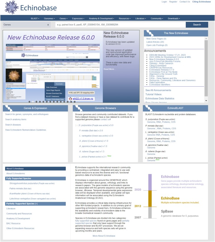
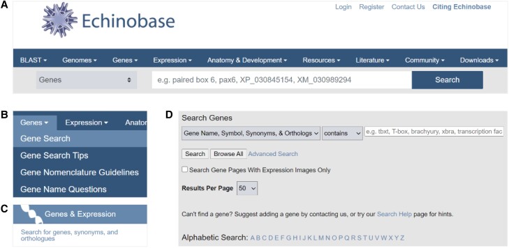
A News carousel and Announcements on the Echinobase landing page (https://www.echinobase.org/) provide current notices for the community regarding news and Echinobase content (Fig. 1). The landing page provides rapid access to information and is designed to allow intuitive navigation with multiple redundant paths for users to interact with the content (Fig. 1). For example, to quickly search for a gene of interest, the Search bar can be used directly as it defaults to gene searches. If a more detailed search is desired, the Gene Search menu item can be used. The Genes and Expression tile block on the landing page also has a link to Search for genes (Fig. 2). The Tutorial Videos (https://www.echinobase.org/echinobase/static-echinobase/HowTo.jsp) provide users with an overview of Echinobase, short step-by-step instructions for using JBrowse, and videos highlighting some of the features of Echinobase.
Fig. 1.
The Echinobase landing page.
Fig. 2.
Gene Search options from the landing page. A) Direct search using the search bar. B) Gene Search using the drop-down menu. C) Search for genes link from the Genes & Expression block. D) Both the drop-down and the link take users to the Search Genes page.
There are drop-down menus and tile blocks to access genomic content, including Genes and Expression data, Genomes, and BLAST. The ECAO anatomy terms can be searched directly from the search bar or accessed from the Anatomy and Development drop-down menu or the information block at the bottom of the landing page. Also on the menu bar are dropdowns that link to Resources, Literature, and Community searches and the Downloads menu. The latter provides access to Gene Page Reports and other data files for download.
Echinobase gene pages
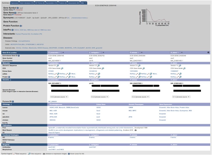
A gene search leads to a Summary page where various data types are collected and displayed (Fig. 3). Summary pages make extensive use of tabs to reduce clutter. At the top of each Summary page, the symbol, name, synonyms, and function of the gene are shown, followed by protein information and links. A graph displays transcript levels across developmental time for S. purpuratus (Tu et al. 2014). The main table on the gene page provides detailed information regarding gene ID and location, sequence visualization and BLAST functionality, a JBrowse genome browser link, and additional information and links regarding orthology, publications, and curated reagents (antibodies, morpholinos, and gRNAs). Additional tabs provide data regarding Expression, Gene Literature, GO Terms, Nucleotides (genomic and mRNA), Proteins, Interactants (literature cocitation), and a Wiki tab for miscellaneous notes regarding the gene.
Fig. 3.
A sample gene page shows all of the information gathered and displayed on the Summary tab. Species-agnostic information such as gene symbol, synonyms, molecular function, and disease associations are displayed at the top, while species-specific content is arranged in vertical columns below this more general content. Other tabs can be used to access more specific supporting information. Note that the gene in this example is orthologous S. purpuratus, L. variegatus, P. miniata, and A. planci. The species displayed, and the order in which they are displayed, can be set by users using the dropdowns on top of each column.
Multispecies integration
Echinobase uses S. purpuratus as the anchor species for orthology, as this species is more widely used for experimental studies than any other echinoderm and has a high-quality genome assembly. Orthology between S. purpuratus and humans is used to assign gene symbols and names. Currently, there is a gene page for every S. purpuratus gene model, and if there is a 1:1 ortholog in another species, then the tabulated information for that species is included. Orthology is determined using a DIOPT (Drosophila RNAi Screening Center Integrative Ortholog Prediction Tool)-based comparison between humans and S. purpuratus (Foley et al. 2021). Protein sequences are analyzed using several tools, and a minimum threshold of 3 tools was selected for identifying orthologs. Seven tools were used to compare S. purpuratus and human proteins for determining gene names and gene symbols; this approach identified 3,617 one-to-one orthologs. Five tools were used to compare S. purpuratus proteins with those of other echinoderms, and this lateral mapping was used to assign orthologs in other echinoderms (Fig. 3). This approach identified several thousand orthologs in L. variegatus (6,577), P. miniata (5,936), and A. planci (6,247). In the future, the display will be expanded to include genes with one-to-many and many-to-one relationships with human genes. Paralog analysis for S. purpuratus (i.e. genes duplicated in urchins relative to a human reference gene) has been completed, and the display of these paralogs on the gene pages is under development.
Genomes, full and partial support
Echinobase only hosts genomes that have been submitted and annotated by NCBI. Experimentally relevant species and high-quality genome assemblies are hosted with 2 levels of support, full and partial. Full support involves full genome integration in the database, including gene pages, as well as BLAST, browsing via JBrowse, and inclusion in weekly data download reports. Full support represents a significant resource commitment and therefore must be approved by the Scientific Advisory Board. Partial support has BLAST, JBrowse, and genomic data download support but no gene page integration making this integration a much more rapid, and a less resource-dependent, process. The new S. purpuratus v5.0 (purple sea urchin), P. miniata v3.0 (bat star), and L. variegatus v3.0 (green sea urchin) genomes are available along with the A. planci v1.0 (crown-of-thorns sea star) genome and are fully supported. The A. japonica v1.0 (feather star), As. rubens v1.3 (sugar star), and L. pictus v2.1 (painted urchin) genomes are partially supported (JBrowse, BLAST, and Download). Links to other echinoderm genomes available at NCBI are listed on the EchinoWiki as Links to Additional Echinoderm Datasets.
Gene nomenclature
Naming of genes is a multistep process that follows guidelines prepared by the Echinobase Nomenclature Steering Committee and Working Group (Beatman et al. 2021). Curators have manually assigned names to another set of genes that are prominent in the published literature, including those that are components of developmental GRNs. Echinoderm gene symbols and names are lowercase and italicized. For protein-coding genes, orthology is the determining factor; if there is no human ortholog, then a hierarchy has been established for assigning the appropriate name (https://www.echinobase.org/echinobase/static/gene/geneNomenclature.jsp). Currently, this is a manual process. When the gene symbol remains a LOC ID, the gene name that was assigned by NCBI is available to provide some information regarding gene function.
Gene expression, regulation, and manipulation
Our understanding of the evolution and function of genomes and genes is improved by providing high-quality, accurate genome assemblies, ortholog identification, and collection and annotation of transcriptomes and regulatory sequences, chromatin accessibility, and other functional genomics datasets. Developmental RNA-seq data from S. purpuratus (Tu et al. 2014) are displayed as TPM (transcripts per million) vs time (in hours post-fertilization) plots on the gene pages. RNA-seq and ATAC-seq developmental expression datasets can be visualized in JBrowse as tracks or downloaded. As more data become available at NCBI, it will be updated on Echinobase. Recent additions for S. purpuratus include DNA methylation tracks ordered by stage, updated transcription factor–binding site tracks with linkouts to the CIS-BP database for all motifs, whole embryo ATAC-seq across different stages of L. variegatus, and RNA-seq tracks across different stages of L. pictus. Additionally, a JBrowse track was created to align historic S. purpuratus transcript models, which are identified by the prefix, WHL.
Users are also invited to submit data directly to Echinobase for incorporation as a JBrowse track. Bigwig file format is optimal, with identifiers that match the current JBrowse coordinates. Genome-wide maps of enhancer RNAs across multiple developmental stages are displayed as JBrowse tracks and are useful for enhancer identification and analysis (Khor et al. 2021). Identification and testing of cis-regulatory elements (Khor and Ettensohn 2022) and inducible gene expression methods (Khor and Ettensohn 2023; Zueva and Hinman 2023) allow for more detailed analysis of genomic elements involved in the regulation of transcription.
Anatomy resources
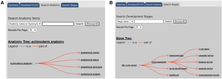
The current ECAO describes anatomical entities and developmental stages of S. purpuratus, the most widely studied echinoderm. Curators use the ontology to label data in a consistent manner. The ECAO is searchable and relates each developmental stage to the stages that immediately precede and follow it (Fig. 4). Hierarchical structural and developmental relationships among anatomical entities are also included, as are the developmental stages when each entity is present.
Fig. 4.
The ECAO serves as a scaffold that can have data attached to it using spatial and temporal dimensions. A) The anatomy can be searched, and items are organized in a hierarchy of spatial structures. B) The developmental stages are arranged in a temporal hierarchy.
Resources, literature, community, and downloads
The Echinobase Resources serve to support the community by collecting data, protocols, and other resources that are then shared using the EchinoWiki and Download site. The EchinoWiki has a wide range of information, including protocols and reagents. Antibodies and morpholinos are manually curated and displayed at the end of the gene page allowing for the rapid identification of gene-specific reagents for use in experiments. For genome-editing experiments, gRNAs that have been published are listed on gene pages and CRISPR (Clustered Regularly Interspaced Short Palindromic Repeats) methods are available on EchinoWiki.
The downloads available include all of the genomes and the Gene Page Reports. The literature associated with a set of echinoderm search terms has been collected (Karimi et al. 2021) and associated with both genes and tissues in the ECAO and will be updated weekly, making Echinobase a destination for relevant literature. Links in publication reports jump directly to gene pages, tissue descriptions, author pages, and more. In order to support the Community and to enable interdisciplinary and collaborative studies, research, descriptions, and contact information of community members and groups are available and searchable. Echinobase also supports the posting of available relevant job positions.
Future directions
Echinobase is a critically important source of genomic information related to echinoderms. Without it, most of the important research resources that have been developed over the past decade (including the genome sequences themselves) would be almost useless. The continual improvement of this vital resource is therefore of the highest priority. Echinobase has unique and important roles in: (1) providing a paradigm for the integration of diverse types of biological data across multiple species and large evolutionary time scales in a single knowledgebase and (2) supporting GRN biology, including the use of multispecies data to study GRN evolution. Echinobase also leads the way in working with highly polymorphic genomes, which are typical for many species with large population sizes. Echinobase therefore serves as a technical resource to scientists establishing new organisms for genomic studies. As a medium-sized community, we are also well placed to test new approaches for data sharing. Echinobase therefore serves as an important resource for a wide community beyond those researchers who work on echinoderms.
Echinobase priorities are developed based on regular input from the research community, gathered in Town Hall Meetings and Surveys, and from the Scientific Advisory Board. Current priorities include the following.
Genomes
A major goal is to add additional genome assemblies of species of echinoderms used as model organisms for genomic research. The genomes of echinoderms are large and polymorphic. Efforts to sequence and assemble them have often served as test cases for this kind of effort in general. The community has recently requested that annotated genomes of L. pictus and Paracentrotus lividus be made available on Echinobase. The L. pictus genome annotations are currently available with partial support.
Gene annotations and ortholog identities
A central objective of many research programs in our community is to assay gene expression and function. Efforts should be made to generally improve gene annotations (e.g. splicing isoforms, noncoding RNAs, translation starts sites, and UTRs). This will globally improve the utility of Echinobase for all researchers and facilitate CRISPR gRNA design (Lin and Su 2016; Cui et al. 2017; Lin et al. 2019). A DIOPT-like pipeline has been developed for the ortholog prediction of S. purpuratus to humans (for gene names) and to other fully supported echinoderm species on Echinobase. This analysis will be expanded to include PhylomeDB phylogenetic analysis. GO terms will be expanded to improve gene expression studies, and visualization tools will be incorporated for the display of data.
The number of user-generated echinoderm transcriptomes is ballooning. There are currently 292 echinoderm transcriptomes in the NCBI sequence read archive representing 81 species. Most of these were collected for an explicit experimental purpose, and no consolidation has been undertaken. Thus, a huge amount of data is lost to the experimentalist. A further goal therefore is to collate transcriptomes from these many sources to provide high-quality reference transcriptomes from multiple species and, where possible, provide details of time points and tissue types.
Echinoderm embryo ontology
The ECAO is currently being expanded from S. purpuratus to include 2 additional, widely used euechinoids (L. variegatus and P. lividus), a representative (and the most widely studied) cidaroid (Eucidaris tribuloides), and the bat star (P. miniata). The integration of the developmental stages and anatomical features of diverse echinoderm species into a single, unified, developmental ontology (the Echinoderm Embryo Ontology) will provide a unique and powerful tool for the curation of diverse, gene-related information across the phylum and the comparative analysis of data from multiple species.
Resource sharing
Echinobase serves as a centralized resource for sharing protocols, reagents, and community news. Therefore, the knowledgebase must remain current. The EchinoWiki is available for posting protocols and validated reagents. Antibodies, morpholinos, and gRNAs are continuously curated and incorporated into gene pages as these reagents appear in publications. Links to gRNA tools will also be provided. Images are curated from articles and an image gallery is available. The Announcements on the landing page serve to notify the community of events and news.
Genome-wide curation of regulatory sequences
The community is very interested in the genome-wide curation of noncoding, transcriptional regulatory sequences for many species, as determined from ATAC-seq profiles and other genomic signatures. Echinoderms are famous for the ease with which synchronous embryo cultures can be obtained, suiting them perfectly for the developmental profiling of chromatin architecture. Most importantly, echinoderm embryos are unusually well suited for functional cis-regulatory analyses of gene expression, an essential component of GRN studies. Such data are emerging from many labs, and it will be crucial to provide genome browser tracks or other portals to these data on Echinobase. This will greatly facilitate improved annotations of functional, noncoding DNA and the use of echinoderms for regulatory functional genomics. Such data are also routinely needed by researchers from other model systems and, in particular, the growing body of researchers performing comparative functional genomics that would like to use this major phylum in their analyses.
Single-cell RNA-seq
A major goal is to further incorporate gene expression data, including single-cell RNA-seq data, into Echinobase. New endeavors to include spatial and quantitative expression should be included for S. purpuratus and other important experimental species. Significant individual lab efforts are directed at identifying spatial and quantitative gene expression profiles that can benefit the community as a whole. Providing these data in a format that can be readily accessed and cross-referenced from multiple species will aid comprehensive syntheses of GRN analyses, including for researchers from other communities. These should, as much as possible, also follow standards for other model systems outside of the echinoderms, to facilitate broader accessibility. Controlled vocabularies for developmental anatomy, and developmental stages, should be developed with the intent of coordinating with other taxa.
Maintenance and updating
As the types and quantity of data expand, it becomes imperative to remodel the Echinobase web information system to ensure that it remains easily accessible to researchers, regardless of their experience with echinoderms. This will include the use of uniform nomenclature and searching tools, as well as intuitive links to external resources and databases. Efforts will be made to seek input from researchers in other systems and, in particular, other genomic web resource developers, to stimulate outreach efforts to service a broader community. The goal is to increase the impact of Echinobase and echinoderm research and to ensure that researchers from other communities can take advantage of the work done using echinoderms. These recommendations address critically important needs identified by the community, seek to make the best use of current resources, and are directed at enhancing the unique strengths of the echinoderm model system for the coming decade.
Education and outreach
We have produced a number of papers and tutorial videos to help our users in accessing information on Echinobase. The availability of sea urchin gametes and the ease of their manipulation have made the sea urchin a popular source of educational material for many years. There are 2 widely used and complementary educational websites. “Sea Urchin Embryology” (https://depts.washington.edu/embryology/) provides essential information concerning animal procurement and handling, gamete collection, and fertilization, as well as detailed protocols for simple wet-lab exercises related to fertilization and early development. “Virtual Urchin” (https://depts.washington.edu/vurchin/) supports unique, interactive, web-based educational modules related to sea urchin development, including a virtual lab bench for simulating complex experimental manipulations. “Embryology Experiment” kits are commercially available from Carolina Biological Supply Company and Gulf Specimen Marine Lab, attesting to the widespread use of sea urchin gametes and embryos as educational materials.
Supplementary Material
Contributor Information
Cheryl A Telmer, Department of Biological Sciences, Carnegie Mellon University, Pittsburgh, PA 15213, USA.
Kamran Karimi, Department of Biological Sciences, University of Calgary, Calgary, AB, Canada T2N 1N4.
Macie M Chess, Department of Biological Sciences, Carnegie Mellon University, Pittsburgh, PA 15213, USA.
Sergei Agalakov, Department of Biological Sciences, University of Calgary, Calgary, AB, Canada T2N 1N4.
Bradley I Arshinoff, Department of Biological Sciences, University of Calgary, Calgary, AB, Canada T2N 1N4.
Vaneet Lotay, Department of Biological Sciences, University of Calgary, Calgary, AB, Canada T2N 1N4.
Dong Zhuo Wang, Department of Biological Sciences, University of Calgary, Calgary, AB, Canada T2N 1N4.
Stanley Chu, Department of Biological Sciences, University of Calgary, Calgary, AB, Canada T2N 1N4.
Troy J Pells, Department of Biological Sciences, University of Calgary, Calgary, AB, Canada T2N 1N4.
Peter D Vize, Department of Biological Sciences, University of Calgary, Calgary, AB, Canada T2N 1N4.
Veronica F Hinman, Department of Biological Sciences, Carnegie Mellon University, Pittsburgh, PA 15213, USA.
Charles A Ettensohn, Department of Biological Sciences, Carnegie Mellon University, Pittsburgh, PA 15213, USA.
Data availability
All data in this article are available at https://www.echinobase.org or at the given URLs.
Funding
The development of Echinobase is funded by the Eunice Kennedy Shriver National Institute of Child Health and Human Development (NICHD), Grant No. P41 HD095831, awarded to VFH, CAE, and PDV.
Literature cited
- Alicea-Delgado M, García-Arrarás JE. 2021. Wnt/β-catenin signaling pathway regulates cell proliferation but not muscle dedifferentiation nor apoptosis during sea cucumber intestinal regeneration. Dev Biol. 480:105–113. doi: 10.1016/j.ydbio.2021.08.011. [DOI] [PMC free article] [PubMed] [Google Scholar]
- Andrikou C, Pai C-Y, Su Y-H, Arnone MI. 2015. Logics and properties of a genetic regulatory program that drives embryonic muscle development in an echinoderm. eLife. 4:e07343. doi: 10.7554/eLife.07343. [DOI] [PMC free article] [PubMed] [Google Scholar]
- Annunziata R, Andrikou C, Perillo M, Cuomo C, Arnone MI. 2019. Development and evolution of gut structures: from molecules to function. Cell Tissue Res. 377(3):445–458. doi: 10.1007/s00441-019-03093-9. [DOI] [PubMed] [Google Scholar]
- Annunziata R, Perillo M, Andrikou C, Cole AG, Martinez P, Arnone MI. 2014. Pattern and process during sea urchin gut morphogenesis: the regulatory landscape. Genesis. 52(3):251–268. doi: 10.1002/dvg.22738. [DOI] [PubMed] [Google Scholar]
- Arshinoff BI, Cary GA, Karimi K, Foley S, Agalakov S, Delgado F, Lotay VS, Ku CJ, Pells TJ, Beatman TR, et al. 2022. Echinobase: leveraging an extant model organism database to build a knowledgebase supporting research on the genomics and biology of echinoderms. Nucleic Acids Res. 50(D1):D970–D979. doi: 10.1093/nar/gkab1005. [DOI] [PMC free article] [PubMed] [Google Scholar]
- Beatman TR, Buckley KM, Cary GA, Hinman VF, Ettensohn CA. 2021. A nomenclature for echinoderm genes. Database (Oxford). 2021:baab052. doi: 10.1093/database/baab052. [DOI] [PMC free article] [PubMed] [Google Scholar]
- Ben-Tabou de-Leon S. 2022. The evolution of biomineralization through the co-option of organic scaffold forming networks. Cells. 11(4):595. doi: 10.3390/cells11040595. [DOI] [PMC free article] [PubMed] [Google Scholar]
- Briggs E, Wessel GM. 2006. In the beginning…animal fertilization and sea urchin development. Dev Biol. 300(1):15–26. doi: 10.1016/j.ydbio.2006.07.014. [DOI] [PubMed] [Google Scholar]
- Buckley KM, Ho ECH, Hibino T, Schrankel CS, Schuh NW, Wang G, Rast JP. 2017. IL17 factors are early regulators in the gut epithelium during inflammatory response to Vibrio in the sea urchin larva. eLife. 6:e23481. doi: 10.7554/eLife.23481. [DOI] [PMC free article] [PubMed] [Google Scholar]
- Buckley KM, Rast JP. 2015. Diversity of animal immune receptors and the origins of recognition complexity in the deuterostomes. Dev Comp Immunol. 49(1):179–189. doi: 10.1016/j.dci.2014.10.013. [DOI] [PubMed] [Google Scholar]
- Buckley KM, Rast JP. 2017. An organismal model for gene regulatory networks in the gut-associated immune response. Front Immunol. 8:1297. doi: 10.3389/fimmu.2017.01297. [DOI] [PMC free article] [PubMed] [Google Scholar]
- Buckley KM, Rast JP. 2019. Immune activity at the gut epithelium in the larval sea urchin. Cell Tissue Res. 377(3):469–474. doi: 10.1007/s00441-019-03095-7. [DOI] [PubMed] [Google Scholar]
- Buckley KM, Schuh NW, Heyland A, Rast JP. 2019. Analysis of immune response in the sea urchin larva. Methods Cell Biol. 150:333–355. doi: 10.1016/bs.mcb.2018.10.009. [DOI] [PubMed] [Google Scholar]
- Byrne M. 2020. The link between autotomy and CNS regeneration: echinoderms as non-model species for regenerative biology. Bioessays. 42(3):e1900219. doi: 10.1002/bies.201900219. [DOI] [PubMed] [Google Scholar]
- Byrne M, Koop D, Cisternas P, Strbenac D, Yang JY, Wray GA. 2015. Transcriptomic analysis of nodal- and BMP-associated genes during juvenile development of the sea urchin Heliocidaris erythrogramma. Mar Genomics. 24:41–45. doi: 10.1016/j.margen.2015.05.019. [DOI] [PubMed] [Google Scholar]
- Cameron RA, Samanta M, Yuan A, He D, Davidson E. 2009. SpBase: the sea urchin genome database and web site. Nucleic Acids Res. 37(Database):D750–D754. doi: 10.1093/nar/gkn887. [DOI] [PMC free article] [PubMed] [Google Scholar]
- Carlisle JA, Swanson WJ. 2021. Molecular mechanisms and evolution of fertilization proteins. J Exp Zool B Mol Dev Evol. 336(8):652–665. doi: 10.1002/jez.b.23004. [DOI] [PubMed] [Google Scholar]
- Cary GA, Cameron RA, Hinman VF. 2018. EchinoBase: tools for echinoderm genome analyses. Methods Mol Biol. 1757:349–369. doi: 10.1007/978-1-4939-7737-6_12. [DOI] [PubMed] [Google Scholar]
- Cary GA, McCauley BS, Zueva O, Pattinato J, Longabaugh W, Hinman VF. 2020. Systematic comparison of sea urchin and sea star developmental gene regulatory networks explains how novelty is incorporated in early development. Nat Commun. 11(1):6235. doi: 10.1038/s41467-020-20023-4. [DOI] [PMC free article] [PubMed] [Google Scholar]
- Chassé H, Boulben S, Glippa V, Pontheaux F, Cormier P, Morales J. 2019. In vivo analysis of protein translation activity in sea urchin eggs and embryos. Methods Cell Biol. 151:335–352. doi: 10.1016/bs.mcb.2018.10.008. [DOI] [PubMed] [Google Scholar]
- Chessel A, De Crozé N, Molina MD, Taberner L, Dru P, Martin L, Lepage T. 2023. RAS-independent ERK activation by constitutively active KSR3 in non-chordate metazoa. Nat Commun. 14(1):3970. doi: 10.1038/s41467-023-39606-y. [DOI] [PMC free article] [PubMed] [Google Scholar]
- Chiaramonte M, Arizza V, Russo R. 2019. Evolutionary conserved pathway of the innate immune response after a viral insult in Paracentrotus lividus sea urchin. Int J Immunogenet. 46(3):192–202. doi: 10.1111/iji.12424. [DOI] [PubMed] [Google Scholar]
- Chiaramonte M, Russo R, Costa C, Bonaventura R, Zito F. 2020. PI3K inhibition highlights new molecular interactions involved in the skeletogenesis of Paracentrotus lividus embryos. Biochim Biophys Acta Mol Cell Res. 1867(1):118558. doi: 10.1016/j.bbamcr.2019.118558. [DOI] [PubMed] [Google Scholar]
- Chiarelli R, Scudiero R, Memoli V, Roccheri MC, Martino C. 2022. Toxicity of vanadium during development of sea urchin embryos: bioaccumulation, calcium depletion, ERK modulation and cell-selective apoptosis. Int J Mol Sci. 23(11):6239. doi: 10.3390/ijms23116239. [DOI] [PMC free article] [PubMed] [Google Scholar]
- Cui M, Lin C-Y, Su Y-H. 2017. Recent advances in functional perturbation and genome editing techniques in studying sea urchin development. Brief Funct Genomics. 16(5):309–318. doi: 10.1093/bfgp/elx011. [DOI] [PubMed] [Google Scholar]
- Cui M, Siriwon N, Li E, Davidson EH, Peter IS. 2014. Specific functions of the Wnt signaling system in gene regulatory networks throughout the early sea urchin embryo. Proc Natl Acad Sci U S A. 111(47):E5029–E5038. doi: 10.1073/pnas.1419141111. [DOI] [PMC free article] [PubMed] [Google Scholar]
- Czarkwiani A, Taylor J, Oliveri P. 2022. Neurogenesis during brittle star arm regeneration is characterised by a conserved set of key developmental genes. Biology (Basel). 11(9):1360. doi: 10.3390/biology11091360. [DOI] [PMC free article] [PubMed] [Google Scholar]
- Damle S, Davidson EH. 2011. Precise cis-regulatory control of spatial and temporal expression of the alx-1 gene in the skeletogenic lineage of S. purpuratus. Dev Biol. 357(2):505–517. doi: 10.1016/j.ydbio.2011.06.016. [DOI] [PMC free article] [PubMed] [Google Scholar]
- Damle SS, Davidson EH. 2012. Synthetic in vivo validation of gene network circuitry. Proc Natl Acad Sci U S A. 109(5):1548–1553. doi: 10.1073/pnas.1119905109. [DOI] [PMC free article] [PubMed] [Google Scholar]
- Davidson EH, Rast JP, Oliveri P, Ransick A, Calestani C, Yuh CH, Minokawa T, Amore G, Hinman V, Arenas-Mena C, et al. 2002a. A genomic regulatory network for development. Science. 295(5560):1669–1678. doi: 10.1126/science.1069883. [DOI] [PubMed] [Google Scholar]
- Davidson EH, Rast JP, Oliveri P, Ransick A, Calestani C, Yuh CH, Minokawa T, Amore G, Hinman V, Arenas-Mena C, et al. 2002b. A provisional regulatory gene network for specification of endomesoderm in the sea urchin embryo. Dev Biol. 246(1):162–190. doi: 10.1006/dbio.2002.0635. [DOI] [PubMed] [Google Scholar]
- Day TC, Márquez-Zacarías P, Bravo P, Pokhrel AR, MacGillivray KA, Ratcliff WC, Yunker PJ. 2022. Varied solutions to multicellularity: the biophysical and evolutionary consequences of diverse intercellular bonds. Biophys Rev (Melville). 3(2):021305. doi: 10.1063/5.0080845. [DOI] [PMC free article] [PubMed] [Google Scholar]
- Dutkowski J, Ideker T. 2011. Protein networks as logic functions in development and cancer. PLoS Comput Biol. 7(9):e1002180. doi: 10.1371/journal.pcbi.1002180. [DOI] [PMC free article] [PubMed] [Google Scholar]
- Dylus DV, Czarkwiani A, Stångberg J, Ortega-Martinez O, Dupont S, Oliveri P. 2016. Large-scale gene expression study in the ophiuroid Amphiura filiformis provides insights into evolution of gene regulatory networks. Evodevo. 7(1):2. doi: 10.1186/s13227-015-0039-x. [DOI] [PMC free article] [PubMed] [Google Scholar]
- Erkenbrack EM. 2016. Divergence of ectodermal and mesodermal gene regulatory network linkages in early development of sea urchins. Proc Natl Acad Sci U S A. 113(46):E7202–E7211. doi: 10.1073/pnas.1612820113. [DOI] [PMC free article] [PubMed] [Google Scholar]
- Erkenbrack EM, Ako-Asare K, Miller E, Tekelenburg S, Thompson JR, Romano L. 2016. Ancestral state reconstruction by comparative analysis of a GRN kernel operating in echinoderms. Dev Genes Evol. 226(1):37–45. doi: 10.1007/s00427-015-0527-y. [DOI] [PubMed] [Google Scholar]
- Erkenbrack EM, Davidson EH, Peter IS. 2018. Conserved regulatory state expression controlled by divergent developmental gene regulatory networks in echinoids. Development. 145(24):dev167288. doi: 10.1242/dev.167288. [DOI] [PMC free article] [PubMed] [Google Scholar]
- Fernandez-Valverde SL, Aguilera F, Ramos-Díaz RA. 2018. Inference of developmental gene regulatory networks beyond classical model systems: new approaches in the post-genomic era. Integr Comp Biol. 58(4):640–653. doi: 10.1093/icb/icy061. [DOI] [PubMed] [Google Scholar]
- Fisher M, James-Zorn C, Ponferrada V, Bell AJ, Sundararaj N, Segerdell E, Chaturvedi P, Bayyari N, Chu S, Pells T, et al. 2023. Xenbase: key features and resources of the Xenopus model organism knowledgebase. Genetics. 224(1):iyad018. doi: 10.1093/genetics/iyad018. [DOI] [PMC free article] [PubMed] [Google Scholar]
- Foley S, Ku C, Arshinoff B, Lotay V, Karimi K, Vize PD, Hinman V. 2021. Integration of 1:1 orthology maps and updated datasets into Echinobase. Database (Oxford). 2021:baab030. doi: 10.1093/database/baab030. [DOI] [PMC free article] [PubMed] [Google Scholar]
- Formery L, Peluso P, Kohnle I, Malnick J, Thompson JR, Pitel M, Uhlinger KR, Rokhsar DS, Rank DR, Lowe CJ. 2023. Molecular evidence of anteroposterior patterning in adult echinoderms. Nature. 623(7987):555–561. doi: 10.1038/s41586-023-06669-2. [DOI] [PubMed] [Google Scholar]
- Foster S, Oulhen N, Fresques T, Zaki H, Wessel G. Distinct mechanisms of germ cell factor regulation for an inductive germ cell fate. bioRxiv 479164. 10.1101/2022.02.04.479164, preprint: not peer reviewed. [DOI]
- Foster S, Oulhen N, Wessel G. 2020. A single cell RNA sequencing resource for early sea urchin development. Development. 147(17):dev191528. doi: 10.1242/dev.191528. [DOI] [PMC free article] [PubMed] [Google Scholar]
- Fresques TM, Wessel GM. 2018. Nodal induces sequential restriction of germ cell factors during primordial germ cell specification. Development. 145(2):dev155663. doi: 10.1242/dev.155663. [DOI] [PMC free article] [PubMed] [Google Scholar]
- Garrett AD, Brennan RS, Steinhart AL, Pelletier AM, Pespeni MH. 2020. Unique genomic and phenotypic responses to extreme and variable ph conditions in purple urchin larvae. Integr Comp Biol. 60(2):318–331. doi: 10.1093/icb/icaa072. [DOI] [PubMed] [Google Scholar]
- Gökirmak T, Campanale JP, Shipp LE, Moy GW, Tao H, Hamdoun A. 2012. Localization and substrate selectivity of sea urchin multidrug (MDR) efflux transporters. J Biol Chem. 287(52):43876–43883. doi: 10.1074/jbc.M112.424879. [DOI] [PMC free article] [PubMed] [Google Scholar]
- Hansen JN, Gong A, Wachten D, Pascal R, Turpin A, Jikeli JF, Kaupp UB, Alvarez L. 2021. Multifocal imaging for precise, label-free tracking of fast biological processes in 3D. Nat Commun. 12(1):4574. doi: 10.1038/s41467-021-24768-4. [DOI] [PMC free article] [PubMed] [Google Scholar]
- Hatleberg WL, Hinman VF. 2021. Modularity and hierarchy in biological systems: using gene regulatory networks to understand evolutionary change. Curr Top Dev Biol. 141:39–73. doi: 10.1016/bs.ctdb.2020.11.004. [DOI] [PubMed] [Google Scholar]
- Henson JH, Samasa B, Burg EC. 2019. High resolution imaging of the cortex isolated from sea urchin eggs and embryos. Methods Cell Biol. 151:419–432. doi: 10.1016/bs.mcb.2019.01.005. [DOI] [PubMed] [Google Scholar]
- Henson JH, Samasa B, Shuster CB, Wikramanayake AH. 2021. The nanoscale organization of the Wnt signaling integrator Dishevelled in the vegetal cortex domain of an egg and early embryo. PLoS One. 16(5):e0248197. doi: 10.1371/journal.pone.0248197. [DOI] [PMC free article] [PubMed] [Google Scholar]
- Hinman VF, Nguyen AT, Cameron RA, Davidson EH. 2003. Developmental gene regulatory network architecture across 500 million years of echinoderm evolution. Proc Natl Acad Sci U S A. 100(23):13356–13361. doi: 10.1073/pnas.2235868100. [DOI] [PMC free article] [PubMed] [Google Scholar]
- Hinman VF, Yankura KA, McCauley BS. 2009. Evolution of gene regulatory network architectures: examples of subcircuit conservation and plasticity between classes of echinoderms. Biochim Biophys Acta. 1789(4):326–332. doi: 10.1016/j.bbagrm.2009.01.004. [DOI] [PubMed] [Google Scholar]
- Hogan JD, Keenan JL, Luo L, Ibn-Salem J, Lamba A, Schatzberg D, Piacentino ML, Zuch DT, Core AB, Blumberg C, et al. 2020. The developmental transcriptome for Lytechinus variegatus exhibits temporally punctuated gene expression changes. Dev Biol. 460(2):139–154. doi: 10.1016/j.ydbio.2019.12.002. [DOI] [PubMed] [Google Scholar]
- Israel JW, Martik ML, Byrne M, Raff EC, Raff RA, McClay DR, Wray GA. 2016. Comparative developmental transcriptomics reveals rewiring of a highly conserved gene regulatory network during a major life history switch in the sea urchin genus Heliocidaris. PLoS Biol. 14(3):e1002391. doi: 10.1371/journal.pbio.1002391. [DOI] [PMC free article] [PubMed] [Google Scholar]
- Karimi K, Agalakov S, Telmer CA, Beatman TR, Pells TJ, Arshinoff BI, Ku CJ, Foley S, Hinman VF, Ettensohn CA, et al. 2021. Classifying domain-specific text documents containing ambiguous keywords. Database (Oxford). 2021:baab062. doi: 10.1093/database/baab062. [DOI] [PMC free article] [PubMed] [Google Scholar]
- Karimi K, Vize PD. The Virtual Xenbase: transitioning an online bioinformatics resource to a private cloud. Database (Oxford). 2014:bau108. doi: 10.1093/database/bau108 [DOI] [PMC free article] [PubMed] [Google Scholar]
- Khadka A, Martínez-Bartolomé M, Burr SD, Range RC. 2018. A novel gene's role in an ancient mechanism: secreted frizzled-related protein 1 is a critical component in the anterior-posterior Wnt signaling network that governs the establishment of the anterior neuroectoderm in sea urchin embryos. Evodevo. 9(1):1. doi: 10.1186/s13227-017-0089-3. [DOI] [PMC free article] [PubMed] [Google Scholar]
- Khor JM, Ettensohn CA. 2017. Functional divergence of paralogous transcription factors supported the evolution of biomineralization in echinoderms. eLife. 6:e32728. doi: 10.7554/eLife.32728. [DOI] [PMC free article] [PubMed] [Google Scholar]
- Khor JM, Ettensohn CA. 2022. Architecture and evolution of the cis-regulatory system of the echinoderm kirrelL gene. eLife. 11:e72834. doi: 10.7554/eLife.72834. [DOI] [PMC free article] [PubMed] [Google Scholar]
- Khor JM, Ettensohn CA. 2023. An optimized Tet-On system for conditional control of gene expression in sea urchins. Development. 150(1):dev201373. doi: 10.1242/dev.201373. [DOI] [PMC free article] [PubMed] [Google Scholar]
- Khor JM, Guerrero-Santoro J, Douglas W, Ettensohn CA. 2021. Global patterns of enhancer activity during sea urchin embryogenesis assessed by eRNA profiling. Genome Res. 31(9):1680–1692. doi: 10.1101/gr.275684.121. [DOI] [PMC free article] [PubMed] [Google Scholar]
- Krumlauf R, Wilkinson DG. 2021. Segmentation and patterning of the vertebrate hindbrain. Development. 148(15):dev186460. doi: 10.1242/dev.186460. [DOI] [PMC free article] [PubMed] [Google Scholar]
- Kubo A, Suzuki N, Yuan X, Nakai K, Satoh N, Imai KS, Satou Y. 2010. Genomic cis-regulatory networks in the early Ciona intestinalis embryo. Development. 137(10):1613–1623. doi: 10.1242/dev.046789. [DOI] [PubMed] [Google Scholar]
- Levin N, Yamakawa S, Morino Y, Wada H. 2022. Perspectives on divergence of early developmental regulatory pathways: insight from the evolution of echinoderm double negative gate. Curr Top Dev Biol. 146:1–24. doi: 10.1016/bs.ctdb.2021.10.001. [DOI] [PubMed] [Google Scholar]
- Li A, Espinoza J, Hamdoun A. 2020. Inhibitory effects of neurotoxin β-N-methylamino-L-alanine on fertilization and early development of the sea urchin Lytechinus pictus. Aquat Toxicol. 221:105425. doi: 10.1016/j.aquatox.2020.105425. [DOI] [PubMed] [Google Scholar]
- Lin C-Y, Oulhen N, Wessel G, Su Y-H. 2019. CRISPR/Cas9-mediated genome editing in sea urchins. Methods Cell Biol. 151:305–321. doi: 10.1016/bs.mcb.2018.10.004. [DOI] [PMC free article] [PubMed] [Google Scholar]
- Lin C-Y, Su Y-H. 2016. Genome editing in sea urchin embryos by using a CRISPR/Cas9 system. Dev Biol. 409(2):420–428. doi: 10.1016/j.ydbio.2015.11.018. [DOI] [PubMed] [Google Scholar]
- Martik ML, McClay DR. 2015. Deployment of a retinal determination gene network drives directed cell migration in the sea urchin embryo. eLife. 4:e08827. doi: 10.7554/eLife.08827. [DOI] [PMC free article] [PubMed] [Google Scholar]
- Martik ML, McClay DR. 2017. New insights from a high-resolution look at gastrulation in the sea urchin, Lytechinus variegatus. Mech Dev. 148:3–10. doi: 10.1016/j.mod.2017.06.005. [DOI] [PMC free article] [PubMed] [Google Scholar]
- Martino C, Chiarelli R, Bosco L, Roccheri MC. 2017. Induction of skeletal abnormalities and autophagy in Paracentrotus lividus sea urchin embryos exposed to gadolinium. Mar Environ Res. 130:12–20. doi: 10.1016/j.marenvres.2017.07.007. [DOI] [PubMed] [Google Scholar]
- Massri AJ, Greenstreet L, Afanassiev A, Berrio A, Wray GA, Schiebinger G, McClay DR. 2021. Developmental single-cell transcriptomics in the Lytechinus variegatus sea urchin embryo. Development. 148(19):dev198614. doi: 10.1242/dev.198614. [DOI] [PMC free article] [PubMed] [Google Scholar]
- Masullo T, Biondo G, Natale MD, Tagliavia M, Bennici CD, Musco M, Ragusa MA, Costa S, Cuttitta A, Nicosia A. 2021. Gene expression changes after parental exposure to metals in the sea urchin affect timing of genetic programme of embryo development. Biology (Basel). 10(2):103. doi: 10.3390/biology10020103. [DOI] [PMC free article] [PubMed] [Google Scholar]
- McClay DR. 2022. Development of a larval nervous system in the sea urchin. Curr Top Dev Biol. 146:25–48. doi: 10.1016/bs.ctdb.2021.10.002. [DOI] [PMC free article] [PubMed] [Google Scholar]
- Meaders JL, Burgess DR. 2020. Microtubule-based mechanisms of pronuclear positioning. Cells. 9(2):505. doi: 10.3390/cells9020505. [DOI] [PMC free article] [PubMed] [Google Scholar]
- Medina-Feliciano JG, García-Arrarás JE. 2021. Regeneration in echinoderms: molecular advancements. Front Cell Dev Biol. 9:768641. doi: 10.3389/fcell.2021.768641. [DOI] [PMC free article] [PubMed] [Google Scholar]
- Meyer A, Hinman V. 2022. The arm of the starfish: the far-reaching applications of Patiria miniata as a model system in evolutionary, developmental, and regenerative biology. Curr Top Dev Biol. 147:523–543. doi: 10.1016/bs.ctdb.2022.01.006. [DOI] [PubMed] [Google Scholar]
- Meyer A, Ku C, Hatleberg W, Telmer CA, Hinman V. New hypotheses of cell type diversity and novelty from comparative single cell and nuclei transcriptomics in echinoderms. bioRxiv 490935. 10.1101/2022.05.06.490935, preprint: not peer reviewed. [DOI] [PMC free article] [PubMed]
- Meyer A, Ku C, Hatleberg WL, Telmer CA, Hinman V. 2023. New hypotheses of cell type diversity and novelty from orthology-driven comparative single cell and nuclei transcriptomics in echinoderms. eLife. 12:e80090. doi: 10.7554/eLife.80090. [DOI] [PMC free article] [PubMed] [Google Scholar]
- Nesbit KT, Fleming T, Batzel G, Pouv A, Rosenblatt HD, Pace DA, Hamdoun A, Lyons DC. 2019. The painted sea urchin, Lytechinus pictus, as a genetically-enabled developmental model. Methods Cell Biol. 150:105–123. doi: 10.1016/bs.mcb.2018.11.010. [DOI] [PMC free article] [PubMed] [Google Scholar]
- Nogueira LS, Domingos-Moreira FXV, Klein RD, Bianchini A, Wood CM. 2021. Influence of environmentally relevant concentrations of Zn, Cd and Ni and their binary mixtures on metal uptake, bioaccumulation and development in larvae of the purple sea urchin Strongylocentrotus purpuratus. Aquat Toxicol. 230:105709. doi: 10.1016/j.aquatox.2020.105709. [DOI] [PubMed] [Google Scholar]
- Paganos P, Voronov D, Musser JM, Arendt D, Arnone MI. 2021. Single-cell RNA sequencing of the Strongylocentrotus purpuratus larva reveals the blueprint of major cell types and nervous system of a non-chordate deuterostome. eLife. 10:e70416. doi: 10.7554/eLife.70416. [DOI] [PMC free article] [PubMed] [Google Scholar]
- Papadogiannis V, Pennati A, Parker HJ, Rothbächer U, Patthey C, Bronner ME, Shimeld SM. 2022. Hmx gene conservation identifies the origin of vertebrate cranial ganglia. Nature. 605(7911):701–705. doi: 10.1038/s41586-022-04742-w. [DOI] [PMC free article] [PubMed] [Google Scholar]
- Parker HJ, Krumlauf R. 2020. A Hox gene regulatory network for hindbrain segmentation. Curr Top Dev Biol. 139:169–203. doi: 10.1016/bs.ctdb.2020.03.001. [DOI] [PubMed] [Google Scholar]
- Perillo M, Oulhen N, Foster S, Spurrell M, Calestani C, Wessel G. 2020. Regulation of dynamic pigment cell states at single-cell resolution. eLife. 9:e60388. doi: 10.7554/eLife.60388. [DOI] [PMC free article] [PubMed] [Google Scholar]
- Perillo M, Swartz SZ, Pieplow C, Wessel GM. 2023. Molecular mechanisms of tubulogenesis revealed in the sea star hydro-vascular organ. Nat Commun. 14(1):2402. doi: 10.1038/s41467-023-37947-2. [DOI] [PMC free article] [PubMed] [Google Scholar]
- Perillo M, Swartz SZ, Wessel GM. 2022. A conserved node in the regulation of Vasa between an induced and an inherited program of primordial germ cell specification. Dev Biol. 482:28–33. doi: 10.1016/j.ydbio.2021.11.007. [DOI] [PMC free article] [PubMed] [Google Scholar]
- Peter IS, Davidson EH. 2009. Modularity and design principles in the sea urchin embryo gene regulatory network. FEBS Lett. 583(24):3948–3958. doi: 10.1016/j.febslet.2009.11.060. [DOI] [PMC free article] [PubMed] [Google Scholar]
- Peter IS, Davidson EH. 2010. The endoderm gene regulatory network in sea urchin embryos up to mid-blastula stage. Dev Biol. 340(2):188–199. doi: 10.1016/j.ydbio.2009.10.037. [DOI] [PMC free article] [PubMed] [Google Scholar]
- Peter IS, Davidson EH. 2017. Assessing regulatory information in developmental gene regulatory networks. Proc Natl Acad Sci U S A. 114(23):5862–5869. doi: 10.1073/pnas.1610616114. [DOI] [PMC free article] [PubMed] [Google Scholar]
- Pieplow A, Dastaw M, Sakuma T, Sakamoto N, Yamamoto T, Yajima M, Oulhen N, Wessel GM. 2021. CRISPR-Cas9 editing of non-coding genomic loci as a means of controlling gene expression in the sea urchin. Dev Biol. 472:85–97. doi: 10.1016/j.ydbio.2021.01.003. [DOI] [PMC free article] [PubMed] [Google Scholar]
- Piovani L, Czarkwiani A, Ferrario C, Sugni M, Oliveri P. 2021. Ultrastructural and molecular analysis of the origin and differentiation of cells mediating brittle star skeletal regeneration. BMC Biol. 19(1):9. doi: 10.1186/s12915-020-00937-7. [DOI] [PMC free article] [PubMed] [Google Scholar]
- Rafiq K, Cheers MS, Ettensohn CA. 2012. The genomic regulatory control of skeletal morphogenesis in the sea urchin. Development. 139(3):579–590. doi: 10.1242/dev.073049. [DOI] [PubMed] [Google Scholar]
- Ragusa MA, Costa S, Cuttitta A, Gianguzza F, Nicosia A. 2017. Coexposure to sulfamethoxazole and cadmium impairs development and attenuates transcriptional response in sea urchin embryo. Chemosphere. 180:275–284. doi: 10.1016/j.chemosphere.2017.04.030. [DOI] [PubMed] [Google Scholar]
- Range RC. 2018. Canonical and non-canonical Wnt signaling pathways define the expression domains of frizzled 5/8 and frizzled 1/2/7 along the early anterior-posterior axis in sea urchin embryos. Dev Biol. 444(2):83–92. doi: 10.1016/j.ydbio.2018.10.003. [DOI] [PMC free article] [PubMed] [Google Scholar]
- Reinardy HC, Chapman J, Bodnar AG. 2016. Induction of innate immune gene expression following methyl methanesulfonate-induced DNA damage in sea urchins. Biol Lett. 12(2):20151057. doi: 10.1098/rsbl.2015.1057. [DOI] [PMC free article] [PubMed] [Google Scholar]
- Rothenberg EV. 2021. Single-cell insights into the hematopoietic generation of T-lymphocyte precursors in mouse and human. Exp Hematol. 95:1–12. doi: 10.1016/j.exphem.2020.12.005. [DOI] [PMC free article] [PubMed] [Google Scholar]
- Rothenberg EV, Göttgens B. 2021. How haematopoiesis research became a fertile ground for regulatory network biology as pioneered by Eric Davidson. Curr Opin Hematol. 28(1):1–10. doi: 10.1097/MOH.0000000000000628. [DOI] [PMC free article] [PubMed] [Google Scholar]
- Sánchez Alvarado A. 2012. Cellular hyperproliferation and cancer as evolutionary variables. Curr Biol. 22(17):R772–R778. doi: 10.1016/j.cub.2012.08.008. [DOI] [PMC free article] [PubMed] [Google Scholar]
- Satoh N, Hisata K, Foster S, Morita S, Nishitsuji K, Oulhen N, Tominaga H, Wessel GM. 2022. A single-cell RNA-seq analysis of Brachyury-expressing cell clusters suggests a morphogenesis-associated signal center of oral ectoderm in sea urchin embryos. Dev Biol. 483:128–142. doi: 10.1016/j.ydbio.2022.01.005. [DOI] [PubMed] [Google Scholar]
- Sea Urchin Genome Sequencing Consortium; Sodergren E, Weinstock GM, Davidson EH, Cameron RA, Gibbs RA, Angerer RC, Angerer LM, Arnone MI, Burgess DR, et al. 2006. The genome of the sea urchin Strongylocentrotus purpuratus. Science. 314(5801):941–952. doi: 10.1126/science.1133609. [DOI] [PMC free article] [PubMed] [Google Scholar]
- Shashikant T, Khor JM, Ettensohn CA. 2018a. Global analysis of primary mesenchyme cell cis-regulatory modules by chromatin accessibility profiling. BMC Genomics. 19(1):206. doi: 10.1186/s12864-018-4542-z. [DOI] [PMC free article] [PubMed] [Google Scholar]
- Shashikant T, Khor JM, Ettensohn CA. 2018b. From genome to anatomy: the architecture and evolution of the skeletogenic gene regulatory network of sea urchins and other echinoderms. Genesis. 56(10):e23253. doi: 10.1002/dvg.23253. [DOI] [PMC free article] [PubMed] [Google Scholar]
- Shipp LE, Hamdoun A. 2012. ATP-binding cassette (ABC) transporter expression and localization in sea urchin development. Dev Dyn. 241(6):1111–1124. doi: 10.1002/dvdy.23786. [DOI] [PMC free article] [PubMed] [Google Scholar]
- Slota LA, McClay DR. 2018. Identification of neural transcription factors required for the differentiation of three neuronal subtypes in the sea urchin embryo. Dev Biol. 435(2):138–149. doi: 10.1016/j.ydbio.2017.12.015. [DOI] [PMC free article] [PubMed] [Google Scholar]
- Slota LA, Miranda EM, McClay DR. 2019. Spatial and temporal patterns of gene expression during neurogenesis in the sea urchin Lytechinus variegatus. Evodevo. 10(1):2. doi: 10.1186/s13227-019-0115-8. [DOI] [PMC free article] [PubMed] [Google Scholar]
- Smith LC, Davidson EH. 1994. The echinoderm immune system. Characters shared with vertebrate immune systems and characters arising later in deuterostome phylogeny. Ann N Y Acad Sci. 712(1):213–226. doi: 10.1111/j.1749-6632.1994.tb33575.x. [DOI] [PubMed] [Google Scholar]
- Spurrell M, Oulhen N, Foster S, Perillo M, Wessel G. 2023. Gene regulatory divergence amongst echinoderms underlies appearance of pigment cells in sea urchin development. Dev Biol. 494:13–25. doi: 10.1016/j.ydbio.2022.11.008. [DOI] [PMC free article] [PubMed] [Google Scholar]
- Sun Z, Ettensohn CA. 2014. Signal-dependent regulation of the sea urchin skeletogenic gene regulatory network. Gene Expr Patterns. 16(2):93–103. doi: 10.1016/j.gep.2014.10.002. [DOI] [PubMed] [Google Scholar]
- Sun Z, Ettensohn CA. 2017. TGF-β sensu stricto signaling regulates skeletal morphogenesis in the sea urchin embryo. Dev Biol. 421(2):149–160. doi: 10.1016/j.ydbio.2016.12.007. [DOI] [PubMed] [Google Scholar]
- Tarsis K, Gildor T, Morgulis M, Ben-Tabou de-Leon S. 2022. Distinct regulatory states control the elongation of individual skeletal rods in the sea urchin embryo. Dev Dyn. 251(8):1322–1339. doi: 10.1002/dvdy.474. [DOI] [PMC free article] [PubMed] [Google Scholar]
- Tsironis I, Paganos P, Gouvi G, Tsimpos P, Stamopoulou A, Arnone MI, Flytzanis CN. 2021. Coup-TF: a maternal factor essential for differentiation along the embryonic axes in the sea urchin Paracentrotus lividus. Dev Biol. 475:131–144. doi: 10.1016/j.ydbio.2020.12.012. [DOI] [PubMed] [Google Scholar]
- Tu Q, Cameron RA, Davidson EH. 2014. Quantitative developmental transcriptomes of the sea urchin Strongylocentrotus purpuratus. Dev Biol. 385(2):160–167. doi: 10.1016/j.ydbio.2013.11.019. [DOI] [PMC free article] [PubMed] [Google Scholar]
- Verd B, Monk NA, Jaeger J. 2019. Modularity, criticality, and evolvability of a developmental gene regulatory network. eLife. 8:e42832. doi: 10.7554/eLife.42832. [DOI] [PMC free article] [PubMed] [Google Scholar]
- Vyas H, Schrankel CS, Espinoza JA, Mitchell KL, Nesbit KT, Jackson E, Chang N, Lee Y, Warner J, Reitzel A, et al. 2022. Generation of a homozygous mutant drug transporter (ABCB1) knockout line in the sea urchin Lytechinus pictus. Development. 149(11):dev200644. doi: 10.1242/dev.200644. [DOI] [PMC free article] [PubMed] [Google Scholar]
- Wang L, Koppitch K, Cutting A, Dong P, Kudtarkar P, Zeng J, Cameron RA, Davidson EH. 2019. Developmental effector gene regulation: multiplexed strategies for functional analysis. Dev Biol. 445(1):68–79. doi: 10.1016/j.ydbio.2018.10.018. [DOI] [PMC free article] [PubMed] [Google Scholar]
- Wessel GM, Wong JL. 2009. Cell surface changes in the egg at fertilization. Mol Reprod Dev. 76(10):942–953. doi: 10.1002/mrd.21090. [DOI] [PMC free article] [PubMed] [Google Scholar]
- Wilkinson MD, Dumontier M, Aalbersberg IJ, Appleton G, Axton M, Baak A, Blomberg N, Boiten JW, da Silva Santos LB, Bourne PE, et al. 2016. The FAIR guiding principles for scientific data management and stewardship. Sci Data. 3(1):160018. doi: 10.1038/sdata.2016.18. [DOI] [PMC free article] [PubMed] [Google Scholar]
- Wolff A, Hinman V. 2021. The use of larval sea stars and sea urchins in the discovery of shared mechanisms of metazoan whole-body regeneration. Genes (Basel). 12(7):1063. doi: 10.3390/genes12071063. [DOI] [PMC free article] [PubMed] [Google Scholar]
- Wozniak KL, Carlson AE. 2020. Ion channels and signaling pathways used in the fast polyspermy block. Mol Reprod Dev. 87(3):350–357. doi: 10.1002/mrd.23168. [DOI] [PMC free article] [PubMed] [Google Scholar]
- Yamazaki A, Yamakawa S, Morino Y, Sasakura Y, Wada H. 2021. Gene regulation of adult skeletogenesis in starfish and modifications during gene network co-option. Sci Rep. 11(1):20111. doi: 10.1038/s41598-021-99521-4. [DOI] [PMC free article] [PubMed] [Google Scholar]
- Zheng M, Zueva O, Hinman VF. 2022. Regeneration of the larval sea star nervous system by wounding induced respecification to the Sox2 lineage. eLife. 11:e72983. doi: 10.7554/eLife.72983. [DOI] [PMC free article] [PubMed] [Google Scholar]
- Zmasek CM, Godzik A. 2013. Evolution of the animal apoptosis network. Cold Spring Harb Perspect Biol. 5(3):a008649. doi: 10.1101/cshperspect.a008649. [DOI] [PMC free article] [PubMed] [Google Scholar]
- Zmasek CM, Zhang Q, Ye Y, Godzik A. 2007. Surprising complexity of the ancestral apoptosis network. Genome Biol. 8(10):R226. doi: 10.1186/gb-2007-8-10-r226. [DOI] [PMC free article] [PubMed] [Google Scholar]
- Zueva O, Hinman V. 2023. Inducible in vivo genome editing in the sea star Patiria miniata. bioRxiv 523328. 10.1101/2023.01.09.523328, preprint: not peer reviewed. [DOI]
Associated Data
This section collects any data citations, data availability statements, or supplementary materials included in this article.
Supplementary Materials
Data Availability Statement
All data in this article are available at https://www.echinobase.org or at the given URLs.


