Abstract
MutS homologs, identified in nearly all bacteria and eukaryotes, include the bacterial proteins MutS1 and MutS2 and the eukaryotic MutS homologs 1 to 7, and they often are involved in recognition and repair of mismatched bases and small insertion/deletions, thereby limiting illegitimate recombination and spontaneous mutation. To explore the relationship of MutS2 to other MutS homologs, we examined conserved protein domains. Fundamental differences in structure between MutS2 and other MutS homologs suggest that MutS1 and MutS2 diverged early during evolution, with all eukaryotic homologs arising from a MutS1 ancestor. Data from MutS1 crystal structures, biochemical results from MutS2 analyses, and our phylogenetic studies suggest that MutS2 has functions distinct from other members of the MutS family. A mutS2 mutant was constructed in Helicobacter pylori, which lacks mutS1 and mismatch repair genes mutL and mutH. We show that MutS2 plays no role in mismatch or recombinational repair or deletion between direct DNA repeats. In contrast, MutS2 plays a significant role in limiting intergenomic recombination across a range of donor DNA tested. This phenotypic analysis is consistent with the phylogenetic and biochemical data suggesting that MutS1 and MutS2 have divergent functions.
MutS homologs (MSH) have been identified in most prokaryotic and all eukaryotic organisms examined. Prokaryotes have two homologs (MutS1 and MutS2), whereas seven MSH proteins (MSH1 to MSH7) have been identified in eukaryotes (16, 19, 23). The homodimer MutS1 and heterodimers MSH2-MSH3 and MSH2-MSH6 are primarily involved in mitotic mismatch repair, whereas MSH4-MSH5 is involved in resolution of Holliday junctions during meiosis (1, 64). All members of the MutS family contain the highly conserved Walker A/B ATPase domain (16), and many share a common mechanism of action. MutS1, MSH2-MSH3, MSH2-MSH6, and MSH4-MSH5 dimerize to form sliding clamps, and recognition of specific DNA structures or lesions results in ADP/ATP exchange (27, 45, 49, 64).
The function of the second prokaryotic homolog, MutS2, is unknown. Sequence analyses reveal fundamental differences between MutS2 and other MutS family members (19). MutS2 proteins contain a conserved C-terminal domain of ∼250 amino acid residues not found in other MutS homologs and lack the conserved N-terminal region present in most of the other MutS family members (43). According to one hypothesis, MutS2 is more closely related to the meiotic recombination proteins MSH4 and MSH5, while MutS1 is more closely related to MSH2, -3, and -6 (19). This hypothesis suggests a gene duplication event early in the evolution of MutS, resulting in the two main MutS lineages, with MSH4 and MSH5 branching with MutS2 and MSH2, -3, and -6 branching with MutS1. Consistent with this hypothesis, MutS2 has been shown to not play a role in mismatch repair (13, 59, 69). However, arguing against this hypothesis is the lack of homology between MutS2 and MSH4-MSH5. According to another hypothesis, all eukaryotic MutS homologs evolved from one ancestor, MutS1, and MutS2 diverged well before the MutS1 homolog was introduced into eukaryotes (16). Based on that hypothesis, MutS2 may have a function distinct from that of MutS1 as well as MSH4-MSH5.
To explore these hypotheses further, we examined the domains conserved between all MutS homologs. To study the function of MutS2, we constructed a mutS2 mutant in the gram-negative bacterium Helicobacter pylori. H. pylori is a common colonizer of the human gastric mucosa (52) and has a high level of genetic diversity among isolates from unrelated hosts, as well as among isolates from a single host (35, 61, 63). Consistent with this diversity, H. pylori lacks homologs to the mismatch repair protein MutS1 and interacting components MutH and MutL (4, 68), making it an ideal candidate for study of MutS2 function.
In this report, we provide evidence that the MutS2 is substantially diverged from MutS1 and the eukaryotic MSH proteins, and we confirm that MutS2 is not involved in mismatch repair nor in recombinational repair and deletions between direct repeats. We demonstrate that mutS2 mutants have a significantly increased frequency of intergenomic recombination compared to wild-type strains. Although MutS2 and other MutS homologs share a common overall function of preserving genomic integrity, they have substantial differences not only in structure but also in function. Our results provide evidence against previous hypotheses proposing that MutS2 is closely related to MSH4-MSH5 and instead suggest that MutS2 has a separate function.
MATERIALS AND METHODS
Amino acid alignment and phylogenetic analyses of MutS homologs.
Amino acid sequences of MutS1, MutS2, and eukaryotic MutS homologs were retrieved from GenBank (www.ncbi.nih.gov). Conserved domains of MutS1, MutS2, and eukaryotic MutS homologs were identified with the Simple Modular Architectural Research Tool (SMART) program (http://smart.embl-heidelberg.de) (42), which encompasses several protein domain databases. Sequences with homology to the small MutS-related (SMR) domain were retrieved using PSI-BLAST (5). Amino acid sequences were aligned using ClustalX (66) with the Gonnet 250 protein weight matrix and visualized with Genedoc (www.psc.edu/biomed/genedoc). Phylogenetic trees were constructed with the program Mega 2.1 (38, 39), by using the neighbor-joining method (60) with 1,000 bootstrap replicates.
Bacterial strains and plasmids.
The H. pylori strains used in this study (Table 1) were routinely grown on Trypticase soy agar (TSA) plates at 37°C in a 5% CO2 incubator. Escherichia coli strain HB101 was used for construction and cloning of the plasmids (Table 1) and was grown in Luria-Bertani media at 37°C.
TABLE 1.
Plasmids and H. pylori strains used in this study
| Plasmid or strain | Relevant characteristics | Source or reference |
|---|---|---|
| Plasmid | ||
| pVacAKm | vacA::aphA in pGEMT-Easy | 33 |
| pRecAKm | recA::aphA in pGEMT-Easy | 33 |
| pMutS2 | HP0621 in pGEMT-Easy | This work |
| pMutS2Km | mutS2::aphA in pGEMT-Easy | This work |
| pVacA0 | vacA with 0-bp deletion cassette | 6 |
| pVacA50 | vacA with 50-bp deletion cassette | 6 |
| pVacA100 | vacA with 100-bp deletion cassette | 6 |
| pMutS2-0 | mutS2 with 0-bp deletion cassette | This work |
| pMutS25-0 | mutS2 with 50-bp deletion cassette | This work |
| pMutS2-100 | mutS2 with 100-bp deletion cassette | This work |
| pAD1 | ureAB fragment in pUC18 | 4 |
| pADC-MutS2 | pAD1 with mutS2 and CAT cassette | This work |
| pRpsLWT | 800-bp rpsL fragment in pGEM3-ZF | This work |
| pRpsLSS | 800-bp rpsL fragment (A128G) in pGEM3-ZF | This work |
| pAD1-Cat | pAD1 with CAT cassette | This work |
| H. pylori strain | ||
| JP26 | Wild-type strain | 33 |
| JP26/vacA::aphA | ΔvacA with aphA insertion | 33 |
| JP26/recA::aphA | ΔrecA with aphA insertion | 33 |
| JP26/mutS2::aphA | ΔmutS2 with aphA insertion | This work |
| JP26 mutS2comp | mutS2::aphA complemented with mutS2 in ureAB locus | This work |
| JP26/vacA::0 | vacA interrupted with 0-bp deletion cassette | 6 |
| JP26/vacA::50 | vacA interrupted with 50-bp deletion cassette | 6 |
| JP26/vacA::100 | vacA interrupted with 100-bp deletion cassette | 6 |
| JP26/mutS2::0 | mutS2 interrupted with 0-bp deletion cassette | This work |
| JP26/mutS2::50 | mutS2 interrupted with 50-bp deletion cassette | This work |
| JP26/mutS2:100 | mutS2 interrupted with 100-bp deletion cassette | This work |
Construction of H. pylori mutants used to assess susceptibility to UV, intergenomic recombination frequencies, and spontaneous mutation frequencies.
A fragment of HP0621 (mutS2 homolog) was amplified by PCR using primers MutS3 and MutS5, based on sequenced strain 26695 (Table 2) and cloned into pGEMT-Easy (Promega, Madison, WI) to create pMutS2. Inverse PCR using primers MutinvF1 and MutinvR1 (Table 2) created BamHI restriction sites, and the aphA cassette, conferring kanamycin resistance (Kmr), was used to interrupt the open reading frame (ORF) to create pMutS2Km. Since it is not involved in recombination, the vacA locus (HP0887) was chosen as a control for the presence of the aphA cassette (8, 33). The recA locus (HP0153) was interrupted as another control, since several phenotypes related to this study have been characterized (7, 67). pVacAKm and pRecAKm, as previously constructed (33), were used to transform HP strain JP26, as described previously (71), to create JP26 vacA::aphA and JP26 recA::aphA. Strain JP26 also was transformed to Kmr with pMutS2Km to create JP26 mutS2::aphA. Chromosomal DNA was isolated from the putative mutant strains, and the correct insertion of the aphA cassette into the expected ORF was confirmed by PCR in each case, as described previously (33).
TABLE 2.
Oligonucleotide primers used in this study
| Primer designation | Genomic locationa | Primer sequence (5′ → 3′)b |
|---|---|---|
| MutS5 | 668833-668855 | AAGAGATCGCTTTGCAAAGAGC |
| MutS3 | 666264-666287 | GCTAAAGATTTTAAGGGTTTTGG |
| MutinvF1 | 667275-667296 | CGGGATCCTGAAGAAAAAGAACGGCCCAC |
| MutinvR1 | 667337-667354 | CGGGATCCCTGCCATTAACACGCTCAAGC |
| MutSXbaI | 668698-668679 | GCTCTAGATGTCAGACGCTCCAAAAAG |
| MutSSmaI | 666281-666296 | TCCCCCGGGTTTTGGCTCAATAGC |
Location based on the sequence of strain 26695 (68).
Restriction sites underlined; EcoRI (GGATCC), XbaI (TCTAGA), and SmaI (CCCGGG).
Construction of H. pylori mutants to assess intragenomic deletion frequencies.
A unique BamHI site was created in pMutS2 and pVacA using inverse PCR, with primers based on sequenced strain 26695 (Table 2), and each ORF was subsequently interrupted with a deletion cassette containing either 0 (control)-, 50-, or 100-bp repeats to create pMutS2-0, -50, and -100 and pVacA-0, -50, and -100. The construction and use of the deletion cassettes has been described previously (8). H. pylori strain JP26 was subsequently transformed to chloramphenicol resistance (Catr) with these plasmids by natural transformation to create JP26 MutS2::0, -50, and -100 and JP26 vacA::0, -50, and -100. Chromosomal DNA was isolated from mutant strains, and the correct insertion of the deletion cassette into the expected ORF was confirmed by PCR in each case.
Assay of intragenomic deletion frequencies.
To assess recombination frequencies in the H. pylori strains containing the deletion or control cassettes, the cells were grown on TSA plates for 48 h at 37°C (5% CO2), allowing for deletions to occur, and then harvested, washed twice in phosphate-buffered saline (PBS), and spread in 25-, 100-, and 200-μl aliquots on brucella agar (BA) plates supplemented with newborn calf serum (NCS) and 25 μg/ml kanamycin, as described previously (8, 33). As further controls, 200 ul from each suspension was inoculated to BA plates containing NCS, kanamycin (25 μg/ml), and chloramphenicol (20 μg/ml); as expected, in no experiments were strains with double resistance identified, confirming the specificity of the deletion process (8). Total CFU and numbers of Kmr deletion mutants were determined by plating serial dilutions onto TSA plates or TSA plates with kanamycin (25 μg/ml), respectively. Plates were incubated at 37°C in 5% CO2 for 96 h, colonies were counted, and deletion frequencies were calculated.
Assay of spontaneous mutation rates.
To assess spontaneous mutation rates in H. pylori, rifampin-sensitive H. pylori strains were diluted onto TSA plates and grown for 5 days at 37°C (5% CO2). Since a point mutation in rpoB confers resistance to rifampin (29), rifampin resistance was chosen as a marker for spontaneous mutation. For each strain, nine colonies were chosen and expanded onto new TSA plates, allowing mutations to occur. After an additional 48 h of growth, strains were harvested into saline and serially diluted onto either BA plates containing 10% NCS and rifampin (7.5 μg/ml) or TSA plates. Plates were incubated at 37°C (5% CO2) for 96 h, colonies were counted, and spontaneous mutation frequencies were calculated. A total of nine spontaneous mutation frequencies were calculated for both wild-type and mutS2::aphA H. pylori strains, and an algorithm based on the method of Lea and Coulson was used to determine rates (41).
Assay of recovery from DNA damage.
H. pylori cells to be tested were grown on TSA plates for 48 h and suspended in 1 ml brucella broth. Equal amounts of suspension were inoculated on TSA plates to produce 100 to 500 CFU per plate. Cells then were exposed to UV at 312 nm (Stratagene Transluminator; Stratagene, La Jolla, CA) for 0 to 90 s and then incubated at 37°C in 5% CO2 for 96 h. Colonies were counted and percent survival was calculated. Ciprofloxacin E-test strips (AB Biodisk, Solna, Sweden) were used to determine MICs for both wild-type and mutant H. pylori strains, according to the manufacturer's instructions. Plates were incubated for 48 h, and MIC determinations were repeated at least five times for each sample.
Assays of intergenomic recombination using PCR fragments and plasmids as donor DNA.
H. pylori strains were grown on TSA plates at 37°C (5% CO2). After 48 h, the recipient H. pylori cells were harvested into 1 ml of PBS, and 50 μl of the resulting suspension was combined with 100 ng donor DNA by spotting onto a TSA plate. Donor DNA was either an 800-bp PCR product of H. pylori rpsL from streptomycin-resistant (Str) strain JP26 with A128G, a plasmid (pRpsLSR) containing the 800-bp rpsL sequence with A128G, a plasmid (pRpsLSS) containing the 800-bp wild-type rpsL sequence as a negative control, or with a plasmid (pAD1-Cat) containing the chloramphenicol resistance cassette (cat) in the middle of the ureAB fragment, as described previously (33). After incubation for 24 h at 37°C in 5% CO2, the transformation mixture was harvested into 1 ml PBS, and 100 μl of the appropriate serial dilutions were plated onto either TSA plates or BA plates containing 10% NCS and 25 μg/μl streptomycin (for cells transformed with rpsL fragment or plasmid) or 20 μg/μl chloramphenicol (for cells transformed with pAD1-Cat). The plates were incubated for 4 days at 37°C in 5% CO2, and the total recombination frequency was determined by the number of Str or Cmr colonies divided by the total CFU. As a negative control, H. pylori strains with no DNA added were also tested in parallel with each experiment; no colonies were seen in any case.
Assay of intergenomic recombination using chromosomal DNA.
Assays were performed as described above, with chromosomal DNA from a 26695 Str strain (A128G in rpsL) as the donor DNA. Chromosomal DNA was prepared using a standard phenol-chloroform extraction protocol, and 1 μg of donor DNA was used for each transformation.
Complementation of the JP26 mutS2::aphA mutant.
Plasmid pADC-MutS2, with ORF HP0621 placed downstream of the ureAB promoter, was constructed by the same methods as described previously (6) and then used to introduce HP0621 in trans into the genome of mutant JP26 mutS2::aphA via natural transformation to create JP26 mutS2comp (see Fig. S1A in the supplemental material). Primers MutSXbaI and MutSSmaI, used to amplify the mutS2 gene, are listed in Table 2. Transformants were selected based on Catr, and the correct insertion of mutS2 and flanking regions into ureA downstream of the ureAB promoter was confirmed by PCR of the chromosomal DNA.
RESULTS
Analyses of MutS family homologs.
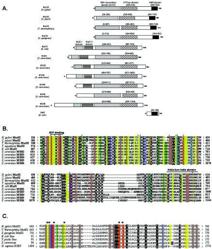
The conserved domains found in MutS1, MutS2, and eukaryotic MutS homologs, based on the SMART database (http://smart.embl-heidelberg.de) (42), with cross-references to PFam data (26), are shown in schematic form for representative organisms (Fig. 1A). E. coli and Saccharomyces cerevisiae were selected as representative species for MutS1 and MSH, respectively. H. pylori, Campylobacter jejuni, Thermus thermophilus, and Streptococcus pyogenes were selected as representative species for MutS2. MutS2 from H. pylori has strong homology to MutS2 from the closely related species C. jejuni as well as to those of S. pyogenes and T. thermophilus, where MutS2 nicking endonuclease activity has been previously demonstrated (25). MutS2 lacks the conserved N-terminal region found in other MutS homologs, which contain MutS domains I and II (Fig. 1A). MutS domain I is critical for mismatch recognition, as evidenced by the crystal structures of E. coli and Thermus aquaticus MutS-DNA complexes (11, 40, 51), and is present in all MutS homologs that play a role in mitotic mismatch repair (32) (Fig. 1A). The absence of MutS domain I in MutS2 is consistent with the lack of mismatch repair activity shown in prior studies (13, 46). MutS domain II is a conserved intervening region hypothesized to have a structural role, linking MutS domain I to the DNA association domain (11, 40, 51).
FIG. 1.
Genetic analyses of mutS1 and mutS2 homologs. A. Schematic of MutS homologs. All sequences were obtained from GenBank, and conserved protein domains were identified using PFam (26) and the SMART program (http://smart.embl-heidelberg.de) (42). B. Amino acid alignment of the ATPase domain of MutS homologs. The ATPase domain of MutS1 from E. coli and T. aquaticus, MSH2, -3, -4, and -6 from S. cerevisiae, and MutS2 from Listeria monocytogenes and H. pylori were aligned with ClustalX and visualized using the program Genedoc (www.psc.edu/biomed/genedoc) in physiochemical properties shading mode, which divides residues into 12 groups representing three hierarchies. The first hierarchy is based on size, the second is based electrical charge for the polar amino acids, and the third, for nonpolar amino acids, is based on aromaticity. The highly conserved ATP binding domain (73) and putative helix-turn-helix domain (12, 54) are indicated with a black line. C. Amino acid alignment of SMR domains. The SMR domain of H. pylori MutS2 was aligned with SMR domains from other MutS2 and Smr proteins. Also included were SMR domains from hypothetical proteins in Caenorhabditis elegans and Drosophila melanogaster and the human Bcl-3 binding protein (B3BP), which has recently been shown to possess nicking endonuclease activity (72). Alignments were constructed and visualized as described for panel B. Asterisks indicate residues with 100% conservation.
The DNA association domain and ATPase domain are conserved in all MutS homologs, including MutS2 (Fig. 1A). Alignment of the ATPase domain containing the helix-turn-helix motif, a region of dimer contact in MutS1 and eukaryotic MSH, shows substantial conservation between homologs (32) (Fig. 1B). That MutS2 also contains this motif suggests that it may function as a dimer as well. The DNA association domain shows a lesser degree of conservation between homologs (data not shown). Crystal structures of MutS1 suggest that this domain forms a major portion of the sliding clamp, with positive residues forming loose hydrophilic associations with DNA. That the sliding clamp structure, common to MutS1, MSH2-MSH3, MSH2-MSH6, and MSH4-MSH5 dimers, shows less conservation is consistent with evidence that MutS dimers associate with a variety of DNA structures, including DNA mismatches, nucleotide loops, and Holliday junctions, and this may reflect differences in DNA structural recognition (27, 45, 49, 64).
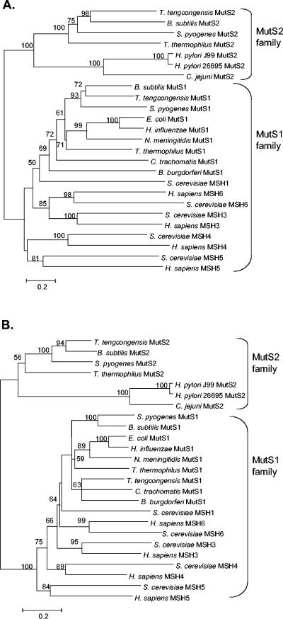
Since the DNA association domain is important in the recognition of various DNA structures (32), we hypothesized that if these proteins were linked, MutS2 would associate with MSH4-MSH5. To test this hypothesis, a phylogeny was constructed using the DNA association domain of representative MutS1, MutS2, and MSH1-MSH6 sequences (Fig. 2A). Our results show that MSH4-MSH5 clusters with MutS1 and other eukaryotic MSH proteins and not with MutS2, indicating substantial differences. Phylogenetic analyses of the MutS ATPase domain (Fig. 2B) also show a deep branching of MutS2 from the MutS1/MSH family. These results support the hypothesis that MutS2 and MutS1 diverged early during prokaryotic evolution and that MSH1-MSH7 subsequently evolved from a MutS1 ancestor. Therefore, the function of MutS2 cannot be inferred based on phylogeny.
FIG. 2.
Phylogenetic analysis of MutS family domains. A. The DNA association domain, conserved throughout MutS homologs, was identified using the SMART database (42), where it is annotated as MutSd. A phylogenetic tree was constructed using the amino acid sequences of MutS1 from nine representative bacterial species, MutS2 from six representative bacterial species, and MSH2 to MSH6 from S. cerevisiae and H. sapiens (Table 3). B. subtilis, S. pyogenes, and T. tengcongensis were selected, since they possess both MutS1 and MutS2. The program Mega 2.1 (38) was used to construct the phylogeny, by using the neighbor-joining method with 1,000 bootstrap replicates. B. The ATPase/dimerization domain, highly conserved throughout MutS homologs, was identified by using the SMART database (42), where it is annotated as MutSac. A phylogenetic tree was constructed as in panel A, with 1,000 bootstrap replicates.
MutS2 contains a SMR domain at its C terminus that is not present in other MutS family members (Fig. 1A). The SMR domain is highly conserved (43, 48), even between distantly related species, and is present as either a small protein, represented by E. coli Smr (Fig. 1A), or as the C terminus of MutS2. The fact that the species with Smr does not possess MutS2, and vice versa (43), was supported by a more extensive sequence analyses (Table 3). The SMR domain has nicking endonuclease activity in T. thermophilus (25), and a human protein, B3BP, containing a SMR domain has similar function (72). Alignment of the Smr domain, using sequences identified through PSI-BLAST, indicates that H. pylori MutS2 possesses the SMR domain (Fig. 1C), suggesting similar function for H. pylori MutS2.
TABLE 3.
Conservation of mutS1 and mutS2 homologs in representative prokaryotic species
| Species | Presencea
|
||
|---|---|---|---|
| MutS1 | Smr fragment | MutS2 | |
| Mycoplasma genitalium | — | — | — |
| Mycoplasma pneumoniae | — | — | — |
| Mycobacterium bovis | — | — | — |
| Mycobacterium tuberculosis | — | — | — |
| Campylobacter jejuni | — | — | NP_282202 |
| Helicobacter pylori 26695 | — | — | O25338 |
| Helicobacter pylori J99 | — | — | Q9ZLL4 |
| Wolinella succinogenes | — | — | Q7M8Q5 |
| Chlamydia pneumoniae | Q9Z6W5 | — | — |
| Rickettsia prowazekii | CAA14759 | — | — |
| Bordetella pertussis | Q7VY01 | Q7VW03 | — |
| Neisseria meningitidis | Q9JWT7 | NC_283999 | — |
| Escherichia coli | NP_417213 | P77458 | — |
| Salmonella enterica serovar Typhi | P10339 | P67246 | — |
| Yersinia pestis | Q8ZBQ3 | NP_993742 | — |
| Haemophilus influenzae | Q6Q131 | NP_43958 | — |
| Pasteurella multocida | P57952 | NP_245328 | — |
| Vibrio cholerae | Q9KUI6 | NP_231750 | — |
| Treponema pallidum | O83348 | O83680 | — |
| Aquifex aeolicus | O66652 | — | O67287 |
| Bacillus subtilis | P49849 | — | P94545 |
| Borrelia burgdorferi | O51737 | — | O51125 |
| Clostridium perfringens | Q8XL87 | — | Q8XJ80 |
| Geobacter sulfurreducens | AAR35199 | — | NP_951605 |
| Lactobacillus plantarum | CAD64627 | — | CAD64608 |
| Listeria innocua | Q92BV3 | — | NP_470532 |
| Staphylococcus aureus | BAB57458 | — | NP_645844 |
| Streptococcus pyogenes | Q8K5J5 | — | AAK34559 |
| Synechocystis sp. (strain PCC6803) | P73769 | — | P73625 |
| Symbiobacterium thermophilum | YP_075586 | — | YP_074944 |
| Thermotoga maritima | P74926 | — | Q9X105 |
| Thermoanaerobacter tengcongensis | Q8R9D0 | — | NP_622975 |
| Thermus thermophilus | P61671 | — | YP_005251 |
Genbank accession numbers are provided if the homolog is present; —, not present.
With the exception of the obligate intracellular organisms Rickettsia prowazekii and Chlamydia pneumoniae, organisms with mutS1 also possess either smr or mutS2 but not both (27), and our analysis of 33 bacterial species found no exceptions to this rule (Table 3). The Mycobacterium and Mycoplasma species were the only organisms with no homologs of either mutS1, mutS2, or smr. The closely related Wolinella succinogenes and Helicobacter and Campylobacter species (20) were the only organisms found to possess MutS2 but not MutS1.
Role of MutS2 in mismatch repair.
To determine whether MutS2 is involved in mismatch repair in H. pylori, a mutS2 mutant was constructed (see Fig. S1B in the supplemental material), and mutation rates for wild-type and mutS2 mutant H. pylori strains were calculated. Our results show no difference in mutation rates between wild-type and mutS2 H. pylori strains (see Fig. S1B in the supplemental material), consistent with the studies of mutation frequencies in wild-type and mutS2 H. pylori strains by Bjorkholm et al. (13).
Effect of MutS2 on recombinational repair.
We hypothesize that H. pylori MutS2 has the ability to bind DNA through its DNA association domain and to introduce nicks through the endonuclease activity present in its SMR domain. Since nicking endonucleases have been identified in several DNA repair pathways involving DNA damage (31, 47, 70), we asked whether MutS2 could recognize and process damaged DNA structures. Wild-type and mutS2 strains were examined for susceptibility to two types of DNA damaging agents, UV and ciprofloxacin (see Fig. S2 in the supplemental material; also Table 4). UV radiation creates interstrand cross-links in DNA as well as strand breaks that can halt progression of the replication fork. Ciprofloxacin binds to DNA and creates ternary complexes with either DNA gyrase or topoisomerase (18, 36), resulting in lesions that impede replication (14, 44). Recombinational repair restores the DNA template by recognizing and replacing damaged DNA via recombination. Mutations in recombination proteins, such as recA and ruvABC, display high susceptibility to UV and fluoroquinolones (67). As expected, H. pylori recA mutants were more susceptible than the wild type to both UV and fluoroquinolone-induced DNA damage (see Fig. S2 in the supplemental material; also Table 4). The fact that the mutS2 mutant did not show increased susceptibility to UV or fluoroquinolone-induced DNA damage provides evidence that MutS2 is not involved in recombinational repair of damaged DNA.
TABLE 4.
Susceptibility of wild-type and mutant H. pylori strains to ciprofloxacin
| Strain designation | Ciprofloxacin MIC (μg/ml)a |
|---|---|
| JP26 | 0.11 ± 0.03 |
| JP26 mutS2::aphA | 0.12 ± 0.03 |
| JP26 recA::aphA | 0.04 ± 0.01 |
H. pylori strains were assayed for inhibition within lawns on TSA plates containing a ciprofloxacin E-test strip (AB Biodisk), in concentrations from 0.002 to 32 μg/ml. Results shown are the means (± standard deviations) of five replicate determinations.
Effect of MutS2 on deletions between flanking DNA repeats.
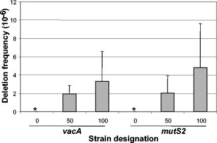
In silico analysis of the H. pylori genome reveals the existence of numerous, nonrandomly distributed, proximate, direct DNA repeat sequences that are “hot spots” for intragenomic DNA rearrangements (2, 8, 9, 57). Generation of loose DNA ends, that can participate in recombination events, are believed to promote deletion and duplication of intervening sequences between such direct DNA repeats. Certain recombination proteins, such as H. pylori RecG, reduce the frequency of deletion events between direct repeats, presumably by recognizing and unwinding intermediary DNA structures before deletion events reach completion (33). We examined whether MutS2 also could influence such recombination by using a previously validated deletion cassette with identical DNA sequence (IDS) repeats of 0 (control), 50, or 100 bp (8). Although insertion of the complete cassette into a host H. pylori strain confers resistance to chloramphenicol, recombination between the flanking identical DNA repeats may delete the chloramphenicol cassette, changing the phenotype to kanamycin resistance and chloramphenicol susceptibility (8). To assess the effect of mutS2 on intragenomic recombination between direct repeats, the deletion cassette was introduced into mutS2 or vacA (as a control). Compared to the control strain, the mutS2 mutant showed no significant difference in deletion frequencies (Fig. 3). These results suggest that MutS2 does not recognize or interfere with DNA intermediates formed during a deletion event.
FIG. 3.
Deletion frequency in H. pylori wild-type and mutant strains. The chloramphenicol/kanamycin resistance cassette flanked by identical repeats (IDS) of 0, 50, or 100 bp was inserted into vacA (control) or mutS2, insertion was confirmed, and deletion frequencies were calculated, as described previously (8). Asterisks indicate that no deletions were detected (frequency < 10−8). As expected (8), H. pylori strains with the cassette in vacA or mutS2 showed progressively higher deletion frequencies with increasing size of the IDS. Strains with the deletion cassette in mutS2 do not show any significant difference in deletion frequency between flanking DNA repeats of 50 and 100 bp compared to control (vacA) mutants with comparable cassettes. Bars represent means ± standard deviations for four to six replicate experiments.
Effect of mutS2 on intergenomic recombination.
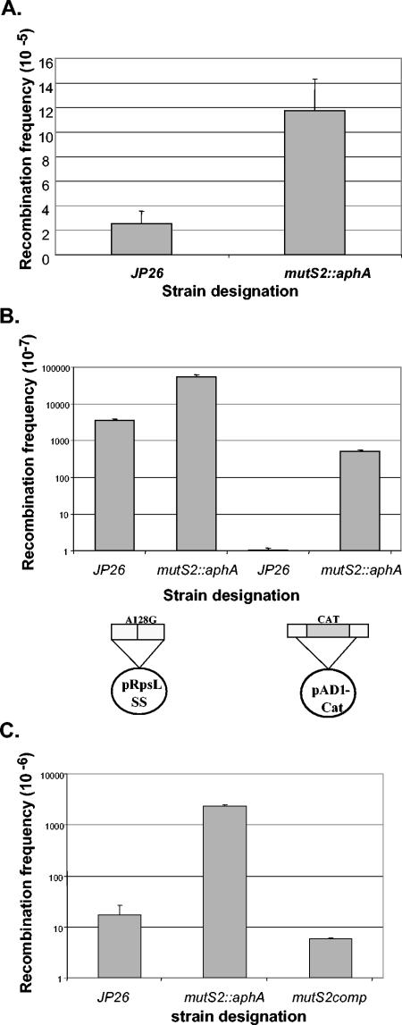
To explore the role of MutS2 in intergenomic recombination events in the naturally competent H. pylori (6), several forms of donor DNA were used to transform H. pylori wild-type and mutant strains. First, we examined transformation frequencies using donor chromosomal DNA with a point mutation in rpsL (A128G) to confer streptomycin resistance (Fig. 4A). The mutS2::aphA strain had a fivefold higher transformation frequency than the wild type (P < 0.05).
FIG. 4.
Intergenomic recombination frequencies of H. pylori wild-type and mutant strains using chromosomal, plasmid, or PCR product DNA. A. H. pylori strains JP26 and JP26/mutS2::aphA were transformed to streptomycin resistance by using chromosomal DNA from Str 26696, which has a point mutation (A128G) in the rpsL gene. Bars represent the means of four experiments. The mutS2::aphA mutant shows a significantly (P < 0.05) higher transformation frequency than wild-type JP26. B. H. pylori strains JP26 and mutS2::aphA were transformed with either pRpsLSS, which contains the 800-bp A128 rpsL fragment, or pAD1-CAT, which contains the ureAB promoter and downstream regions interrupted by a chloramphenicol resistance cas-sette, as shown in the schematic. For both pRpsLSS and pAD1-Cat donor DNA, the mutS2::aphA strain was transformed at a significantly (P < 0.05) higher frequency than wild-type JP26. Overall, pRpsLSS transforms both the wild-type and mutS2 mutant strains at 2 to 3 log10 higher frequency (P < 0.05) than pAD1-Cat. C. H. pylori strains JP26, mutS2::aphA, and mutS2comp were transformed to streptomycin resistance with an 800-bp A128G rpsL PCR product; bars represent the means of four to eight replicate experiments. The mutS2::aphA strain shows a 2 log10 higher transformation frequency (P < 0.05) than wild-type JP26. Complementation of JP26 mutS2::aphA with mutS2 in trans restored the recombination frequency to a level not significantly different from wild-type JP26.
To determine whether the higher transformation frequencies noted in the mutS2::aphA strains with chromosomal donor DNA were more widely applicable, plasmid DNA was used to transform H. pylori (Fig. 4B). A plasmid (pSS) with an 800-bp rpsL fragment containing the same A128G mutation at its midpoint was used to transform H. pylori wild-type and mutS2 mutant strains. The mutS2::aphA strain displayed a significantly higher (16-fold) transformation frequency than wild-type JP26 (P < 0.05). Plasmid pWT, which contains the same 800-bp sequence without the A128G mutation, was used as a control and no transformants were detected, as expected (data not shown). Another form of donor DNA used was pAD1-Cat, which contains the 585-bp ureAB promoter region and the 312-bp downstream regions interrupted by a CAT cassette. Again, the mutS2::aphA strain had a significantly higher (520-fold) transformation frequency than the wild type (P < 0.05).
Examining recombination frequencies with a linear 800-bp rpsL PCR product showed that, as for other forms of donor DNA, the mutS2::aphA strain had a significantly higher (137-fold) transformation frequency than wild-type JP26 (P < 0.05) (Fig. 4C). To determine whether the results were specific to mutation of mutS2 and not due to polar effects, the mutS2::aphA mutant strain was complemented in trans by expressing mutS2 downstream of a strong (ureA) promoter (Fig. 4A). Complementation restored recombination frequency to wild-type levels, confirming that the observed phenotype is specific to the MutS2 mutation and not due to polar effects (Fig. 4C).
DISCUSSION
Our analysis of conserved protein domains in MutS family members reveals substantial differences between MutS2 and other MutS homologs, suggesting corresponding differences in biological function. By targeting phylogenetic analyses to a region specific for DNA association, the large differences in protein sequence that confound construction of a phylogenetic tree could be overcome. The results provide further evidence that MutS2 and MutS1 diverged early during prokaryotic evolution and that MutS1 was introduced into eukaryotes subsequent to this event, resulting in the evolution of MSH1 to MSH7. Phylogenetic analyses of the DNA association domains, and phenotypic analyses of a MutS2 mutant, argue against the hypothesis that MSH4-MSH5 and MutS2 evolved together. MSH4 and MSH5 are involved in promoting crossover of DNA during recombination (30, 37, 64). Our results for MutS2 in H. pylori indicate an opposing role, limiting recombination. In total, neither the phylogenetic or experimental results support previous hypotheses (19) suggesting that MSH4-MSH5 arose from an ancestral MutS2.
The SMR domain of MutS2, which is not present in other MutS homologs (Fig. 1A), has extensive sequence conservation, suggesting an important conserved function. In T. thermophilus MutS2, the SMR domain binds double-stranded DNA (dsDNA) and possesses endonuclease activity (25). In humans, the Bcl-3 binding protein (B3BP) has a single C-terminal SMR domain with nicking endonuclease activity, and it is believed to be involved in DNA repair and/or recombination (72).
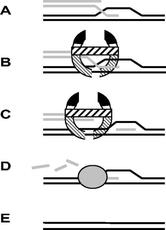
Nicking endonucleases participate in multiple repair processes, including excision of damaged DNA, mismatch repair, and possibly class-switch immunoglobulin gene recombination and somatic hypermutation (31, 47, 70). These enzymes also participate in strand displacement reactions, introducing a site-specific nick into DNA, and a polymerase is used to displace the parental strand (50). We hypothesize that MutS2 acts similarly (Fig. 5), with the DNA association domain forming a loose clamp-like structure that recognizes recombination intermediates. Recent evidence shows that MutS2 has the ability to recognize and bind dsDNA (25, 53). The presence of a conserved helix-turn-helix domain in MutS2 (Fig. 2B) suggests that, as with other MutS homologs, it functions as a dimer. We propose that once recognition occurs, the Smr domain introduces a nick into the DNA, and the nick is subsequently extended, possibly by an exonuclease, resulting in displacement of the invading strand and abortion of potentially illegitimate recombination events.
FIG. 5.
Model of how MutS2 might limit intergenomic recombination. A. RecA-mediated strand invasion of donor DNA, which is essential for recombination events, forms a D-loop. B. MutS2, shown as a dimer via interaction at its ATPase/dimerization domains (thick gray lines), recognizes the primary recombination structure via its DNA association domains (thin gray lines). C. SMR endonuclease activity (represented in black) introduces a nick into the DNA. D. Recruitment of other proteins, possibly a repair polymerase such as PolA, with exonuclease activity, removes the invading DNA and fills in the gap. E. Restoration of original dsDNA.
That MutS2 mutants are hyperrecombinant regardless of the type of donor DNA (fragment, plasmid, or chromosome) supports the hypothesis that it recognizes a DNA structure common to all intergenomic recombination events (Fig. 5). That this hyperrecombinant phenotype is not dependent on the extent of homology in the donor DNA (point mutation versus nonhomologous cassette [Fig. 4B; also reference 53]) further suggests that MutS2 acts at an early step in recombination, possibly during strand invasion, to recognize and abort illegitimate recombination events; in this view, MutS2 mutation allows recombination to proceed unhindered, regardless of homology. That MutS2 is not involved in deletions between direct repeats suggests that it does not inhibit slipped-strand mispairing of homologous DNA, which is a common mechanism of deletion formation and does not require DNA strand exchange (3, 8); rather, MutS2 function may be specific to RecA-mediated recombination events (53). MutS2 inhibition of initiation of DNA strand exchange reactions (53) is consistent with this hypothesis. Since illegitimate recombination events also can be aborted by MutS1, through a separate mechanism (74), MutS1 presence may mask the hyperrecombinant phenotype of a MutS2 mutant. H. pylori, one of few organisms with MutS2 but not MutS1, is an ideal candidate for the study of MutS2 function.
That organisms lack MutS1, including the closely related W. succinogenes and Campylobacter and Helicobacter species (20), and the Mycoplasma and Mycobacterium species (Table 3), suggests that they may gain a selective advantage from the absence of MutS1-directed mismatch repair. Numerous phase-variable loci are present in these organisms, facilitating variation of antigenic structures at the cell surface (10, 15, 20, 28, 55, 56, 58, 62). As such, H. pylori, C. jejuni, W. succinogenes, and the Mycoplasma species may be regarded as multicellular populations that continually generate variants, particularly through phase variation events, that would be inhibited if mismatch repair were present. The Mycobacterium genome also contains an unusually high number of frameshifts, as well as repetitive sequence elements and repeat regions (8, 65). Clinical isolates of M. tuberculosis indicate that chromosomal rearrangements such as duplications, deletions, and inversions contribute significantly to variation of this species within its obligate human host (21, 22, 24, 34). Such events are facilitated by a lack of MutS1-directed mismatch repair (32, 74). Furthermore, M. tuberculosis is similar to H. pylori in that both species occupy niches in which other microbiota are underrepresented or absent, resulting in limited opportunities for lateral gene transfer from exogenous sources (65). Therefore, loss of mismatch repair and the subsequent increased opportunity for chromosomal rearrangements and diversification within the population can facilitate microevolution. Since a high degree of mutation may decrease overall fitness (17), by possessing MutS2 function, H. pylori and its related campylobacters can limit the amount of diversification that occurs. In this respect, MutS1 and MutS2 may both function to preserve genomic integrity.
Supplementary Material
Footnotes
Supplemental material for this article may be found at http://jb.asm.org/.
REFERENCES
- 1.Acharya, S., T. Wilson, S. Gradia, M. F. Kane, S. Guerrette, G. T. Marsischky, R. Kolodner, and R. Fishel. 1996. hMSH2 forms specific mispair-binding complexes with hMSH3 and hMSH6. Proc. Natl. Acad. Sci. USA 93:13629-13634. [DOI] [PMC free article] [PubMed] [Google Scholar]
- 2.Achaz, G., E. P. Rocha, P. Netter, and E. Coissac. 2002. Origin and fate of repeats in bacteria. Nucleic Acids Res. 30:2987-2994. [DOI] [PMC free article] [PubMed] [Google Scholar]
- 3.Albertini, A. M., M. Hofer, M. P. Calos, and J. H. Miller. 1982. On the formation of spontaneous deletions: the importance of short sequence homologies in the generation of large deletions. Cell 29:319-328. [DOI] [PubMed] [Google Scholar]
- 4.Alm, R. A., and T. J. Trust. 1999. Analysis of the genetic diversity of Helicobacter pylori: the tale of two genomes. J. Mol. Med. 77:834-846. [DOI] [PubMed] [Google Scholar]
- 5.Altschul, S. F., T. L. Madden, A. A. Schaffer, J. Zhang, Z. Zhang, W. Miller, and D. J. Lipman. 1997. Gapped BLAST and PSI-BLAST: a new generation of protein database search programs. Nucleic Acids Res. 25:3389-3402. [DOI] [PMC free article] [PubMed] [Google Scholar]
- 6.Ando, T., D. A. Israel, K. Kusugami, and M. J. Blaser. 1999. HP0333, a member of the dprA family, is involved in natural transformation in Helicobacter pylori. J. Bacteriol. 181:5572-5580. [DOI] [PMC free article] [PubMed] [Google Scholar]
- 7.Aras, R. A., W. Fischer, G. I. Perez-Perez, M. Crosatti, T. Ando, R. Haas, and M. J. Blaser. 2003. Plasticity of repetitive DNA sequences within a bacterial (type IV) secretion system component. J. Exp. Med. 198:1349-1360. [DOI] [PMC free article] [PubMed] [Google Scholar]
- 8.Aras, R. A., J. Kang, A. I. Tschumi, Y. Harasaki, and M. J. Blaser. 2003. Extensive repetitive DNA facilitates prokaryotic genome plasticity. Proc. Natl. Acad. Sci. USA 100:13579-13584. [DOI] [PMC free article] [PubMed] [Google Scholar]
- 9.Aras, R. A., T. Takata, T. Ando, A. van der Ende, and M. J. Blaser. 2001. Regulation of the HpyII restriction-modification system of Helicobacter pylori by gene deletion and horizontal reconstitution. Mol. Microbiol. 42:369-382. [DOI] [PubMed] [Google Scholar]
- 10.Bacon, D. J., C. M. Szymanski, D. H. Burr, R. P. Silver, R. A. Alm, and P. Guerry. 2001. A phase-variable capsule is involved in virulence of Campylobacter jejuni 81-176. Mol. Microbiol. 40:769-777. [DOI] [PubMed] [Google Scholar]
- 11.Biswas, I., and P. Hsieh. 1997. Interaction of MutS protein with the major and minor grooves of a heteroduplex DNA. J. Biol. Chem. 272:13355-13364. [DOI] [PubMed] [Google Scholar]
- 12.Biswas, I., G. Obmolova, M. Takahashi, A. Herr, M. A. Newman, W. Yang, and P. Hsieh. 2001. Disruption of the helix-u-turn-helix motif of MutS protein: loss of subunit dimerization, mismatch binding and ATP hydrolysis. J. Mol. Biol. 305:805-816. [DOI] [PubMed] [Google Scholar]
- 13.Bjorkholm, B., M. Sjolund, P. G. Falk, O. G. Berg, L. Engstrand, and D. I. Andersson. 2001. Mutation frequency and biological cost of antibiotic resistance in Helicobacter pylori. Proc. Natl. Acad. Sci. USA 98:14607-14612. [DOI] [PMC free article] [PubMed] [Google Scholar]
- 14.Chen, C. R., M. Malik, M. Snyder, and K. Drlica. 1996. DNA gyrase and topoisomerase IV on the bacterial chromosome: quinolone-induced DNA cleavage. J. Mol. Biol. 258:627-637. [DOI] [PubMed] [Google Scholar]
- 15.Citti, C., and R. Rosengarten. 1997. Mycoplasma genetic variation and its implication for pathogenesis. Wien. Klin. Wochenschr. 109:562-568. [PubMed] [Google Scholar]
- 16.Culligan, K. M., G. Meyer-Gauen, J. Lyons-Weiler, and J. B. Hays. 2000. Evolutionary origin, diversification and specialization of eukaryotic MutS homolog mismatch repair proteins. Nucleic Acids Res. 28:463-471. [DOI] [PMC free article] [PubMed] [Google Scholar]
- 17.de Visser, J. A. 2002. The fate of microbial mutators. Microbiology 148:1247-1252. [DOI] [PubMed] [Google Scholar]
- 18.Drlica, K., and X. Zhao. 1997. DNA gyrase, topoisomerase IV, and the 4-quinolones. Microbiol. Mol. Biol. Rev. 61:377-392. [DOI] [PMC free article] [PubMed] [Google Scholar]
- 19.Eisen, J. A. 1998. A phylogenomic study of the MutS family of proteins. Nucleic Acids Res. 26:4291-4300. [DOI] [PMC free article] [PubMed] [Google Scholar]
- 20.Eppinger, M., C. Baar, G. Raddatz, D. H. Huson, and S. C. Schuster. 2004. Comparative analysis of four Campylobacterales. Nat. Rev. Microbiol. 2:872-885. [DOI] [PubMed] [Google Scholar]
- 21.Fang, Z., C. Doig, D. T. Kenna, N. Smittipat, P. Palittapongarnpim, B. Watt, and K. J. Forbes. 1999. IS6110-mediated deletions of wild-type chromosomes of Mycobacterium tuberculosis. J. Bacteriol. 181:1014-1020. [DOI] [PMC free article] [PubMed] [Google Scholar]
- 22.Fang, Z., C. Doig, A. Rayner, D. T. Kenna, B. Watt, and K. J. Forbes. 1999. Molecular evidence for heterogeneity of the multiple-drug-resistant Mycobacterium tuberculosis population in Scotland (1990 to 1997). J. Clin. Microbiol. 37:998-1003. [DOI] [PMC free article] [PubMed] [Google Scholar]
- 23.Fishel, R., and T. Wilson. 1997. MutS homologs in mammalian cells. Curr. Opin. Genet. Dev. 7:105-113. [DOI] [PubMed] [Google Scholar]
- 24.Fleischmann, R. D., D. Alland, J. A. Eisen, L. Carpenter, O. White, J. Peterson, R. DeBoy, R. Dodson, M. Gwinn, D. Haft, E. Hickey, J. F. Kolonay, W. C. Nelson, L. A. Umayam, M. Ermolaeva, S. L. Salzberg, A. Delcher, T. Utterback, J. Weidman, H. Khouri, J. Gill, A. Mikula, W. Bishai, W. R. Jacobs, Jr., J. C. Venter, and C. M. Fraser. 2002. Whole-genome comparison of Mycobacterium tuberculosis clinical and laboratory strains. J. Bacteriol. 184:5479-5490. [DOI] [PMC free article] [PubMed] [Google Scholar]
- 25.Fukui, K., R. Masui, and S. Kuramitsu. 2004. Thermus thermophilus MutS2, a MutS paralogue, possesses an endonuclease activity promoted by MutL. J. Biochem. (Tokyo) 135:375-384. [DOI] [PubMed] [Google Scholar]
- 26.Gasteiger, E., E. Jung, and A. Bairoch. 2001. SWISS-PROT: connecting biomolecular knowledge via a protein database. Curr. Issues Mol. Biol. 3:47-55. [PubMed] [Google Scholar]
- 27.Gradia, S., S. Acharya, and R. Fishel. 1997. The human mismatch recognition complex hMSH2-hMSH6 functions as a novel molecular switch. Cell 91:995-1005. [DOI] [PubMed] [Google Scholar]
- 28.Guerry, P., C. M. Szymanski, M. M. Prendergast, T. E. Hickey, C. P. Ewing, D. L. Pattarini, and A. P. Moran. 2002. Phase variation of Campylobacter jejuni 81-176 lipooligosaccharide affects ganglioside mimicry and invasiveness in vitro. Infect. Immun. 70:787-793. [DOI] [PMC free article] [PubMed] [Google Scholar]
- 29.Heep, M., S. Odenbreit, D. Beck, J. Decker, E. Prohaska, U. Rieger, and N. Lehn. 2000. Mutations at four distinct regions of the rpoB gene can reduce the susceptibility of Helicobacter pylori to rifamycins. Antimicrob. Agents Chemother. 44:1713-1715. [DOI] [PMC free article] [PubMed] [Google Scholar]
- 30.Hollingsworth, N. M., L. Ponte, and C. Halsey. 1995. MSH5, a novel MutS homolog, facilitates meiotic reciprocal recombination between homologs in Saccharomyces cerevisiae but not mismatch repair. Genes Dev. 9:1728-1739. [DOI] [PubMed] [Google Scholar]
- 31.Honjo, T., K. Kinoshita, and M. Muramatsu. 2002. Molecular mechanism of class switch recombination: linkage with somatic hypermutation. Annu. Rev. Immunol. 20:165-196. [DOI] [PubMed] [Google Scholar]
- 32.Hsieh, P. 2001. Molecular mechanisms of DNA mismatch repair. Mutat. Res. 486:71-87. [DOI] [PubMed] [Google Scholar]
- 33.Kang, J., D. Tavakoli, A. Tschumi, R. A. Aras, and M. J. Blaser. 2004. Effect of host species on recG phenotypes in Helicobacter pylori and Escherichia coli. J. Bacteriol. 186:7704-7713. [DOI] [PMC free article] [PubMed] [Google Scholar]
- 34.Kato-Maeda, M., P. J. Bifani, B. N. Kreiswirth, and P. M. Small. 2001. The nature and consequence of genetic variability within Mycobacterium tuberculosis. J. Clin. Investig. 107:533-537. [DOI] [PMC free article] [PubMed] [Google Scholar]
- 35.Kersulyte, D., H. Chalkauskas, and D. E. Berg. 1999. Emergence of recombinant strains of Helicobacter pylori during human infection. Mol. Microbiol. 31:31-43. [DOI] [PubMed] [Google Scholar]
- 36.Khodursky, A. B., and N. R. Cozzarelli. 1998. The mechanism of inhibition of topoisomerase IV by quinolone antibacterials. J. Biol. Chem. 273:27668-27677. [DOI] [PubMed] [Google Scholar]
- 37.Kneitz, B., P. E. Cohen, E. Avdievich, L. Zhu, M. F. Kane, H. Hou, Jr., R. D. Kolodner, R. Kucherlapati, J. W. Pollard, and W. Edelmann. 2000. MutS homolog 4 localization to meiotic chromosomes is required for chromosome pairing during meiosis in male and female mice. Genes Dev. 14:1085-1097. [PMC free article] [PubMed] [Google Scholar]
- 38.Kumar, S., K. Tamura, I. B. Jakobsen, and M. Nei. 2001. MEGA2: molecular evolutionary genetics analysis software. Bioinformatics 17:1244-1245. [DOI] [PubMed] [Google Scholar]
- 39.Kumar, S., K. Tamura, and M. Nei. 2004. MEGA3: integrated software for molecular evolutionary genetics analysis and sequence alignment. Brief. Bioinformatics 5:150-163. [DOI] [PubMed] [Google Scholar]
- 40.Lamers, M. H., A. Perrakis, J. H. Enzlin, H. H. Winterwerp, N. de Wind, and T. K. Sixma. 2000. The crystal structure of DNA mismatch repair protein MutS binding to a G x T mismatch. Nature 407:711-717. [DOI] [PubMed] [Google Scholar]
- 41.Lea, E. A., and D. E. Coulson. 1949. The distribution of the numbers of mutants in bacterial populations. J. Genet. 49:264-285. [DOI] [PubMed] [Google Scholar]
- 42.Letunic, I., R. R. Copley, S. Schmidt, F. D. Ciccarelli, T. Doerks, J. Schultz, C. P. Ponting, and P. Bork. 2004. SMART 4.0: towards genomic data integration. Nucleic Acids Res. 32:D142-D144. [DOI] [PMC free article] [PubMed] [Google Scholar]
- 43.Malik, H. S., and S. Henikoff. 2000. Dual recognition-incision enzymes might be involved in mismatch repair and meiosis. Trends Biochem. Sci. 25:414-418. [DOI] [PubMed] [Google Scholar]
- 44.Mamber, S. W., B. Kolek, K. W. Brookshire, D. P. Bonner, and J. Fung-Tomc. 1993. Activity of quinolones in the Ames Salmonella TA102 mutagenicity test and other bacterial genotoxicity assays. Antimicrob. Agents Chemother. 37:213-217. [DOI] [PMC free article] [PubMed] [Google Scholar]
- 45.Marra, G., and P. Schar. 1999. Recognition of DNA alterations by the mismatch repair system. Biochem. J. 338:1-13. [PMC free article] [PubMed] [Google Scholar]
- 46.Mennecier, S., G. Coste, P. Servant, A. Bailone, and S. Sommer. 2004. Mismatch repair ensures fidelity of replication and recombination in the radioresistant organism Deinococcus radiodurans. Mol. Genet. Genomics 272:460-469. [DOI] [PubMed] [Google Scholar]
- 47.Moolenaar, G. F., K. L. Franken, P. van de Putte, and N. Goosen. 1997. Function of the homologous regions of the Escherichia coli DNA excision repair proteins UvrB and UvrC in stabilization of the UvrBC-DNA complex and in 3′-incision. Mutat. Res. 385:195-203. [DOI] [PubMed] [Google Scholar]
- 48.Moreira, D., and H. Philippe. 1999. Smr: a bacterial and eukaryotic homologue of the C-terminal region of the MutS2 family. Trends Biochem. Sci. 24:298-300. [DOI] [PubMed] [Google Scholar]
- 49.Natrajan, G., M. H. Lamers, J. H. Enzlin, H. H. Winterwerp, A. Perrakis, and T. K. Sixma. 2003. Structures of Escherichia coli DNA mismatch repair enzyme MutS in complex with different mismatches: a common recognition mode for diverse substrates. Nucleic Acids Res. 31:4814-4821. [DOI] [PMC free article] [PubMed] [Google Scholar]
- 50.Nuovo, G. J. 2000. In situ strand displacement amplification: an improved technique for the detection of low copy nucleic acids. Diagn. Mol. Pathol. 9:195-202. [DOI] [PubMed] [Google Scholar]
- 51.Obmolova, G., C. Ban, P. Hsieh, and W. Yang. 2000. Crystal structures of mismatch repair protein MutS and its complex with a substrate DNA. Nature 407:703-710. [DOI] [PubMed] [Google Scholar]
- 52.Peek, R. M., Jr., and M. J. Blaser. 2002. Helicobacter pylori and gastrointestinal tract adenocarcinomas. Nat. Rev. Cancer 2:28-37. [DOI] [PubMed] [Google Scholar]
- 53.Pinto, V. A., A. Mathieu, S. Marsin, X. Veaute, E. J. O'Rourke, A. Labigne, and P. Radicella. 2004. Suppression of homologous and homeologous recombination by bacterial MutS2 protein. Mol. Cell 17:113-120. [DOI] [PubMed] [Google Scholar]
- 54.Pochart, P., D. Woltering, and N. M. Hollingsworth. 1997. Conserved properties between functionally distinct MutS homologs in yeast. J. Biol. Chem. 272:30345-30349. [DOI] [PubMed] [Google Scholar]
- 55.Prendergast, M. M., D. R. Tribble, S. Baqar, D. A. Scott, J. A. Ferris, R. I. Walker, and A. P. Moran. 2004. In vivo phase variation and serologic response to lipooligosaccharide of Campylobacter jejuni in experimental human infection. Infect. Immun. 72:916-922. [DOI] [PMC free article] [PubMed] [Google Scholar]
- 56.Rocha, E. P., and A. Blanchard. 2002. Genomic repeats, genome plasticity and the dynamics of Mycoplasma evolution. Nucleic Acids Res. 30:2031-2042. [DOI] [PMC free article] [PubMed] [Google Scholar]
- 57.Rocha, E. P., A. Danchin, and A. Viari. 1999. Analysis of long repeats in bacterial genomes reveals alternative evolutionary mechanisms in Bacillus subtilis and other competent prokaryotes. Mol. Biol. Evol. 16:1219-1230. [DOI] [PubMed] [Google Scholar]
- 58.Rosengarten, R., and K. S. Wise. 1990. Phenotypic switching in mycoplasmas: phase variation of diverse surface lipoproteins. Science 247:315-318. [DOI] [PubMed] [Google Scholar]
- 59.Rossolillo, P., and A. M. Albertini. 2001. Functional analysis of the Bacillus subtilis yshD gene, a mutS paralogue. Mol. Gen. Genet. 264:809-818. [DOI] [PubMed] [Google Scholar]
- 60.Saitou, N., and M. Nei. 1987. The neighbor-joining method: a new method for reconstructing phylogenetic trees. Mol. Biol. Evol. 4:406-425. [DOI] [PubMed] [Google Scholar]
- 61.Salama, N., K. Guillemin, T. K. McDaniel, G. Sherlock, L. Tompkins, and S. Falkow. 2000. A whole-genome microarray reveals genetic diversity among Helicobacter pylori strains. Proc. Natl. Acad. Sci. USA 97:14668-14673. [DOI] [PMC free article] [PubMed] [Google Scholar]
- 62.Salaun, L., B. Linz, S. Suerbaum, and N. J. Saunders. 2004. The diversity within an expanded and redefined repertoire of phase-variable genes in Helicobacter pylori. Microbiology 150:817-830. [DOI] [PubMed] [Google Scholar]
- 63.Smeets, L. C., N. L. Arents, A. A. van Zwet, C. M. Vandenbroucke-Grauls, T. Verboom, W. Bitter, and J. G. Kusters. 2003. Molecular patchwork: chromosomal recombination between two Helicobacter pylori strains during natural colonization. Infect. Immun. 71:2907-2910. [DOI] [PMC free article] [PubMed] [Google Scholar]
- 64.Snowden, T., S. Acharya, C. Butz, M. Berardini, and R. Fishel. 2004. hMSH4-hMSH5 recognizes Holliday junctions and forms a meiosis-specific sliding clamp that embraces homologous chromosomes. Mol. Cell 15:437-451. [DOI] [PubMed] [Google Scholar]
- 65.Springer, B., P. Sander, L. Sedlacek, W. D. Hardt, V. Mizrahi, P. Schar, and E. C. Bottger. 2004. Lack of mismatch correction facilitates genome evolution in mycobacteria. Mol. Microbiol. 53:1601-1609. [DOI] [PubMed] [Google Scholar]
- 66.Thompson, J. D., T. J. Gibson, F. Plewniak, F. Jeanmougin, and D. G. Higgins. 1997. The CLUSTAL_X Windows interface: flexible strategies for multiple sequence alignment aided by quality analysis tools. Nucleic Acids Res. 25:4876-4882. [DOI] [PMC free article] [PubMed] [Google Scholar]
- 67.Thompson, S. A., and M. J. Blaser. 1995. Isolation of the Helicobacter pylori recA gene and involvement of the recA region in resistance to low pH. Infect. Immun. 63:2185-2193. [DOI] [PMC free article] [PubMed] [Google Scholar]
- 68.Tomb, J. F., O. White, A. R. Kerlavage, R. A. Clayton, G. G. Sutton, R. D. Fleischmann, K. A. Ketchum, H. P. Klenk, S. Gill, B. A. Dougherty, K. Nelson, J. Quackenbush, L. Zhou, E. F. Kirkness, S. Peterson, B. Loftus, D. Richardson, R. Dodson, H. G. Khalak, A. Glodek, K. McKenney, L. M. Fitzegerald, N. Lee, M. D. Adams, J. C. Venter, et al. 1997. The complete genome sequence of the gastric pathogen Helicobacter pylori. Nature 388:539-547. [DOI] [PubMed] [Google Scholar]
- 69.Vijayvargia, R., and I. Biswas. 2002. MutS2 family protein from Pyrococcus furiosus. Curr. Microbiol. 44:224-228. [DOI] [PubMed] [Google Scholar]
- 70.Wang, H., and J. B. Hays. 2002. Mismatch repair in human nuclear extracts. Quantitative analyses of excision of nicked circular mismatched DNA substrates, constructed by a new technique employing synthetic oligonucleotides. J. Biol. Chem. 277:26136-26142. [DOI] [PubMed] [Google Scholar]
- 71.Wang, Y., K. P. Roos, and D. E. Taylor. 1993. Transformation of Helicobacter pylori by chromosomal metronidazole resistance and by a plasmid with a selectable chloramphenicol resistance marker. J. Gen. Microbiol. 139:2485-2493. [DOI] [PubMed] [Google Scholar]
- 72.Watanabe, N., S. Wachi, and T. Fujita. 2003. Identification and characterization of BCL-3-binding protein: implications for transcription and DNA repair or recombination. J. Biol. Chem. 278:26102-26110. [DOI] [PubMed] [Google Scholar]
- 73.Worth, L., Jr., T. Bader, J. Yang, and S. Clark. 1998. Role of MutS ATPase activity in MutS,L-dependent block of in vitro strand transfer. J. Biol. Chem. 273:23176-23182. [DOI] [PubMed] [Google Scholar]
- 74.Yang, W. 2000. Structure and function of mismatch repair proteins. Mutat. Res. 460:245-256. [DOI] [PubMed] [Google Scholar]
Associated Data
This section collects any data citations, data availability statements, or supplementary materials included in this article.


