Abstract
Cells depend on their endolysosomal system for nutrient uptake and downregulation of plasma membrane proteins. These processes rely on endosomal maturation, which requires multiple membrane fusion steps. Early endosome fusion is promoted by the Rab5 GTPase and its effector, the hexameric CORVET tethering complex, which is homologous to the lysosomal HOPS. How these related complexes recognize their specific target membranes remains entirely elusive. Here, we solve the structure of CORVET by cryo-electron microscopy and revealed its minimal requirements for membrane tethering. As expected, the core of CORVET and HOPS resembles each other. However, the function-defining subunits show marked structural differences. Notably, we discover that unlike HOPS, CORVET depends not only on Rab5 but also on phosphatidylinositol-3-phosphate (PI3P) and membrane lipid packing defects for tethering, implying that an organelle-specific membrane code enables fusion. Our data suggest that both shape and membrane interactions of CORVET and HOPS are conserved in metazoans, thus providing a paradigm how tethering complexes function.
Subject terms: Cryoelectron microscopy, Cryoelectron microscopy, Membrane fusion, Biophysics
Endosomal biogenesis is vital for eukaryotic cell physiology and relies on membrane fusion promoted by the CORVET tethering complex. Here, the authors solved the structure of CORVET by cryo-EM and reveal its minimal requirements for membrane tethering.
Introduction
Eukaryotic cells maintain an elaborate endomembrane system of organelles. This interconnected network depends on the vesicular transport and relies on conserved types of machinery for vesicle generation at the donor organelle and fusion at the acceptor organelle1–3. For proper intracellular recognition, each organelle exhibits distinct membrane compositions and shapes. Identity markers include specific phosphoinositides (PIPs) in the lipid bilayer4 and distinct peripheral membrane proteins such as Rab GTPases (Rabs)3,5. Together, these elements guide trafficking effector proteins to their specific membrane and enable the effective direction of vesicles to their acceptor organelles4,6–8.
Rabs are an integral part of the membrane fusion cascade, as their depletion leads to impaired membrane trafficking9,10. As switch-like proteins, Rabs only interact with their effector proteins upon activation by guanine nucleotide exchange factors (GEFs) and binding to GTP while requiring a GTPase-activating protein (GAP) to hydrolyze GTP for their inactivation3,5,11–13.
The most prominent effector proteins are tethering and fusion factors and complexes14–18. They tether the membranes and recruit Sec1/Munc18 proteins (SM proteins) to promote the zippering of SNAREs from each membrane into a four-helix bundle to trigger fusion14,17–19.
The hexameric HOPS and CORVET are evolutionarily conserved tethering complexes within the endolysosomal system of eukaryotic cells16,20–24. Both share four subunits. Vps11 and Vps18 form the central core to which Vps33 and Vps16 are attached as an SM module to promote SNARE assembly25,26. The two unique subunits determine the respective Rab specificity of the tethering complexes22–24,27–29. In CORVET, Vps3 and Vps8 bind to Rab5 on early endosomes (EE)22,23,27, while in HOPS, Vps41 and Vps39 interact with the Rab7-like Ypt7 in yeast, and with Rab2 and lysosomal Arl8 in metazoan HOPS16,18,20,21,30–33. The specificity of the distinctive subunits is fundamental to the different roles of the two complexes within the endolysosomal system. CORVET detects Rab5, which decorates early endosomes and, therefore, functions in the fusion of endocytic vesicles and among EEs. HOPS promotes the fusion of late endosomes, autophagosomes, and vacuoles. Given their central position in the endolysosomal pathway, it is not surprising that CORVET34 and HOPS have been linked to diseases and infections16,35–39.
So far, most mechanistic insights into endolysosomal tethers have been obtained on the yeast HOPS complex. Proteoliposome assays and in vivo analyses revealed how it catalyzes fusion through tethering of Ypt7-decorated membranes and promoting the assembly of membrane-anchored SNAREs40–44. How CORVET works remains however unclear, and any discussions on similarity between HOPS and CORVET are speculative in the absence of structural insights.
Here, we present the structure of CORVET that resolves this puzzle. The overall structure of CORVET is conserved and highly similar to HOPS45, while the Rab/membrane-interacting subunits, which control the respective functions of the two complexes, are remarkably different. Unlike HOPS, which solely depends on Ypt7 to tether membranes, CORVET requires PI3P and loose membrane packing in addition to its Rab5 GTPase for tethering. In combination, our study provides structural and functional evidence of how a multisubunit tethering complex perceives the combinatorial code of the membrane environment. As CORVET and HOPS share a similar shape and composition, and their subunits are highly homologous throughout eukaryotes, we predict that analogous tethering complexes from other organisms, including humans, will be similar if not identical to the yeast complexes.
Results
Cryo-EM structure of the CORVET complex and its specific subunits
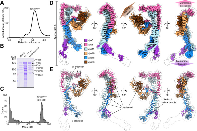
CORVET was produced in yeast cells and isolated via a FLAG tag on Vps8 using affinity chromatography followed by size exclusion chromatography (SEC) with high purity (Fig. 1A, B). The molecular weight of the purified complex measured by mass photometry (656 kDa) agreed with the predicted weight of 658 kDa (Fig. 1C).
Fig. 1. Purification and cryo-EM structure of the yeast CORVET complex.
A Size exclusion chromatography (SEC) of the affinity-purified CORVET. The representative result of three independent experiments is shown. B SDS-PAGE analysis of purified CORVET. Protein samples from affinity purification (eluate) and before and after SEC are shown. n = 3 independent experiments were performed. C Mass photometry analysis of the peak fractions from SEC of CORVET. D Overall architecture of the CORVET complex. Composite cryo-EM map generated from local refinement maps (see Figs. S2, S3) is colored by subunits assigned (Vps3, violet; Vps8, pink; Vps11, light blue; Vps16, sand; Vps18 blue; Vps33, brown). The consensus maps used for local refinements are low-pass-filtered and shown as a transparent envelope with a black outline. E Molecular model of CORVET fitted into the low-pass-filtered consensus map.
Negative stain electron microscopy (EM) revealed evenly distributed single particles of CORVET with no apparent aggregates in the sample (Supplementary Fig. 1A), allowing further single-particle cryo-EM analysis (Supplementary Fig. 1B, 2). From 33882 micrographs, 219391 selected particles provided a consensus map with 4.6 Å resolution. Local refinements further improved the resolution to 3.8 Å (SNARE-binding module, Supplementary Fig. 3, Supplementary Table 3). A composite map of all local refinements (Fig. 1D, Supplementary Fig. 2) enabled model building of the CORVET structure (Figs. 1E, 2, Supplementary Fig. 4, Supplementary Movie 1, Supplementary Table 3).
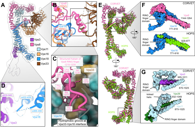
Fig. 2. Interactions of CORVET functional subunits with the core of the complex.
A Model of the CORVET complex viewed from the side in ribbon representation. The low-pass-filtered consensus map is shown by a black outline. The subunits are colored as in Fig. 1. B Zoomed-in view of the Vps33-Vps8-Vps16 interface with Vps8 and Vps18 RING finger domains indicated. C Zoomed-in view of the Vps8 structured hairpin sandwiched between Vps33 and Vps18. D Interactions between the α-solenoids of Vps18 and Vps3. Associated semi-transparent cryo-EM densities are zoned around the molecular models and colored accordingly. E Structural alignment of the CORVET molecular model (pink, this study) with the HOPS model (light green, PDB:7ZU0) viewed from two sides. F Close-up views of C-terminal two-helix bundle and RING finger domains of CORVET Vps8 and Vps18 (top) and HOPS Vps41 and Vps18 (bottom). G Close-up views of C-terminal two-helix bundle and RING finger domains of CORVET Vps3 and Vps11 (top) and HOPS Vps39 and Vps11 (bottom).
CORVET and HOPS share strong structural homology. Their respective cores, consisting of identical subunits (Vps11 and Vps18), are virtually indistinguishable from each other (Fig. 2E). A similar scenario is found for the SNARE-binding module (Vps33-Vps16) which branches off sideways from the core.
Reflecting their diverse functions within the endolysosomal system, those parts of the complexes that are responsible for target recognition show marked differences (Fig. 3). Vps3 and Vps8 are specific to CORVET and located at the distal ends of the complex (Fig. 1D, E). Each interlocks with the core through long C-terminal α-helices, reminiscent to the HOPS-specific Vps39 and Vps41 subunits (Figs. 1E, 2E–G, 3).
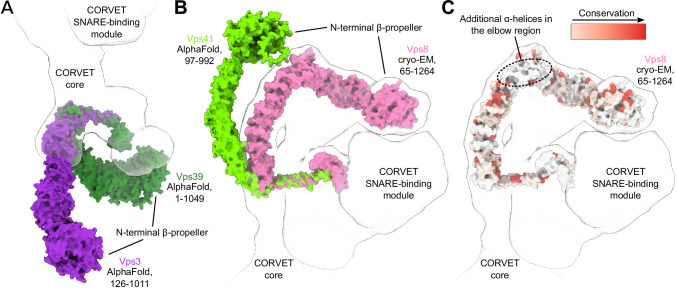
Fig. 3. Rab-binding subunits of CORVET compared to HOPS.
A Structural alignment of CORVET Vps3 (violet, AlphaFold prediction) with HOPS Vps39 (dark green, AlphaFold prediction). B Structural alignment of CORVET Vps8 (pink, cryo-EM structure from this study) with HOPS Vps41 (light green, AlphaFold prediction). C Vps8 subunit colored by conservation according to the sequence alignment with Vps41. In (A–C), the models of proteins are fitted into the semi-transparent volume (black outline) generated from the CORVET structure.
The extended Vps3 subunit is located at one end of the structure, but substantial flexibility in this region (Supplementary Fig. 5, Supplementary Movies 2–4) limited the achievable resolution. As such, the N-terminal part of Vps3, carrying the β-propeller, is only visible at lower thresholds (Fig. 1E, black outline). Similarly, to the analogous subunit Vps39 in HOPS, the C-terminal α-helix of Vps3 is anchored to CORVET’s core through an interaction with the RING finger domain and the long α-helix located at the C-terminus of Vps11 (Fig. 2E, G). In HOPS the RING finger domain of Vps11 tightly interacts with the C-terminal portion of Vps39 that follows the long α-helix. In contrast, the CORVET subunit Vps3 lacks any further C-terminal extensions after the long α-helix, thus significantly reducing the contact area in the Vps3-Vps11 interface. Furthermore, we observe a high flexibility of Vps3 and a more distant positioning from the complex core, when compared to HOPS (Figs. 1D, E, 3A). Interestingly, mutations in the Vps3-Vps11 interface can have profound impacts on the complex. As shown by mass spectrometry, CORVET variants carrying mutations in Vps11 (vps11-1, vps11-3)46 destabilize the complex (Supplementary Fig. 6A–F). In case of the vps11-3 allele the attachment of the Vps3 subunit to the core is abolished (Supplementary Fig. 6C, D), whereas the vps11-1 allele disassembles the entire complex (Supplementary Fig. 6E, F).
Vps8, the second Rab-binding subunit of CORVET, is located at the opposite end of the complex (Fig. 1D, E). Vps8 exhibits a characteristic elbow configuration, which turns its N-terminal half towards the β-propeller of Vps11. In the equivalent Vps41 subunit of HOPS, no such coordination is observed (Fig. 3B). In fact, Vps41 is curved in the opposing direction and exhibits higher flexibility than Vps845. Importantly, Vps8 carries 282 additional amino acids compared to Vps41. Sequence alignment revealed that the main portion of these residues are inserted as 8 additional α-helices into the elbow region of Vps8 (Fig. 3C).
The attachment of Vps8 to the core of the complex is similar to Vps3 and established through the long C-terminal α-helices and RING finger domains of the Vps8 and Vps18 subunits (Figs. 1E, 2A, E, F). This C-terminal helical bundle formed by Vps18 and Vps8 is followed by a pair of semi-symmetrically interacting RING finger domains (Fig. 2F, upper panel), reminiscent to the Vps11-Vps39 interface of HOPS (at the opposing end of the complex, compare Fig. 2 panels F and G). Mutations in this interface (vps18-1) disassemble the entire complex (Supplementary Fig. 6G, H), similar to the Vps3-Vps11 interface (Supplementary Fig. 6E, F). The C-terminus of Vps8 not only anchors the subunit to the core but also contributes to the attachment of the SNARE-binding module (Fig. 2A–C), analogous to Vps41 in HOPS. Distinctively, the binding interface between the core and the SNARE-binding module is larger in CORVET, which apparently increases stability. As in HOPS, Vps33 interacts with the RING finger domain of Vps18, and the N-terminal portion of Vps16 α-solenoid connects with the Vps8-Vps18 C-terminal long α-helical bundle (Fig. 2A–C, E). Moreover, a hairpin protruding from the Vps8 RING finger domain is sandwiched in a hydrophobic groove between Vps33 and Vps18 (Fig. 2B, C), clamping the SNARE-binding unit from both sides together with the C-terminus of Vps18 (Fig. 2A, F upper panel). A similar interface likely exists in human HOPS, where the analogous Vps41 subunit also has a RING finger domain, which is lacking in yeast Vps4124,47.
Structural and functional implications of Vps8 interactions with Vps11 β-propeller
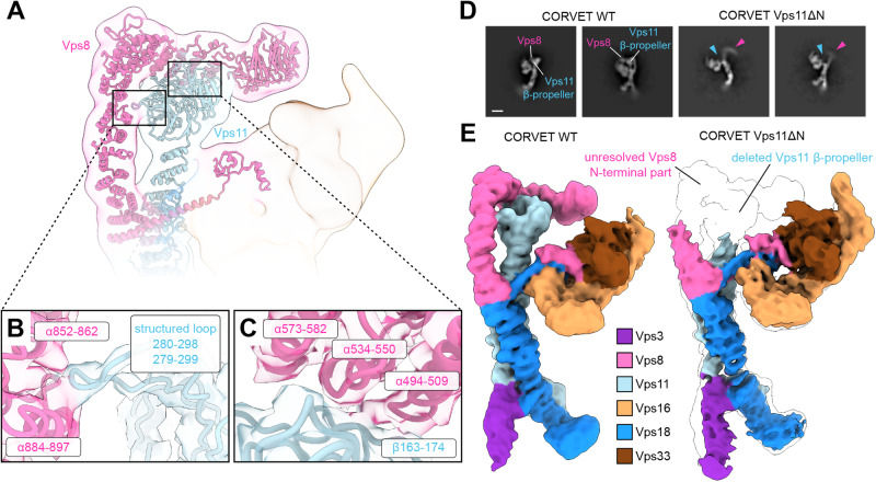
The hallmark of CORVET is the Vps8 subunit with its elbow shaped configuration. In addition to the C-terminal interaction with Vps18, and distinctive from HOPS, the α-solenoid of Vps8 has two additional interfaces with Vps11 (Figs. 1D, E, 4A). The first interface is between two α-helices of Vps8 (residues 852-862 and 884-897) and a structured loop at the Vps11 β-propeller (residues 279-299) (Fig. 4B), which stabilizes the upright section of the α-solenoid before the elbow. The second interface after the elbow connects three α-helices of Vps8 (residues 494-509, 534-550, 573-582) and the outward side of Vps11 β-propeller (residues 163-174) (Fig. 4C).
Fig. 4. CORVET-specific Vps8-Vps11 N-terminal interface.
A Molecular models of CORVET Vps8 and Vps11 in ribbon representation fitted into the semi-transparent low-pass-filtered consensus cryo-EM map colored by subunits as in Fig. 1. B, C Zoomed-in views of the interface sites between Vps8 and Vps11 with interacting structural elements indicated. Associated semi-transparent cryo-EM densities are zoned around the molecular models and colored accordingly. D Representative cryo-EM 2D class averages of CORVET wild-type and Vps11ΔN mutant. Structural differences in the regions of Vps8 and Vps11 N-termini are indicated. E Comparison of CORVET wild-type and Vps11ΔN mutant cryo-EM densities colored by subunits as in Fig. 1. The mutant cryo-EM density is overlayed on the wild-type density (transparent, black outline) highlighting the structural differences.
As these interfaces are distinct to CORVET, we investigated them closely. A mutant complex lacking the Vps11 β-propeller (CORVET Vps11ΔN, residues 1-335) could be purified like the wild-type complex (Supplementary Fig. 7A, C). Cryo-EM analysis indicates that this mutant not only lacks the β-propeller of Vps11, but shows poor resolution of N-terminal part of Vps8 (Fig. 4D, E). We attribute this to increased flexibility in Vps8 due to the missing interface with Vps11.
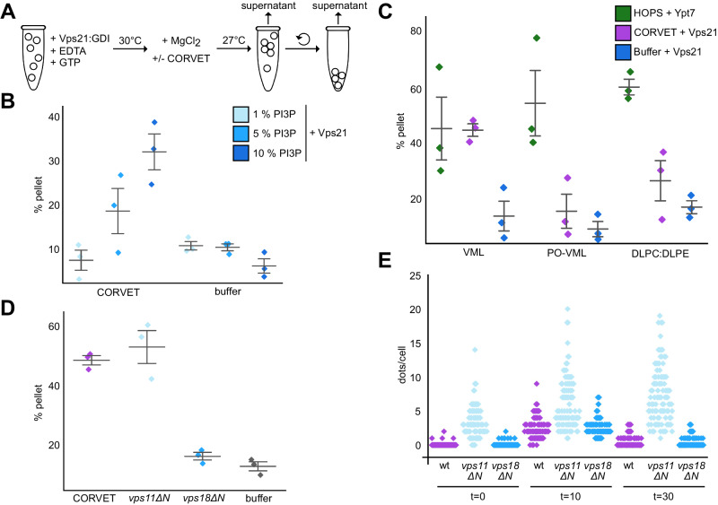
We next explored if the corresponding mutation in CORVET affects its function. For HOPS, reconstituted tethering and fusion assays have been established42,48,49, whereas comparable assays were missing so far for CORVET22,42,50. We thus set up a similar fluorescence-based tethering assay for CORVET (Fig. 5A) using liposomes loaded with Rab5(Vps21)-GTP45,51,52, including a screen for membrane conditions. Initially, we used vacuole mimicking lipid mix (VML) established for vacuole fusion assays53 for the generation of liposomes (see “Methods”). Due to the unsaturated C18:2 lipids in the phospholipids, the corresponding liposomes have a more fluid membrane with packaging defects. This analysis revealed that CORVET requires relatively high PI3P (10 mol%) concentrations for efficient tethering (Fig. 5B). To ask if membrane lipid packing defects contribute to efficient CORVET activity in tethering, we kept the VML composition for liposomes with the established PI3P concentration, but altered the acyl chain composition to palmitoyl (C16:0) oleolyl (C18:1) (PO) phospholipids (PO-VML), or used a more simple DL-phosphatidylcholine (PC) and DL-phosphatidylethanolamine (PE) mixture with the same PI3P concentration for liposomes (DLPC:DLPE). CORVET was only able to tether liposomes containing PI3P and the more fluid VML mixture, but none of the other liposomes (Fig. 5C). This contrasts to HOPS, where the GTP-loaded Rab7 GTPase Ypt7 is sufficient for tethering liposomes regardless of the composition (Fig. 5C). We thus conclude that CORVET requires multiple factors—PI3P, Vps21 and membrane packaging defects—to bind membranes.
Fig. 5. Functional analysis of CORVET-mediated membrane tethering.
A Scheme of the CORVET tethering assay (see “Methods” for details). B Tethering assay with phosphatidylinositol-3-phosphate (PI3P) titration. Prenylated Vps21 was incorporated in its GTP-loaded form into fluorescently labelled liposomes containing 1, 5, or 10 mol % PI3P. Liposomes were incubated with purified CORVET complex or a buffer control. Tethering activity was determined as described in materials and methods. Colored diamonds indicate single measurements. Bars indicate mean +/− 1 SE. Further tethering and statistics in Supplements. C Tethering assay with 1,2-dilinoleoyl-sn-glycero-3-phosphocholine (DLPC) and 1,2-dilinoleoyl-sn-glycero-3-phosphoethanolamine (DLPE) (DL-lipids) and palmitoyl/oleolyl (PO)-lipids. Liposomes containing 18:2/18:2 (DL-lipids) or 16:0/18:1 PO-lipids and L-α-phosphatidylinositol (SoyPI), PI3P, ergosterol and 1,2-dipalmitoyl-sn-glycero-3-(cytidine diphosphate) (DAG) or liposomes containing DLPC and DLPE and PI3P were fluorescently labelled. Prior to incubation with HOPS or CORVET liposomes were preloaded with prenylated and GTP-loaded Ypt7 or Vps21 out of the GDP dissociation inhibitor (GDI) complex. Activity was measured according to materials and methods. Bars indicate mean +/− 1 SE. Colored diamonds indicate single measurements. D Tethering assay with CORVET carrying N-terminally truncated Vps11 and Vps18 subunits. Fluorescently labelled liposomes, decorated with prenylated and GTP-loaded Vps21, were incubated with wild-type or mutated CORVET variants. Tethering activity was measured as described in materials and methods. Bars indicate mean +/− 1 SE. Colored diamonds indicate single measurements. For all tethering assays (B–D), tree (n = 3) independent measurements were conducted for every condition tested. E Quantification of Mup1 uptake assay. Mup1 was GFP-tagged in wild-type, or cells expressing truncated vps11ΔN or vps18ΔN. Cells were grown in synthetic medium lacking methionine and imaged by fluorescence microscopy (t = 0), before shifting to media containing methionine for 10 min (t = 10) and 30 min (t = 30). The number of GFP-positive dots per cell is plotted for each strain. Colored diamonds indicate single cells (n = 100 cells were used for every experimental condition).
We then compared wild-type CORVET to mutants lacking either the Vps11 or Vps18 β-propeller (Vps11∆N and Vps18∆N, respectively) in tethering assays. Surprisingly, the more flexible Vps11∆N complex was still functional in tethering on membranes with high PI3P, whereas the Vps18∆N was inactive (Fig. 5D). The Vps18∆N was fully assembled as it was purified as a hexamer like the wild type (Supplementary Fig. 7B), suggesting that the Vps18 β-propeller itself is critical for efficient tethering.
To determine if mutations in the β-propeller of Vps11 or Vps18 affect CORVET function, we used corresponding cells for possible endolysosomal defects (Supplementary Fig. 8). We initially analyzed vacuole morphology as CORVET deletion mutants have enlarged vacuoles54–56, but observed no difference to wild-type. However, cells expressing Vps11∆N grew slower on plates containing endolysosomal stressors like ZnCl2, indicating possible defects (Supplementary Fig. 8B). To analyze early endocytic trafficking, we followed the transport of the GFP-tagged methionine transporter Mup1 from the plasma membrane via endosomal dots to the vacuole (Fig. 5E, Supplementary Fig. 8C). Whereas Mup1 was efficiently transported to the vacuole in wild-type and Vps18∆N cells, it accumulated in GFP-positive dots over long periods in Vps11∆N cells, suggesting impaired endosomal transport toward the vacuole in this mutant in vivo.
Interaction of CORVET with membranes
To analyze how CORVET interacts with membranes, we first used in silico prediction of interactions of CORVET’s subunits Vps8 or Vps3 with the Rab5 GTPase Vps21. Our AlphaFold modeling57 of the Vps3-Vps21 and Vps8-Vps21 interactions (Supplementary Fig. 9A–F) suggested binding sites on the peripheral areas of Vps8 and Vps3 α-solenoids, which would be accessible by membrane-bound Vps21 via its 10 nm long hypervariable domain (not shown in the prediction). The predicted interaction mode is similar to that in the HOPS complex45, involving analogous conserved residues localized to the switch regions in the Rab GTPases (Supplementary Fig. 9A–G), which are known to mediate Rab-effector binding12,58.
To visualize CORVET on membranes, we incubated the complex with PI3P-containing liposomes loaded with Vps21-GTP and analyzed these by negative-stain microscopy. We observed CORVET particles coating the vesicle surfaces, mostly in an upright position (Supplementary Fig. 9H). Some particles also appeared flat on the membranes, possibly via Rab binding of both membrane-binding subunits in CORVET. The liposomes incubated without CORVET were indeed free of any decoration (Supplementary Fig. 9I). We conclude that CORVET is like HOPS on membranes preferentially upright prior to tethering.
Discussion
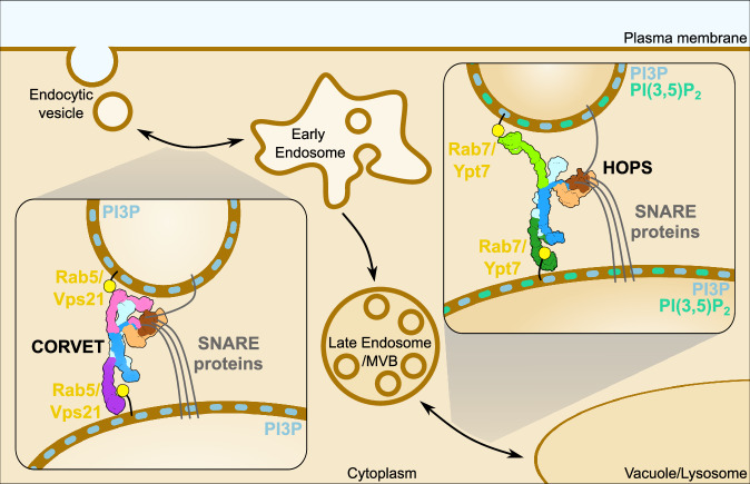
In this study, we solved the structure of the crucial metazoan endolysosomal tethering complex CORVET. We show that CORVET and HOPS have modular architecture sharing an identical core with the attached SNARE-binding module and distinct complex-specific membrane-binding subunits. Importantly, we uncovered that CORVET differs strongly from HOPS in that it requires (i) PI3P, (ii) membrane packaging defects, and (iii) Rab5 (Vps21) for its efficient tethering function. Our data suggest that tethering complexes thus read out their membrane environment at several levels to gain organelle-specificity (Fig. 6).
Fig. 6. Mechanistic model of membrane tethering by CORVET and HOPS within the endolysosomal pathway.
CORVET is responsible for early-endosomal fusion events and selectively relies on the Rab5 (Vps21) and phosphatidylinositol-3-phosphate (PI3P) presence in the membranes for function. In contrast, the lysosomal tether HOPS requires Rab7 (Ypt7) and, potentially, different lipid composition (e.g. phosphatidylinositol 3,5-bisphosphate (PI(3,5)P2)) to promote membrane fusion. The cartoon, including the SNARE proteins, Rab GTPases, and membranes, is not drawn to scale.
As in HOPS, subunit interactions in CORVET rely on the presence of C-terminal long α-helices and RING finger domains. As CORVET Vps3 misses a C-terminal RING finger domain, its association is weaker than Vps39 binding in HOPS, at least in solution22,28. This is confirmed by our analysis of the Vps11-specific mutant alleles, which trigger CORVET disassembly in vivo (Supplementary Fig. 6C–F). It has been shown that RING finger domains of HOPS and CORVET subunits have ubiquitin ligase activity59. As RING finger domains are structural elements of HOPS and CORVET, they may function as quality control elements to monitor the assembly of the complex. This could also explain, why an intermediate complex between HOPS and CORVET exists, which maintains the core part and can swap functional subunits such as Vps41/Vps8 or Vps39/Vps3, while the free subunit may fulfill auxiliary functions22,60. When Vps8 alone is overproduced, cells accumulate MVBs proximal to the vacuole27, suggesting that Vps8 can constitute a minimal tether. Interestingly, a minimal CORVET complex, lacking Vps11 as a central subunit, was proposed in Drosophila61, which is, however, incompatible with the identified Vps11-Vps18 core of CORVET and HOPS. It is thus not yet clear if miniCORVET is indeed a minimal CORVET or may have additional subunits.
One striking difference between CORVET and HOPS is the rigid apposition of Vps8 to the Vps11 β-propeller. This tight interaction can explain, why overproduced Vps8 in Drosophila can outcompete Vps41 and thus prevent HOPS assembly as it will likely sequester the available Vps11 pool62. The Vps8 apposition to the core and the Vps8-Vps11 contacts appear to be important for full CORVET activity in early endocytic transport in vivo. Interestingly, a reported point mutation in the α-solenoid of Vps8 might cause a destabilizing of the Vps8-Vps11 interaction29. We demonstrate this by our structural (Fig. 4) and functional (Fig. 5) analyses of CORVET Vps11∆N. Indeed, a deletion of the β-propeller domain of core subunits such as Vps11 and Vps18 will affect both CORVET and HOPS in vivo. In this regard, we were surprised that the CORVET Vps18∆N mutant, in contrast to Vps11∆N, was largely inactive in the tethering assay (Fig. 5D), whereas the comparable HOPS complex was functional45. This suggests that Vps18 either contributes to membrane recognition by Vps3 within CORVET or to the structural integrity of the complex. Curiously, in contrast to the in vitro results, introduction of the vps18∆N mutation did not have an effect in vivo, while the vps11∆N mutation caused significant endosomal defects (Fig. 5E, Supplementary Fig. 8). This difference may be due to the fact that in vivo, the vps11ΔN mutation impacts not only CORVET but also HOPS, while the vps18ΔN mutant may be complemented by additional cellular factors not included in our in vitro assays. Further precise analyses, such as introducing point mutations into specific subunits of CORVET and HOPS, and establishing an in vitro fusion assay for CORVET, will help to better explain the discrepancies between the in vitro and in vivo situations, as well as the specific functional differences between HOPS and CORVET. In either case, we can only speculate, how membranes are decoded by CORVET and how the β-propeller domains of Vps11 and Vps18 contribute. One idea is that the β-propeller of the core subunits Vps11 and Vps18 bind Rab5 GTPases together with the Rab5-GTP specific subunits Vps3 and Vps8, thus optimally positioning them for direct PI3P and membrane binding.
We show that CORVET depends on multiple membrane cues, whereas HOPS can tether any membrane carrying the Rab GTPase Ypt7. For membrane binding, CORVET may thus require specific lipids and membrane packaging defects, either through high curvature or due to the membrane composition6,63, in addition to Rab5, which is needed for CORVET localization in vivo55. In line with this, predicted Rab binding sites of CORVET and HOPS are largely conserved and located at the distal exposed ends of the complexes. However, Rab5 also binds and stimulates the Vps34 PI-3-kinase complex, which establishes a suitable membrane environment required for CORVET recognition64,65. Consequently, loss of Rab5 (or its homolog in yeast) will possibly affect both the CORVET binding and the specific membrane environment.
Currently, we have no clear understanding, how tethering complexes function on membranes to promote tethering and fusion. Our analyses of HOPS suggest that the complex is positioned upright on membranes when bound to the Rab7-like Ypt7 protein66. Here, we used liposomes and find CORVET also mostly upright on membranes (Supplementary Fig. 9H). This suggests that direct interactions with the lipid bilayer strongly contribute to the positioning of CORVET, making it ready for tethering and subsequent promotion of SNARE-mediated fusion, while Rab GTPases may further stabilize the complex.
Our analysis of CORVET reveals that its core architecture largely resembles HOPS, yet the orientation and structure of the Rab-specific subunits and their decoding of membranes strongly differs between both complexes. Nevertheless, though interchangeable assemblies of CORVET and HOPS are possible, the four shared central subunits, Vps11, 18, 33 and 16, are found in all species67. We are thus convinced that the overall arrangement and function of HOPS and CORVET as a modular membrane fusion machinery is evolutionary conserved. Future studies will take advantage of our structural insights to determine, how these complexes promote SNARE-mediated membrane fusion.
Methods
Yeast strains
Yeast strains and oligonucleotides used in this study are listed in Supplementary Table 1 and Supplementary Table 2 respectively. In general, CORVET subunits were expressed under the control of the GAL1 promoter according to the standard protocol68. For subunit truncation (Vps11 or Vps18) of the N-terminal part, the GAL1 promotor was inserted into the genome at the respective position. The 3x-FLAG Tag was attached to the CORVET subunit Vps8, except for the Vps18ΔN mutant.
Protein expression and purification from Escherichia coli
Rab GTPases for tethering assays were expressed in Escherichia coli BL21 (DE3) Rosetta cells. Cultures were grown in Luria broth (LB) medium complemented with 35 µg/ml kanamycin or 100 µg/ml ampicillin and 30 µg/ml chloramphenicol. Cultures were induced with 0.5 mM Isopropyl-β-D-thiogalactoside (IPTG) and incubated overnight at 16 °C. Cells were harvested by centrifugation (4800 × g, 10 min, 4 °C) and resuspended in buffer (150 mM NaCl, 50 mM HEPES/NaOH, pH 7.4, 10% glycerol, 1 mM PMSF, and 0.5-fold protease inhibitor mixture [PIC]) prior to lysis in a Microfluidizer (Microfluidics Inc). Crude lysates were centrifugated at 25,000 × g, 30 min, 4 °C. Supernatants were incubated with glutathione Sepharose (GSH) fast flow beads (GE Healthcare) for GST-tagged proteins or nickel–nitriloacetic acid (Ni-NTA) agarose (Qiagen) for His-tagged proteins (2 h, 4 °C). Proteins were eluted with buffer (150 mM NaCl, 50 mM HEPES/NaOH, pH 7.4, 10% glycerol) containing either 25 mM glutathione or 300 mM imidazole. Buffer was exchanged via a PD10 column (GE Healthcare). For tag cleavage, TEV or PreScission protease was added after washing and incubated overnight. Proteins were stored at −80 °C.
Purification of 3xFLAG-tagged CORVET complex variants
CORVET tethering complex variants were purified according to the standard FLAG-purification protocol as previously described45 with minor changes. Two liters medium (yeast peptone (YP), containing 2% galactose (v/v)) were inoculated with 6 ml of an overnight preculture. Cultures were grown for 24 h and harvested by centrifugation (4800 × g, 10 min, 4 °C). Cell pellets were washed with cold CORVET purification buffer (CPB, 300 mM NaCl, 20 mM HEPES/NaOH, pH 7.4, 1.5 mM MgCl2, and 10% (v/v) glycerol). Pellets were resuspended in CORVET lysis buffer (CLB, CPB supplemented with 1 mM phenylmethylsulfonylfluoride (PMSF), 1× FY protease inhibitor mix (Serva) and 1 mM dithiothreitol (AppliChem GmbH)). Cell suspension was dropwise frozen in liquid nitrogen before lysis in a freezer mill cooled with liquid nitrogen (SPEX SamplePrep LLC). For purification, the powder was thawed on ice and resuspended in CLB, followed by two steps of centrifugation at 5000 and 15,000 × g at 4 °C for 10 and 20 min. The supernatant was combined with anti-FLAG M2 affinity gel (Sigma–Aldrich) and placed on a nutator for 45 min at 4 °C. Beads were centrifugated (500 × g, 1 min, 4 °C) and transferred to a 2.5 ml MoBiCol column (MoBiTec). Samples were washed with 25 ml CPB before FLAG-peptide was added, followed by incubation on a turning wheel for 40 min at 4 °C. The eluate was concentrated in a Vivaspin 100 kDa MWCO concentrator (Sartorius), which was incubated with CPB containing 1% TX-100. Concentrated sample was applied to a Superose 6 Increase 15/150 column (Cytiva) for size exclusion chromatography (SEC). Fractions were eluted in 50 μl using ÄKTA go purification system (Cytiva). Peak fraction was used for further analysis.
Mass photometry analysis
Mass photometry experiments were performed using a Refeyn TwoMP (Refeyn Ltd). Data were acquired using AcquireMP software and analyzed using DiscoverMP v2023 (both Refeyn Ltd). High Precision Cover Glasses (Marienfeld) were used for sample analysis. Perforated silicone gaskets were placed on the coverslips to form wells for every sample to be measured. Samples were evaluated at a final concentration of 10 nM in a total volume of 20 μl in the buffer used for SEC. Calibration was performed using β-amylase (Carl Roth).
ALFA pulldowns for mass spectrometry
Vps8-ALFA pulldowns were performed in the same way as previously described45. One liter YP medium containing 2% glucose (v/v) was inoculated with an overnight preculture. Cells were grown to OD600 1 at 26 °C, followed by 1 h heat shock at 38 °C. Cells were harvested by centrifugation (4800 × g for 10 min at 4 °C). Pellets were washed with cold Pulldown buffer (PB, 150 mM KAc, 20 mM HEPES/NaOH, pH 7.4, 5% (v/v) glycerol and 25 mM CHAPS). Cells were resuspended in a 1:1 ratio (w/v) in PB supplemented with Complete Protease Inhibitor Cocktail (Roche) and afterward dropwise frozen in liquid nitrogen before lysis in 6875D LARGE FREEZER/MILL (SPEX SamplePrep LLC). Powder was thawed on ice and resuspended in PB, followed by two centrifugation steps at 5000 and 15,000 × g at 4 °C for 10 and 20 min. Supernatant was added to 12.5 µl prewashed ALFA Selector ST beads (2500 × g, 2 min, 4 °C) (NanoTag Biotechnologies) and incubated for 15 min at 4 °C while rotating on a turning wheel. Afterwards, beads were washed two times in PB and four times in PB without CHAPS. Samples were digested and prepared according to the PreOmics ST 96x Kit (iST Kit, preomics), using LysC as protease. Dried samples were resuspended in 10 µl LC-Load. 1 µl of the final sample was analysed in reversed-phase chromatography, performed on a Thermo Ultimate 3000 RSLCnano system, connected to a Q ExactivePlus mass spectrometer (Thermo Fisher Scientific) through a nano-electrospray ion source. 50 cm PepMap C18 easy spray columns (Thermo Fisher Scientific) with an inner diameter of 75 μm were used and kept at 40 °C. Peptides were eluted with a linear gradient of acetonitrile from 10 to 35% in 0.1% formic acid for 118 min at a constant flow rate of 300 nl/min, which was followed by electrospraying into the mass spectrometer. The mass spectra were acquired on the Q-Exactive Plus. The maximum injection time was set to 50 ms, the target value to 3,000,000 at a resolution of 70,000 at m/z = 200. From this, the ten most intense multiply charged ions (z = 2) from the survey scan were selected with an isolation width of 1.6 Th as well as fragments with higher energy collision dissociation with normalized collision energies of 2769. Target values for MS/MS were set at 100,000, with maximum injection time of 80 ms at a resolution of 17,500 at m/z = 200. The dynamic exclusion of the overall sequenced peptides was set at 20 s to avoid repetitive sequencing. The resulting MS spectra were analyzed using MaxQuant (v2.0.3.0, www.maxquant.org/)70,71. Data were analyzed using Persus (v2.0.11, www.maxquant.org/persus)72. Significances in the volcano plot of the Perseus software package corresponds to a given FDR which was determined by a permutation-based method73.
Negative stain analysis
Samples of wild-type CORVET, mutant CORVET variants, and CORVET incubated with liposomes were examined by negative-stain electron microscopy. 3 μl of sample at a protein concentration of approximately 0.05 mg/ml was applied onto freshly glow-discharged carbon-coated copper grids with plastic support, blotted from the side and stained using 2% (w/v) uranyl formate solution as previously described74. Negative-stain micrographs were collected manually on a JEM-2100Plus transmission electron microscope (JEOL) operated at 200 kV and equipped with a XAROSA CMOS 20-megapixel camera (EMSIS) at a nominal magnification of 30,000 (3.12 Å per pixel). The data was analyzed using ImageJ v1.52k75 and cryoSPARC v3 and v476.
Cryo-EM sample preparation and data acquisition
For cryo-EM, 3 μL of freshly purified CORVET samples at a protein concentration of approximately 0.8 mg/ml were applied to glow-discharged C-Flat grids (R1.2/1.3 3Cu-50) (EMS) and immediately plunge frozen in liquid ethane using a Vitrobot Mark IV (Thermo Fisher Scientific) with the environmental chamber set at 100% humidity and 4 °C.
Micrographs were collected automatically with EPU v3.7.1. (Thermo Fisher Scientific), using a Glacios cryo-transmission electron microscope (Thermo Fisher Scientific) operated at 200 kV and equipped with a Selectris energy filter and a Falcon 4 detector (both Thermo Fisher Scientific). Data were recorded in Electron Event Representation (EER) mode at a nominal magnification of 130,000 (0.924 Å per pixel) in the defocus range of −0.8 to −1.8 μm with an exposure time of 7.50 s resulting in a total electron dose of approximately 50 e− Å−2.
Cryo-EM image processing
All cryo-EM data were preprocessed in cryoSPARC Live, and further processing was performed in cryoSPARC v3 and v476 (Supplementary Fig. 2). For all collected movies, motion correction (EER upsampling factor 1, EER number of fractions 40) and contrast transfer function (CTF) estimation were performed using cryoSPARC Live implementations. After micrograph curation, 33,882 micrographs were included for further data analysis.
For the determination of the wild-type structure, all collected datasets were preprocessed in a similar manner and combined at the final steps of image processing (Supplementary Fig. 2). Briefly, particles were selected by the template picker implemented in CryoSPARC Live, using well-defined 2D classes obtained from preliminary CORVET datasets as templates, and additional particle picking was performed using the Topaz wrapper77. For particle picking, micrographs from sessions 1–5 were combined, while the micrographs from sessions 6 and 7 were used separately. After particle duplicate removal and particle extraction in a box size of 882 pixels (Fourier-cropped to 128 pixels, resulting in a pixel size of 6.37 Å per pixel), two rounds of 2D classification were performed for all particle stacks obtained from each of the picking jobs to eliminate bad picks (Supplementary Fig. 2). After 2D classifications, duplicate particles were removed again and a round of ab-initio reconstruction with 5 classes followed by heterogeneous refinement was performed for each stack of particles resulted from sessions 1–5, session 6, and session 7. Each of these three heterogeneous refinement jobs produced one best class revealing all of the subunits of CORVET in the map. Such a class resulted from session 7 was further refined individually using NU refinement78 and particles from it (158,046 particles) were afterwards combined with the particles from the best classes of the heterogeneous refinement jobs from sessions 1-5 (93661 particles) and 6 (216338 particles). A round of heterogeneous refinement with three classes was performed using these combined particles in a box size of 882 pixels (Fourier-cropped to 224 pixels, pixel size 3.64 Å per pixel), which resulted in two good classes reaching resolution of 7.4 Å (Gold Standard Fourier Shell Correlation (GSFSC) value of 0.143). These two classes (174,263 and 198,464 particles) were combined and further refined using NU refinement in a box size of 882 pixels (Fourier-cropped to 400 pixels, pixel size 2.04 Å per pixel). The obtained reconstruction resolved to 4.6 Å (GSFSC = 0.143) was further classified using a round of heterogeneous refinement with 5 classes. The best class that reached resolution of 6.7 Å (GSFSC = 0.143) was used for a NU refinement providing a consensus map (219391 particles) at 4.6 Å resolution (GSFSC = 0.143). This consensus map was further used for local refinements of distinct areas of the structure. For local refinements, five masks were generated using UCSF ChimeraX 1.7.179 and particles were again re-extracted for some of the parts of the structure in a box size of 882 pixels (Fourier-cropped to 512 pixels, pixel size 1.59 Å per pixel) (Supplementary Fig. 2). A composite map was generated from the local refinement maps using the “volume maximum” command in UCSF ChimeraX.
All maps were subjected to unsupervised B-factor sharpening within cryoSPARC. No symmetry was applied during processing. The quality of the consensus and local refinement maps is shown in Figs. S3 and S4. All GSFSC curves and angular distribution plots were generated within cryoSPARC (Supplementary Fig. 3). The local resolutions of the consensus and local refinement maps (Supplementary Fig. 3) were estimated in cryoSPARC and analyzed in UCSF ChimeraX. Dataset statistics are provided in Supplementary Table 3. The cryo-EM analysis of the CORVET Vps11ΔN mutant was performed similarly to the wild type (Supplementary Fig. 7D).
Model building and refinement
The structures of Vps11, Vps16, Vps18, and Vps33 from the structure of the HOPS complex (PDB: 7ZU0) and the AlphaFold structure predictions of Vps8 and Vps3 (Uniprot: P39702/P23643 respectively) were manually fitted into the consensus and local-refinement maps using the “Fit in Map” tool in UCSF ChimeraX and used as a starting model. Most of the structure was modelled as poly-alanine sequences using the PDB Tools job in PHENIX 1.2080, except for the regions of the structure with the highest resolution obtained (SNARE-binding module, Vps8 α-solenoid, N-terminal half of Vps11). In addition, several protein fragments without well resolved corresponding EM densities were removed from the model (including the N-terminal half of Vps3). The models of the CORVET subunits were then manually adjusted and refined in Coot81 and combined into a single model of the full complex. Subsequently, iterative rounds of real space refinement82 of the model against the composite map of CORVET in PHENIX were performed, followed by manual adjustments in Coot 0.9.6. Model validation was done using MolProbity83 in PHENIX. Models and maps were visualized, and figures were prepared in UCSF ChimeraX and Inkscape v1.3. Model refinement and validation statistics are provided in Supplementary Table 3.
Liposome preparation
For the preparation of liposomes, the following lipids were used. 1,2-dilinoleoyl-sn-glycero-3-phosphocholine (18:2 PC), 1-palmitoyl-2-oleoyl-glycero-3-phosphocholine (POPC) 1,2-dilinoleoyl-sn-glycero-3-phosphoethanolamine (18:2 PE), 1-palmitoyl-2-oleoyl-sn-glycero-3-phosphoethanolamine (POPE), L-α-phosphatidylinositol (SoyPI), 1,2-dilinoleoyl-sn-glycero-3-phospho-L-serine (18:2 PS), 1-palmitoyl-2-oleoyl-sn-glycero-3-phospho-L-serine (POPS), 1,2-dilinoleoyl-sn-glycero-3-phosphate (18:2 PA), 1-palmitoyl-2-oleoyl-sn-glycero-3-phosphate (POPA), 1,2-dipalmitoyl-sn-glycero-3-(cytidine diphosphate) (DAG) were purchased from Avanti Polar Lipids (Alabama, USA). Phosphatidylinositol 3-phosphate (PI3P) was purchased from Echelon Biosciences Inc. ATTO488-1,2-dipalmitoyl-sn-glycero-3-phosphoethanolamine (ATTO488) was obtained from ATTO-TEC GmbH. Ergosterol was purchased from Sigma Aldrich.
For tethering assays and negative stain, lipid films, containing 37.6 or 42.6 mol% 18:2 PC/ POPC, 18 mol% 18:2 PE/ POPE, 18 mol% SoyPI, 10 or 5 mol% PI(3)P, 4.4 mol% 18:2 PS/ POPS, 2 mol% 18:2 PA/ POPA, 8 mol% Ergosterol, 1 mol% DAG and 1 mol% ATTO488, were evaporated by a SpeedVac (CHRIST). Lipid films prepared for the negative stain EM analysis did not contain dye and the 18:2 PC content was raised accordingly. Lipid films were resuspended in buffer (25 mM HEPES/NaOH, pH 7.4, 135 mM NaCl). Unilamellar vesicles were obtained through seven freeze and thaw cycles in liquid nitrogen. Liposomes were extruded through polycarbonate filters to 100 nm for tethering assays and 30 nm for negative stain EM analysis (400, 200, 100, 50 and 30 nm pore size) using a hand extruder (Avanti Polar Lipids, Inc.).
Tethering assay
CORVET-mediated tethering assays were done as before42. ATTO488 labelled liposomes were prepared and loaded with prenylated Vps21 or Ypt752. 100 nmole liposomes were incubated with 100 pmole pVps21:GDI/Ypt7:GDI in the presence of 1 mM GTP and 20 mM EDTA for 30 min at 30 °C, before the addition of 25 mM MgCl2. 100 nM CORVET/HOPS or buffer (HEPES/NaOH, pH 7.4, 300 mM NaCl, 1.5 mM MgCl2) were incubated with 0.17 mM Rab-loaded liposomes for 10 min at 27 °C. Liposomes were sedimented by centrifugation at 2500 × g for 5 min. The tethered liposomes in the pellet fraction were determined by comparing the ATTO488 fluorescent signal of the supernatant before and after centrifugation using a SpectraMax M3 fluorescence plate reader (Molecular Devices). Statistical analyses were performed with Origin 2023 v10.0.
Imaging of yeast cells using fluorescence microscopy
Cells were grown to the exponential phase in synthetic complete medium supplemented with all amino acids (SDC), or lacking methionine (SDS-met) [0.675% (w/v) yeast nitrogen base without amino acids, 2.0% (w/v) glucose, 0.075% (w/v) CSM (MPBiomedicals)]. Cells were imaged in SDC or SDC-met using the Zeiss Axioscope 5, with a PLAN-Apochromat 100x/1.40 Oil DIC M27 objective and an axiocam 702 mono (1.0x) camera.
All images were processed and quantified using ImageJ. Statistical analyses were performed with Origin 2023 v10.0 software. Vacuoles were stained using 30 µM FM4-64 (Molecular Probes Inc., Eugene, OR) for 30 min. Cells were washed twice with medium, prior to 1 h incubation at 30 °C84.
Reporting summary
Further information on research design is available in the Nature Portfolio Reporting Summary linked to this article.
Supplementary information
Description of Additional Supplementary Files
Source data
Acknowledgements
The authors thank Jannis Schoppe for help with the initial purifications of the CORVET sample, Dovile Januliene for help with cryo-EM experiments, Kilian Schnelle for help with computing and data processing, and all members of the Ungermann and Moeller lab for feedback. This work was supported by a grant of the DFG (UN111/5-6; MO2752/3-6), the SFB 944 and SFB 1557 (C.U.; F.F., A.M.), the DFG INST190/196-1 FUGG (A.M.) and the BMBF/DLR 01ED2010 (A.M.).
Author contributions
D.S., C.K. and N.S. conceived and designed all experiments together with A.M. and C.U. D.S. conducted all cryo-EM analyses. C.K. and N.S. did all biochemical and cell biology. experiments. F.F. performed mass spectrometry analysis with support of S.W. A.P. gave technical support on biochemical and cell biology experiments. L.L. gave conceptional and technical advice on biochemical and cell biology experiments. The manuscript was written by D.S., C.U. and A.M. with contributions from all authors.
Peer review
Peer review information
Nature Communications thanks the anonymous reviewers for their contribution to the peer review of this work. A peer review file is available.
Funding
Open Access funding enabled and organized by Projekt DEAL.
Data availability
The data that support this study are available from the corresponding authors upon request. The cryo-EM maps have been deposited in the Electron Microscopy Data Bank (EMDB) under accession codes EMD-18701 (CORVET composite map); EMD-18702 (CORVET Vps8-Vps11 local refinement map); EMD-18703 (CORVET Vps8 β-propeller local refinement map); EMD-18704 (CORVET SNARE binding module local refinement map); EMD-18705 (CORVET core local refinement map); EMD-18706 (CORVET Vps18 β-propeller local refinement map); EMD-18707 (CORVET consensus map); EMD-18708 (CORVET Vps11ΔN mutant map). The atomic coordinates have been deposited in the Protein Data Bank (PDB) under accession code 8QX8 (CORVET tethering complex). The previously published structure of HOPS tethering complex with the accession code 7ZU0 has been used in this study. The source data underlying Figs. 1a–c, 5b–e and Supplementary Figs. 6b–h, 7a–c, 8 are provided as a Source Data file. Source data are provided with this paper.
Competing interests
The authors declare no competing interests.
Footnotes
Publisher’s note Springer Nature remains neutral with regard to jurisdictional claims in published maps and institutional affiliations.
These authors contributed equally: Dmitry Shvarev, Caroline König, Nicole Susan.
Change history
10/10/2024
A Correction to this paper has been published: 10.1038/s41467-024-53199-0
Contributor Information
Christian Ungermann, Email: cu@uos.de.
Arne Moeller, Email: arne.moeller@uos.de.
Supplementary information
The online version contains supplementary material available at 10.1038/s41467-024-49137-9.
References
- 1.Rout, M. P. & Field, M. C. The evolution of organellar coat complexes and organization of the eukaryotic cell. Annu. Rev. Biochem.86, 637–657 (2017). [DOI] [PubMed] [Google Scholar]
- 2.Gomez-Navarro, N. & Miller, E. A. COP-coated vesicles. Curr. Biol.26, R54–R57 (2016). [DOI] [PubMed] [Google Scholar]
- 3.Borchers, A.-C., Langemeyer, L. & Ungermann, C. Who’s in control? Principles of Rab GTPase activation in endolysosomal membrane trafficking and beyond. J. Cell Biol.220, e202105120 (2021). [DOI] [PMC free article] [PubMed] [Google Scholar]
- 4.Posor, Y., Jang, W. & Haucke, V. Phosphoinositides as membrane organizers. Nat. Rev. Mol. Cell Biol.23, 797–816 (2022). [DOI] [PMC free article] [PubMed] [Google Scholar]
- 5.Goody, R. S., Müller, M. P. & Wu, Y.-W. Mechanisms of action of Rab proteins, key regulators of intracellular vesicular transport. Biol. Chem.398, 565–575 (2017). [DOI] [PubMed] [Google Scholar]
- 6.Bigay, J. & Antonny, B. Curvature, lipid packing, and electrostatics of membrane organelles: defining cellular territories in determining specificity. Dev. Cell23, 886–895 (2012). [DOI] [PubMed] [Google Scholar]
- 7.Herrmann, E., Langemeyer, L., Auffarth, K., Ungermann, C. & Kümmel, D. Targeting of the Mon1-Ccz1 Rab guanine nucleotide exchange factor to distinct organelles by a synergistic protein and lipid code. J. Biol. Chem.299, 102915 (2023). [DOI] [PMC free article] [PubMed] [Google Scholar]
- 8.Wilmes, S. & Kümmel, D. Insights into the role of the membranes in Rab GTPase regulation. Curr. Opin. Cell Biol.83, 102177 (2023). [DOI] [PubMed] [Google Scholar]
- 9.Singer-Krüger, B. et al. Role of three rab5-like GTPases, Ypt51p, Ypt52p, and Ypt53p, in the endocytic and vacuolar protein sorting pathways of yeast. J. Cell Biol.125, 283–298 (1994). [DOI] [PMC free article] [PubMed] [Google Scholar]
- 10.Wichmann, H., Hengst, L. & Gallwitz, D. Endocytosis in yeast: evidence for the involvement of a small GTP-binding protein (Ypt7p). Cell71, 1131–1142 (1992). [DOI] [PubMed] [Google Scholar]
- 11.Wandinger-Ness, A. & Zerial, M. Rab proteins and the compartmentalization of the endosomal system. Cold Spring Harb. Persp. Biol.6, a022616–a022616 (2014). [DOI] [PMC free article] [PubMed] [Google Scholar]
- 12.Barr, F. A. Rab GTPases and membrane identity: causal or inconsequential? J. Cell Biol.202, 191–199 (2013). [DOI] [PMC free article] [PubMed] [Google Scholar]
- 13.Hutagalung, A. H. & Novick, P. J. Role of Rab GTPases in membrane traffic and cell physiology. Physiol. Rev.91, 119–149 (2011). [DOI] [PMC free article] [PubMed] [Google Scholar]
- 14.Ungermann, C. & Kümmel, D. Structure of membrane tethers and their role in fusion. Traffic20, 479–490 (2019). [DOI] [PubMed] [Google Scholar]
- 15.Spang, A. Membrane tethering complexes in the endosomal system. Front. Cell Dev. Biol.4, 35 (2016). [DOI] [PMC free article] [PubMed] [Google Scholar]
- 16.Beek, J., van der, Jonker, C., Welle, R., van der, Liv, N. & Klumperman, J. CORVET, CHEVI and HOPS – multisubunit tethers of the endo-lysosomal system in health and disease. J. Cell Sci.132, jcs189134 (2019). [DOI] [PubMed] [Google Scholar]
- 17.Zhang, Y. & Hughson, F. M. Chaperoning SNARE folding and assembly. Annu. Rev. Biochem.90, 1–23 (2021). [DOI] [PMC free article] [PubMed] [Google Scholar]
- 18.Wickner, W. & Rizo, J. A cascade of multiple proteins and lipids catalyzes membrane fusion. Mol. Biol. Cell28, 707–711 (2017). [DOI] [PMC free article] [PubMed] [Google Scholar]
- 19.Risselada, H. J. & Mayer, A. SNAREs, tethers and SM proteins: how to overcome the final barriers to membrane fusion? Biochem. J.477, 243–258 (2020). [DOI] [PubMed] [Google Scholar]
- 20.Seals, D. F., Eitzen, G., Margolis, N., Wickner, W. T. & Price, A. A Ypt/Rab effector complex containing the Sec1 homolog Vps33p is required for homotypic vacuole fusion. Proc. Natl Acad. Sci. USA97, 9402–9407 (2000). [DOI] [PMC free article] [PubMed] [Google Scholar]
- 21.Wurmser, A. E., Sato, T. K. & Emr, S. D. New component of the vacuolar class C-Vps complex couples nucleotide exchange on the Ypt7 Gtpase to snare-dependent docking and fusion. J. Cell Biol.151, 551–562 (2000). [DOI] [PMC free article] [PubMed] [Google Scholar]
- 22.Peplowska, K., Markgraf, D. F., Ostrowicz, C. W., Bange, G. & Ungermann, C. The CORVET tethering complex interacts with the yeast Rab5 Homolog Vps21 and is involved in endo-lysosomal biogenesis. Dev. Cell12, 739–750 (2007). [DOI] [PubMed] [Google Scholar]
- 23.Perini, E. D., Schaefer, R., Stöter, M., Kalaidzidis, Y. & Zerial, M. Mammalian CORVET is required for fusion and conversion of distinct early endosome subpopulations. Traffic15, 1366–1389 (2014). [DOI] [PubMed] [Google Scholar]
- 24.van der Kant, R. et al. Characterization of the mammalian CORVET and HOPS complexes and their modular restructuring for endosome specificity. J. Biol. Chem.290, 30280–30290 (2015). [DOI] [PMC free article] [PubMed] [Google Scholar]
- 25.Baker, R. W. et al. A direct role for the Sec1/Munc18-family protein Vps33 as a template for SNARE assembly. Science349, 1111–1114 (2015). [DOI] [PMC free article] [PubMed] [Google Scholar]
- 26.Baker, R. W. & Hughson, F. M. Chaperoning SNARE assembly and disassembly. Nat. Rev. Mol. Cell Biol.17, 465–479 (2016). [DOI] [PMC free article] [PubMed] [Google Scholar]
- 27.Markgraf, D. F. et al. The CORVET subunit Vps8 cooperates with the Rab5 homolog Vps21 to induce clustering of late endosomal compartments. Mol. Biol. Cell20, 5276–5289 (2009). [DOI] [PMC free article] [PubMed] [Google Scholar]
- 28.Ostrowicz, C. W. et al. Defined subunit arrangement and rab interactions are required for functionality of the HOPS tethering complex. Traffic11, 1334–1346 (2010). [DOI] [PubMed] [Google Scholar]
- 29.Plemel, R. L. et al. Subunit organization and Rab interactions of Vps-C protein complexes that control endolysosomal membrane traffic. Mol. Biol. Cell22, 1353–1363 (2011). [DOI] [PMC free article] [PubMed] [Google Scholar]
- 30.Wilkin, M. et al. Drosophila HOPS and AP-3 complex genes are required for a deltex-regulated activation of notch in the endosomal trafficking pathway. Dev. Cell15, 762–772 (2008). [DOI] [PubMed] [Google Scholar]
- 31.Pols, M. S., Brink, Cten, Gosavi, P., Oorschot, V. & Klumperman, J. The HOPS proteins hVps41 and hVps39 are required for homotypic and heterotypic late endosome fusion. Traffic14, 219–232 (2013). [DOI] [PubMed] [Google Scholar]
- 32.Takáts, S. et al. Interaction of the HOPS complex with Syntaxin 17 mediates autophagosome clearance in Drosophila. Mol. Biol. Cell25, 1338–1354 (2014). [DOI] [PMC free article] [PubMed] [Google Scholar]
- 33.Jiang, P. et al. The HOPS complex mediates autophagosome-lysosome fusion through interaction with syntaxin 17. Mol. Biol. Cell25, 1327–1337 (2014). [DOI] [PMC free article] [PubMed] [Google Scholar]
- 34.Bayram, Y. et al. Molecular etiology of arthrogryposis in multiple families of mostly Turkish origin. J. Clin. Investig.126, 762–778 (2016). [DOI] [PMC free article] [PubMed] [Google Scholar]
- 35.Miao, G. et al. ORF3a of the COVID-19 virus SARS-CoV-2 blocks HOPS complex-mediated assembly of the SNARE complex required for autolysosome formation. Dev. Cell56, 427–442.e5 (2021). [DOI] [PMC free article] [PubMed] [Google Scholar]
- 36.van der Welle, R. E. N. et al. Neurodegenerative VPS41 variants inhibit HOPS function and mTORC1‐dependent TFEB/TFE3 regulation. EMBO Mol. Med.13, e13258 (2021). [DOI] [PMC free article] [PubMed] [Google Scholar]
- 37.Sofou, K. et al. Bi‐allelic VPS16 variants limit HOPS/CORVET levels and cause a mucopolysaccharidosis‐like disease. EMBO Mol. Med.13, e13376 (2021). [DOI] [PMC free article] [PubMed] [Google Scholar]
- 38.Anderson, J., Walker, G. & Pu, J. BORC-ARL8-HOPS ensemble is required for lysosomal cholesterol egress through NPC2. Mol. Biol. Cell33, ar81 (2022). [DOI] [PMC free article] [PubMed] [Google Scholar]
- 39.Sindhwani, A. et al. Salmonella exploits the host endolysosomal tethering factor HOPS complex to promote its intravacuolar replication. PLoS Pathog.13, e1006700–e1006746 (2017). [DOI] [PMC free article] [PubMed] [Google Scholar]
- 40.Stroupe, C., Hickey, C. M., Mima, J., Burfeind, A. S. & Wickner, W. Minimal membrane docking requirements revealed by reconstitution of Rab GTPase-dependent membrane fusion from purified components. Proc. Natl Acad. Sci. USA106, 17626–17633 (2009). [DOI] [PMC free article] [PubMed] [Google Scholar]
- 41.Mima, J., Hickey, C. M., Xu, H., Jun, Y. & Wickner, W. Reconstituted membrane fusion requires regulatory lipids, SNAREs and synergistic SNARE chaperones. EMBO J.27, 2031–2042 (2008). [DOI] [PMC free article] [PubMed] [Google Scholar]
- 42.Lürick, A. et al. Multivalent Rab interactions determine tether-mediated membrane fusion. Mol. Biol. Cell28, 322–332 (2017). [DOI] [PMC free article] [PubMed] [Google Scholar]
- 43.D’Agostino, M., Risselada, H. J., Lürick, A., Ungermann, C. & Mayer, A. A tethering complex drives the terminal stage of SNARE-dependent membrane fusion. Nature551, 634–638 (2017). [DOI] [PubMed] [Google Scholar]
- 44.Bröcker, C. et al. Molecular architecture of the multisubunit homotypic fusion and vacuole protein sorting (HOPS) tethering complex. Proc. Natl Acad. Sci. USA109, 1991–1996 (2012). [DOI] [PMC free article] [PubMed] [Google Scholar]
- 45.Shvarev, D. et al. Structure of the HOPS tethering complex, a lysosomal membrane fusion machinery. eLife11, e80901 (2022). [DOI] [PMC free article] [PubMed] [Google Scholar]
- 46.Peterson, M. R. & Emr, S. D. The class C Vps complex functions at multiple stages of the vacuolar transport pathway. Traffic2, 476–486 (2001). [DOI] [PubMed] [Google Scholar]
- 47.Radisky, D., Snyder, W., Emr, S. & Kaplan, J. Characterization of VPS41, a gene required for vacuolar trafficking and high-affinity iron transport in yeast. Proc. Natl Acad. Sci. USA94, 5662–5666 (1997). [DOI] [PMC free article] [PubMed] [Google Scholar]
- 48.Zick, M. & Wickner, W. T. A distinct tethering step is vital for vacuole membrane fusion. eLife3, e03251 (2014). [DOI] [PMC free article] [PubMed] [Google Scholar]
- 49.Ho, R. & Stroupe, C. The HOPS/Class C Vps complex tethers high‐curvature membranes via a direct protein–membrane interaction. Traffic17, 1078–1090 (2016). [DOI] [PubMed] [Google Scholar]
- 50.Balderhaar, H. Jkleine et al. The CORVET complex promotes tethering and fusion of Rab5/Vps21-positive membranes. Proc. Natl Acad. Sci. USA110, 3823–3828 (2013). [DOI] [PMC free article] [PubMed] [Google Scholar]
- 51.Langemeyer, L. et al. A conserved and regulated mechanism drives endosomal Rab transition. eLife9, e56090 (2020). [DOI] [PMC free article] [PubMed] [Google Scholar]
- 52.Langemeyer, L., Perz, A., Kümmel, D. & Ungermann, C. A guanine nucleotide exchange factor (GEF) limits Rab GTPase–driven membrane fusion. J. Biol. Chem.293, 731–739 (2018). [DOI] [PMC free article] [PubMed] [Google Scholar]
- 53.Zick, M., Stroupe, C., Orr, A., Douville, D. & Wickner, W. T. Membranes linked by trans-SNARE complexes require lipids prone to non-bilayer structure for progression to fusion. eLife3, e01879–e01879 (2014). [DOI] [PMC free article] [PubMed] [Google Scholar]
- 54.Raymond, C., Howald-Stevenson, I., Vater, C. & Stevens, T. Morphological classification of the yeast vacuolar protein sorting mutants: evidence for a prevacuolar compartment in class E vps mutants. Mol. Biol. Cell3, 1389–1402 (1992). [DOI] [PMC free article] [PubMed] [Google Scholar]
- 55.Cabrera, M. et al. Functional separation of endosomal fusion factors and the class C core vacuole/endosome tethering (CORVET) complex in endosome biogenesis*. J. Biol. Chem.288, 5166–5175 (2013). [DOI] [PMC free article] [PubMed] [Google Scholar]
- 56.Banta, L. M., Robinson, J. S., Klionsky, D. J. & Emr, S. D. Organelle assembly in yeast: characterization of yeast mutants defective in vacuolar biogenesis and protein sorting. J. Cell Biol.107, 1369–1383 (1988). [DOI] [PMC free article] [PubMed] [Google Scholar]
- 57.Evans, R. et al. Protein complex prediction with AlphaFold-Multimer. Preprint at https://www.biorxiv.org/content/10.1101/2021.10.04.463034v2 (2022).
- 58.Müller, M. P. & Goody, R. S. Molecular control of Rab activity by GEFs, GAPs and GDI. Small GTPases9, 5–21 (2018). [DOI] [PMC free article] [PubMed] [Google Scholar]
- 59.Segala, G. et al. Vps11 and Vps18 of Vps-C membrane traffic complexes are E3 ubiquitin ligases and fine-tune signalling. Nat. Commun.10, 1833 (2019). [DOI] [PMC free article] [PubMed] [Google Scholar]
- 60.Montoro, A. G. et al. Subunit exchange among endolysosomal tethering complexes is linked to contact site formation at the vacuole. Mol. Biol. Cell32, br14 (2021). [DOI] [PMC free article] [PubMed] [Google Scholar]
- 61.Lõrincz, P. et al. MiniCORVET is a Vps8-containing early endosomal tether in Drosophila. eLife5, 1705 (2016). [DOI] [PMC free article] [PubMed] [Google Scholar]
- 62.Lőrincz, P. et al. Vps8 overexpression inhibits HOPS-dependent trafficking routes by outcompeting Vps41/Lt. eLife8, e45631 (2019). [DOI] [PMC free article] [PubMed] [Google Scholar]
- 63.Holthuis, J. C. M. & Menon, A. K. Lipid landscapes and pipelines in membrane homeostasis. Nature510, 48–57 (2014). [DOI] [PubMed] [Google Scholar]
- 64.Christoforidis, S. et al. Phosphatidylinositol-3-OH kinases are Rab5 effectors. Nat. Cell Biol.1, 249–252 (1999). [DOI] [PubMed] [Google Scholar]
- 65.Tremel, S. et al. Structural basis for VPS34 kinase activation by Rab1 and Rab5 on membranes. Nat. commun.12, 1564 (2021). [DOI] [PMC free article] [PubMed] [Google Scholar]
- 66.Füllbrunn, N. et al. Nanoscopic anatomy of dynamic multi-protein complexes at membranes resolved by graphene-induced energy transfer. eLife10, e62501 (2021). [DOI] [PMC free article] [PubMed] [Google Scholar]
- 67.Klinger, C. M., Klute, M. J. & Dacks, J. B. Comparative genomic analysis of multi-subunit tethering complexes demonstrates an ancient pan-eukaryotic complement and sculpting in apicomplexa. PLoS ONE8, e76278 (2013). [DOI] [PMC free article] [PubMed] [Google Scholar]
- 68.Janke, C. et al. A versatile toolbox for PCR-based tagging of yeast genes: new fluorescent proteins, more markers and promoter substitution cassettes. Yeast21, 947–962 (2004). [DOI] [PubMed] [Google Scholar]
- 69.Olsen, J. V. et al. Higher-energy C-trap dissociation for peptide modification analysis. Nat. Methods4, 709–712 (2007). [DOI] [PubMed] [Google Scholar]
- 70.Cox, J. & Mann, M. MaxQuant enables high peptide identification rates, individualized p.p.b.-range mass accuracies and proteome-wide protein quantification. Nat. Biotech.26, 1367–1372 (2008). [DOI] [PubMed] [Google Scholar]
- 71.Cox, J. et al. Andromeda: a peptide search engine integrated into the MaxQuant environment. J. Proteome Res.10, 1794–1805 (2011). [DOI] [PubMed] [Google Scholar]
- 72.Tyanova, S. et al. The Perseus computational platform for comprehensive analysis of (prote)omics data. Nat. Methods13, 731–740 (2016). [DOI] [PubMed] [Google Scholar]
- 73.Tusher, V. G., Tibshirani, R. & Chu, G. Significance analysis of microarrays applied to the ionizing radiation response. Proc. Natl Acad. Sci. USA98, 5116–5121 (2001). [DOI] [PMC free article] [PubMed] [Google Scholar]
- 74.Januliene, D. & Moeller, A. Structure and function of membrane proteins. Meth. Mol. Biol.2302, 153–178 (2021). [DOI] [PubMed] [Google Scholar]
- 75.Schneider, C. A., Rasband, W. S. & Eliceiri, K. W. NIH Image to ImageJ: 25 years of image analysis. Nat. Methods9, 671–675 (2012). [DOI] [PMC free article] [PubMed] [Google Scholar]
- 76.Punjani, A., Rubinstein, J. L., Fleet, D. J. & Brubaker, M. A. cryoSPARC: algorithms for rapid unsupervised cryo-EM structure determination. Nat. Methods14, 290–296 (2017). [DOI] [PubMed] [Google Scholar]
- 77.Bepler, T. et al. Positive-unlabeled convolutional neural networks for particle picking in cryo-electron micrographs. Nat. Methods16, 1153–1160 (2019). [DOI] [PMC free article] [PubMed] [Google Scholar]
- 78.Punjani, A., Zhang, H. & Fleet, D. J. Non-uniform refinement: adaptive regularization improves single-particle cryo-EM reconstruction. Nat. Methods17, 1214–1221 (2020). [DOI] [PubMed] [Google Scholar]
- 79.Pettersen, E. F. et al. UCSF ChimeraX: structure visualization for researchers, educators, and developers. Protein Sci.30, 70–82 (2020). [DOI] [PMC free article] [PubMed] [Google Scholar]
- 80.Liebschner, D. et al. Macromolecular structure determination using X‐rays, neutrons and electrons: recent developments in Phenix. Acta Crystallogr Sect. D.75, 861–877 (2019). [DOI] [PMC free article] [PubMed] [Google Scholar]
- 81.Emsley, P., Lohkamp, B., Scott, W. G. & Cowtan, K. Features and development of Coot. Acta Crystallogr. Sect. D. Biol. Crystallogr.66, 486–501 (2010). [DOI] [PMC free article] [PubMed] [Google Scholar]
- 82.Afonine, P. V. et al. Real-space refinement in PHENIX for cryo-EM and crystallography. Acta Crystallogr. Sect. D. Struct. Biol.74, 531–544 (2018). [DOI] [PMC free article] [PubMed] [Google Scholar]
- 83.Williams, C. J. et al. MolProbity: more and better reference data for improved all‐atom structure validation. Protein Sci.27, 293–315 (2018). [DOI] [PMC free article] [PubMed] [Google Scholar]
- 84.Vida, T. A. & Emr, S. D. A new vital stain for visualizing vacuolar membrane dynamics and endocytosis in yeast. J. Cell Biol.128, 779–792 (1995). [DOI] [PMC free article] [PubMed] [Google Scholar]
Associated Data
This section collects any data citations, data availability statements, or supplementary materials included in this article.
Supplementary Materials
Description of Additional Supplementary Files
Data Availability Statement
The data that support this study are available from the corresponding authors upon request. The cryo-EM maps have been deposited in the Electron Microscopy Data Bank (EMDB) under accession codes EMD-18701 (CORVET composite map); EMD-18702 (CORVET Vps8-Vps11 local refinement map); EMD-18703 (CORVET Vps8 β-propeller local refinement map); EMD-18704 (CORVET SNARE binding module local refinement map); EMD-18705 (CORVET core local refinement map); EMD-18706 (CORVET Vps18 β-propeller local refinement map); EMD-18707 (CORVET consensus map); EMD-18708 (CORVET Vps11ΔN mutant map). The atomic coordinates have been deposited in the Protein Data Bank (PDB) under accession code 8QX8 (CORVET tethering complex). The previously published structure of HOPS tethering complex with the accession code 7ZU0 has been used in this study. The source data underlying Figs. 1a–c, 5b–e and Supplementary Figs. 6b–h, 7a–c, 8 are provided as a Source Data file. Source data are provided with this paper.


