Deciding whether to treat a scar or leave it alone depends on accurate diagnosis of scar type and scar site, symptoms, severity, and stigma
Each year in the developed world 100 million patients acquire scars, some of which cause considerable problems, as a result of 55 million elective operations and 25 million operations after trauma.1 There are an estimated 11 million keloid scars and four million burn scars, 70% of which occur in children.1 Global figures are unknown but doubtless much higher. People with abnormal skin scarring may face physical, aesthetic, psychological, and social consequences that may be associated with substantial emotional and financial costs. This article reviews the spectrum of abnormal scar types, a range of problems associated with scarring, and provides advice on assessment, treatment, and new therapeutic developments.
Summary points
Skin scars are the normal and inevitable outcome of mammalian tissue repair
Skin scarring covers a wide spectrum of clinical phenotypes from normal fine lines to abnormal widespread, atrophic, hypertrophic, and keloid scars and scar contractures.
Abnormal scars can cause unpleasant symptoms and be aesthetically distressing, disfiguring, and psychosocially and functionally disabling
Appropriate treatment depends on scar type and aetiology. Options vary from leaving alone to using a combination of corticosteroids, surgical excision, and radiotherapy
Recent advances in understanding of the biological basis of embryonic skin healing has led to the development of new drugs to prevent scarring
Method
This article is based on our scientific and clinical experiences in dermal scarring and on selected articles in recent issues of journals on plastic and reconstructive surgery, dermatology, and wound healing. Key terms included keloid disease, hypertrophic scars, and contractures, plus diagnosis, prevention, and treatment.
Why do we scar?
Scars are the end point of the normal continuum of mammalian tissue repair. The ideal end point would be total regeneration, with the new tissue having the same structural, aesthetic, and functional attributes as the original uninjured skin. Scarless skin healing occurs in early mammalian embryos,2 and complete regeneration occurs in lower vertebrates, such as salamanders, and invertebrates.3
What, if any, are the advantages of scarring, and why do we scar? We hypothesise that wound healing is evolutionarily optimised for speed of healing under dirty conditions, where a multiply redundant, compensating, rapid inflammatory response with overlapping cytokine and inflammatory cascades allows the wound to heal quickly to prevent infection and future wound breakdown. A scar may therefore be the price we pay for evolutionary survival after wounding.
Skin scarring: the clinical problem
Scars arise after almost every dermal injury—rare exceptions include tattoos, superficial scratches, and hopefully venepunctures. Scars are often considered trivial, but they can be disfiguring and aesthetically unpleasant and cause severe itching, tenderness, pain, sleep disturbance, anxiety, depression, and disruption of daily activities.4 Other psychosocial sequelae include development of post-traumatic stress reactions,5 loss of self esteem,6 and stigmatisation,7 leading to diminished quality of life. Physical deformity as a result of skin scar contractures can be disabling.8 Many scars take two to three years to pale and mature.
In spite of media suggestions to the contrary, scars cannot yet be made to disappear. Many patients arrive at plastic surgery clinics with unrealistic expectations. Clinical judgment is required when considering treatment, balancing the potential benefits of the various treatments available against the likelihood of a poor response and possible iatrogenic complications. The evidence base for the use of many current treatments is poor, and some may have only placebo benefit.
There is considerable quantitative and qualitative variation in scarring potential between individuals and even within the same individual9: scars are normally worst in the deltoid and sternal regions and best in intraoral tissues, reflecting biological and mechanical differences between such sites. Injury in adolescents and young adults normally results in worse scarring than does similar injury in elderly people, reflecting the altered inflammatory and cytokine profile of old wounds, which in many respects resemble those of the early embryo.10 Individuals with pigmented skin are more prone to severe skin scarring than white people.11
The spectrum of skin scar types
Skin tissue repair results in a broad spectrum of scar types, ranging from a “normal” fine line (fig 1) to a variety of abnormal scars, including widespread scars, atrophic scars, scar contractures, hypertrophic scars, and keloid scars.
Figure 1.
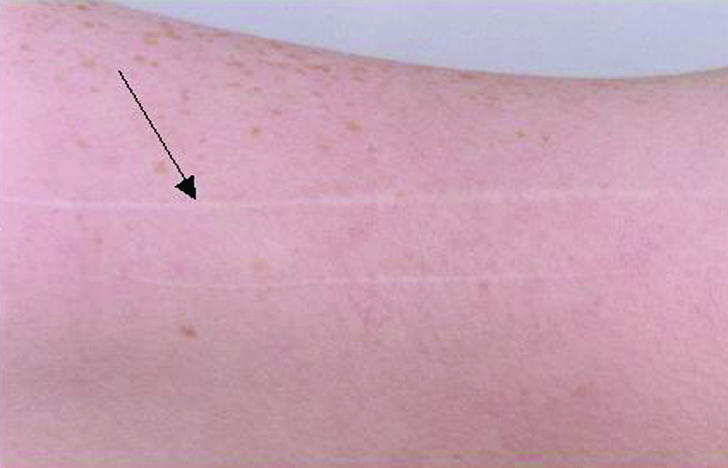
A fine line scar in forearm of a white man after a knife wound
Widespread (stretched) scars
appear when the fine lines of surgical scars gradually become stretched and widened, (fig 2) which usually happens in the three weeks after surgery.9 They are typically flat, pale, soft, symptomless scars often seen after knee or shoulder surgery.12 Stretch marks (abdominal striae) after pregnancy are variants of widespread scars in which there has been injury to the dermis and subcutaneous tissues but the epidermis is unbreached. There is no elevation, thickening, or nodularity in mature widespread scars, which distinguishes them from hypertrophic scars.
Figure 2.
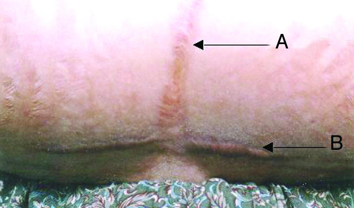
Widespread scar (A) in the midline after surgical incision and a hypertrophic scar (B) after transverse surgical incision in an Asian woman
Atrophic scars
are flat and depressed below the surrounding skin. They are generally small and often round with an indented or inverted centre (fig 3), and commonly arise after acne or chickenpox.13
Figure 3.
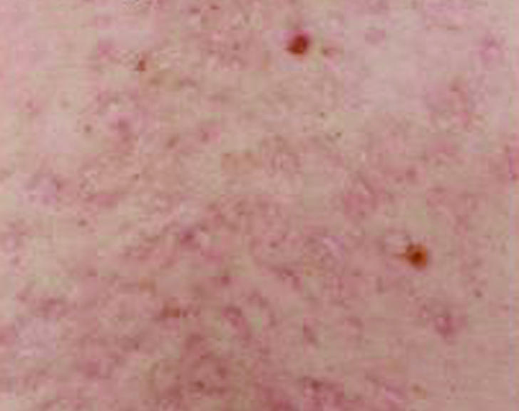
Atrophic scars after acne in upper back and shoulder area of a white man
Scar contractures—
Scars that cross joints or skin creases at right angles are prone to develop shortening or contracture. Scar contractures occur when the scar is not fully matured, often tend to be hypertrophic, and are typically disabling and dysfunctional (fig 4). They are common after burn injury across joints or skin concavities.
Figure 4.
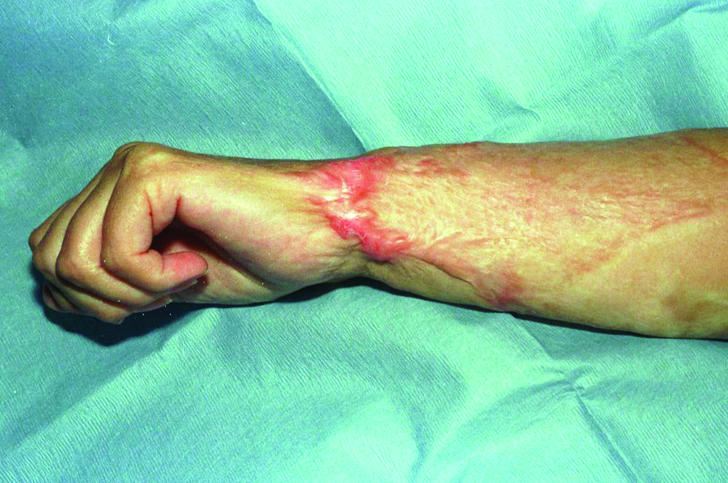
Scar contracture of forearm and wrist after a burn injury to a white woman
Raised skin scars
Raised skin scars are described as hypertrophic or keloid scars.14
Hypertrophic scars
are raised scars that remain within the boundaries of the original lesion, generally regressing spontaneously after the initial injury (fig 5).15 Hypertrophic scars are often red, inflamed, itchy, and even painful. They typically occur after burn injury on the trunk and extremities.
Figure 5.
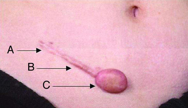
Presence of multiple scar types in same anatomical site of a white woman after surgery—widespread scar (A), hypertrophic scar (B), and keloid scar (C)
Keloid scars
are raised scars that spread beyond the margins of the original wound and invade the surrounding normal skin in a way that is site specific. Ear lobe keloids often grow as large lobules (fig 6), central sternal keloids commonly develop a butterfly shape, and deltoid keloids tend to extend vertically. A keloid continues to grow over time, does not regress spontaneously, and almost invariably recurs after simple excision. It is difficult to apply the term keloid until a scar has been present for at least a year, although there is no precise time interval. Histologically, keloids have a swirling nodular pattern of collagen fibres.16
Figure 6.
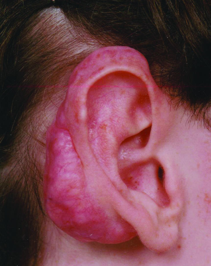
Right ear keloid scar after reduction surgery for prominent ears in a white man
Scars that are difficult to categorise have been termed intermediate scars.17 However, if a raised scar is still emerging after a year, a true keloid is a potential diagnosis, whereas hypertrophic scars should show some evidence of regression within this time. Keloids may be inflamed, itchy, and painful, especially during their growth phase. Common presentations are in the ear lobe after ear piercing, the deltoid after vaccination, and the sternum after acne, chickenpox, trauma, or surgery. Keloids are unique to humans, and there seems to be some genetic predisposition, with dark skinned races being more prone to them, though there are few large epidemiological studies. They develop predominantly in people aged 10-30 years, with an apparent predilection for emergence and deterioration during puberty and pregnancy.11
Structured scar assessment
Accurate scar assessment is essential for diagnosis and for starting, monitoring, and evaluating a therapeutic strategy for scar management. The cause and course of scar development are important—is the scar getting better or worse? A decision whether to treat will depend on:
1. Site (anatomical location of the scar)
2. Symptoms (pain, itching, etc)
3. Severity of functional impairment (such as joint mobility)
4. Stigma (how much is the patient disturbed?)
The severity of scars is often judged by eye but can be assessed quantitatively with a scar assessment guide such as the Vancouver scar scale18 or the Manchester scar proforma, a validated method for scar assessment and monitoring (see example on bmj.com). The exact anatomical location of scars are recorded, as are their number and size per site and a description of their margins, surface, colour, and texture.19 From these a score is compiled, with the lower the score the better the scar. A standardised colour photograph of the scar lesion at each consultation provides a reference to evaluate effectiveness of treatment since changes occur slowly.
The presence of a positive family history, previous abnormal scarring in the same or other anatomical sites, poor response to treatment or recurrence of scarring, specific anatomical locations (such as the sternum), large size, prolonged inflammation, and severe symptoms are associated with abnormal scarring.
Current methods of treating problematic scars
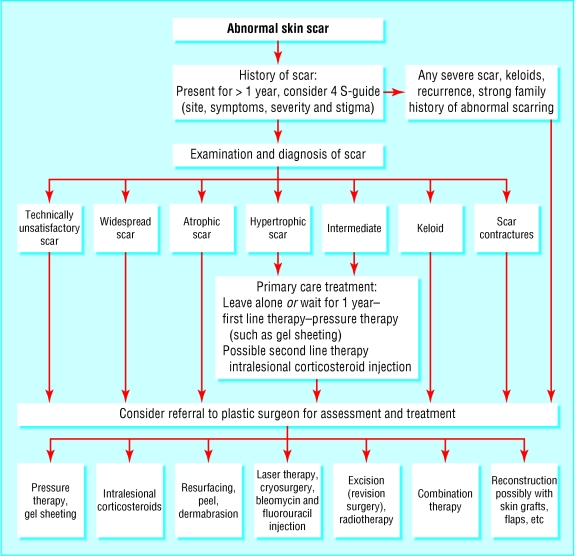
A simple plan of treatment can be offered with three courses of action—non-invasive treatment, invasive treatment, and leave alone management (fig 7).
Figure 7.
Flow chart for managing scars from diagnosis to treatment
Non-invasive options
include use of compression therapy (such as pressure garments with or without gel sheeting); static and dynamic splints; acrylic casts; masks and clips; application of a variety of oils, lotions, and creams; antihistamine drugs; hydrotherapy; and psychosocial counselling and advice.20,21 Silicon sheeting, with or without adhesive, has become popular.22 Massage therapy is often advocated but lacks evidence of benefit. All the above treatments are empirical.22 Without proper trials, benefits are difficult to quantify objectively, although even a placebo benefit may be appreciated by patients.
Invasive treatments
include surgical excision and resuture. Generally, revision should be considered only if the surgeon thinks that more favourable conditions for wound healing can be provided than on the first occasion (less inflammation, better technique). Intralesional corticosteroid injection20 is widely used but is prone to complications (fat atrophy, dermal thinning, and pigment changes). Other treatments that have been advocated with variable outcomes include injections of fluorouracil,23 interferon gamma,24 and bleomycin,25 radiotherapy,26 laser therapy,27 and cryosurgery.28
Additional educational resources
Most scientific and clinical papers on scarring are published in journals of plastic and reconstructive surgery, dermatology, and specific wound healing. Examples include:
British Journal of Plastic Surgery (www.hbuk.co.uk/journals/bjps/)
Plastic Reconstructive Surgery Journal (www.plasreconsurg.com/)
Journal of Investigative Dermatology (www.jidonline.org/)
British Journal of Dermatology (www.blacksci.co.uk/∼cgilib/jnlpage.bin?Journal=BJD)
Wound Repair and Regeneration (www.blacksci.co.uk/∼cgilib/jnlpage.asp?Journal=wrr)
Journal of Wound Care (www.journalofwoundcare.com/nav?page=jowc)
BMJ archive
Harding KG, Morris HaotL, Patel GK. Science, medicine, and the future: healing chronic wounds. BMJ 2002;324:160-3
Useful websites
British Association of Plastic Surgeons (www.baps.co.uk)
American Society of Plastic Surgeons and Plastic Surgery Educational Foundation (www.plasticsurgery.org)
Wound Healing Society (www.woundheal.org)
European Tissue Repair Society (www.etrs.org)
UK Wound Care Society (www.woundcaresociety.org)
British Association of Skin Camouflage (www.skin-camouflage.net)
Changing Faces (UK registered charity for help with disfigurement) (www.changingfaces.co.uk)
Recommended reading
Clark RAF. Molecular and cellular biology of wounds. 2nd ed. London: Plenum Press, 1996
Cherry GW, Hughes MA, Leaper DJ, Ferguson MWJ. Wound healing. In: Morris PJ, Wood WC, eds. Oxford textbook of surgery. 2nd ed. Oxford: Oxford University Press, 2001:129-59.
Garg HG, Longaker MT, eds. Scarless wound healing. New York: Marcel Dekker, 2000:213-26.
Ferguson MWJ, Leigh IM. Wound healing. In: Champion RH, Burton JL, Burns DA, Breathnach SM, eds. Textbook of dermatology. 6th ed. Oxford: Blackwell Science, 1998:337-56.
Ferguson MWJ, Whitby DJ, Shah M, Armstrong J, Siebert JW, Longaker MT. Scar formation: the spectral nature of fetal and adult wound repair. Plast Reconstr Surg 1996;97:854-60
Leave alone management—
Most scars are best left undisturbed by invasive treatment for a year to mature before any judgment is made on their appearance. Monitoring (wait and watch) will allow ongoing assessment of appearance, symptoms, and psychological impact, and reassurance is important. Some scars are best left alone in the long term. Informed, shared decision making with patients may help reduce inappropriate demands for treatment.
When such management plans are applied to specific scar types, certain patterns emerge:
Treatment of widespread scars is mostly by revision surgery to narrow the width of the scar. Revised scars in anatomical sites subject to excessive tissue mobility such as limbs can benefit from splinting29
Atrophic scars have been improved with chemical peels, cutaneous laser resurfacing, dermabrasion, punch excisions, and the use of soft tissue biological and alloplastic biological fillers30
It is important to distinguish between hypertrophic and keloid scars as inappropriate management can lead to recurrence and larger scars. Simple surgical excision of keloid scars has a 50%-80% risk of recurrence.31 A combination of surgery with either intralesional corticosteroid injection or radiotherapy has been the mainstay of treatment32
For scar contractures, surgical release with splinting, acrylic casting, and compression therapy may be required. Full thickness and split or partial thickness skin grafts and, perhaps more effectively, local and free flaps are used for reconstruction of difficult and extensive scars and contractures.33
Future perspectives
By subtly altering the regulatory growth factor cascades involved in wound healing—for example, increasing the ratio of cytokines such as transforming growth factor β3 compared with factors β1 and β2—the endogenous embryonic regenerative response can be restored without any adverse consequences on wound strength, healing rates, or incidence of wound infection.34–36 Transforming growth factor β3, neutralising antibodies to transforming growth factors β1 and β2, and mannose-6-phosphate are all in early stage human clinical trials for preventing skin scarring. Thus, future drug treatments hold the promise of substantially improving the cosmetic outcome of injury, trauma, or elective surgery, with scarring no longer being an inevitable consequence of skin healing.
Supplementary Material
Acknowledgments
Research summarised in this manuscript has been supported by a variety of grants from the MRC, Wellcome Trust, and the Biotechnology and Biological Sciences Research Council. AB is an MRC fellow.
Footnotes
Competing interests: MWJF is the co-founder and chief executive officer of Renovo. DAMcG is a member of the scientific and clinical advisory board of Renovo.
An example of a scar assessment guide apears on bmj.com
References
- 1.Sund B. New developments in wound care. London: PJB Publications; 2000. pp. 1–255. . (Clinica Report CBS 836.) [Google Scholar]
- 2.Ferguson MWJ, Whitby DJ, Shah M, Armstrong J, Siebert JW, Longaker MT. Scar formation: the spectral nature of fetal and adult wound repair. Plast Reconstr Surg. 1996;97:854–860. doi: 10.1097/00006534-199604000-00029. [DOI] [PubMed] [Google Scholar]
- 3.Brockes JP, Kumar A, Velloso CP. Regeneration as an evolutionary variable. J Anat. 2001;199(Pt 1-2):3–11. doi: 10.1046/j.1469-7580.2001.19910003.x. [DOI] [PMC free article] [PubMed] [Google Scholar]
- 4.Bell L, McAdams T, Morgan R, Parshley PF, Pike RC, Riggs P, et al. Pruritus in burns: a descriptive study. J Burn Care Rehabil. 1988;9:305–308. [PubMed] [Google Scholar]
- 5.Taal L, Faber AW. Posttraumatic stress and maladjustment among adult burn survivors 1 to 2 years postburn. Part II: the interview data. Burns. 1998;24:399–405. doi: 10.1016/s0305-4179(98)00053-9. [DOI] [PubMed] [Google Scholar]
- 6.Robert R, Meyer W, Bishop S, Rosenberg L, Murphy L, Blakeney P. Disfiguring burn scars and adolescent self-esteem. Burns. 1999;25:581–585. doi: 10.1016/s0305-4179(99)00065-0. [DOI] [PubMed] [Google Scholar]
- 7.Dorfmuller M. Psychological management and after-care of severely burned patients. Unfallchirurg. 1995;98:213–217. [PubMed] [Google Scholar]
- 8.Woo SH, Seul JH. Optimizing the correction of severe postburn hand deformities by using aggressive contracture releases and fasciocutaneous free-tissue transfers. Plast Reconstr Surg. 2001;107:1–8. doi: 10.1097/00006534-200101000-00001. [DOI] [PubMed] [Google Scholar]
- 9.Sommerlad BC, Creasey JM. The stretched scar: a clinical and histological study. Br J Plast Surg. 1978;31:34–45. doi: 10.1016/0007-1226(78)90012-7. [DOI] [PubMed] [Google Scholar]
- 10.Ashcroft GS, Horan MA, Ferguson MWJ. Aging alters the inflammatory and endothelial cell adhesion molecule profiles during human cutaneous wound healing. Lab Invest. 1998;78:47–58. [PubMed] [Google Scholar]
- 11.Murray JC, Pollack SV, Pinnell SR. Keloids: a review. J Am Acad Dermatol. 1981;4:461–470. doi: 10.1016/s0190-9622(81)70048-3. [DOI] [PubMed] [Google Scholar]
- 12.Rudolph R. Widespread scars, hypertrophic scars and keloids. Clin Plast Surg. 1987;14:253–260. [PubMed] [Google Scholar]
- 13.Tsao SS, Dover JS, Arndt KA, Kaminer MS. Scar management: keloid, hypertrophic, atrophic and acne scars. Semin Cutan Med Surg. 2002;21:46–75. doi: 10.1016/s1085-5629(02)80719-2. [DOI] [PubMed] [Google Scholar]
- 14.McGrouther DA. Hypertrophic or keloid scars? Eye. 1994;8(Pt 2):200–203. doi: 10.1038/eye.1994.46. [DOI] [PubMed] [Google Scholar]
- 15.Peacock EE, Madden JW, Trier WC. Biologic basis for the treatment of keloids and hypertrophic scars. South Med J. 1970;63:755–760. doi: 10.1097/00007611-197007000-00002. [DOI] [PubMed] [Google Scholar]
- 16.Ehrlich HP, Desmouliere A, Diegelmann RF, Cohen IK, Compton CC, Garner WL, et al. Morphological and immunochemical differences between keloid and hypertrophic scar. Am J Pathol. 1994;145:105–113. [PMC free article] [PubMed] [Google Scholar]
- 17.Muir IF. On the nature of keloid and hypertrophic scars. Br J Plast Surg. 1990;43:61–69. doi: 10.1016/0007-1226(90)90046-3. [DOI] [PubMed] [Google Scholar]
- 18.Powers PS, Sarkar S, Goldgof DB, Cruse CW, Tsap LV. Scar assessment: current problems and future solutions. J Burn Care Rehabil. 1999;20:54–60. doi: 10.1097/00004630-199901001-00011. [DOI] [PubMed] [Google Scholar]
- 19.Beausang E, Flyod H, Dunn KW, Orton CI, Ferguson MWJ. A new quantitative scale for clinical scar assessment. Plast Reconstr Surg. 1998;102:1954–1961. doi: 10.1097/00006534-199811000-00022. [DOI] [PubMed] [Google Scholar]
- 20.Niessen FB, Spauwen PH, Schalkwijk J, Kon M. On the nature of hypertrophic scars and keloids: a review. Plast Reconstr Surg. 1999;104:1435–1458. doi: 10.1097/00006534-199910000-00031. [DOI] [PubMed] [Google Scholar]
- 21.Van den Helder CJ, Hage JJ. Sense and nonsense of scar creams and gels. Aesthetic Plast Surg. 1994;18:307–313. doi: 10.1007/BF00449800. [DOI] [PubMed] [Google Scholar]
- 22.Mustoe TA, Cooter RD, Gold MH, Hobbs FD, Ramelet AA, Shakespeare PG, et al. International Advisory Panel on Scar Management. International clinical recommendations on scar management. Plast Reconstr Surg. 2002;110:560–571. doi: 10.1097/00006534-200208000-00031. [DOI] [PubMed] [Google Scholar]
- 23.Uppal RS, Khan U, Kakar S, Talas G, Chapman P, McGrouther DA. The effects of a single dose of 5-fluorouracil on keloid scars: a clinical trial of timed wound irrigation after extralesional excision. Plast Reconstr Surg. 2001;108:1218–1224. doi: 10.1097/00006534-200110000-00018. [DOI] [PubMed] [Google Scholar]
- 24.Broker BJ, Rosen D, Amsberry J, Schmidt R, Sailor L, Pribitkin EA, et al. Keloid excision and recurrence prophylaxis via intradermal interferon-gamma injections: a pilot study. Laryngoscope. 1996;106(12 Pt 1):1497–1501. doi: 10.1097/00005537-199612000-00010. [DOI] [PubMed] [Google Scholar]
- 25.Espana A, Solano T, Quintanilla E. Bleomycin in the treatment of keloids and hypertrophic scars by multiple needle punctures. Dermatol Surg. 2001;27:23–27. [PubMed] [Google Scholar]
- 26.Clavere P, Bedane C, Bonnetblanc JM, Bonnafoux-Clavere A, Rousseau J. Postoperative interstitial radiotherapy of keloids by iridium 192: a retrospective study of 46 treated scars. Dermatology. 1997;195:349–352. doi: 10.1159/000245986. [DOI] [PubMed] [Google Scholar]
- 27.Alster TS, Williams CM. Treatment of keloid sternotomy scars with 585 nm flashlamp-pumped pulsed-dye laser. Lancet. 1995;345:1198–1200. doi: 10.1016/s0140-6736(95)91989-9. [DOI] [PubMed] [Google Scholar]
- 28.Zouboulis CC, Zouridaki E, Rosenberger A, Dalkowski A. Current developments and uses of cryosurgery in the treatment of keloids and hypertrophic scars. Wound Repair Regen. 2002;10:98–102. doi: 10.1046/j.1524-475x.2002.02111.x. [DOI] [PubMed] [Google Scholar]
- 29.Jordan RB, Daher J, Wasil K. Splints and scar management for acute and reconstructive burn care. Clin Plast Surg. 2000;27:71–85. [PubMed] [Google Scholar]
- 30.Hirsch RJ, Lewis AB. Treatment of acne scarring. Semin Cutan Med Surg. 2001;20:190–198. doi: 10.1053/sder.2001.27557. [DOI] [PubMed] [Google Scholar]
- 31.Darzi MA, Chowdri NA, Kaul SK, Khan M. Evaluation of various methods of treating keloids and hypertrophic scars: a 10-year follow-up study. Br J Plast Surg. 1992;45:374–379. doi: 10.1016/0007-1226(92)90008-l. [DOI] [PubMed] [Google Scholar]
- 32.Urioste SS, Arndt KA, Dover JS. Keloids and hypertrophic scars: review and treatment strategies. Semin Cutan Med Surg. 1999;18:159–171. doi: 10.1016/s1085-5629(99)80040-6. [DOI] [PubMed] [Google Scholar]
- 33.Adant JP, Bluth F, Jacquemin D. Reconstruction of neck burns. A long-term comparative study between skin grafts, skin expansion and free flaps. Acta Chir Belg. 1998;98:5–9. [PubMed] [Google Scholar]
- 34.Border WA, Noble NA. Targeting TGF-beta for treatment of disease. Nat Med. 1995;1:1000–1001. doi: 10.1038/nm1095-1000. [DOI] [PubMed] [Google Scholar]
- 35.O'Kane S, Ferguson MWJ. Transforming growth factor betas and wound healing. Int J Biochem Cell Biol. 1997;29:63–78. doi: 10.1016/s1357-2725(96)00120-3. [DOI] [PubMed] [Google Scholar]
- 36.Ferguson MWJ. Scar wars. Br Dent J. 2002;192:475. doi: 10.1038/sj.bdj.4801404. [DOI] [PubMed] [Google Scholar]
Associated Data
This section collects any data citations, data availability statements, or supplementary materials included in this article.


