Abstract
Magnesium (Mg) is involved in essential cellular and physiological processes. Globally, inadequate consumption of Mg is widespread among populations, especially those who consume processed foods, and its homeostasis is impaired in obese individuals and type 2 diabetes patients. Since Mg deficiency triggers oxidative stress and chronic inflammation, common features of several frequent chronic non-communicable diseases, interest in this mineral is growing in clinical medicine as well as in biomedicine. To date, very little is known about the role of Mg deficiency in adipose tissue. In obesity, the increase in fat tissue leads to changes in the release of cytokines, causing low-grade inflammation and macrophage infiltration. Hypomagnesemia in obesity can potentiate the excessive production of reactive oxygen species, mitochondrial dysfunction, and decreased ATP production. Importantly, Mg plays a role in regulating intracellular calcium concentration and is involved in carbohydrate metabolism and insulin receptor activity. This narrative review aims to consolidate existing knowledge, identify research gaps, and raise awareness of the critical role of Mg in supporting adipose tissue metabolism and preventing oxidative stress.
Keywords: hypomagnesemia, antioxidants, calcium antagonism, low-grade chronic inflammation, obesity
1. Introduction
Adipose tissue (AT) has historically been classified into two types: white adipose tissue (WAT) and brown adipose tissue (BAT). In addition, recently, beige adipose tissue has been described [1]. WAT represents most AT in adult humans and is the body’s main energy storage. At the same time, BAT dissipates energy as a defense against cold and maintains energy balance for the whole body. Beige adipose tissue is located within WAT but shares similar features with BAT. WAT comprises many cell types, such as adipocytes (the most abundant), preadipocytes, fibroblasts, stem cells, macrophages, and capillary endothelial cells (ECs). Of note, an intense reciprocal dynamic communication exists between ECs and adipocytes. Endothelial transfer of plasma constituents and biological signals via secretory signaling molecules and microvesicles to the adipocytes [2] is crucial for metabolic homeostasis. Conversely, adipocytes release various bioactive molecules that influence endothelial function. The role of adipose tissues in modulating vascular homeostasis is attracting more and more attention, because most blood vessels are surrounded by a functionally specialized aggregate of AT, termed perivascular adipose tissue (PVAT). PVAT is an established regulator of vascular function through its release of gases, such as nitric oxide (NO) and hydrogen sulfide, and adipokines. While in physiological conditions, PVAT is vasculo-protective, its dysregulation contributes to vascular dysfunction since it releases inflammatory mediators that readily promote oxidative stress (OS) and the acquisition of an inflammatory phenotype of vascular cells [3].
Magnesium (Mg) deficiency is one of the many factors involved in promoting OS and inflammation. Globally, the intake of Mg is inadequate, and this trend is widespread across populations. Subclinical Mg deficiency has been observed mainly in populations that consume processed foods, and it has also been observed in obese individuals [4]. In obesity, the expansion of AT results in the alteration of its secretion of cytokines, initiating a cascade of metabolic changes that favor macrophage infiltration mediated and sustained by low-grade chronic inflammation (LGCI). Moreover, the pro-inflammatory phenotype of hypertrophic/hyperplastic adipocytes in obesity promotes EC activation, contributing to LGCI [2]. Proper Mg intake can directly limit OS by reducing pro-oxidants and enhancing antioxidant capacity. It can also indirectly reduce inflammation [5,6].
We performed a literature review using MEDLINE, PubMed, EMBASE, and Web of Science to assess the impact of Mg on OS and inflammation of AT, focusing on obese subjects. We meticulously analyzed the full-text articles, selecting the most pertinent studies for inclusion in this review.
2. Oxidative Stress, Inflammation, and AT Dysfunction
Our understanding of the role of reactive oxygen species (ROS) in cellular biology has evolved considerably, revealing their role as essential regulators in the cellular signaling network. In physiological conditions, homeostatic ROS are secondary messengers in various intracellular signaling pathways, involving programmed cell death or necrosis, gene expression regulation, and innate and adaptive immune responses [7]. In addition, ROS are implicated in body weight control because they are crucial for the response of hypothalamic neurons to fluctuations in levels of bodily metabolic fuels such as glucose, free fatty acids, and amino acids [8].
ROS are generated during several biochemical processes, such as mitochondrial ATP synthesis, the activity of NADPH oxidases, nitric oxide synthases (NOSs) and microsomal cytochrome P450 oxidases, Fenton reaction, peroxisomal β-oxidation, prostaglandin synthesis, and others [9]. However, mitochondria are the main source of cellular ROS [10]. The rate of ROS production by isolated mitochondria is dependent on the metabolic state and is inversely related to the coupling between the respiratory chain and ATP synthesis [11,12]. Adipocytes adapt to dynamic changes in ROS levels and use them as second messengers. As an example, hydrogen peroxide has been found to mimic the action of insulin in adipocytes [9].
ROS accumulation directly contributes to the pathophysiology of several chronic inflammatory diseases, causing lipid peroxidation, DNA damage, protein oxidation, irreversible mitochondrial damage, inadequate ATP production, and indirectly, activating the nuclear factor-κB (NF-κB) pathway and, therefore, inflammation [13]. Inflammation and oxidative stress are two related processes: one can promote the other, leading to a toxic feedback system. A rich antioxidant arsenal tightly regulates ROS production. The body’s antioxidant defenses include a variety of water-soluble and fat-soluble compounds, such as enzymes, proteins, glutathione, urate, vitamins C and E, and beta-carotene, which work together to neutralize the effects of pro-oxidants. Antioxidant enzymes include catalase (CAT), glutathione reductase (GR), thioredoxin reductase (TrxR), heme oxygenase-1 (HO-1), superoxide dismutase (SOD), glutathione peroxidase (GPx), peroxiredoxin (Prx), paraoxonases (PON), and NAD(P)H: quinone oxidoreductase 1 (NQO1). However, the prolonged and uncontrolled production of ROS can overcome the body’s antioxidant defense system, generating OS. In obesity, systemic OS is due to excessive superoxide generation from nicotinamide adenine dinucleotide phosphate (NADPH) oxidase (NOX), uncoupled endothelial nitric oxide synthase (eNOS), and reduced antioxidant defense [4]. Exposure to OS can lead to the damage of proteins by promoting their carbonylation. This process is an irreversible non-enzymatic modification that can cause protein dysfunction and aggregation. In the case of obesity, higher levels of carbonylated proteins appear to be linked to mitochondrial dysfunction in adipocytes, which may be relevant to the development of insulin resistance (IR) and type 2 diabetes [9].
In obesity, AT expands by a combination of an increase in adipocyte size and number. There may be a difference in the rates of growth between AT and vascularity, leading to reduced blood flow in the area. This can create an oxygen gradient and cause a relatively low-oxygen environment in the growing AT [14]. Research has shown that this hypoxia can alter the expression and release of adipokines in human adipocytes because of an increased expression of the hypoxia-inducible factor 1 alpha [15] and intracellular calcium (Ca) concentration oscillations [14]. Increased intracellular Ca concentration regulates the hypoxia-induced release of leptin, vascular endothelial growth factor, and interleukin (IL)-6, while the release of adiponectin is NF-κB-dependent.
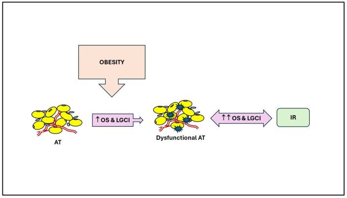
These adipokines are pro-oxidant and pro-inflammatory, except adiponectin. In normal-weight individuals, small adipocytes effectively store fatty acids as triglycerides (TG). Excessive caloric intake can lead to an overload of the metabolic system, causing an increase in TG storage and the enlargement of adipocytes. In addition, OS in the WAT disturbs its redox balance and impacts its function by impairing adipogenesis, inducing IR, and causing adipocyte hypertrophy [9] (Figure 1).
Figure 1.
Relationships between AT dysfunction, oxidative stress, inflammation, and insulin resistance in obesity. LGCI: low-grade chronic inflammation; OS: oxidative stress; IR: insulin resistance; ↑: increase.
Hypertrophic adipocytes exhibit a decreased density of insulin receptors and an elevated level of β-3 adrenergic receptors. This facilitates the migration of monocytes to the visceral adipose stroma, initiating a proinflammatory cycle between adipocytes and monocytes, causing tissue dysfunction and contributing to obesity-related inflammation and comorbidities [16]. Physiologically, monocytes reach the adipose tissue during development, become resident AT macrophages, and regulate the metabolic activity of adipocytes and their precursors to maintain AT homeostasis and efficient function. Importantly, they display an anti-inflammatory M2 phenotype [17]. On the contrary, in obese individuals, AT macrophages tend to polarize to a pro-inflammatory M1 phenotype. M1 macrophages release chemokines, such as monocyte chemoattractant protein-1 (MCP-1), and pro-inflammatory cytokines, such as IL-1β, IL-6, and tumor necrosis factor-α (TNF-α), contributing to LGCI [18]. These cytokines not only promote their local proliferation but also recruit more macrophages and retain them in the AT to the point that the proportion of macrophages significantly increases in the adipose tissue of obese people [19]. Obesity-related metabolic inflammation in AT gradually impacts other organs through lipid and inflammatory mediators. The surplus of circulating TG and free fatty acids causes an accumulation of activated lipids in the muscle, disrupting functions such as mitochondrial oxidative phosphorylation and insulin-stimulated glucose transport, ultimately leading to peripheral IR and further potentiating OS [18]. In addition, nutrient overload alters AT metabolism towards increased lipid accumulation and glycolytic ATP synthesis in conjunction with decreased mitochondrial biogenesis. Overeating also activates inflammatory responses in the liver, skeletal muscle, pancreas, and hypothalamus, thus contributing to diminished insulin sensitivity and systemic IR in obese individuals [4]. In obesity, IR is often associated with hyperinsulinemia, increased visceral adiposity, metabolic dyslipidemia with high TG levels, low HDL cholesterol levels, and hypertension, features collectively referred to as metabolic syndrome (MetS). The cluster of conditions defining MetS increases the risk of coronary heart disease and type 2 diabetes [4].
3. Mg
Mg is a mineral macro-element that functions as a crucial signaling element and metabolite in cell physiology [20,21]. Mg participates in metabolizing lipids, proteins, carbohydrates, and nucleic acids [22]. Mg also mitigates the effects of OS and maintains cell membrane stability [23]. At the organ level, Mg is essential to maintain proper bone density and glucose tolerance, especially in diabetic patients, to relieve neurological and psychiatric disorders, to regulate blood pressure, to prevent ischemic heart disease and strokes, and to maintain an adequate skeletal muscular mass, principally in the elderly [22,24]. Mg should be consumed following the appropriate dietary recommendations to maintain these physiological functions. The recommended dietary allowance (RDA) for Mg in the USA is 310–320 and 410–420 mg per day for females and males, respectively, while in Europe, the European Food Safety Authority (EFSA) recommends 300 and 350 mg per day for women and men, respectively [24]. Globally, the reduction in dietary Mg intake is mainly due to (i) the effect of global warming that reduces Mg amounts in crops; (ii) the impact of cereals’ milling that drastically reduces Mg content in the grains (e.g., whole wheat flour 120 mg/100 g to refined wheat flour 20 mg/100 g of product); and (iii) the change in the dietary pattern of the global population from diets rich in vegetables, whole grains, fruit, and nuts rich in Mg to a Western dietary pattern rich in refined grains, sugars, saturated fats, additives and poor in veggies and fruits [24]. Mg dietary intake decreases with the increasing westernization of the dietary pattern worldwide. Beyond dietary intake, pre-existing pathological conditions (such as impaired gastrointestinal absorption, inflammatory bowel disease, colon cancer, and gastric bypass), diabetes mellitus, renal disorders and hydro-electrolyte imbalances, genetic factors, alcohol abuse, and the use of certain medications result in chronic Mg deficiency [25,26,27,28,29,30,31]. Aging is a significant risk factor for Mg deficiency. Despite stable Mg requirements across age groups [32,33], older adults do not consume enough Mg, regardless of gender and race [34]. Furthermore, aging reduces the efficiency of Mg absorption and active renal reabsorption [33]. In older adults, an imbalance in Mg can lead to greater susceptibility to age-related diseases like cardiovascular disease and diabetes but may also contribute to the aging process itself [35]. This is not surprising considering that Mg deficiency causes OS, which is one of the hallmarks of aging [36].
Also, strenuous physical activity can lead to Mg deficiency [37]. This factor should be considered when determining the appropriate Mg levels for active individuals. Exercise triggers a shift in Mg within the body tissues to support metabolic demands. Mg transits between plasma, adipocytes, and skeletal muscle during and post aerobic workouts. The extent of movement depends on cell energy level production. Following exercise, Mg moves back to the bloodstream from tissues by drawing from bone, muscle, and adipose tissue. Although tubular reabsorption mechanisms act to minimize urinary losses of Mg, post-workout urine Mg excretion rises. These changes affect Mg levels in various body fluids and tissues. Inadequate Mg intake, especially for athletes, can impair performance and worsen exercise effects [37]. As with all essential nutrients, Mg deficiency can be corrected by increasing Mg intake through diet and/or supplementation. To correct hypomagnesemia, supplementation of 250 to 600 mg per day of Mg, preferably in the form of organic Mg salts due to their higher bioavailability, is useful [18]. Even better, an increase in dietary Mg can help correct hypomagnesemia [24]. As an example, Mg enteric absorption from almonds is much higher compared to that from Mg supplements [38]. Accordingly, adolescents’ adherence to the Mediterranean diet, which is high in Mg, increases Mg levels and helps prevent obesity [39]. In addition, an Mg food source is more effective than supplements in decreasing all-cause mortality [40].
4. Mg Deficiency, Inflammation, and Oxidative Stress
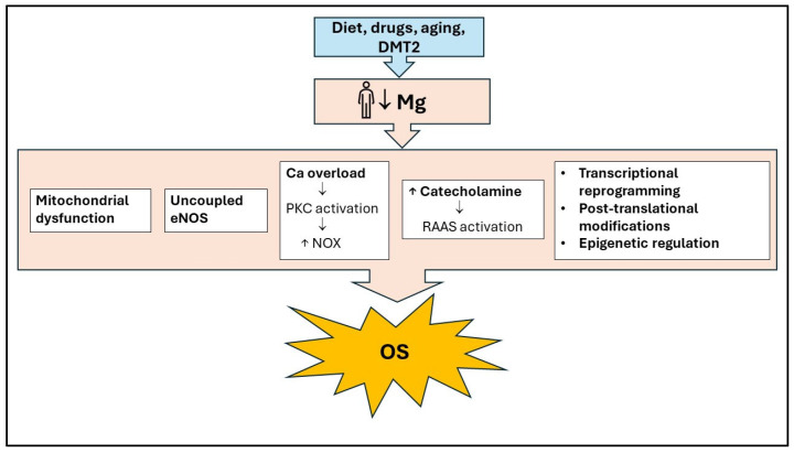
Mg deficiency causes excessive production of ROS, mainly due to mitochondrial dysfunction, abnormal calcium homeostasis, and activation of the renin-angiotensin-aldosterone system [6] (Figure 2). Furthermore, the antioxidant defense system is impaired, and ATP synthase is downregulated, causing a decrease in ATP production (Figure 1). In AT, Mg regulates intracellular Ca concentration by blocking the opening of the L-type Ca channel, which is controlled by the Mg-binding sites. The excess of intracellular Ca, in turn, results in the activation of Ca-dependent processes such as the release of inflammatory cytokines and activation of NOX by phosphorylation of protein kinase C (PKC), activation of NOS and calcium-dependent calmodulin complex, and hence increased OS [41] (Figure 2).
Figure 2.
The main cellular mechanisms of Mg’s preventive action against oxidative stress. Mg deficiency leads to oxidative stress by causing mitochondrial dysfunction, abnormal calcium homeostasis, eNOS uncoupling, Renin-Angiotensin-Aldosterone System activation, and changes in transcription and translation processes. Ca: Calcium; DMT2: Diabetes Mellitus type 2; eNOS, Endothelial Nitric Oxide Synthase; OS: Oxidative Stress; NOX: NADPH oxidase; RAAS: Renin-Angiotensin-Aldosterone System; ↑: increase; ↓: decrease.
Intracellular Mg also acts as an essential cofactor of several enzymes involved in carbohydrate metabolism, regulating the activity of those that catalyze phosphorylation reactions and acting as part of the Mg–ATP complex, necessary for the action of enzymes that participate in glycolysis. Thus, the appropriate Mg concentration is essential for the tyrosine kinase activity of the insulin receptor and, therefore, for the autophosphorylation of the β subunit of this receptor and phosphorylation of its substrates [42].
The glutathione system constitutes the primary mechanism for mitigating OS within the body. Its activity is closely related to the redox state of other low-molecular-weight thiols, such as cysteine, cysteine-glycine, and homocysteine. This system operates at both intracellular and plasma levels, and the redox state of the plasma pool is in equilibrium with that of the intracellular pool. Glutathione is the primary intracellular antioxidant. It can be released from tissue, contributing to maintaining other thiols in the reduced form. The reducing power of water-soluble thiols is necessary to keep some fat-soluble antioxidants (such as vitamin E and coenzyme Q) in their active, reduced form. Mg is an obligatory cofactor in glutathione synthesis and all biosynthetic reactions involving ATP. Additionally, hypomagnesemia contributes to a reduction in the expression and activity of antioxidant enzymes (such as GPx, SOD, and CAT), leading to a decrease in cell and tissue concentrations of antioxidants and an increase in the production of the ROS hydrogen peroxide and superoxide anion by inflammatory cells [23].
The production of antioxidants, the expression of anti-oxidative enzymes, and AT metabolism have been reported to be regulated by circadian rhythms [43]. The circadian rhythm generally fluctuates in a daily cycle of about 24 h, a period of light and dark, in response to abiotic and biotic factors. The circadian clock is a network of molecular clocks in central and peripheral tissues that orchestrates biological processes in adapting to everyday environmental changes. The central circadian clock is in the hypothalamic suprachiasmatic nuclei, while the peripheral clocks are in other tissues, including the kidney, liver, blood vessels, and AT [43]. Accumulating data from both human and experimental animal models suggest that AT function and OS have an innate connection with the intrinsic biological clock [43]. Interestingly, daily Mg fluxes regulate cellular timekeeping and energy balance [44].
Recent studies have demonstrated that obesity leads to increased OS in both humans and animals [45]. The level of OS in the body was found to be directly associated with fat mass in both humans and rodents [46]. In obese individuals, markers of mitochondrial OS, such as protein carbonyls, lipid peroxidation products including malondialdehyde, and production of ROS, were elevated in AT [47]. Considering the various properties of Mg that contribute to maintaining metabolic and redox balance in AT, an adequate level of Mg in this tissue can prevent OS and its effects on obesity.
5. Role of Mg in Adipose Tissue: Preclinical Evidence
Mg deficiency in rodents contributes to accelerated catabolism, partly by generating insulin resistance, but the metabolic phenotype analysis has not been disclosed [48,49,50]. Controversies exist about the effect of an Mg-deficient diet on body mass. In some studies, Mg dietary restriction had no impact on body mass and adiposity index [49,50], while in others, a decrease in body mass was reported because of total lean body mass decrease without changes in total body fat. These findings were attributed to reduced appetite and decreased food consumption [51,52].
Table 1 summarizes the preclinical evidence on the role of Mg in AT.
An interesting study has highlighted how age affects AT in either Mg-deficient rats or those supplemented with Mg [53]. In rats fed a standard diet, the significant increase in AT weight observed during aging was related to a rise in both the size and number of adipocytes. In young rats, Mg deficiency did not change the size of the adipocytes but increased their number (30% more than the standard diet or supplementation), suggesting that a low Mg status contributes to improving the lipid storage capacity of AT. In aged rats, a Mg-deficient diet led to relative hypotrophy of adipocytes, counterbalanced by the rise in their number. In brief, inadequate Mg intake affected the size and number of fat cells in both young and aged rats.
Notably, Mg restriction abrogates weight gain in rats exposed to a sucrose-rich diet, mainly because de novo lipogenesis is reduced under Mg deficiency [54,55]. In this experimental model, low Mg intake exacerbates OS in sucrose-fed rats, leading to increased oxidative damage to unsaturated lipids in membranes and amino acids in proteins [55]. In parallel, the activities of the SOD, glutathione-S-transferase, and catalase decline considerably in animals under a low Mg/sucrose-rich diet [56]. Moreover, a low dietary Mg intake prevents high-fat diet-induced obesity in mice by enhancing the expression of genes involved in β oxidation and by elevating Ucp1 levels in BAT, with the consequent increased thermogenesis [57]. The two events are linked to elevated β3-adrenergic receptor expression in WAT and BAT.
Mg intake must be adequate from the early stages of development. Maternal Mg deficiency in mice irreversibly increases the visceral adiposity of the offspring, which shows higher expression of fatty acid synthase and fatty acyl transport protein 1, liver, and adipose tissue together with increased levels of plasma TNF-α [58].
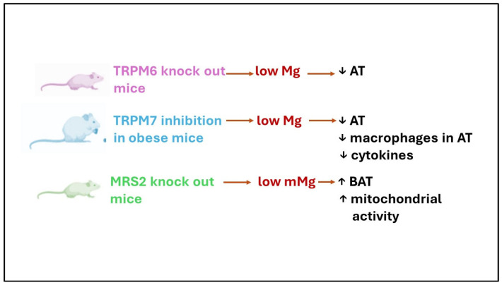
Interesting hints about the complex role of Mg in the AT derive from Mg transporter/channel knock-out mice (Figure 3). Transient receptor potential ion channel for melastatin (TRPM) 6, a kinase-coupled ion channel, is essential in the intestine for maintaining organismal Mg levels, while its closest relative, TRPM7, is considered indispensable for cellular Mg homeostasis. TRPM6−/− mice, which develop severe hypomagnesemia because of deficient intestinal Mg uptake, are entirely devoid of abdominal and subcutaneous fat depots and present clear signs of catabolic metabolism [48]. In obese mice, pharmacological inhibition of TRPM7 reduces body weight by reducing the adipose mass and prevents insulin resistance (Figure 3). Of particular interest is the finding that the percentage of macrophage in WAT is significantly reduced, and this correlates with the downregulation of the pro-inflammatory cytokines IL1β and IL6 and the chemokine MCP-1 [59].
Figure 3.
A summary of the effects of inhibiting certain Mg transporters/channels in mice. TRPM: transient receptor potential ion channels for melastatin; MRS2: mitochondrial RNA splicing 2; mMg: mitochondrial Mg; ↓: decrease; ↑: increase.
Similarly, serum levels of TNF-α, IL-6, IL-1β, and MCP-1 were upregulated in obese mice and reduced by the conditional knock-out of TRPM7 in the AT [60]. These results point to TRPM7 as the link between obesity and inflammation. Indeed, since low Mg upregulates TRPM7, many of the effects of Mg deficiency, including inflammation and OS, can be attributed to increased TRPM7 and the activation of its kinase domain [60]. A positive feedback loop exists between OS levels and TRPM7’s expression and function. Elevated OS increases TRPM7 expression [61,62], while TRPM7 overexpression induces the accumulation of intracellular ROS and inflammation [63,64]. In addition, since the promoter region of TRPM7 contains binding sites for NF-κB, LGCI upregulates TRPM7 [59].
Mitochondria are central to health and disease [65]. They are involved in cell metabolism and regulate ion homeostasis, cell growth, redox status, and cell signaling. Moreover, they are intracellular Mg stores [66]. Mg, pivotal for protein and ATP synthesis and various metabolic pathways, enters the mitochondria through the Mitochondrial RNA Splicing 2 (MRS2) channel anchored to the inner mitochondrial membrane [67]. MRS2−/− mice on a Western diet do not gain weight and show enhanced mitochondrial activity and increased BAT [68]. Accordingly, the knockdown of MRS2 in human cells leads to decreased mitochondrial uptake of Mg and metabolic reprogramming [69].
Table 1.
Preclinical evidence on the role of Mg in adipose tissue.
| Study | Type of Animal | Treatment | Treatment Duration |
Effects |
|---|---|---|---|---|
| Devaux et al. [53] | Male Sprague Dawley young rats (YR) vs. old rats (OR) |
Diet with Mg deficiency | 22 mo | YR adipocytes hyperplasia; OR hypotrophy of adipocytes |
| Boparai et al. [55] | Male Wistar rats | High sucrose (HS); low Mg (LM); HSLM | 12 wk | HSLM ↑ of TBARS and PCO (plasma and liver) |
| Kurstjens et al. [57] | Male C57BL6/J mice | normal Mg Low-fat diet (NMLFD); NM High fat diet (NMHFD); LMLFD; LMHFD | 17 wk | LM ameliorates HFD-induced obesity, fasting glucose ↓, insulin sensitivity ↑; absence of liver steatosis; ↑ BAT Ucp1 m-RNA expression, and higher body temperature |
| Zhong et al. [59] | Flox and ATKO mice | HFD; TRPM7 inhibition | 16 wk | ATKO have less body weight than Flox, ↓ % of macrophage in WAT, IL-1β, IL-6, MCP-1 |
| Madaris et al. [68] | Male WT and MRS2−/− KO mice | HFD; Western Diet (WD) | 12 mo | MRS2−/− KO in WD no weight gain and ↑ mitochondrial activity and BAT |
| Choudary et al. [56] | Male Wistar rats | LM; HS; HSLM | 3 mo | ↓ SOD, catalase and GST in HSLM |
ATKO = adipocyte-specific TRPM7 knock-out; BAT = brown adipose tissue; GST = glutathione-S-transferase; HFD = high fat diet; HS = high sucrose; IL = interleukin; KO = knock-out; LFD = low fat diet; LM = low Mg; MCP-1 = monocyte chemoattractive protein 1; MRS2 = mitochondrial RNA splicing 2; NM = normal Mg; OR = old rats; PCO = protein carbonyls; SOD = superoxide dismutase; TBARS = thiobarbituric acid reactive substances; TRPM7 = transient receptor potential ion channels for melastatin; WAT = white adipose tissue; WD = western diet; WT = wild-type; YR = young rats; ↑ = increase; ↓ = decrease; wk = weeks; mo = months.
Very little is known about the effect of low Mg on cultured adipocytes. Mg deficiency impairs insulin-dependent glucose metabolism in isolated rat adipocytes [70]. In mature 3T3-L1 adipocytes, Mg deficiency diminishes GLUT4 translocation and decreases glycolysis upon insulin stimulation because of the lack of Akt activation [71]. Currently, no data are available about redox balance and the secretion of cytokines and adipokines in Mg-deficient adipocytes. In an in vitro model of brown adipocyte differentiation, increasing extracellular Mg tends to enhance the expression of PR domain containing 16 (PRDM 16) and PPARγ coactivator 1-alpha (PGC1-α), two adipogenic factors, thus suggesting the inhibition of brown adipocyte differentiation through a calcium antagonistic effect [72].
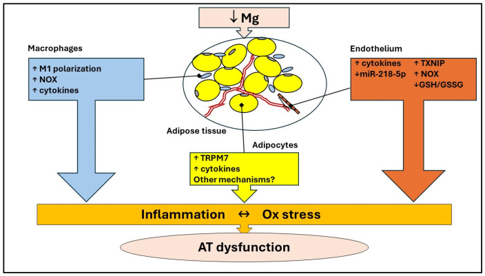
Within the complexity and cellular heterogeneity of AT, it should also be recalled that low Mg promotes a pro-oxidant and pro-inflammatory phenotype in endothelial cells [73,74] (Figure 4). These data are further supported by the powerful effect of different Mg concentrations on endothelial transcriptome [75]. Specifically, low Mg markedly perturbs inflammatory pathways, events that adversely impact adipocyte metabolism, insulin sensitivity, and plasticity. Moreover, Mg reduces endothelial apoptosis in rats with preeclampsia by upregulating miR-218-5p, which targets the HMGB1 pathway, known to be pro-inflammatory [76] (Figure 4). Macrophages are also sensitive to Mg deficiency, which upregulates the M1 subtype markers and induces the secretion of inflammatory cytokines [77] (Figure 4). Briefly, Mg deficiency drives all cells to promote and maintain inflammation. The good news is that most of these detrimental effects are reversible when Mg homeostasis is restored.
Figure 4.
Crosstalk between different cell types in AT. In adipocytes, Mg deficiency upregulates TRMP7, which appears to elevate cytokine levels. Moreover, low Mg promotes a pro-oxidant and pro-inflammatory phenotype in endothelial cells and induces M1 macrophage polarization, an event associated with the increase in NOX and the release of high amounts of pro-inflammatory cytokines. GSH: reduced glutathione; GSSG oxidized glutathione; NOX: NADPH oxidase; TRPM: transient receptor potential ion channels for melastatin; TXNIP: Thioredoxin Interacting Protein; ↑: increase; ↓: decrease.
While Mg is known to control transcription and translation [21], an aspect that is often neglected is its role in regulating chromatin dynamics [78]. Interestingly, Mg serves as a cofactor for methionine adenosyl transferase 1A (MAT1A), an enzyme responsible for producing S-adenosylmethionine (SAM). SAM is the primary methyl donor within cells and is crucial for numerous methylation reactions, including those involving DNA and proteins. Mg also influences the activity of enzymes that catalyze the methylation and demethylation of DNA, such as DNA methyltransferases (DNMTs) [79] and ten-eleven translocation (TET) enzymes [80]. Therefore, Mg emerges as a mineral linked to epigenetics, as demonstrated in pregnant rats, where Mg deficiency induces metabolic complications in their offspring by altering cytosine methylation in the promoter region of the hepatic hydroxysteroid dehydrogenase-2 gene [81], which contributes to fatty acid metabolism [82]. Recently, a proper intake of Mg has been proposed as a beneficial measure to prevent epigenetic changes that lead to impaired cardiovascular function [83]. At this point, a question arises: since epigenetic signatures are potentially reversible, are the reversible effects observed in vitro upon restoration of Mg homeostasis due to epigenetic regulation? More research is needed to find a proper answer. Understanding DNA methylation patterns in response to Mg is crucial for enhancing knowledge of potential prevention strategies by modifying nutritional status in at-risk populations.
6. Mg, Oxidative Stress, and Inflammation: Clinical Evidence
A search for articles on Mg and OS in AT or obesity resulted in a limited number of clinical studies investigating the role of Mg in preventing the generation of OS and inflammation in obese individuals. Some studies evaluated the links between Mg and anthropometric indices. Toprak et al. show that supplementing obese hypomagnesemic individuals (BMI > 30) with Mg oxide (365 mg die) for 3 months normalizes magnesemia, ameliorates metabolic profiles, and reduces waist circumference [84]. Indeed, a recent systematic review and meta-analysis of clinical trials concluded that Mg supplementation is associated with lower waist circumference only in obese subjects [85]. These results are in accordance with the data from the Mexican National Health and Nutrition Survey (ENSANUT), showing that increased dietary Mg intake is linked to lower body mass index and waist circumference [86]. On the contrary, higher Mg intake was not associated with differences in anthropometric indices in Iranian adults [87]. Of interest, increased fat mass is linked to lower Mg levels also in childhood obesity, suggesting that adipose tissue plays a crucial role in maintaining Mg balance [88]. Similar results were recently obtained in 189 Mexican schoolchildren [89].
At the same time, weight reduction may impact Mg levels. Mikalseni et al. [90] examined variations in serum Mg levels among obese individuals with and without diabetes following weight loss from dietary modifications and bariatric surgery. A moderate weight loss resulting from lifestyle changes caused a 5% increase in serum Mg levels in both diabetic and non-diabetic individuals. Following bariatric surgery, Mg levels remained stable in non-diabetic patients but continued to rise in diabetic patients. Six months after bariatric surgery, these two groups had no significant difference in serum Mg concentration. In obese individuals, diabetes is likely the primary cause of low Mg levels. Research by Lecube et al. [91] revealed that 48% of diabetic individuals and 15% of non-diabetic individuals were hypomagnesemic. There were notable negative correlations between Mg levels and fasting blood sugar, HbA1c, HOMA-IR, and BMI. Following bariatric surgery, serum Mg levels increased only in patients whose diabetes had resolved. However, there was no change in Mg levels among those who did not achieve glycemic control, with no discernible variations in weight loss outcomes between the two groups. Reduced Mg intake and increased urinary Mg loss are significant contributors to Mg deficiency in individuals with type 2 diabetes, while Mg absorption and retention seem to remain stable [92]. Both hyperglycemia and hyperinsulinemia can elevate urinary Mg excretion, whereas adequate metabolic control is linked to decreased Mg loss through urine. Mg also plays a crucial role in the regulation of insulin signaling. The clinical manifestation of a chronic Mg deficiency includes post-receptor insulin resistance, leading to decreased glucose utilization in cells. This exacerbates the existing insulin sensitivity reduction in type 2 diabetes and worsens hyperglycemia, potentially resulting in an elevated urinary excretion of Mg in a self-reinforcing cycle. Another potential connection between Mg deficiency and decreased insulin sensitivity lies in the existence of OS and LGCI, commonly prevalent in conditions correlated with Mg deficiency, including diabetes, hypertension, metabolic syndrome, and aging [92].
Pointing our attention to the links between impaired Mg homeostasis and inflammation, an analysis of the NHANES data from 1999–2002 on more than 10,000 adults found a 40% increase in elevated C-reactive protein (CRP), a hallmark of chronic inflammatory diseases, in individuals consuming less than the RDA for Mg [93]. Subjects consuming less than 50% of the RDA for Mg but supplementing with more than 50 mg per day were 22% less likely to have elevated serum CRP levels compared to those not taking a supplement [94].
Mg did not seem to exert any effect on systemic inflammatory markers, such as CRP, MCP-1, IL-6, and adiponectin [95] in 95 overweight and obese subjects supplemented with Mg glycinate 360 mg/die + vitamin D 1000 UI/die or only vitamin D 1000 UI/die vs. placebo for 12 weeks. Analogously, no effects on the levels of hs-CRP, IL-6, TNFα, sICAM-1, sVCAM-1, and E-selectin were observed in a cross-over study on 14 subjects supplemented with Mg citrate (500 mg elemental Mg/d) for 4 weeks [96]. On the contrary, a meta-analysis shows that Mg supplementation significantly reduces serum CRP levels [97]. Notably, the authors suggest designing RCTs with a larger sample size and a more extended follow-up period to give unequivocal answers [97]. More recently, a systematic review and meta-analysis has summarized the state of the art of 17 randomized control trials investigating the effects of Mg supplementation versus placebo on serum parameters of inflammation. The authors concluded that Mg supplementation significantly reduced plasma C reactive protein, fibrinogen, tartrate-resistant acid phosphatase type 5, tumor necrosis factor-ligand superfamily member 13B, ST2 protein, and IL-1 [98].
The relation between Mg and OS has been overlooked. In a study involving 23 individuals, Mg oxide supplementation (500 mg/die) for 28 days significantly decreased DNA oxidative damage of blood lymphocytes [99]. In a study on patients with hypertension, potassium and Mg citrate supplement (20 meq Mg = 243 mg per day) for 4 weeks brought a significant decrease in urinary 8-isoprostane, a stable end-product of lipid peroxidation, compared to placebo (13.5 ± 5.7 vs. 21.1 ± 10.5 ng/mg Cr) [100]. On the contrary, a RCT performed in women affected by polycystic ovary syndrome showed that 250 mg of Mg for 8 weeks did not produce an appreciable decrease or increase in total antioxidant capacity (TAC), despite a reduction achieved in the waist circumference of the subjects [101]. In a study involving children with atopic asthma, those aged 7 or younger received 200 mg of Mg citrate, while those older than 7 received 290 mg. The study found that after 12 weeks of treatment, there was a significant increase in reduced glutathione (GSH) concentration. However, there were no changes in the ratio of reduced to oxidized GSH, and no effects were observed on oxidized hemoglobin in plasma and whole blood [102]. The study conducted by de Oliveira and colleagues [103] examined the impact of Mg in mitigating OS among individuals with obesity using the levels of thiobarbituric acid-reactive substances (TBARS) in plasma and erythrocytes as markers of OS. The findings suggest that obese patients exhibit decreased Mg consumption in their diets, leading to hypomagnesuria as a compensatory response. While the plasma concentration of TBARS was notably higher in obese patients in comparison to the control group, there was no correlation between Mg levels and OS markers. In a study by Morais and colleagues [23], it was found that obese individuals tend to consume a low Mg diet, but this does not seem to affect their plasma and erythrocyte Mg levels. Additionally, the average plasma TBARS concentration was higher in obese women compared to the control group. However, it was observed that there is a negative correlation between Mg levels in erythrocytes and plasma TBARS, indicating the impact of Mg status on oxidative stress indicators in overweight women.
The studies collectively suggest Mg’s antioxidant and anti-inflammatory effects in various situations. However, they are limited in number and have been conducted on a small number of subjects. Additionally, baseline nutritional status is often not considered in supplementation studies. Supplementation of essential nutrients like Mg has a positive effect, especially in individuals with deficiencies (Table 2). In individuals with good nutritional status, it probably has no effect. Therefore, further well-designed studies are needed to elucidate the role of Mg in modulating OS in the whole body and obese AT.
Table 2.
Clinical evidence of the effects of Mg supplementation on oxidative and inflammatory markers and insulin function.
| Study | Year | Type of Trial |
Mg mg Per Day |
Mg Formulation |
Timing of Administration (Weeks) |
N° Subjects | Subjects’ Description | Effects |
|---|---|---|---|---|---|---|---|---|
| Cheung et al. [87] | 2022 | RCT DB, parallel | 360 | Mg glycinate | 12 | 95 | Healthy ow and ob 25 < BMI < 40 |
↑ in Vit D absorption and ↓ systolic BP, no effects on IL-6; MCP-1, adiponectin, and CRP |
| Toprak et al. [76] | 2017 | RCT DB, parallel | 365 | Mg oxide | 12 | 128 | Hypomagnesemic, pre-diabetic, ob with mild-to-moderate CKD | ↓ of IR; HOMA-IR; HbA1c; insulin; WC and UA with an ↑ albumin and serum Mg level |
| Chacko et al. [88] | 2011 | RCT DB, cross-over | 500 | Mg citrate | 4 | 14 | Healthy ow BMI > 25 | ↓ fasting C-peptide and insulin; no effects on inflammatory markers |
| Petrovic’ et al. [91] | 2016 | CT, parallel | 500 | Mg oxide | 4 | 23 | Young male rugby student vs. sedentary student | ↓ DNA oxidative damage in lymphocyte |
| Vongpatanasin et al. [92] | 2016 | RCT DB, cross-over | 243 | Potassium Mg citrate | 4 | 30 | Pre- or hypertensive subjects | ↓ of urinary 8-isoprostane |
| Mousavi et al. [93] | 2021 | RCT DB, parallel | 250 | Mg oxide | 8 | 84 | PCOS women BMI < 35 | No effect on TAC, ↓ CRP |
| Bede et al. [94] | 2008 | RCT DB, parallel | 200–290 | Mg citrate | 12 | 40 | Children with atopic asthma | ↑ GSH, no effect on GSH/GSSG |
BP = Blood Pressure; BMI = Body mass index; CKD = Chronic kidney disease; CRP = C-Reactive Protein; DB = Double Blind; HbA1c = Hemoglobin A1C; HOMA-IR = homeostatic model assessment for insulin resistance; GSH = reduced glutathione; GSSG = oxidized glutathione; IL-6 = Interleukin 6; IR = Insulin Resistance; MCP-1 = Monocyte Chemoattractant Protein-1; Mg = magnesium; ob = obese; ow = over-weight; PCOS = polycystic ovarian syndrome; RCT = Randomized Controlled Trial; TAC = total antioxidant capacity; UA = Uric Acid; WC = Waist Circumference; ↑ = increase; ↓ = decrease.
7. Conclusions and Future Directions
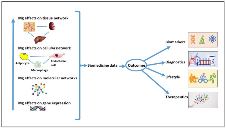
In this narrative literature review, we highlight the role of Mg in regulating AT metabolism and its potential impact on preventing OS and LGCI in AT and obesity. This preventive action of Mg is primarily attributed to its role in maintaining mitochondrial function, supporting antioxidant defenses, and acting as a calcium antagonist. Nevertheless, despite extensive research in major databases, we found limited studies that directly or indirectly investigate these topics at preclinical and clinical levels. Studies should be conducted on the contribution of Mg in regulating adipogenesis and in modulating the function of adipocytes challenged with inflammatory cytokines and/or unbalanced levels of adipokines. New insights could be gained from omics studies conducted both in vivo and in vitro. This knowledge can contribute to the development of innovative treatment strategies to maintain health and prevent diseases (Figure 5). We hope that this review will inspire future research to delve deeper into the role of Mg in regulating metabolism and ROS production in AT.
Figure 5.
Biomedical research on Mg’s potential impact on health and disease prevention. Data from studies on the interaction between different cell types and between the various organs might result in the identification of biomarkers and strategies to prevent AT dysfunction and, in general, diseases.
Abbreviations
adipose tissue (AT); brown adipose tissue (BAT); body mass index (BMI); calcium (Ca); cat-alase (CAT); C-reactive protein (CRP); endothelial cells (ECs); endothelial nitric oxide synthase (eNOS); European Food Safety Authority (EFSA); fatty acid synthase (FAS); fatty acyl transport protein 1 (FATP 1); glutathione (GSH); glutathione peroxidase (GPx); glutathione reductase (GR); heme oxygenase-1 (HO-1); insulin resistance (IR); interleukin (IL); low-grade chronic inflammation (LGCI); magnesium (Mg); mitochondrial RNA splicing 2 (MRS2); monocyte chemoattractant pro-tein-1 (MCP-1); NADPH oxidase (NOX); NAD(P)H: quinone oxidoreductase 1 (NQO1); nitric oxide synthase (NOS); nuclear factor-κB (NF-κB); oxidative stress (OS); paraoxonases (PON); perivascular adipose tissue (PVAT); peroxiredoxin (Prx); PPARγ coactivator 1-alpha (PGC1-α); PR domain containing 16 (PRDM 16); protein kinase C (PKC); reactive oxygen species (ROS); recommended dietary allowance (RDA); superoxide dismutase (SOD); thiobarbituric acid-reactive substances (TBARS); thioredoxin reductase (TrxR); Total Antioxidant Capacity (TAC); transient receptor po-tential ion channels for melastatin (TRPM); triglycerides (TG); tumor necrosis factor-α (TNF-α); white adipose tissue (WAT).
Author Contributions
Conceptualization, R.C. and J.A.M.; writing—original draft preparation, R.C., M.D.P., G.P. and J.A.M.; writing—review and editing, R.C., M.D.P., G.P. and J.A.M.; supervision, R.C. and J.A.M. All authors have read and agreed to the published version of the manuscript.
Conflicts of Interest
The authors declare no conflicts of interest.
Funding Statement
This research received no external funding.
Footnotes
Disclaimer/Publisher’s Note: The statements, opinions and data contained in all publications are solely those of the individual author(s) and contributor(s) and not of MDPI and/or the editor(s). MDPI and/or the editor(s) disclaim responsibility for any injury to people or property resulting from any ideas, methods, instructions or products referred to in the content.
References
- 1.Chen Y., Pan R., Pfeifer A. Fat tissues, the brite and the dark sides. Pflugers Arch. Eur. J. Physiol. 2016;468:1803–1807. doi: 10.1007/s00424-016-1884-8. [DOI] [PMC free article] [PubMed] [Google Scholar]
- 2.Chaurasiya V., Nidhina Haridas P.A., Olkkonen V.M. Adipocyte-endothelial cell interplay in adipose tissue physiology. Biochem. Pharmacol. 2024;222:116081. doi: 10.1016/j.bcp.2024.116081. [DOI] [PubMed] [Google Scholar]
- 3.Cheng C.K., Ding H., Jiang M., Yin H., Gollasch M., Huang Y. Perivascular adipose tissue: Fine-tuner of vascular redox status and inflammation. Redox Biol. 2023;62:102683. doi: 10.1016/j.redox.2023.102683. [DOI] [PMC free article] [PubMed] [Google Scholar]
- 4.Zocchi M., Della Porta M., Lombardoni F., Scrimieri R., Zuccotti G.V., Maier J.A., Cazzola R. A Potential Interplay between HDLs and Adiponectin in Promoting Endothelial Dysfunction in Obesity. Biomedicines. 2022;10:1344. doi: 10.3390/biomedicines10061344. [DOI] [PMC free article] [PubMed] [Google Scholar]
- 5.Liu M., Dudley S.C. Magnesium, Oxidative Stress, Inflammation, and Cardiovascular Disease. Antioxidants. 2020;9:907. doi: 10.3390/antiox9100907. [DOI] [PMC free article] [PubMed] [Google Scholar]
- 6.Zheltova A.A., Kharitonova M.V., Iezhitsa I.N., Spasov A.A. Magnesium deficiency and oxidative stress: An update. BioMedicine. 2016;6:8–14. doi: 10.7603/s40681-016-0020-6. [DOI] [PMC free article] [PubMed] [Google Scholar]
- 7.Schieber M., Chandel N.S. ROS Function in Redox Signaling and Oxidative Stress. Curr. Biol. 2014;24:R453. doi: 10.1016/j.cub.2014.03.034. [DOI] [PMC free article] [PubMed] [Google Scholar]
- 8.Horvath T.L., Andrews Z.B., Diano S. Fuel utilization by hypothalamic neurons: Roles for ROS. Trends Endocrinol. Metab. 2009;20:78–87. doi: 10.1016/j.tem.2008.10.003. [DOI] [PubMed] [Google Scholar]
- 9.Castro J.P., Grune T., Speckmann B. The two faces of reactive oxygen species (ROS) in adipocyte function and dysfunction. Biol. Chem. 2016;397:709–724. doi: 10.1515/hsz-2015-0305. [DOI] [PubMed] [Google Scholar]
- 10.Zhao R.Z., Jiang S., Zhang L., Yu Z. Bin Mitochondrial electron transport chain, ROS generation and uncoupling. Int. J. Mol. Med. 2019;44:3. doi: 10.3892/IJMM.2019.4188. [DOI] [PMC free article] [PubMed] [Google Scholar]
- 11.Boveris A., Oshino N., Chance B. The cellular production of hydrogen peroxide. Biochem. J. 1972;128:617. doi: 10.1042/bj1280617. [DOI] [PMC free article] [PubMed] [Google Scholar]
- 12.Nègre-Salvayre A., Hirtz C., Carrera G., Cazenave R., Troly M., Salvayre R., Pénicaud L., Casteilla L. A role for uncoupling protein-2 as a regulator of mitochondrial hydrogen peroxide generation. FASEB J. 1997;11:809–815. doi: 10.1096/fasebj.11.10.9271366. [DOI] [PubMed] [Google Scholar]
- 13.Song P., Zou M.H. Atherosclerosis: Risks, Mechanisms, and Therapies. Wiley; Hoboken, NJ, USA: 2015. Roles of Reactive Oxygen Species in Physiology and Pathology; pp. 379–392. [DOI] [Google Scholar]
- 14.Al-Anazi A., Parhar R., Saleh S., Al-Hijailan R., Inglis A., Al-Jufan M., Bazzi M., Hashmi S., Conca W., Collison K., et al. Intracellular calcium and NF-kB regulate hypoxia-induced leptin, VEGF, IL-6 and adiponectin secretion in human adipocytes. Life Sci. 2018;212:275–284. doi: 10.1016/j.lfs.2018.10.014. [DOI] [PubMed] [Google Scholar]
- 15.Wang B., Wood I.S., Trayhurn P. Dysregulation of the expression and secretion of inflammation-related adipokines by hypoxia in human adipocytes. Pflugers Arch. Eur. J. Physiol. 2007;455:479–492. doi: 10.1007/s00424-007-0301-8. [DOI] [PMC free article] [PubMed] [Google Scholar]
- 16.Li X., Ren Y., Chang K., Wu W., Griffiths H.R., Lu S., Gao D. Adipose tissue macrophages as potential targets for obesity and metabolic diseases. Front. Immunol. 2023;14:1153915. doi: 10.3389/fimmu.2023.1153915. [DOI] [PMC free article] [PubMed] [Google Scholar]
- 17.Liang W., Qi Y., Yi H., Mao C., Meng Q., Wang H., Zheng C. The Roles of Adipose Tissue Macrophages in Human Disease. Front. Immunol. 2022;13:908749. doi: 10.3389/fimmu.2022.908749. [DOI] [PMC free article] [PubMed] [Google Scholar]
- 18.Piuri G., Zocchi M., Della Porta M., Ficara V., Manoni M., Zuccotti G.V., Pinotti L., Maier J.A., Cazzola R. Magnesium in Obesity, Metabolic Syndrome, and Type 2 Diabetes. Nutrients. 2021;13:320. doi: 10.3390/nu13020320. [DOI] [PMC free article] [PubMed] [Google Scholar]
- 19.Chavakis T., Alexaki V.I., Ferrante A.W. Macrophage function in adipose tissue homeostasis and metabolic inflammation. Nat. Immunol. 2023;24:757–766. doi: 10.1038/s41590-023-01479-0. [DOI] [PubMed] [Google Scholar]
- 20.Trapani V., Rosanoff A., Baniasadi S., Barbagallo M., Castiglioni S., Guerrero-Romero F., Iotti S., Mazur A., Micke O., Pourdowlat G., et al. The relevance of magnesium homeostasis in COVID-19. Eur. J. Nutr. 2022;61:625–636. doi: 10.1007/s00394-021-02704-y. [DOI] [PMC free article] [PubMed] [Google Scholar]
- 21.Touyz R.M., Baaij J.H.F. De Hoenderop, J.G.J. Magnesium Disorders. N. Engl. J. Med. 2024;390:1998–2009. doi: 10.1056/NEJMra1510603. [DOI] [PubMed] [Google Scholar]
- 22.Pelczyńska M., Moszak M., Bogdański P. The Role of Magnesium in the Pathogenesis of Metabolic Disorders. Nutrients. 2022;14:1714. doi: 10.3390/nu14091714. [DOI] [PMC free article] [PubMed] [Google Scholar]
- 23.Morais J.B.S., Severo J.S., dos Santos L.R., de Sousa Melo S.R., de Oliveira Santos R., de Oliveira A.R.S., Cruz K.J.C., do Nascimento Marreiro D. Role of Magnesium in Oxidative Stress in Individuals with Obesity. Biol. Trace Elem. Res. 2017;176:20–26. doi: 10.1007/s12011-016-0793-1. [DOI] [PubMed] [Google Scholar]
- 24.Cazzola R., Della Porta M., Manoni M., Iotti S., Pinotti L., Maier J.A. Going to the roots of reduced magnesium dietary intake: A tradeoff between climate changes and sources. Helyon. 2020;6:e05390. doi: 10.1016/j.heliyon.2020.e05390. [DOI] [PMC free article] [PubMed] [Google Scholar]
- 25.Rondanelli M., Faliva M.A., Gasparri C., Peroni G., Naso M., Picciotto G., Riva A., Nichetti M., Infantino V., Alalwan T.A., et al. Micronutrients Dietary Supplementation Advices for Celiac Patients on Long-Term Gluten-Free Diet with Good Compliance: A Review. Medicina. 2019;55:337. doi: 10.3390/medicina55070337. [DOI] [PMC free article] [PubMed] [Google Scholar]
- 26.Galland L. Magnesium and inflammatory bowel disease. Magnesium. 1988;7:78–83. [PubMed] [Google Scholar]
- 27.Dinicolantonio J.J., O’keefe J.H., Wilson W. Subclinical magnesium deficiency: A principal driver of cardiovascular disease and a public health crisis Coronary artery disease. Open Hear. 2018;5:668. doi: 10.1136/openhrt-2017-000668. [DOI] [PMC free article] [PubMed] [Google Scholar]
- 28.Bateman S.W. A Quick Reference on Magnesium. Vet. Clin. N. Am. Small Anim. Pract. 2017;47:235–239. doi: 10.1016/j.cvsm.2016.09.002. [DOI] [PubMed] [Google Scholar]
- 29.Chrysant S.G. Proton pump inhibitor-induced hypomagnesemia complicated with serious cardiac arrhythmias. Expert Rev. Cardiovasc. Ther. 2019;17:345–351. doi: 10.1080/14779072.2019.1615446. [DOI] [PubMed] [Google Scholar]
- 30.Maguire D., Ross D.P., Talwar D., Forrest E., Naz Abbasi H., Leach J.P., Woods M., Zhu L.Y., Dickson S., Kwok T., et al. Low serum magnesium and 1-year mortality in alcohol withdrawal syndrome. Eur. J. Clin. Investig. 2019;49:e13152. doi: 10.1111/eci.13152. [DOI] [PubMed] [Google Scholar]
- 31.Grochowski C., Blicharska E., Baj J., Mierzwínska A., Brzozowska K., Forma A., MacIejewski R. Serum iron, magnesium, copper, and manganese levels in alcoholism: A systematic review. Molecules. 2019;24:1361. doi: 10.3390/molecules24071361. [DOI] [PMC free article] [PubMed] [Google Scholar]
- 32.Hunt C.D., Johnson L.K. Magnesium requirements: New estimations for men and women by cross-sectional statistical analyses of metabolic magnesium balance data. Am. J. Clin. Nutr. 2006;84:843–852. doi: 10.1093/ajcn/84.4.843. [DOI] [PubMed] [Google Scholar]
- 33.Barbagallo M., Belvedere M., Dominguez L.J. Magnesium homeostasis and aging. Magnes. Res. 2009;22:235–246. doi: 10.1684/mrh.2009.0187. [DOI] [PubMed] [Google Scholar]
- 34.Ford E.S., Mokdad A.H. Dietary magnesium intake in a national sample of US adults. J. Nutr. 2003;133:2879–2882. doi: 10.1093/jn/133.9.2879. [DOI] [PubMed] [Google Scholar]
- 35.Hartwig A. Role of magnesium in genomic stability. Mutat. Res. Fundam. Mol. Mech. Mutagen. 2001;475:113–121. doi: 10.1016/S0027-5107(01)00074-4. [DOI] [PubMed] [Google Scholar]
- 36.López-Otín C., Blasco M.A., Partridge L., Serrano M., Kroemer G. Hallmarks of aging: An expanding universe. Cell. 2023;186:243–278. doi: 10.1016/j.cell.2022.11.001. [DOI] [PubMed] [Google Scholar]
- 37.Nielsen F.H., Lukaski H.C., Nielsen F.H. Update on the relationship between magnesium and exercise. Magnes. Res. 2006;19:180–189. [PubMed] [Google Scholar]
- 38.Fine K.D., Santa Ana C.A., Porter J.L., Fordtran J.S. Intestinal absorption of magnesium from food and supplements. J. Clin. Invest. 1991;88:396–402. doi: 10.1172/JCI115317. [DOI] [PMC free article] [PubMed] [Google Scholar]
- 39.Kocaadam-Bozkurt B., Karaçil Ermumcu M.Ş., Erdoğan Gövez N., Bozkurt O., Akpinar Ş., Mengi Çelik Ö., Köksal E., Tek N.A. Association between adherence to the Mediterranean diet with anthropometric measurements and nutritional status in adolescents. Nutr. Hosp. 2023;40:368–376. doi: 10.20960/nh.04545. [DOI] [PubMed] [Google Scholar]
- 40.Chen F., Du M., Blumberg J.B., Chui K.K.H., Ruan M., Rogers G., Shan Z., Zeng L., Zhang F.F. Association Between Dietary Supplement Use, Nutrient Intake, and Mortality Among US Adults: A Cohort Study. Ann. Intern. Med. 2019;170:604. doi: 10.7326/M18-2478. [DOI] [PMC free article] [PubMed] [Google Scholar]
- 41.Arancibia-Hernández Y.L., Hernández-Cruz E.Y., Pedraza-Chaverri J. Magnesium (Mg2+) Deficiency, Not Well-Recognized Non-Infectious Pandemic: Origin and Consequence of Chronic Inflammatory and Oxidative Stress-Associated Diseases. Cell. Physiol. Biochem. 2023;57:1–23. doi: 10.33594/000000603. [DOI] [PubMed] [Google Scholar]
- 42.Kostov K. Effects of magnesium deficiency on mechanisms of insulin resistance in type 2 diabetes: Focusing on the processes of insulin secretion and signaling. Int. J. Mol. Sci. 2019;20:1351. doi: 10.3390/ijms20061351. [DOI] [PMC free article] [PubMed] [Google Scholar]
- 43.Man A.W.C., Xia N., Li H. Circadian Rhythm in Adipose Tissue: Novel Antioxidant Target for Metabolic and Cardiovascular Diseases. Antioxidants. 2020;9:968. doi: 10.3390/antiox9100968. [DOI] [PMC free article] [PubMed] [Google Scholar]
- 44.Feeney K.A., Hansen L.L., Putker M., Olivares-Yañez C., Day J., Eades L.J., Larrondo L.F., Hoyle N.P., O’Neill J.S., Van Ooijen G. Daily magnesium fluxes regulate cellular timekeeping and energy balance. Nature. 2016;532:375–379. doi: 10.1038/nature17407. [DOI] [PMC free article] [PubMed] [Google Scholar]
- 45.Zhou Y., Li H., Xia N. The Interplay Between Adipose Tissue and Vasculature: Role of Oxidative Stress in Obesity. Front. Cardiovasc. Med. 2021;8:650214. doi: 10.3389/fcvm.2021.650214. [DOI] [PMC free article] [PubMed] [Google Scholar]
- 46.Furukawa S., Fujita T., Shimabukuro M., Iwaki M., Yamada Y., Nakajima Y., Nakayama O., Makishima M., Matsuda M., Shimomura I. Increased oxidative stress in obesity and its impact on metabolic syndrome. J. Clin. Investig. 2017;114:1752–1761. doi: 10.1172/JCI21625. [DOI] [PMC free article] [PubMed] [Google Scholar]
- 47.Chattopadhyay M., Khemka V.K., Chatterjee G., Ganguly A., Mukhopadhyay S., Chakrabarti S. Enhanced ROS production and oxidative damage in subcutaneous white adipose tissue mitochondria in obese and type 2 diabetes subjects. Mol. Cell. Biochem. 2015;399:95–103. doi: 10.1007/s11010-014-2236-7. [DOI] [PubMed] [Google Scholar]
- 48.Chubanov V., Ferioli S., Wisnowsky A., Simmons D.G., Leitzinger C., Einer C., Jonas W., Shymkiv Y., Bartsch H., Braun A., et al. Epithelial magnesium transport by TRPM6 is essential for prenatal development and adult survival. eLife. 2016;5:e20914. doi: 10.7554/eLife.20914. [DOI] [PMC free article] [PubMed] [Google Scholar]
- 49.Suárez A., Pulido N., Casla A., Casanova B., Arrieta F.J., Rovira A. Impaired tyrosine-kinase activity of muscle insulin receptors from hypomagnesaemic rats. Diabetologia. 1995;38:1262–1270. doi: 10.1007/BF00401757. [DOI] [PubMed] [Google Scholar]
- 50.Sales C.H., Dos Santos A.R., Cintra D.E.C., Colli C. Magnesium-deficient high-fat diet: Effects on adiposity, lipid profile and insulin sensitivity in growing rats. Clin. Nutr. 2014;33:879–888. doi: 10.1016/j.clnu.2013.10.004. [DOI] [PubMed] [Google Scholar]
- 51.Bertinato J., Lavergne C., Rahimi S., Rachid H., Vu N.A., Plouffe L.J., Swist E. Moderately Low Magnesium Intake Impairs Growth of Lean Body Mass in Obese-Prone and Obese-Resistant Rats Fed a High-Energy Diet. Nutrients. 2016;8:253. doi: 10.3390/nu8050253. [DOI] [PMC free article] [PubMed] [Google Scholar]
- 52.Durlach J., Pagès N., Bac P., Bara M., Guiet-Bara A. Magnesium research: From the beginnings to today. Magnes. Res. 2004;17:163–168. [PubMed] [Google Scholar]
- 53.Devaux S., Adrian M., Laurant P., Berthelot A., Quignard-Boulangé A. Dietary magnesium intake alters age-related changes in rat adipose tissue cellularity. Magnes. Res. 2016;29:175–183. doi: 10.1684/mrh.2016.0406. [DOI] [PubMed] [Google Scholar]
- 54.Chaudhary D.P., Boparai R.K., Bansal D.D. Effect of a low magnesium diet on in vitro glucose uptake in sucrose fed rats. Magnes. Res. 2007;20:187–195. [PubMed] [Google Scholar]
- 55.Boparai R.K., Kiran R., Bansal D.D. Insinuation of exacerbated oxidative stress in sucrose-fed rats with a low dietary intake of magnesium: Evidence of oxidative damage to proteins. Free Radic. Res. 2007;41:981–989. doi: 10.1080/10715760701447892. [DOI] [PubMed] [Google Scholar]
- 56.Chaudhary D.P., Boparai R.K., Bansal D.D. Implications of oxidative stress in high sucrose low magnesium diet fed rats. Eur. J. Nutr. 2007;46:383–390. doi: 10.1007/s00394-007-0677-4. [DOI] [PubMed] [Google Scholar]
- 57.Kurstjens S., van Diepen J.A., Overmars-Bos C., Alkema W., Bindels R.J.M., Ashcroft F.M., Tack C.J.J., Hoenderop J.G.J., de Baaij J.H.F. Magnesium deficiency prevents high-fat-diet-induced obesity in mice. Diabetologia. 2018;61:2030–2042. doi: 10.1007/s00125-018-4680-5. [DOI] [PMC free article] [PubMed] [Google Scholar]
- 58.Venu L., Padmavathi I.J.N., Kishore Y.D., Bhanu N.V., Rao K.R., Sainath P.B., Ganeshan M., Raghunath M. Long-term effects of maternal magnesium restriction on adiposity and insulin resistance in rat pups. Obesity. 2008;16:1270–1276. doi: 10.1038/oby.2008.72. [DOI] [PubMed] [Google Scholar]
- 59.Zhong W., Ma M., Xie J., Huang C., Li X., Gao M. Adipose-specific deletion of the cation channel TRPM7 inhibits TAK1 kinase-dependent inflammation and obesity in male mice. Nat. Commun. 2023;14:491. doi: 10.1038/s41467-023-36154-3. [DOI] [PMC free article] [PubMed] [Google Scholar]
- 60.Liu M., Dudley S.C. Beyond Ion Homeostasis: Hypomagnesemia, Transient Receptor Potential Melastatin Channel 7, Mitochondrial Function, and Inflammation. Nutrients. 2023;15:3920. doi: 10.3390/nu15183920. [DOI] [PMC free article] [PubMed] [Google Scholar]
- 61.Baldoli E., Castiglioni S., Maier J.A.M. Regulation and Function of TRPM7 in Human Endothelial Cells: TRPM7 as a Potential Novel Regulator of Endothelial Function. PLoS ONE. 2013;8:e59891. doi: 10.1371/journal.pone.0059891. [DOI] [PMC free article] [PubMed] [Google Scholar]
- 62.Scrimieri R., Locatelli L., Cazzola R., Maier J.A.M., Cazzaniga A. Reactive oxygen species are implicated in altering magnesium homeostasis in endothelial cells exposed to high glucose. Magnes. Res. 2019;32:54–62. doi: 10.1684/mrh.2019.0456. [DOI] [PubMed] [Google Scholar]
- 63.Miller B.A. The role of TRP channels in oxidative stress-induced cell death. J. Membr. Biol. 2006;209:31–41. doi: 10.1007/s00232-005-0839-3. [DOI] [PubMed] [Google Scholar]
- 64.Liu M., Liu H., Feng F., Krook-Magnuson E., Dudley S.C. TRPM7 kinase mediates hypomagnesemia-induced seizure-related death. Sci. Rep. 2023;13:7855. doi: 10.1038/s41598-023-34789-2. [DOI] [PMC free article] [PubMed] [Google Scholar]
- 65.Javadov S., Kozlov A.V., Camara A.K.S. Mitochondria in Health and Diseases. Cells. 2020;9:1177. doi: 10.3390/cells9051177. [DOI] [PMC free article] [PubMed] [Google Scholar]
- 66.Kubota T., Shindo Y., Tokuno K., Komatsu H., Ogawa H., Kudo S., Kitamura Y., Suzuki K., Oka K. Mitochondria are intracellular magnesium stores: Investigation by simultaneous fluorescent imagings in PC12 cells. Biochim. Biophys. Acta Mol. Cell Res. 2005;1744:19–28. doi: 10.1016/j.bbamcr.2004.10.013. [DOI] [PubMed] [Google Scholar]
- 67.Li M., Li Y., Lu Y., Li J., Lu X., Ren Y., Wen T., Wang Y., Chang S., Zhang X., et al. Molecular basis of Mg2+ permeation through the human mitochondrial Mrs2 channel. Nat. Commun. 2023;14:4713. doi: 10.1038/s41467-023-40516-2. [DOI] [PMC free article] [PubMed] [Google Scholar]
- 68.Madaris T.R., Venkatesan M., Maity S., Stein M.C., Vishnu N., Venkateswaran M.K., Davis J.G., Ramachandran K., Uthayabalan S., Allen C., et al. Limiting Mrs2-dependent mitochondrial Mg2+ uptake induces metabolic programming in prolonged dietary stress. Cell Rep. 2023;42:112155. doi: 10.1016/j.celrep.2023.112155. [DOI] [PMC free article] [PubMed] [Google Scholar]
- 69.Lai L.T.F., Balaraman J., Zhou F., Matthies D. Cryo-EM structures of human magnesium channel MRS2 reveal gating and regulatory mechanisms. Nat. Commun. 2023;14:7207. doi: 10.1038/s41467-023-42599-3. [DOI] [PMC free article] [PubMed] [Google Scholar]
- 70.Kandeel F.R., Balon E., Scott S., Nadler J.L. Magnesium deficiency and glucose metabolism in rat adipocytes. Metabolism. 1996;45:838–843. doi: 10.1016/S0026-0495(96)90156-0. [DOI] [PubMed] [Google Scholar]
- 71.Oost L.J., Kurstjens S., Ma C., Hoenderop J.G.J., Tack C.J., de Baaij J.H.F. Magnesium increases insulin-dependent glucose uptake in adipocytes. Front. Endocrinol. 2022;13:986616. doi: 10.3389/fendo.2022.986616. [DOI] [PMC free article] [PubMed] [Google Scholar]
- 72.Pramme-Steinwachs I., Jastroch M., Ussar S. Extracellular calcium modulates brown adipocyte differentiation and identity. Sci. Rep. 2017;7:8888. doi: 10.1038/s41598-017-09025-3. [DOI] [PMC free article] [PubMed] [Google Scholar]
- 73.Fedele G., Castiglioni S., Trapani V., Zafferri I., Bartolini M., Casati S.M., Ciuffreda P., Wolf F.I., Maier J.A. Impact of Inducible Nitric Oxide Synthase Activation on Endothelial Behavior under Magnesium Deficiency. Nutrients. 2024;16:1406. doi: 10.3390/nu16101406. [DOI] [PMC free article] [PubMed] [Google Scholar]
- 74.Ferrè S., Baldoli E., Leidi M., Maier J.A.M. Magnesium deficiency promotes a pro-atherogenic phenotype in cultured human endothelial cells via activation of NFkB. Biochim. Biophys. Acta. 2010;1802:952–958. doi: 10.1016/j.bbadis.2010.06.016. [DOI] [PubMed] [Google Scholar]
- 75.Almousa L.A., Salter A.M., Castellanos M., May S.T., Langley-Evans S.C. The Response of the Human Umbilical Vein Endothelial Cell Transcriptome to Variation in Magnesium Concentration. Nutrients. 2022;14:3586. doi: 10.3390/nu14173586. [DOI] [PMC free article] [PubMed] [Google Scholar]
- 76.Zheng J., Tian M., Liu L., Jia X., Sun M., Lai Y. Magnesium sulfate reduces vascular endothelial cell apoptosis in rats with preeclampsia via the miR-218-5p/HMGB1 pathway. Clin. Exp. Hypertens. 2022;44:159–166. doi: 10.1080/10641963.2021.2013492. [DOI] [PubMed] [Google Scholar]
- 77.Sun L., Li X., Xu M., Yang F., Wang W., Niu X. In vitro immunomodulation of magnesium on monocytic cell toward anti-inflammatory macrophages. Regen. Biomater. 2020;7:391–401. doi: 10.1093/rb/rbaa010. [DOI] [PMC free article] [PubMed] [Google Scholar]
- 78.Ohyama T. New aspects of magnesium function: A key regulator in nucleosome self-assembly, chromatin folding and phase separation. Int. J. Mol. Sci. 2019;20:4232. doi: 10.3390/ijms20174232. [DOI] [PMC free article] [PubMed] [Google Scholar]
- 79.Bist P., Rao D.N. Identification and mutational analysis of Mg2+ binding site in EcoP15I DNA methyltransferase: Involvement in target base eversion. J. Biol. Chem. 2003;278:41837–41848. doi: 10.1074/jbc.M307053200. [DOI] [PubMed] [Google Scholar]
- 80.Zhang X., Zhang Y., Wang C., Wang X. TET (Ten-eleven translocation) family proteins: Structure, biological functions and applications. Signal Transduct. Target. Ther. 2023;8:297. doi: 10.1038/s41392-023-01537-x. [DOI] [PMC free article] [PubMed] [Google Scholar]
- 81.Takaya J., Iharada A., Okihana H., Kaneko K. Magnesium deficiency in pregnant rats alters methylation of specific cytosines in the hepatic hydroxysteroid dehydrogenase-2 promoter of the offspring. Epigenetics. 2011;6:573–578. doi: 10.4161/epi.6.5.15220. [DOI] [PubMed] [Google Scholar]
- 82.Yang Y., Han A., Wang X., Yin X., Cui M., Lin Z. Lipid metabolism regulator human hydroxysteroid dehydrogenase-like 2 (HSDL2) modulates cervical cancer cell proliferation and metastasis. J. Cell. Mol. Med. 2021;25:4846–4859. doi: 10.1111/jcmm.16461. [DOI] [PMC free article] [PubMed] [Google Scholar]
- 83.Gharipour M., Mani A., Baghbahadorani M.A., de Souza Cardoso C.K., Jahanfar S., Sarrafzadegan N., de Oliveira C., Silveira E.A. How Are Epigenetic Modifications Related to Cardiovascular Disease in Older Adults? Int. J. Mol. Sci. 2021;22:9949. doi: 10.3390/ijms22189949. [DOI] [PMC free article] [PubMed] [Google Scholar]
- 84.Toprak O., Kurt H., Sarl Y., Şarklş C., Us H., Klrlk A. Magnesium replacement improves the metabolic profile in obese and pre-diabetic patients with mild-to-moderate chronic kidney disease: A 3-month, randomised, double-blind, placebo-controlled study. Kidney Blood Press. Res. 2017;42:33–42. doi: 10.1159/000468530. [DOI] [PubMed] [Google Scholar]
- 85.Rafiee M., Ghavami A., Rashidian A., Hadi A., Askari G. The effect of magnesium supplementation on anthropometric indices: A systematic review and dose–response meta-analysis of clinical trials. Br. J. Nutr. 2021;125:644–656. doi: 10.1017/S0007114520003037. [DOI] [PubMed] [Google Scholar]
- 86.Castellanos-Gutiérrez A., Sánchez-Pimienta T.G., Carriquiry A., Da Costa T.H.M., Ariza A.C. Higher dietary magnesium intake is associated with lower body mass index, waist circumference and serum glucose in Mexican adults. Nutr. J. 2018;17:114. doi: 10.1186/s12937-018-0422-2. [DOI] [PMC free article] [PubMed] [Google Scholar]
- 87.Mirrafiei A., Jabbarzadeh B., Hosseini Y., Djafarian K., Shab-Bidar S. No association between dietary magnesium intake and body composition among Iranian adults: A cross-sectional study. BMC Nutr. 2022;8:39. doi: 10.1186/s40795-022-00535-6. [DOI] [PMC free article] [PubMed] [Google Scholar]
- 88.Van Eyck A., Ledeganck K.J., Vermeiren E., De Lamper A., Eysackers M., Mortier J., Van Vliet M.P., Broere P., Roebersen M., France A., et al. Body composition helps to elucidate the different origins of low serum magnesium in children with obesity compared to children with type 1 diabetes. Eur. J. Pediatr. 2023;182:3743–3753. doi: 10.1007/s00431-023-05046-5. [DOI] [PubMed] [Google Scholar]
- 89.Rios-Lugo M.J., Serafín-Fabián J.I., Hernández-Mendoza H., Klünder-Klünder M., Cruz M., Chavez-Prieto E., Martínez-Navarro I., Vilchis-Gil J., Vazquez-Moreno M. Mediation effect of body mass index on the association between serum magnesium level and insulin resistance in children from Mexico City. Eur. J. Clin. Nutr. 2024 doi: 10.1038/s41430-024-01447-3. [DOI] [PubMed] [Google Scholar]
- 90.Mikalsen S.M., Bjørke-Monsen A.L., Whist J.E., Aaseth J. Improved Magnesium Levels in Morbidly Obese Diabetic and Non-diabetic Patients After Modest Weight Loss. Biol. Trace Elem. Res. 2019;188:45–51. doi: 10.1007/s12011-018-1349-3. [DOI] [PubMed] [Google Scholar]
- 91.Lecube A., Baena-Fustegueras J.A., Fort J.M., Pelegrí D., Hernández C., Simó R. Diabetes is the main factor accounting for hypomagnesemia in obese subjects. PLoS ONE. 2012;7:e30599. doi: 10.1371/journal.pone.0030599. [DOI] [PMC free article] [PubMed] [Google Scholar]
- 92.Barbagallo M. Magnesium and type 2 diabetes. World J. Diabetes. 2015;6:1152. doi: 10.4239/wjd.v6.i10.1152. [DOI] [PMC free article] [PubMed] [Google Scholar]
- 93.Nielsen F.H. Dietary Magnesium and Chronic Disease. Adv. Chronic Kidney Dis. 2018;25:230–235. doi: 10.1053/j.ackd.2017.11.005. [DOI] [PubMed] [Google Scholar]
- 94.King D.E., Mainous Iii A.G., Geesey M.E., Egan B.M., Rehman S. Magnesium supplement intake and C-reactive protein levels in adults. Nutr. Res. 2006;26:193–196. doi: 10.1016/j.nutres.2006.05.001. [DOI] [Google Scholar]
- 95.Cheung M.M., Dall R.D., Shewokis P.A., Altasan A., Volpe S.L., Amori R., Singh H., Sukumar D. The effect of combined magnesium and vitamin D supplementation on vitamin D status, systemic inflammation, and blood pressure: A randomized double-blinded controlled trial. Nutrition. 2022;99:111674. doi: 10.1016/j.nut.2022.111674. [DOI] [PubMed] [Google Scholar]
- 96.Chacko S.A., Sul J., Song Y., Li X., LeBlanc J., You Y., Butch A., Liu S. Magnesium supplementation, metabolic and inflammatory markers, and global genomic and proteomic profiling: A randomized, double-blind, controlled, crossover trial in overweight individuals. Am. J. Clin. Nutr. 2011;93:463–473. doi: 10.3945/ajcn.110.002949. [DOI] [PMC free article] [PubMed] [Google Scholar]
- 97.Mazidi M., Rezaie P., Banach M. Effect of magnesium supplements on serum C-reactive protein: A systematic review and meta-analysis. Arch. Med. Sci. 2018;14:707–716. doi: 10.5114/aoms.2018.75719. [DOI] [PMC free article] [PubMed] [Google Scholar]
- 98.Veronese N., Pizzol D., Smith L., Dominguez L.J., Barbagallo M. Effect of Magnesium Supplementation on Inflammatory Parameters: A Meta-Analysis of Randomized Controlled Trials. Nutrients. 2022;14:679. doi: 10.3390/nu14030679. [DOI] [PMC free article] [PubMed] [Google Scholar]
- 99.Petrović J., Stanić D., Dmitrašinović G., Plećaš-Solarović B., Ignjatović S., Batinić B., Popović D., Pešić V. Magnesium supplementation diminishes peripheral blood lymphocyte DNA oxidative damage in athletes and sedentary young man. Oxid. Med. Cell Longev. 2016;2016:2019643. doi: 10.1155/2016/2019643. [DOI] [PMC free article] [PubMed] [Google Scholar]
- 100.Vongpatanasin W., Peri-Okonny P., Velasco A., Arbique D., Wang Z., Ravikumar P., Adams-Huet B., Moe O.W., Pak C.Y.C. Effects of Potassium Magnesium Citrate Supplementation on 24-Hour Ambulatory Blood Pressure and Oxidative Stress Marker in Prehypertensive and Hypertensive Subjects. Am. J. Cardiol. 2016;118:849–853. doi: 10.1016/j.amjcard.2016.06.041. [DOI] [PMC free article] [PubMed] [Google Scholar]
- 101.Mousavi R., Alizadeh M., Jafarabadi M.A., Heidari L., Nikbakht R., Rezaei H.B., Karandish M. Effects of Melatonin and/or Magnesium Supplementation on Biomarkers of Inflammation and Oxidative Stress in Women with Polycystic Ovary Syndrome: A Randomized, Double-Blind, Placebo-Controlled Trial. Biol. Trace Elem. Res. 2022;200:1010–1019. doi: 10.1007/s12011-021-02725-y. [DOI] [PubMed] [Google Scholar]
- 102.Bede O., Nagy D., Surányi A., Horváth I., Szlávik M., Gyurkovits K. Effects of magnesium supplementation on the glutathione redox system in atopic asthmatic children. Inflamm. Res. 2008;57:279–286. doi: 10.1007/s00011-007-7077-3. [DOI] [PubMed] [Google Scholar]
- 103.De Oliveira A.R., Cruz K.J., Morais J., Severo J.S., Beserra J.B., dos Santos L.R., de Sousa M., Stéfany R., Luz L.M., de Sousa L.A., et al. Association Between Magnesium and Oxidative Stress in Patients with Obesity. Curr. Nutr. Food Sci. 2020;16:743–748. doi: 10.2174/1573401315666190730123842. [DOI] [Google Scholar]


