Abstract
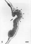
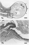
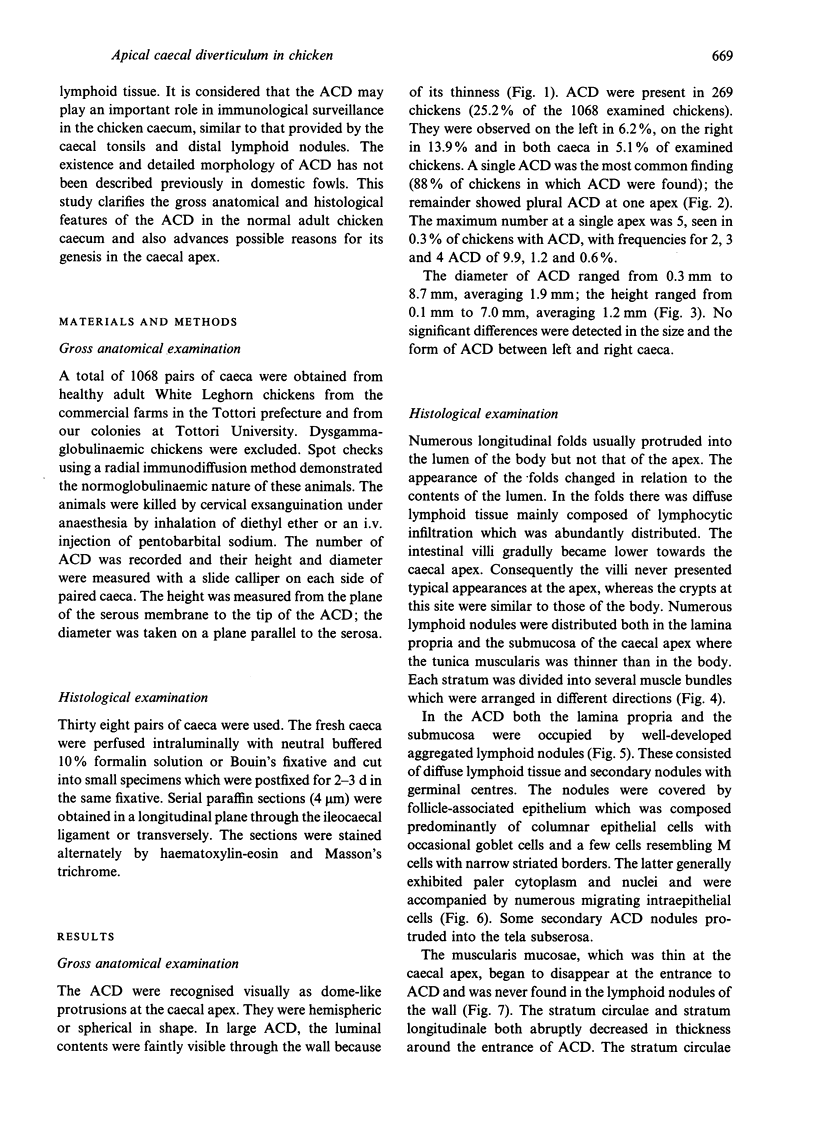
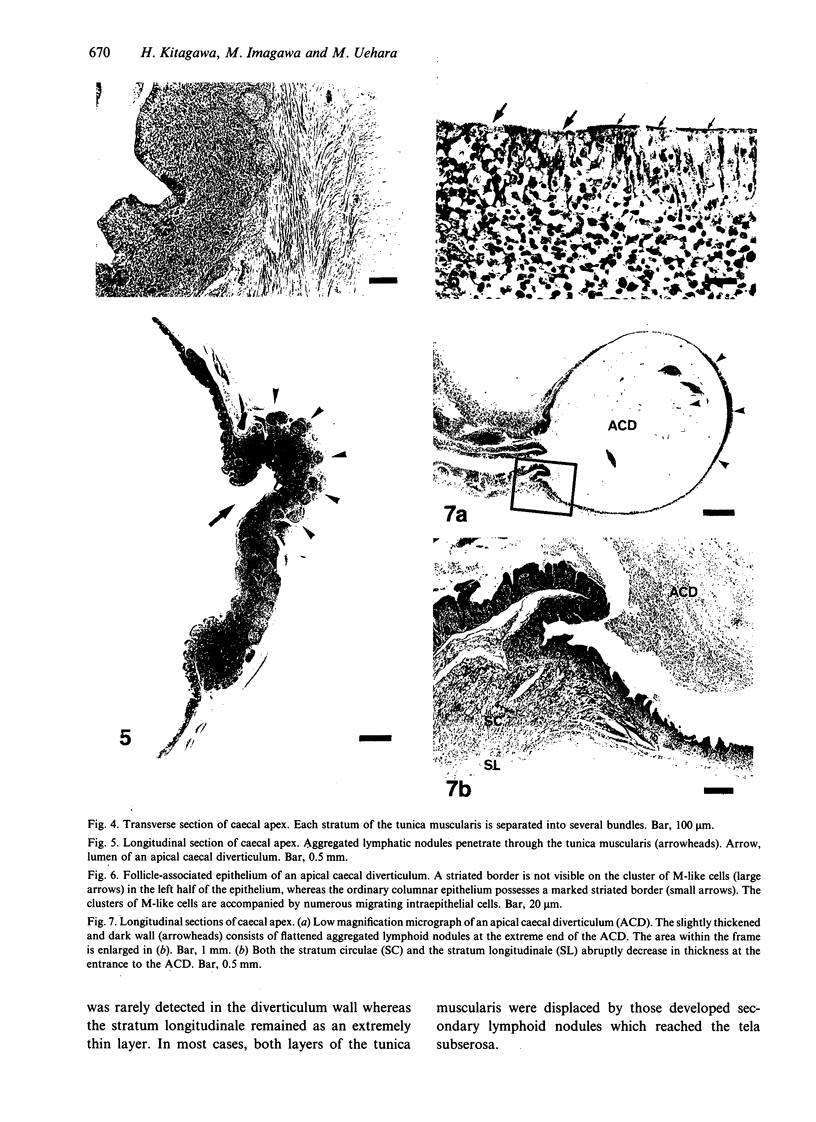
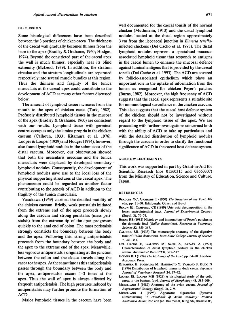
A peculiar structure, observed as a dome-like protrusion at the apex of the caecum, was investigated macroscopically and histologically in healthy White Leghorn chickens. It was hemispheric or spherical in shape and as it consisted of a lumen with a wall occupied by lymphoid tissue, this structure was designated the apical caecal diverticulum (ACD). ACD were detected in 25.2% of examined chickens and had a mean diameter and height of 1.9 mm and 1.2 mm respectively. Histologically, both the lamina propria mucosae and the submucosa of ACD consisted of well developed aggregated lymphoid nodules. Each nodule was covered by follicle-associated epithelium which contained cells resembling M cells. Some secondary nodules extended into the subserosa. The muscularis mucosae and the stratum circulae of the tunica muscularis disappeared near the entrance to ACD. The stratum longitudinale also gradually decreased in thickness around the entrance, becoming an extremely thin layer in the diverticulum wall. At the caecal apex, each stratum of the tunica muscularis was thinner than in the caecal body and separated into several muscle bundles. These bundles were occasionally displaced by developed lymphoid nodules, causing them to protrude into the subserosa. The high frequency of ACD suggests that caecal apex may be sites for immunological surveillance in the chicken caecum. In addition to the intense and frequent antiperistalsis at the apex suggested by Yasukawa (1959), possible causes for the formation of ACD included (1) the fragility of the tunica muscularis at the ACD, and (2) the local removal of the physical supporting structures by the development of lymphoid nodules.
Full text
PDF

Images in this article
Selected References
These references are in PubMed. This may not be the complete list of references from this article.
- Braun E. J., Campbell C. E. Uric acid decomposition in the lower gastrointestinal tract. J Exp Zool Suppl. 1989;3:70–74. doi: 10.1002/jez.1402520512. [DOI] [PubMed] [Google Scholar]
- Burns R. B. Histology and immunology of Peyer's patches in the domestic fowl (Gallus domesticus). Res Vet Sci. 1982 May;32(3):359–367. [PubMed] [Google Scholar]
- Kitamura H., Sugimura M., Hashimoto Y., Yamano S., Kudo N. Distribution of lymphatic tissues in duck caeca. Jpn J Vet Res. 1976 May;24(1-2):37–42. [PubMed] [Google Scholar]
- Mead G. C. Microbes of the avian cecum: types present and substrates utilized. J Exp Zool Suppl. 1989;3:48–54. doi: 10.1002/jez.1402520508. [DOI] [PubMed] [Google Scholar]
- Turk D. E. The anatomy of the avian digestive tract as related to feed utilization. Poult Sci. 1982 Jul;61(7):1225–1244. doi: 10.3382/ps.0611225. [DOI] [PubMed] [Google Scholar]
- del Cacho E., Gallego M., Sanz A., Zapata A. Characterization of distal lymphoid nodules in the chicken caecum. Anat Rec. 1993 Dec;237(4):512–517. doi: 10.1002/ar.1092370411. [DOI] [PubMed] [Google Scholar]


