Abstract
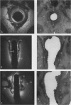
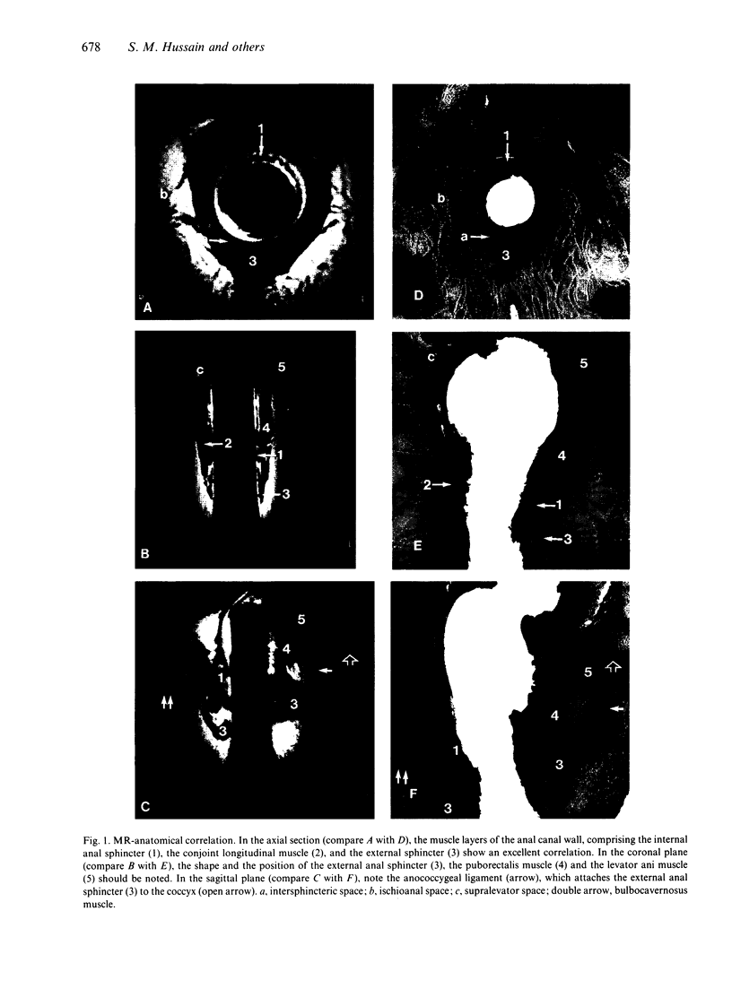
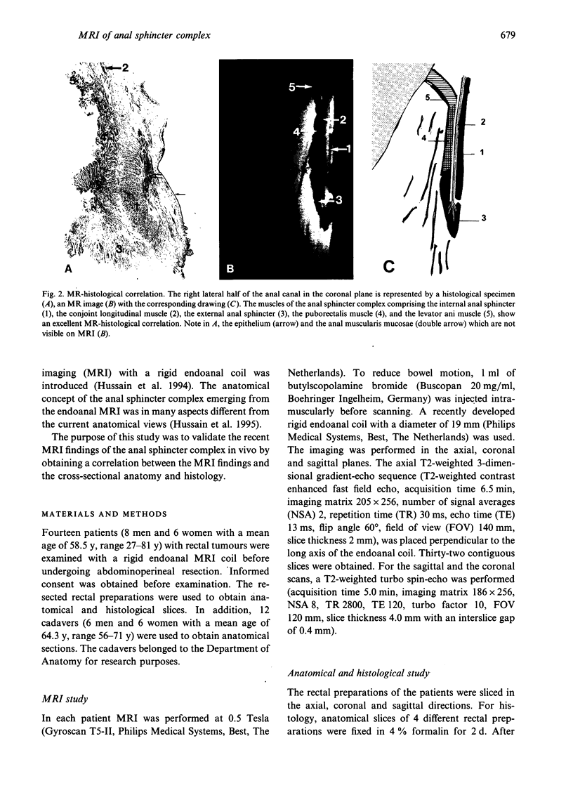
The purpose of this study was to correlate the in vivo endoanal MRI findings of the anal sphincter with the cross-sectional anatomy and histology. Fourteen patients with rectal tumours were examined with a rigid endoanal MR coil before undergoing abdominoperineal resection. In addition, 12 cadavers were used to obtain cross-sectional anatomical sections. The images were correlated with the histology and anatomy of the resected rectal specimens as well as with the cross-sectional anatomical sections of the 12 cadavers. The findings in 8 patients, 11 rectal preparations, and 10 cadavers, could be compared. In these cases, there was an excellent correlation between endoanal MRI and the cross-sectional cadaver anatomy and histology. With endoanal MRI, all muscle layers of the anal canal wall, comprising the internal anal sphincter, longitudinal muscle, the external anal sphincter and the puborectalis muscle were clearly visible. The levator ani muscle and ligamentous attachments were also well demonstrated. The perianal anatomical spaces, containing multiple septae, were clearly visible. In conclusion, endoanal MRI is excellent for visualising the anal sphincter complex and the findings show a good correlation with the cross-sectional anatomy and histology.
Full text
PDF
Images in this article
Selected References
These references are in PubMed. This may not be the complete list of references from this article.
- Ayoub S. F. Anatomy of the external anal sphincter in man. Acta Anat (Basel) 1979;105(1):25–36. doi: 10.1159/000145103. [DOI] [PubMed] [Google Scholar]
- Choen S., Burnett S., Bartram C. I., Nicholls R. J. Comparison between anal endosonography and digital examination in the evaluation of anal fistulae. Br J Surg. 1991 Apr;78(4):445–447. doi: 10.1002/bjs.1800780418. [DOI] [PubMed] [Google Scholar]
- Dalley A. F., 2nd The riddle of the sphincters. The morphophysiology of the anorectal mechanism reviewed. Am Surg. 1987 May;53(5):298–306. [PubMed] [Google Scholar]
- GOLIGHER J. C., LEACOCK A. G., BROSSY J. J. The surgical anatomy of the anal canal. Br J Surg. 1955 Jul;43(177):51–61. doi: 10.1002/bjs.18004317707. [DOI] [PubMed] [Google Scholar]
- Hussain S. M., Stoker J., Laméris J. S. Anal sphincter complex: endoanal MR imaging of normal anatomy. Radiology. 1995 Dec;197(3):671–677. doi: 10.1148/radiology.197.3.7480737. [DOI] [PubMed] [Google Scholar]
- Law P. J., Bartram C. I. Anal endosonography: technique and normal anatomy. Gastrointest Radiol. 1989 Fall;14(4):349–353. doi: 10.1007/BF01889235. [DOI] [PubMed] [Google Scholar]
- Oh C., Kark A. E. Anatomy of the external anal sphincter. Br J Surg. 1972 Sep;59(9):717–723. doi: 10.1002/bjs.1800590909. [DOI] [PubMed] [Google Scholar]
- Shafik A. A new concept of the anatomy of the anal sphincter mechanism and the physiology of defecation. The external anal sphincter: a triple-loop system. Invest Urol. 1975 Mar;12(5):412–419. [PubMed] [Google Scholar]
- Sultan A. H., Kamm M. A., Hudson C. N., Thomas J. M., Bartram C. I. Anal-sphincter disruption during vaginal delivery. N Engl J Med. 1993 Dec 23;329(26):1905–1911. doi: 10.1056/NEJM199312233292601. [DOI] [PubMed] [Google Scholar]
- Sultan A. H., Nicholls R. J., Kamm M. A., Hudson C. N., Beynon J., Bartram C. I. Anal endosonography and correlation with in vitro and in vivo anatomy. Br J Surg. 1993 Apr;80(4):508–511. doi: 10.1002/bjs.1800800435. [DOI] [PubMed] [Google Scholar]


