Abstract
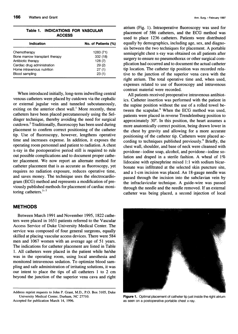
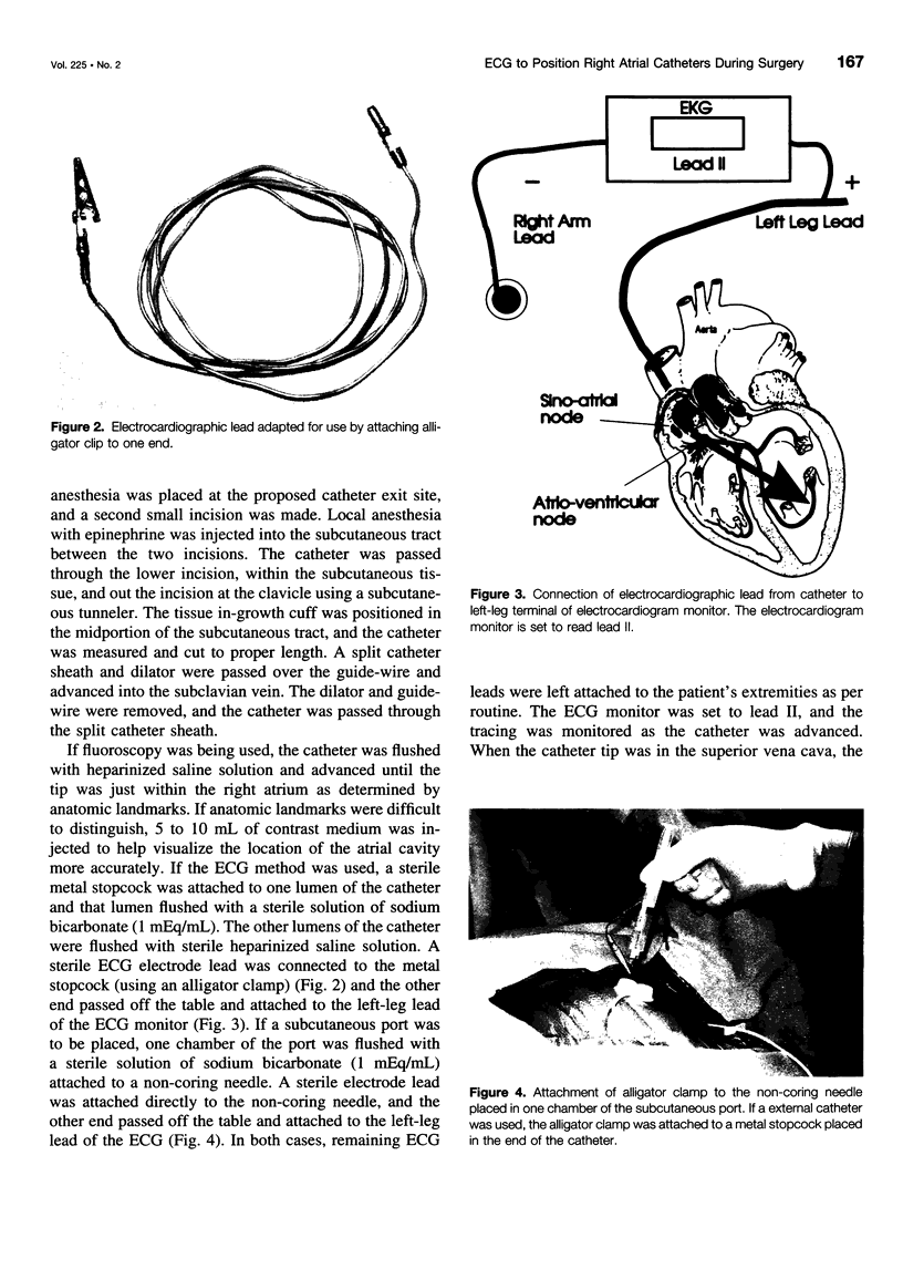
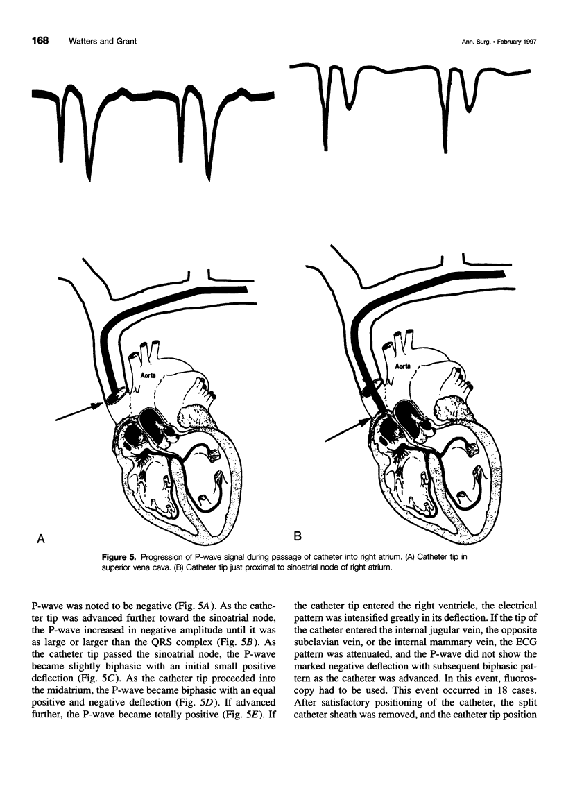
OBJECTIVE: This study evaluated the accuracy of placing right atrial catheters using an electrocardiographic (ECG) technique. SUMMARY BACKGROUND DATA: Placement of right atrial catheters for vascular access is a common operative procedure. Accurate placement is essential for proper function. Previous placement techniques have used fluoroscopy, which is both time consuming and hazardous. METHODS: The accuracy of placement of 1236 right atrial catheters using an ECG technique was compared to placement of 586 catheters using fluoroscopy between March 1991 and November 1995. In the ECG technique, the catheter was flushed with sodium bicarbonate. A sterile left-leg ECG lead was attached to the catheter with the other ECG leads applied normally. On advancing the catheter through the superior vena cava, the P-wave amplitude (lead II) increased in negative deflection until greater than the QRS complex. Passing the sinoatrial node, the P-wave developed an initial positive then negative deflection. The catheter was positioned so the P-wave was biphasic, representing a position midway between the sinoatrial and atrioventricular nodes. For the fluoroscopic technique, catheters were positioned under direct observation just within the atrium estimated from cardiac contour. Use of contrast was optional if atrial anatomy was unclear. RESULTS: Postoperative portable chest x-rays showed the ECG method to position the catheter tip within the right atrium just as accurately (average, 1.9 +/- 1.3 cm) as with the use of fluoroscopy (average, 1.1 +/- 1.6 cm). The ECG method eliminated an average of 20 seconds of radiation exposure, an average of 3.0 minutes operating room time (p < 0.04), avoided all risks of contrast dye, and saved $279.10 per case. CONCLUSIONS: The ECG method is a satisfactory alternative to that of fluoroscopy for placement of long-term central venous catheters into the right atrium.
Full text
PDF
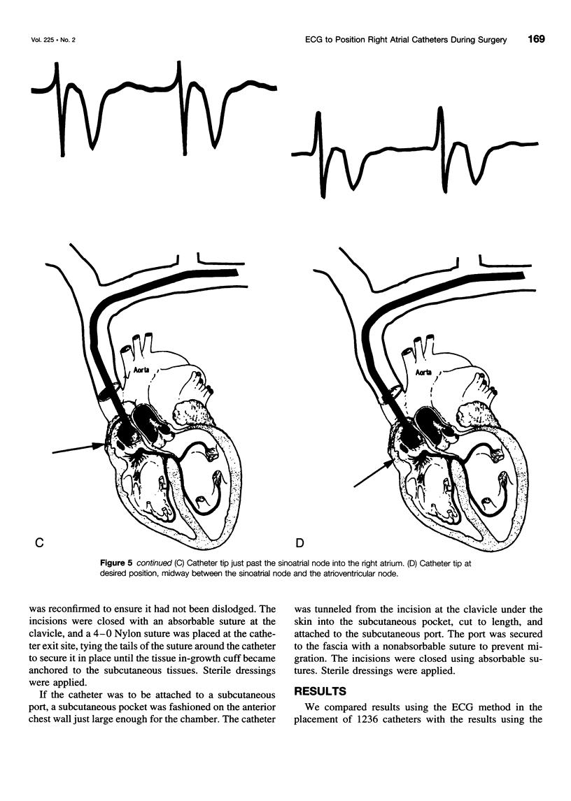
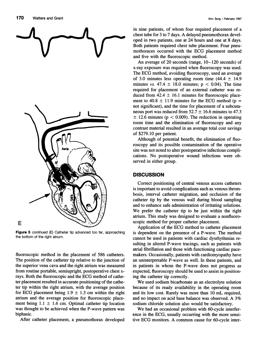
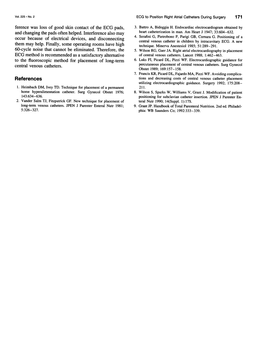
Images in this article
Selected References
These references are in PubMed. This may not be the complete list of references from this article.
- Francis K. R., Picard D. L., Fajardo M. A., Pizzi W. F. Avoiding complications and decreasing costs of central venous catheter placement utilizing electrocardiographic guidance. Surg Gynecol Obstet. 1992 Sep;175(3):208–211. [PubMed] [Google Scholar]
- Heimbach D. M., Ivey T. D. Technique for placement of a permanent home hyperalimentation catheter. Surg Gynecol Obstet. 1976 Oct;143(4):634–636. [PubMed] [Google Scholar]
- Luks F. I., Picard D. L., Pizzi W. F. Electrocardiographic guidance for percutaneous placement of central venous catheters. Surg Gynecol Obstet. 1989 Aug;169(2):157–158. [PubMed] [Google Scholar]
- Serafini G., Pietrobono P., Parigi G. B., Cornara G. Posizionamento mirato di catetere venoso centrale nel bambino per mezzo di elettrocardiogramma endocavitario. Nuova tecnica. Minerva Anestesiol. 1985 Jun;51(6):289–291. [PubMed] [Google Scholar]
- Vander Salm T. J., Fitzpatrick G. F. New technique for placement of long-term venous catheters. JPEN J Parenter Enteral Nutr. 1981 Jul-Aug;5(4):326–327. doi: 10.1177/0148607181005004326. [DOI] [PubMed] [Google Scholar]
- Wilson R. G., Gaer J. A. Right atrial electrocardiography in placement of central venous catheters. Lancet. 1988 Feb 27;1(8583):462–463. doi: 10.1016/s0140-6736(88)91247-0. [DOI] [PubMed] [Google Scholar]


