Abstract
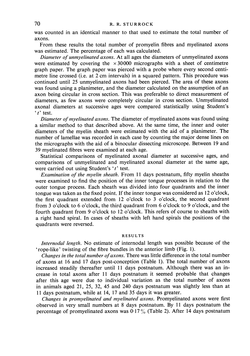
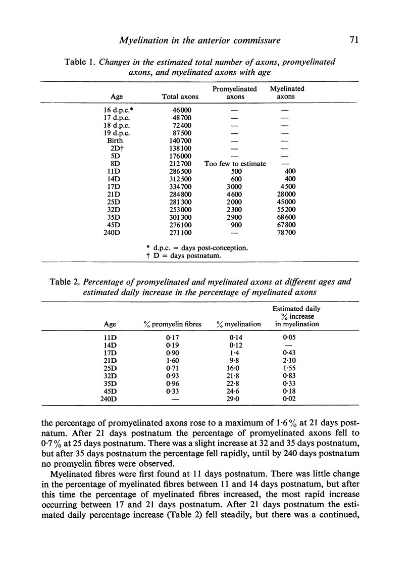
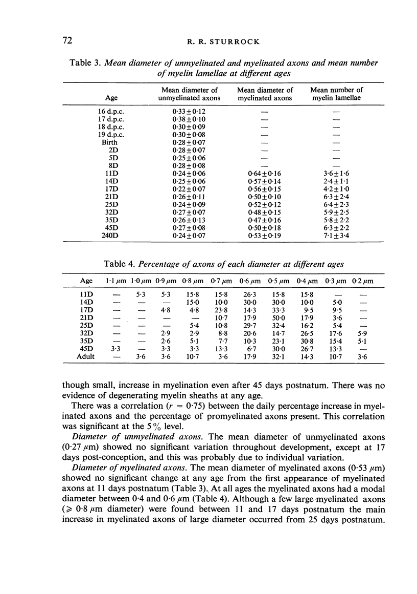
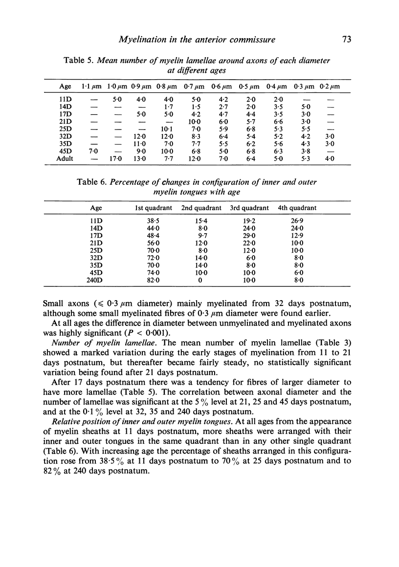
An electron microscopic study of myelination was carried out in the anterior limb of the anterior commissure of the mouse brain. The total number of axons increased from 48 700 at 17 days post-conception to 286 500 at 11 days postnatum. The first evidence of myelination was the presence of a few promyelin fibres at 8 days postnatum. Myelinated axons were first found at 11 days postnatum. The most rapid increase in myelinated fibres occurred between 17 and 21 days postnatum, but myelination continued to increase even after 45 days postnatum. There was no change in mean diameter (0-27 mum) of unmyelinated axons after 18 days post-conception. The mean diameter of myelinated axons (0-53 mum) also showed no variation with age. The modal diameter of myelinated axons lay between 0-4 and 0-6 mum. Small fibres (0-2-0-3 mum) myelinated around 32-35 days postnatum. The greatest increase in large myelinated axons (larger than or equal to 0-8 mum) occurred after 25 days postnatum. At all ages sheaths with outer and inner tongues in the same quadrant predominated and by 240 days postnatum 80% of sheaths showed this configuration.
Full text
PDF
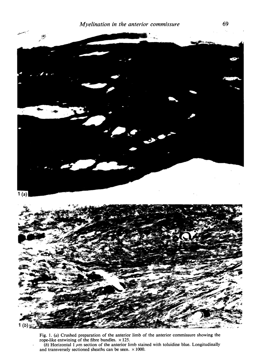
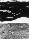
Images in this article
Selected References
These references are in PubMed. This may not be the complete list of references from this article.
- Fleischhauer K., Wartenberg H. Elektronenmikroskopische Untersuchungen über das Wachstum der Nervenfasern und über das Auftreten von Markscheiden im Corpus callosum der Katze. Z Zellforsch Mikrosk Anat. 1967;83(4):568–581. [PubMed] [Google Scholar]
- Fraher J. P. A quantitative study of anterior root fibres during early myelination. J Anat. 1972 May;112(Pt 1):99–124. [PMC free article] [PubMed] [Google Scholar]
- Hildebrand C. Ultrastructural and light-microscopic studies of the developing feline spinal cord white matter. II. Cell death and myelin sheath disintegration in the early postnatal period. Acta Physiol Scand Suppl. 1971;364:109–144. doi: 10.1111/j.1365-201x.1971.tb10980.x. [DOI] [PubMed] [Google Scholar]
- Matthews M. A., Duncan D. A quantitative study of morphological changes accompanying the initiation and progress of myelin production in the dorsal funiculus of the rat spinal cord. J Comp Neurol. 1971 May;142(1):1–22. doi: 10.1002/cne.901420102. [DOI] [PubMed] [Google Scholar]
- PETERS A. FURTHER OBSERVATIONS ON THE STRUCTURE OF MYELIN SHEATHS IN THE CENTRAL NERVOUS SYSTEM. J Cell Biol. 1964 Feb;20:281–296. doi: 10.1083/jcb.20.2.281. [DOI] [PMC free article] [PubMed] [Google Scholar]
- Sturrock R. R. A light microscope study of glial necrosis with age in the anterior limb of the anterior commissure of the pre and postnatal mouse. J Anat. 1974 Jul;117(Pt 3):469–474. [PMC free article] [PubMed] [Google Scholar]
- Sturrock R. R. Histogenesis of the anterior limb of the anterior commissure of the mouse brain. 3. An electron microscopic study of gliogenesis. J Anat. 1974 Feb;117(Pt 1):37–53. [PMC free article] [PubMed] [Google Scholar]
- Sturrock R. R. Histogenesis of the anterior limb of the anterior commissure of the mouse brain. I. A quantitative study of changes in the glial population with age. J Anat. 1974 Feb;117(Pt 1):17–25. [PMC free article] [PubMed] [Google Scholar]


