

Abstract
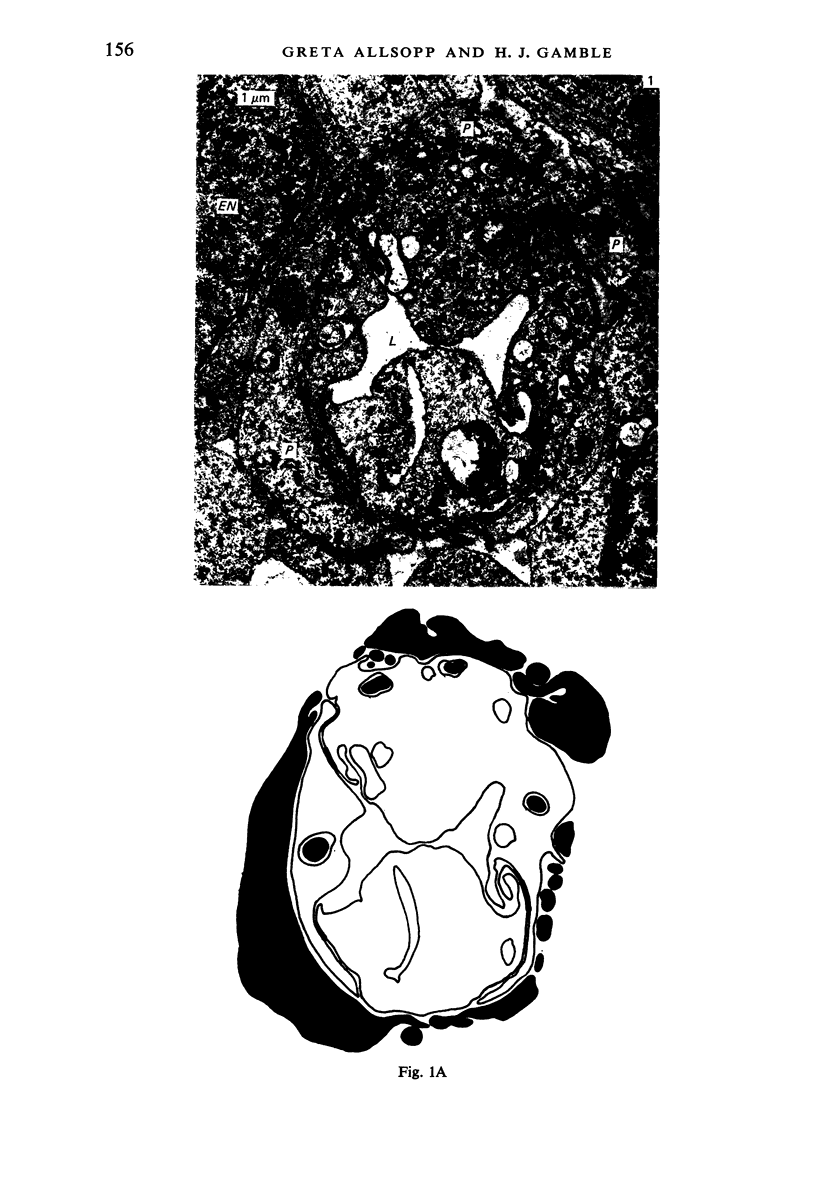
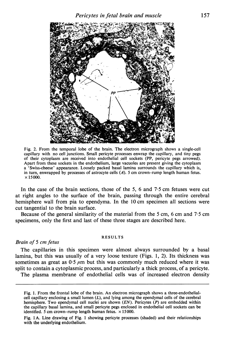
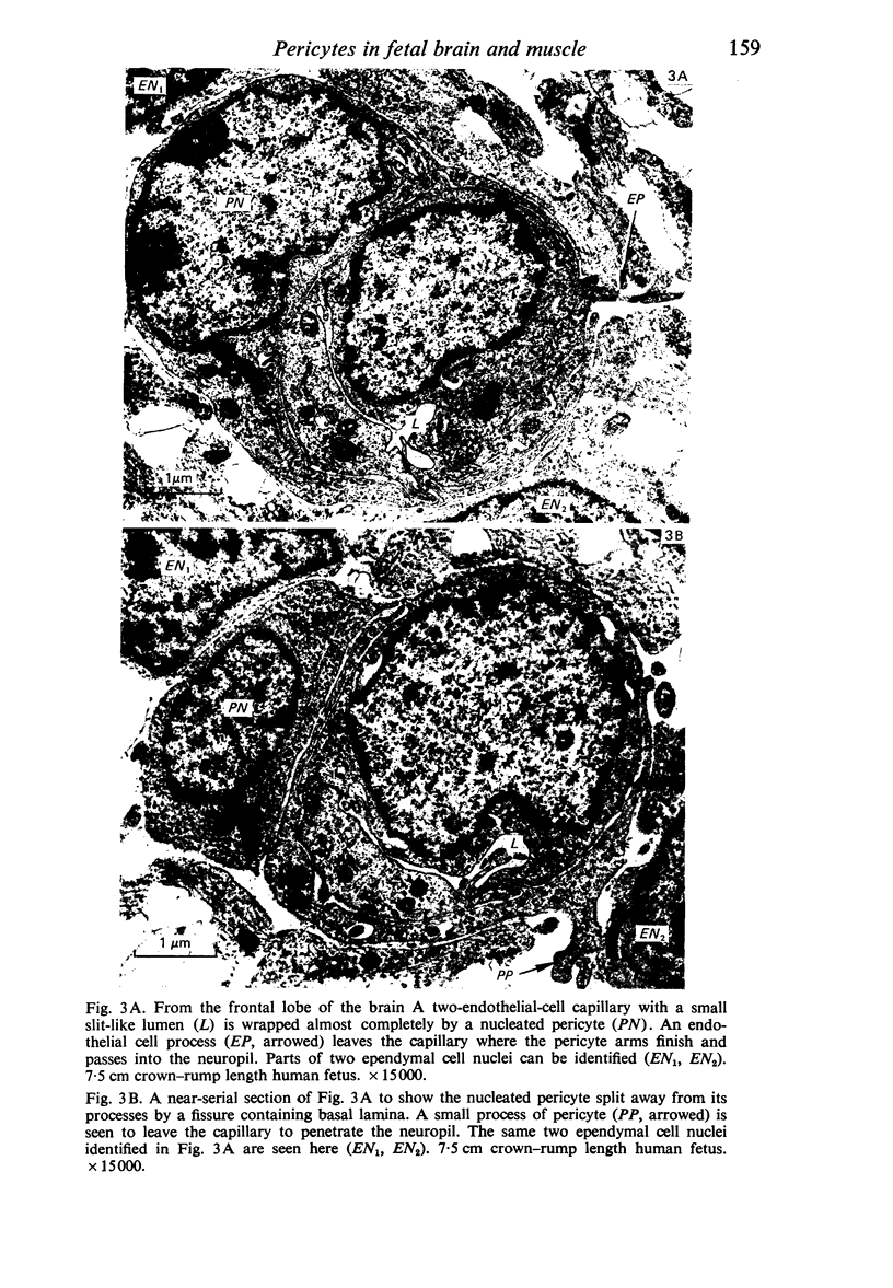
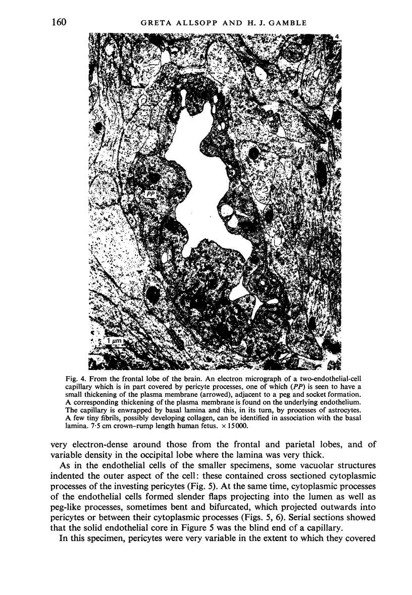
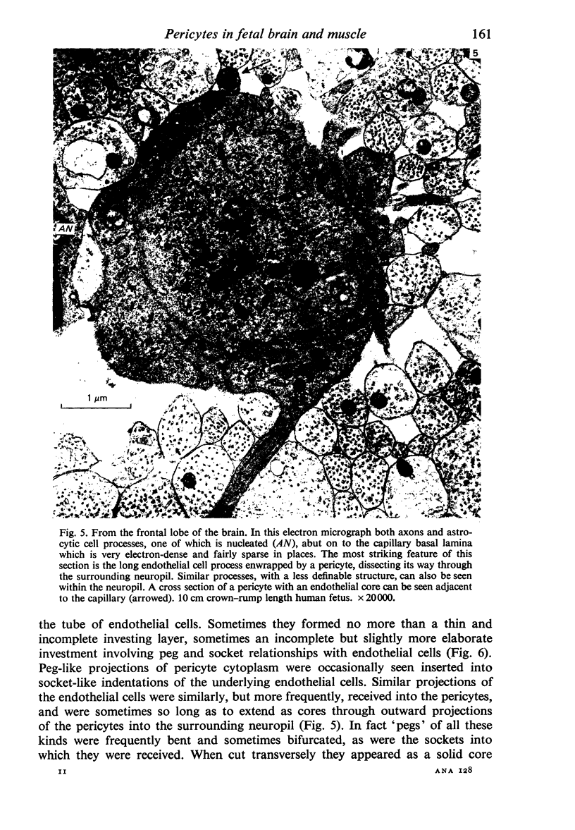
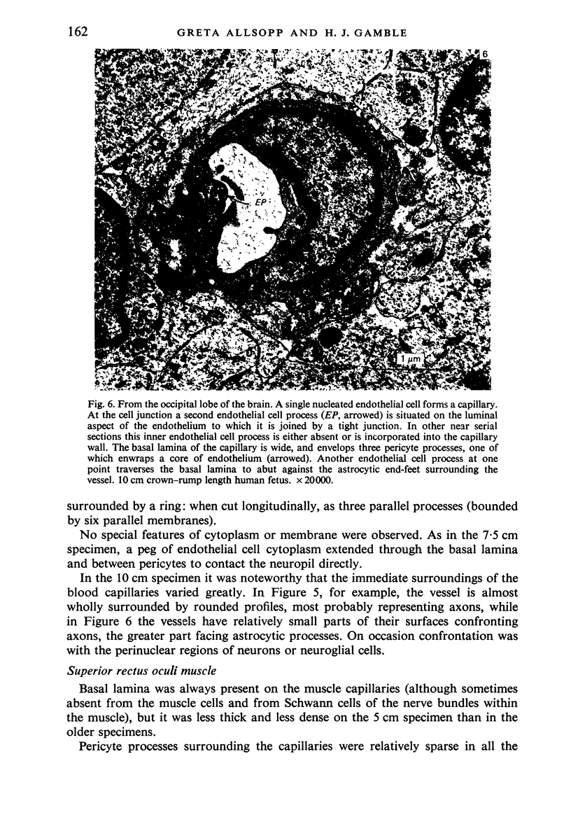
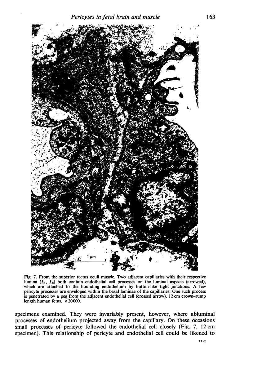
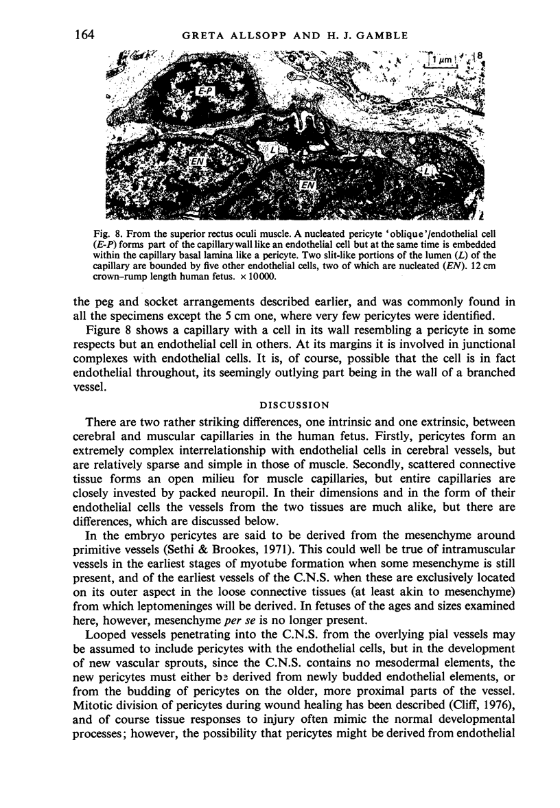
Capillaries of the developing cerebral cortex were examined by electron microscopy and compared with those developing in the superior rectus oculi muscle. The pericytes of cerebral vessels were found to be of very elaborate form and involved in complex 'peg and socket' relationships with endothelial cells. They were numerous and formed an almost complete investment of the endothelium. In the muscle, by contrast, pericytes were rare, although often of complex form. Cerebral capillaries were always completely and closely invested by neuropil, in marked contrast to the very loose connective tissues in the vicinity of muscle capillaries. The significance of the findings is discussed.
Full text
PDF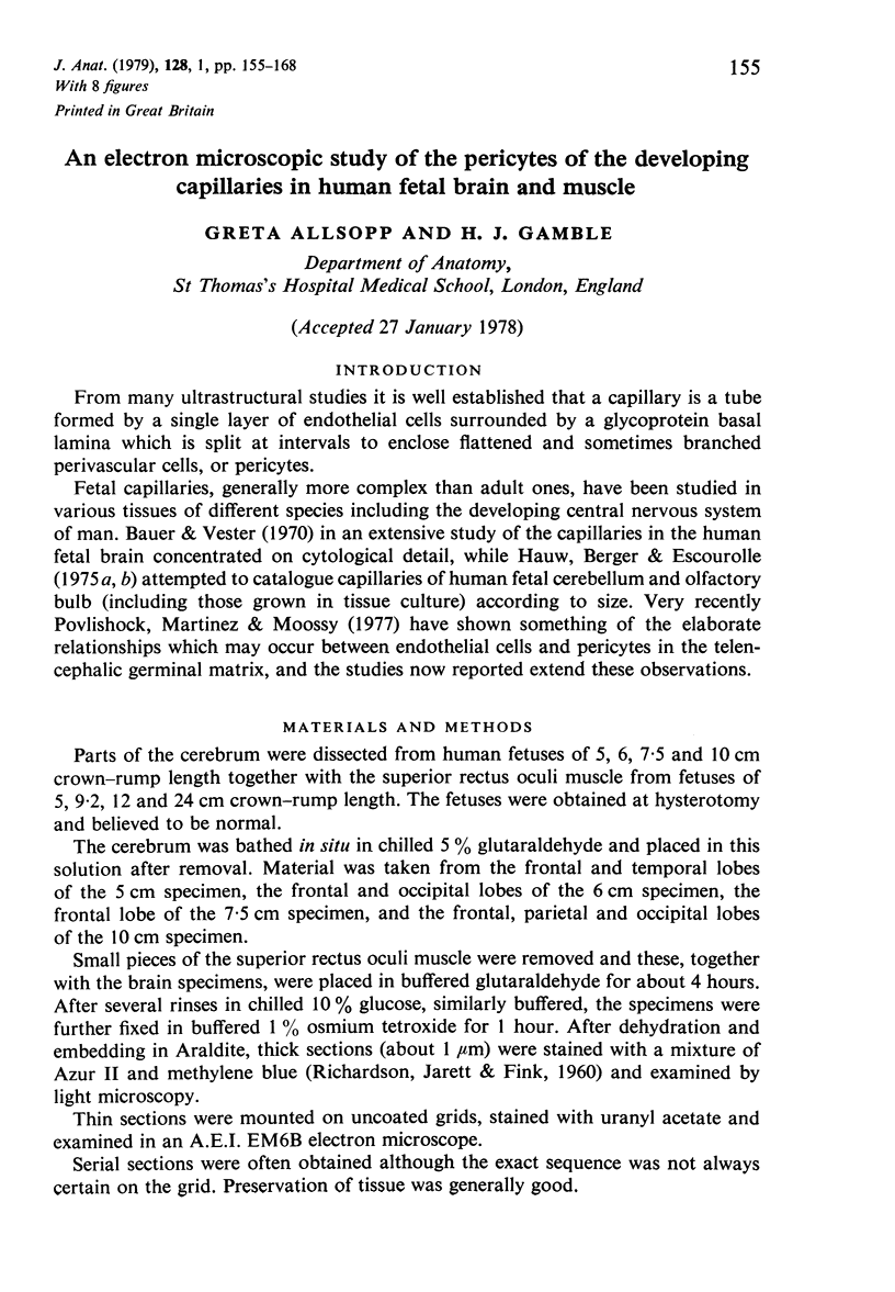
Images in this article
Selected References
These references are in PubMed. This may not be the complete list of references from this article.
- Bauer K. F., Vester G. Das elektronenmikroskopische Bild der Hirnkapillaren menschlicher Feten. Fortschr Neurol Psychiatr Grenzgeb. 1970 Jun;38(6):269–318. [PubMed] [Google Scholar]
- Crocker D. J., Murad T. M., Geer J. C. Role of the pericyte in wound healing. An ultrastructural study. Exp Mol Pathol. 1970 Aug;13(1):51–65. doi: 10.1016/0014-4800(70)90084-5. [DOI] [PubMed] [Google Scholar]
- Dyson S. E., Jones D. G., Kendrick W. L. Some observations on the ultrastructure of developing rat cerebral capillaries. Cell Tissue Res. 1976 Oct 19;173(4):529–542. doi: 10.1007/BF00224312. [DOI] [PubMed] [Google Scholar]
- FARQUHAR M. G., PALADE G. E. Glomerular permeability. II. Ferritin transfer across the glomerular capillary wall in nephrotic rats. J Exp Med. 1961 Nov 1;114:699–716. doi: 10.1084/jem.114.5.699. [DOI] [PMC free article] [PubMed] [Google Scholar]
- Forbes M. S., Rennels M. L., Nelson E. Ultrastructure of pericytes in mouse heart. Am J Anat. 1977 May;149(1):47–70. doi: 10.1002/aja.1001490105. [DOI] [PubMed] [Google Scholar]
- Hauw J. J., Berger B., Escourolle R. Ultrastructural observations on human cerebral capillaries in organ culture. Cell Tissue Res. 1975 Nov 7;163(2):133–150. doi: 10.1007/BF00221722. [DOI] [PubMed] [Google Scholar]
- Hauw J., Berger B., Escourolle R. Electron microscopic study of the developing capillaries of human brain. Acta Neuropathol. 1975;31(3):229–242. doi: 10.1007/BF00684562. [DOI] [PubMed] [Google Scholar]
- KUWABARA T., COGAN D. G. Retinal vascular patterns. VI. Mural cells of the retinal capillaries. Arch Ophthalmol. 1963 Apr;69:492–502. doi: 10.1001/archopht.1963.00960040498013. [DOI] [PubMed] [Google Scholar]
- Matsusaka T. Tridimensional views of the relationship of pericytes to endothelial cells of capillaries in the human choroid and retina. J Electron Microsc (Tokyo) 1975;24(1):13–18. [PubMed] [Google Scholar]
- Povlishock J. T., Martinez A. J., Moossy J. The fine structure of blood vessels of the telencephalic germinal matrix in the human fetus. Am J Anat. 1977 Aug;149(4):439–452. doi: 10.1002/aja.1001490402. [DOI] [PubMed] [Google Scholar]
- RICHARDSON K. C., JARETT L., FINKE E. H. Embedding in epoxy resins for ultrathin sectioning in electron microscopy. Stain Technol. 1960 Nov;35:313–323. doi: 10.3109/10520296009114754. [DOI] [PubMed] [Google Scholar]
- Roy S., Hirano A., Kochen J. A., Zimmerman H. M. The fine structure of cerebral blood vessels in chick embryo. Acta Neuropathol. 1974;30(4):277–285. doi: 10.1007/BF00697010. [DOI] [PubMed] [Google Scholar]
- SCHOEFL G. I. STUDIES ON INFLAMMATION. III. GROWING CAPILLARIES: THEIR STRUCTURE AND PERMEABILITY. Virchows Arch Pathol Anat Physiol Klin Med. 1963 Nov 8;337:97–141. [PubMed] [Google Scholar]
- Sethi N., Brookes M. Ultrastructure of the blood vessels in the chick allantois and chorioallantois. J Anat. 1971 May;109(Pt 1):1–15. [PMC free article] [PubMed] [Google Scholar]
- Stensaas L. J. Pericytes and perivascular microglial cells in the basal forebrain of the neonatal rabbit. Cell Tissue Res. 1975 May 20;158(4):517–541. doi: 10.1007/BF00220217. [DOI] [PubMed] [Google Scholar]