



Abstract
Antibody drug conjugates (ADCs) are effective targeted therapeutics but are limited in their ability to incorporate less-potent payloads, varied drug mechanisms of action, different drug-release mechanisms and tunable drug-to-antibody ratios. Here we introduce a technology to overcome these limitations called “antibody–bottlebrush prodrug conjugates” (ABCs). ABCs consist of an IgG1 monoclonal antibody (mAb) covalently conjugated to the terminus of a compact bivalent bottlebrush prodrug, with payloads bound through cleavable linkers and polyethylene glycol branches. This design enables the synthesis of ABCs with tunable drug-to-antibody ratios (DARs) up to two orders-of-magnitude greater than those of traditional ADCs. We demonstrate the functional flexibility and manufacturing efficiency of this technology by synthesizing >10 different ABCs targeting either HER2 or MUC1 with drug potencies spanning several orders-of-magnitude; imaging agents for ABC visualization; and photocatalysts for proximity-based labeling of the ABC interactome. ABCs display high target engagement, cell uptake, and improved efficacy in tumor models compared with conventional HER2-targeted ADCs, suggesting promise for clinical translation.
Cancer is a leading cause of death worldwide and while many transformative cancer therapies have been developed in recent decades, the number of cancer deaths continues to increase, motivating the search for novel targeted therapies.1 Amongst the targeted therapy platforms in development for cancer treatment, antibody drug conjugates (ADCs), consisting of a mAb conjugated to a small-molecule drug through a covalent linker, have demonstrated clinical success and future potential.2–13 More than a dozen ADCs have been approved worldwide, and hundreds of ADCs are in preclinical/clinical development.13–18 Despite these successes,5,19,20 traditional ADCs face inherent limitations that may hinder their broader applicability and versatility.21,22 For example, the payload scope of ADCs is limited to highly potent (<~10 nM IC50) cytotoxic agents with few MoAs, e.g., microtubule inhibition and DNA damage. As a result, significant toxicities and resistance can be observed,22,23 and the use of inherently more selective payloads, payload MoAs, or payload combinations is difficult. Because the payloads of ADCs are directly conjugated to amino acid sidechains of the mAb through short linkers, the average number of payloads that can be conjugated per mAb (i.e., the drug-to-antibody ratio or “DAR”) before deterioration of ADC properties is limited to ≤ 8 (Fig. 1a).10–14,21,22,25–29 While having a higher DAR may not always be better from a therapeutic perspective, particularly when highly potent payloads are used,30 a higher DAR may be required to use payloads that are ~10–100-fold less potent than traditional ADC payloads while maintaining a practical mAb dose (e.g., all clinically used ADCs so far are given at ~1–10 mg/kg doses of mAb).
Figure 1. Construction and in vitro evaluation of ABCs.
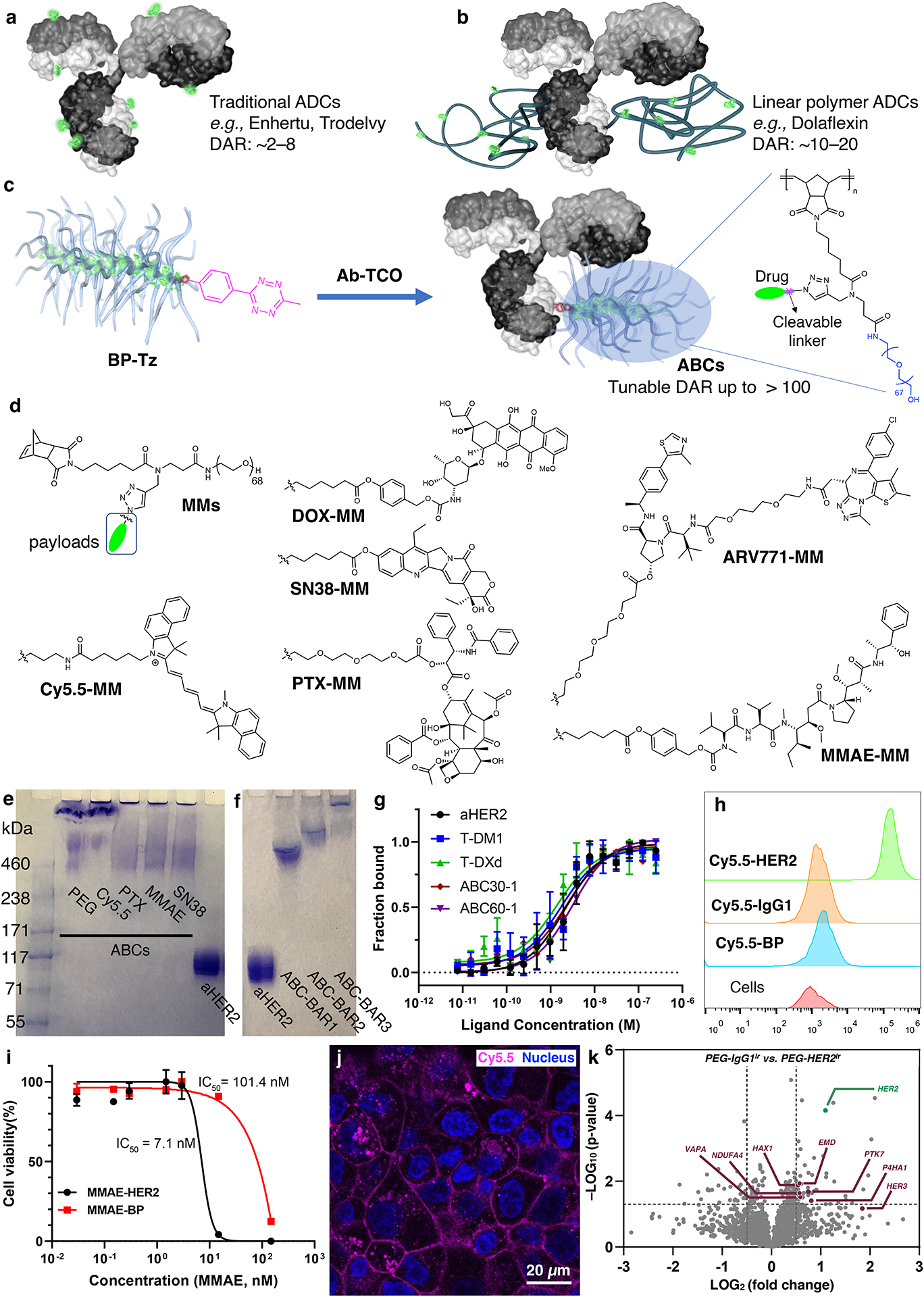
a. Traditional ADCs feature DARs of ~2–8. b. ADCs based on linear polymer–drug conjugates with drugs distributed on the pendants of hydrophilic polymer chains have been developed. The surface exposure of payloads and chemical heterogeneity in such systems may limit DAR and payload diversity. c. Formation of ABCs through “click” conjugation between Ab-TCO and BP-Tz enabled by a ROMP terminator (enyne-PEG12-Tz). ABCs comprise a mAb covalently conjugated to the end of one or more BPDs (BPD-to-antibody ratio or “BAR” = 1 shown in Figure), the latter of which has hydrophilic polymer chains (blue; PEG in this work) and drugs (green) and cleavable linkers (pink) attached to backbone repeat units, leading to a compact, homogenous microstructure that enables DAR values up to ~135 for a wide range of mechanistically distinct payloads. d. Chemical structures of the 6 different dye and drug–linker-containing MMs used in this work. e. Non-reducing SDS-PAGE gel comparing ABCs with different payloads. f. Non-reducing SDS-PAGE gel for isolated ABCs with different BAR values. g. Binding affinities of various HER2-targeted ADCs and ABCs as measured by MST. ABC30–1 refers to an ABC with DAR = 30 and BAR = 1. ABC60–1 refer to an ABC with DAR = 60 and BAR = 1. Results are presented as mean ± SEM (n = 3 technical replicates). h. In vitro characterization of the BT-474 cell targeting ability of ABCs as measured by flow cytometry (25 μg/mL of ABC, 1 h incubation, 5% Cy5.5 labeled ABC with BAR = 1). The x-axis represents the Cy5.5 fluorescence intensity. i. Cytotoxicity assay (MTT) comparing ABC MMAE-HER2 and BPD MMAE-BP after 24 h incubation, showing greater potency for the HER2-targeted ABC. Results are presented as mean ± SEM (n = 3 biological replicates). j. Confocal microscopy image showing BT-474 cell binding and uptake of Cy5.5-HER2 (50 μg/mL, 6 h incubation, 1% Cy5.5 labeled ABC). Magenta is Cy5.5; blue is Hoechst staining of the nucleus. k. Label-free quantitative proteomics for analyzing the targeted interactome of PEG-HER2Ir against an isotype PEG-IgG1Ir ABC on BT474 cells. Three biological replicates were created for each condition. In such constructs, the Ir-containing photocatalyst was conjugated to the antibodies (Trastuzumab or IgG1) via NHS ester-lysine coupling. Then, the PEG-BP was conjugated onto these antibodies to provide PEG-HER2Ir and PEG-IgG1Ir ABCs. Previously reported interactors of HER2 are identified in burgundy with dashed lines indicating statistical cutoff of log2(fold change) > 0.5, −log(P-value) > 1.3. Normalization was performed via median subtraction and a volcano plot was generated using a t-test for statistical significance. Contaminants were filtered manually for image clarity.
Accessing tunable and ultra-high DAR values with a well-defined molecular system that does not preclude mAb function may allow the use of payloads with varied MoAs and antigen targets in a tunable, modular fashion. Attachment or encapsulation of payloads into polymers or nanoparticles that are then conjugated to antibodies is a promising way to achieve this goal (Fig. 1b); however, such designs often lack payload and linker versatility due to the surface-exposed nature of the payloads, which inextricably link the physical properties of the payload–linker to the physical properties of the construct.31,32 A design that facilitates straightforward access to antibody–targeted delivery constructs with predictable physical properties regardless of payload and linker identity could broaden the landscape for cancer therapeutics.
We explored the use of molecular bottlebrush prodrugs (BPDs)33–37 as a solution to this challenge. BPDs are synthetic polymers with mAb-like dimensions (~10 nm diameter and controlled length) that feature branched pendants containing a hydrophilic polymer chain (e.g., polyethylene glycol (PEG)) and a payload–linker unit attached to a compact polymer backbone. The PEG chains of BPDs shield the payload–linker units, thus conferring consistent physical properties to the BPD regardless of the payload–linker composition and backbone length.8–11 Moreover, the average number of PEG–payload–linker units, which would control the DAR after mAb conjugation, can be varied by controlling the BPD synthesis stoichiometry, with straightforward access to values ranging from ~10–100. Additionally, the microstructure of BPDs enables chemical differentiation of the BPD backbone ends from the surface, which we leverage here to introduce a single “click” chemistry38–41 functional group for high-yielding conjugation to one mAb molecule per BPD. Finally, the efficient nature of BPD synthesis facilitates the use of a wide range of payloads and payload combinations, linkers, and antibodies, allowing rapid optimization in various disease contexts.35–37
Here, we report the synthesis and evaluation of ABCs in vitro and in vivo (Fig. 1c). First, we develop a synthetic method to terminate the ends of BPDs with reactive handles for selective and efficient “click” bioconjugation to mAbs using either stochastic (Lys) or site-specific (Cys) methods (Fig. 1c). Then, we demonstrate the synthesis, in vitro, and in vivo evaluation of ABCs based on 6 payload–linker combinations with distinct MoAs and two mAbs that target tumor antigens (HER2 and MUC1). We show that ABCs can have high target-specific engagement and cell uptake with average DAR values up to ~135, which leads to higher efficacy compared to non-targeted BPDs and clinical HER2-targeted ADCs despite having payloads that are up to ~100-fold less potent. Interactome micromapping experiments, enabled by the installation of photosensitizers into ABCs, show that ABCs engage their expected antigen targets in vitro. ABCs achieve good tumor regression with no discernable toxicities in several in vivo efficacy studies using HER2+ and MUC1+ cell-line-derived xenograft murine models. Moreover, ABCs with SN-38 payloads and DAR 60 display improved efficacy in a low-antigen-expressing tumor model compared to T-DXd, an Enhertu biosimilar with DAR ~ 8, at the same mAb dose and using a clinically relevant payload dose.
Results
Synthesis of ABCs via a modular “click” chemistry approach
Our strategy for the synthesis of ABCs involves conjugation of a pre-synthesized BPD to a mAb (Fig. 1c). While this approach would be highly modular, allowing, in principle, for variation of the payload–linker and mAb, we recognized the challenges of coupling nanoscale objects of similar sizes under the necessarily mild conditions and low reaction concentrations needed for mAb functionalization. Moreover, the chosen conjugation reaction must be orthogonal to the BPD payloads–linkers and compatible with BPD synthesis. To our knowledge, the conjugation of bottlebrush polymers to mAbs has not been demonstrated; however, “click” chemistry reactions have been developed precisely to tackle such challenges.38–41 We tested various possible methods for installation of click chemistry compatible functional groups onto the ends of model bottlebrush polymers (lacking payloads and linkers) with polynorbornene backbones (number-average degrees of polymerization, DP = 60) and PEG sidechains with molecular weights of 3k (total bottlebrush MW = 200 k) (see Supplementary Information for discussion; Supplementary Scheme 1–4, Supplementary Fig. 1–2). A versatile protocol was identified wherein bottlebrush polymer synthesis via ring-opening metathesis polymerization (ROMP) of suitable macromonomers (MMs, Fig. 1d) is quenched by an enyne terminator bearing a 6-methyl-1,2,4,5-tetrazine (enyne-PEG12-Tz, Supplementary Scheme 3 and 4),42,43 yielding a bottlebrush polymer (“BP-Tz”) with a single tetrazine on its backbone end (Fig. 1c, Supplementary Fig. 3). BP-Tz is set up for coupling to a trans-cycloctene (TCO)-functionalized mAb via inverse electron demand Diels–Alder cycloaddition, one of the most efficient biorthogonal “click” reactions.44–46 Lysine and cysteine are the most common amino acids used for ADC payload conjugation; here, as a proof-of-concept, we first appended TCOs to serum IgG1 via a lysine amidation reaction to provide IgG1-TCO. Notably, mixing BP-Tz and IgG1-TCO together (3:1) in neutral phosphate-buffered saline (PBS) at room temperature for 24 h gave quantitative consumption of IgG1-TCO to form conjugates PEG-IgG1 as a mixture of species with different numbers of BPs per mAb, as determined by SDS-page gel electrophoresis (Supplementary Fig. 4).
Encouraged by these results, we studied the conjugation of BP-Tz to the HER2-targeting mAb trastuzumab, which is currently used as a therapeutic mAb (Herceptin) and is the basis of clinically approved ADCs such as Kadcyla and Enhertu.47 Amidation of trastuzumab with TCO gave HER2-TCO (Supplementary Fig. 5), which was subsequently coupled to BP-Tz under the same conditions as described above to provide BP-HER2 as confirmed by SDS-PAGE (Fig. 1e). Next, we synthesized a Tz-terminated, cyanine-5.5 (Cy5.5) dye-labeled bottlebrush polymer (Cy5.5-Tz) and three BPDs with different payload–linkers: paclitaxel (PTX-Tz), monomethylauristatin (MMAE-Tz), and SN-38 (SN38-Tz), to test for conjugation to HER2-TCO; SDS-PAGE showed consumption of HER2-TCO within 24 hours in all cases (Fig. 1e), providing fluorophore-labeled Cy5.5-HER2 and ABCs MMAE-HER2, PTX-HER2, and SN38-HER2, with varied payload mechanisms and potencies spanning over two orders-of-magnitude.48
In ABCs, the DAR is determined by the BPD backbone DP as well as the number of BPDs per mAb (herein referred to as the “BPD-to-Antibody Ratio” or BAR). We hypothesized that BAR could be controlled by varying the conjugation reaction stoichiometry and that the steric hindrance of coupling a second or third bottlebrush to a mAb may favor 1:1 or 2:1 BAR values. To test this hypothesis, conjugation reactions of BP-Tz to IgG1-TCO and HER2-TCO at feed ratios from 1:1 to 4:1 were conducted (Extended Data Fig. 1). When a 1:1 ratio of BP-Tz to TCO-mAb was used, the major conjugation product had BAR = 1. By contrast, when a 2:1 ratio was used, the mAb was consumed and mostly BAR = 1 or 2 products formed. Further increasing the reaction stoichiometry gave higher BAR values. Notably, due to size and charge differences between conjugates with 0, 1, 2, or 3 BPs, we could isolate conjugates with majority BAR = 1, 2, or 3 to study the effects of BAR on antigen binding (Fig. 1f and Supplementary Fig. 6). Dynamic light scattering (DLS) and cryogenic electron microscopy (cryo-EM) showed that BP-HER2 conjugates were ~25 nm in diameter (Supplementary Fig. 7), which is reasonable given the dimensions of each BP and mAb (~10–15 nm). We note that while BAR could be controlled using this approach, the exact site of conjugation to the mAb in these cases is not controlled, as lysine conjugation is a stochastic process. To demonstrate that ABC synthesis is compatible with “site-specific” mAb conjugation techniques, a similar sequence of reactions was carried out using engineered trastuzumab with 2 reactive cysteine residues (Extended Data Fig. 2 and Supplementary Fig. 8), providing site-specifically modified ABCs with BAR = 1 or 2.
ABCs show strong targeting and potency in vitro
We investigated ABC–HER2 binding affinity using microscale thermophoresis (MST) and ELISA assays. Unlike protein conjugates based on flexible linear polymers, which can lead to sterically occluded protein function as molecular weight increases due to folding of the polymer around the biomolecule,49 the rigid backbones of BPs may provide compact structures that extend away from the mAb,50 minimizing impacts on mAb function. In support of this hypothesis, PEG-HER2 with BAR = 1 and DP up to 60 displayed solution binding constants by MST that were not significantly different from trastuzumab and commercial HER2–targeted ADCs TDM-1 (Kadcyla) and T-DXd (Enhertu) (Fig. 1g). Similar trends were observed using ELISA assays (Supplementary Fig. 9) though the ABCs showed moderately lower binding affinities compared to MST, which we attribute to surface occlusion effects inherent to the ELISA assay. Minor reductions in binding affinity and the compact, stable BP architecture could facilitate release of ABCs from perivascular regions of the tumor periphery and facilitate deeper tumor penetration.51,52
Target-mediated uptake of Cy5.5-HER2 into HER2+ BT474 and SKBR-3 cells was investigated using flow cytometry (Fig. 1h, Extended Data Fig. 1–3). Conjugates with average BAR ≤ 3 displayed similar levels of SKBR-3 uptake after 1 h, while BAR ~ 4 showed significantly less cell uptake, suggesting that BPD conjugation has minimal impacts on cell uptake under these conditions. Notably, Cy5.5-HER2 was taken up by cells to a >100-fold greater extent than non-HER2-targeting variants Cy5.5-IgG1 or Cy5.5-BP alone (Fig. 1h). Cy5.5-HER2 uptake was also increased in SKOV-3 cells, which are HER2 positive despite expressing ~10-fold less HER2 compared to SKBR-353 (Extended Data Fig. 4a). Finally, uptakes of Cy5.5-HER2 and Cy5.5-BP were similar in HER2-negative MCF10A cells after different times (10 min–60 min) with varied concentrations (100–250 μg/mL), confirming the importance of HER2 expression. Confocal fluorescence imaging supported these findings (Fig. 1j), with Cy5.5-HER2 showing substantially greater cell surface binding and uptake compared to Cy5.5-IgG1 and Cy5.5-BP (Extended Data Fig. 3). Further, site-specific cysteine-conjugated Cy5.5-HER2 ABCs (BAR = 1) exhibited BT-474 cell uptake similar to the lysine-conjugated Cy5.5-HER2 (Extended Data Fig. 2), demonstrating that both conjugation approaches give ABCs capable of efficient cell uptake.
The in vitro cytotoxicities of MMAE-HER2, SN38-HER2, PTX-HER2, and doxorubicin-based ABC DOX-HER2 were compared to the analogous BPDs lacking trastuzumab (Fig. 1i, Extended Data Fig. 3 and 4). ABCs were more potent than their corresponding BPDs in HER2+ cell lines SKBR3, SKOV3, and BT-474. For example, MMAE-HER2 and MMAE-BPD exhibited half-maximal inhibitory concentrations (IC50) of 7.1 nM and 101.4 nM (Fig. 1i), respectively, after incubation for 24 h with BT-474 cells. By contrast, ABCs and BPDs displayed similar potencies after incubation for 24 h and 72 h with HER2– MCF10A cells, which agrees well with their uptake behavior in these cell lines (Fig. 1j and Extended Data Fig. 4). Further, the use of microenvironment mapping (μMap) technology paired with PEG-HER2Ir identifies the ABC interactome through label-free quantitative proteomic analysis (Fig. 1k and Supplementary Fig. 10).54 μMap analysis supports the high fidelity targeting of the ABC, as HER2 and its interactors (including HER3, PTK7 etc, typical proteins are highlighted in Fig. 1k) are well-preserved (defined by log2(fold change) > 0.5, P < 0.05) even with the conjugation of the bottlebrush and photocatalyst to trastuzumab. No previous study, to our knowledge, has employed μMap technology with mAb–polymer conjugates, highlighting the synthetic modularity of ABC manufacturing.
We note that the ester-based linkers used for these ABCs and BPDs can cleave via slow hydrolysis (Supplementary Fig. 11), akin to the clinical ADC Trodelvy®, but their cleavage rates can potentially be accelerated in the presence of hydrolase enzymes present in lysosomal compartments;36,53 thus, while extracellular hydrolysis likely plays a role in their potencies, these cytotoxicity results suggest that antigen-mediated cell uptake augments drug release, leading to greater potency within 24 h than the BPD alone. BPD synthesis is compatible with a wide variety of payload linkers,34–37,55 including peptides that are often used for ADCs,56 suggesting ample room for future ABC linker optimization in the future. Notably, it was recently suggested that the efficacy of Enhertu in low-HER2-expressing tumors is due to extracellular drug release, suggesting that both intracellular and extracellular drug release play important roles in the efficacy of targeted drug conjugates.57
Imaging ABC cell uptake and payload release
The cell uptake and drug release mechanisms of ABCs in BT-474 cells as a function of time were investigated using confocal fluorescence microscopy (Extended Data Fig. 5a). After a short incubation time (10 min, Extended Data Fig. 5a, top row), Cy5.5-HER2 was observed to bind to the cell surface. Cell surface binding increased substantially over 4 h, with ABCs appearing inside of the cells (Extended Data Fig. 5a, 2nd row). Most of the ABCs were internalized into endosomal/lysosomal compartments after 24 h and 72 h (Extended Data Fig. 5a, 3rd and 4th rows; Supplementary Fig. 12).
Leveraging the modularity of ABC synthesis, we prepared an ABC SN38-Cy5.5-HER2 featuring ~45 releasable SN-38 payloads and non-releasable Cy5.5 dyes, thereby enabling simultaneous therapy and imaging. Confocal fluorescence microscopy imaging studies were conducted with this construct, where the intrinsic fluorescence of SN-38 was leveraged to image the payload independently of the Cy5.5-labeled ABC. After 4 h incubation with SN38-Cy5.5-HER2, BT-474 cells were washed to remove unbound ABC, and fluorescence signals were collected over time (Extended Data Fig. 5b). After 4 h, the SN-38 signal overlapped with the Cy5.5 signal, suggesting that the payload is bound to the ABC (Extended Data Fig. 5b, top row). After 24 h, the SN-38 signal is still largely colocalized with the ABC signal, but substantial amounts of SN-38 appear in the cell nuclei separate from the ABC, consistent with payload release and transport to the nucleus where SN-38 functions as a topoisomerase I inhibitor (Extended Data Fig. 5b, 2nd row). This separation of SN-38 from the ABC and transport to the nucleus is further enhanced at 72 h (Extended Data Fig. 5b, 3rd row).
Altogether, these results combined with μMap results (vide supra) suggest that ABCs function in a manner that mirrors ADCs (Extended Data Fig. 6): first, ABCs bind to the cell surface through mAb–antigen recognition. Then, bound ABCs are internalized via receptor-mediated endocytosis and the covalently attached drugs are released through linker (e.g., ester) cleavage. Finally, the drugs escape the membrane-encapsulated endosomal/lysosomal vesicles and transport to the relevant intracellular location (e.g., the nucleus) where they can exert their intended function based on their MoA. As noted above, cell surface-bound or unbound ABCs likely also release payload extracellularly in the tumor microenvironment, which can contribute to the ‘bystander effect’ and improve efficacy in low antigen expressing tumors.57 Extracellular, particularly systemic, release could increase the risk of off-target toxicity, which can be mitigated in the future by designing cleavable linkers that respond more selectively to triggers within the tumor microenvironment.4,22,56 Overall, the mechanistic information presented in Extended Data Fig. 5 is observable due to the versatility of ABC synthesis, where both Cy5.5 and SN-38 were incorporated into one BPD via copolymerization. In the future, this strategy can be used to enable mAb-targeted combination therapies wherein a single ABC could carry a defined number and ratio of multiple payloads.
ABCs show long half-lives and tumor accumulation
Encouraged by the in vitro cell culture results described above, we considered other aspects of ABC function that will become important for their further translation. For example, interactions between the mAb Fc domain and the neonatal fragment crystallizable receptor (FcRn) play an important role in enhancing the circulation half-life of mAbs and ADCs.58 FcRn binding assays (Fig. 2a) suggested that ABCs preserve this function, with affinities similar to unmodified Trastuzumab and other HER2-targeted ADCs. Next, we assessed the blood pharmacokinetics (PK) of Cy5.5-HER2, Cy5.5-IgG1, Cy5.5-BP, and Cy5.5-labeled trastuzumab (Cy5.5-TmAb) in NCR nude mice following tail-vein injection; >40% injected dose of each ABC and BPD construct was still present in circulation after 24 h (Fig. 2b) and >30% remained after 3 d (Fig. 2b,c). By contrast, Cy5.5-TmAb was not detectable in blood after 3d (Fig. 2c). Ex vivo tissue biodistribution (BD) was assessed as a function of time following tail-vein injection into NCR nude mice bearing subcutaneous BT474 tumors (80 ± 10 mm3). While Cy5.5-HER2, Cy5.5-IgG1, and Cy5.5-BP displayed similar BD within 12 h (Extended Data Fig. 7), the HER2-targeted ABC Cy5.5-HER2 showed substantially greater tumor accumulation over the course of 36 h and 72 h compared to the non-targeted controls (Fig. 2d–f). By contrast, Cy5.5-TmAb displayed significant accumulation in the liver (Supplementary Fig. 13). These results suggest that the extended circulation and tumor targeting of ABCs may confer beneficial therapeutic effects in vivo. Moreover, they highlight how direct conjugation of hydrophobic molecules to mAbs, e.g., Cy5.5-TmAb, can disrupt mAb function, whereas shifting the hydrophobic payloads to BPs can preserve mAb function.
Figure 2. Pharmacokinetics (PK) and biodistribution (BD) of ABCs.

a. FcRn binding assay comparing HER2-targeted ABCs to Trastuzumab (aHER2) and ADCs T-DM1 and T-DXd. ABC60–1 refers to an ABC with DAR = 60 and BAR = 1. ABC60–2 refers to an ABC with DAR = 60 and BAR = 2. b. Blood PK studies comparing BPD Cy5.5-BP, non-HER2-targeted Cy5.5-IgG1, and Cy5.5-HER2 as assessed by fluorescence imaging of blood samples (n = 3 biological replicates). c. Total percentage of injected dose in blood 72 h post-injection of each construct, as assessed by fluorescence imaging (n = 3 biological replicates). d. Ex vivo images of different organs and tumors of mice bearing subcutaneous BT-474 tumors 72 h post administration of each construct. Only HER2-targeted Cy5.5-HER2 shows selective tumor accumulation over this timescale. e. Quantitative BD results obtained via fluorescence quantification 72 h post administration to mice bearing subcutaneous BT-474 tumors (n = 3 biological replicates). f. Zoomed in BD results for tumor tissue fluorescence signal 72 h post-injection. Results are presented as mean ± SEM (n = 3 biological replicates). Statistical analysis was done using a 2-tailed t-test. For these statistical tests, ** denotes P < 0.01.
ABCs show efficacy across broad drug potencies
Next, we turned to investigating the efficacy and safety of ABCs with various drugs in murine tumor models. MMAE is one of the most widely used drugs for clinical ADCs;59 thus, to assess whether ABCs could function in vivo, we began our studies using MMAE-based ABCs as a model system. Notably, MMAE-HER2 was much more potent than MMAE-BP toward BT474 cells in vitro toward HER2+ BT474 cells (Extended Data Fig. 8a). Thus, we examined ABC MMAE-HER2 (DAR ~ 135; BAR ~ 3), MMAE-IgG1 (DAR ~ 135; BAR ~ 3; non-HER2-targeted ABC), MMAE-BP (BPD only), and saline controls in vivo using a BT474 mouse xenograft model. Each construct was administered via tail-vein injection into mice (n = 4 per group) bearing subcutaneous BT474 tumors once weekly for four weeks (5 mg/kg mAb; 1.7 mg/kg MMAE per dose). This antibody dose was selected for consistency with reported preclinical ADC efficacy studies and following common clinical dosing of ADCs (~1–10 mg/kg)60,61 While the tumors in the control group grew continuously over the course of 40 d (Fig. 3a, Fig. Extended Data Fig. 8b), and the non-targeted controls MMAE-IgG1 and MMAE-BP significantly retarded tumor growth, presumably via tumor accumulation through the enhanced permeability and retention effect,62 MMAE-HER2 displayed superior tumor regression as determined by tumor volume, ex vivo weight, and histopathologic evaluation of the xenograft site where no tumor cells were visible (Fig. 3c, Extended Data Fig. 8c and 9). Body weight measurements suggested that all the constructs were well-tolerated at the doses given (Fig. 3b). Moreover, histopathologic evaluation of major organs (heart, lung, liver, spleen, and kidney) revealed no signs of toxicity (Extended Data Fig. 9).
Figure 3. Efficacy and safety of ABCs incorporating diverse payloads with different potencies in HER2+ BT-474 tumor-bearing mice.

a. Tumor volume, b. body weight, and c. tumor weight measurements after 40 d for mice given MMAE-based constructs (5 mg/kg mAb; 1.7 mg/kg MMAE per dose). Mice were dosed once a week for a total of 4 doses as illustrated via green arrows in panel a. n = 4 mice/group. d. Tumor volume, e. body weight, and f. survival curves for mice given SN-38-based constructs (5 mg/kg mAb or 1.1 mg/kg SN38 per dose). Mice were dosed twice a week with a total of 7 doses (dose schedule is illustrated in Extended Data Fig. 8d). n = 6 mice/group. g. Tumor volume, h. body weight, and i. and tumor weight measurements after 40 d for mice given DOX-based constructs (5 mg/kg mAb or 1.9 mg/kg DOX per dose). Mice were dosed once per week for a total of 4 doses as illustrated by the green arrows in panel g. n = 6 mice/group. Results are presented as mean ± SEM. Statistical analysis was done using a 2-tailed t-test. For these statistical tests, NS denotes non-significant; *, P < 0.05; **, P < 0.01.
While the above study was encouraging, the ultra-high DAR of MMAE-HER2 leads to a drug dose that is substantially greater than the doses of MMAE in ADCs given to patients in the clinic. Thus, we sought to investigate less potent payloads, which would potentially benefit more from ultra-high DAR of ABCs while maintaining clinically relevant drug doses. Similar studies were conducted using SN38-HER2. SN38 is given in prodrug form (irinotecan) in the clinic, it is the payload in the clinical ADC sacituzumab govitecan (Trodelvy®),27,28 and it represents a much broader class of popular TopoI-inhibitor ADC payloads, e.g., Dxd and exatecan. For comparison in vitro, we also prepared an ADC SN38-ADC with SN38 (DAR ~ 2.5) and trastuzumab as the targeting moiety, using a similar cleavable ester linker. In vitro cytotoxicity studies showed that the ABC HER2-SN38 exhibits significantly higher potency when normalized to the antibody concentration (Supplementary Fig. 14), as expected due to the larger drug dose. Then, mice (n = 6) bearing BT474 tumors were given SN38-HER2 and the non-targeted analogs SN38-IgG1 and SN38-BP (5 mg/kg mAb; 1.1 mg/kg SN-38) via tail-vein injection twice weekly for seven total doses. SN38-HER2 displayed superior tumor regression (Fig. 3d and Extended Data Fig. 8d) and led to 100% survival (Fig.3f) after 280 d. By contrast, the mice in the control and non-trastuzumab groups displayed substantial tumor growth (Fig. 3d) and comparably poor survival outcomes (Fig. 3f). The constructs were well-tolerated as determined by body weight measurements (Fig. 3e); notably, the drug doses used here are similar to those used clinically for SN-38 prodrugs like irinotecan.
Encouraged by these studies, we next investigated ABCs with DOX as the payload. DOX is a topoisomerase II poison used clinically in liposomal and free drug forms for various solid and liquid cancers; its low potency (~10-fold less than SN-38; ~100-fold less than MMAE) makes it a poor candidate for traditional ADCs and attempts to use it in this context failed.10,63 For example, the DOX-based ADC BR-96 was given at very high doses of 700 mg/m2 (~17.3 mg/kg) of mAb in the clinic.63 Such high doses can potentially lead to diminishing therapeutic returns64 and a narrow therapeutic window. To maintain the ~1–10 mg/kg mAb dose used for ADCs in the clinic today while also enabling much less potent payloads like DOX, a higher DAR could be advantageous (Supplementary Fig. 15). BT474 tumor-bearing mice (n = 6) given DOX-HER2 (5 mg/kg mAb; 1.9 mg/kg of DOX) once per week for 4 weeks showed no visible tumor at the xenograft site (Fig. 3g and 3i, Extended Data Fig. 8e–g); non-targeted DOX-IgG1 and DOX-BP showed no efficacy at the same doses. Here again, the constructs were well-tolerated (Fig. 3h) at the doses given, which are similar to the clinical doses of free DOX.
Efficacy comparison between ABCs and clinical ADCs
Having shown that ABCs outperform their non-targeted IgG1- and BPD counterparts, we proceeded to benchmark them against commercially available ADCs.47 First, we compared the single-dose efficacy of ABCs MMAE-HER2, SN38-HER2, DOX-HER2 and PEG-HER2 (i.e., no payload) to Kadcyla (payload = maytansine; DAR = 3.8) at the same antibody dose (5 mg/kg) using the BT474 model described above. While PEG-HER2 displayed some efficacy, presumably due to HER2 antagonism analogous to Herceptin, each payload-containing ABC and T-DM1 performed much better (Fig. 4a). Tumor volume comparisons after 40 d revealed that while the ABCs displayed a clear efficacy–payload potency trend, all three ABCs exhibited superior efficacy compared to Kadcyla (Fig. 4b and 4c); ex vivo images (Fig. 4d) and tumor mass (Fig. 4e and 4f) measurements generally supported these results. We attribute the performance of DOX-HER2, which has a much less potent (~100x) payload compared to Kadcyla, to its high DAR, which allows administering the same mAb dose while achieving an effective payload dose.
Figure 4. Efficacy of HER2-targeted ABCs with different payloads and commercial HER2-targeted ADCs T-DM1 and T-DXd in high and low HER2 tumors.

a. Tumor volumes versus time for MMAE, SN-38, and DOX-based ABCs compared to T-DM1 and a non-payload-containing PEG bottlebrush–Trastuzumab conjugate PEG-HER2 in HER2+ subcutaneous BT-474 tumor bearing mice. When tumor volumes reached ~100 mm3, mice were randomized into treatment/control groups (5 mg/kg mAb per dose; mice were dosed on day 0 as illustrated by the green arrow on the x-axis; n = 5 mice/group). b. Tumor volumes after 40 d for all groups. A1, B1, C1, and D1 are the statistical analyses with the control group; A1: P = 0.0075 (**), B1: P = 0.0052 (**), C1: P = 0.0052 (**), D1: P = 0.0062 (**). A2, B2, C2, and D2 are the statistical analyses with the PEG-HER2 group; A2: P = 0.0194 (*), B2: P = 0.0097 (**), C2: P = 0.0099 (**), D2: P = 0.0137 (*). c. Zoomed-in view of tumor volumes for payload-containing ABCs and T-DM1, showing that ABCs display payload-potency-dependent efficacy and improved efficacy for all payloads compared to T-DM1. d. Ex vivo image of tumors from each group at day 40. e. Tumor weight measurement after 40 d for all groups. A1, B1, C1, and D1 are the statistical analyses with the control group; A1: P = 0.0027 (**), B1: P = 0.0016 (**), C1: P = 0.0017 (**), D1: P = 0.0023 (**). A2, B2, C2, and D2 are the statistical analyses with the PEG-HER2 group; A2: P = 0.094 (NS), B2: P = 0.0432 (*), C2: P = 0.0473 (*), D2: P = 0.0757 (NS). f. Zoomed-in view of tumor volumes for payload-containing ABCs and T-DM1, showing that ABCs display payload-potency-dependent efficacy and improved efficacy for all payloads compared to T-DM1. g. Tumor volumes versus times for BT-474 tumor-bearing mice following a single dose of SN38-HER2, non-targeted SN38-IgG1, or T-DXd. Once tumor volumes reached ~175 mm3, mice were randomized into treatment/control groups. Mice were given a single dose (5 mg/kg mAb) of each construct intravenously at day 0 (n = 5 mice/group; control group: n = 4). h. Tumor volumes versus times for NCR nude mice bearing orthotopic, low-HER2-expressing HCC70 tumors following injection of SN38-HER2 or T-DXd (n = 8 mice/group). At tumor volumes of ~100 mm3, mice were randomized into treatment/control groups. Mice were administered each construct intravenously once every 10 days for 3 total doses as indicated by the green arrows. Results are presented as mean ± SEM. Statistical analysis was done using a 2-tailed t-test. For these statistical tests, NS denotes non-significant; *, P < 0.05; **, P < 0.01; ***, P < 0.001.
Encouraged by these findings, we next compared an SN38-HER2 ABC (BAR = 1; DAR = 60; Note: This construct was selected to further lower the dose of SN-38 compared to the study described above) and the ADC T-DXd (payload = DXd; DAR = 7.7), a biosimilar of the drug Enhertu that also uses a TopoI inhibitor payload (DXd). In the high HER2+ BT-474 tumor model with initial tumor sizes of ~200 mm3, both constructs displayed excellent efficacy (Fig. 4g); however, in an orthotopic tumor model with very low HER2 expression (HCC70;65 5 mg/kg antibody for each construct given every 10 days for 3 total doses), SN38-HER2 displayed superior efficacy compared T-Dxd (Fig. 4h). We attribute this performance to the ultra-high DAR of SN38-HER2, which delivers a greater number of TopoI inhibitor drug molecules to the tumor following either antigen-mediated uptake or extracellular release57 when given at the same mAb dose. Here again, all the constructs were well-tolerated at the doses given (Supplementary Fig. 16), which are again within the range of clinical doses of SN-38 prodrugs.
ABCs enable targeted protein degradation using a PROTAC
Given the therapeutic efficacy demonstrated by ABCs with payloads from a diverse range of drugs that are used in clinical ADCs (MMAE and SN-38) or given as free drugs in the clinic (DOX), we set out to apply ABCs for the targeted delivery of a potential next-generation payload class: proteolysis targeting chimeras (PROTACs).66–69 While PROTACs have garnered enormous attention in recent years, their physical properties and PK limitations have necessitated the use of frequent, high doses (e.g., daily doses of 10–100 mg/kg) to achieve in vivo efficacy in murine models70–73 and have posed significant translational challenges;66–69 antibody conjugation (e.g. PROTACs-based ADCs) could potentially overcome these challenges by imparting improved PK and enhancing target cell uptake.74 Here, we selected ARV771 to test this idea, as it is one of the most extensively studied PROTACs, degrading bromodomain and extra-terminal motif (BET) proteins responsible for regulating gene expression, ultimately leading to cell death. The Von Hippel-Lindau (VHL) protein binding moiety of ARV771 features a hydroxyl group conducive to facile linker conjugation. Thus, following the same general strategy outlined above, we prepared ABC ARV771-HER2 (DAR ~ 135; BAR ~ 3; Supplementary Fig. 17a) and its non-targeted analogs ARV771-IgG1 and ARV771-BP. ARV771-HER2 displayed greater potency in vitro following 48 h incubation compared to its non-targeted ARV771-BP (Supplementary Fig. 17b) and its MoA of BRD4 degradation was confirmed, though higher doses of ABC compared to ARV771 were needed to achieve similar BRD4 degradation due to the slow release of ARV771 from the ABC (Supplementary Fig. 17c). We then compared the efficacy of ARV771-HER2 to its non-targeted counterparts, ARV771 alone, and a related PROTAC ARV825 that leverages cereblon rather than VHL to achieve BRD4 degradation, using the BT-474 tumor model (Fig. 5a). Each construct was given via tail-vein injection once weekly for 3 weeks (5 mg/kg mAb; 3 mg/kg ARV771); all were well-tolerated at this dose (Fig. 5b); however, only ABC ARV771-HER2 displayed efficacy in this model at this dose (Fig. 5a,c), leading to nearly complete disappearance of the tumor at the xenograft site (Fig. 5d,e), while the non-targeted constructs displayed no substantial efficacy. These results suggest that ABCs could facilitate infrequent, low dosing of PROTACs to substantially improve their efficacy in vivo.
Figure 5. Efficacy and safety of ABCs incorporating the PROTAC payload ARV771 in HER2+ BT-474 tumor-bearing mice.

a. Tumor volume measurement, b. Body weight measurement, c. tumor volumes at day 30, d. Ex vivo images of tumors at day 30, and e. tumor weight measurements for mice bearing subcutaneous BT-474 tumors. Once tumors reached ~100 mm3, mice were randomized into treatment versus control groups. Mice were given each construct intravenously once per week for 3 total doses, as indicated by the green arrows in panel a (n = 5 mice/group). Note that two PROTACs were tested as free drugs: ARV771 and ARV825 for comparison to ABC ARV771-HER2. At the doses given, the free PROTACs and non-targeted controls are not effective while ARV771-HER2 leads to near tumor eradication. Results are presented as mean ± SEM. Statistical analysis was done using a 2-tailed t-test. For these statistical tests, NS denotes non-significant; *, P < 0.05.
ABCs are compatible with other mAbs for targeting
Finally, we sought to demonstrate the modularity and scope of ABCs beyond HER2 targeting. MUC1 was selected as a potential target; MUC1 is overexpressed in diverse cancer types including ovarian, lung, and breast cancers,75 and it currently ranks as the second most promising antigen among 75 candidates according to the National Cancer Institute (NCI).76 Here, Cy5.5-BP, MMAE-BP, and PTX-BP were conjugated to a TCO-functionalized anti-MUC1 mAb following the same protocols used above for Trastuzumab conjugation to BPDs, giving MUC1-targeted ABCs Cy5.5-MUC1, MMAE-MUC1, and PTX-MUC1 (DAR ~ 135; BAR ~ 3; Supplementary Fig. 18a). Cy5.5-MUC1 exhibited ~100x improved cell engagement (MUC1+ CAOV3 ovarian cancer cells) by flow cytometry compared to non-targeted controls (Supplementary Fig. 18b), while PTX-MUC1 and MMAE-MUC1 displayed greater potency in vitro following 48 h incubation compared to its non-targeted controls (Supplementary Fig. 18c and 18d). Moreover, Cy5.5-MUC1 displayed greater CAOV3 tumor accumulation in vivo as determined via ex vivo imaging (Fig. 6a).
Figure 6. BD, efficacy and safety of MUC1-targeted ABCs in ovarian cancer (CAOV3) tumor-bearing mice.

a. Ex vivo BD 72 h after administration showing increased fluorescence in tumors of mice given MUC1-targeted Cy5.5-MUC1. b. tumor volume versus time, c. body weight, d. ex vivo tumor volumes at day 60, e. ex vivo images of tumors at day 60, and f. tumor weights for NCR nude mice bearing subcutaneous CAOV3 tumors (n = 6 mice/group). At tumor volumes of ~80 mm3, mice were randomized into treatment/control groups. Mice were given each construct intravenously once per week for 4 total doses as indicated by the green arrows in panel b. Results are presented as mean ± SEM (n = 6 mice/group). Statistical analysis was done using a 2-tailed t-test. For these statistical tests, **** denotes P < 0.0001.
Encouraged by these results, we studied the efficacy of MMAE-MUC1 in NCR mice bearing subcutaneous CAOV3 tumors (3.75 mg/kg mAb; 1.28 mg/kg payload; every 7 d for 4 total doses). Like the findings shown above for HER2-targeted ABCs, MMAE-MUC1 substantially outperformed its non-targeted counterparts leading to no visible tumor at the xenograft site over 60 d while being well tolerated (Fig. 6b–6f).
Discussion
We present Antibody Bottlebrush prodrug Conjugates (ABCs) that enable the modular synthesis of targeted cancer therapeutics. ABCs are synthesized through a bottlebrush pro-drug (BPD) termination reaction that installs one “click” chemistry functional handle onto the BPD chain end; subsequent bioconjugation using either stochastic or site-specific reactions provides a simple and convenient strategy to generate ABCs with different drug compositions and targeting agents, as demonstrated herein using multiple payloads and 2 different targeting monoclonal antibodies. ABCs are demonstrated to show efficacy in vivo with traditional ADC payloads (e.g., MMAE and SN38) and drug with mechanisms of action (MoAs) that have not yet been successfully used in clinical ADCs (e.g., DOX and ARV771). ABC synthesis facilitates rapid, consistent manufacturing, as demonstrated here for different drugs, combinations of drugs and imaging agents (e.g., SN-38 and Cy5.5), and antigen targets (HER2 and MUC1). Moreover, selected ABCs display superior efficacy in murine models compared to clinical ADCs with no observable toxicities. Given the modularity of ABC manufacturing, we expect it will be possible to readily extend these concepts to payload combinations and alternative targeting agents, such as small molecules, peptides, or even cells, in the future.77
Because ABCs move the payloads from the antibody surface onto the backbone of the BPDs, they introduce several features compared to standard ADCs, but they also raise questions. For example, in contrast to ADCs, which are limited to drug-to-antibody ratios (DARs) of ~8 due to the inherent functionality of mAbs, ABCs can achieve 1–2 orders-of-magnitude greater DAR values. Traditionally, higher DARs can lead to deterioration of ADC physical properties and, as a result, having a higher DAR does not necessarily translate to improved ADC function.78 ABCs allow for access to ultra-high DARs with potentially reduced impacts on physical properties, thereby opening access to regimes of payload diversity for targeted therapeutics. For example, we propose that for low potency payloads, i.e., payloads that are ~10–100-fold less potent than current ADC payloads, which represent the majority of small molecule anticancer drugs, increasing DAR may be the only way to practically utilize such payloads by compensating for their lower per-payload potency. Thus, being able to access ultra-high DAR values could facilitate the use of a broader landscape of drugs in the ABC context, including payloads with improved therapeutic windows imparted by their MoA. In the future, combining mAb-based surface antigen targeting, which creates a therapeutic window through cell-selective delivery, with MoA-driven payload targeting, where the therapeutic window is generated through targeting of biological pathways that drive disease, may enable more effective and safer cancer therapies by leveraging both biological and pharmacological selectivity. Nevertheless, these potential benefits of ABCs raise questions, such as how the BPD will potentially impact the safety, BD, and PK properties of ABCs compared to ADCs as they progress toward clinical applications. BPDs are designed to achieve tunable, high DARs while staying as compact as possible and by presenting PEG sidechains that may confer PK and safety advantages. Moreover, bottlebrush polymers have been shown to display advantageous PK and tissue penetration properties when compared to spherical or linear counterparts of similar molecular weight.79–81 Nevertheless, ABCs are larger than ADCs, which may alter their PK and BD. If necessary, smaller PEG sidechains can be used to further compress the BPD size, though at some point, the beneficial “shielding” of the payload and linker sidechains may be compromised if these sidechains are too short. Other translational questions, such as long-term clearance and immunogenicity of ABCs, must be examined, though we are encouraged by the fact that PEGylated therapeutics are widely used in the clinic, and bottlebrush polymers and BPDs have been shown to be safe and non-immunogenic in various preclinical assays.82 Moreover, the PEG chains of BPDs can be exchanged for other hydrophilic, non-immunogenic components, such as polyoxazolines,83 and backbone degradability to facilitate tissue clearance can be built into BPD constructs through simple copolymerization reactions;84 investigating the impacts of such compositional changes on ABC function will guide the future of ABC design. Additionally, questions related to payload–linker release deserve further detailed investigation in the future. Here, we leverage ester-based functional groups as cleavable linkers in ABCs, which function through a combination of hydrolysis and potential accelerated cleavage upon cell uptake. While such linkers are viable for ADC development, as demonstrated with Trodelvy®, most clinical ADCs utilize peptide-based linkers that display improved serum stability. While it is straightforward to incorporate peptide-based linkers into BPDs as well,56 their impacts on ABC function and comparison to the ester-based linkers reported here have not yet been investigated; identifying combinations of linkers and payloads that combine good serum stability with rapid release upon cell uptake will be a continued avenue of investigation.
Methods.
ABC conjugation and purification
ABCs were prepared by incubation of BP-Tz and Ab-TCO at different ratios in PBS at room temperature for 24 h. The synthesis details of the BT-Tz and Ab-TCO are described in the Supplemental Information (Section 2). The conjugation was monitored by SDS-PAGE. ABCs were purified by fast protein liquid chromatography (FPLC) with a cationic exchange column or size exclusion column. The details for ABC purification through FPLC are described below. The crude reaction mixtures were filtered through sterile 0.2 μm filters. FPLC analyses were performed on a BioRad NGC Quest10 Plus system with BioRad ENrich SEC 70 or SEC 650 columns at a flow rate of 1 mL/min with 1X PBS buffer as the mobile phase, and a Cytiva HiTrap™ SP HP cationic exchange column (5 mL) attached separately at a flow rate of 5 mL/min with 0.02 M acetate buffer (pH = 4.5) as the mobile phase. Finally, the conjugates were concentrated, and the salt was removed by ultracentrifuge with cutoff = 10 k.
Descriptions of assays used for ABC characterization
Payload release assay:
Stock solutions of MMs were prepared in PBS at 5 mg/mL. Aliquots of 100 μL of these solutions were then added to 2 mL LC/MS vials. The vials were incubated at 37 °C in an oven. At pre-determined time points, one vial was removed from the oven and allowed to cool to room temperature (rt). 100 μL DMSO were added; the resulting sample was analyzed by LC/MS.
SDS PAGE gel:
Samples were mixed with 4x non-reducing loading buffer. Specifically, 9 μL of each sample (mAb, crude or purified ABCs) were mixed with 3 μL of loading buffer and each sample was loaded on acrylamide gel for gel electrophoresis.
ELISA assays:
ELISA assays were performed using 96-well plates coated with tag-free Human HER2 (ACROBiosystems) at 1 μg/mL (100 μL/well) in coating buffer (pH 9.4 bicarb buffer, Thermo Scientific). Samples were incubated for 1 hour, with the highest concentration being 40 μg/mL. Blocking solution is 2% BSA in 0.05% Tween-20 in TBS, pH 7.4 (Thermo Scientific); washing solution is 0.05% Tween-20 in TBS, pH 7.4; antibody and sample buffer are 0.5% BSA in 0.05% Tween-20 in TBS, pH 7.4; detection antibody is Peroxidase AffiniPure Goat Anti-Human IgG (Jackson ImmunoResearch); substrate solution (Thermo Scientific) was used following manufacturer protocol (mix equal volumes of the TMB solution and the peroxide solution); 2 M H2SO4 was used as stop solution. Data was acquired using a Tecan M200.
MST binding assays:
MST assays were performed on a Monolith instrument (Nanotemper) following manufacturer’s protocol (https://www.qd-taiwan.com/products/nanotemper.html). The highest concentration of sample used is 250 nM. The fluorescent HER2 was generated using His-Tag Human HER2 (ACROBiosystems) and the corresponding His-Tag labelling kit RED-tris-NTA (Nanotemper). The labelling reaction was done in PBS, and the incubation time was 30 min.
FcRn Binding assays:
FcRn assays were performed using the Lumit FcRn Binding Immunoassay (Promega) following the manufacturer’s protocols. Sample pH was adjusted to 6.0; the control antibody standards covered an IgG concentration range of 0.004–4000 μg/mL (pre-dilution). 25 μL of Tracer-LgBiT solution, 25 μL of control antibody or sample, and 50 μL of hFcRn-SmBiT solution were added into the wells of a white 96-well plate sequentially. Cover the plate with a plate seal and mix gently on a plate shaker (300–400rpm) for 45 minutes at room temperature. 3 mL of FcRn assay buffer and 60 μL of Lumit™ FcRn detection substrate A were mixed into a reservoir to create Lumit™ FcRn detection reagent. Add 25 μL of Lumit™ FcRn detection reagent from the reservoir to each plate well. The plate was incubated at room temperature for 5 minutes. Then, read the plate on a Tecan M200 luminometer to acquire the data.
Cell studies
Cell culture:
BT-474 (clone 5), SKBR3, SKOV-3, MCF-10A, HCC-70, and CAOV-3 cells were cultured and used in the present work. BT-474 cells were grown in RPMI 1640 supplemented with 5% FBS, 100 units/mL of penicillin, 100 μg/mL of streptomycin, 2 mM of L-glutamine and incubated in a humidified 37 °C, 5% CO2 incubator. SKOV-3, MCF-10A, CAOV-3, and HCC70 cells were grown in DMEM:F12 media supplemented with 5% FBS, 100 units/mL of penicillin, 100 μg/mL of streptomycin and 2 mM of L-glutamine and incubated in a humidified 5% CO2 incubator at 37 °C. SKBR-3 cells were grown in McCoy’s media supplemented with 10% FBS, 100 units/mL of penicillin, and 100 μg/mL of streptomycin and grown at 37 °C in 5% CO2 incubator.
Flow cytometry:
To evaluate selective cell targeting abilities, Cy5.5 (1% or 5%) dye-labeled materials (BPD, Ctrl-ABC, or ABC) were used for fluorescence. Different cells (BT-474, SKBR, SKOV-3, MCF-10A, CAOV-3) were treated with BPD, control ABC, or ABC at different concentrations in 0.2 million cells in 0.2 mL media at 37 °C in 5% CO2 incubator. The cells were then washed twice with PBS, stained with zombie yellow, washed, and resuspended in FACS buffer. The samples were then subjected to flow cytometry on a BD FACSymphony A3 and the data collected were analyzed on FACSDiva (v.9.0) and Flowjo (v.10.9) Software. A representative FACS gating strategy for flow cytometry analysis is shown in Supplementary Figure 19.
Cytotoxicity assays:
Different cells (including MCF10A, SKOV-3, SKBR3, BT474, CAOV-3) were seeded into 96-well tissue culture plates at a density of 15,000 cells/well/100 μL sample and incubated at 37 °C. After 24 h, the culture media was replaced, and the cells were treated with different concentrations of ABCs, controls, or BPD samples in 100 μL media (10 μL sample solution with different concentrations + 90 μL medium). At the desired time interval (24 h, 48 h, 72 h, or 5 days), the medium was removed, and the cells were cultured by 100 μL 10% MTT (5 mg/mL) in a medium solution for another 4 h. Then, the solution was discarded, and the remaining crystals were dissolved by 100 μL DMSO. The solution was subjected to absorbance measurement with SpectraMax M3 at 590 nm. Cell death was measured by the MTT assay in triplicate. To enhance targeting efficiency, cytotoxicity assays can be performed by incubating cells with samples for a specific period (e.g., several hours to 1–2 days), followed by replacing the drug-containing media with fresh media and continuing incubation for an additional 2 to 4 days.
Confocal microscopy:
Cell internalization studies were performed with BT-474 cells, seeded at 150,000 cells/mL in glass-bottomed Petri dishes, and cultured for 24 h at 37 °C in a 5% CO2 incubator. Cells were washed three times with PBS buffer and incubated with 1 mL media containing ABC, BPD, or Ctrl-ABC with different concentrations at 37 °C for different times (e.g. 10 min, 30 min, or 4 h). Then, the media was replaced with fresh stock, and the cells were further incubated at different times (e.g. 0 h, 24 h, 72 h). Afterward, the cells were washed three times with PBS buffer. Then, the cell nucleus was stained with or without Hoechst 33342 (8 μM) and the cell membrane was stained with CellMask™ Green for CLSM analysis. In addition, for the drug release experiment, HER2-SN38 ABC was incubated with the pre-cultured cells in glass-bottomed Petri dishes for 4 hours. Then, the media was replaced with fresh stock and the cells were incubated at different times (e.g. 0 h, 24 h, 72 h) for CLSM analysis. Live cell imaging was performed using a Nikon Spectral A1R confocal microscope.
Proteomics studies:
Labeling reagents and iridium photocatalyst conjugated ABCs generation.
Biotin-PEG3-diazirine probe and Ir-G3 DBCO photocatalyst were prepared as previously described.85,86 The Ir-G3 DBCO catalyst was conjugated to either trastuzumab or IgG1 pre-functionalized with NHS-PEG3-azide (BroadPharm, BP-21605) using previously described methods.86 Then, the surfaces of the Ir-conjugated antibodies were modified with TCO functionality through the reaction with TCO-PEG12-NHS according to Section 2.4. PEG-based BP-Tz (DP = 60) was conjugated onto these antibodies for PEG-HER2Ir or PEG-IgG1Ir ABCs. These ABCs were purified by FPLC according to Section 2.5.
μMap proximity labeling with ABCs.
BT474 cells were seeded onto 10 cm dishes with three biological replicates for each condition and grown to ~90% confluency on the day of labeling. Cell media was gently aspirated, and dishes were then rinsed (0.5 mL) then incubated (1 mL) with 0.05% trypsin-EDTA (ThermoFisher, 23500054) until cells detached from dishes. Cells were resuspended in complete media and transferred to 15 mL conical centrifuge tubes (Olympus Plastics, 28–101). Samples were centrifuged at 400 × g for 3 minutes at 4 °C in a Sorvall Legend XTR (ThermoFisher, 75004521). The supernatant was gently aspirated, and cells were resuspended in 1 mL of ice cold DPBS (ThermoFisher, 14190144) and transferred to 1.5 mL Axygen Maxymum Recovery tubes (MCT-150-LC). Cell suspensions were centrifuged using the same conditons and resuspended in 1 mL of fresh, ice-cold DPBS. Samples were then incubated with 5 μg of either PEG-HER2Ir or PEG-IgG1Ir and incubated in the dark with rotation at 4 °C for one hour. Samples were then centrifuged and washed 3 × 1 mL ice-cold DPBS before being resuspended in 1 mL ice-cold 250 μM Biotin-PEG3-diazirine in DPBS. Samples were incubated in the dark with rotation at 4 °C for 10 minutes and then irradiated at 4 °C with M2 photoreactors (Acceled; outfitted with a 450 nm LED plate, 100% intensity, corresponding to an output of 2.2W). Samples were then washed 2× 1 mL ice-cold DPBS. Samples were then lysed and prepared for proteomics as previously detailed.87
MS-based proteomics and data analysis.
Label-free, data-independent acquisition (DIA) proteomics was performed using a Bruker TimsTOF Pro 2 connected to a nanoElute LC. For each sample, ~100 ng of protein was injected onto a trap column (C18 Pepmap; 5 μM particle size, 5 mm length and 300 μM internal diameter), followed by separation via an analytical column (C18 ReproSil AQ; 1.9 μM particle size, 100 mm length, and 75 μM internal diameter). Peptides were eluted via an acetonitrile/water gradient at a column temperature of 40 °C (buffer A = 0.1% formic acid/water, buffer B = 0.1% formic acid/acetonitrile; flow rate = 0.5 μL min−1; gradient start at 2% buffer B then increase to 35% buffer B over 20 min, then increase to 95% buffer B over 0.5 min, hold at 95% buffer B for 2.25 min). Scans were performed in positive ion, dia-PASEF (parallel accumulation serial fragmentation) mode over a m/z range of 100–1,700 with a ramp time of 100 ms, accumulation time of 100 ms, and a duty cycle of 100% ramp rate of 9.43 Hz, MS averaging set to 1. Absolute thresholds were set to 5,000 for mobility peaks and 10 for MS peaks. Data were collected with Bruker Compass HyStar v.6.2.
The resulting raw data (.d files) were then processed via DIA-NN 1.8.188,89 via the following parameters: trypsin/P digestion three missed cleavages, three maximum variable modifications, N-term M excision, Ox(M), Ac(N-term) and C-carbamidomethylation, peptide length range of 7–30, precursor charge range 1–4, m/z range 300–1,800, fragment ion range 200–1,800, mass accuracy and MS accuracy both set to 10, precursor false discovery rate set to 1%. Within the DIA-NN algorithm, the following settings are applied: ‘use iso-topologues’, ‘MBR’ (match between runs), ‘no shared spectra’ and ‘heuristic protein inference’. A spectral library was used, which was generated in DIA-NN from all known human proteins (in silico spectral library—generated in DIA-NN via FASTA of Uniprot human proteome UP000005640—options selected were ‘FASTA digest for library-free search/library generation’ and ‘deep learning-based spectra, retention times and ion mobilities prediction’, other parameters same as described above). After processing, the resulting matrix.pg files were worked up in Perseus (v.2.0.7.0),90 where intensities were inputted as ‘main,’ while the other descriptors were listed as ‘categorical.’ Intensities were transformed by log2, and data were annotated to the appropriate condition (either PEG-HER2Ir or PEG-IgG1Ir). Here, missing values were then replaced from a normal distribution (width = 0.3, down-shift = 1.8, separately for each column). Normalization was performed via median subtraction and a volcano plot was generated using a t-test for statistical significance. Contaminants were filtered manually for image clarity. Resulting volcano plot was plotted in GraphPad Prism 9.5 for final figure. HER2 interactome comparison was performed through cross-referencing with BioGRID v4.4.237.
Animal studies
Animal usage:
All experiments involving animals were reviewed and approved by the MIT Committee for Animal Care (CAC). All mice were housed in a specific pathogen-free (SPF) facility under standard conditions, including a 12-hour light/dark cycle, ambient temperature maintained at ~18–26 °C, and relative humidity between 30–70%. In vivo tumor imaging of the subcutaneous model (BT-474 and CAOV-3 cell lines; NCR Nude mouse (female), Taconic) was performed at the Imaging Facility Center of the David H Koch Institute for Integrative Cancer Research at MIT. NCR nude mice (female, 8–12 weeks old, Taconic; n = 3–4 mice/group) were used for pharmacokinetic and biodistribution studies. All animals received an alfalfa-free diet (TestDiet) at least 2 weeks before the start of the studies to minimize auto-fluorescence. No estrogen was used for these experiments.
Subcutaneous and orthotopic tumor models:
NCR nude mice (female, 8–12 weeks old, Taconic) were used for the generation of the xenograft cancer models. Mice were injected subcutaneously (BT-474 and CAOV-3 cells) or orthotopically (HCC-70 cells) with 2 million cells. Tumor growth was monitored for 2–4 weeks until appropriate cumulative diameters (~ 1 cm) were achieved. Tumor-bearing mice were then randomly distributed to receive different treatments (n = 5–8 mice/group).
Pharmacokinetics (PK) and biodistribution (BD) studies:
ABC and BPD solutions (5.0 mg in PBS, injected as 0.25 mL of a 20 mg/mL solution) were prepared, passed through sterile 0.2 μm filters (Nalgene, PES membrane), and administered into NCR nude mice (n = 3/group) via iv or ip injection. Blood samples were taken at pre-determined time point of up to 72 h after administration via cardiac puncture after euthanasia in a CO2 chamber. The blood samples were subjected to fluorescence imaging (IVIS, Cy5.5 lex/lem = 640/700 nm) for analysis of blood-compartment PK, which was then fitted into a two-component model using standard procedures.2,7 To determine BD profiles, organs from these same NCR nude mice were harvested and subjected to fluorescence imaging (IVIS, Cy5.5 lex/lem = 640/700 nm).
In vivo efficacy studies:
BT-474, HCC-70, and CAOV-3 cells were cultured following the protocol described above to a final confluency of 80%. Cells were then harvested, mixed with Geltrex (for BT-474 and CAOV-3 cells) or Matrigel (for HCC-70 cells) and sterile pH 7.4 PBS buffer (1 : 1), filtered through sterile 0.2 μm filters, and injected subcutaneously (2.0 × 10⁶ cells) into the hind flank of NCR-nude mice (for BT-474 and CAOV-3 cells) or orthotopically into the 4th mammary pad (for HCC-70 cells). Tumor growth was monitored for 1–3 weeks until appropriate cumulative diameters (~1 cm, measured by a digital caliper) were achieved. Tumor-bearing mice were then randomized into groups of n = 5–8 and given i.v. or i.p. injections of 100 μL of materials at varying concentrations (to give 5 mg/kg mAb) with different administration schedules as illustrated in the Figures of the main text. Tumor growth was then accessed via caliper measurements. We note that, given the relatively low potency and established safety of BPDs on their own, and for convenience, we used ABCs prepared directly from mAb conjugation reactions, without subsequent separation of unconjugated BPD (~30%), for in vivo efficacy studies. The doses stated in the text include this excess BPD; thus, with further purification to remove BPDs, it is expected that these constructs may perform similarly at lower total payload doses. The exception to this practice was in the studies comparing ABCs to T-DXd, where the ABCs were rigorously separated from BPD.
Statistics and Reproducibility:
In vitro and in vivo signals measured from ABCs, Ctrl ABCs, and BPDs are reported as mean ± standard error of the mean. Statistical analysis was done using a 2-tailed t-test. Results from representative experiments, such as micrographs, were independently repeated with similar outcomes. Specifically, Figure 1e was reproduced in 3 independent experiments, Figure 1f in 2, Figure 1h in 3, and Figure 1j in 2.
Extended Data
Extended Data Figure 1. ABC synthesis using different stoichiometries of BP-Tz and Ab-TCO.

a. Non-reducing SDS-PAGE analysis of model IgG1-based ABCs as a function of synthesis stoichiometry. BAR = brush–antibody ratio. b. Non-reducing SDS-PAGE analysis of Trastuzumab-based ABCs as a function of synthesis stoichiometry. c. Flow cytometry histograms showing Cy5.5-HER2 uptake into HER2+ BT-474 cells as a function of synthesis stoichiometry (i.e., the constructs are present as a mixture of BAR values). The x-axis represents the Cy5.5 fluorescence intensity. Since the number of Cy5.5 dyes per ABC increases with BAR, histograms are normalized to the total Cy5.5 loading for each construct to enable comparison (normalized to 40 µg/mL Cy5.5-BP, 1 h). d. Flow cytometry histograms for the same constructs shown in panel c normalized by antibody dose (normalized to 10 µg/mL mAb, 1 h). The x-axis represents the Cy5.5 fluorescence intensity. e. and f. Flow cytometry histograms for BT-474 cell uptake of isolated Cy5.5-HER2 ABCs with different BAR values (25 µg/mL, 1 h) (i.e., ABCs with each BAR were separated from the synthesis mixture. Results are presented as mean ± SEM (n = 3 biological replicates). Statistical analysis was done using a 2-tailed t-test. For these statistical tests, **** denotes P < 0.001.
Extended Data Figure 2. HER2-targeted ABCs synthesized via site-specific cysteine conjugation.

a. Non-reducing SDS-PAGE analysis of site-specific Cy5.5-HER ABCs. Two independent experiments were performed with similar results. b. Flow cytometry histograms showing enhance BT474 cell uptake for site-specific Cy5.5-HER2 ABC compared to BPD alone. The x-axis represents the Cy5.5 fluorescence intensity. c. and d. Quantification of cell uptake based on mean fluorescence intensity from flow cytometry (n = 3 biological replicates). e. and f. Flow cytometry histogram (The x-axis represents the Cy5.5 fluorescence intensity.) and quantification, respectively, for cell uptake studies comparing site-specific Cy5.5-HER2 to stochastically functionalized lysine-based Cy5.5-HER2 ABC with BAR of 1 (BT474 cells, 20 µg/mL, 60 min incubation). Both ABCs have the same 5% Cy5.5 concentration. ABCK is the lysine-conjugated Cy5.5-HER2 ABC prepared from commercial Trastuzumab. We note that this ABC was stored at 4 °C for ~1.5 years prior to this study, demonstrating excellent long-term storage stability. T-ABCK is a stochastic Lys conjugate prepared using the engineered Trastuzumab designed for cysteine conjugation; this construct is designed to rule out differences in cell uptake between Lys-conjugated commercial Trastuzumab and the engineered antibody. Finally, T-ABCC is the site-specific cysteine conjugate Cy5.5-HER2. Results are presented as mean ± SEM (n = 3 biological replicates). Statistical analysis was done using a 2-tailed t-test. For these statistical tests, NS denotes non-significant; **, P < 0.01.
Extended Data Figure 3. HER2+ BT474 cell uptake and toxicity experiments comparing C5.5-labeled constructs.

a. Cell uptake studies compared Cy5.5-HER2 to non-targeted controls Cy5.5-IgG1 and Cy5.5-BP each containing 1% Cy5.5 loading (40 µg/mL, 60 min incubation, BAR = 3 for ABCs). The x-axis represents the Cy5.5 fluorescence intensity. b. Cell uptake for similar constructs with 5% Cy5.5 loadings (40 µg/mL, 60 min incubation, BAR = 3 for ABCs). The x-axis represents the Cy5.5 fluorescence intensity. c. Cytotoxicity of PTX-HER2 ABC compared to non-targeted PTX-BP (BPD only), a mixture of PTX-BPD and Trastuzumab (aHER2), and Trastuzumab (aHER2) alone for 24 h incubation. Results are presented as mean ± SEM (n = 3 biological replicates). The ABC PTX-HER2 displays improved cytotoxicity compared to controls. d. Confocal fluorescence microscopy images showed improved cell engagement and uptake for Cy5.5-HER2 compared to non-targeted controls (50 μg/mL, 6 h incubation, 1% Cy5.5 labeled ABC). Two independent experiments were performed with similar results.
Extended Data Figure 4. In vitro targeting abilities of ABCs across cell lines with different HER2 expression.

a) Flow cytometry histograms demonstrating that Cy5.5-HER2 ABC uptake is dependent on HER2 expression. The x-axis represents the Cy5.5 fluorescence intensity. Cell uptake was studied using cell lines with varied HER2 expression: MCF-10A (HER2–); SKOV-3 (HER2 medium); SKBR-3 (HER2 high). Cells were treated with Cy5.5-HER2 ABC or non-targeting Cy5.5-BP polymer (1% Cy5.5 labeling) under the conditions listed at the bottom of the figure (varied times and concentrations). (b-d) Cytotoxicity results for HER2-targeted ABCs comprising different payloads in cell lines with varied HER2 expression. Results are presented as mean ± SEM (n = 3 biological replicates). b. MTT assay results for PTX-HER2 compared to non-targeted PTX-BP in HER2 high, medium, and negative (from left to right), respectively, cell lines following 24 h and 72 h incubation. c. HER2+ SKBR3 cell viability results for ABCs with different payloads (from left to right: MMAE, SN-38, and DOX). d. HER2– MCF-10A cell viability results for ABCs with different payloads (from left to right: MMAE, SN-38, and DOX).
Extended Data Figure 5. Imaging of cell uptake and payload release in BT-474 cells.

a. Confocal fluorescence images of BT-474 cells incubated with Cy5.5-HER2 ABC (20 μg/mL) for different times. b. Confocal fluorescence images of BT-474 cells incubated with “theranostic” ABC SN38-Cy5.5-HER2 (50 μg/mL) for different times. White arrows point to cell nuclei where SN38 has localized following release from the ABC. In panels a and b, 4 h + 24 h and 4 h + 72 h mean that the cells were incubated with Cy5.5-HER2 or SN38-Cy5.5-HER2, respectively, for 4 h. Then, the cells were washed with PBS buffer three times and then further incubated for an additional 24 h or 72 h before imaging. Two independent experiments were performed with similar results.
Extended Data Figure 6. Proposed ABC cell uptake and drug release mechanism.

First, ABCs bind to the cell surface through antibody-antigen interactions (upper left). Then, bound ABCs enter the cells through receptor-mediated endocytosis. Inside the endosome or lysosome, covalently attached payloads are released with rapid kinetics and subsequently diffuse to regions of the cell (e.g., the nucleus or cytosol) to perform their MoA. Graphic created with BioRender.com.
Extended Data Figure 7. Time-dependent biodistribution (BD) studies.

a. Ex vivo images of organs from mice (n = 3) at different time points following administration Cy5.5-HER2 and non-targeted controls Cy5.5-IgG1 and Cy5.5-BP. b. Quantification of time-dependent BD for targeted and non-targeted constructs as quantified by fluorescence imaging. The bottom bar graphs correspond to the tumor fluorescence signals for different treatment groups, showing substantially greater tumor accumulation for HER2-targetd ABC Cy5.5-HER2. Results are presented as mean ± SEM (n = 3 biological replicates). Statistical analysis was done using a 2-tailed t-test. For these statistical tests, NS denotes non-significant; *, P < 0.05; **, P < 0.01.
Extended Data Figure 8. In vitro and in vivo evaluation of the anticancer efficiency of ABCs with different payloads on BT-474 cells or xenograft tumor model.

a. Cell viability studies for ABCs with different payloads compared to their nontargeting BPs (top and middle row, from left to right: MMAE, SN-38, and DOX; top row: 2 days incubation; bottom row: 5 days incubation) and different ADCs (bottom row, from left to right: T-DM1 and T-DXd, 2 d or 5 d incubation). Each data point represents the mean of three independent replicates (n = 3 biological replicates). b. BT-474 tumor volumes at day 40 for mice given MMAE-based ABCs and non-targeted controls (n = 4 mice/group). c. Ex vivo images of the tumors at day 40 for mice given MMAE-based constructs. d. Enlarged Figure 3d for tumor volume measurement with dose schedule illustrated via green arrows (n = 6 mice/group). e. SDS-PAGE analysis of ABCs with DOX as the payload. f. BT-474 tumor volumes at day 40 for mice given DOX-based ABCs and non-targeted controls (n = 6 mice/group). g. Ex vivo images of the tumors at day 40 for mice given DOX-based constructs. Results are presented as mean ± SEM. Statistical analysis was done using a 2-tailed t-test. For these statistical tests, NS denotes non-significant; *, P < 0.05; **, P < 0.01.
Extended Data Figure 9. Microscopic images of mice organs and xenografts examined after ABCs treatments on the BT-474 xenograft tumor model.

Tissue sections were formalin-fixed paraffin-embedded (FFPE), stained with hematoxylin and eosin (H&E), and evaluated by a board-certified veterinary pathologist. a. No sign of toxicity was observed in the heart, lung, liver, spleen, and kidney supporting a good safety profile. b. Xenografts with different magnifications. Tumor cells were only visible in the PBS, MMAE-BP, and MMAE-IgG1 groups. Tumor-bearing mice were treated with MMAE-based constructs (5 mg/kg mAb; 1.7 mg/kg MMAE per dose). Mice were dosed once a week for a total of 4 doses as illustrated via green arrows in panel a of Figure 3. Scale bar = 300 μm. For each representative histology image, tissue sections from 4 mice were analyzed independently, with 3 slices examined per mouse, yielding similar results across all samples.
Supplementary Material
Acknowledgements.
We thank the National Institutes of Health (2R01CA220468-06A1) and the Yosemite-American Cancer Society Award (YACS-24-1343208-01-YACS) for support. B. L. thanks the Ludwig Center of the Koch Institute for Integrative Cancer Research at MIT for a postdoctoral fellowship and the Koch Institute Frontier Research Program for support. Z.H.B., P.A.R., and D.W.C.M. are thankful for the financial support provided by the National Institutes of Health, National Institute of General Medical Sciences (R35GM134897-04), the Princeton Catalysis Initiative, Princeton University, and kind gifts from Merck, Janssen, BMS, Genentech, Genmab, and Pfizer. N.J. is supported by The Department of Defense (DOD Award W81XWH-22-1-1122). We are grateful for the use of open-access tools including DIA-NN, Perseus, and BioGRID.
Footnotes
Competing interests. J.A.J., H.V.-T.N., and Y.J. are shareholders of Window Therapeutics. D.W.C.M. declares an ownership interest in the company Dexterity Pharma, which has commercialized materials used in this work. The remaining authors declare no competing interests.
Data Availability
All data supporting the findings of this study are available within the Article, its Supplementary Information, on Figshare (doi:10.6084/m9.figshare.29414048),91 and can be obtained from the corresponding authors upon reasonable request.
References
- 1.Siegel RL, Miller KD, Fuchs HE, Jemal A Cancer statistics, 2022. CA. Cancer J. Clin 72, 7–33 (2022). [DOI] [PubMed] [Google Scholar]
- 2.Wang Z, Li H, Gou L, Li W, Wang Y Antibody–drug conjugates: Recent advances in payloads. Acta Pharm. Sin. B 13, 4025–4059 (2023). [DOI] [PMC free article] [PubMed] [Google Scholar]
- 3.Hobson AD Chapter One - Antibody drug conjugates beyond cytotoxic payloads. Progress in Medicinal Chemistry. 62, 1–59 (2023). [DOI] [PubMed] [Google Scholar]
- 4.Tarantino P, Ricciuti B, Pradhan SM & Tolaney SM Optimizing the safety of antibody-drug conjugates for patients with solid tumours. Nat. Rev. Clin. Oncol 20, 558–576 (2023). [DOI] [PubMed] [Google Scholar]
- 5.Dumontet C, Reichert JM, Senter PD, Lambert JM & Beck A Antibody-drug conjugates come of age in oncology. Nat. Rev. Drug Discov 22, 641–661 (2023). [DOI] [PubMed] [Google Scholar]
- 6.Chalouni C & Doll S Fate of Antibody-Drug Conjugates in Cancer Cells. J. Exp. Clin. Cancer Res 37, 20 (2018). [DOI] [PMC free article] [PubMed] [Google Scholar]
- 7.Tashima T Delivery of Drugs into Cancer Cells Using Antibody-Drug Conjugates Based on Receptor-Mediated Endocytosis and the Enhanced Permeability and Retention Effect. Antibodies 11, 78 (2022). [DOI] [PMC free article] [PubMed] [Google Scholar]
- 8.ADCs’ revival. Nat. Biotechnol 41, 740–740 (2023). [DOI] [PubMed] [Google Scholar]
- 9.Tarantino P et al. Antibody–drug conjugates: Smart chemotherapy delivery across tumor histologies. CA. Cancer J. Clin 72, 165–182 (2022). [DOI] [PubMed] [Google Scholar]
- 10.Beck A, Goetsch L, Dumontet C & Corvaïa N Strategies and challenges for the next generation of antibody–drug conjugates. Nat. Rev. Drug Discov 16, 315–337 (2017). [DOI] [PubMed] [Google Scholar]
- 11.Drago JZ, Modi S & Chandarlapaty S Unlocking the potential of antibody–drug conjugates for cancer therapy. Nat. Rev. Clin. Oncol 18, 327–344 (2021). [DOI] [PMC free article] [PubMed] [Google Scholar]
- 12.Gan HK, van den Bent M, Lassman AB, Reardon DA & Scott AM Antibody–drug conjugates in glioblastoma therapy: the right drugs to the right cells. Nat. Rev. Clin. Oncol 14, 695–707 (2017). [DOI] [PubMed] [Google Scholar]
- 13.Fu Z, Li S, Han S, Shi C & Zhang Y Antibody drug conjugate: the “biological missile” for targeted cancer therapy. Signal Transduct. Target. Ther 7, 1–25 (2022). [DOI] [PMC free article] [PubMed] [Google Scholar]
- 14.Tong JTW, Harris PWR, Brimble MA & Kavianinia I An Insight into FDA Approved Antibody-Drug Conjugates for Cancer Therapy. Molecules 26, 5847 (2021). [DOI] [PMC free article] [PubMed] [Google Scholar]
- 15.Criscitiello C, Morganti S & Curigliano G Antibody–drug conjugates in solid tumors: a look into novel targets. J. Hematol. Oncol 14, 20 (2021). [DOI] [PMC free article] [PubMed] [Google Scholar]
- 16.Nagayama A, Ellisen LW, Chabner B & Bardia A Antibody–Drug Conjugates for the Treatment of Solid Tumors: Clinical Experience and Latest Developments. Target. Oncol 12, 719–739 (2017). [DOI] [PubMed] [Google Scholar]
- 17.Jabbour E, Paul S & Kantarjian H The clinical development of antibody–drug conjugates — lessons from leukaemia. Nat. Rev. Clin. Oncol 18, 418–433 (2021). [DOI] [PubMed] [Google Scholar]
- 18.Yu B & Liu D Antibody-drug conjugates in clinical trials for lymphoid malignancies and multiple myeloma. J. Hematol. Oncol 12, 94 (2019). [DOI] [PMC free article] [PubMed] [Google Scholar]
- 19.Zhao P et al. Recent advances of antibody drug conjugates for clinical applications. Acta Pharm. Sin. B 10, 1589–1600 (2020). [DOI] [PMC free article] [PubMed] [Google Scholar]
- 20.Nessler I, Menezes B & Thurber GM Key metrics to expanding the pipeline of successful antibody–drug conjugates. Trends Pharmacol. Sci 42, 803–812 (2021). [DOI] [PMC free article] [PubMed] [Google Scholar]
- 21.Mckertish CM & Kayser V Advances and Limitations of Antibody Drug Conjugates for Cancer. Biomedicines 9, 872 (2021). [DOI] [PMC free article] [PubMed] [Google Scholar]
- 22.Su Z et al. Antibody–drug conjugates: Recent advances in linker chemistry. Acta Pharm. Sin. B 11, 3889–3907 (2021). [DOI] [PMC free article] [PubMed] [Google Scholar]
- 23.Abelman RO, Wu B, Spring LM, Ellisen LW, Bardia A Mechanisms of Resistance to Antibody–Drug Conjugates. Cancers 15, 1278 (2023). [DOI] [PMC free article] [PubMed] [Google Scholar]
- 24.Nguyen TD, Bordeau BM, Balthasar JP Mechanisms of ADC Toxicity and Strategies to Increase ADC Tolerability. Cancers 15, 713 (2023). [DOI] [PMC free article] [PubMed] [Google Scholar]
- 25.Chudasama V, Maruani A & Caddick S Recent advances in the construction of antibody–drug conjugates. Nat. Chem 8, 114–119 (2016). [DOI] [PubMed] [Google Scholar]
- 26.Lyon RP et al. Reducing hydrophobicity of homogeneous antibody-drug conjugates improves pharmacokinetics and therapeutic index. Nat. Biotechnol 33, 733–735 (2015). [DOI] [PubMed] [Google Scholar]
- 27.Adams E, Wildiers H, Neven P & Punie K Sacituzumab govitecan and trastuzumab deruxtecan: two new antibody-drug conjugates in the breast cancer treatment landscape. ESMO Open 6, 100204 (2021). [DOI] [PMC free article] [PubMed] [Google Scholar]
- 28.Goldenberg DM & Sharkey RM Sacituzumab govitecan, a novel, third-generation, antibody-drug conjugate (ADC) for cancer therapy. Expert Opin. Biol. Ther 20, 871–885 (2020). [DOI] [PubMed] [Google Scholar]
- 29.Modi S et al. Trastuzumab Deruxtecan in Previously Treated HER2-Positive Breast Cancer. N. Engl. J. Med 382, 610–621 (2020). [DOI] [PMC free article] [PubMed] [Google Scholar]
- 30.Rubahamya B, Dong S, Thurber GM Clinical translation of antibody drug conjugate dosing in solid tumors from preclinical mouse data. Sci. Adv 10, eadk1894 (2024). [DOI] [PMC free article] [PubMed] [Google Scholar]
- 31.Kukkar D, Kukkar P, Kumar V, Hong J, Kim KHA Deep, Recent advances in nanoscale materials for antibody-based cancer theranostics. Biosens. Bioelectron 173, 112787 (2021). [DOI] [PubMed] [Google Scholar]
- 32.Yurkovetskiy AV et al. Dolaflexin: A Novel Antibody–Drug Conjugate Platform Featuring High Drug Loading and a Controlled Bystander Effect. Mol. Cancer Ther 20, 885–895 (2021). [DOI] [PubMed] [Google Scholar]
- 33.Müllner M Molecular polymer bottlebrushes in nanomedicine: therapeutic and diagnostic applications. Chem. Commun 58, 5683–5716 (2022). [DOI] [PubMed] [Google Scholar]
- 34.Johnson JA et al. Core-Clickable PEG-Branch-Azide Bivalent-Bottle-Brush Polymers by ROMP: Grafting-Through and Clicking-To. J. Am. Chem. Soc 133, 559–566 (2011). [DOI] [PMC free article] [PubMed] [Google Scholar]
- 35.Golder MR et al. Reduction of liver fibrosis by rationally designed macromolecular telmisartan prodrugs. Nat. Biomed. Eng 2, 822–830 (2018). [DOI] [PMC free article] [PubMed] [Google Scholar]
- 36.Bhagchandani SH et al. Engineering kinetics of TLR7/8 agonist release from bottlebrush prodrugs enables tumor-focused immune stimulation. Sci. Adv 9, eadg2239 (2023). [DOI] [PMC free article] [PubMed] [Google Scholar]
- 37.Liao L, Liu J, Dreaden EC, et al. A Convergent Synthetic Platform for Single-Nanoparticle Combination Cancer Therapy: Ratiometric Loading and Controlled Release of Cisplatin, Doxorubicin, and Camptothecin. J. Am. Chem. Soc 136, 5896–5899 (2014). [DOI] [PMC free article] [PubMed] [Google Scholar]
- 38.Kolb HC, Finn MG & Sharpless KB Click Chemistry: Diverse Chemical Function from a Few Good Reactions. Angew. Chem. Int. Ed 40, 2004–2021 (2001). [DOI] [PubMed] [Google Scholar]
- 39.Sletten EM & Bertozzi CR Bioorthogonal chemistry: fishing for selectivity in a sea of functionality. Angew. Chem. Int. Ed 48, 6974–6998 (2009). [DOI] [PMC free article] [PubMed] [Google Scholar]
- 40.Agard NJ, Prescher JA & Bertozzi CR A strain-promoted [3 + 2] azide-alkyne cycloaddition for covalent modification of biomolecules in living systems. J. Am. Chem. Soc 126, 15046–15047 (2004). [DOI] [PubMed] [Google Scholar]
- 41.Thirumurugan P, Matosiuk D & Jozwiak K Click Chemistry for Drug Development and Diverse Chemical–Biology Applications. Chem. Rev 113, 4905–4979 (2013). [DOI] [PubMed] [Google Scholar]
- 42.Ogba OM, Warner NC, O’Leary DJ & Grubbs RH Recent advances in ruthenium-based olefin metathesis. Chem. Soc. Rev 47, 4510–4544 (2018). [DOI] [PMC free article] [PubMed] [Google Scholar]
- 43.Fu L, Zhang T, Fu G & Gutekunst WR Relay Conjugation of Living Metathesis Polymers. J. Am. Chem. Soc 140, 12181–12188 (2018). [DOI] [PubMed] [Google Scholar]
- 44.Knall AC & Slugovc C Inverse electron demand Diels–Alder (iEDDA)-initiated conjugation: a (high) potential click chemistry scheme. Chem. Soc. Rev 42, 5131–5142 (2013). [DOI] [PubMed] [Google Scholar]
- 45.Oliveira BL, Guo Z & Bernardes GJL Inverse electron demand Diels–Alder reactions in chemical biology. Chem. Soc. Rev 46, 4895–4950 (2017). [DOI] [PubMed] [Google Scholar]
- 46.Blackman ML, Royzen M & Fox JM Tetrazine ligation: fast bioconjugation based on inverse-electron-demand Diels-Alder reactivity. J. Am. Chem. Soc 130, 13518–13519 (2008). [DOI] [PMC free article] [PubMed] [Google Scholar]
- 47.Oh DY & Bang YJ HER2-targeted therapies — a role beyond breast cancer. Nat. Rev. Clin. Oncol 17, 33–48 (2020). [DOI] [PubMed] [Google Scholar]
- 48.Nakada T, Sugihara K, Jikoh T, Abe Y & Agatsuma T The Latest Research and Development into the Antibody–Drug Conjugate, [fam-] Trastuzumab Deruxtecan (DS-8201a), for HER2 Cancer Therapy. Chem. Pharm. Bull 67, 173–185 (2019). [DOI] [PubMed] [Google Scholar]
- 49.Hou Y, & Lu H Protein PEPylation: A New Paradigm of Protein–Polymer Conjugation. Bioconjug. Chem 30, 1604–1616 (2019). [DOI] [PubMed] [Google Scholar]
- 50.Chen P, Yun W, Sun T, Lin J, & Zhang K Enabling safer, more potent oligonucleotide therapeutics with bottlebrush polymer conjugates. J. Control. Release 366, 44–51 (2024). [DOI] [PMC free article] [PubMed] [Google Scholar]
- 51.Yu X et al. Reducing affinity as a strategy to boost immunomodulatory antibody agonism. Nature 614, 539–547 (2023). [DOI] [PubMed] [Google Scholar]
- 52.Oostindie SC, Lazar GA, Schuurman J & Parren PWHI Avidity in antibody effector functions and biotherapeutic drug design. Nat. Rev. Drug Discov 21, 715–735 (2022). [DOI] [PMC free article] [PubMed] [Google Scholar]
- 53.Gall VA et al. Trastuzumab Increases HER2 Uptake and Cross-Presentation by Dendritic Cells. Cancer Res. 77, 5374–5383 (2017). [DOI] [PMC free article] [PubMed] [Google Scholar]
- 54.Geri JB et al. Microenvironment mapping via Dexter energy transfer on immune cells. Science 367, 1091–1097 (2020). [DOI] [PMC free article] [PubMed] [Google Scholar]
- 55.Vohidov F et al. Design of BET Inhibitor Bottlebrush Prodrugs with Superior Efficacy and Devoid of Systemic Toxicities. J. Am. Chem. Soc 143, 4714–4724 (2021). [DOI] [PMC free article] [PubMed] [Google Scholar]
- 56.Zafar H, Liu B, Nguyen HVT & Johnson JA Caspase-3-Responsive, Fluorogenic Bivalent Bottlebrush Polymers. ACS Macro Lett. 13, 571–576 (2024). [DOI] [PMC free article] [PubMed] [Google Scholar]
- 57.Tsao L et al. Effective extracellular payload release and immunomodulatory interactions govern the therapeutic effect of trastuzumab deruxtecan (T-DXd) Nat. Commun 16, 3167 (2025). [DOI] [PMC free article] [PubMed] [Google Scholar]
- 58.Pyzik M, Kozicky LK, Gandhi AK & Blumberg RS The therapeutic age of the neonatal Fc receptor. Nat. Rev. Immunol 23, 415–432 (2023). [DOI] [PMC free article] [PubMed] [Google Scholar]
- 59.Akaiwa M, Dugal-Tessier J & Mendelsohn BA Antibody-Drug Conjugate Payloads; Study of Auristatin Derivatives. Chem. Pharm. Bull 68, 201–211 (2020). [DOI] [PubMed] [Google Scholar]
- 60.Junutula JR et al. Site-specific conjugation of a cytotoxic drug to an antibody improves the therapeutic index. Nat. Biotechnol 26, 925–932 (2008). [DOI] [PubMed] [Google Scholar]
- 61.Rubahamya B, Dong S & Thurber GM Clinical translation of antibody drug conjugate dosing in solid tumors from preclinical mouse data. Sci. Adv 10, eadk1894 (2024). [DOI] [PMC free article] [PubMed] [Google Scholar]
- 62.Ejigah V et al. Approaches to Improve Macromolecule and Nanoparticle Accumulation in the Tumor Microenvironment by the Enhanced Permeability and Retention Effect. Polymers 14, 2601 (2022). [DOI] [PMC free article] [PubMed] [Google Scholar]
- 63.Tolcher AW et al. Randomized Phase II Study of BR96-Doxorubicin Conjugate in Patients with Metastatic Breast Cancer. J. Clin. Oncol 17, 478 (1999). [DOI] [PubMed] [Google Scholar]
- 64.Hendrikx JJMA et al. Fixed Dosing of Monoclonal Antibodies in Oncology. The Oncologist 22, 1212 (2017). [DOI] [PMC free article] [PubMed] [Google Scholar]
- 65.Li Y et al. Targeted immunotherapy for HER2-low breast cancer with 17p loss. Sci. Transl. Med 13, eabc6894 (2021). [DOI] [PMC free article] [PubMed] [Google Scholar] [Retracted]
- 66.Burslem GM & Crews CM Proteolysis-Targeting Chimeras as Therapeutics and Tools for Biological Discovery. Cell 181, 102–114 (2020). [DOI] [PMC free article] [PubMed] [Google Scholar]
- 67.Dale B et al. Advancing targeted protein degradation for cancer therapy. Nat. Rev. Cancer 21, 638–654 (2021). [DOI] [PMC free article] [PubMed] [Google Scholar]
- 68.Wu T et al. Targeted protein degradation as a powerful research tool in basic biology and drug target discovery. Nat. Struct. Mol. Biol 27, 605–614 (2020). [DOI] [PMC free article] [PubMed] [Google Scholar]
- 69.Li X & Song Y Proteolysis-targeting chimera (PROTAC) for targeted protein degradation and cancer therapy. J. Hematol. Oncol 13, 50 (2020). [DOI] [PMC free article] [PubMed] [Google Scholar]
- 70.Raina K et al. PROTAC-induced BET protein degradation as a therapy for castration-resistant prostate cancer. Proc. Natl. Acad. Sci. USA 113, 7124–7129 (2016). [DOI] [PMC free article] [PubMed] [Google Scholar]
- 71.del M. Noblejas-López M et al. Activity of BET-proteolysis targeting chimeric (PROTAC) compounds in triple negative breast cancer. J. Exp. Clin. Cancer Res 38, 383 (2019). [DOI] [PMC free article] [PubMed] [Google Scholar]
- 72.Sun X et al. A chemical approach for global protein knockdown from mice to non-human primates. Cell Discov. 5, 1–13 (2019). [DOI] [PMC free article] [PubMed] [Google Scholar]
- 73.del M. Noblejas-López M et al. MZ1 co-operates with trastuzumab in HER2 positive breast cancer. J. Exp. Clin. Cancer Res 40, 106 (2021). [DOI] [PMC free article] [PubMed] [Google Scholar]
- 74.Dragovich PS Degrader-antibody conjugates. Chem. Soc. Rev 51, 3886–3897 (2022). [DOI] [PubMed] [Google Scholar]
- 75.Chen W et al. MUC1: Structure, Function, and Clinic Application in Epithelial Cancers. Int. J. Mol. Sci 22, 6567 (2021). [DOI] [PMC free article] [PubMed] [Google Scholar]
- 76.Cheever MA et al. The Prioritization of Cancer Antigens: A National Cancer Institute Pilot Project for the Acceleration of Translational Research. Clin. Cancer Res 15, 5323–5337 (2009). [DOI] [PMC free article] [PubMed] [Google Scholar]
- 77.Liu B et al. An organometallic swap strategy for bottlebrush polymer–protein conjugate synthesis. Chem. Commun 60, 4238–4241 (2024). [DOI] [PMC free article] [PubMed] [Google Scholar]
- 78.Hamblett KJ et al. Effects of Drug Loading on the Antitumor Activity of a Monoclonal Antibody Drug Conjugate. Clin. Cancer Res 10, 7063–7070 (2004). [DOI] [PubMed] [Google Scholar]
- 79.Bai S et al. Cylindrical polymer brushes-anisotropic unimolecular micelle drug delivery system for enhancing the effectiveness of chemotherapy. Bioact. Mater 6, 2894–2904 (2021). [DOI] [PMC free article] [PubMed] [Google Scholar]
- 80.Rabanel JM, Mirbagheri M, Olszewski M et al. Deep Tissue Penetration of Bottle-Brush Polymers via Cell Capture Evasion and Fast Diffusion. ACS Nano 16, 21583–21599 (2022). [DOI] [PubMed] [Google Scholar]
- 81.Strasser P et al. , Degradable Bottlebrush Polypeptides and the Impact of their Architecture on Cell Uptake, Pharmacokinetics, and Biodistribution In Vivo. Small 19, 2300767 (2023). [DOI] [PMC free article] [PubMed] [Google Scholar]
- 82.Qi Y et al. A brush-polymer/exendin-4 conjugate reduces blood glucose levels for up to five days and eliminates poly(ethylene glycol) antigenicity. Nat. Biomed. Eng 1, 0002 (2016). [DOI] [PMC free article] [PubMed] [Google Scholar]
- 83.Gil Alvaradejo G et al. Polyoxazoline-Based Bottlebrush and Brush-Arm Star Polymers via ROMP: Syntheses and Applications as Organic Radical Contrast Agents. ACS Macro Lett. 8, 473–478 (2019). [DOI] [PMC free article] [PubMed] [Google Scholar]
- 84.Shieh P, Nguyen HVT, Johnson JA Tailored silyl ether monomers enable backbone-degradable polynorbornene-based linear, bottlebrush, and star copolymers through ROMP. Nat. Chem 11, 1124–1132 (2019). [DOI] [PMC free article] [PubMed] [Google Scholar]
Methods-only references
- 85.Trowbridge AD et al. Small molecule photocatalysis enables drug target identification via energy transfer. Proc. Natl Acad. Sci. USA 119, e2208077119 (2022). [DOI] [PMC free article] [PubMed] [Google Scholar]
- 86.Oakley JV et al. Radius measurement via super-resolution microscopy enables the development of a variable radii proximity labeling platform. Proc. Natl. Acad. Sci. USA 119, e2203027119 (2022). [DOI] [PMC free article] [PubMed] [Google Scholar]
- 87.Pan C, Knutson SD, Huth SW, MacMillan DWC μMap proximity labeling in living cells reveals stress granule disassembly mechanism. Nat. Chem. Biol 21, 490–500 (2025). [DOI] [PMC free article] [PubMed] [Google Scholar]
- 88.Demichev V, Messner CB, Vernardis SI, Lilley KS, Ralser M DIA-NN: neural networks and interference correction enable deep proteome coverage in high throughput. Nat. Methods 17, 41–44 (2020). [DOI] [PMC free article] [PubMed] [Google Scholar]
- 89.Demichev V et al. dia-PASEF data analysis using FragPipe and DIA-NN for deep proteomics of low sample amounts. Nat. Commun 13, 3944 (2022). [DOI] [PMC free article] [PubMed] [Google Scholar]
- 90.Tyanova S et al. The Perseus computational platform for comprehensive analysis of (prote)omics data. Nat. Methods 13, 731–740 (2016). [DOI] [PubMed] [Google Scholar]
- 91.Liu B et al. Data for “antibody-bottlebrush prodrug conjugates for targeted cancer therapy”. figshare. Dataset. 10.6084/m9.figshare.29414048 (2025). [DOI] [PMC free article] [PubMed] [Google Scholar]
Associated Data
This section collects any data citations, data availability statements, or supplementary materials included in this article.
Supplementary Materials
Data Availability Statement
All data supporting the findings of this study are available within the Article, its Supplementary Information, on Figshare (doi:10.6084/m9.figshare.29414048),91 and can be obtained from the corresponding authors upon reasonable request.