Abstract
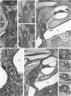
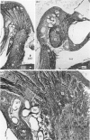
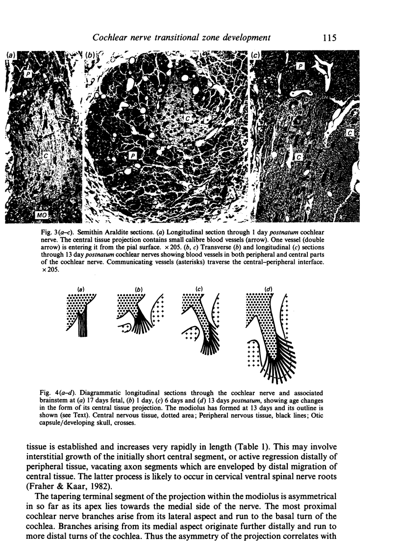
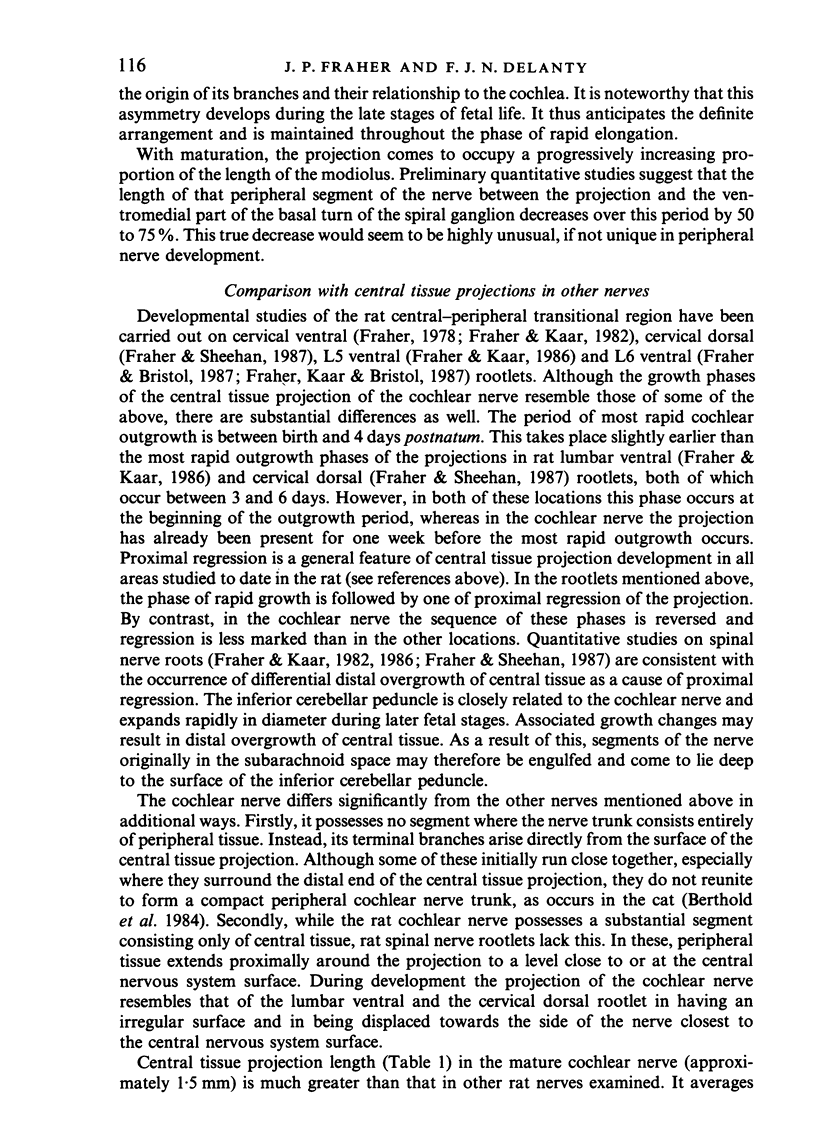
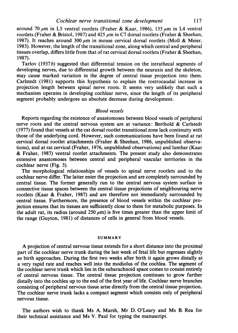
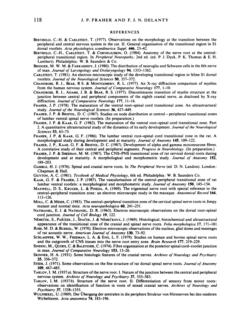
A projection of central nervous tissue extends for a short distance into the proximal part of the cochlear nerve trunk during the last week of fetal life but regresses slightly as birth approaches. During the first two weeks after birth it again grows distally at a very rapid rate and reaches well into the modiolus of the cochlea. The segment of the cochlear nerve trunk which lies in the subarachnoid space comes to consist entirely of central nervous tissue. The central tissue projection continues to grow further distally into the cochlea up to the end of the first year of life. Cochlear nerve branches consisting of peripheral nervous tissue arise directly from the central tissue projection. The cochlear nerve trunk lacks a compact segment which consists only of peripheral nervous tissue.
Full text
PDF
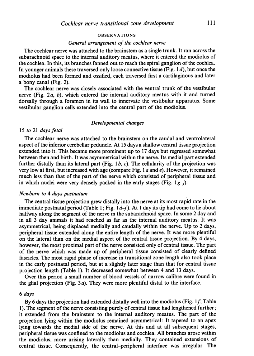
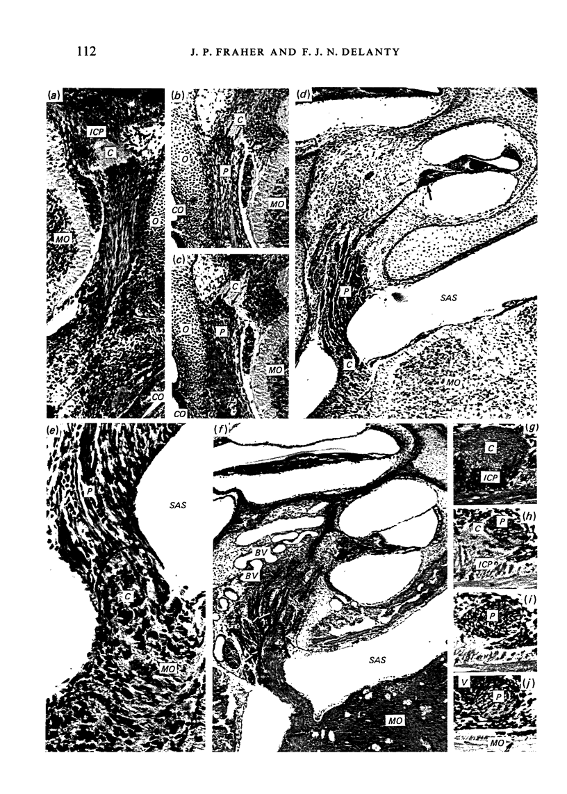
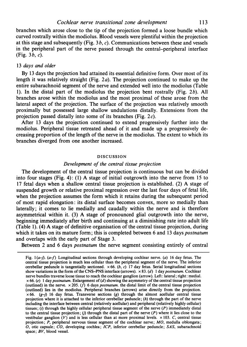
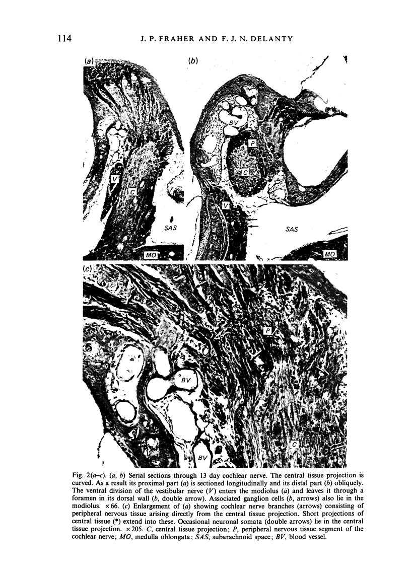
Images in this article
Selected References
These references are in PubMed. This may not be the complete list of references from this article.
- Berthold C. H., Carlstedt T. Observations on the morphology at the transition between the peripheral and the central nervous system in the cat. II. General organization of the transitional region in S1 dorsal rootlets. Acta Physiol Scand Suppl. 1977;446:23–42. [PubMed] [Google Scholar]
- Bridger M. W., Farkashidy J. The distribution of neuroglia and schwann cells in the 8th nerve of man. J Laryngol Otol. 1980 Dec;94(12):1353–1362. doi: 10.1017/s0022215100090186. [DOI] [PubMed] [Google Scholar]
- Carlstedt T. An electron-microscopical study of the developing transitional region in feline S1 dorsal rootlets. J Neurol Sci. 1981 Jun;50(3):357–372. doi: 10.1016/0022-510x(81)90148-9. [DOI] [PubMed] [Google Scholar]
- Chandross R. J., Adams J. B., Bear R. S. Discontinuous transition of myelin structure at the junction between central and peripheral components of the eighth cranial nerve, as disclosed by X-ray diffraction. J Comp Neurol. 1978 Jan 1;177(1):11–15. doi: 10.1002/cne.901770103. [DOI] [PubMed] [Google Scholar]
- Chandross R. J., Bear R. S., Montgomery R. L. An X-ray diffraction comparison of myelins from the human nervous system. J Comp Neurol. 1978 Jan 1;177(1):1–9. doi: 10.1002/cne.901770102. [DOI] [PubMed] [Google Scholar]
- Fraher J. P., Kaar G. F. The lumbar ventral root-spinal cord transitional zone in the rat. A morphological study during development and at maturity. J Anat. 1986 Apr;145:109–122. [PMC free article] [PubMed] [Google Scholar]
- Fraher J. P., Kaar G. F. The maturation of the ventral root-spinal cord transitional zone. Part 2. A quantitative ultrastructural study of the dynamics of its early development. J Neurol Sci. 1982 Jan;53(1):63–75. doi: 10.1016/0022-510x(82)90080-6. [DOI] [PubMed] [Google Scholar]
- Fraher J. P., Sheehan M. M. The CNS-PNS transitional zone of rat cervical dorsal roots during development and at maturity. A morphological and morphometric study. J Anat. 1987 Jun;152:189–203. [PMC free article] [PubMed] [Google Scholar]
- Fraher J. P. The maturation of the ventral root-spinal cord transitional zone. An ultrastructural study. J Neurol Sci. 1978 May;36(3):427–449. doi: 10.1016/0022-510x(78)90049-7. [DOI] [PubMed] [Google Scholar]
- Kaar G. F., Fraher J. P. The vascularisation of the central-peripheral transitional zone of rat lumbar ventral rootlets: a morphological and morphometric study. J Anat. 1987 Feb;150:145–154. [PMC free article] [PubMed] [Google Scholar]
- Maxwell D. S., Kruger L., Pineda A. The trigeminal nerve root with special reference to the central-peripheral transition zone: an electron microscopic study in the macaque. Anat Rec. 1969 May;164(1):113–125. doi: 10.1002/ar.1091640108. [DOI] [PubMed] [Google Scholar]
- Moll C., Meier C. The central-peripheral transition zone of cervical spinal nerve roots in Jimpy mutant and normal mice. Light- and electron-microscopic study. Acta Neuropathol. 1983;60(3-4):241–251. doi: 10.1007/BF00691872. [DOI] [PubMed] [Google Scholar]
- Nemecek S., Parízek J., Spacek J., Nemecková J. Histological, histochemical and ultrastructural appearance of the transitional zone of the cranial and spinal nerve roots. Folia Morphol (Praha) 1969;17(2):171–181. [PubMed] [Google Scholar]
- Ross M. D., Burkel W. Electron microscopic observations of the nucleus, glial dome, and meninges of the rat acoustic nerve. Am J Anat. 1971 Jan;130(1):73–91. doi: 10.1002/aja.1001300106. [DOI] [PubMed] [Google Scholar]
- Schlaepfer W. W., Freeman L. A., Eng L. F. Studies of human and bovine spinal nerve roots and the outgrowth of CNS tissues into the nerve root entry zone. Brain Res. 1979 Nov 16;177(2):219–229. doi: 10.1016/0006-8993(79)90773-x. [DOI] [PubMed] [Google Scholar]
- Sindou M., Quoex C., Baleydier C. Fiber organization at the posterior spinal cord-rootlet junction in man. J Comp Neurol. 1974 Jan 1;153(1):15–26. doi: 10.1002/cne.901530103. [DOI] [PubMed] [Google Scholar]
- Steer J. M. Some observations on the fine structure of rat dorsal spinal nerve roots. J Anat. 1971 Sep;109(Pt 3):467–485. [PMC free article] [PubMed] [Google Scholar]
- Wulfhekel U. Der Ubergang der zentralen in die periphere Struktur von Hirnnerven bei den niederen Wirbeltieren. Acta Anat (Basel) 1969;74(2):183–196. [PubMed] [Google Scholar]


