Abstract
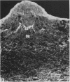
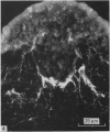
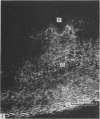
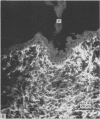
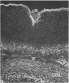
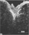
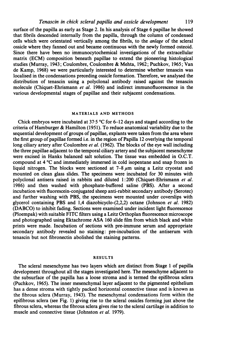
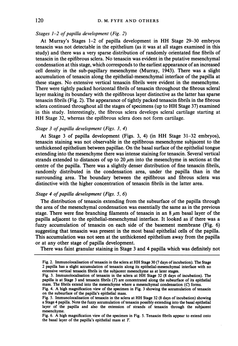
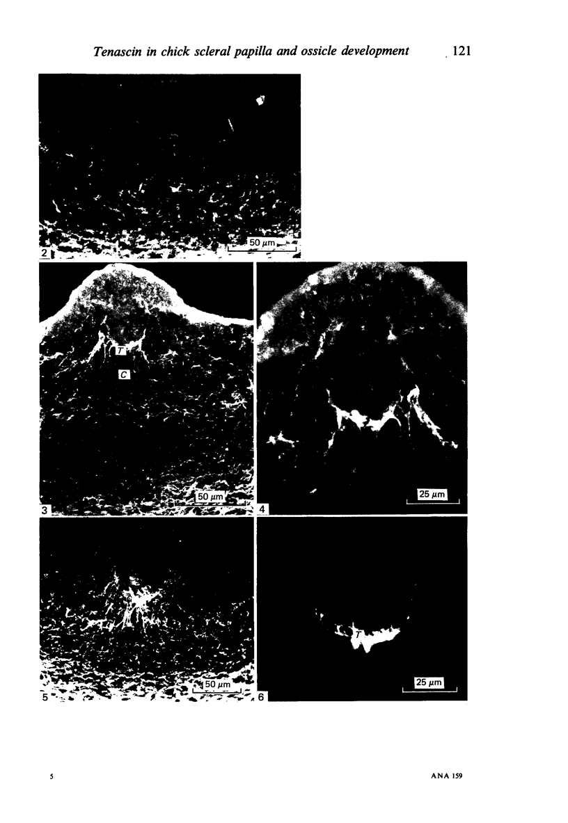
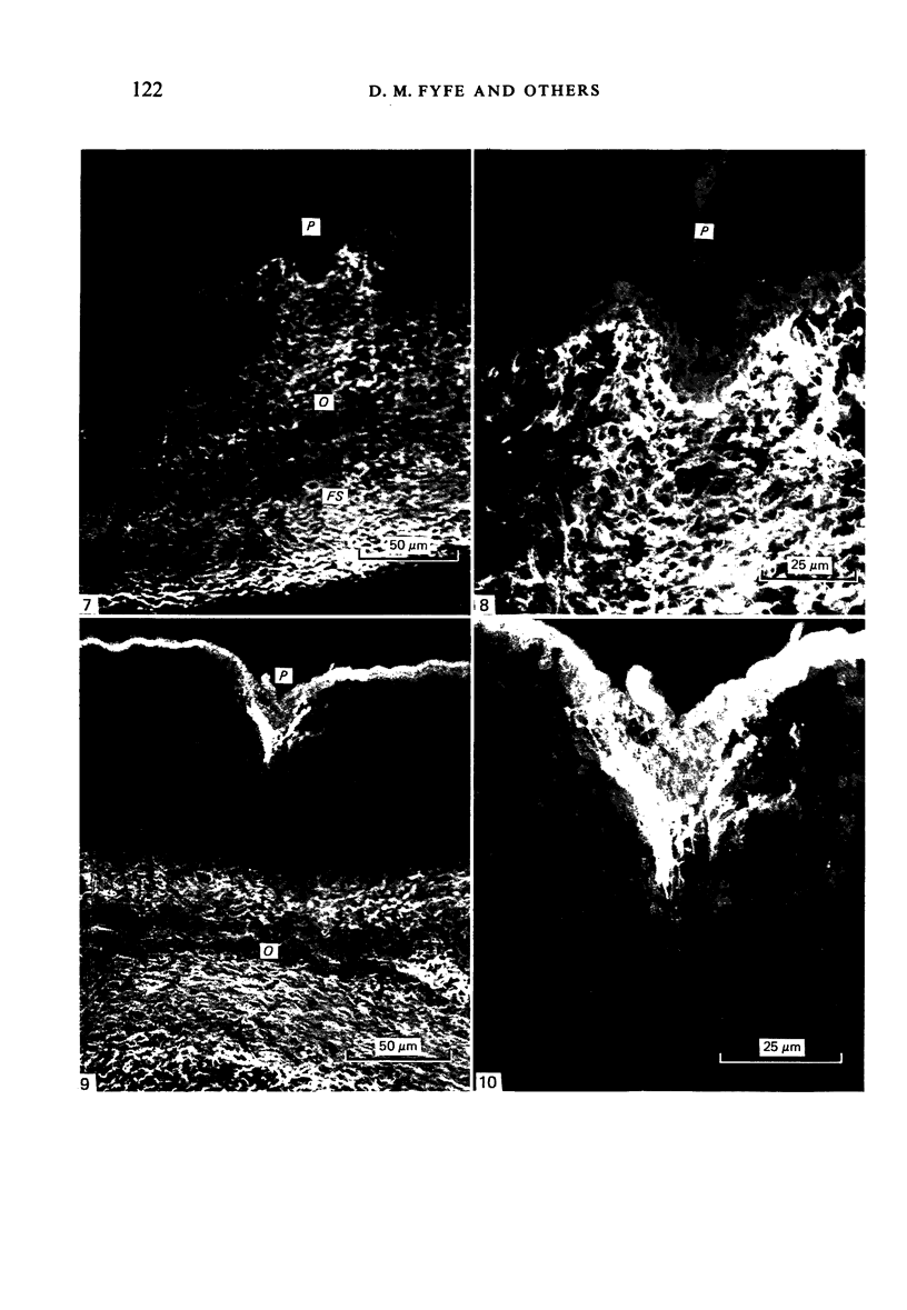
We have used a polyclonal antibody against tenascin (a 240 kDa extracellular matrix glycoprotein) and indirect immunofluorescence to investigate the distribution of tenascin in cryostat sections of the chick sclera during the six stages of scleral papilla development (Murray, 1943) and formation of membrane bone anlagen (mesenchymal condensations between 6-12 days of incubation - HH Stage 29-37). At Stages 1 and 2, when the papilla was a slight thickening in the conjunctival epithelium and mesenchymal condensation formation was initiated, tenascin was sparse in the sclera. At Stage 3, when the papilla had an epithelial mass intruding into the mesenchyme, there was an accumulation of tenascin fibrils along the subsurface of the papilla with fibrils extending from this region into the mesenchymal condensation. Interestingly, tenascin was sparse between papillae and between mesenchymal condensations. The Stage 4 papilla had a similar localisation of tenascin fibrils and in addition fine arborizing fibrils of tenascin were observed within the basal epithelia of the papilla adjacent to the epithelial-mesenchymal interface (which suggested that the fibrils infiltrated through the basement membrane region). The Stage 5 and 6 papillae had a column of vertical tenascin fibrils extending from the subsurface of the papilla to the interior mesenchyme corresponding exactly to the location of the mesenchymal condensation which was then forming the anlage of the ossicle in a flat bed about 100 microns from the conjunctival surface. The column of tenascin disappeared as the osteoid appeared in the ossicular bed on the 12th day of incubation but a dense accumulation of tenascin remained along the subsurface of the papilla. With the exception of tenascin fluorescence in the basal region of the Stage 4 papilla, tenascin fibrils were not observed in the other stages of papilla development in the epithelium covering areas between the mesenchymal condensation. This restricted distribution of tenascin may be important in the morphogenesis of scleral papillae and scleral ossicles.
Full text
PDF
Images in this article
Selected References
These references are in PubMed. This may not be the complete list of references from this article.
- Bronner-Fraser M. Distribution of latex beads and retinal pigment epithelial cells along the ventral neural crest pathway. Dev Biol. 1982 May;91(1):50–63. doi: 10.1016/0012-1606(82)90007-0. [DOI] [PubMed] [Google Scholar]
- Bronner-Fraser M. Latex beads as probes of a neural crest pathway: effects of laminin, collagen, and surface charge on bead translocation. J Cell Biol. 1984 Jun;98(6):1947–1960. doi: 10.1083/jcb.98.6.1947. [DOI] [PMC free article] [PubMed] [Google Scholar]
- COULOMBRE A. J., COULOMBRE J. L. The skeleton of the eye. I. Conjunctival papillae and scleral ossicles. Dev Biol. 1962 Dec;5:382–401. doi: 10.1016/0012-1606(62)90020-9. [DOI] [PubMed] [Google Scholar]
- Chiquet-Ehrismann R., Mackie E. J., Pearson C. A., Sakakura T. Tenascin: an extracellular matrix protein involved in tissue interactions during fetal development and oncogenesis. Cell. 1986 Oct 10;47(1):131–139. doi: 10.1016/0092-8674(86)90374-0. [DOI] [PubMed] [Google Scholar]
- Chiquet M., Fambrough D. M. Chick myotendinous antigen. I. A monoclonal antibody as a marker for tendon and muscle morphogenesis. J Cell Biol. 1984 Jun;98(6):1926–1936. doi: 10.1083/jcb.98.6.1926. [DOI] [PMC free article] [PubMed] [Google Scholar]
- Fyfe D. M., Hall B. K. The origin of the ectomesenchymal condensations which precede the development of the bony scleral ossicles in the eyes of embryonic chicks. J Embryol Exp Morphol. 1983 Feb;73:69–86. [PubMed] [Google Scholar]
- Johnson G. D., Davidson R. S., McNamee K. C., Russell G., Goodwin D., Holborow E. J. Fading of immunofluorescence during microscopy: a study of the phenomenon and its remedy. J Immunol Methods. 1982 Dec 17;55(2):231–242. doi: 10.1016/0022-1759(82)90035-7. [DOI] [PubMed] [Google Scholar]
- Johnston M. C., Noden D. M., Hazelton R. D., Coulombre J. L., Coulombre A. J. Origins of avian ocular and periocular tissues. Exp Eye Res. 1979 Jul;29(1):27–43. doi: 10.1016/0014-4835(79)90164-7. [DOI] [PubMed] [Google Scholar]
- Mauger A., Demarchez M., Herbage D., Grimaud J. A., Druguet M., Hartmann D., Sengel P. Immunofluorescent localization of collagen types I and III, and of fibronectin during feather morphogenesis in the chick embryo. Dev Biol. 1982 Nov;94(1):93–105. doi: 10.1016/0012-1606(82)90072-0. [DOI] [PubMed] [Google Scholar]
- Murray P. D. The development of the conjunctival papillae and of the scleral bones in the embryo chick. J Anat. 1943 Apr;77(Pt 3):225–240.2. [PMC free article] [PubMed] [Google Scholar]
- Silver M. H., Foidart J. M., Pratt R. M. Distribution of fibronectin and collagen during mouse limb and palate development. Differentiation. 1981;18(3):141–149. doi: 10.1111/j.1432-0436.1981.tb01115.x. [DOI] [PubMed] [Google Scholar]
- Thesleff I., Barrach H. J., Foidart J. M., Vaheri A., Pratt R. M., Martin G. R. Changes in the distribution of type IV collagen, laminin, proteoglycan, and fibronectin during mouse tooth development. Dev Biol. 1981 Jan 15;81(1):182–192. doi: 10.1016/0012-1606(81)90361-4. [DOI] [PubMed] [Google Scholar]
- Van de Kamp M. Fine structural analysis of the conjunctival papillae in the chick embryo: a reassessment of their morphogenesis and developmental significance. J Exp Zool. 1968 Dec;169(4):447–461. doi: 10.1002/jez.1401690407. [DOI] [PubMed] [Google Scholar]
- van de Kamp M., Hilfer S. R. Cell proliferation in condensing scleral ectomesenchyme associated with the conjunctival papillae in the chick embryo. J Embryol Exp Morphol. 1985 Aug;88:25–37. [PubMed] [Google Scholar]
- von der Mark K., von der Mark H., Timpl R., Trelstad R. L. Immunofluorescent localization of collagen types I, II, and III in the embryonic chick eye. Dev Biol. 1977 Aug;59(1):75–85. doi: 10.1016/0012-1606(77)90241-x. [DOI] [PubMed] [Google Scholar]


