Full text
PDF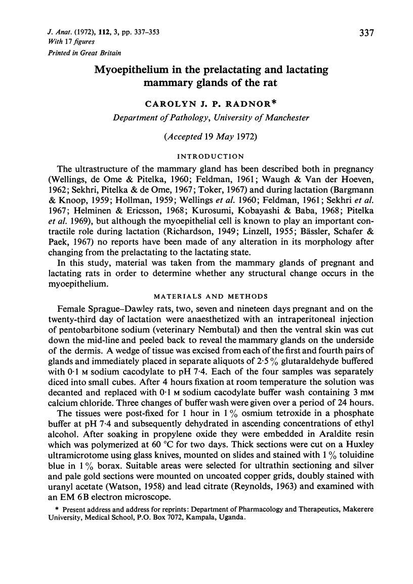
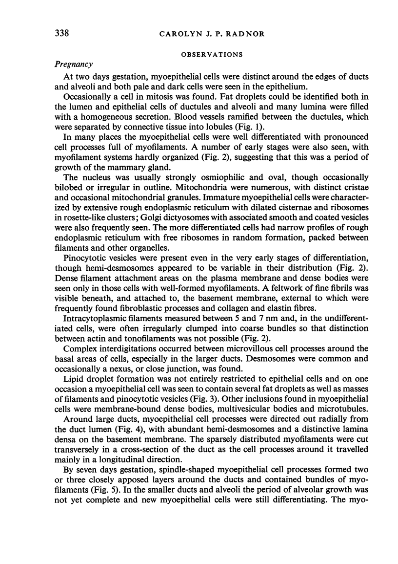
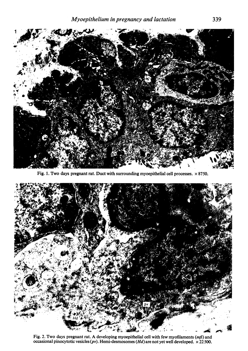
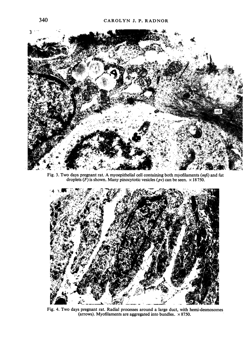
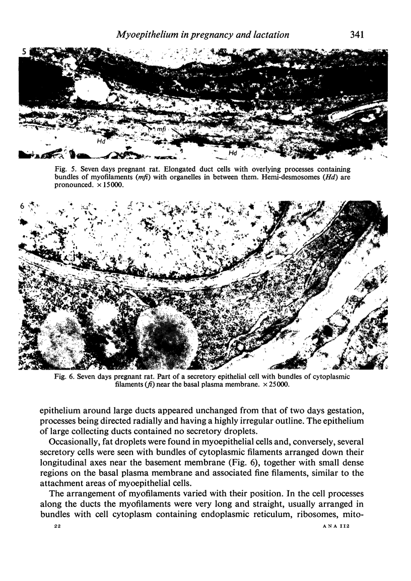
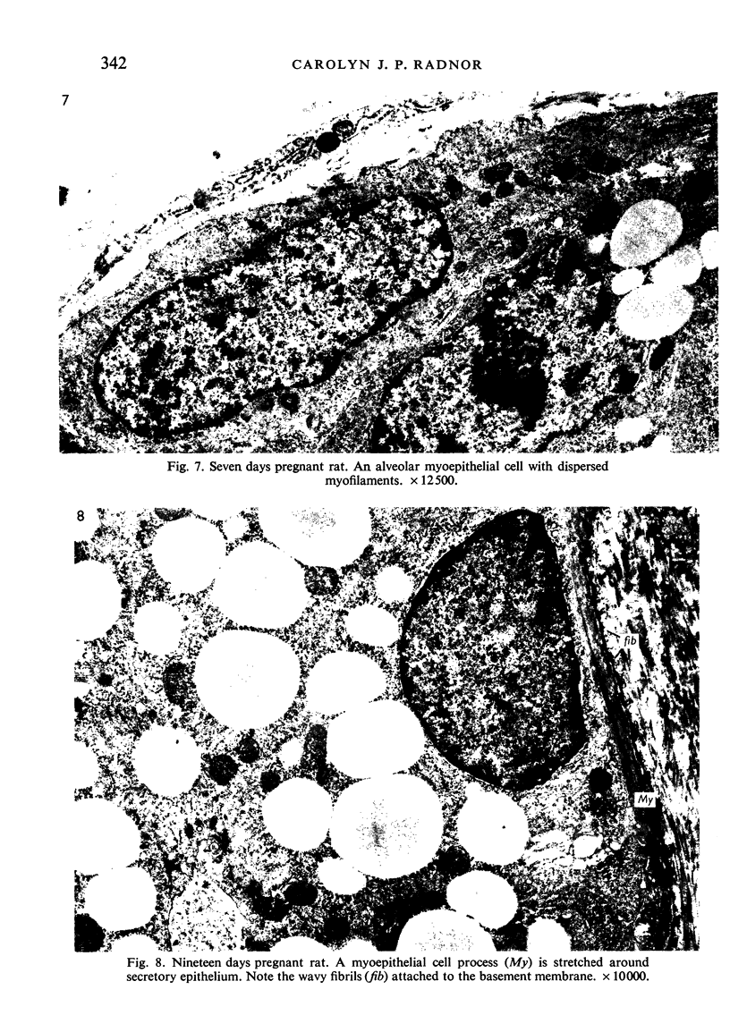
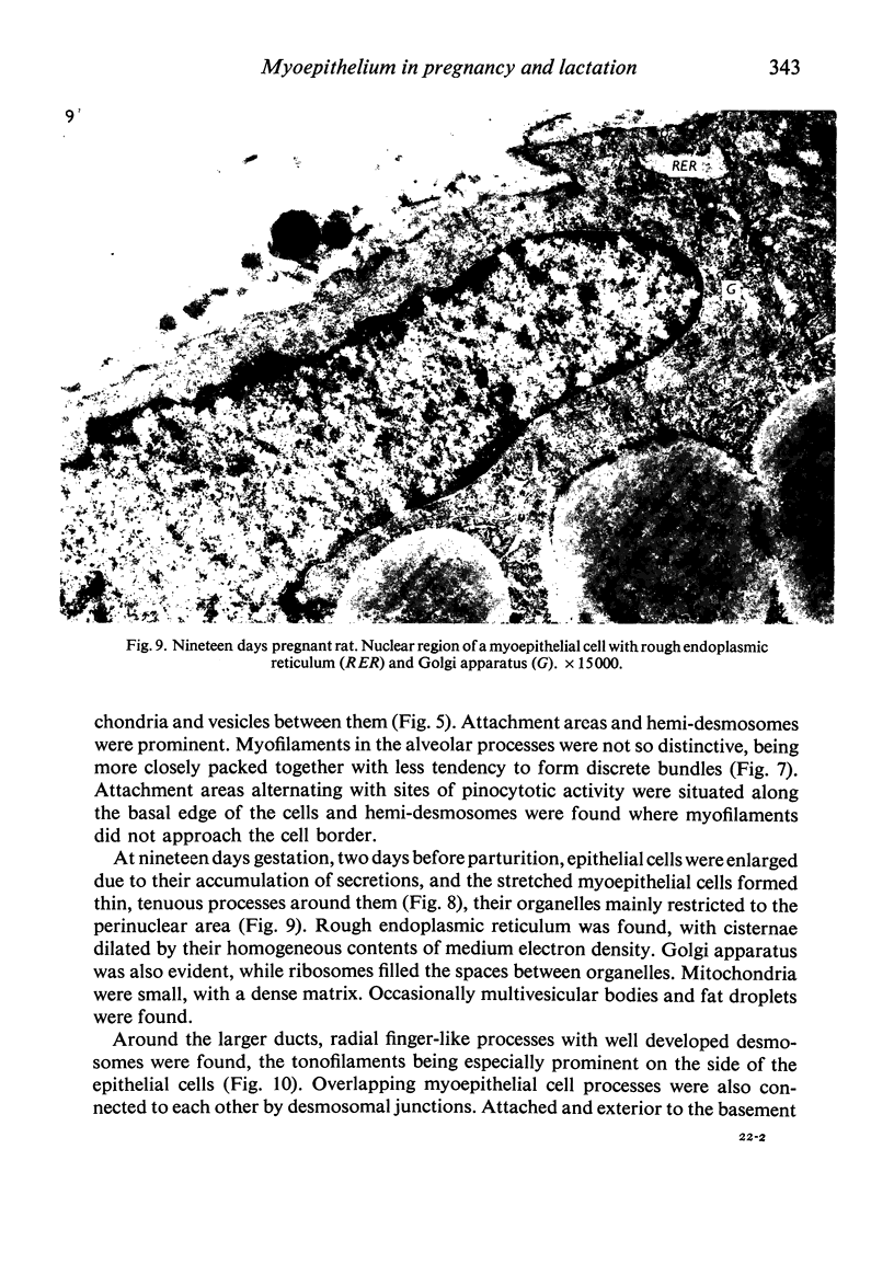
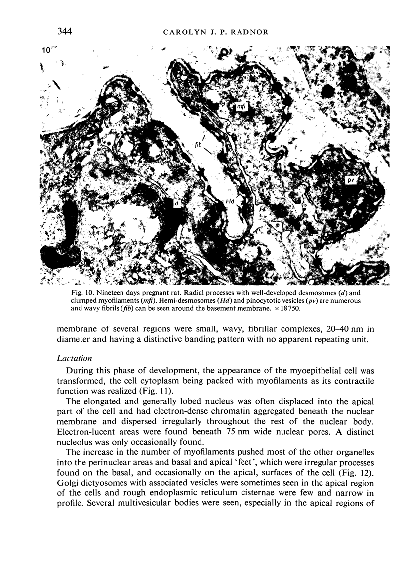
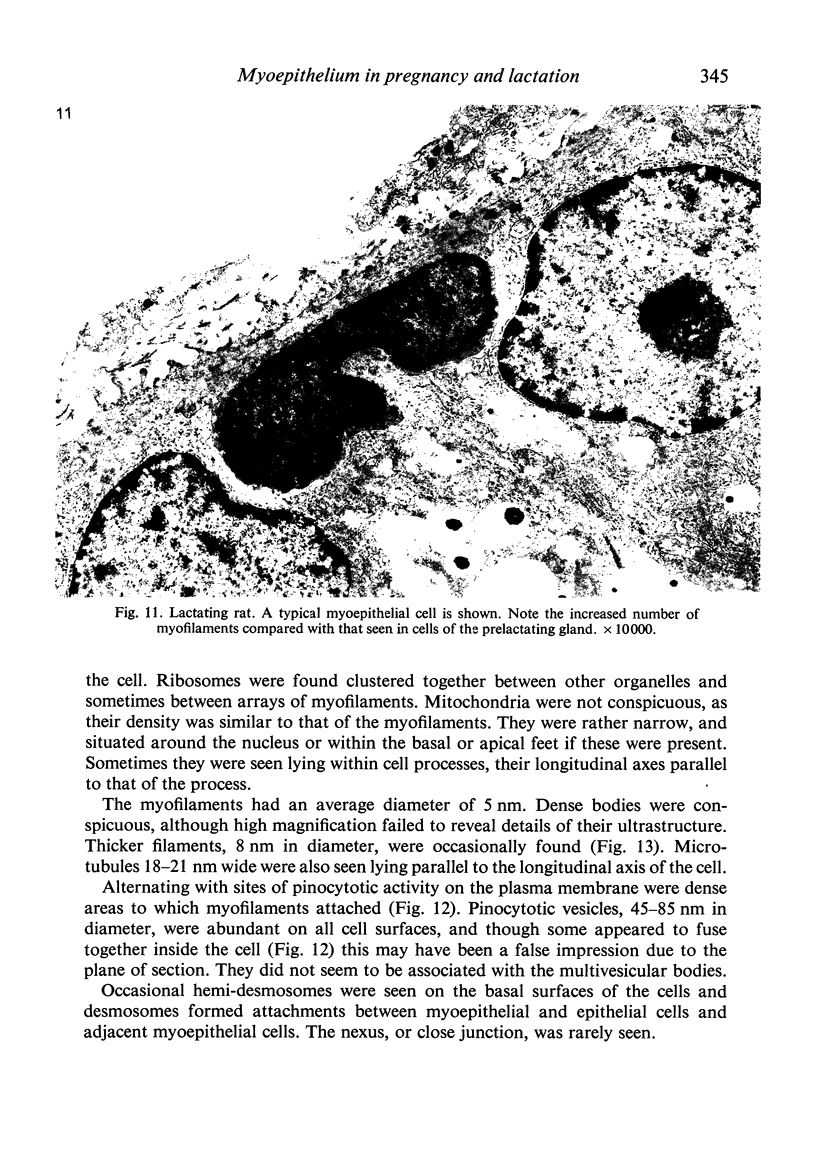
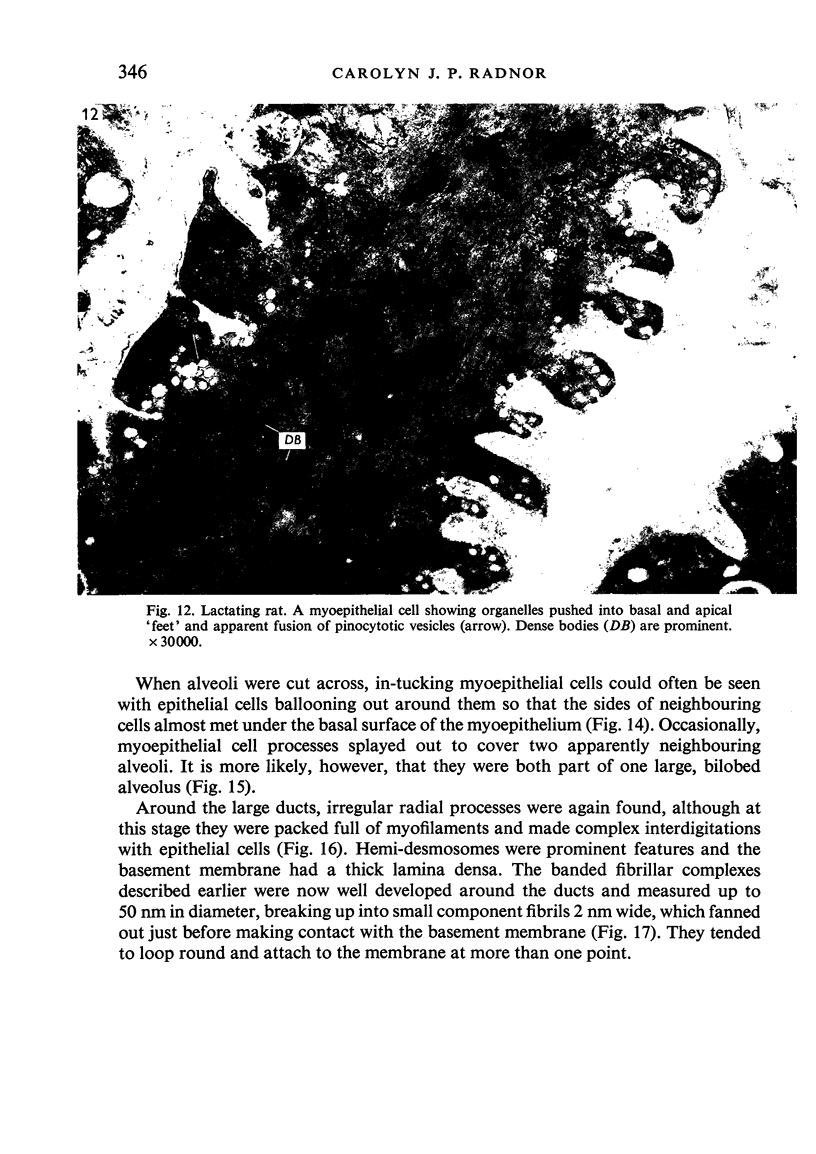
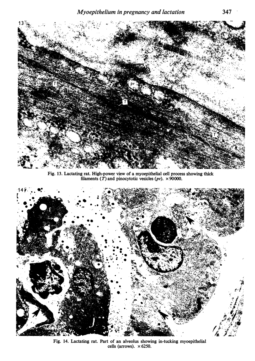
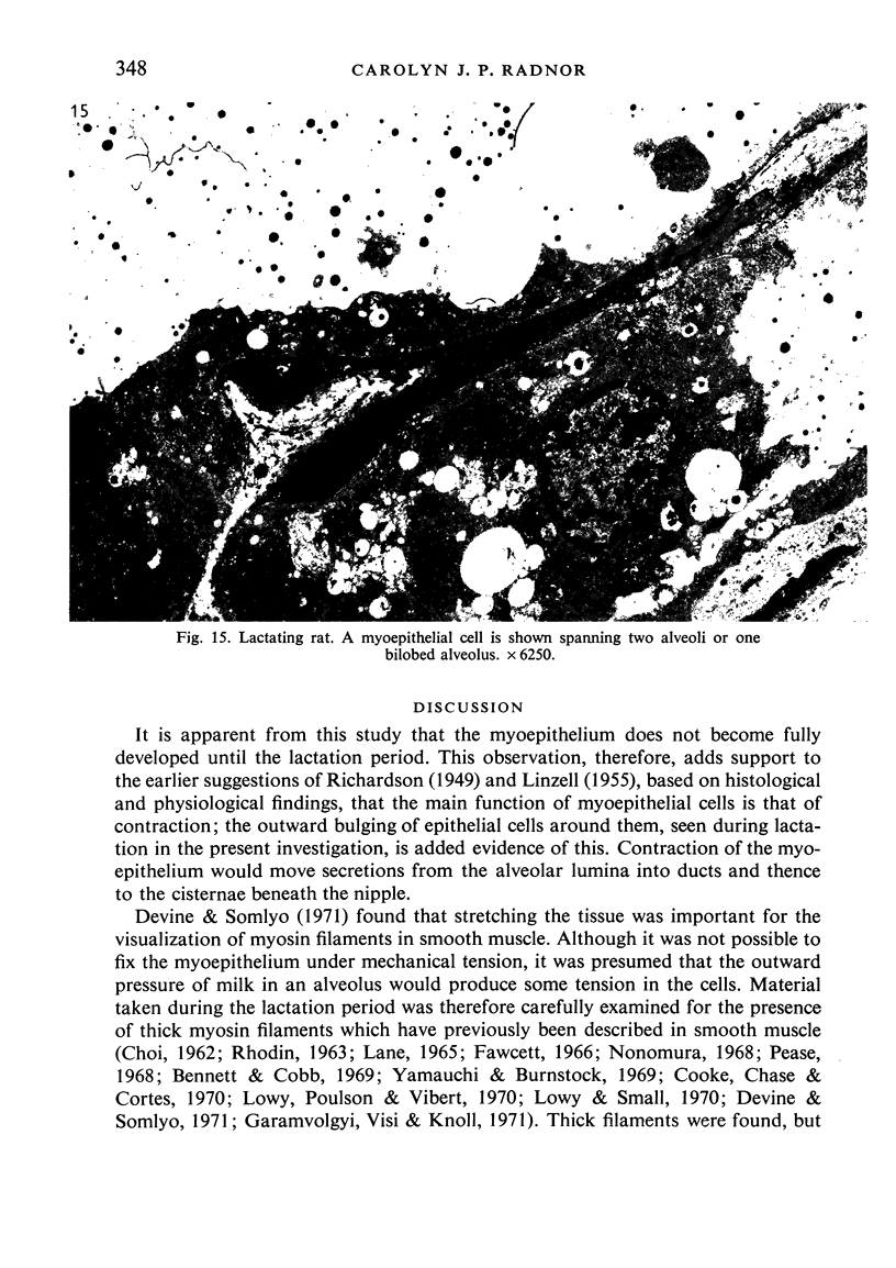
Images in this article
Selected References
These references are in PubMed. This may not be the complete list of references from this article.
- BARGMANN W., KNOOP A. Uber die Morphologie der Milchsekretion; lichtund elektronenmikroskopische Studien an der Milchdrüse Ratte. Z Zellforsch Mikrosk Anat. 1959;49(3):344–388. [PubMed] [Google Scholar]
- Bennett T., Cobb J. L. Studies on the avian gizzard: morphology and innervation of the smooth muscle. Z Zellforsch Mikrosk Anat. 1969;96(2):173–185. doi: 10.1007/BF00338765. [DOI] [PubMed] [Google Scholar]
- Bässler R., Schäfer A., Paek S. Elektronenmikroskopische und histochemische Untersuchungen zur Morphologie und Funktion myoepithelialer Zellen. Verh Dtsch Ges Pathol. 1967;51:301–308. [PubMed] [Google Scholar]
- Cooke P. H., Chase R. H., Cortés J. M. Thick filaments resembling myosin in electrophoretically-extracted vertebrate smooth muscle. Exp Cell Res. 1970 May;60(2):237–246. doi: 10.1016/0014-4827(70)90510-0. [DOI] [PubMed] [Google Scholar]
- DEWEY M. M., BARR L. A STUDY OF THE STRUCTURE AND DISTRIBUTION OF THE NEXUS. J Cell Biol. 1964 Dec;23:553–585. doi: 10.1083/jcb.23.3.553. [DOI] [PMC free article] [PubMed] [Google Scholar]
- Devine C. E., Somlyo A. P. Thick filaments in vascular smooth muscle. J Cell Biol. 1971 Jun;49(3):636–649. doi: 10.1083/jcb.49.3.636. [DOI] [PMC free article] [PubMed] [Google Scholar]
- Ellis R. A. Fine structure of the myoepithelium of the eccrine sweat glands of man. J Cell Biol. 1965 Dec;27(3):551–563. doi: 10.1083/jcb.27.3.551. [DOI] [PMC free article] [PubMed] [Google Scholar]
- FELDMAN J. D. Fine structure of the cow's udder during gestation and lactation. Lab Invest. 1961 Mar-Apr;10:238–255. [PubMed] [Google Scholar]
- Garamvölgyi N., Vizi E. S., Knoll J. The regular occurrence of thick filaments in stretched mammalian smooth muscle. J Ultrastruct Res. 1971 Jan;34(1):135–143. doi: 10.1016/s0022-5320(71)90009-8. [DOI] [PubMed] [Google Scholar]
- Girardie J. Fonction catabolique de l'épithélium mammaire. Etude histochimique et ultrastructurale. Z Zellforsch Mikrosk Anat. 1967;80(3):385–412. [PubMed] [Google Scholar]
- Helminen H. J., Ericsson J. L. Studies on mammary gland involution. I. On the ultrastructure of the lactating mammary gland. J Ultrastruct Res. 1968 Nov;25(3):193–213. doi: 10.1016/s0022-5320(68)80069-3. [DOI] [PubMed] [Google Scholar]
- Kurosumi K., Kobayashi Y., Baba N. The fine structure of mammary glands of lactating rats, with special reference to the apocrine secretion. Exp Cell Res. 1968 Apr;50(1):177–192. doi: 10.1016/0014-4827(68)90406-0. [DOI] [PubMed] [Google Scholar]
- LINZELL J. L. Some observations on the contractile tissue of the mammary glands. J Physiol. 1955 Nov 28;130(2):257–267. doi: 10.1113/jphysiol.1955.sp005408. [DOI] [PMC free article] [PubMed] [Google Scholar]
- Lane B. P. Alterations in the cytologic detail of intestinal smooth muscle cells in various stages of contraction. J Cell Biol. 1965 Oct;27(1):199–213. doi: 10.1083/jcb.27.1.199. [DOI] [PMC free article] [PubMed] [Google Scholar]
- Lane B. P. Localization of products of ATP hydrolysis in mammalian smooth muscle cells. J Cell Biol. 1967 Sep;34(3):713–720. doi: 10.1083/jcb.34.3.713. [DOI] [PMC free article] [PubMed] [Google Scholar]
- Lowy J., Poulsen F. R., Vibert P. J. Myosin filaments in vertebrate smooth muscle. Nature. 1970 Mar 14;225(5237):1053–1054. doi: 10.1038/2251053a0. [DOI] [PubMed] [Google Scholar]
- Lowy J., Small J. V. The organization of myosin and actin in vertebrate smooth muscle. Nature. 1970 Jul 4;227(5253):46–51. doi: 10.1038/227046a0. [DOI] [PubMed] [Google Scholar]
- Nonomura Y. Myofilaments in smooth muscle of guinea pigs taenia coli. J Cell Biol. 1968 Dec;39(3):741–745. doi: 10.1083/jcb.39.3.741. [DOI] [PMC free article] [PubMed] [Google Scholar]
- Palade G. E., Farquhar M. G. A special fibril of the dermis. J Cell Biol. 1965 Oct;27(1):215–224. doi: 10.1083/jcb.27.1.215. [DOI] [PMC free article] [PubMed] [Google Scholar]
- Pitelka D. R., Kerkof P. R., Gagné H. T., Smith S., Abraham S. Characteristics of cells dissociated from mouse mammary glands. I. Method of separation and morphology of parenchymal cells from lactating glands. Exp Cell Res. 1969 Sep;57(1):43–62. doi: 10.1016/0014-4827(69)90365-6. [DOI] [PubMed] [Google Scholar]
- REYNOLDS E. S. The use of lead citrate at high pH as an electron-opaque stain in electron microscopy. J Cell Biol. 1963 Apr;17:208–212. doi: 10.1083/jcb.17.1.208. [DOI] [PMC free article] [PubMed] [Google Scholar]
- ROSTGAARD J., BARRNETT R. J. FINE STRUCTURE LOCALIZATION OF NUCLEOSIDE PHOSPHATASES IN RELATION TO SMOOTH MUSCLE CELLS AND UNMYELINATED NERVES IN THE SMALL INTESTINE OF THE RAT. J Ultrastruct Res. 1964 Aug;11:193–207. doi: 10.1016/s0022-5320(64)80103-9. [DOI] [PubMed] [Google Scholar]
- Sekhri K. K., Pitelka D. R., DeOme K. B. Studies of mouse mammary glands. I. Cytomorphology of the normal mammary gland. J Natl Cancer Inst. 1967 Sep;39(3):459–490. [PubMed] [Google Scholar]
- Susi F. R., Belt W. D., Kelly J. W. Fine structure of fibrillar complexes associated with the basement membrane in human oral mucosa. J Cell Biol. 1967 Aug;34(2):686–690. doi: 10.1083/jcb.34.2.686. [DOI] [PMC free article] [PubMed] [Google Scholar]
- Toker C. Observations on the ultrastructure of a mammary ductule. J Ultrastruct Res. 1967 Nov;21(1):9–25. doi: 10.1016/s0022-5320(67)80003-0. [DOI] [PubMed] [Google Scholar]
- WATSON M. L. Staining of tissue sections for electron microscopy with heavy metals. J Biophys Biochem Cytol. 1958 Jul 25;4(4):475–478. doi: 10.1083/jcb.4.4.475. [DOI] [PMC free article] [PubMed] [Google Scholar]
- WAUGH D., VAN DER HOEVEN E. Fine structure of the human adult female breast. Lab Invest. 1962 Mar;11:220–228. [PubMed] [Google Scholar]
- WELLINGS S. R., DEOME K. B., PITELKA D. R. Electron microscopy of milk secretion in the mammary gland of the C3H/Crgl mouse. I. Cytomorphology of the prelactating and the lactating gland. J Natl Cancer Inst. 1960 Aug;25:393–421. [PubMed] [Google Scholar]
- Yamauchi A., Burnstock G. Post-natal development of smooth muscle cells in the mouse vas deferens. A fine structural study. J Anat. 1969 Jan;104(Pt 1):1–15. [PMC free article] [PubMed] [Google Scholar]


