Abstract
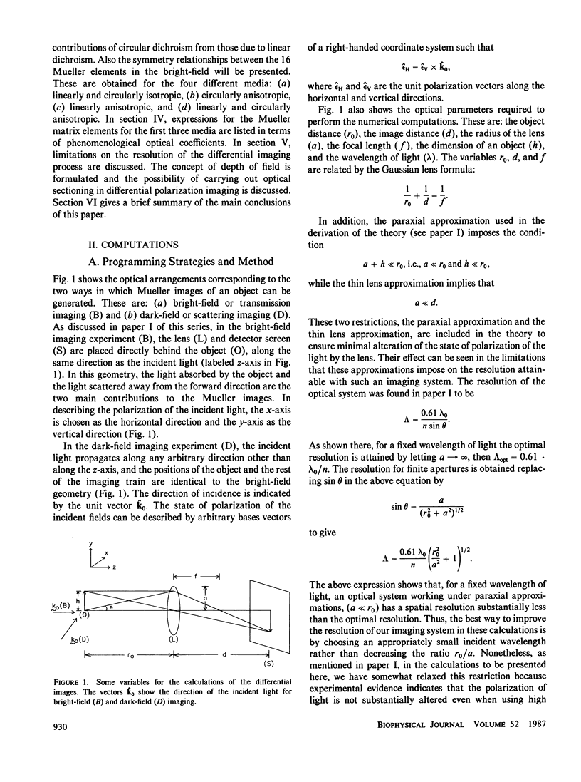
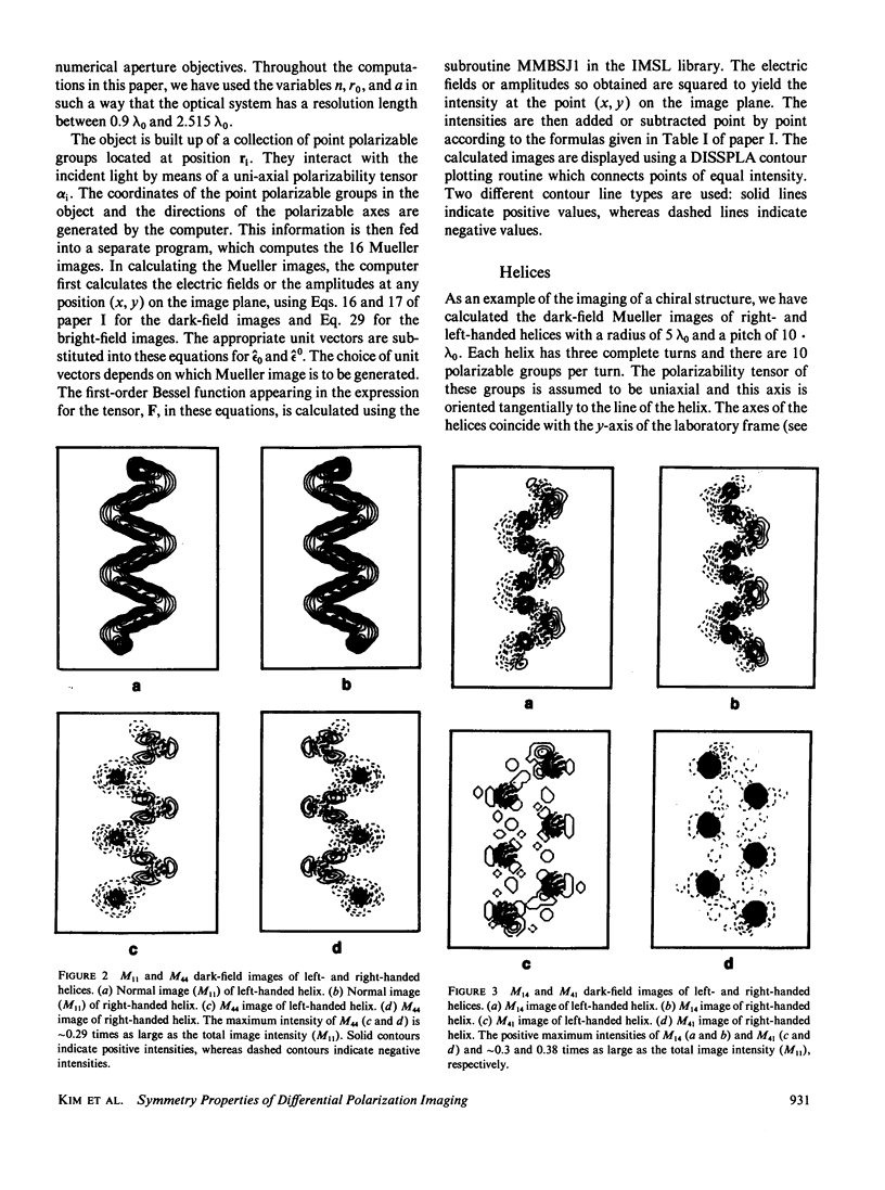
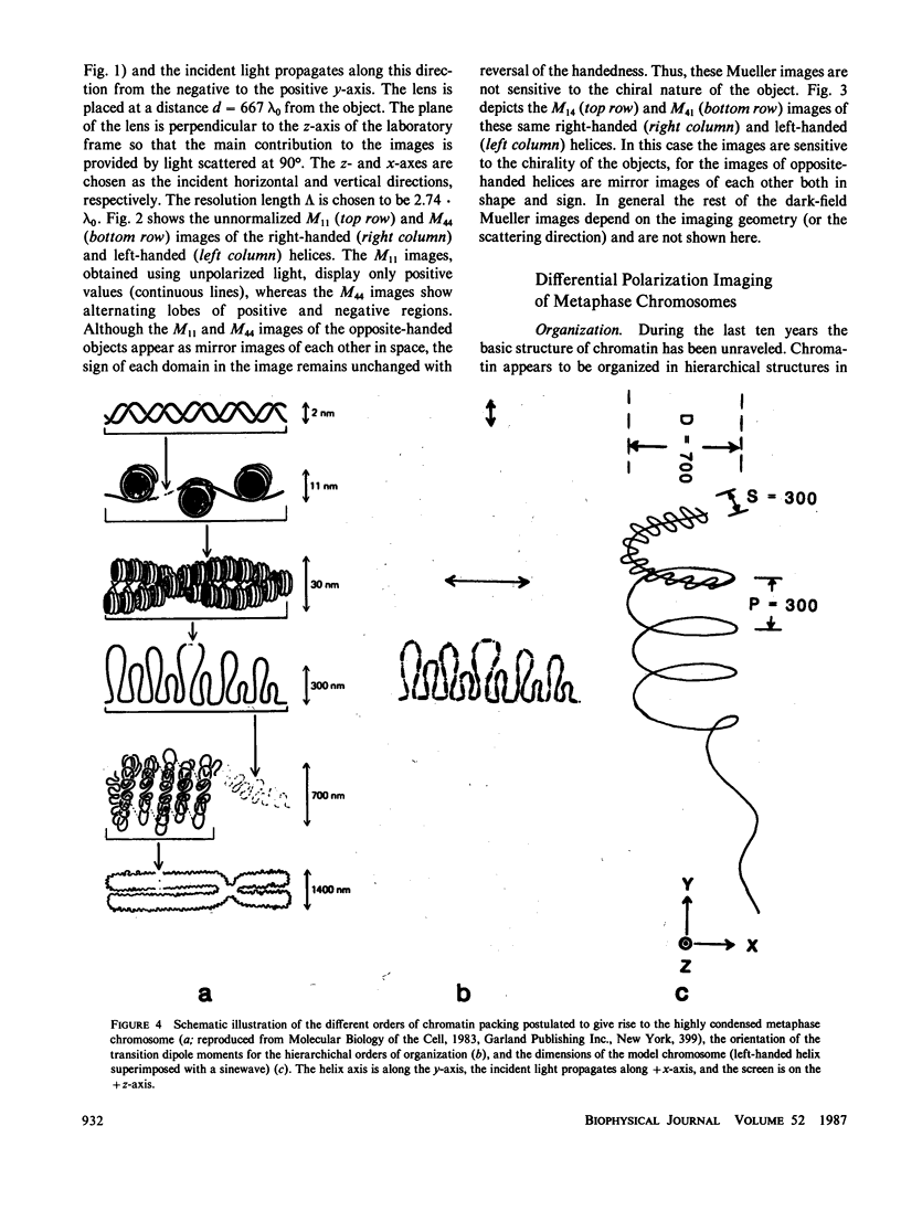
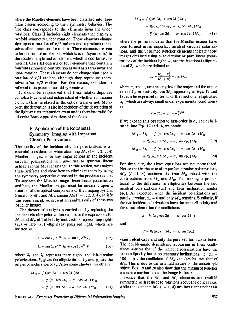
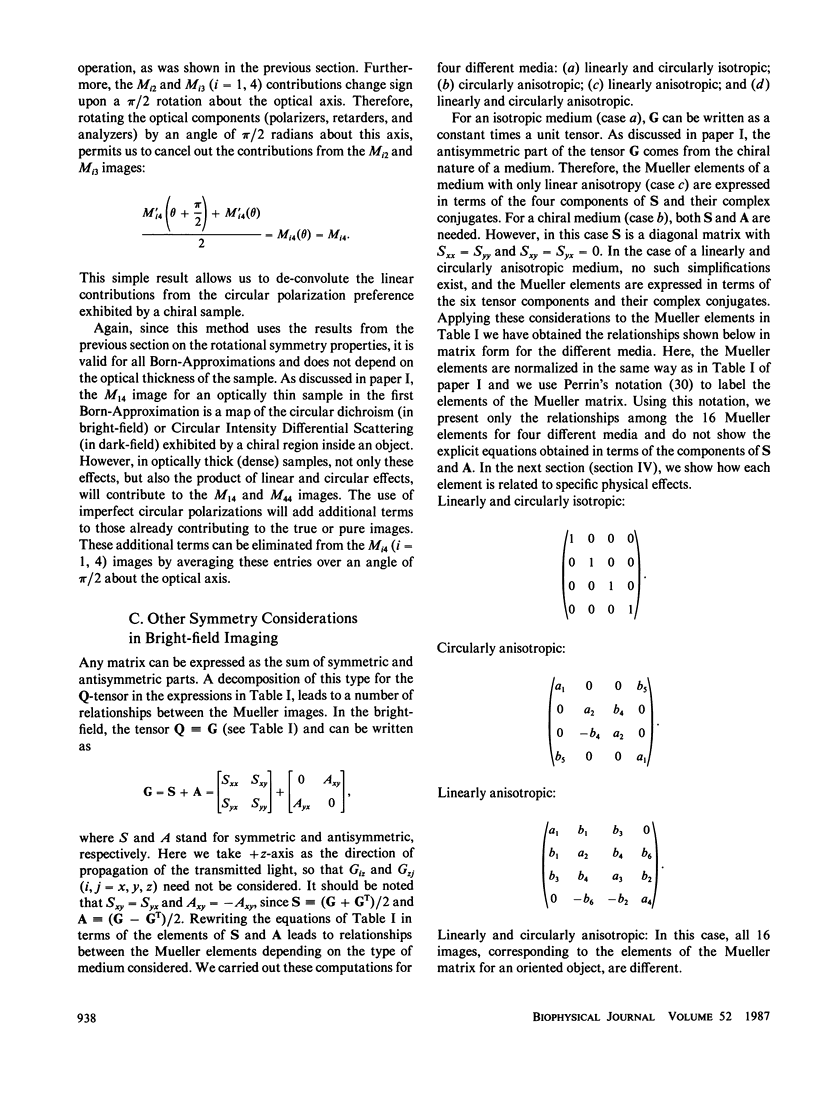
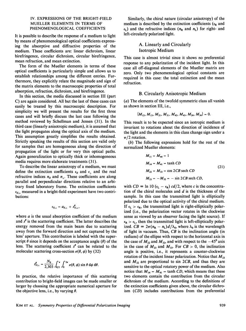
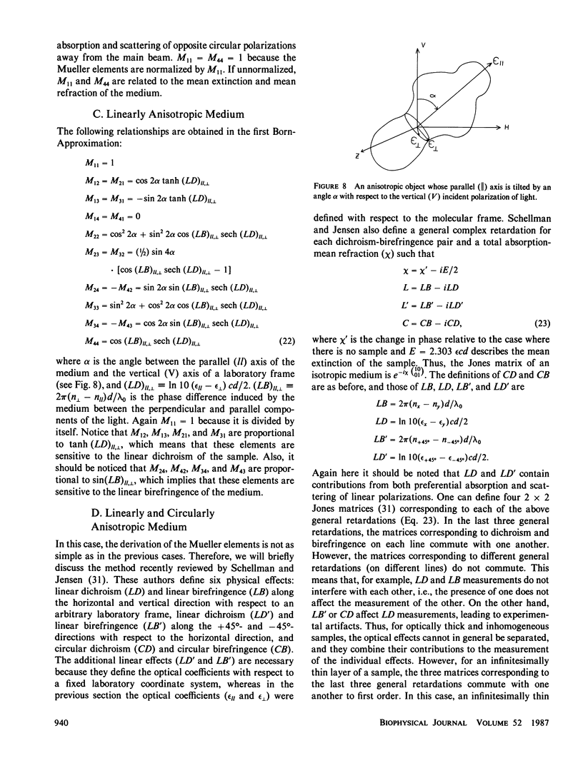
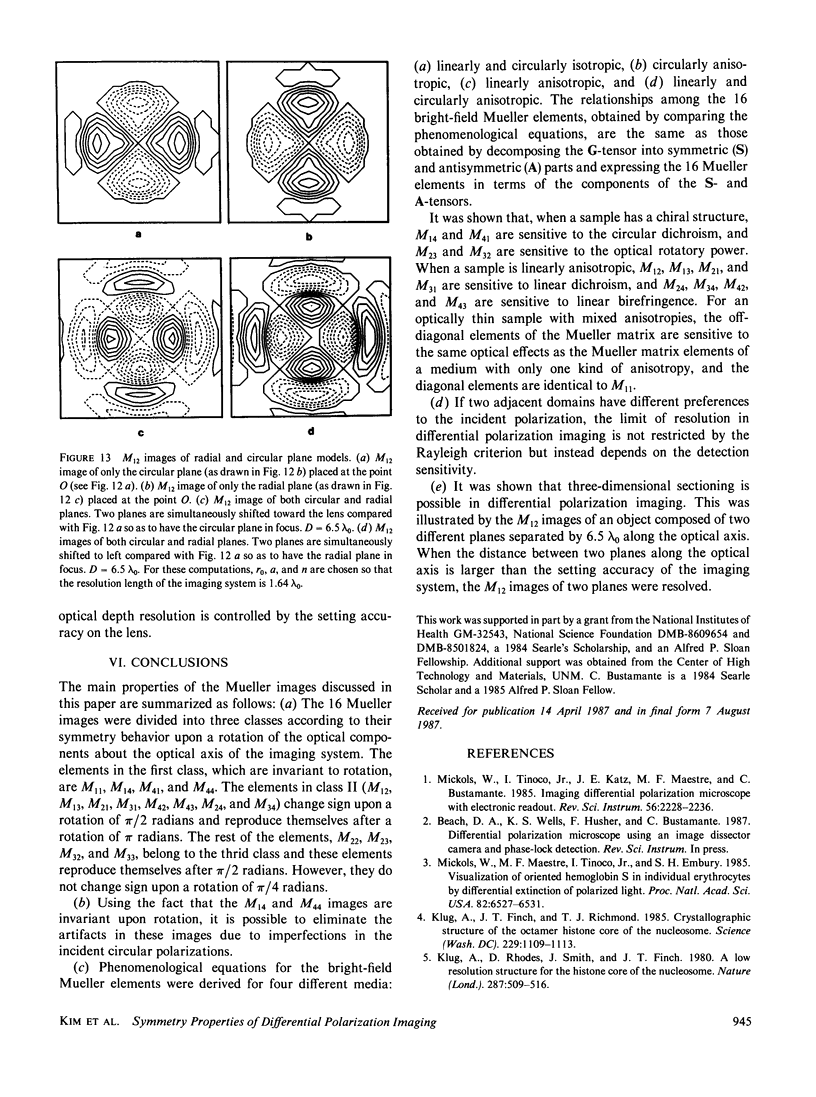
Various differential polarization images or Mueller images of model objects are generated using the equations derived in the previous paper (paper I of this series). These calculated images include models of the higher-order organization of metaphase chromosomes, and show the applicability of the differential polarization imaging method to the elucidation of complex molecular organizations. Then, the symmetry behavior of the Mueller matrix elements upon infinitesimal rotations of the optical components about the optical axis of the imaging system is presented. It is shown that the rotational properties of the Mueller images can be used to eliminate the linear polarization contributions to the M14 and M44 images, which appear when these images are generated with imperfect circular polarizations. The relationships between the 16 bright-field Mueller images for four different media, i.e., linearly and circularly isotropic, circularly anisotropic, linearly anisotropic, and linearly and circularly anisotropic, are also derived. For the first three cases simple relationships between the Mueller images are found and phenomenological equations in terms of the optical coefficients are derived. In the last case there are no specific relationships between the Mueller images and instead we briefly present Schellman and Jensen's method for treating this type of medium. The criterion of spatial resolution between adjacent domains of different optical anisotropy is then derived. It is found that in transitions between domains of opposite anisotropy the classical Rayleigh limit must be replaced by a magnitude criterion which depends on the limits of the sensitivity of the detection. Finally, the feasibility of optical sectioning in differential polarization imaging is demonstrated.
Full text
PDF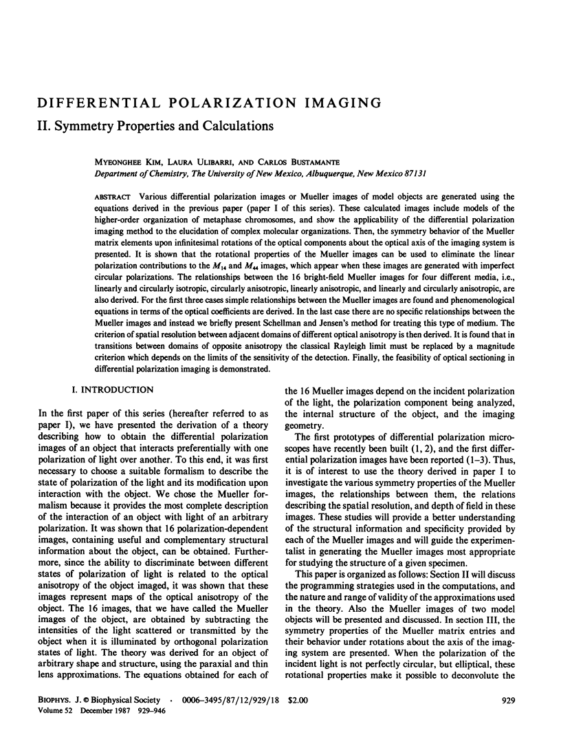
Selected References
These references are in PubMed. This may not be the complete list of references from this article.
- Barbashov S. F., Glotov B. O., Nikolaev L. G. Evidence for attachment of interphase chromatin to the nuclear matrix via matrix-bound nucleosomes. Biochim Biophys Acta. 1984 Jun 16;782(2):177–186. doi: 10.1016/0167-4781(84)90022-8. [DOI] [PubMed] [Google Scholar]
- Bentley G. A., Finch J. T., Lewit-Bentley A. Neutron diffraction studies on crystals of nucleosome cores using contrast variation. J Mol Biol. 1981 Feb 5;145(4):771–784. doi: 10.1016/0022-2836(81)90314-4. [DOI] [PubMed] [Google Scholar]
- Bustamante C., Tinoco I., Jr, Maestre M. F. Circular differential scattering can be an important part of the circular dichroism of macromolecules. Proc Natl Acad Sci U S A. 1983 Jun;80(12):3568–3572. doi: 10.1073/pnas.80.12.3568. [DOI] [PMC free article] [PubMed] [Google Scholar]
- Butler P. J. A defined structure of the 30 nm chromatin fibre which accommodates different nucleosomal repeat lengths. EMBO J. 1984 Nov;3(11):2599–2604. doi: 10.1002/j.1460-2075.1984.tb02180.x. [DOI] [PMC free article] [PubMed] [Google Scholar]
- Earnshaw W. C., Halligan N., Cooke C., Rothfield N. The kinetochore is part of the metaphase chromosome scaffold. J Cell Biol. 1984 Jan;98(1):352–357. doi: 10.1083/jcb.98.1.352. [DOI] [PMC free article] [PubMed] [Google Scholar]
- Finch J. T., Brown R. S., Richmond T., Rushton B., Lutter L. C., Klug A. X-ray diffraction study of a new crystal form of the nucleosome core showing higher resolution. J Mol Biol. 1981 Feb 5;145(4):757–769. doi: 10.1016/0022-2836(81)90313-2. [DOI] [PubMed] [Google Scholar]
- Glotov B. O., Nikolaev L. G., Dashkevich V. K., Barbashov S. F. Histone crosslinking patterns indicate dynamic binding of histone H1 in chromatin. Biochim Biophys Acta. 1985 Mar 20;824(3):185–193. doi: 10.1016/0167-4781(85)90047-8. [DOI] [PubMed] [Google Scholar]
- Huang H. C., Cole R. D. The distribution of H1 histone is nonuniform in chromatin and correlates with different degrees of condensation. J Biol Chem. 1984 Nov 25;259(22):14237–14242. [PubMed] [Google Scholar]
- Ibel K., Klingholz R., Strätling W. H., Bogenberger J., Fittler F. Neutron diffraction of chromatin in interphase nuclei and metaphase chromosomes. Eur J Biochem. 1983 Jun 15;133(2):315–319. doi: 10.1111/j.1432-1033.1983.tb07464.x. [DOI] [PubMed] [Google Scholar]
- Jordano J., Nieto M. A., Palacián E. Dissociation of nucleosomal particles by chemical modification. Equivalence of the two binding sites for H2A.H2B dimers. J Biol Chem. 1985 Aug 5;260(16):9382–9384. [PubMed] [Google Scholar]
- Karnik P. S. Correlation between phosphorylated H1 histone and condensed chromatin in Planococcus citri. FEBS Lett. 1983 Oct 31;163(1):128–131. doi: 10.1016/0014-5793(83)81178-8. [DOI] [PubMed] [Google Scholar]
- Klug A., Lutter L. C., Rhodes D. Helical periodicity of DNA on and off the nucleosome as probed by nucleases. Cold Spring Harb Symp Quant Biol. 1983;47(Pt 1):285–292. doi: 10.1101/sqb.1983.047.01.034. [DOI] [PubMed] [Google Scholar]
- Klug A., Rhodes D., Smith J., Finch J. T., Thomas J. O. A low resolution structure for the histone core of the nucleosome. Nature. 1980 Oct 9;287(5782):509–516. doi: 10.1038/287509a0. [DOI] [PubMed] [Google Scholar]
- Langmore J. P., Paulson J. R. Low angle x-ray diffraction studies of chromatin structure in vivo and in isolated nuclei and metaphase chromosomes. J Cell Biol. 1983 Apr;96(4):1120–1131. doi: 10.1083/jcb.96.4.1120. [DOI] [PMC free article] [PubMed] [Google Scholar]
- Lebkowski J. S., Laemmli U. K. Evidence for two levels of DNA folding in histone-depleted HeLa interphase nuclei. J Mol Biol. 1982 Apr 5;156(2):309–324. doi: 10.1016/0022-2836(82)90331-x. [DOI] [PubMed] [Google Scholar]
- León P., Macaya G. Properties of DNA rosettes and their relevance to chromosome structure. Chromosoma. 1983;88(4):307–314. doi: 10.1007/BF00292908. [DOI] [PubMed] [Google Scholar]
- Mickols W., Maestre M. F., Tinoco I., Jr, Embury S. H. Visualization of oriented hemoglobin S in individual erythrocytes by differential extinction of polarized light. Proc Natl Acad Sci U S A. 1985 Oct;82(19):6527–6531. doi: 10.1073/pnas.82.19.6527. [DOI] [PMC free article] [PubMed] [Google Scholar]
- Mirzabekov A. D., Bavykin S. G., Karpov V. L., Preobrazhenskaya O. V., Ebralidze K. K., Tuneev V. M., Melnikova A. F., Goguadze E. G., Chenchick A. A., Beabealashvili R. S. Structure of nucleosomes, chromatin, and RNA polymerase-promoter complex as revealed by DNA-protein cross-linking. Cold Spring Harb Symp Quant Biol. 1983;47(Pt 1):503–510. doi: 10.1101/sqb.1983.047.01.060. [DOI] [PubMed] [Google Scholar]
- Mitra S., Sen D., Crothers D. M. Orientation of nucleosomes and linker DNA in calf thymus chromatin determined by photochemical dichroism. Nature. 1984 Mar 15;308(5956):247–250. doi: 10.1038/308247a0. [DOI] [PubMed] [Google Scholar]
- Nielsen P. E., Matsuoka Y., Nordén B. J. Stepwise unfolding of chromatin by urea. A flow linear dichroism and photoaffinity labeling study. Eur J Biochem. 1985 Feb 15;147(1):65–68. doi: 10.1111/j.1432-1033.1985.tb08719.x. [DOI] [PubMed] [Google Scholar]
- Prusov A. N., Polyakov VYu, Zatsepina O. V., Chentsov YuS, Fais D. Rosette-like structures from nuclei with condensed (chromomeric) chromatin but not from nuclei with diffuse (nucleomeric or nucleosomic) chromatin. Cell Biol Int Rep. 1983 Oct;7(10):849–858. doi: 10.1016/0309-1651(83)90189-3. [DOI] [PubMed] [Google Scholar]
- Seki S., Nakamura T., Oda T. Supranucleosomal fiber loops of chicken erythrocyte chromatin. J Electron Microsc (Tokyo) 1984;33(2):178–181. [PubMed] [Google Scholar]
- Wang J. C. The path of DNA in the nucleosome. Cell. 1982 Jul;29(3):724–726. doi: 10.1016/0092-8674(82)90433-0. [DOI] [PubMed] [Google Scholar]
- Weith A. The fine structure of euchromatin and centromeric heterochromatin in Tenebrio molitor chromosomes. Chromosoma. 1985;91(3-4):287–296. doi: 10.1007/BF00328224. [DOI] [PubMed] [Google Scholar]


