Abstract
1. During respiratory efforts against a closed airway, the afferent activity of vagal fibres from pulmonary stretch receptors does not appreciably increase during the inspiratory phase because the lung is prevented from expanding.
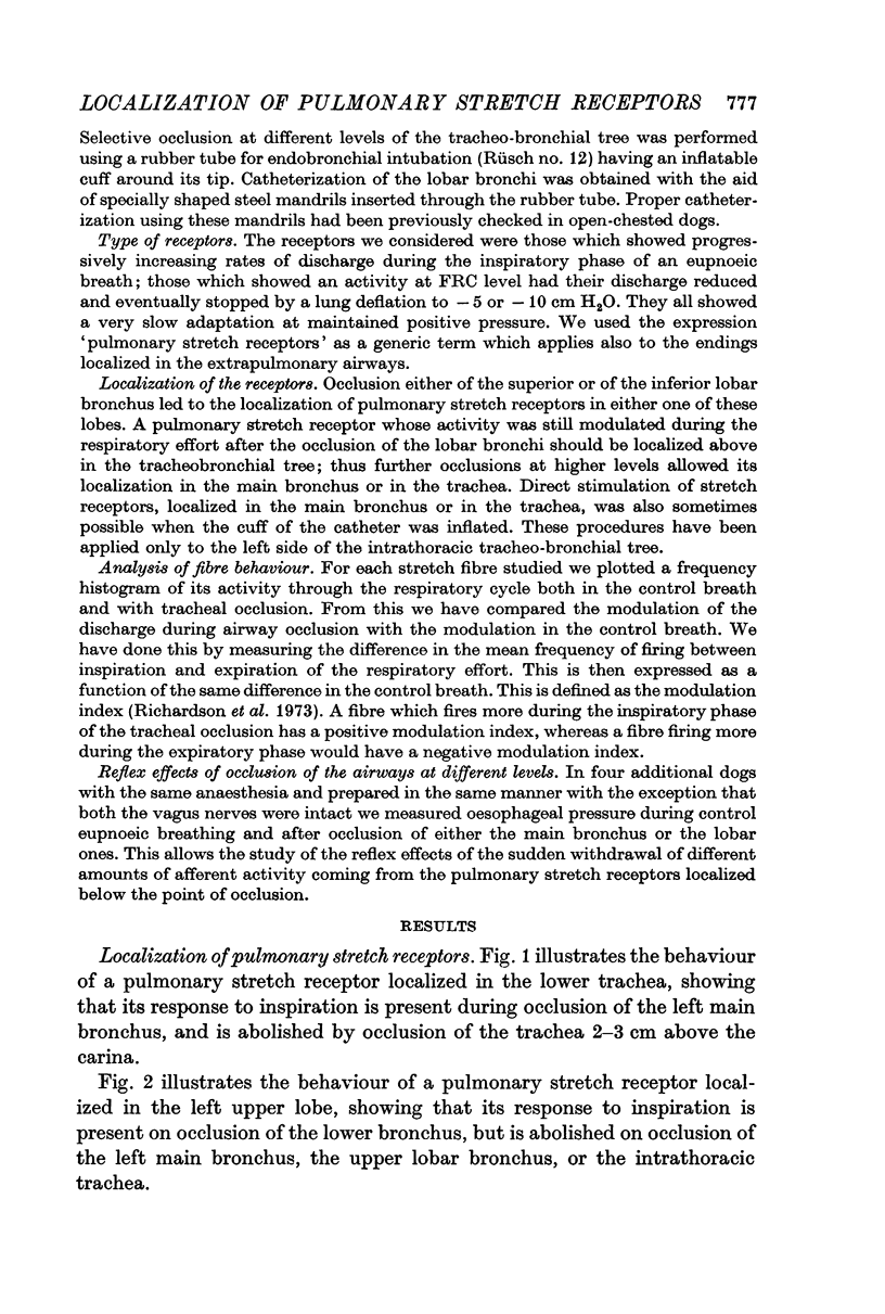
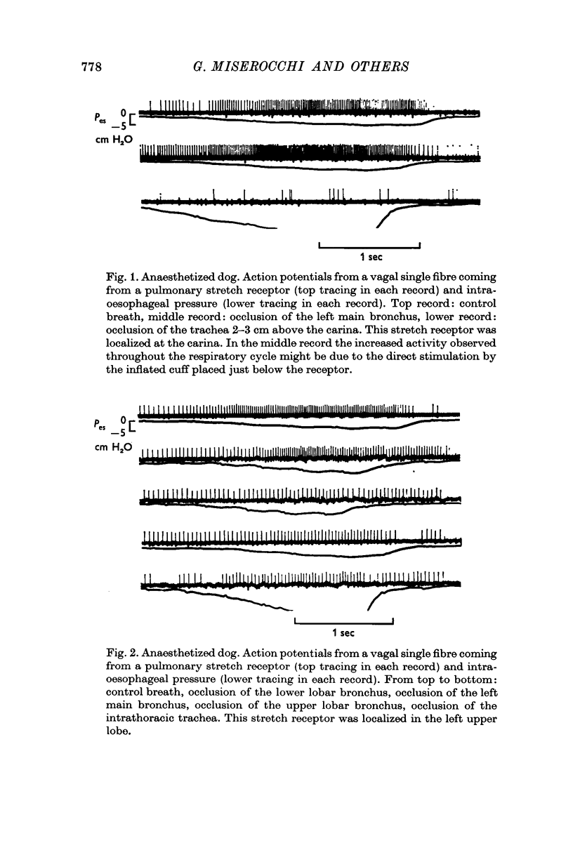
2. Occlusion at different levels of the airways allows the localization of pulmonary stretch receptors in the tracheo-bronchial tree.
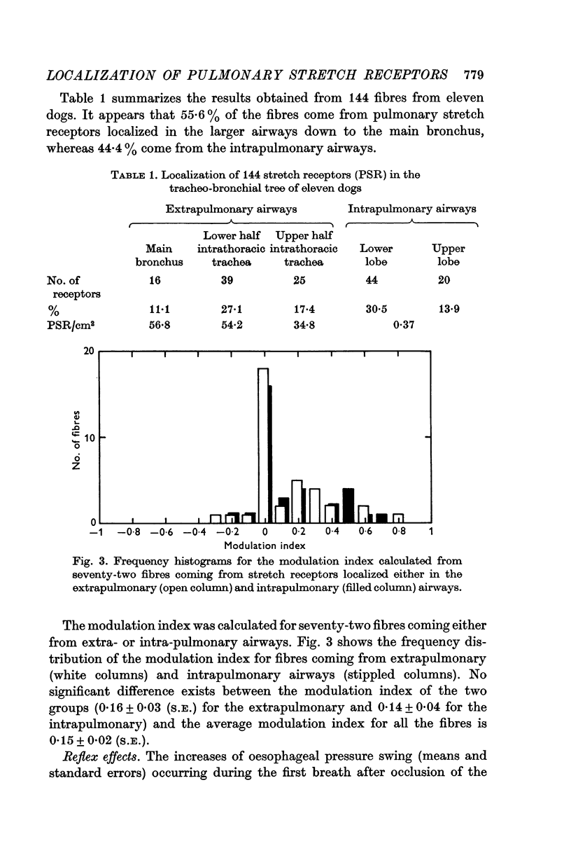
3. 144 fibres from pulmonary stretch receptors on the left side of the tracheo-bronchial tree have been studied in eleven dogs and their localization was as follows: 17·4% in the upper half of the intrathoracic trachea, 27·1% in the lower half of the intrathoracic trachea and the carina, 11·1% in the main bronchus, 13·9% in the upper lobe and 30·5% in the lower lobe.
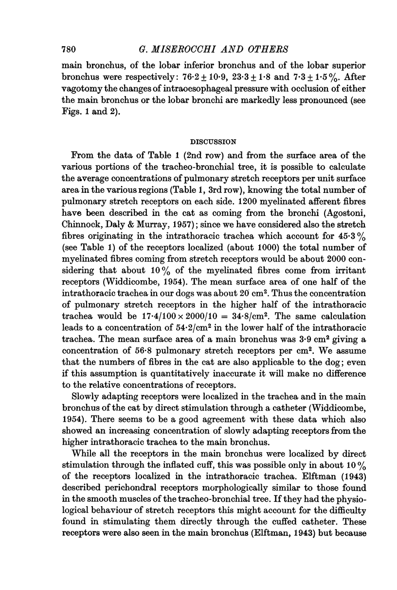
4. From the surface area of the tracheo-bronchial tree at different levels on the assumption of a total of 2000 stretch receptors on each side, their average concentration was as follows: 34·8% receptors/cm2 in the upper half of the intrathoracic trachea, 54·2/cm2 in the lower half of the intrathoracic trachea, 56·8/cm2 in the main bronchus, 0·37/cm2 in the intrapulmonary airways.
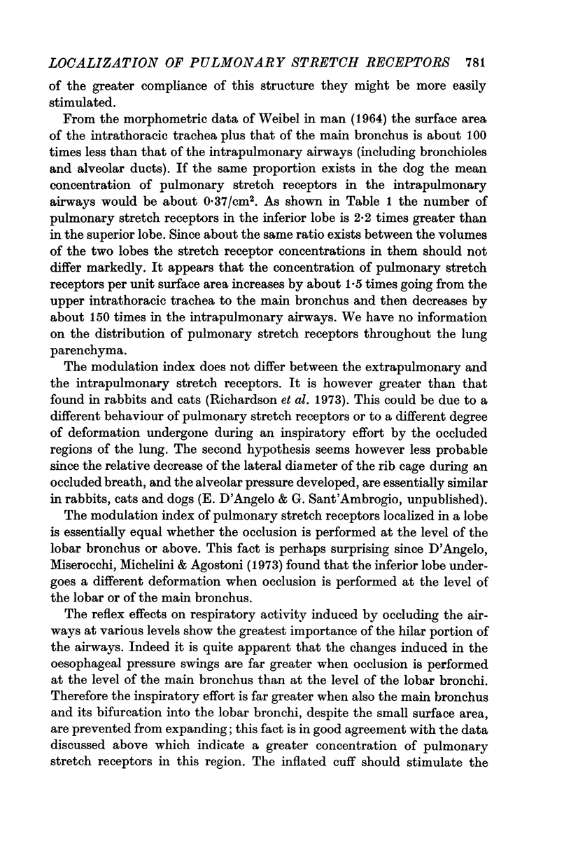
5. Occlusion of the main bronchus caused an increase of the eupnoeic oesophageal pressure swing by about 75% whereas occlusion of the inferior lobar bronchus led to an increase of only 20%. Therefore the reflex effects induced on the respiratory activity by occluding the airways at various levels show the greatest importance of the hilar portions of the airways where the concentration of pulmonary stretch receptors has been found to be greater.
Full text
PDF
Selected References
These references are in PubMed. This may not be the complete list of references from this article.
- AGOSTONI E., CHINNOCK J. E., DE DALY M. B., MURRAY J. G. Functional and histological studies of the vagus nerve and its branches to the heart, lungs and abdominal viscera in the cat. J Physiol. 1957 Jan 23;135(1):182–205. doi: 10.1113/jphysiol.1957.sp005703. [DOI] [PMC free article] [PubMed] [Google Scholar]
- Miserocchi G., D'Angelo E., Michelini S., Agostoni E. Displacements of the lung hilum, pleural surface pressure and alveolar morphology. Respir Physiol. 1972 Oct;16(2):161–174. doi: 10.1016/0034-5687(72)90048-5. [DOI] [PubMed] [Google Scholar]
- Richardson P. S., Sant'Ambrogio G., Mortola J., Bianconi R. The activity of lung afferent nerves during tracheal occlusion. Respir Physiol. 1973 Jul;18(2):273–283. doi: 10.1016/0034-5687(73)90056-x. [DOI] [PubMed] [Google Scholar]
- WEIDMANN H., BUCHER K. Zur Frage der Spezifität der vagalen Dehnungsreceptoren in der Lunge. Helv Physiol Pharmacol Acta. 1951;9(1):94–100. [PubMed] [Google Scholar]
- WIDDICOMBE J. G. Receptors in the trachea and bronchi of the cat. J Physiol. 1954 Jan;123(1):71–104. doi: 10.1113/jphysiol.1954.sp005034. [DOI] [PMC free article] [PubMed] [Google Scholar]
- WIDDICOMBE J. G. The activity of pulmonary stretch receptors during bronchoconstriction, pulmonary oedema, atelectasis and breathing against a resistance. J Physiol. 1961 Dec;159:436–450. doi: 10.1113/jphysiol.1961.sp006819. [DOI] [PMC free article] [PubMed] [Google Scholar]


