

Abstract
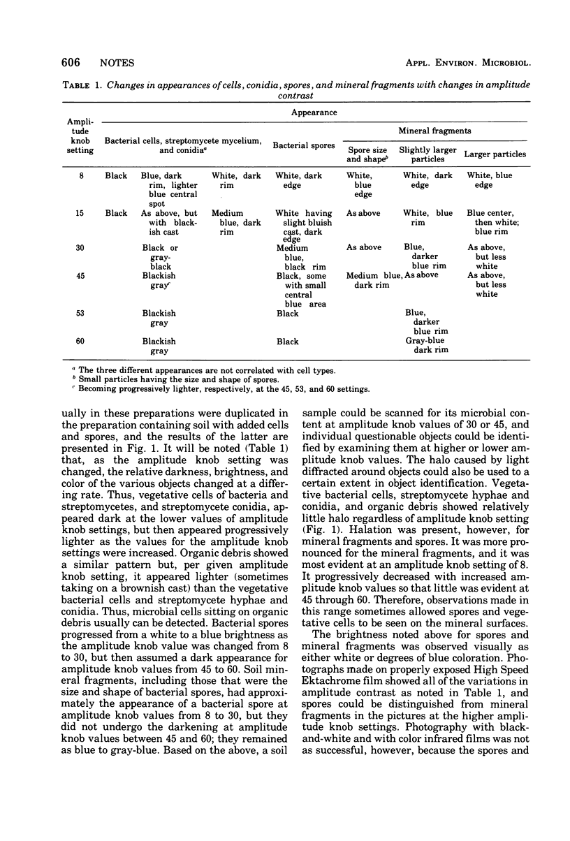
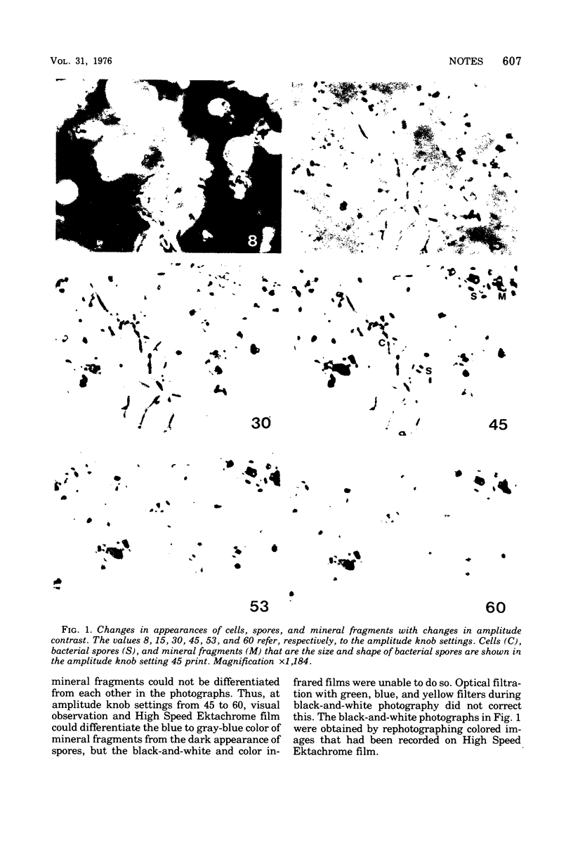
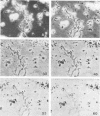
A new type of phase microscope was used to detect and observe the microflora in soil and to differentiate cells and spores from soil debris. This microscope provides a continuous variation in the amplitude ratios between undeviated and deviated beams of light, and the microbial cells, spores, and debris in soil change in appearance to a differing degree in response to the changes in amplitude ratio.
Full text
PDF
Images in this article
Selected References
These references are in PubMed. This may not be the complete list of references from this article.
- ALEXANDER F. E., JACKSON R. M. Examination of soil micro-organisms in their natural environment. Nature. 1954 Oct 16;174(4433):750–751. doi: 10.1038/174750b0. [DOI] [PubMed] [Google Scholar]
- Babiuk L. A., Paul E. A. The use of fluorescein isothiocyanate in the determination of the bacterial biomass of grassland soil. Can J Microbiol. 1970 Feb;16(2):57–62. doi: 10.1139/m70-011. [DOI] [PubMed] [Google Scholar]
- CASIDA L. E., Jr On the isolation and growth of individual microbial cells from soil. Can J Microbiol. 1962 Feb;8:115–119. doi: 10.1139/m62-015. [DOI] [PubMed] [Google Scholar]
- Casida L. E., Jr Infrared color photography: selective demonstration of bacteria. Science. 1968 Jan 12;159(3811):199–200. doi: 10.1126/science.159.3811.199. [DOI] [PubMed] [Google Scholar]
- Casida L. E., Jr Infrared color photomicrography of soil microorganisms. Can J Microbiol. 1975 Nov;21(11):1892–1893. doi: 10.1139/m75-276. [DOI] [PubMed] [Google Scholar]
- Casida L. E., Jr Interval scanning photomicrography of microbial cell populations. Appl Microbiol. 1972 Jan;23(1):190–192. doi: 10.1128/am.23.1.190-192.1972. [DOI] [PMC free article] [PubMed] [Google Scholar]
- Casida L. E., Jr Microorganisms in unamended soil as observed by various forms of microscopy and staining. Appl Microbiol. 1971 Jun;21(6):1040–1045. doi: 10.1128/am.21.6.1040-1045.1971. [DOI] [PMC free article] [PubMed] [Google Scholar]
- Casida L. E., Jr Observation of microorganisms in soil and other natural habitats. Appl Microbiol. 1969 Dec;18(6):1065–1071. doi: 10.1128/am.18.6.1065-1071.1969. [DOI] [PMC free article] [PubMed] [Google Scholar]
- Conn H. J. USE OF THE MICROSCOPE IN STUDYING THE ACTIVITIES OF BACTERIA IN SOIL. J Bacteriol. 1929 Jun;17(6):399–405. doi: 10.1128/jb.17.6.399-405.1929. [DOI] [PMC free article] [PubMed] [Google Scholar]
- McElroy L. J., Casida L. E., Jr An evaluation of rhodamine-labeled lysozyme as a fluorescent stain for in situ soil bacteria. Can J Microbiol. 1972 Jun;18(6):933–936. doi: 10.1139/m72-143. [DOI] [PubMed] [Google Scholar]
- Millar W. N., Casida L. E., Jr Microorganisms in soil as observed by staining with rhodamine-labeled lysozyme. Can J Microbiol. 1970 May;16(5):305–307. doi: 10.1139/m70-055. [DOI] [PubMed] [Google Scholar]
- Pital A., Janowitz S. L., Hudak C. E., Lewis E. E. Direct fluorescent labeling of microorganisms as a possible life-detection technique. Appl Microbiol. 1966 Jan;14(1):119–123. doi: 10.1128/am.14.1.119-123.1966. [DOI] [PMC free article] [PubMed] [Google Scholar]
- SKINNER F. A., JONES P. C., MOLLISON J. E. A comparison of a direct- and a plate counting technique for the quantitative estimation of soil micro-organisms. J Gen Microbiol. 1952 May;6(3-4):261–271. doi: 10.1099/00221287-6-3-4-261. [DOI] [PubMed] [Google Scholar]