Abstract
Aim—Solid and papillary epithelial neoplasm (SPEN) is an uncommon pancreatic tumour. Very rarely it has also been described outside the pancreas, usually arising from heterotopic pancreatic tissue. This report summarises all the published extrapancreatic SPENs and documents the sixth such case arising from heterotopic pancreatic tissue of the transverse mesocolon in a 15 year old girl.
Methods/Results—Histological and immunohistochemical examination revealed typical papillary and solid areas composed of columnar, cuboidal, and round cells, which were focally positive for vimentin, cytokeratin, neurone specific enolase, carcinoembryonic antigen, α1-antitrypsin, α1-antichymotrypsin, and negative for neuroendocrine markers (neurofilament, PGP 9.5, chromogranin A, synaptophysin, and S100), p53, and oestrogen and progesterone receptors. Electron microscopy showed scant zymogen but no neurosecretory granules. In agreement with the flow cytometric result of diploidy, comparative genomic hybridisation (CGH) did not reveal loss or gain of genetic material, and the in situ hybridisation analysis of the RB1 and p53 genes revealed no abnormality in the 13q and 17p arms.
Conclusions—Immunohistochemical and electron microscopic data support exocrine differentiation. The CGH and the flow cytometric results suggest a subtle, yet unknown genetic change, rather than a large genetic alteration. RB1 and p53 in situ hybridisation ruled out the role of deletion at these sites in the pathogenesis of SPEN. Interestingly, review of the published and the present heterotopic pancreatic SPENs identified the mesocolon as the most common anatomical site (four of six), despite the very rare occurrence of ectopic pancreatic tissue at this site.
Key Words: solid papillary epithelial neoplasm • heterotopic/ectopic pancreas • mesocolon
Full Text
The Full Text of this article is available as a PDF (314.1 KB).
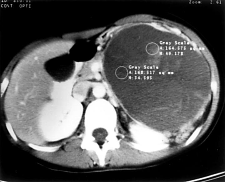
Figure 1 Axial computed tomography scan of the upper abdomen shows a circumscribed tumour in the transverse mesocolon, which dislocates but does not infiltrate the surrounding organs.

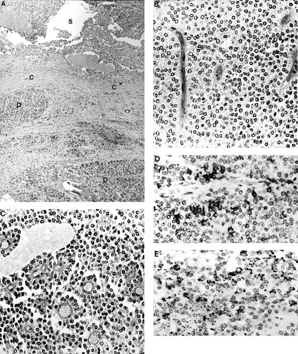
Figure 2 (A) Low power photomicrograph of the tumour (S) showing the fibrous pseudocapsule (C) and the ectopic pancreatic tissue (P). Tumour cells are arranged in (B) solid sheets or (C) form pseudopapillary structures around the blood vessels. Two characteristic immunophenotypic features were: (D) the strong, focal KL-1 positivity, and (E) the focal, granular or dot-like α-1-antichymotrypsin positivity.

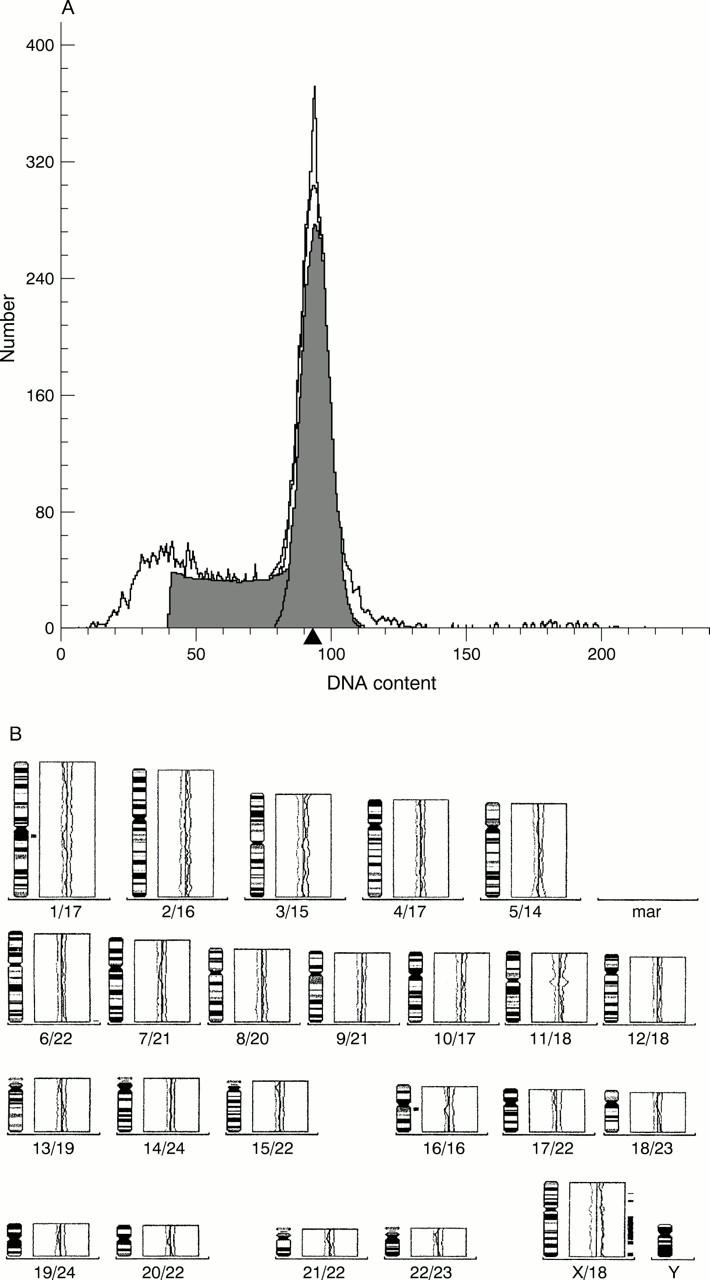
Figure 3 (A) Characteristic single diploid peak on the histogram. (B) Comparative genomic hybridisation shows no gain or loss in the genetic material.


