Abstract
Cells of higher eukaryotes process within minutes double strand breaks (DSBs) in their genome using a non-homologous end joining (NHEJ) apparatus that engages DNA-PKcs, Ku, DNA ligase IV, XRCC4 and other as of yet unidentified factors. Although chemical inhibition, or mutation, in any of these factors delays processing, cells ultimately remove the majority of DNA DSBs using an alternative pathway operating with an order of magnitude slower kinetics. This alternative pathway is active in mutants deficient in genes of the RAD52 epistasis group and frequently joins incorrect ends. We proposed, therefore, that it reflects an alternative form of NHEJ that operates as a backup (B-NHEJ) to the DNA-PK-dependent (D-NHEJ) pathway, rather than homology directed repair of DSBs. The present study investigates the role of Ku in the coordination of these pathways using as a model end joining of restriction endonuclease linearized plasmid DNA in whole cell extracts. Efficient, error-free, end joining observed in such in vitro reactions is strongly inhibited by anti-Ku antibodies. The inhibition requires DNA-PKcs, despite the fact that Ku efficiently binds DNA ends in the presence of antibodies, or in the absence of DNA-PKcs. Strong inhibition of DNA end joining is also mediated by wortmannin, an inhibitor of DNA-PKcs, in the presence but not in the absence of Ku, and this inhibition can be rescued by pre-incubating the reaction with double stranded oligonucleotides. The results are compatible with a role of Ku in directing end joining to a DNA-PK dependent pathway, mediated by efficient end binding and productive interactions with DNA-PKcs. On the other hand, efficient end joining is observed in extracts of cells lacking DNA-PKcs, as well as in Ku-depleted extracts in line with the operation of alternative pathways. Extracts depleted of Ku and DNA-PKcs rejoin blunt ends, as well as homologous ends with 3′ or 5′ protruding single strands with similar efficiency, but addition of Ku suppresses joining of blunt ends and homologous ends with 3′ overhangs. We propose that the affinity of Ku for DNA ends, particularly when cooperating with DNA-PKcs, suppresses B-NHEJ by quickly and efficiently binding DNA ends and directing them to D-NHEJ for rapid joining. A chromatin-based model of DNA DSB rejoining accommodating biochemical and genetic results is presented and deviations between in vitro and in vivo results discussed.
INTRODUCTION
Endogenous cellular processes and exogenous factors such as ionizing radiation (IR) can generate double strand breaks (DSB) in the DNA that undermine genomic integrity. Three enzymatically distinct processes, homology directed repair (HDR), single strand annealing (SSA) and non-homologous end joining (NHEJ) can, in principle, repair DNA DSBs (1–4). In cells of higher eukaryotes, repair of IR-induced DNA DSBs is dominated by a fast component operating with half times of the order of a few minutes. This form of rejoining is severely compromised by defects in any of the constituents of DNA-PK, DNA-PKcs and Ku (5–7). Very similar defects are also observed in cells defective in DNA ligase IV (8). Genetic, as well as a wealth of biochemical studies assign these proteins to the same end-joining pathway, which is characterized by an ability to remove DNA DSBs from the genome with extremely fast kinetics. We proposed the term D-NHEJ, for this pathway of DNA DSB rejoining.
Despite the prevalence of D-NHEJ, reports by us and others indicate that cells with defects in components of either DNA-PK, or DNA ligase IV complex are able to rejoin the majority of IR-induced DNA DSBs utilizing an alternative pathway operating with 20–30-fold slower kinetics (7–9). A slow pathway of DNA DSBs rejoining is frequently discernible in wild-type cells as well, but it becomes well defined and assumes a dominant role when D-NHEJ is compromised through defects in participating factors, or when inhibitors against DNA-PK, such as wortmannin, are administered.
Because HDR is normally operating with kinetics of the order of hours, it is in principle possible that the slow component seen after inhibition of D-NHEJ reflects this process. We tested this hypothesis using the hyper- recombinogenic DT40 chicken cell line and a set of mutants defective in homologous recombination (HR) (10,11). DT40 cells rejoin IR-induced DNA DSBs with kinetics similar to those of other vertebrate cells displaying 1000-fold lower levels of HR (6). In addition, knockouts of RAD51B, RAD52 and RAD54 rejoin DNA DSBs with kinetics similar to the wild type, as does also a conditional knock out mutant of RAD51 (6). While a significant reduction in the fast component of rejoining is observed in Ku70–/– DT40 cells, a double mutant Ku70–/–/RAD54–/– shows similar half times to Ku70–/– cells (6).
Thus, increases by several orders of magnitude in the capacity of cells to carry out HR, or defects in the proteins involved, fail to alter the rejoining kinetics in a way compatible with an involvement of HDR in the slow component of DNA DSBs rejoining, even when D-NHEJ is severely compromised (6). Furthermore, rejoining of DNA DSBs with slow kinetics is associated with the joining of incorrect ends, an effect not compatible with the operation of HDR (12). To accommodate these observations, we hypothesized that the slow component of DNA DSBs rejoining reflects an alternative pathway of NHEJ, which we termed backup (initially basic), B-NHEJ—to differentiate from D-NHEJ. According to this model, at least two distinct NHEJ pathways cooperate to remove IR-induced DNA DSBs from the genome of higher eukaryotes (6,9,13)
We inquired whether biochemical studies utilizing as substrate plasmid DNA digested by a restriction endonuclease to generate different types of ends, and cellular extracts as a source of enzyme, can recapitulate aspects of the operation of the two pathways of DNA DSB rejoining. Here, we examine this possibility by studying the role of Ku in NHEJ using an in vitro plasmid-based DNA end-joining assay.
Ku is a heterodimer of 69 and 86 kDa polypeptides called Ku70 and Ku80, respectively (3,14). Ku has high affinity for binding to DNA ends without exhibiting sequence specificity and it is thought that Ku binding on DNA ends is protective against nuclease digestion (15–17). The crystal structure of Ku indicates that Ku70 and Ku80 fold in a similar fashion and bind to each other to generate an asymmetrical ring like structure with an aperture large enough to allow DNA to be threaded through (18). Ku transfers between DNA ends and translocates along the DNA in an ATP-independent manner allowing several Ku dimers to bind on a single DNA molecule (19–21). The DNA-PK activity is required for Ku to enter DNA when a Ku-DNA-PKcs complex assembles on DNA ends (22). As a regulatory subunit, Ku stimulates the DNA-PK kinase activity by binding to DNA ends (23,24). Ku and DNA-PKcs are thought to recruit other DNA repair proteins like XRCC4/ligase IV to DNA ends and to stimulate the DNA end joining by mammalian DNA ligases (25–27). Evidence has been presented that Ku is functioning as an alignment factor that brings and holds the DNA ends together to facilitate their metabolic restoration and rejoining (28,29). Ku also plays a critical role in telomere capping in mammalian cells (30,31) and yeast (32,33), and is involved in telomeric silencing (34,35). These diverse functions of Ku made it an appropriate initial focus for our studies.
MATERIALS AND METHODS
Cell lines and extract preparation
HeLa cells were grown either as monolayer or suspension cultures in Joklik’s modified MEM (S-MEM) supplemented with 5% bovine calf serum. M059-J cells were grown as monolayer cultures in DMEM/F12 medium supplemented with 10% bovine calf serum. All experiments were performed with whole cell extract (WCE). For HeLa extract-preparation a 1–30 l suspension of cells was grown in spinner flasks to 0.5–1 × 10⁶ cells/ml and collected by centrifugation. For M059-J extract-preparation, 100 dishes (100 mm, 20 ml growth medium, 0.5 × 10⁶ cells) were prepared and cells were allowed to grow to ∼4 × 10⁶ cells/dish before harvesting by trypsinization. The very slow growth characteristics of M059K cells, the isogenic wild-type control of M059J cells, made extract preparation difficult. Therefore, in this work HeLa cells are used as a wild-type control for M059J cells. For further processing, cells from both cell lines were washed once in ice-cold PBS and subsequently in five packed cell volumes of cold hypotonic buffer (10 mM HEPES, 5 mM KCl, 1.5 mM MgCl2, 0.2 mM phenylmethylsulfonyl fluoride and 0.5 mM DTT). The cell pellet was resuspended in 2 vol of hypotonic buffer and, after 10 min in ice, disrupted in a Dounce homogenizer (15 strokes with a B pestle). Subsequently, 3 M KCl were slowly added to the homogenate to a final concentration of 0.5 M KCl and, after 30 min on ice, centrifuged for 40 min at 14 000 r.p.m. at 4°C to obtain the WCE. To remove DNA from the extract, 0.1 vol of DEAE Sepharose, equilibrated in dialysis buffer (25 mM HEPES, pH 7.5, 100 mM KCl, 1 mM EDTA, 10% glycerol, 0.2 mM PMSF and 0.5 mM DTT), was added and the mixture was gently rotated at 4°C for 30 min. Extract was cleared by centrifugation at 12 000 r.p.m. for 5 min and dialyzed overnight in dialysis buffer. After centrifugation, extract was aliquoted, snap frozen and stored at –80°C.
Proteins and antibodies
DNA-PKcs and DNA-PK were purified from HeLa nuclear extracts as previously described (36). Baculovirus-produced, purified, recombinant Ku was a gift of Dr William S. Dynan (University of Georgia at Augusta, Medical College of Georgia, GA). Antibodies against XRCC4 and DNA ligase IV were kindly provided by Dr Steven Jackson (Cambridge University, UK). Mouse monoclonal antibodies against Ku80 (Ab-3) and DNA-PKcs (Ab-2) were purchased from Neomarkers (Union City, CA). Goat polyclonal antibody against Ku70 (c-19) was purchased from Santa Cruz Biotechnology (Santa Cruz, CA).
Western blot analysis and immunodepletion
For western blot, cell extracts were electrophoresed on 10% SDS–polyacrylamide gels, transferred to a PVDF membrane and probed by the ECL-Plus kit as recommended by the manufacturer (Amersham Biosciences). Signal was detected using the ‘Storm’ or the ‘Typhoon’ (Molecular Dynamics).
For Ku-immunodepletion, 20 µl of anti-Ku serum (OY), or normal human serum (NHS) were added to 50 µl of protein A-Sepharose beads in 250 µl of DB buffer (20 mM HEPES, pH 7.9, 100 mM KCl, 20% glycerol, 0.2 mM EDTA, 0.5 mM DTT and 0.5 mM PMSF) with 50 µg/ml BSA (37). Mixture was incubated under constant rotation at 4°C overnight. Subsequently, beads were washed three times with dialysis buffer, and incubated with 100–200 µl HeLa or M059-J WCE for 2 h at 4°C. After centrifugation, the supernatants were used for quantification of depletion and DNA end joining, whereas the beads were washed three times with dialysis buffer before analysis by western blotting. When necessary, additional depletion cycles were carried out until practically complete depletion was achieved.
Plasmid and oligonucleotides
Supercoiled plasmid pSP65 (3 kb, Promega) was prepared using CsCl2/EtBr gradients. It was used as a substrate in DNA end-joining reactions following digestion with SalI to generate linearized DNA. In some experiments, pSP65 was linearized using other restriction endonucleases as indicated.
Double stranded DNA oligonucleotides used in electrophoretic mobility shift assay (EMSA) experiments were generated by annealing two single stranded complementary oligonucleotides. Their names and sequences are: OA 5′-GGCCGCACGCGTCCACCATGGGGTACAA-3′ and OB 5′-GTAGTTGTACCCCATGGTGGACGCGTGC-3′. OA and OB were annealed as described (38) to form a duplex DNA with four-base 5′ overhanging ends.
DNA end joining
End-joining reactions were performed in 20 mM HEPES–KOH (pH 7.5), 10 mM MgCl2, 80 mM KCl, 1 mM ATP, 1 mM DTT, 0.25 µg of DNA (12.5 ng/µl) and 0–20 µg (0–1 µg/µl) of HeLa, or M059-J WCE in a final volume of 20 µl at 25°C for 1 h. Reactions were terminated by adding 2 µl of 5% SDS, 2 µl of 0.5 M EDTA and 1 µl of proteinase E (10 mg/ml), then incubated for 1 h at 37°C. One half of the reaction was loaded on a 0.7% agarose gel and run at 45 V (2 V/cm) for 5 h. Gels were stained in SYBR Gold (Molecular Probes) and scanned in a FluorImager (Molecular Dynamics). For quantification of rejoining the ImageQuant software (Molecular Dynamics) was used to calculate the percent of input plasmid found in dimers and other higher order polymers. The values obtained are included in the figures below the corresponding gels.
Wortmannin (Sigma) was prepared in DMSO at 10 mM and diluted in 10% DMSO to 20 µM immediately before use. Diluted wortmannin (1 µl) was added to the reactions and incubated for 10 min at 4°C prior to the addition of ATP and DNA. This pre-treatment and sequence of reagent addition was found to maximize the effect of wortmannin.
Electrophoretic mobility shift assay
Standard EMSA was performed to measure Ku activity using a radiolabeled double-stranded 32mer DNA-probe prepared as described above. Cell extract was incubated with 0.2 ng end-labeled DNA probe in DNA binding buffer (10 mM Tris–HCl, pH 7.5, 1 mM EDTA, 1 mM DTT, 100 mM NaCl, 0.5% glycerol) at 25°C for 30 min, then electrophoresed on a 6% polyacrylamide gel in 0.5× TBE buffer. The gel was dried and analyzed in the ‘Storm’ (Molecular Dynamics). When antibodies were included in the reactions, they were incubated with the complete reaction for 30 min on ice, before addition of the probe.
Assay of DNA-PK activity
The assay used to measure DNA-PK activity has been described (39). Briefly, the peptide EPPLSQEAFADLWKK, corresponding to 11–24 amino acids of human p53 with threonine 18 and serine 20 changed to alanine was used as substrate. Cell extracts are mixed with 200 µM peptide, with or without 10 µg/ml sonicated calf thymus DNA in 50 mM HEPES (pH 7.5), 10 mM MgCl2, 50 mM KCl, 0.2 mM EGTA in the final volume of 18 µl. A radioactively-labeled ATP solution is made by mixing 1 µl of [γ-32P]ATP (3000 Ci/mmol, 10 µCi/µl) in 49 µl unlabeled 5 mM ATP. Reactions are started by adding 2 µl of ATP solution and incubating at 30°C for 30 min; they are stopped by adding 20 µl stop solution (30% acetic acid, 1 mM ATP). Subsequently, 20 µl of the mixture are spotted on Whatman p81 phosphocellulose paper and washed four times in 10% acetic acid for 10 min. 32P incorporation is measured in a Scintillation Counter (Packard). Results are analyzed to calculate net incorporation of ATP (in pmol) by subtracting incorporation in reactions assembled without DNA (background).
RESULTS
Anti-Ku antibodies inhibit DNA end joining only in the presence of DNA-PKcs
We designed experiments to investigate the requirement for Ku in DNA end joining in vitro, as assayed by the ligation of restriction endonuclease-linearized plasmid in reactions assembled with WCEs. We selected reaction conditions supporting optimal DNA end joining under the assumption that this will enable us to assay for a spectrum of processes operating in the cell for DNA DSB removal, and for the potential role of Ku in these processes. This was important since the present study was designed to explore the operation of alternative, backup pathways of DNA end joining, as well as the potential interplay and hierarchical structure between such pathways. For such a goal, conditions forcing DNA end joining to the operation of a single pathway are not desirable, despite their usefulness in characterizing factors involved in a specific pathway (27,40,41).
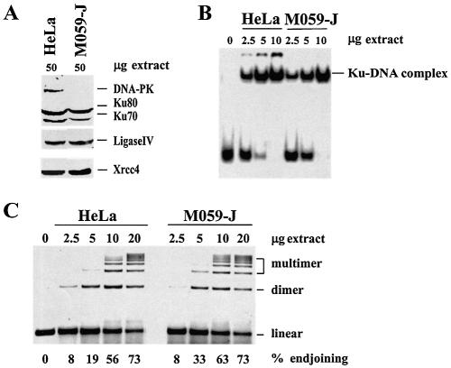
Because extensive genetic and biochemical studies indicate a direct interaction and cooperation between Ku and DNA-PKcs (42), we first examined how the presence of the latter protein influences DNA end joining. For the experiments, we employed WCEs prepared from actively growing DNA-PK-proficient HeLa cells, and DNA-PK-deficient M059-J cells (43,44) as described in Materials and Methods. As a first step in the initial characterization of these extracts, we carried out western blotting to assess the abundance of Ku. The results in Figure 1A indicate that the levels of the two Ku subunits, Ku70 and Ku80, are only slightly lower in extracts (50 µg) of M059-J cells as compared to extracts of HeLa cells. Other components of the DNA-PK-dependent pathway of NHEJ, such as DNA ligase IV, and XRCC4 are present at similar levels in the extracts of these cell lines. As expected, however, DNA-PKcs is undetectable in extracts of M059-J cells.
Figure 1.
Levels and activity of Ku, as well as DNA end joining in WCEs of HeLa and M059-J cells. (A) Western blot showing the levels of Ku70, Ku80, DNA ligase IV, XRCC4 and DNA-PKcs in extracts of HeLa and M059-J cells. Fifty micrograms of extract were loaded per lane. Note the comparable abundance of Ku, DNA ligase IV and XRCC4 in the extracts of the two cell lines, as well as the absence of DNA-PKcs in extracts from M059-J cells. (B) EMSA with extracts of HeLa and M059-J cells. 0.2 ng of a 32 bp, 32P-labeled, double stranded DNA probe was incubated with different amounts of extract at 25°C for 30 min and loaded on a 6% polyacrylamide gel. The lower band indicates unbound probe, whereas the higher bands indicate DNA–protein complexes. Note the comparable DNA binding activity present in HeLa and M059-J cell extracts. (C) DNA end joining in extracts of HeLa and M059-J cells. Reactions were assembled with the indicated amount of extract and 0.25 µg plasmid substrate and incubated at 25°C for 1 h. Products were analyzed by electrophoresis in a 0.7% agarose gel run at 45 V (2 V/cm) for 5 h. Gels were stained in SYBR Gold and scanned in a FluorImager (Molecular Dynamics). Quantification was carried out using the ImageQuant software (Molecular Dynamics). The results were used to calculate the percent of plasmid migrating as a dimer and other higher order forms and is given at the bottom of the gel (% end joining). The migration distances of the input plasmid substrate (linear), as well as of dimers and other higher order forms (multimers) are indicated. Note the high level of DNA end-joining activity in the extracts of both cell lines.
For a functional characterization of Ku activity in extracts of HeLa and M059-J cells, we carried out EMSA. Figure 1B shows results of reactions assembled with 0–10 µg extract. Effective DNA binding is observed in reactions assembled with extracts of both cell lines, with a slightly higher activity seen again in extracts of HeLa cells. The slower mobility forms in reactions assembled with extracts of HeLa cells are higher order complexes comprising, in addition to the DNA probe and Ku, probably also DNA-PKcs, as they are undetectable in reactions assembled with extracts of M059-J cells.
Having established comparable levels and activity for Ku protein in the extracts of the two cell lines, we studied next DNA end joining using SalI-digested pSP65 plasmid. The results in Figure 1C show strong DNA end-joining activity as a function of HeLa extract concentration resulting in the formation of dimers, trimers and other higher order multimers. Formation of circles (relaxed or supercoiled) is not detectable under the conditions employed here. More than 70% of the input plasmid is ligated in reactions assembled with 20 µg extract suggesting high levels of activity that can be directly quantified by DNA fluorescence. Thus, use of DNA end labeling, Southern blotting or other methods employed to assess low levels of activity, or low amount of DNA substrate are not required. Restriction analysis of reaction products (not shown) indicates an approximately equal representation of tail-to-tail and head-to-tail end joining, with a slightly smaller fraction of head-to-head end joining. Since random end joining would produce twice as many head-to-tail products than head-to-head or tail-to-tail, our results suggest a preference for head-to-head and particularly for tail-to-tail end joining.
Efficient DNA end joining in a concentration-dependent manner is also observed in reactions assembled with extracts of M059-J cells. Under the conditions employed here, 20 µg extracts of M059-J cells also rejoin >70% of the input plasmid. Thus, despite the absence of DNA-PKcs, DNA end-joining activity remains high. It has been previously shown that when the concentration of Mg2+ is adjusted to 0.5 mM (compared to 10 mM in our reactions), only DNA-PK-dependent DNA end joining occurs (40). At this low Mg2+ concentration DNA end joining is reduced by >90% (8). As mentioned above, it is our working hypothesis that reactions assembled at Mg2+ concentrations allowing optimal DNA end joining are preferable for the goals of our study. In addition, the concentration of Mg2+ employed is closer to that present in the nucleus of the cell: 2–4 mM during interphase and 4–17 mM during M-phase (45). Restriction analysis of the reaction products (not shown) indicates an approximately equal representation for tail-to-tail, head-to-tail and head-to-head end joining, and thus similar end-joining preferences to those found with HeLa cell extracts. Taken together, the above results indicate similar levels of Ku and DNA end-joining activities in extracts of HeLa and M059-J cells, despite the absence of DNA-PKcs from the latter.
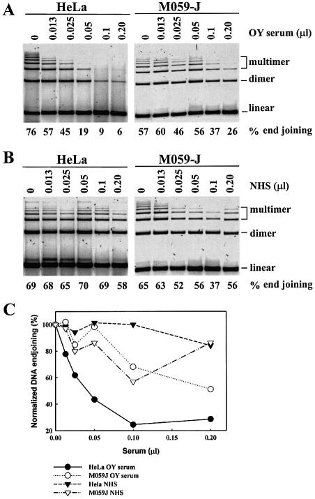
As a first step in the evaluation of the role of Ku in DNA end joining in vitro we employed as a tool sera from individuals with polymyositis-scleroderma overlap syndrome. These sera contain antibodies against Ku that are known to inhibit signal joint formation during in vitro V(D)J recombination (46) and in vitro DNA end joining (37,47). Increasing amounts of OY serum were added to reactions containing extract, but no substrate DNA or ATP, and were incubated at 25°C for 10 min. Subsequently, DNA end joining was initiated by adding DNA and ATP and reactions were incubated at 25°C for 1 h. In reactions assembled with extracts of HeLa cells, OY serum strongly inhibits DNA end joining (Fig. 2A), whereas a control serum from a normal individual (NHS) is without a significant effect (Fig. 2B). Figure 2C shows a quantitative evaluation of the above results, after normalization to the untreated controls, for reactions assembled with OY serum (closed circles), or NHS (closed triangles). At 0.1 µl per reaction, OY serum inhibits >80% of the DNA end-joining activity, whereas NHS has no measurable effect. Higher amounts do not further enhance this effect suggesting a component in DNA end joining that is refractory to inhibition by this antibody. EMSA demonstrates an interaction between Ku and antibodies present in the serum (supershift) in the range of concentrations used for DNA end joining, confirming an interaction between antibodies in the OY serum and the Ku antigen (results not shown). Notably, antibodies do not prevent end binding of Ku. These results are in line with an involvement of Ku in DNA end joining and suggest that, when present in the extract, Ku engages in the reaction. However, interaction with antibodies inhibits the normal functions of the protein and blocks DNA end joining without preventing end binding.
Figure 2.
Serum of polymyositis-scleroderma overlap syndrome patients inhibits in vitro DNA end joining. (A) Twenty micrograms of extract of either HeLa or M059-J cells were incubated in 16 µl of DNA end-joining reaction-mixture (without DNA and ATP) in which 2 µl of serum from OY patient were added, after appropriate dilution in phosphate buffered saline, to achieve the final levels in µl serum per reaction indicated on top of the gel, and were incubated at 25°C for 10 min. After this pre-incubation period, DNA and ATP were added in half of the reaction-mixture to assay for DNA end joining, whereas the remaining reaction-mixture was processed for EMSA (results not shown). Note the inhibition of DNA end joining with increasing serum concentration in reactions assembled with extracts of HeLa cells and the reduced inhibition in reactions assembled with extracts of M059-J cells. (B) Similar to (A), but for reactions assembled in the presence of identical amounts of NHS from a healthy individual. (C) Quantitative evaluation of the DNA end-joining results shown in (A) and (B) using ImageQuant. Results are shown normalized to the percent rejoining measured for each set of reactions when incubated in the absence of serum. Actual values for percent end joining are shown at the bottom of the gels in (A) and (B). Note the inhibition in DNA end joining with increasing amount of OY serum in the reaction, and the difference in the effect between M059-J and HeLa cells, particularly when considering the effect of NHS.
Treatment of extracts of M059-J cells with OY serum, under practically identical conditions, leads to different results. Although some inhibition is observed with increasing amounts of OY serum (Fig. 2A), the inhibition is significantly reduced compared to reactions assembled with HeLa cell extracts. Furthermore, a small effect is seen in reactions assembled with NHS (Fig. 2B) and when the results obtained in these two sets of reactions are directly compared quantitatively (Fig. 3C, open symbols) the specific effect of OY serum (open triangles) is only slightly larger than the non-specific effect of NHS (open circles). Since the results in Figure 1 indicate comparable abundance and activity for Ku protein in HeLa and M059-J cell extracts, the reduced inhibition in extracts of the latter cells, can be attributed to the absence of DNA-PKcs. Thus, DNA-PKcs stabilizes the interactions of Ku with the DNA and directs end joining to a Ku dependent pathway.
Figure 3.
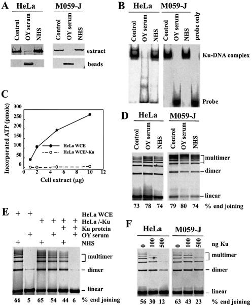
DNA end joining in extracts of HeLa and M059-J cells after depletion of Ku. (A) Two hundred microgram extracts of HeLa or M059-J cells (∼20 µl) were incubated with a 1:1 mixture of Sepharose A and G beads (20 µl) that had been pre-incubated overnight with 5 µl of OY serum, or NHS. The extract-beads mixture was incubated at 4°C for 1 h and beads were removed by centrifugation. The procedure was repeated a second time and extracts were analyzed by western blotting for the levels of Ku70. Similar results were obtained when Ku80 was assayed (not shown). Note the practically complete depletion of Ku in extracts treated with OY serum and the unchanged levels of the protein in extracts treated with NHS compared to those observed in untreated controls. The beads-fraction contains large amounts of Ku protein after treatment with OY serum, but no protein after treatment with NHS. (B) EMSA analysis under the conditions outlined in Figure 1 for extracts of HeLa and M059-J cells treated as described in (A). Note the absence of detectable DNA binding in extracts treated with OY serum. (C) DNA-PK activity in HeLa WCE and Ku-depleted HeLa WCE (HeLa WCE/–Ku). 1, 2.5, 5, 10 µg of cell extract was mixed in 20 µl DNA-PK reaction buffer with and without calf thymus DNA. Reactions were incubated at 30°C for 30 min. The DNA-PK activity was calculated as described in Materials and Methods. (D) DNA end joining in reactions assembled with untreated control extracts (20 µg), or equal amounts of extracts treated with either OY serum, or NHS. End-joining activity is indicated at the bottom of the gel as % end joining. Note the efficient end joining in Ku-depleted cell extracts of either HeLa or M059-J cells. (E) Twenty micrograms of extract of either HeLa WCE, Ku-depleted HeLa WCE, or Ku-depleted HeLa WCE supplemented with 50 ng purified recombinant Ku protein were incubated in 18 µl of DNA end-joining reaction-mixture (without DNA and ATP) with 0.1 µl of OY serum, or 0.1 µl of NHS at 25°C for 10 min. After this pre-incubation period, DNA and ATP were added to start the reaction. Note that OY serum does not inhibit the DNA end joining in Ku-depleted HeLa WCE, and that the inhibition can be rescued by recombinant Ku. (F) DNA end joining in reactions assembled with extracts of HeLa or M059-J cells (10 µg) and increasing amounts of purified Ku. Percent end joining is indicated at the bottom of the gel. Note the inhibitory effect of Ku that is less pronounced in reactions assembled with extracts of M059-J cells.
DNA end joining in Ku-depleted extracts
To further evaluate the role of Ku in DNA end joining, we tested extracts of HeLa and M059-J cells after immunodepletion with OY serum. After two cycles of depletion by immunoprecipitation, Ku is barely detectable in extracts of either HeLa or M059-J cells (Fig. 3A) and is found practically exclusively in the beads. Similar treatment with NHS has no effect on the level of Ku in the extract of either cell line. The depletion of Ku indicated in Figure 3A is corroborated by the DNA binding results in Figure 3B and the DNA-PK activity measurements in Figure 3C. Strong gel shift is observed in control cell extracts, as well as in extracts treated with NHS. On the other hand, DNA binding is not detectable in Ku-depleted extracts of either HeLa or M059-J cells. Similarly robust DNA-PK activity is detected in HeLa cell extracts only before Ku depletion (Fig. 3C). Thus, by several measures non-detectable levels of Ku remain in the depleted extracts.
Despite the practically complete removal of Ku, DNA end joining is not diminished in reactions assembled with depleted HeLa cell extracts (Fig. 3D). This observation suggests the presence in HeLa cell extracts of factors efficiently catalyzing DNA end joining in the absence of Ku, probably via an alternative DNA end-joining pathway. The results of Figures 2 and 3 in aggregate suggest, on the one hand, a dominant engagement of Ku in the end-joining reaction, manifested by the strong inhibition by Ku-specific antibodies, and on the other hand, the ability of the cell to perform efficient end joining in the absence of Ku. These apparently contradictory observations are analyzed in more detail in the Discussion.
To confirm that the residual end joining observed in depleted extracts is not due to remaining traces of Ku, we tested the effect of OY serum. The results in Figure 3E demonstrate that OY serum has only a very small effect in reactions assembled with depleted extract, but inhibits almost completely end joining in reactions assembled with non-treated extract. Notably, addition of recombinant Ku to reactions assembled with depleted extract rescues the sensitivity of the end-joining reaction to OY serum. These results are consistent with a quantitative removal of Ku from the extract and a specific function of OY serum through interaction with Ku.
A small increase in DNA end-joining activity is occasionally observed after Ku depletion, implying that Ku may actually inhibit the alternative Ku-independent pathway. To examine this possibility we supplemented Ku-depleted HeLa cell extracts with recombinant Ku. The results in Figure 3F show that high Ku concentrations have a strongly inhibitory effect on DNA end joining and suggest that at physiological concentrations, Ku may actually suppress alternative pathways of DNA end joining. On the other hand, it is also possible that at high concentrations, DNA becomes overloaded with Ku, which interrupts subsequent productive interactions with other factors and stops the normal progression of the repair reaction. Very similar observations are also made in reactions assembled using extracts of M059-J cells, although recombinant Ku is less inhibitory than in HeLa cells at similar concentrations (Fig. 3F). These observations are also in line with the operation of alternative, Ku-independent, pathways of DNA end joining operating in the absence of DNA-PKcs, and reinforce the important role of DNA-PKcs in the functions of Ku.
Fidelity of end joining and joining of different DNA ends in Ku-depleted extracts
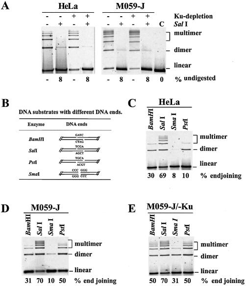
Although the results presented above suggest efficient DNA end joining in the absence of Ku and DNA-PKcs, it remains open whether other aspects of the reaction, for example end-preservation, are altered in the absence of these repair factors. This is particularly relevant, as Ku may protect DNA ends from processing by nucleases, and there is evidence that it can function as an alignment factor during DNA end joining (28,29). To address this question we evaluated fidelity of end joining of SalI-digested plasmid by isolating products and subjecting them to re-digestion with the same restriction endonuclease. Figure 4A shows the results obtained. Efficient and error-free DNA end joining is observed in reactions assembled with HeLa cell extracts, with 92% of the products redigested with SalI (8% undigested). This indicates a high probability for preservation during end joining of the restriction endonuclease recognition sequence. Neither the efficiency of the reaction nor the digestibility of the products are changing after Ku-depletion, suggesting that the fidelity of end joining remains high and similar to that of non-depleted extracts. Similar results are also obtained in reactions assembled with extracts of M059-J cells (Fig. 4A), suggesting that DNA-PKcs, in the presence or absence of Ku, is not altering the joining fidelity of this type of DNA ends.
Figure 4.
The role of Ku in the fidelity of DNA end joining, as well as in the joining of different types of DNA ends. (A) Reactions were assembled under standard conditions using normal or Ku-depleted extracts (20 µg) of either HeLa or M059-J cells and DNA end joining was allowed to take place at 25°C for 1 h (lanes marked –Sal I). After gel electrophoresis, products were purified and the fidelity of end joining assayed by re-digestion with SalI. The lanes marked +Sal I show the digested products. Percent- undigested plasmid was calculated by ImageQuant and the values are shown at the bottom of the gel. Note that products of the end joining reaction remain sensitive to SalI under all conditions examined (92% digestibility) suggesting end joining without sequence modification around the end. (B) Types of ends generated and recognition sequences of BamHI, SalI, PstI and SmaI. (C) DNA end joining in HeLa cell extracts under standard conditions using as substrate pSP65 plasmid, linearized with the indicated restriction endonucleases. Percent end joining is shown at the bottom of the gel. Note the reduced end joining with substrate prepared by digestion with BamHI, PstI and SmaI. (D) As in (C) but with extracts of M059-J cells. Note the increase in DNA end joining, as compared to HeLa cells, with substrate prepared by digestion with PstI. (E) As in (D) but with Ku-depleted M059-J cell extracts (using OY serum). Note that the end-bias of the joining reaction is significantly reduced when compared to HeLa cells.
It is well documented that the efficiency of in vitro DNA end joining is dependent on the type of ends. To investigate whether Ku contributes to this dependence, we used different restriction endonucleases to generate blunt ends, or ends with 5′ or 3′ overhangs (Fig. 4B), and evaluated end joining in extracts of HeLa or M059-J cells, as well as in Ku-depleted extracts of M059-J cells. The results obtained with HeLa cells are shown in Figure 4C. It is evident that 5′ protruding DNA ends (protruding single strands, PSS) generated by SalI are joined with the highest efficiency (69% substrate conversion). Although BamHI also produces 5′ protruding ends, substrate conversion is only 30%, mainly as a result of a reduction in trimers and other higher order forms. An even lower efficiency of DNA end joining (10% substrate conversion) is observed with the 3′ protruding DNA ends produced by PstI, and substrate conversion drops to 8% for blunt ends generated by SmaI.
Extracts of M059-J cells join DNA ends generated by SalI, BamHI and SmaI with efficiency practically indistinguishable from that of HeLa cell extracts. However, ends generated by PstI are rejoined nearly five times more efficiently than in HeLa cell extracts (Fig. 4D). Depletion of Ku from M059-J cell extracts leaves joining of ends generated by SalI and PstI unchanged as compared to non-depleted extracts, increases slightly (from 30 to 50%) joining of BamHI ends, but increases >3-fold (from 10 to 30%) joining of blunt ends produced by SmaI. Thus, in the absence of Ku and DNA-PKcs, the end-preference for DNA end joining of normal cell extracts is nearly eliminated, suggesting that these factors partly determine the end-specificity of the rejoining reaction, probably by restricting the joining of certain types of ends. We have no information on the fidelity of end joining for the different types of ends assayed in the experiments described above.
Ku is essential for wortmannin inhibition of DNA end joining
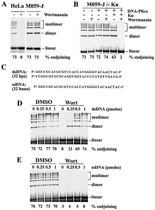
Wortmannin was originally characterized as a PI-3 kinase inhibitor, but was later shown to also inhibit at higher concentrations other kinases of the PI-3 kinase family, such as DNA-PK, ATM and ATR (48,49). Wortmannin inhibits in vitro DNA end joining in reactions similar to those presented above, and this observation is interpreted as an indication for dependence of the reaction on DNA-PK. We investigated the role of Ku in this inhibition. Reaction mixtures containing extracts as indicated were treated for 10 min with 1 µM wortmannin or DMSO, prior to the addition of plasmid DNA and ATP to initiate the reaction. Figure 5A shows that, in agreement with previous observations, 1 µM wortmannin completely inhibits DNA end joining in reactions assembled with extracts of HeLa cells. This effect is likely to be entirely and exclusively due to the presence of DNA-PKcs in the extract, as the inhibitor has no effect on DNA end joining in extracts of M059-J cells that lack DNA-PKcs. Thus, although active end joining occurs in M059-J cells that lack DNA-PKcs, end joining initiated in the presence of this kinase requires this activity for completion and implies involvement of DNA-PKcs in the overall reaction.
Figure 5.
Inhibition of DNA end joining by wortmannin requires Ku and can be reversed by short dsDNA oligonucleotides. (A) DNA end joining in reactions assembled with 20 µg extract of HeLa or M059-J cells in the presence or absence of 1 µM wortmannin. Percent end joining is given at the bottom of the gel. Reaction mixtures containing the cell extract, but no substrate DNA or ATP were incubated with wortmannin or DMSO (the solvent of wortmannin) at 25°C for 10 min. End joining was initiated by adding DNA and ATP and was allowed to take place at 25°C for 1h. Note the strong inhibition of DNA end joining by wortmannin in extracts of HeLa cells, and the lack of inhibition in extracts of M059-J cells. (B) Reactions were assembled with Ku-depleted extracts of M059-J cells (20 µg) in the presence or absence of wortmannin as outlined in (A). Two hundred nanograms of purified DNA-PKcs, or 100 ng of purified Ku were added to the indicated reactions starting from and including the pre-incubation period. Percent end joining is given at the bottom of the gel. Note that wortmannin induced inhibition requires Ku. See Figure 3D for results of reactions assembled with Ku, with or without wortmannin. (C) Sequence of the double stranded and single stranded oligonucleotides (32mers) used in the DNA end joining reactions shown in (D) and (E). (D) DNA end joining in reactions assembled under standard conditions with extracts (20 µg) of HeLa cells in the presence or absence of 1 µM wortmannin and the indicated amounts of the dsDNA shown in (C). Reaction mixtures containing extract but no ATP or substrate DNA were incubated with wortmannin or DMSO at 25°C for 10 min. Subsequently the indicated amount of dsDNA was added and the reaction was incubated at 25°C for an additional 10 min. Finally, substrate DNA (0.25 µg per reaction, ∼1.1 pmol) and ATP were added to initiate end joining. Percent end joining is given at the bottom of the gel. Note the lack of inhibition in the presence of dsDNA alone, and the reversion of the wortmannin effect by more than 0.5 pmol dsDNA. (E) As in (D) but with reactions supplemented with the indicated amounts of single-stranded DNA. Note the lack of effect on DNA end joining under these conditions, in the presence or absence of wortmannin.
We examined the role of Ku in wortmannin-induced inhibition of DNA end joining by supplementing Ku-depleted M059-J cell extracts with purified DNA-PKcs and/or Ku and evaluating DNA end joining in the presence or absence of drug. As expected, Ku-depleted extracts of M059-J cells support effective DNA end joining that remains unaffected by wortmannin treatment. Thus, Ku-depleted extracts of M059-J cells respond to wortmannin as non-depleted extracts of the same cells. Addition of 100 ng purified DNA-PKcs to the reaction produces no detectable effect on DNA end joining and leaves the extracts refractory to inhibition by wortmannin. However, addition of 100 ng Ku together with 100 ng DNA-PKcs produces a slight inhibition in DNA end joining (likely an effect of Ku, see above) but render the reaction fully sensitive to wortmannin. Thus, Ku is required for wortmannin-induced inhibition of DNA end joining in the presence of DNA-PKcs.
The inhibition of DNA end joining by wortmannin can be explained as a blockade of the end joining process by the inactive kinase that remains trapped at the DNA ends and prevents further processing by the DNA-PK-dependent pathway (22), or the diversion of the end joining process to DNA-PK-independent backup pathways. To examine whether other factors present in the extract and involved in DNA end joining are also compromised in their function by this inhibition, for example by binding to the inactivated DNA-PK complex, and to test the status of DNA-PK-independent pathways of DNA end joining in extracts treated with wortmannin, we carried out the following experiment: reactions were assembled with extracts of HeLa cells and treated with 1 µM wortmannin, or the equivalent amount of DMSO, in the absence of substrate DNA and ATP, for 10 min at 25°C. Subsequently, different amounts of dsDNA (a 32mer) were added (Fig. 5C) and reactions were further incubated at 25°C for 10 min. Finally, substrate DNA and ATP were added to initiate end joining and reaction mixtures incubated at 25°C for 1 h. We inquired whether the double stranded 32mer would titer the inactive DNA-PK but leave alternative pathways of end joining in an operational state. Figure 5D shows the results obtained. Increasing amounts of dsDNA has no detectable inhibitory effect on the DNA end-joining reaction in the absence of wortmannin in the range between 0.25 and 1 pmol despite the expected titration of Ku and DNA-PKcs. This is in line with the activity measured in the extracts in the absence of Ku or DNA-PKcs. In reactions assembled with wortmannin-treated extracts no DNA end joining is observed in the control, in line with the above-presented results. However, as the amount of dsDNA added in the reaction increases, DNA end joining takes place and at 0.5 pmol dsDNA, end joining is at levels similar to those of untreated controls. Thus, increasing amounts of dsDNA progressively allows plasmid DNA end joining in wortmannin-treated extracts, probably by tittering inhibitory DNA-PK complexes that block access to the plasmid ends of components of alternative end-joining pathways. Of note, the presence of dsDNA up to 1 pmol is not affecting end joining by alternative pathways suggesting that blunt DNA ends do not deplete extract of these activities and that there is an abundance of such activities in the extract. Some inhibition of DNA end joining is however observed above 2 pmol dsDNA (not shown). Addition of ssDNA in the reactions under similar conditions does not relieve wortmannin inhibition (Fig. 5E), confirming a requirement for dsDNA and the role of DNA-PK in wortmannin-induced inhibition of end joining.
DISCUSSION
Genetic studies quantitatively evaluating the kinetics of rejoining of radiation-induced DNA DSBs in wild-type cells of higher eukaryotes, in mutants deficient in DNA-PKcs, Ku or DNA ligase IV, as well as after treatment with wortmannin provide evidence for the operation of at least two pathways of NHEJ with distinct properties that we have termed D-NHEJ and B-NHEJ (see Introduction). D-NHEJ is fast (t1/2: 5– 30 min), requires DNA-PKcs, Ku, DNA ligase IV and probably XRCC4 and rejoins DNA ends without excessive degradation (12). B-NHEJ on the other hand is slow (t1/2: 2– 20 h), may involve degradation of DNA ends (12), and utilizes as of yet unidentified components. The two pathways operate in a complementary fashion and cooperate to remove IR-induced DNA DSBs in a well-defined hierarchical order. D-NHEJ is dominant and normally removes the majority of DNA DSBs. When D-NHEJ is compromised as a result of mutation in a participating factor, by inhibitors, or by the absence of an essential component from the vicinity of the DNA DSB, B-NHEJ acts as a backup to remove practically completely DNA DSBs, albeit with slower kinetics (9). The biochemical experiments presented in the previous section extend and complement the genetic studies by reproducing several important, albeit not all, of the features of these pathways.
Alternative pathways of NHEJ
Active end joining is observed in Ku-depleted extracts of HeLa cells, in extracts from M059-J cells that lack DNA-PKcs, or in Ku-depleted extracts of M059-J cells that lack both DNA-PKcs and Ku. Thus, the absence of key components of D-NHEJ is not compromising end joining in line with the operation of an alternative, DNA-PK-independent pathway that may be operationally equivalent to the genetically defined B-NHEJ (5,6,8,9,13).
Extensive literature addressing DNA end joining from a different angle also points to the operation of alternative pathways. Seminal work by Roth and Wilson first demonstrated the extraordinary ability of mammalian cells to join with similar efficiency matched or mismatched ends of transfected DNA either with a pathway utilizing direct ligation, or a pathway relying on terminal homologies of a few nucleotides (50,51). With the characterization of mutants deficient in components of D-NHEJ this work was extended to investigate the roles of specific factors in the end joining process. In agreement with the biochemical data presented here, end joining of transfected DNA remained at levels similar to those of wild-type controls in cells deficient in DNA-PKcs (52–54), Ku (54,55), XRCC4 (54,55) or DNA ligase IV (54) pointing to pathways operating efficiently in the absence of these proteins. Notably, when the generated junctions were examined by sequencing, a clear shift was observed in the mutants from homology independent to homology dependent end joining (54,55).
Also, in vitro data obtained using assays similar to the one employed here provided evidence for the operation of alternative pathways of DNA end joining and have implicated Ku in the fidelity of the process, as well as in the inhibition of the pathway utilizing microhomologies (28,37,54,56). In addition, V(D)J recombination, despite its stricter dependence on DNA-PK and DNA ligase IV/XRCC4, can also utilize alternative, DNA-PK-independent pathways that rely on microhomologies for the formation of non-standard V(D)J recombination products (54). Finally, alternative pathways of DNA DSB end joining have been suggested from genetic studies involving the generation of DNA ligase IV and Ku70-deficient DT40 chicken B-cells (57). Thus the evidence for alternative pathways of NHEJ is ample and quite diverse. There are distinct parallels between microhomology- dependent end joining and B-NHEJ that require further study. These studies will be benefit substantially by a biochemical characterization of B-NHEJ.
The dominant nature of D-NHEJ
D-NHEJ dominates NHEJ in vivo as long as its key factors are not compromised by either genetic mutation or chemical inhibition. This implies that DNA ends generated either endogenously, or by exogenous agents such as IR and other DNA damaging agents, are quickly bound and processed by factors involved in this pathway. Indeed this appears to be the case for both Ku and DNA-PKcs in the in vitro studies presented here (Figs 2 and 5). These observations suggest a priority binding of these factors to DNA ends, in line with the documented in vivo dominance of D-NHEJ and the known properties of DNA-PKcs and Ku (3,58). Inhibition of DNA end joining in vitro by antibodies against Ku has also been reported for reactions assembled with extracts of Xenopus eggs (37), and inhibition of DNA end joining by wortmannin in extracts of human cells and Xenopus eggs (22,59). Thus, the results presented here are not specific for the model systems and the conditions employed.
Of note, the results show a strong functional interdependence between DNA-PKcs and Ku on DNA end joining. Thus, inhibition of end joining by anti-Ku antibodies is not effective in the absence of DNA-PKcs suggesting that the kinase facilitates the stalling of subsequent steps in the end joining reaction despite the fact that a ligase other than DNA ligase IV must be employed (8). A recent report demonstrates loss of DNA-PKcs requirement in reactions assembled with purified DNA ligase IV/XRCC4, Ku and a fraction containing Mre11/Rad50/NBS1 (41). The significance of this observation for our results is not clear at present.
Further indication for coordination in the functions of DNA-PKcs and Ku provides the observation that these factors impose restrictions in the joining efficiency between different types of ends. The bias observed here against joining of 3′ protruding ends in the presence of Ku, although not always detectable (40), if active in vivo may be of consequence for the 3′ protruding strands generated during the processing of DNA ends by HDR. For example, it may reduce a non-productive competition between NHEJ and HDR. The same results also suggest that the role of Ku as an alignment factor (28,29) depends on the types of DNA ends presented to the end-joining machinery, and that this function is modulated by additional factors in the extract. This is probably why Ku inhibits joining of blunt ends in our setting, whereas it facilitates joining of blunt ends in reactions assembled with purified DNA ligases (25).
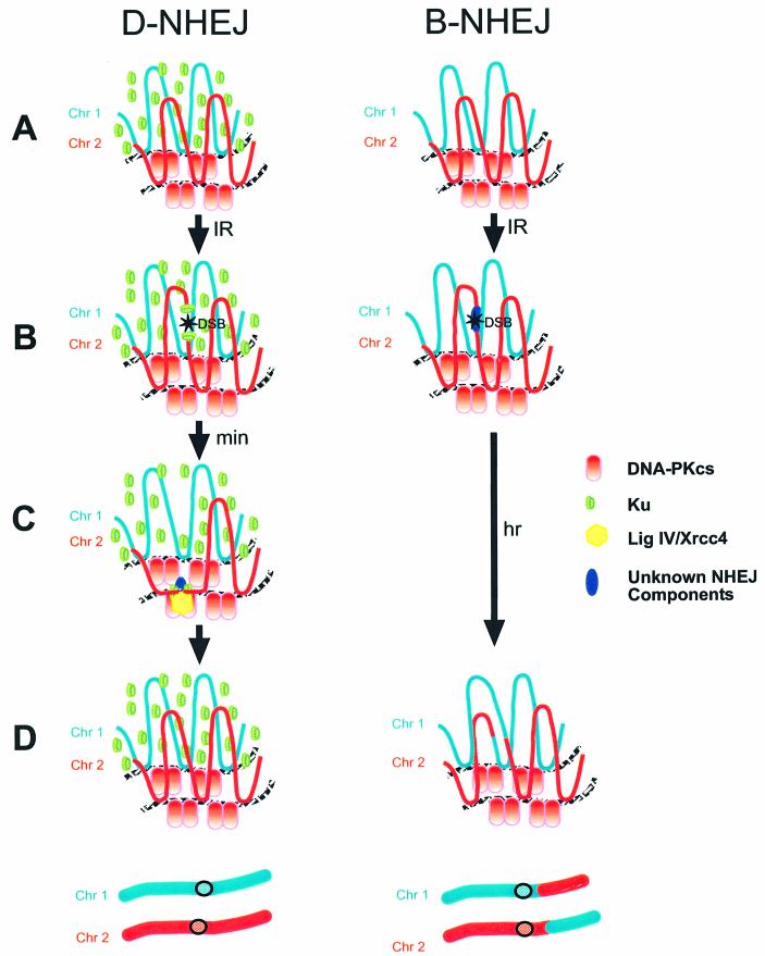
The model in Figure 6 recapitulates the interpretation of the results presented here; it is a further development of a previously proposed model (9) and addresses the processing of IR-induced DNA DSBs by D-NHEJ and B-NHEJ in the context of chromatin. In the presence of Ku and DNA-PKcs, DNA ends are quickly captured and chromatin structure is locally altered (not shown) to facilitate synapsis. This is the rate-limiting step of the reaction and is considered to occur close to the nuclear matrix mediated by a complex set of protein–protein interactions between DNA-PKcs, Ku, DNA ligase IV, XRCC4, histones and other unidentified factors. End joining is catalyzed by DNA ligase IV. The high speed of the process and local changes in chromatin structure facilitate joining of correct ends and suppress the formation of chromosome aberrations. In the absence of Ku or DNA-PKcs, DNA ends are not captured and local changes in chromatin structure fail to occur. Ends remain open and are processed by components of B-NHEJ with slow kinetics (hours) because of inefficient synapsis at random locations in the nucleus. The long-lived DNA ends may interact with other DNA ends in the vicinity, generated by IR or other processes, causing the formation of chromosome aberrations. In this model, quick capture and local processing of the DNA ends through optimized protein–protein interactions and chromatin conformation changes is the central mechanism by which joining errors are prevented during D-NHEJ. Although this model awaits rigorous testing it is relevant that affinity chromatography with ATC clusters, typically found in scaffold or matrix attachment regions, almost exclusively binds a complex containing poly(ADP-ribose) polymerase and DNA-PK (60).
Figure 6.
A hypothetical model for the operation of D-NHEJ and B-NHEJ in chromatin-packed DNA. D-NHEJ: (A) neighboring chromatin loops of two chromosomes (Chr 1 and Chr 2) with attachments to the nuclear matrix are shown. In the absence of DNA DSBs, DNA-PKcs is drawn attached to the nuclear matrix and Ku (5–10-fold excess) distributed throughout the nucleus. (B) Exposure to IR induces a DSB in Chr 2 that is recognized by Ku. This recognition step and the availability of DNA-PKcs allow the cell to direct the rejoining of the DSB to the D-NHEJ pathway. (C) Within minutes after induction, interactions between Ku and DNA-PKcs, on the one hand, and chromatin remodeling, on the other hand, facilitate synapsing of the ends and joining by DNA ligase IV/XRCC4, a process also facilitated by as of yet uncharacterized factors. Optimal interaction of factors and processes involved in this step require the chromatin context to ensure the exceptional speed characterizing this pathway of end joining. (D) Although the rejoining process does not ensure restoration of the original sequence in the vicinity of the DSB, the extremely rapid processing favors synapsing of correct ends and suppresses thus the formation of exchange-type chromosome aberrations. Therefore, when chromosome integrity is the endpoint, D-NHEJ can be considered an error-free process. B-NHEJ: In the absence of Ku (intermediate results are expected in case of Ku haploinsufficiency), DSBs are induced by IR with similar efficiency, but are not directed to D-NHEJ despite the presence of DNA-PKcs. Under these conditions, DSBs are recognized and processed by as yet uncharacterized components of B-NHEJ that can also operate outside the chromatin context. Although B-NHEJ is capable of processing the great majority of IR-induced DNA DSBs, it does so with kinetics an order of magnitude slower than D-NHEJ, presumably because of inefficient synapsing of DNA ends. As a result the DNA ends remain open for long periods of time and can be partly degraded and/or interact with DNA DSBs in neighboring loops induced randomly, or through the replication of damaged DNA at a later time point, to form exchanges. Exchanges can also be formed through the interaction between two IR-induced breaks that are brought into proximity after irradiation by changes in chromatin conformation. (D) An exchange between loops from different chromosomes can lead to a reciprocal exchange (shown), or to the formation of a dicentrics chromosome and an acentric fragment (not shown).
Kinetics and fidelity of B-NHEJ in vivo and in vitro
The kinetic advantage of D-NHEJ in vivo (see Introduction and above) is not reproduced in vitro, where both pathways operate with similar kinetics. We attribute this difference to the loss of the kinetic advantage in the synapsis step of D-NHEJ (see model in Fig. 6), as synapsis may not be rate limiting at the high DNA concentrations used in vitro. Although our results indicate no reduction in end-joining efficiency in the absence of Ku, other experiments show a reduction in circle formation (28,37,61). The significance of circle formation in in vitro DNA end-joining assays and its correlation, if any, with in vivo rejoining of DNA DSBs remains to be established.
The in vitro assay utilized in the present study, as well as in previous reports (37,40,62), fails to reproduce the reduced fidelity of B-NHEJ in vivo (see Introduction) and the cancer prone nature of alternative pathways of NHEJ in mice (63–65). We propose that this may also be a reflection of the artificial kinetic advantage of B-NHEJ under the in vitro conditions employed here. Indeed, a fidelity reduction has been reported in the absence of Ku under conditions supporting end joining less efficiently than in our experiments (28,56). It is possible that even in vitro, slow end joining allows processing of the ends by an exonuclease (likely a 3′ to 5′ exonuclease) causing a reduction in fidelity and that this processing is modulated by Ku. That reduced fidelity in the absence of Ku may be seen in in vitro systems only under conditions of kinetic disadvantage is also supported by the high fidelity observed in experiments evaluating end joining of transfected plasmid DNA in Ku-deficient cells (55).
Acknowledgments
ACKNOWLEDGEMENTS
Work was supported by grants RO1 CA42026, RO1 CA56706 awarded from NIH, DHHS, and a grant from the IFORES program of the University of Duisburg-Essen.
REFERENCES
- 1.Haber J.E. (1999) Gatekeepers of recombination. Nature, 398, 665–667. [DOI] [PubMed] [Google Scholar]
- 2.Jackson S.P. (2002) Sensing and repairing DNA double-strand breaks. Carcinogenesis, 23, 687–696. [DOI] [PubMed] [Google Scholar]
- 3.Doherty A.J. and Jackson,S.P. (2001) DNA repair: how KU makes ends meet. Curr. Biol., 11, R920–R924. [DOI] [PubMed] [Google Scholar]
- 4.Thompson L.H. and Schild,D. (2001) Homologous recombinational repair of DNA ensures mammalian chromosome stability. Mutat. Res., 477, 131–153. [DOI] [PubMed] [Google Scholar]
- 5.DiBiase S., Zeng,Z.-C., Chen,R., Hyslop,P.A., Curran,W.,Jr and Iliakis,G. (2000) DNA-dependent protein kinase stimulates an independently active, nonhomologous, end-joining apparatus. Cancer Res., 60, 1245–1253. [PubMed] [Google Scholar]
- 6.Wang H., Zeng,Z.-C., Bui,T.-A., Sonoda,E., Takata,M., Takeda,S. and Iliakis,G. (2001) Efficient rejoining of radiation-induced DNA double-strand breaks in vertebrate cells deficient in genes of the RAD52 epistasis group. Oncogene, 20, 2212–2224. [DOI] [PubMed] [Google Scholar]
- 7.Nevaldine B., Longo,J.A. and Hahn,P.J. (1997) The scid defect results in much slower repair of DNA double-strand breaks but not high levels of residual breaks. Radiat. Res., 147, 535–540. [PubMed] [Google Scholar]
- 8.Wang H., Zhao-Chong,Z., Perrault,A.R., Cheng,X., Qin,W. and Iliakis,G. (2001) Genetic evidence for the involvement of DNA ligase IV in the DNA-PK-dependent pathway of non-homologous end joining in mammalian cells. Nucleic Acids Res., 29, 1653–1660. [DOI] [PMC free article] [PubMed] [Google Scholar]
- 9.DiBiase S.J., Zeng,Z.-C., Chen,R., Hyslop,T., Curran,W.J.,Jr and Iliakis,G. (2000) DNA-dependent protein kinase stimulates an independently active, nonhomologous, end-joining apparatus. Cancer Res., 60, 1245–1253. [PubMed] [Google Scholar]
- 10.Takata M., Sasaki,M.S., Sonoda,E., Morrison,C., Hashimoto,M., Utsumi,H., Yamaguchi-Iwai,Y., Shinohara,A. and Takeda,S. (1998) Homologous recombination and non-homologous end-joining pathways of DNA double-strand break. EMBO J., 17, 5497–5508. [DOI] [PMC free article] [PubMed] [Google Scholar]
- 11.Sonoda E., Sasaki,M.S., Buerstedde,J.-M., Bezzubova,O., Shinohara,A., Ogawa,H., Takata,M., Yamaguchi-Iwai,Y. and Takeda,S. (1998) Rad51-deficient vertebrate cells accumulate chromosomal breaks prior to cell death. EMBO J., 17, 598–608. [DOI] [PMC free article] [PubMed] [Google Scholar]
- 12.Loebrich M., Rydberg,B. and Cooper,P.K. (1995) Repair of X-ray-induced DNA double-strand breaks in specific Not I restriction fragments in human fibroblasts: joining of correct and incorrect ends. Proc. Natl Acad. Sci. USA, 92, 12050–12054. [DOI] [PMC free article] [PubMed] [Google Scholar]
- 13.Asaad N.A., Zeng,Z.-C., Guan,J., Thacker,J. and Iliakis,G. (2000) Homologous recombination as a potential target for caffeine radiosensitization in mammalian cells: reduced caffeine radiosensitization in XRCC2 and XRCC3 mutants. Oncogene, 19, 5788–5800. [DOI] [PubMed] [Google Scholar]
- 14.Dynan W.S. and Yoo,S. (1998) Interaction of ku protein and DNA-dependent protein kinase catalytic subunit with nucleic acids. Nucleic Acids Res., 26, 1551–1559. [DOI] [PMC free article] [PubMed] [Google Scholar]
- 15.Blier P.R., Griffith,A.J., Craft,J. and Hardin,J.A. (1993) Binding of Ku protein to DNA. Measurement of affinity for ends and demonstration of binding to nicks. J. Biol. Chem., 268, 7594–7601. [PubMed] [Google Scholar]
- 16.Falzon M., Fewell,J.W. and Kuff,E.L. (1993) EBP-80, a transcription factor closely resembling the human autoantigen Ku, recognizes single- to double-strand transitions in DNA. J. Biol. Chem., 268, 10546–10552. [PubMed] [Google Scholar]
- 17.Pang D., Yoo,S., Dynan,W.S., Jung,M. and Dritschilo,A. (1997) Ku proteins join DNA fragments as shown by atomic force microscopy. Cancer Res., 57, 1412–1415. [PubMed] [Google Scholar]
- 18.Walker J.R., Corpina,R.A. and Goldberg,J. (2001) Structure of the Ku heterodimer bound to DNA and its implications for double-strand break repair. Nature, 412, 607–614. [DOI] [PubMed] [Google Scholar]
- 19.de Vries E., van Driel,W., Bergsma,W.G., Arnberg,A.C. and van der Vliet,P.C. (1989) HeLa nuclear protein recognizing DNA termini and translocating on DNA forming a regular DNA-multimeric protein complex. J. Mol. Biol., 208, 65–78. [DOI] [PubMed] [Google Scholar]
- 20.Zhang W.-W. and Yaneva,M. (1992) On the mechanisms of Ku protein binding to DNA. Biochem. Biophys. Res. Commun., 186, 574–579. [DOI] [PubMed] [Google Scholar]
- 21.Bliss T.M. and Lane,D.P. (1997) Ku selectively transfers between DNA molecules with homologous ends. J. Biol. Chem., 272, 5765–5773. [DOI] [PubMed] [Google Scholar]
- 22.Calsou P., Frit,P., Humbert,O., Muller,C., Chen,D.J. and Salles,B. (1999) The DNA-dependent protein kinase catalytic activity regulates DNA end processing by means of Ku entry into DNA. J. Biol. Chem., 274, 7848–7856. [DOI] [PubMed] [Google Scholar]
- 23.Dvir A., Peterson,S.R., Knuth,M.W., Lu,H. and Dynan,W.S. (1992) Ku autoantigen is the regulatory component of a template-associated protein kinase that phosphorylates RNA polymerase II. Proc. Natl Acad. Sci. USA, 89, 11920–11924. [DOI] [PMC free article] [PubMed] [Google Scholar]
- 24.Gottlieb T.M. and Jackson,S.P. (1993) The DNA-dependent protein kinase: requirement for DNA ends and association with Ku antigen. Cell, 72, 131–142. [DOI] [PubMed] [Google Scholar]
- 25.Ramsden D.A. and Gellert,M. (1998) Ku protein stimulates DNA end joining by mammalian DNA ligases: a direct role for Ku in repair of DNA double-strand breaks. EMBO J., 17, 609–614. [DOI] [PMC free article] [PubMed] [Google Scholar]
- 26.McElhinny S.A., Snowden,C.M., McCarville,J. and Ramsden,D. (2000) Ku recruits the XRCC4-ligase IV complex to DNA ends. Mol. Cell. Biol., 20, 2996–3003. [DOI] [PMC free article] [PubMed] [Google Scholar]
- 27.Lee K.-J., Huang,J., Takeda,Y. and Dynan,W.S. (2000) DNA ligase IV and XRCC4 form a stable mixed tetramer that functions synergistically with other repair factors in a cell-free end-joining system. J. Biol. Chem., 275, 34787–34796. [DOI] [PubMed] [Google Scholar]
- 28.Feldmann E., Schmiemann,V., Goedecke,W., Reichenberger,S. and Pfeiffer,P. (2000) DNA double-strand break repair in cell-free extracts from Ku80-deficient cells: implications for Ku serving as an alignment factor in non-homologous DNA end joining. Nucleic Acids Res., 28, 2585–2596. [DOI] [PMC free article] [PubMed] [Google Scholar]
- 29.Thode S., Schafer,A., Pfeiffer,P. and Vielmetter,W. (1990) A novel pathway of DNA end-to-end joining. Cell, 60, 921–928. [DOI] [PubMed] [Google Scholar]
- 30.Bianchi A. and de Lange,T. (1999) Ku binds telomeric DNA in vitro.J. Biol. Chem., 274, 21223–21227. [DOI] [PubMed] [Google Scholar]
- 31.Hsu H.L., Gilley,D., Galande,S.A., Hande,M.P., Allen,B., Kim,S.H., Li,G.C., Campisi,J., Kohwi-Shigematsu,T. and Chen,D.J. (2000) Ku acts in a unique way at the mammalian telomere to prevent end joining. Genes Dev., 14, 2807–2812. [DOI] [PMC free article] [PubMed] [Google Scholar]
- 32.Boulton S.J. and Jackson,S.P. (1996) Identification of a Saccharomyces cerevisiae Ku80 homologue: roles in DNA double strand break rejoining and in telomeric maintenance. Nucleic Acids Res., 24, 4639–4648. [DOI] [PMC free article] [PubMed] [Google Scholar]
- 33.Porter S.E., Greenwell,P.W., Ritchie,K.B. and Petes,T.D. (1996) The DNA-binding protein Hdf1p (a putative Ku homologue) is required for maintaining normal telomere length in Saccharomyces cerevisiae. Nucleic Acids Res., 24, 582–585. [DOI] [PMC free article] [PubMed] [Google Scholar]
- 34.de Lange T. (2001) Telomere capping—one strand fits all. Science, 292, 1075–1076. [DOI] [PubMed] [Google Scholar]
- 35.de Lange T. (2002) Protection of mammalian telomeres. Oncogene, 21, 532–540. [DOI] [PubMed] [Google Scholar]
- 36.Hartley K.O., Gell,D., Smith,G.C.M., Zhang,H., Divecha,N., Connelly,M.A., Admon,A., Lees-Miller,S.P., Anderson,C.W. and Jackson,S.P. (1995) DNA-dependent protein kinase catalytic subunit: a relative of phosphatidylinositol 3-kinase and the ataxia telangiectasia gene product. Cell, 82, 849–856. [DOI] [PubMed] [Google Scholar]
- 37.Labhart P. (1999) Ku-dependent nonhomologous DNA end joining in xenopus egg extracts. Mol. Cell. Biol., 19, 2585–2593. [DOI] [PMC free article] [PubMed] [Google Scholar]
- 38.Hammarsten O. and Chu,G. (1998) DNA-dependent protein kinase: DNA binding and activation in the absence of Ku. Proc. Natl Acad. Sci. USA, 95, 525–530. [DOI] [PMC free article] [PubMed] [Google Scholar]
- 39.Lees-Miller S.P., Sakaguchi,K., Ullrich,S.J., Appella,E. and Anderson,C.W. (1992) Human DNA-activated protein kinase phosphorylates serines 15 and 37 in the amino-terminal transactivation domain of human p53. Mol. Cell. Biol., 12, 5041–5049. [DOI] [PMC free article] [PubMed] [Google Scholar]
- 40.Baumann P. and West,S.C. (1998) DNA end-joining catalyzed by human cell-free extracts. Proc. Natl Acad. Sci. USA, 95, 14066–14070. [DOI] [PMC free article] [PubMed] [Google Scholar]
- 41.Huang J. and Dynan,W.S. (2002) Reconstruction of the mammalian DNA double-strand break end-joining reaction reveals a requirement for an Mre11/Rad50/NBS1-containing fraction. Nucleic Acids Res., 30, 1–8. [DOI] [PMC free article] [PubMed] [Google Scholar]
- 42.Smith G.C.M. and Jackson,S.P. (1999) The DNA-dependent protein kinase. Genes Dev., 13, 916–934. [DOI] [PubMed] [Google Scholar]
- 43.Lees-Miller S.P., Godbout,R., Chan,D.W., Weinfeld,M., Day,R.S.,III, Barron,G.M. and Allalunis-Turner,J. (1995) Absence of p350 subunit of DNA-activated protein kinase from a radiosensitive human cell line. Science, 267, 1183–1185. [DOI] [PubMed] [Google Scholar]
- 44.Allalunis-Turner M.J., Barron,G.M., Day,R.S., Dobler,K.D. and Mirzayans,R. (1993) Isolation of two cell lines from a human malignant glioma specimen differing in sensitivity to radiation and chemotherapeutic drugs. Radiat. Res., 134, 349–354. [PubMed] [Google Scholar]
- 45.Strick R., Strissel,P.L., Gavrilov,K. and Levi-Setti,R. (2001) Cation-chromatin binding as shown by ion microscopy is essential for the structural integrity of chromosomes. J. Cell Biol., 155, 899–910. [DOI] [PMC free article] [PubMed] [Google Scholar]
- 46.Cortes P., Weis-Garcia,F., Misulovin,Z., Nussenzweig,A., Lai,J.-S., Li,G., Nussenzweig,M.C. and Baltimore,D. (1996) In vitro V(D)J recombination: Signal joint formation. Proc. Natl Acad. Sci. USA, 93, 14008–14013. [DOI] [PMC free article] [PubMed] [Google Scholar]
- 47.Labhart P. (1999) Nonhomologous DNA end joining in cell-free systems. Eur. J. Biochem., 265, 849–861. [DOI] [PubMed] [Google Scholar]
- 48.Powis G., Bonjouklian,R., Berggren,M.M., Gallegos,A., Abraham,R., Ashendel,C., Zalkow,L., Matter,W.F., Dodge,J., Grindley,G. et al. (1994) Wortmannin, a potent and selective inhibitor of phosphatidylinositol-3-kinase. Cancer Res., 54, 2419–2423. [PubMed] [Google Scholar]
- 49.Sarkaria J.N., Tibbetts,R.S., Busby,E.C., Kennedy,A.P., Hill,D.E. and Abraham,R.T. (1998) Inhibition of phosphoinositide 3-kinase related kinases by the radiosensitizing agent wortmannin. Cancer Res., 58, 4375–4382. [PubMed] [Google Scholar]
- 50.Roth D.B., Porter,T.M. and Wilson,J.H. (1985) Mechanisms of nonhomologous recombination in mammalian cells. Mol. Cell. Biol., 5, 2599–2607. [DOI] [PMC free article] [PubMed] [Google Scholar]
- 51.Roth D.B. and Wilson,J.H. (1986) Nonhomologous recombination in mammalian cells: role for short sequence homologies in the joining reaction. Mol. Cell. Biol., 6, 4295–4304. [DOI] [PMC free article] [PubMed] [Google Scholar]
- 52.Chang C., Biedermann,K.A., Mezzina,M. and Brown,J.M. (1993) Characterization of the DNA double strand break repair defect in scid mice. Cancer Res., 53, 1244–1248. [PubMed] [Google Scholar]
- 53.Harrington J., Hsieh,C.L., Gerton,J., Bosma,G. and Lieber,M.R. (1992) Analysis of the defect in DNA end joining in the murine scid mutation. Mol. Cell. Biol., 12, 4758–4768. [DOI] [PMC free article] [PubMed] [Google Scholar]
- 54.Verkaik N.S., Esveldt-van Lange,R.E.E., van Heemst,D., Brüggenwirth,H.T., Hoeijmakers,J.H.J., Zdzienicka,M.Z. and van Gent,D.C. (2002) Different types of V(D)J recombination and end-joining defects in DNA double-strand break repair mutant mammalian cells. Eur. J. Immunol., 32, 701–709. [DOI] [PubMed] [Google Scholar]
- 55.Kabotyanski E.B., Gomelsky,L., Han,J.-O., Stamato,T.D. and Roth,D.B. (1998) Double-strand break repair in Ku86- and XRCC4-deficient cells. Nucleic Acids Res., 26, 5333–5342. [DOI] [PMC free article] [PubMed] [Google Scholar]
- 56.Chen S., Inamdar,K.V., Pfeiffer,P., Feldmann,E., Hannah,M.F., Yu,Y., Lee,J.-W., Zhou,T., Lees-Miller,S.P. and Povirk,L.F. (2001) Accurate in vitro end joining of a DNA double strand break with partially cohesive 3′-overhangs and 3′-phosphoglycolate termini. Effect of Ku on repair fidelity. J. Biol. Chem., 276, 24323–24330. [DOI] [PubMed] [Google Scholar]
- 57.Adachi N., Ishino,T., Ishii,Y., Takeda,S. and Koyama,H. (2001) DNA ligase IV-deficient cells are more resistant to ionizing radiation in the absence of Ku70: implications for DNA double-strand break repair. Proc. Natl Acad. Sci. USA, 98, 12109–12113. [DOI] [PMC free article] [PubMed] [Google Scholar]
- 58.Featherstone C. and Jackson,S.P. (1999) Ku, a DNA repair protein with multiple cellular functions? Mutat. Res., 434, 3–15. [DOI] [PubMed] [Google Scholar]
- 59.Gu X.-Y., Weinfeld,M.A. and Povirk,L.F. (1998) Implication of DNA-dependent protein kinase in an early, essential, local phosphorylation event during end-joining of DNA double-strand breaks in vitro. Biochemistry, 37, 9827–9835. [DOI] [PubMed] [Google Scholar]
- 60.Galande S. and Kohwi-Shigematsu,T. (1999) Poly(ADP-ribose) polymerase and Ku autoantigen form a complex and synergistically bind to matrix attachment sequences. J. Biol. Chem., 274, 20521–20528. [DOI] [PubMed] [Google Scholar]
- 61.Tzung T.-Y. and Rünger,T.M. (1998) Reduced joining of DNA double strand breaks with an abnormal mutation spectrum in rodent mutants of DNA-PKcs and Ku80. Int. J. Radiat. Biol., 73, 469–474. [DOI] [PubMed] [Google Scholar]
- 62.Gu X.-Y., Bennett,R.A.O. and Povirk,L.F. (1996) End-joining of free radical-mediated DNA double-strand breaks in vitro is blocked by the kinase inhibitor Wortmannin at a step preceding removal of damaged 3′ termini. J. Biol. Chem., 271, 19660–19663. [DOI] [PubMed] [Google Scholar]
- 63.Zhu C., Mills,K.D., Ferguson,D.O., Lee,C., Manis,J., Fleming,J., Gao,Y., Morton,C.C. and Alt,F.W. (2002) Unrepaired DNA breaks in p53-deficient cells lead to oncogenic gene amplification subsequent to translocations. Cell, 109, 811–821. [DOI] [PubMed] [Google Scholar]
- 64.Gao Y., Ferguson,D.O., Xie,W., Manis,J.P., Sekiguchi,J.A., Frank,K.M., Chaudhuri,J., Horner,J., DePinho,R.A. and Alt,F.W. (2000) Interplay of p53 and DNA-repair protein XRCC4 in tumorigenesis, genomic stability and development. Nature, 404, 897–900. [DOI] [PubMed] [Google Scholar]
- 65.Difilippantonio M.J., Zhu,J., Chen,H.T., Meffre,E., Nussenzweig,N.C., Max,E.E., Ried,T. and Nussenzweig,A. (2000) DNA repair protein Ku80 suppresses chromosomal aberrations and malignant transformation. Nature, 404, 510–514. [DOI] [PMC free article] [PubMed] [Google Scholar]


