Short abstract
The eukaryotic calpains are a family of calcium-dependent papain-like proteases and their non-enzymatic relatives whose varied physiological functions are beginning to be fully explored.
Abstract
The calpain family is named for the calcium dependence of the papain-like, thiol protease activity of the well-studied ubiquitous vertebrate enzymes calpain-1 (μ-calpain) and calpain-2 (m-calpain). Proteins showing sequence relatedness to the catalytic core domains of these enzymes are included in this ancient and diverse eukaryotic protein family. Calpains are examples of highly modular organization, with several varieties of amino-terminal or carboxy-terminal modules flanking a conserved core. Acquisition of the penta-EF-hand module involved in calcium binding (and the formation of heterodimers for some calpains) seems to be a relatively late event in calpain evolution. Several alternative mechanisms for binding calcium and associating with membranes/phospholipids are found throughout the family. The gene family is expanded in mammals, trypanosomes and ciliates, with up to 26 members in Tetrahymena, for example; in striking contrast to this, only a single calpain gene is present in many other protozoa and in plants. The many isoforms of calpain and their multiple splice variants complicate the discussion and analysis of the family, and challenge researchers to ascertain the relationships between calpain gene sequences, protein isoforms and their distinct or overlapping functions. In mammals and plants it is clear that a calpain plays an essential role in development. There is increasing evidence that ubiquitous calpains participate in a variety of signal transduction pathways and function in important cellular processes of life and death. In contrast to relatively promiscuous degradative proteases, calpains cleave only a restricted set of protein substrates and use complex substrate-recognition mechanisms, involving primary and secondary structural features of target proteins. The detailed physiological significance of both proteolytically active calpains and those lacking key catalytic residues requires further study.
Gene organization and evolutionary history
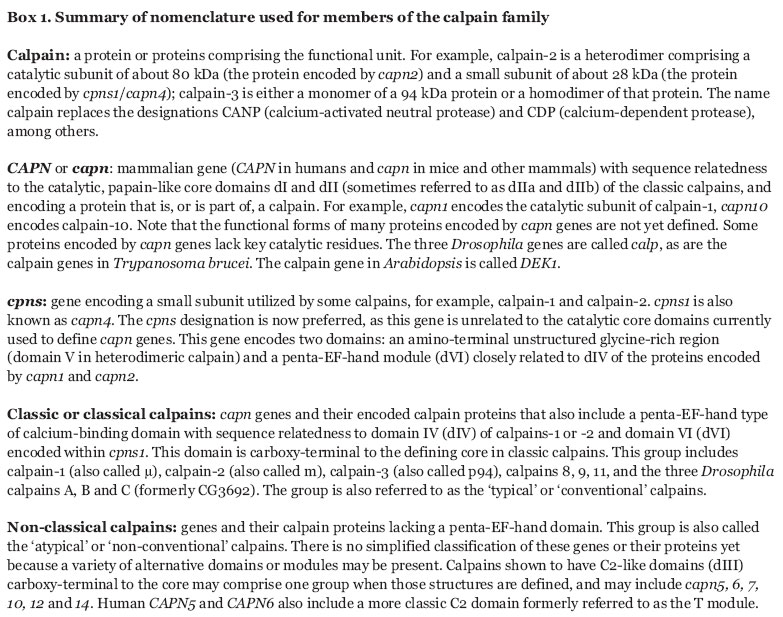
This review focuses on the eukaryotic calpains, although genome databases reveal bacteria, but no archaea, with sequences related to the catalytic core domains (domains dI and dII) of the classical calpains, the criterion used for designating a protein as a calpain. Only single copies of calpain-coding genes are found in the small number of sequenced or partially sequenced protozoan genomes, such as those of the apicomplexan parasites Plasmodium falciparum, Theileria annulata and Cryptosporidium parvum [1-3], and of the amitochondrial parasite Entamoeba histolytica [4]. No calpain-like sequences were identified in the human pathogen Giardia lamblia, a diplomonad often considered to be the most basal eukaryotic organism [5]. Protozoan calpains lack a domain containing EF-hand-type Ca2+-binding sites, as also do plant and fungal calpains, and thus it seems likely that the proposed cysteine protease-calmodulin gene fusion leading to the classical calpain structure (for earlier reviews see [6-8]) occurred exclusively within the animal lineage. The nomenclature recommended for describing calpain proteins and the genes encoding them is summarized in Box 1.
Box 1.

Uniquely within protozoa, the kinetoplastid parasites Trypanosoma brucei, T. cruzi and species of Leishmania, and the ciliate Tetrahymena thermophila [9-12] display expansion of calpain genes. Fourteen genes encoding calpain-related proteins have been identified in T. brucei, 17 in Leishmania major and 15 in T. cruzi [13]. Most of these capn genes are organized as tandem repeats in a small number of gene clusters that are syntenic between T. brucei, T. cruzi and L. major, indicating that most of the observed expansion and diversity was probably generated by gene-duplication events in an ancestral kinetoplastid. The macronuclear genome sequence of the ciliate T. thermophila [12] predicts a surprisingly large number of 26 calpain-like proteins. Analysis of human and mouse genomes has identified 14 members of the calpain family. For the few calpain genes analyzed in mammals, sizes range from 13 to 50 kb with 15 to 28 exons [7]. Phylogenetic trees have been generated for isolated domains [8,14] and for the defining catalytic core domain (dI-dII) in conjunction with the most common, C2-like, auxiliary domain (dIII), of selected species [14,15]. An analysis by Jekely and Friedrich [14] revealed clear segregation of the EF-hand-containing capn gene (Schistosoma, Caenorhabditis elegans CLP-1, Drosophila A/B and the classic vertebrate capn) from the cluster containing capn5(tra3) and capn6 [14]. Possible gene-duplication events may explain the closer evolutionary relationships between the pairs capn2 and capn8, capn3 and capn9, and capn1 and chicken μ/m [14]. Wang et al. [15] also included capn11 and 12 in their phylogenetic analysis, but neither report included capn10. Of interest would be a more detailed analysis of domain dIII sequences in these genes, to determine whether there is a general functional homology between dIII domains that are related to the calcium- and phospholipid-binding domain of protein kinase C (C2-like domains), as is the case in mammalian calpains 1, 2 and 3 [7] and Drosophila calpain B [16].
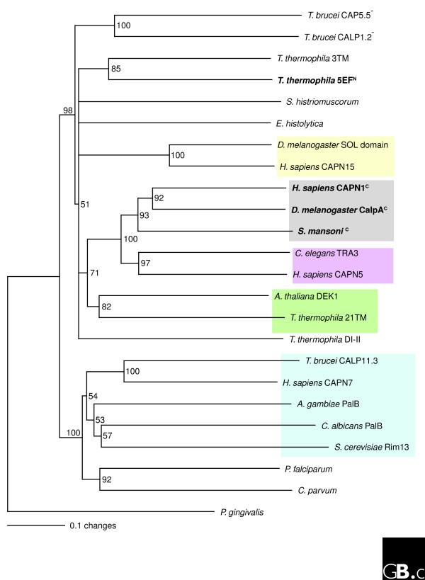
A phylogenetic tree rooted to the calpain-related sequence of the prokaryote Porphyromonas gingivalis and based only on the catalytic core (dI-dII) is shown in Figure 1, and suggests that the EF-hand-containing calpains from animals (carboxy-terminal EF-hands) and Tetrahymena (amino-terminal EF-hands) are phylogenetically well separated. This raises the intriguing possibility that the acquisition of EF-hands occurred through independent gene-fusion events in these groups. The phylogenetic analysis also reveals a close relationship of the Tetrahymena calpain containing 21 transmembrane motifs (21TM) with plant calpain (Arabidopsis DEK1), thus raising the possibility of a common origin for these unusual calpains. Lateral gene transfer from a green alga-type endosymbiont of ciliates is one possible mechanism.
Figure 1.
The phylogenetic relationship of calpains from diverse evolutionary groups of eukaryotes. Only the catalytic core domains (dI-II) were used to construct the tree. Multiple alignments were done with Clustal X and bootstrapped with PAUP4* (1,000 iterations). Only values greater than 50% are indicated. The tree was rooted with the calpain-related sequence from the prokaryote Porphyromonas gingivalis. A minus sign (-) indicates a nonstandard catalytic triad; species names in bold contain EF-hand motifs and the amino- or carboxy-terminal location of the motif is indicated by superscript N or C. Gray box, representative examples of classical calpains; yellow box, calpains containing a carboxy-terminal SOL domain; magenta box, calpains containing an additional carboxy-terminal C2 domain (also referred to as a Tra3 or T domain); green box, calpains containing 21 amino-terminal transmembrane domains; blue box, calpains containing a carboxy-terminal PalB-type domain. Species names: T. brucei, Trypanosoma brucei; T. thermophila, Tetrahymena thermophila; S. histriomuscorum, Sterkiella histriomuscorum (a ciliate); E. histolytica, Entamoeba histolytica; D. melanogaster, Drosophila melanogaster; S. mansoni, Schistosoma mansoni; C. elegans, Caenorhabditis elegans; H. sapiens, Homo sapiens; A. thaliana, Arabidopsis thaliana; A. gambiae, Anopheles gambiae; C. albicans, Candida albicans; S. cerevisiae, Saccharomyces cerevisiae; P. falciparum, Plasmodium falciparum; C. parvum, Cryptosporidium parvum; P. gingivalis, Porphyromonas gingivalis. Calpains listed with unpublished, nonstandard abbreviations: 3TM, three carboxy-terminal transmembrane domains; 5EF, five-EF-hand motifs; 21TM, 21 amino-terminal transmembrane domains; DI-II, domains dI-dII-only calpain without further recognizable motifs. Single calpains have been identified in organisms where only species names are given. Sequences and accession numbers are available in Additional data file 1.
Characteristic structural features
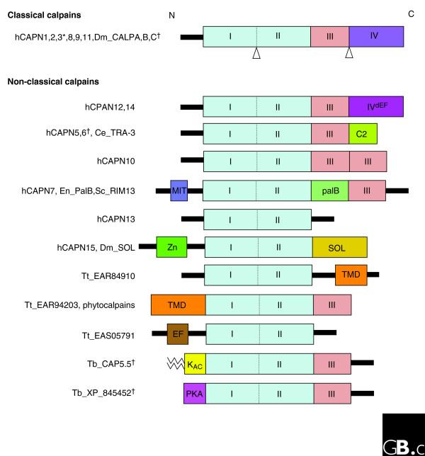
Calpains have a highly modular organization, as illustrated in Figure 2, which shows the types of protein modules and their organization within specific calpains. The catalytic subunit of classical calpains has four domains, of which dI and dII constitute the catalytic core, dIII is a C2-like domain capable of calcium and phospholipid binding, and dIV contains five EF-hand motifs, the fifth serving in some calpains as a dimerization motif for binding to a 'small subunit' (see below) or to form homodimers. The non-classical calpains all have domains dI and dII (by definition), but not all have dIII or dIV, and some contain other types of modules (Figure 2). Although defined by their 'catalytic' core sequence, an increasing number of calpains lack one or more of the essential catalytic amino-acid residues, suggesting functions unrelated to proteolysis. It has been speculated that these 'pseudo-proteases' are involved in regulatory processes [13,17]. A very recent report describes a role for the non-catalytic calpain-6 in the stabilization of microtubules [18].
Figure 2.
A modular architecture is found in all members of the calpain protein family. All the identified human calpain genes (hCAPN) are depicted with selected examples from other species. The presence of domains dI and dII is used to define the family. Domain dIII is defined as the classical calpain C2-like domain; other C2 domains can also be present (see hCAPN5 and 6). Domain dIV is the penta-EF-hand module shared by classical calpains and their small subunit Cpns-1 (where the penta-EF-hand module is known as domain dVI). Domain dV, specific to the small subunit Cpns-1 and without known motifs, is not shown here. The black bars linking modules represent sequences without known motifs and are unique to individual calpains. *The classical calpain hCAPN3 has two insertions, indicated by Δ here. †These proteins have lost key catalytic residues and are predicted to lack protease activity. Species: Dm, Drosophila melanogaster; Ce, Caenorhabiditis elegans; En, Emericella (Aspergillus) nidulans; Sc, Saccharomyces cerevisiae; Tt, Tetrahymena thermophila; Tb, Trypanosoma brucei. Domain abbreviations: C2, protein kinase C conserved region 2 (domain involved in calcium-dependent phospholipid binding); IVdEF, domain dIV with degenerate EF-hand motifs that are unlikely to bind calcium; EF, domain with EF-hand motifs distinct from domain dIV; KAC, kinetoplastid acylated domain (myristic acid and palmitic acid chains are indicated by zigzag lines); MIT, microtubule interacting and trafficking molecule domain; palB, palB-homologous domain; PKA, protein kinase A regulatory subunit domain; SOL, small optic lobe domain; TMD, transmembrane domain; Zn, zinc finger domain. The functions of some of these protein modules are not yet defined. The domain structures were assembled using SMART [79] and the peptidase database MEROPS [80].
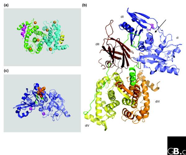
Some of the classical calpains are heterodimers of the 'large' catalytic subunit with the so-called small subunit Cpns-1 (formerly known as Capn-4). Cpns-1 is composed of two domains: dV, an amino-terminal glycine-rich unstructured domain, and dVI, a penta-EF-hand module homologous with dIV of the catalytic subunit. Domain dVI was the first calpain module for which structures were solved in the absence and presence of calcium (reviewed in [7]). These structures provided crucial insight into the nature of heterodimer formation in the classical calpains, anticipated the small contribution of this domain to the calcium-induced conformational change of the holoenzyme, and later revealed details of the interaction of the Cpns-1 protein with a peptide mimicking calpastatin, the endogenous and specific inhibitor of the classic calpains 1 and 2 [19] (Figure 3a).
Figure 3.
Structures of calpain modules and calpain-2. (a) Ribbon diagram of the structure of the penta-EF-hand module (domain dVI) of Cpns-1 from pig. It is shown here as a homodimer (one chain green, one cyan). The short helical peptides (yellow and magenta) are 19-residue mimics of the conserved C peptide of the calpain inhibitor calpastatin bound to dVI in the presence of calcium (orange spheres). The structure is from PDB 1NX1 (Todd et al. [19]). (b) Ribbon diagram of the structure of the rat calpain-2 heterodimer. The catalytic core domains (dI-dII) are in light and dark blue, respectively. Catalytic residues are shown as magenta sticks (with the engineered mutation of C105S) and the arrow designates the active-site cleft between domains dI and dII. Domain dIII (brown) is C2-like. The penta-EF-hand domain dIV of the large subunit (Capn-2) is in yellow, and the similar domain dVI of the small subunit (Cpns-1) is in orange. Domain dV, the amino-terminal glycine-rich region of the small subunit, was truncated by protein engineering; in the human enzyme it is highly flexible and structurally unresolved [21]. The amino-terminal helix and linker loops are in green. The structure is from PDB 1DF0 (Hosfield et al. [20]). The dVI heterodimer in (a) is very similar to that formed between the dIV and dVI domains, and can be used to model this interaction. (c) Ribbon diagram of the structure of the calcium-bound catalytic core (domains dI-dII) of rat calpain-1 based on PDB 1TL9 (Moldoveanu et al. [26]). The bound inhibitor leupeptin is shown as gold, blue and red spheres; the magenta spheres are two calcium ions bound to hitherto unknown sites. All ribbon diagrams were generated using PyMol (DeLano Scientific, Palo Alto, CA, USA).
The determination of the calcium-free structure of calpain-2 from rat and human [20,21] was key to furthering our understanding of the classic calpains (Figure 3b) and revealed unanticipated insights. In contrast to most allo-sterically regulated enzymes, where activation relieves steric hindrance at a pre-formed active site, classic calpains require a conformational change to realign the key residues (Cys, His, Asn) to make them catalytically competent. In addition, domain dIII in calpain-2 shows some structural resemblance to C2 domains, which suggests possible additional mechanisms for binding calcium and phospholipids. Mutagenesis experiments provide evidence for the function of dIII as an electrostatic switch contributing to the maintenance of the catalytic core in an inactive form and the subsequent stabilization of the active enzyme [22,23]. The structure of calpain-2 also provided a platform for modeling the structures of calpain-1 (since confirmed by crystallization and structure determination of a chimeric calpain-1-like enzyme [24]) and of calpain-3 [25].
The isolated catalytic core of calpain-1 (the dI-dII module, referred to as 'mini-calpain') yielded a calcium-bound structure [26] (Figure 3c). Quite surprisingly, in some calpains, for example rat calpain-1, the isolated core showed weak but measurable Ca2+-dependent proteolytic activity, a result of unpredicted and novel calcium-binding sites [26,27]. Comparisons between chimeric enzymes (mixtures of domains from calpains 1 and 2 or 3), the inactive hetero-dimer, and mini-calpains indicate some details of the mechanism of regulation of catalytic function by calcium. Activation of the enzyme core within the heterodimer involves proteolytic removal of the amino-terminal 'anchor' helix (see Figure 3b) or the release of its binding to dVI, weakening of the electrostatic interactions between dIII and dII, and the binding of multiple calcium ions to the EF-hand modules (dIV and dVI) and to dIII, which trigger changes that permit binding of calcium to the calcium-binding sites of the catalytic core. The weakening of the constraints that maintain the dI-dII domains in their 'inactive' positions and the cooperative Ca2+ binding to the core allow the realignment of the core into its active state, in which it bears a substantial structural resemblance to papain. The isolated core also provides a useful reagent for screening calpain inhibitors to find potential drug candidates [26-28].
Localization and function
Calpain function has been investigated by both genetic and cell-biological routes. Table 1 summarizes these studies and their results. The targeted deletion, and more recently the conditional deletion, of the cpns1 gene [7,29] showed that at least one classical calpain is essential for early embryogenesis in mammals. Targeted deletion studies have since shown that capn1 is not essential [30] whereas capn2 is [31]; the function of calpains in development is not yet known, however. Loss of capn9 in NIH3T3 cells results in a more transformed phenotype, as shown by increased growth in soft agar [32], but to our knowledge this gene has not yet been targeted in whole organisms. Multiple genetic defects that truncate, or otherwise inactivate, calpain-3 (also called p94) seem to be a cause of limb-girdle muscular dystrophy type IIa [25,33], thus identifying the importance of calpain-3 in skeletal muscle integrity. Targeted deletion of capn3 in mice produces a model for assessing its role in muscle function and repair [33,34]. Specific splice variants of capn3 occur in the lens of the eye and are linked to the formation of cataracts [35]. One factor contributing to increased susceptibility to type 2 diabetes, a multifactorial disease, may be variations in the capn10 gene. This idea still sparks controversy, as the initial observation identified a polymorphism in a capn10 intron in populations with increased risk for diabetes [7,36,37]. However, studies show that calpain-10 may function in stimulated secretion and/or pancreatic cell death [38,39], and thereby be relevant to this disease. Two non-classical calpains, Tra3 and PalB (orthologs of calpain-5, capn5, and calpain-7, capn7), mediate signal transduction pathways for sex determination in nematodes [40] and adaptation to pH in yeast [41], respectively.
Table 1.
The physiological functions of calpains as revealed by genetics
| Gene disruption or mutation | Model system | Enzyme(s) | Findings and implication for function |
| cpns1 (capn4)-targeted | Mouse | Calpains 1, 2, and probably 9 | Embryonic lethal, therefore some calpain is essential during embryogenesis [29] |
| capn1-targeted | Mouse | Calpain-1 | Viable, fertile mice, some platelet changes, calpain-1 is not essential for development [30] |
| capn2-targeted | Mouse | Calpain-2 | Embryonic lethal, very early, implying essential role for calpain-2 [31] |
| capn3-targeted/naturally occurring mutations | Mouse/human | Calpain-3 | Muscle-repair defects, myopathy/LGMD type IIa [33,34,69] |
| capn9 random homozygous knockout | Mouse cell culture (NIH3T3 cells) | Calpain-9 | Increased cell growth in soft agar suggests that calpain-9 is a potential tumor suppressor [32] |
| capn10 variation in population | Human | Calpain-10 | Potential risk factor for type 2 diabetes; transport of GLUT4 [36,38] |
| dek1-targeted/naturally occurring mutations | Maize/Arabidopsis/tobacco | Phytocalpain | Embryonic lethal in maize, developmental defects [70] |
| tra3 (capn5) mutant and engineered | C. elegans | Tra3 | Sex determination [40] |
| palB (capn7) mutant | Emericella nidulans | PalB | pH signal transduction pathway [41] |
Biochemical and cell-biological studies also provide significant insights into calpain physiology. It is often speculated that calpains function, or become activated, when associated with membranes, despite their predominantly cytoplasmic localization [6,7]. Although membrane binding is not well substantiated for classical calpains, predicted transmembrane segments in phytocalpain and some ciliate calpains suggest an evolutionary link between calpain function and membranes. At least two acylated calpain-like proteins in the kinetoplastids L. major and T. brucei are biochemically associated or co-localize with cellular membranes ([42] and KE, unpublished work). Acylated proteins are often associated with the cytoplasmic face of membranes and lipid rafts, where they are implicated in signal transduction [42,43]. Thus, the small amount of calpain fractionating biochemically with membranes may be the active, physiologically relevant, enzyme population, although suggestions that vertebrate calpains localize to lipid rafts or caveolae require further confirmation. Biophysical studies demonstrate the ability of a conserved peptide (GTAMRILGGVI) located in the amino-terminal domain dV to form a membrane-penetrating α-helical structure [44], providing one mechanism for calpains 1 and 2 to bind to membranes. For many calpains, the C2-like domain (dIII) provides an additional or alternative mechanism for membrane association via its phospholipid-binding properties. A recent study has demonstrated the importance of dIII-mediated membrane binding of calpain-2 in living cells [45]. In addition, the critical self-sealing repair of damaged plasma membranes requires the activity of ubiquitous calpains, which may act to remodel the underlying cortical cytoskeleton [46].
In contrast to relatively promiscuous degradative proteases, calpains cleave only a restricted set of protein substrates and use complex substrate-recognition mechanisms, involving primary and secondary structural features of target proteins. Proteins identified as substrates for calpains include numerous membrane-bound or membrane-associated proteins, such as calcium-ATPase, the epidermal growth factor (EGF) receptor, the ryanodine receptor, the calcium receptor, the NMDA receptor (a glutamic acid receptor), β-integrins, aquaporin, the transporters ABC-A1 and GLUT4, and proteins interfacing with receptors and the cytoskeleton, such as talin, α-spectrin (α-fodrin) and ezrin, among many others (see Table 11 in [7] for a more extensive, though still incomplete, list).
A wide variety of receptors function upstream of the intra-cellular activation of calpains (Table 2). The most thoroughly studied models focus on the roles of calpains in cell motility in response to either EGF [47] or integrin engagement [48]. Additional work links calpains to cell transformation and oncogenesis [49,50]. Knockdown strategies utilizing anti-sense RNAs or small interfering RNAs to study the roles of calpains in cell transformation and in other cellular processes have provided significant evidence for non-redundant, distinctive functions for each ubiquitous calpain isoform. Although less widely studied, there is also increasing evidence for the externalization of calpains and their extracellular contribution to tissue damage in response to toxicants or other factors [51-53]. These destructive roles may relate to the documented involvement of calpains in pathways that trigger apoptosis and/or necrosis [54-59] and, discovered most recently, autophagy [60,61]. Thus, there is considerable evidence for a complex relationship between calpain activity and the functions of both caspases and the proteasome.
Table 2.
Functional diversity of calpains
| Examples of proposed calpain function(s) | Model systems providing key supporting evidence | Calpain implicated (possible substrates) | Selected references |
| Participant in signaling pathways | |||
| EGF-EGFR-induced motility | Mouse NR6 fibroblasts and derivatives, Hs68 (human neonatal foreskin fibroblasts) | Calpain-2 (?) | [7,45,47] |
| Integrin receptor-linked motility | Platelets, bovine aortic endothelial cells, CHO, CHO KI1 and SHI derivative, goldfish fin CAR, and immortalized mouse embryonic fibroblasts deficient in Cpns-1 (capn4-/-) | Calpain-1 leading edge; calpain-2 trailing edge (talin, filamin, spectrin, β-integrin) | [6,48,71] |
| Integrin receptor-linked adhesion | Mouse cell line NIH3T3 | Calpain-2 (ezrin) | [72] |
| Downstream of VEGF | Pulmonary microvascular endothelial cells | Calpain-2 (?) | [73] |
| Responsive to TRPM7 | Flp in T rex 293 derivatives | Calpain-2 (talin) | [74] |
| Adaptation to alkaline environment | Emericella (Aspergillus) nidulans, Candida albicans, Saccharomyces cerevisiae | PalB or Rim13/Clp1 | [41,75] |
| Downstream of endothelin-1 in development | Mice, HeLa cells, NIH3T3 or HEK293 | Calpain-6 (a non-catalytic form; binds to and stabilizes microtubules) | [18] |
| Shear stress induced motility | Human umbilical vein endothelial cells | Calpain-2 (pp125FAK, ezrin) | [76] |
| Cellular transformation and tumorigenesis | |||
| v-src-transformed cells | HT1080 human fibrosarcoma, H1299 non-small cell lung carcinoma | Implied calpains 1 and 2 (FAK, paxillin) | [48,77] |
| capn9 gene disruption | Mouse cell line NIH3T3 | Calpain-9 | [32] |
| Necrosis and/or apoptosis | |||
| Toxicant induced damage to liver or kidney | Rat/mouse exposure to selected toxicants | Extracellular calpain (fibronectin) | [51-53] |
| Neuronal cell death | C. elegans, cell culture (for example, SH-SY5Y), primary cells | Isoforms not defined Bax, Bid, AIF, caspase) | [55-57] |
| Cerebellar granule cell survival | Rat, primary cells | Nuclear localized calpain-2 | [78] |
| Apoptosis of pancreatic islet cells | Islets (human, mouse) MIN6 β cells exposed to ryanodine or palmitate and low glucose | Calpain-10 | [36,38,39] |
| Autophagy/apoptosis switch | Neutrophil, Jurkat, USO2, cpns1-/- MEF | Isoform not defined (Atg5) | [60,61] |
| StrepB-induced apoptosis | Macrophage | Calpain-2 (Bax, Bid) | [58] |
Frontiers
Despite great advances in our knowledge of calpains 1 and 2, much is yet to be learned about the evolution of the family and the range of functions of its members. Genomic sequences from a wide range of organisms document the extreme diversity and modular nature of the calpain protein family. Current evidence suggests that the acquisition of the penta-EF-hand module, characteristic of the classical calpains, may be restricted to animals, but that EF-hands may have been acquired independently in Tetrahymena calpains. The use of different strategies for associating with membranes, such as transmembrane domains, C2-like domains, and acylation, support the importance of membrane association in calpain function. More genomic information from representative organisms, particularly protozoa, is required to better analyze the evolutionary relationships within this family. The proteolytic core module is now relatively well characterized as to structure and function. Distinguishing the overlapping or unique substrate specificities [62] and inhibitor sensitivities of the proteolytically active calpain isoforms is expected to aid the design of studies aimed at determining their roles in cellular pathways. For the family members lacking key catalytic residues, alternative functions await discovery. Future work is also needed to determine how the modules associated with the core influence its function. There is likely to be interplay between protein-protein interactions, membrane binding, calcium binding (in many calpains) and, potentially, post-translational modifications in the modulation of calpain function. Many calpain proteins remain to be purified and characterized biochemically, so the challenge of identifying their relevant binding partners remains.
It is now established that some calpains are components of regulatory networks involved in fundamental processes at cellular (for example, motility) and organismal (for example, embryogenesis) levels. Further work will determine if and when specific isoforms and the multitude of their possible splice variants are expressed in either a tissue-specific or time-dependent manner in cells. Understanding the function(s) of individual isoforms in a variety of physiological contexts - from protozoa to humans - remains the ultimate challenge. RNA interference will continue to make a significant contribution to these goals, and the design of calpain-resistant substrates [63,64] will provide a way of documenting calpain-catalyzed, limited proteolysis in vivo. The future development of biosensors to visualize calpain activity (or activation), like those generated for other signal pathway molecules [65], may also provide a major advance. Efforts to develop cellular calpain 'reporter' substrates have been described [66,67] and the tight binding of calpastatin to active calpains 1 and 2 [6,7,68] may be exploited to develop reporters that selectively recognize the active conformation of these enzymes (D.E.C. and L.M. Vanhooser, unpublished work). More data and new approaches are needed to enhance understanding of the regulation of both proteolytic and non-proteolytic calpains. Careful transcriptional, translational and activity-based profiling - ideally able to detect the variety of splice variants - will be required to establish detailed expression patterns for calpains in relation to embryogenesis, differentiation or other cellular processes. The time is ripe to define the regulatory circuits in which calpains participate, to complete the assessment of their in vivo substrates and to characterize the regulators of all functions of calpains.
Additional data files
Additional data file 1 contains the sequences and accession numbers of the calpain sequences in the phylogenetic tree in Figure 1.
Supplementary Material
The sequences and accession numbers of the calpain sequences in the phylogenetic tree in Figure 1
Acknowledgments
Acknowledgements
D.E.C. acknowledges previous support from the National Science Foundation and current support from the National Institutes of Health (NINDS).
Contributor Information
Dorothy E Croall, Email: dorothy.croall@umit.maine.edu.
Klaus Ersfeld, Email: k.ersfeld@hull.ac.uk.
References
- Gardner MJ, Bishop R, Shah T, de Villiers EP, Carlton JM, Hall N, Ren Q, Paulsen IT, Pain A, Berriman M, et al. Genome sequence of Theileria parva, a bovine pathogen that transforms lymphocytes. Science. 2005;309:134–137. doi: 10.1126/science.1110439. [DOI] [PubMed] [Google Scholar]
- Gardner MJ, Hall N, Fung E, White O, Berriman M, Hyman RW, Carlton JM, Pain A, Nelson KE, Bowman S, et al. Genome sequence of the human malaria parasite Plasmodium falciparum. Nature. 2002;419:498–511. doi: 10.1038/nature01097. [DOI] [PMC free article] [PubMed] [Google Scholar]
- Abrahamsen MS, Templeton TJ, Enomoto S, Abrahante JE, Zhu G, Lancto CA, Deng M, Liu C, Widmer G, Tzipori S, et al. Complete genome sequence of the apicomplexan, Cryptosporidium parvum. Science. 2004;304:441–445. doi: 10.1126/science.1094786. [DOI] [PubMed] [Google Scholar]
- Loftus B, Anderson I, Davies R, Alsmark UC, Samuelson J, Amedeo P, Roncaglia P, Berriman M, Hirt RP, Mann BJ, et al. The genome of the protist parasite Entamoeba histolytica. Nature. 2005;433:865–868. doi: 10.1038/nature03291. [DOI] [PubMed] [Google Scholar]
- McArthur AG, Morrison HG, Nixon JE, Passamaneck NQ, Kim U, Hinkle G, Crocker MK, Holder ME, Farr R, Reich CI, et al. The Giardia genome project database. FEMS Microbiol Lett. 2000;189:271–273. doi: 10.1111/j.1574-6968.2000.tb09242.x. [DOI] [PubMed] [Google Scholar]
- Croall DE, DeMartino GN. Calcium-activated neutral protease (calpain) system: structure, function, and regulation. Physiol Rev. 1991;71:813–847. doi: 10.1152/physrev.1991.71.3.813. [DOI] [PubMed] [Google Scholar]
- Goll DE, Thompson VF, Li H, Wei W, Cong J. The calpain system. Physiol Rev. 2003;83:731–801. doi: 10.1152/physrev.00029.2002. [DOI] [PubMed] [Google Scholar]
- Sorimachi H, Suzuki K. The structure of calpain. J Biochem. 2001;129:653–664. doi: 10.1093/oxfordjournals.jbchem.a002903. [DOI] [PubMed] [Google Scholar]
- Berriman M, Ghedin E, Hertz-Fowler C, Blandin G, Renauld H, Bartholomeu DC, Lennard NJ, Caler E, Hamlin NE, Haas B, et al. The genome of the African trypanosome Trypanosoma brucei. Science. 2005;309:416–422. doi: 10.1126/science.1112642. [DOI] [PubMed] [Google Scholar]
- El-Sayed NM, Myler PJ, Bartholomeu DC, Nilsson D, Aggarwal G, Tran AN, Ghedin E, Worthey EA, Delcher AL, Blandin G, et al. The genome sequence of Trypanosoma cruzi, etiologic agent of Chagas disease. Science. 2005;309:409–415. doi: 10.1126/science.1112631. [DOI] [PubMed] [Google Scholar]
- Ivens AC, Peacock CS, Worthey EA, Murphy L, Aggarwal G, Berriman M, Sisk E, Rajandream MA, Adlem E, Aert R, et al. The genome of the kinetoplastid parasite, Leishmania major. Science. 2005;309:436–442. doi: 10.1126/science.1112680. [DOI] [PMC free article] [PubMed] [Google Scholar]
- Eisen JA, Coyne R, Wu M, Wu D, Thiagarajan M, Wortman JR, Badger JH, Ren Q, Amedeo P, Jones KM, et al. Macronuclear genome sequence of the ciliate Tetrahymena thermophila, a model eukaryote. PLoS Biol. 2006;4:e286. doi: 10.1371/journal.pbio.0040286. [DOI] [PMC free article] [PubMed] [Google Scholar]
- Ersfeld K, Barraclough H, Gull K. Evolutionary relationships and protein domain architecture in an expanded calpain superfamily in kinetoplastid parasites. J Mol Evol. 2005;61:742–757. doi: 10.1007/s00239-004-0272-8. [DOI] [PubMed] [Google Scholar]
- Jekely G, Friedrich P. The evolution of the calpain family as reflected in paralogous chromosome regions. J Mol Evol. 1999;49:272–281. doi: 10.1007/PL00006549. [DOI] [PubMed] [Google Scholar]
- Wang C, Barry JK, Min Z, Tordsen G, Rao AG, Olsen OA. The calpain domain of the maize DEK1 protein contains the conserved catalytic triad and functions as a cysteine proteinase. J Biol Chem. 2003;278:34467–34474. doi: 10.1074/jbc.M300745200. [DOI] [PubMed] [Google Scholar]
- Tompa P, Emori Y, Sorimachi H, Suzuki K, Friedrich P. Domain III of calpain is a Ca2+-regulated phospholipid-binding domain. Biochem Biophys Res Comm. 2001;280:1333–1339. doi: 10.1006/bbrc.2001.4279. [DOI] [PubMed] [Google Scholar]
- Pils B, Schultz J. Inactive enzyme-homologues find new function in regulatory processes. J Mol Biol. 2004;340:399–404. doi: 10.1016/j.jmb.2004.04.063. [DOI] [PubMed] [Google Scholar]
- Tonami K, Kurihara Y, Aburatani H, Uchijima Y, Asano T, Kurihara H. Calpain 6 is involved in microtubule stabilization and cytoskeletal organization. Mol Cell Biol. 2007;27:2548–2561. doi: 10.1128/MCB.00992-06. [DOI] [PMC free article] [PubMed] [Google Scholar]
- Todd B, Moore D, Deivanayagam CC, Lin GD, Chattopadhyay D, Maki M, Wang KK, Narayana SV. A structural model for the inhibition of calpain by calpastatin: crystal structures of the native domain VI of calpain and its complexes with calpastatin peptide and a small molecule inhibitor. J Mol Biol. 2003;328:131–146. doi: 10.1016/S0022-2836(03)00274-2. [DOI] [PubMed] [Google Scholar]
- Hosfield C, Elce J, Davies P, Jia Z. Crystal structure of calpain reveals the structural basis for Ca2+-dependent protease activity and a novel mode of enzyme activation. EMBO J. 1999;18:6880–6889. doi: 10.1093/emboj/18.24.6880. [DOI] [PMC free article] [PubMed] [Google Scholar]
- Strobl S, Fernandez-Catalan C, Braun M, Huber R, Masumoto H, Nakagawa K, Irie A, Sorimachi H, Bourenkow G, Bartunik H, et al. The crystal structure of calcium-free human m-calpain suggests an electrostatic switch mechanism for activation by calcium. Proc Natl Acad Sci USA. 2000;97:588–592. doi: 10.1073/pnas.97.2.588. [DOI] [PMC free article] [PubMed] [Google Scholar]
- Moldoveanu T, Hosfield CM, Lim D, Elce JS, Jia Z, Davies PL. A Ca2+ switch aligns the active site of calpain. Cell. 2002;108:649–660. doi: 10.1016/S0092-8674(02)00659-1. [DOI] [PubMed] [Google Scholar]
- Reverter D, Strobl S, Fernandez-Catalan C, Sorimachi H, Suzuki K, Bode W. Structural basis for possible calcium-induced activation mechanisms of calpains. Biol Chem. 2001;382:753–766. doi: 10.1515/BC.2001.091. [DOI] [PubMed] [Google Scholar]
- Pal GP, De Veyra T, Elce JS, Jia Z. Crystal structure of a micro-like calpain reveals a partially activated conformation with low Ca2+ requirement. Structure. 2003;11:1521–1526. doi: 10.1016/j.str.2003.11.007. [DOI] [PubMed] [Google Scholar]
- Jia Z, Petrounevitch V, Wong A, Moldoveanu T, Davies PL, Elce JS, Beckmann JS. Mutations in calpain 3 associated with limb girdle muscular dystrophy: analysis by molecular modeling and by mutation in m-calpain. Biophys J. 2001;80:2590–2596. doi: 10.1016/S0006-3495(01)76229-7. [DOI] [PMC free article] [PubMed] [Google Scholar]
- Moldoveanu T, Campbell RL, Cuerrier D, Davies PL. Crystal structures of calpain-E64 and -leupeptin inhibitor complexes reveal mobile loops gating the active site. J Mol Biol. 2004;343:1313–1326. doi: 10.1016/j.jmb.2004.09.016. [DOI] [PubMed] [Google Scholar]
- Cuerrier D, Moldoveanu T, Inoue J, Davies PL, Campbell RL. Calpain inhibition by alpha-ketoamide and cyclic hemiacetal inhibitors revealed by X-ray crystallography. Biochemistry. 2006;45:7446–7452. doi: 10.1021/bi060425j. [DOI] [PubMed] [Google Scholar]
- Li Q, Hanzlik R, Weaver R, Schonbrunn E. Molecular mode of action of a covalently inhibiting peptidomimetic on the human calpain protease core. Biochemistry. 2006;45:701–708. doi: 10.1021/bi052077b. [DOI] [PubMed] [Google Scholar]
- Tan Y, Dourdin N, Wu C, De Veyra T, Elce JS, Greer PA. Conditional disruption of ubiquitous calpains in the mouse. Genesis. 2006;44:297–303. doi: 10.1002/dvg.20216. [DOI] [PubMed] [Google Scholar]
- Azam M, Andarabi S, Sahr K, Kamath L, Kuliopulos A, Chisti A. Disruption of the mouse-μ-calpain gene reveals an essential role in platelet function. Mol Cell Biol. 2001;21:2213–2220. doi: 10.1128/MCB.21.6.2213-2220.2001. [DOI] [PMC free article] [PubMed] [Google Scholar]
- Dutt P, Croall DE, Arthur JSC, DeVeyra T, Williams K, Elce JS, Greer PA. m-Calpain is required for preimplantation embryonic development in mice. BMC Dev Biol. 2006;6:3. doi: 10.1186/1471-213X-6-3. [DOI] [PMC free article] [PubMed] [Google Scholar]
- Liu K, Li L, Cohen SN. Antisense RNA-mediated deficiency of the calpain protease, nCL-4, in NIH3T3 cells is associated with neoplastic transformation and tumorigenesis. J Biol Chem. 2000;275:31093–31098. doi: 10.1074/jbc.M005451200. [DOI] [PubMed] [Google Scholar]
- Duguez S, Bartoli M, Richard I. Calpain 3: a key regulator of the sarcomere? FEBS J. 2006;273:3427–3436. doi: 10.1111/j.1742-4658.2006.05351.x. [DOI] [PubMed] [Google Scholar]
- Cohen N, Kudryashova E, Kramerova I, Anderson LV, Beckmann JS, Bushby K, Spencer MJ. Identification of putative in vivo substrates of calpain 3 by comparative proteomics of overex-pressing transgenic and nontransgenic mice. Proteomics. 2006;6:6075–6084. doi: 10.1002/pmic.200600199. [DOI] [PubMed] [Google Scholar]
- Shih M, Ma H, Nakajima E, David LL, Azuma M, Shearer TR. Biochemical properties of lens-specific calpain Lp85. Exp Eye Res. 2006;82:146–152. doi: 10.1016/j.exer.2005.06.011. [DOI] [PubMed] [Google Scholar]
- Turner MD, Cassell PG, Hitman GA. Calpain-10: from genome search to function. Diabetes Metab Res Rev. 2005;21:505–514. doi: 10.1002/dmrr.578. [DOI] [PubMed] [Google Scholar]
- Horikawa Y, Oda N, Cox NJ, Li X, Orho-Melander M, Hara M, Hinokio Y, Lindner TH, Mashima H, Schwarz PE, et al. Genetic variation in the gene encoding calpain-10 is associated with type 2 diabetes mellitus. Nat Genet. 2000;26:163–175. doi: 10.1038/79876. [DOI] [PubMed] [Google Scholar]
- Johnson JD, Han Z, Otani K, Ye H, Zhang Y, Wu H, Horikawa Y, Misler S, Bell GI, Polonsky KS. RyR2 and calpain-10 delineate a novel apoptosis pathway in pancreatic islets. J Biol Chem. 2004;279:24794–24802. doi: 10.1074/jbc.M401216200. [DOI] [PubMed] [Google Scholar]
- Marshall C, Hitman GA, Partridge CJ, Clark A, Ma H, Shearer TR, Turner MD. Evidence that an isoform of calpain-10 is a regulator of exocytosis in pancreatic β-cells. Mol Endocrinol. 2005;19:213–224. doi: 10.1210/me.2004-0064. [DOI] [PubMed] [Google Scholar]
- Sokol SB, Kuwabara PE. Proteolysis in Caenorhabditis elegans sex determination: cleavage of TRA-2A by TRA-3. Genes Dev. 2000;14:901–906. [PMC free article] [PubMed] [Google Scholar]
- Nozawa SR, May GS, Martinez-Rossi NM, Ferreira-Nozawa MS, Coutinho-Netto J, Maccheroni W, Jr, Rossi A. Mutation in a calpain-like protease affects the posttranslational mannosylation of phosphatases in Aspergillus nidulans. Fungal Genet Biol. 2003;38:220–227. doi: 10.1016/S1087-1845(02)00521-2. [DOI] [PubMed] [Google Scholar]
- Tull D, Vince JE, Callaghan JM, Naderer T, Spurck T, McFadden GI, Currie G, Ferguson K, Bacic A, McConville MJ. SMP-1, a member of a new family of small myristoylated proteins in kinetoplastid parasites, is targeted to the flagellum membrane in Leishmania. Mol Biol Cell. 2004;15:4775–4786. doi: 10.1091/mbc.E04-06-0457. [DOI] [PMC free article] [PubMed] [Google Scholar]
- Hertz-Fowler C, Ersfeld K, Gull K. CAP5.5, a life-cycle-regulated, cytoskeleton-associated protein is a member of a novel family of calpain-related proteins in Trypanosoma brucei. Mol Biochem Parasitol. 2001;116:25–34. doi: 10.1016/S0166-6851(01)00296-1. [DOI] [PubMed] [Google Scholar]
- Dennison SR, Dante S, Hauss T, Brandenburg K, Harris F, Phoenix DA. Investigations into the membrane interactions of m-calpain domain V. Biophys J. 2005;88:3008–3017. doi: 10.1529/biophysj.104.049957. [DOI] [PMC free article] [PubMed] [Google Scholar]
- Shao H, Chou J, Baty CJ, Burke NA, Watkins SC, Stolz DB, Wells A. Spatial localization of m-calpain to the plasma membrane by phosphoinositide biphosphate binding during epidermal growth factor receptor-mediated activation. Mol Cell Biol. 2006;26:5481–5496. doi: 10.1128/MCB.02243-05. [DOI] [PMC free article] [PubMed] [Google Scholar]
- Mellgren RL, Zhang W, Miyake K, McNeil PL. Calpain is required for the rapid, calcium-dependent repair of wounded plasma membrane. J Biol Chem. 2007;282:2567–2575. doi: 10.1074/jbc.M604560200. [DOI] [PubMed] [Google Scholar]
- Wells A, Huttenlocher A, Lauffenburger DA. Calpain proteases in cell adhesion and motility. Int Rev Cytol. 2005;245:1–16. doi: 10.1016/S0074-7696(05)45001-9. [DOI] [PubMed] [Google Scholar]
- Carragher NO, Walker SM, Scott Carragher LA, Harris F, Sawyer TK, Brunton VG, Ozanne BW, Frame MC. Calpain 2 and Src dependence distinguishes mesenchymal and amoeboid modes of tumour cell invasion: a link to integrin function. Oncogene. 2006;25:5726–5740. doi: 10.1038/sj.onc.1209582. [DOI] [PubMed] [Google Scholar]
- Franco SJ, Huttenlocher A. Regulating cell migration: calpains make the cut. J Cell Science. 2005;118:3829–3838. doi: 10.1242/jcs.02562. [DOI] [PubMed] [Google Scholar]
- Carragher NO, Frame MC. Focal adhesion and actin dynamics: a place where kinases and proteases meet to promote invasion. Trends Cell Biol. 2004;14:241–249. doi: 10.1016/j.tcb.2004.03.011. [DOI] [PubMed] [Google Scholar]
- Frangié C, Zhang W, Perez J, Dubois YC, Haymann JP, Baud L. Extracellular calpains increase tubular epithelial cell mobility: implications for kidney repair after ischemia. J Biol Chem. 2006;281:26624–26632. doi: 10.1074/jbc.M603007200. [DOI] [PubMed] [Google Scholar]
- Mehendale HM, Limaye PB. Calpain: a death protein that mediates progression of liver injury. Trends Pharm Sci. 2005;26:232–236. doi: 10.1016/j.tips.2005.03.008. [DOI] [PubMed] [Google Scholar]
- Dnyanmote AV, Sawant SP, Lock EA, Latendresse JR, Warbritton AA, Mehendale HM. Calpastatin overexpression prevents progression of S-1,2-dichlorovinyl-l-cysteine (DCVC)-initiated acute renal injury and renal failure (ARF) in diabetes. Toxicol Appl Pharmacol. 2006;215:146–157. doi: 10.1016/j.taap.2006.01.018. [DOI] [PubMed] [Google Scholar]
- Lu T, Xu Y, Mericle MT, Mellgren RL. Participation of the conventional calpains in apoptosis. Biochim Biophys Acta. 2002;1590:16–26. doi: 10.1016/S0167-4889(02)00193-3. [DOI] [PubMed] [Google Scholar]
- Syntichaki P, Tavernarakis N. The biochemistry of neuronal necrosis: rogue biology? Nature Rev Neurosci. 2003;4:672–684. doi: 10.1038/nrn1174. [DOI] [PubMed] [Google Scholar]
- Artal-Sanz M, Tavernarakis N. Proteolytic mechanisms in necrotic cell death and neurodegeneration. FEBS Lett. 2005;579:3287–3296. doi: 10.1016/j.febslet.2005.03.052. [DOI] [PubMed] [Google Scholar]
- Tan Y, Dourdin N, Wu C, De Veyra T, Elce JS, Greer PA. Ubiquitous calpains promote caspase-12 and JNK activation during endoplasmic reticulum stress-induced apoptosis. J Biol Chem. 2006;281:16016–16024. doi: 10.1074/jbc.M601299200. [DOI] [PubMed] [Google Scholar]
- Fettucciari K, Fetriconi I, Mannucci R, Nicoletti I, Bartoli A, Coaccioli S, Marconi P. Group B streptococcus induces macrophage apoptosis by calpain activation. J Immunol. 2006;176:7542–7556. doi: 10.4049/jimmunol.176.12.7542. [DOI] [PubMed] [Google Scholar]
- Raynaud F, Marcilhac A. Implication of calpain in neuronal apoptosis: a possible regulation of Alzheimer's disease. FEBS J. 2006;273:3437–3443. doi: 10.1111/j.1742-4658.2006.05352.x. [DOI] [PubMed] [Google Scholar]
- Demarchi F, Bertoli C, Copetti T, Tanida I, Brancolini C, Eskelinen EL, Schneider C. Calpain is required for macroautophagy in mammalian cells. J Cell Biol. 2006;175:595–605. doi: 10.1083/jcb.200601024. [DOI] [PMC free article] [PubMed] [Google Scholar]
- Yousefi S, Perozzo R, Schmid I, Ziemiecki A, Schaffner T, Scapozza L, Brunner T, Simon HU. Calpain-mediated cleavage of Atg5 switches autophagy to apoptosis. Nature Cell Biol. 2006;8:1124–1132. doi: 10.1038/ncb1482. [DOI] [PubMed] [Google Scholar]
- Cuerrier D, Moldoveanu T, Davies PL. Determination of peptide substrate specificity for {micro}-calpain by a peptide library-based approach: the importance of primed side interactions. J Biol Chem. 2005;280:40632–40641. doi: 10.1074/jbc.M506870200. [DOI] [PubMed] [Google Scholar]
- Franco SJ, Rodgers MA, Perrin BJ, Han J, Bennin DA, Critchley DR, Huttenlocher A. Calpain-mediated proteolysis of talin regulates adhesion dynamics. Nature Cell Biol. 2004;6:977–983. doi: 10.1038/ncb1175. [DOI] [PubMed] [Google Scholar]
- Stabach PR, Cianci CD, Glantz SB, Zhang Z, Morrow JS. Site-directed mutagenesis of alpha II spectrin at codon 1175 modulates its mu-calpain susceptibility. Biochemistry. 1997;36:57–65. doi: 10.1021/bi962034i. [DOI] [PubMed] [Google Scholar]
- Pertz O, Hahn KM. Designing biosensors for Rho family proteins - deciphering the dynamics of Rho family GTPase activation in living cells. J Cell Sci. 2004;117:1313–1318. doi: 10.1242/jcs.01117. [DOI] [PubMed] [Google Scholar]
- Stockholm D, Bartoli M, Sillon G, Bourg N, Davoust J, Richard I. Imaging calpain protease activity by multiphoton FRET in living mice. J Mol Biol. 2005;346:215–222. doi: 10.1016/j.jmb.2004.11.039. [DOI] [PubMed] [Google Scholar]
- Vanderklish PW, Krushel LA, Holst BH, Gally JA, Crossin KL, Edelman GM. Marking synaptic activity in dendritic spines with a calpain substrate exhibiting fluorescence resonance energy transfer. Proc Natl Acad Sci USA. 2000;97:2253–2258. doi: 10.1073/pnas.040565597. [DOI] [PMC free article] [PubMed] [Google Scholar]
- Mucsi Z, Hudecz F, Hollosi M, Tompa P, Friedrich P. Binding-induced folding transitions in calpastatin subdomains A and C. Protein Sci. 2003;12:2327–2336. doi: 10.1110/ps.03138803. [DOI] [PMC free article] [PubMed] [Google Scholar]
- Bartoli M, Bourg N, Stockholm D, Raynaud F, Delevaque A, Han Y, Borel P, Seddik K, Armande N, Richard I. A mouse model for monitoring calpain activity under physiological and pathological conditions. J Biol Chem. 2006;281:39672–39680. doi: 10.1074/jbc.M608803200. [DOI] [PubMed] [Google Scholar]
- Lid S, Olsen L, Nestestog R, Aukerman M, Brown R, Lemmon B, Mucha M, Opsahl-Sorteberg H, Olsen O. Mutation in the Arabidopsis thaliana DEK1 calpain gene perturbs endosperm and embryo development while over-expression affects organ development globally. Planta. 2005;221:339–351. doi: 10.1007/s00425-004-1448-6. [DOI] [PubMed] [Google Scholar]
- Bialkowska K, Kulkarni S, Du X, Goll DE, Saido TC, Fox JE. Evidence that beta3 integrin-induced Rac activation involves the calpain-dependent formation of integrin clusters that are distinct from the focal complexes and focal adhesions that form as Rac and RhoA become active. J Cell Biol. 2000;151:685–696. doi: 10.1083/jcb.151.3.685. [DOI] [PMC free article] [PubMed] [Google Scholar]
- Potter DA, Tirnauer JS, Janssen R, Croall DE, Hughes CN, Fiacco KA, Mier JW, Maki M, Herman IM. Calpain regulates actin remodeling during cell spreading. J Cell Biol. 1998;141:647–662. doi: 10.1083/jcb.141.3.647. [DOI] [PMC free article] [PubMed] [Google Scholar]
- Su Y, Cui Z, Li Z, Block ER. Calpain-2 regulation of VEGF-mediated angiogenesis. FASEB J. 2006;20:1443–1451. doi: 10.1096/fj.05-5354com. [DOI] [PubMed] [Google Scholar]
- Su LT, Agapito MA, Li M, Simonson WT, Huttenlocher A, Habas R, Yue L, Runnels LW. TRPM7 regulates cell adhesion by controlling the calcium-dependent protease calpain. J Biol Chem. 2006;281:11260–11270. doi: 10.1074/jbc.M512885200. [DOI] [PMC free article] [PubMed] [Google Scholar]
- Li M, Martin SJ, Bruno VM, Mitchell AP, Davis DA. Candida albicans Rim13p, a protease required for Rim101p processing at acidic and alkaline pHs. Eukaryot Cell. 2004;3:741–751. doi: 10.1128/EC.3.3.741-751.2004. [DOI] [PMC free article] [PubMed] [Google Scholar]
- Ariyoshi H, Yoshikawa N, Aono Y, Tsuji Y, Ueda A, Tokunaga M, Sakon M, Monden M. Localized activation of m-calpain in migrating human umbilical vein endothelial cells stimulated by shear stress. J Cell Biochem. 2001;81:184–192. doi: 10.1002/1097-4644(20010401)81:1<184::AID-JCB1034>3.0.CO;2-G. [DOI] [PubMed] [Google Scholar]
- Carragher NO, Frame MC. Calpain: a role in cell transformation and migration. Int J Biochem Cell Biol. 2002;34:1539–1543. doi: 10.1016/S1357-2725(02)00069-9. [DOI] [PubMed] [Google Scholar]
- Tremper-Wells B, Vallano ML. Nuclear calpain regulates Ca2+-dependent signaling via proteolysis of nuclear Ca2+/calmodulin-dependent protein kinase type IV in cultured neurons. J Biol Chem. 2005;280:2165–2175. doi: 10.1074/jbc.M410591200. [DOI] [PubMed] [Google Scholar]
- SMART http://smart.embl-heidelberg.de
- MEROPS http://merops.sanger.ac.uk
Associated Data
This section collects any data citations, data availability statements, or supplementary materials included in this article.
Supplementary Materials
The sequences and accession numbers of the calpain sequences in the phylogenetic tree in Figure 1


