Abstract
An active CO2-concentrating mechanism is induced when Chlamydomonas reinhardtii acclimates to limiting inorganic carbon (Ci), either low-CO2 (L-CO2; air level; approximately 0.04% CO2) or very low-CO2 (VL-CO2; approximately 0.01% CO2) conditions. A mutant, ad1, which is defective in the limiting-CO2-inducible, plastid-localized LCIB, can grow in high-CO2 or VL-CO2 conditions but dies in L-CO2, indicating a deficiency in a L-CO2-specific Ci uptake and accumulation system. In this study, we identified two ad1 suppressors that can grow in L-CO2 but die in VL-CO2. Molecular analyses revealed that both suppressors have mutations in the CAH3 gene, which encodes a thylakoid lumen localized carbonic anhydrase. Photosynthetic rates of L-CO2-acclimated suppressors under acclimation CO2 concentrations were more than 2-fold higher than ad1, apparently resulting from a more than 20-fold increase in the intracellular concentration of Ci as measured by direct Ci uptake. However, photosynthetic rates of VL-CO2-acclimated cells under acclimation CO2 concentrations were too low to support growth in spite of a significantly elevated intracellular Ci concentration. We conclude that LCIB functions downstream of CAH3 in the CO2-concentrating mechanism and probably plays a role in trapping CO2 released by CAH3 dehydration of accumulated Ci. Apparently dehydration by the chloroplast stromal carbonic anhydrase CAH6 of the very high internal Ci caused by the defect in CAH3 provides Rubisco sufficient CO2 to support growth in L-CO2-acclimated cells, but not in VL-CO2-acclimated cells, even in the absence of LCIB.
CO2 serves both as the substrate for photosynthesis and as an important signal to regulate plant growth and development, so variable CO2 concentrations can impact photosynthesis, growth, and productivity of plants. Terrestrial C4 plants have developed a CO2-concentrating mechanism (CCM) involving anatomical and biochemical adaptations to accumulate a higher concentration of CO2 as substrate Rubisco and to suppress oxygenation of ribulose-1,5-bisP, a wasteful side reaction. In contrast, a different type of CCM is induced in the unicellular green microalga Chlamydomonas reinhardtii when the supply of dissolved inorganic carbon (Ci; CO2 and HCO3−) for photosynthesis is limited (Beardall and Giordano, 2002; Giordano et al., 2005; Moroney and Ynalvez, 2007; Spalding, 2008). In response to limiting CO2, the CCM uses active Ci transport, both at the plasma membrane and the chloroplast envelope, to accumulate a high concentration of HCO3− within the chloroplast (Palmqvist et al., 1988; Sültemeyer et al., 1988). The thylakoid lumen carbonic anhydrase (CAH3) plays an essential role in the rapid dehydration of the accumulated HCO3− to release CO2 into the pyrenoid, a Rubisco-containing internal compartment of the chloroplast, for assimilation by Rubisco (Price et al., 2002; Spalding et al., 2002).
While a number of genes and proteins essential to the operation of the CCM in C. reinhardtii have been identified, our understanding of Ci uptake and its regulation, as well as other aspects of CCM function is limited. A better understanding of the similar CCM in prokaryotic organisms, specifically the cyanobacteria Synechocystis and Synechococcus, has been gained. At least five different types of Ci transporters have been identified in cyanobacteria, including three HCO3− transporters and two active CO2 uptake systems (Price et al., 2002, 2004).
Recently, at least three distinct CO2-regulated acclimation states were identified in C. reinhardtii based on growth, photosynthesis and gene expression characteristics, a high-CO2 (H-CO2) state (5%–0.5% CO2), low-CO2 (L-CO2) state (air level; 0.4%–0.03% CO2), and very low-CO2 (VL-CO2) state (0.01%–0.005% CO2; Vance and Spalding, 2005). Two allelic HCR (H-CO2-requiring) mutants, pmp1 and ad1, grow as well (pmp1) or nearly as well (ad1) as wild-type cells in both H-CO2 and VL-CO2 conditions while only dying in L-CO2, indicating a deficient Ci transport and/or accumulation system only in the L-CO2 acclimation state (Spalding et al., 1983b, 2002). The defective gene responsible for the pmp1/ad1 phenotype was identified as LCIB, a limiting CO2-inducible gene, the product of which is predicted to be located in the chloroplast stroma and proposed to be involved with chloroplast Ci uptake in L-CO2 conditions (Wang and Spalding, 2006). The LCIB gene product is a member of a small gene family so far only found in a few microalgae species (Spalding, 2008).
To investigate the roles of LCIB in eukaryotic photosynthetic organisms and identify other functional components involved in chloroplast Ci accumulation in C. reinhardtii, we used an insertional mutagenesis approach to select suppressors of the air-dier phenotype of the LCIB mutant ad1. In this study, we describe two ad1 suppressors, ad-su6 and ad-su7, that grow normally in L-CO2 but, unlike ad1, die in VL-CO2. This report also presents data suggesting that the air-dier phenotype of ad1 is suppressed by increased intracellular Ci concentrations in the two suppressors, and suggesting a possible role for LCIB as a CO2 trap rather than having any direct role in chloroplast envelope Ci transport.
RESULTS
Subcellular Localization of LCIB Protein
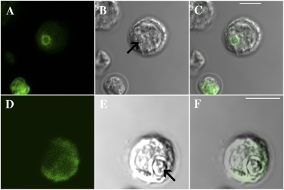
A putative chloroplast localization signal suggests that LCIB may target to the chloroplast. Immunofluorescent detection of LCIB with anti-LCIB antiserum was used, in combination with confocal microscopy, to visualize the subcellular localization of LCIB in cells grown under H-CO2 (5% CO2), L-CO2 (approximately 0.04% CO2), and VL-CO2 (<0.02% CO2). In H-CO2-acclimated cells, LCIB was expressed only at a very low level and was barely detectable (data not shown), while in cells acclimated to L-CO2 or VL-CO2, LCIB protein increased dramatically in abundance, consistent with its reported mRNA accumulation in these cells (Miura et al., 2004). In L-CO2- and VL-CO2-acclimated cells, two different patterns of distribution were observed in the immunofluorescent detection of LCIB protein. Immunofluorescence from LCIB was either dispersed throughout the entire chloroplast stroma or concentrated mainly in a discreet region surrounding the pyrenoid, appearing as a distinct ring structure in virtual longitudinal sections inside individual chloroplasts (Fig. 1; VL-CO2 localization data not shown).
Figure 1.
Immunofluorescent localization of LCIB in C. reinhardtii L-CO2-acclimated CC125 cells. A and D, False color immunofluorescence images of two different cells. B and E, Confocal images of the same cells. C and F, Merged images. The white bars in C and F are 5 μm in length and the arrows in B and E indicate the location of the pyrenoid in each confocal image.
Identification and Genetic Analysis of ad1 Suppressors
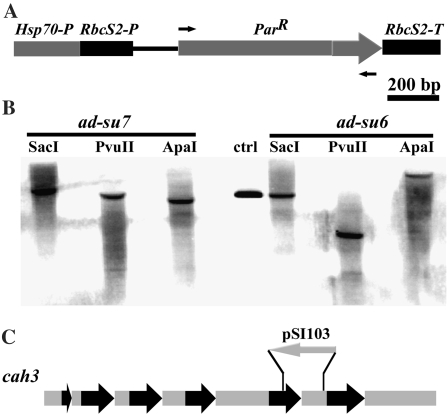
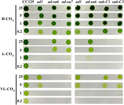
The C. reinhardtii strain chosen for this study was ad1, containing a deletion mutation of LCIB (Wang and Spalding, 2006). This strain cannot grow in L-CO2 but can grow either in H-CO2 or in VL-CO2. To isolate and identify suppressors that can grow in L-CO2, we performed insertional mutagenesis using a ParR-containing plasmid (pSI103) to transform ad1 (Fig. 3A). From approximately 106 transformants, three displayed the suppression phenotype, and two of these, ad-su6 and ad-su7, are described here. These two suppressors exhibited suboptimal growth in L-CO2 where the parental strain ad1 could not grow at all (Fig. 2). Unexpectedly, neither suppressor could survive in VL-CO2, indicating that although the second site suppressor mutations could suppress the L-CO2 lethal, air-dier phenotype of the LCIB mutation in ad1, growth of the two suppressors in VL-CO2 was completely abolished.
Figure 3.
Southern-blot analysis of ad-su6 and ad-su7 probed with an 880-bp PCR fragment of the ParR gene. A, Map of the ParR portion of the pSI103 plasmid used for insertional mutagenesis. Arrows above and below the map indicate the primers used to make the probe. B, Southern analysis of two suppressors, with linearized plasmid as control. Genomic DNA was digested with the indicated restriction enzymes. C, Map of the CAH3 genomic region in ad-su6. Black arrows, Exons; gray arrow, pSI103 insertion; gray bars, introns.
Figure 2.
Growth of ad1 suppressors and CAH3 genomic DNA complemented ad-su6 on minimal plates in H-CO2, L-CO2, and VL-CO2 chambers. Cells grown to logarithmic phase were diluted to the indicated numbers (×103) per 5 μL, spotted on plates, and incubated for 9 d under dim lights. [See online article for color version of this figure.]
The ad-su6 and ad-su7 strains were crossed with wild-type strain CC620 to determine whether the suppression phenotype cosegregated with the inserted ParR gene. More than 150 ZeoR (zeocin resistance, conferred by the BleR insert responsible for the LCIB mutation) random progeny from each cross were screened for their growth in different levels of CO2 and their resistance to paromomycin. In the ad-su6 cross, all 90 random progeny with the suppressor phenotype were paromomycin resistant, while all paromomycin-sensitive progeny exhibited an ad1-like growth phenotype in L-CO2 and VL-CO2, indicating cosegregation of the suppressor phenotype with the ParR insert. Southern analysis with probe specific for the ParR gene indicated a single insert present in ad-su6 (Fig. 3B). Although ad-su7 also contains only one ParR insert, genetic analysis showed the suppressor phenotype was not linked to the insert (data not shown).
Inverse PCR was employed to identify the flanking DNA in ad-su6. This flanking sequence was used in a BLAST search against the C. reinhardtii genome (http://genome.jgi-psf.org/Chlre3/Chlre3.home.html) and the insertion site was shown to be located between exon 5 and intron 5 of CAH3 (Fig. 3C). Further PCR and DNA gel-blot analyses revealed that only 186 nucleotides of CAH3 (63 nt of exon 5 and 123 nt of intron 5) were deleted in ad-su6. Since ad-su6 has the same growth phenotype as ad-su7, we PCR amplified and sequenced CAH3 genomic DNA from ad-su7 and found that two nucleotides were deleted downstream of the ATG translation initiation codon (ATGCGCTCAGCCGTTCTACAACGCGGCCAGGCGCGGCGAGTGTCTTGCCGGGTGAGTGAA; underline indicates deletion mutation in ad-su7), which predicts a premature stop codon (ATGCGCTCAGCCGCTACAACGCGGCCAGGCGCGGCGAGTGTCTTGCCGGGTGAGTGA; underline indicates stop codon).
Expression Patterns of Limiting-CO2-Inducible Genes in ad-su6
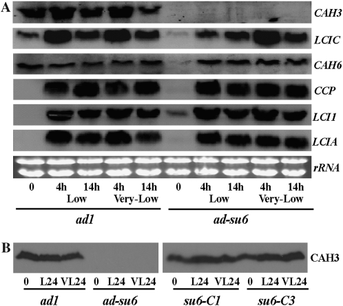
Northern-blot analysis showed that ad-su6 apparently is a hypomorphic mutant for CAH3. In ad1 cells, a transient, slight increase in the CAH3 transcript abundance was observed when cells were shifted from a H-CO2 to either L-CO2 or VL-CO2 atmosphere, and, after a longer acclimation time (14 h), the CAH3 message abundance was decreased (Fig. 4A). In the CAH3 hypomorphic mutant ad-su6, CAH3 transcript was undetectable throughout all CO2 conditions. Western-blot analysis using polyclonal antiserum raised against CAH3 from C. reinhardtii also failed to detect the CAH3 protein in the suppressor mutant ad-su6 cultures (Fig. 4B).
Figure 4.
A, Northern-blot analysis of ad1 and ad-su6. Cells were adapted to L-CO2 or VL-CO2 conditions for 4 and 14 h, respectively, and 10 μg of total RNA was used. B, Immunoblot analysis of whole cell fractions. Cells were either H-CO2 grown or induced 24 h under L-CO2 (L24) or VL-CO2 (VL24) conditions. One hundred micrograms of total protein were analyzed with CAH3 antibody. su6-C1 and su6-C3 are two CAH3 genomic DNA-complemented ad-su6 transformants as described in Figure 2.
Message accumulations of several limiting-CO2-inducible genes also were analyzed. Among all the genes tested, including LCIC, CCP, LCI1, and LCIA, patterns of expression in the suppressor ad-su6 relative to the original LCIB mutant ad1 were not found to be significantly different. The expression level of chloroplast stromal carbonic anhydrase (CAH6) was not affected by CO2 conditions, and the transcript abundance of CAH6 in ad-su6 under limiting-CO2 conditions was comparable to that in ad1 (Fig. 4A).
Complementation of ad-su6
To confirm whether the CAH3 mutation is responsible for the suppressor phenotype, we transformed a genomic DNA fragment containing a wild-type copy of CAH3 into the suppressors ad-su6 and ad-su7 and selected complemented lines that could survive in VL-CO2. Complemented ad-su6 (su6-C1 and su6-C3) and ad-su7 lines showed the same growth phenotype as ad1, growth in VL-CO2 and no growth in L-CO2 (Fig. 2; ad-su7 data not shown). In addition, western-blot analysis showed that complemented ad-su6 lines recovered the expression of CAH3 protein (Fig. 4B). Complementation of the ad-su6 VL-CO2 lethal phenotype also was achieved by expressing CAH3 cDNA under control of the constitutive PsaD promoter and terminator (Fischer and Rochaix, 2001; data not shown).
Photosynthetic Affinity for Ci and Direct Ci Uptake Characteristics
Photosynthetic O2 evolution in response to Ci concentrations for L-CO2- and VL-CO2-acclimated wild-type and various mutant cells was compared (Table I). Walled progeny of mutants were generated for physiological measurements, including the CAH3 mutation of ad-su6 in a wild-type background (wt-su6) or in an ad1 background (ad-su6-1). Consistent with the ad1 air-dier phenotype, the LCIB-defective ad1-1 mutant cells (walled progeny of ad1) acclimated in L-CO2 showed dramatically decreased photosynthetic affinity for Ci compared with wild-type cells acclimated under the same conditions (approximately 11% of wild type). In contrast, when acclimated to VL-CO2, photosynthetic affinity of ad1-1 was increased to approximately 57% to 61% of the wild-type strain (P20 and P50, estimated at 20 and 50 μm total Ci, respectively). Photosynthetic affinity of L-CO2-acclimated ad-su6-1 was significantly higher than ad1-1 at 50 μm total Ci (P50: 0.14 ± 0.02 versus 0.07 ± 0.02, P value < 0.05), while VL-CO2-acclimated ad-su6-1 had a lower relative affinity than ad1-1 (P20: 0.07 ± 0.03 versus 0.27 ± 0.03, P < 0.05) at 20 μm total Ci, both of which are consistent with the phenotypes of these two strains. The aberrant photosynthetic affinity of ad-su6-1 was caused by the CAH3 mutation, since wt-su6 showed the same pattern of photosynthetic affinity in both L-CO2 and VL-CO2 conditions as ad-su6-1.
Table I.
Relative affinity of photosynthetic O2 evolution in cells acclimated to L-CO2 or VL-CO2 for 1 d
Rates of O2 evolution (μmol mg Chl−1 h−1) were determined in pH 7.3 (MOPS-KOH) buffer at 20 μm (V20), 50 μm (V50), and 4,000 μm (V4000) NaHCO3. Relative Ci affinity was calculated as ratios: P20 = V20/V4000 and P50 = V50/V4000. Vmax = V4000.
| Strains | L-CO2
|
VL-CO2
|
||||
|---|---|---|---|---|---|---|
| P20 | P50 | Vmax | P20 | P50 | Vmax | |
| CC125 | 0.38 ± 0.04 | 0.60 ± 0.05 | 180 ± 22 | 0.44 ± 0.05 | 0.63 ± 0.07 | 162 ± 18 |
| ad1-1 | 0.04 ± 0.02 | 0.07 ± 0.02 | 106 ± 18 | 0.27 ± 0.03 | 0.36 ± 0.04 | 82 ± 8 |
| wt-su6 | 0.08 ± 0.01 | 0.15 ± 0.03 | 104 ± 12 | 0.09 ± 0.02 | 0.15 ± 0.02 | 80 ± 9 |
| ad-su6-1 | 0.05 ± 0.02 | 0.14 ± 0.02 | 130 ± 21 | 0.07 ± 0.03 | 0.13 ± 0.04 | 83 ± 10 |
| cia5 | 0.08 ± 0.02 | 0.12 ± 0.03 | 98 ± 18 | 0.06 ± 0.03 | 0.08 ± 0.02 | 72 ± 9 |
Intracellular Ci accumulation in L-CO2-acclimated ad1-1 cells was similar to that of the nonacclimating mutant cia5 (0.26 ± 0.08 mm versus 0.19 ± 0.05 mm), in which the CCM presumably does not function (Table II). An active Ci accumulation mechanism was regained in VL-CO2-acclimated ad1-1 cells, although not as high as the wild-type strain (0.80 ± 0.15 mm versus 1.65 ± 0.25 mm, measured with 20 μm total Ci). The measured intracellular Ci pool in the LCIB-CAH3 double mutant ad-su6-1 was increased more than 20-fold over that of the LCIB single mutant ad1-1 when acclimated in L-CO2 conditions (6.15 ± 1.15 mm versus 0.26 ± 0.08 mm), and the increased Ci accumulation could be attributed to the CAH3 mutation because CAH3 single mutant wt-su6 accumulated the same level of intracellular [Ci] as ad-su6-1 (7.60 ± 1.25 mm versus 6.15 ± 1.15 mm). In VL-CO2-acclimated strains, intracellular [Ci] in ad-su6-1 was only 2.5-fold higher than that in ad1-1 (2.85 ± 0.32 mm versus 0.80 ± 0.15 mm, measured with 20 μm total Ci).
Table II.
Intracellular Ci accumulation in cells acclimated to L-CO2 or VL-CO2 for 1 d
Internal Ci accumulation (mm Ci after 80 s) was determined by silicone oil centrifugation in pH 7.3 (MOPS-KOH) buffer at initial external Ci concentrations of 20 μm and 50 μm [14C]NaHCO3 and a Chl concentration of 25 μg/mL.
| Strains | L-CO2
|
VL-CO2
|
|
|---|---|---|---|
| 50 μm Ci | 20 μm Ci | 50 μm Ci | |
| CC125 | 1.80 ± 0.33 | 1.65 ± 0.25 | 1.78 ± 0.24 |
| ad1-1 | 0.26 ± 0.08 | 0.80 ± 0.15 | 1.43 ± 0.22 |
| wt-su6 | 7.60 ± 1.25 | 2.50 ± 0.33 | 8.10 ± 1.45 |
| ad-su6-1 | 6.15 ± 1.15 | 2.85 ± 0.32 | 5.30 ± 0.58 |
| cia5 | 0.19 ± 0.05 | 0.15 ± 0.04 | 0.30 ± 0.15 |
DISCUSSION
Rubisco, the primary enzyme for photosynthetic CO2 assimilation, is an inefficient catalyst with a low affinity for atmospheric CO2. For most algae, the Rubisco Km(CO2) is greater than 25 μm, so Rubisco is functioning at <20% of capacity at 10 μm CO2 in an air-equilibrated aquatic environment. Many photosynthetic organisms have an inducible CCM that raises the CO2 concentration around the active site of Rubisco several-fold higher than the environmental level, thus improving the efficiency of CO2 assimilation. In the eukaryotic microalga C. reinhardtii, active Ci uptake (mainly as CO2 and HCO3−) at either the plasma membrane or the inner chloroplast envelope is an essential component of the CCM. Although the products of several limiting-CO2-inducible genes have been identified as putative Ci transporters on the chloroplast envelope, including LCIA, LCI1, CCP1/2, and Ycf10, or on the plasma membrane, including HLA3, none of the respective gene products have been definitively determined to transport Ci species (Burow et al., 1996; Chen et al., 1997; Rolland et al., 1997; Im and Grossman, 2001; Miura et al., 2004; Pollock et al., 2004; Mariscal et al., 2006), leaving Ci transport largely a mystery.
Mutants with lesions in the limiting-CO2-induced gene LCIB shed some light on the nature of Ci uptake and accumulation in chloroplasts. Two conditional lethal mutants, pmp1 and ad1, with lesions in LCIB, are defective in Ci accumulation when acclimated in L-CO2 but not when acclimated in VL-CO2, indicating the existence of multiple Ci uptake and accumulation pathways in C. reinhardtii corresponding to multiple limiting-CO2 acclimation states (Vance and Spalding, 2005). LCIB was proposed to be involved in active Ci transport (Spalding et al., 1983b; Wang and Spalding, 2006), although, since it is predicted to be a soluble protein with no significant transmembrane regions, it was acknowledged to be unlikely that LCIB itself is directly involved in transport. Therefore, it was argued that LCIB might be a subunit of a Ci transport complex or might have a regulatory role in Ci uptake and/or accumulation.
In this article, we identified two independent, second-site suppressors of the LCIB mutant air-dier phenotype that restored growth to ad1 in L-CO2 but, unlike ad1 itself, were unable to grow in VL-CO2. Both suppressors have lesions in CAH3, encoding a thylakoid-lumen-located, α-type carbonic anhydrase. The requirement of a thylakoidal CA and an acidic compartment in a functional CCM was first suggested by Pronina and colleagues, and later CAH3 was identified and proposed to catalyze the rapid conversion of HCO3− to CO2 in the acidic thylakoid lumen, with the CO2 then diffusing to the pyrenoid to supply elevated substrate CO2 concentrations for Rubisco (Pronina et al., 1981; Pronina and Semenenko, 1990; Funke et al., 1997; Raven, 1997; Karlsson et al., 1998; Hanson et al., 2003; Moroney and Ynalvez, 2007). Consistent with previous reports (Spalding et al., 1983a; Moroney et al., 1987), the L-CO2-acclimated CAH3 single mutant wt-su6, generated in a cross between the suppressed line and wild type, accumulated a very high internal Ci concentration but was unable to use Ci efficiently for photosynthesis. However, the photosynthetic Ci affinity of this CAH3 single gene mutant wt-su6 still is significantly higher than that of the L-CO2-acclimated LCIB mutant ad1, which accumulates only very low internal Ci levels and cannot grow at all in L-CO2, both traits agreeing with prior reports of the characteristics of the allelic LCIB mutant pmp1 and various CAH3 single mutants (Spalding et al., 1983a, 1983b; Moroney et al., 1987; Suzuki and Spalding, 1989). The LCIB-CAH3 double mutant suppressor ad-su6-1 accumulated the same level of intracellular Ci as the CAH3 single gene mutant wt-su6, strongly arguing against LCIB as being required for or solely responsible for Ci transport into the chloroplast. This effect of combining lesions in CAH3 and LCIB has been reported previously (Spalding et al., 1983c), before the genes themselves were identified and before the full significance of the observation could be appreciated. It was recognized at the time that the lesion in CAH3 was apparently epistatic to the lesion in LCIB, and that the double mutant exhibited a leaky CO2-requiring phenotype in air, similar to the CAH3 single mutant.
Our current work definitively demonstrates that a CAH3 loss-of-function mutation can suppress the air-dier phenotype of LCIB mutants, as well as revealing a novel, VL-CO2 lethal phenotype of the double mutant suppressors. The suppressors exhibited the same levels of photosynthetic Ci affinity and internal Ci accumulation, regardless of whether they were acclimated to L-CO2 or VL-CO2. No significant difference in intracellular Ci accumulation was detected between the L-CO2-acclimated suppressor (LCIB-CAH3 double mutant) and the CAH3 single mutant, indicating the LCIB mutation had no influence on Ci accumulation in the absence of a functional CAH3, which is contrary to the proposed direct involvement of LCIB in Ci transport in L-CO-acclimated cells (Spalding et al., 1983b; Wang and Spalding, 2006), but is consistent with the reported epistatic interaction between these two mutations (Spalding et al., 1983c).
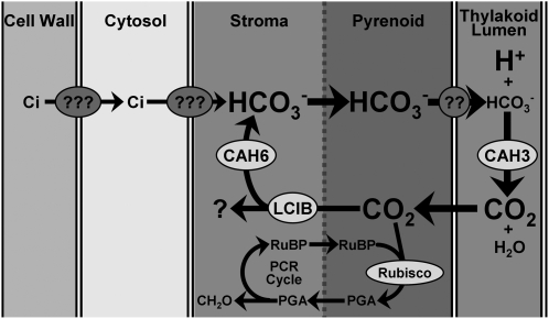
The position of LCIB in the biochemical pathway of Ci uptake and accumulation in C. reinhardtii is informed by the epistatic interaction between the CAH3 and LCIB mutations. It is clear that CAH3 functions to dehydrate HCO3− accumulated in the stroma, although it is not known how HCO3− gains access to the thylakoid lumen. Because CAH3 mutations are epistatic over LCIB mutations, LCIB must act downstream of CAH3, meaning it must act after the accumulated stromal HCO3− is dehydrated to CO2. The localization of LCIB also is inconsistent with a direct role in Ci transport, regardless of whether it is diffusely distributed throughout the stroma or concentrated around the pyrenoid. The combination of LCIB localization and the epistatic interaction between LCIB and CAH3 mutations, together with the clear demonstration that LCIB mutants can transport and accumulate Ci in the absence of functional CAH3, provide a very compelling argument against a direct role for LCIB in Ci transport. Since it is very unlikely LCIB is involved in Ci transport, it appears most likely that LCIB is involved in preventing the loss of CO2 from the chloroplast (Fig. 5). One possibility is that LCIB is involved in sequestering or trapping excess CO2 from CAH3-catalyzed dehydration of HCO3− that might otherwise diffuse out of the chloroplast and be lost. A functionally similar CCM exists in cyanobacteria, and Price et al. (2002) have proposed that two unique hydrophilic proteins, ChpX and ChpY (also named CupA and CupB), are critical components of a thylakoid-based NDH-1 CO2 uptake complex in the model cyanobacterium Synechococcus sp. PCC7942 and aid in the recapture or recycling of CO2 leakage from carboxysomes by CO2 hydration activity in the light (Maeda et al., 2002; Shibata et al., 2001, 2002; Price et al., 2002). Thus LCIB may be part of a functionally analogous but novel CO2 pump/trap and may function mainly to prevent depletion of the HCO3− pool by trapping CO2 from CAH3 dehydration back into the stromal HCO3− pool or by providing a barrier to CO2 diffusion back out of the pyrenoid.
Figure 5.
Simple schematic of the C. reinhardtii CCM, showing the relative position of CAH3 and LCIB in the pathway of Ci transport, HCO3− accumulation, and the subsequent dehydration of HCO3− to produce CO2. PGA, 3-Phosphoglycerate.
Although it is clear that CAH3 mutations are epistatic to LCIB mutations and thus that LCIB must function downstream of CAH3, it is more complicated to explain mechanistically how the characteristics of the individual mutants and the double mutants explain this epistatic interaction. Dehydration, either uncatalyzed or catalyzed by the stromal carbonic anhydrase CAH6, of the very high stromal HCO3− concentration in the LCIB-CAH3 double mutant ad-su6-1 may provide a direct supply of CO2 to Rubisco sufficient to support growth and suppress the air-dier phenotype of ad1-1 in L-CO2-acclimated cells. However, the stromal HCO3− concentration of VL-CO2-acclimated ad-su6-1 also is fairly high, yet apparently fails to provide Rubisco sufficient CO2 to support growth in VL-CO2 conditions. The predicted carboxylation rates for Rubisco (64 μmol CO2 mg−1 Chl h−1 and 41 μmol CO2 mg−1 Chl h−1 for L-CO2- and VL-CO2-acclimated cells, respectively), assuming complete equilibration at pH of 8.5 between HCO3− and CO2 at the observed internal Ci concentrations of 6.15 mm (50 μm external Ci) and 2.9 mm (20 μm external Ci) for L-CO2- and VL-CO2-acclimated cells, respectively, substantially overestimates the observed photosynthetic rates of 18 μmol CO2 mg−1 Chl h−1 and 5.8 μmol CO2 mg−1 Chl h−1 for L-CO2- and VL-CO2-acclimated cells, respectively. On the other hand, calculations assuming an uncatalyzed rate of HCO3− dehydration (CO2 supply rates of 8 μmol CO2 mg−1 Chl h−1 and 2.9 μmol CO2 mg−1 Chl h−1 for L-CO2 and VL-CO2, respectively) underestimate the actual photosynthetic rates (see Spalding and Portis, 1985). Thus the observed photosynthetic rates argue for only limited CAH6-catalyzed dehydration of the accumulated HCO3−, which may explain why an internal Ci concentration of 2.9 mm in VL-CO2-acclimated cells is unable to support growth under VL-CO2 conditions.
A key question is why an observed photosynthetic rate of 18.2 μmol CO2 mg−1 Chl h−1 in L-CO2 allows for substantial growth of the double mutant suppressor but a rate of 5.8 μmol CO2 mg−1 Chl h−1 in VL-CO2 does not. A photosynthetic rate of 5.8 μmol CO2 mg−1 Chl h−1 may simply be below the threshold for survival, a conclusion supported by the observed rates of photosynthesis of three other strains unable to grow, L-CO2-acclimated LCIB mutant ad1 at 50 μm external Ci and VL-CO2-acclimated wild type-su6 (CAH3 mutation alone) and cia5 at 20 μm external Ci, which were 7.4, 7.2, and 4.3 μmol CO2 mg−1 Chl h−1, respectively, and by the minimum photosynthetic rate observed for any strain able to grow, which was 11.8 μmol CO2 mg−1 Chl h−1 (L-CO2-acclimated cia5 at 50 μm external Ci). Based on the reported effect of photon flux density on Ci accumulation (Spalding, 1990), we also would expect substantially lower stromal HCO3− accumulation and thus a lower photosynthetic rate under light conditions used for growth (approximately 100 μmol photons m−2 s−1), which is much lower than that used for photosynthesis and Ci uptake measurements (approximately 800 μmol photons m−2 s−1).
Even though the data presented here help identify the position of LCIB in the pathway for Ci uptake and accumulation by placing its function downstream of CAH3, the actual function of LCIB remains a mystery, as does how the single mutation in LCIB eliminates almost all Ci accumulation to the same extent as the cia5 mutant, in which almost no limiting-CO2-inducible genes are expressed (Fukuzawa et al., 2001; Xiang et al., 2001). LCIB is essential in L-CO2 conditions, and we hypothesize that LCIB might be involved in somehow preventing the loss of CAH3-generated CO2 that is not captured by Rubisco in the pyrenoid, either by preventing its diffusion from the pyrenoid or by recapturing any CO2 escaping from the pyrenoid or from thylakoids outside the pyrenoid. The physiological observations for the single and double mutants are consistent with the role in CO2 trapping, and the immunolocalization of LCIB in the stroma or surrounding the pyrenoid is more consistent with CO2 trapping than with Ci transport. The constitutively expressed chloroplast stromal CAH6 might normally also help retain Ci in the stroma by trapping it as HCO3−. It is tempting to speculate that LCIB and CAH6 might cooperate in trapping CO2 released from the pyrenoid and/or from thylakoids, especially since CAH6 reportedly is differentially concentrated around the pyrenoid (Mitra et al., 2004), as is LCIB, at least under some conditions. It will be interesting to investigate the potential colocalization of LCIB and CAH6, and the generation of CAH6 mutants by either an RNAi approach or by insertional mutagenesis could help to clarify the physiological roles of and any potential interactions between LCIB and CAH6 in the L-CO2 acclimation state.
MATERIALS AND METHODS
Cell Strains and Culture Conditions
Chlamydomonas reinhardtii strains CC125 and CC620 were obtained from the Chlamydomonas Stock Center, Duke University, Durham, NC. The LCIB-defective mutant ad1 was generated by insertional mutagenesis (Wang and Spalding, 2006) and the cia5 mutant was a gift from Dr. Donald P. Weeks (University of Nebraska, Lincoln).
Media and growth conditions for C. reinhardtii strains have been previously described (Wang and Spalding, 2006). All strains were maintained on CO2-minimal plates and kept in H-CO2 (air enriched with 5% [v/v] CO2) chambers at room temperature, under continuous illumination (50 μmol photons m−2 s−1). Liquid cultures were grown on a gyratory shaker under aeration in white light (approximately 100 μmol photons m−2 s−1). For experiments in which cells were shifted from high to limiting CO2 (L-CO2, 0.035%–0.04% or VL-CO2, 0.005%–0.01%) conditions, cells were cultured in CO2-minimal medium aerated with 5% CO2 to a density of approximately 2 × 10⁶ cells/mL and then shifted to aeration with the appropriate limiting CO2 for various times. Very low CO2 was obtained by mixing normal air with compressed, CO2-free air.
Immunolocalization of LCIB Protein
Cells grown under different CO2 conditions were placed on precharged microscope slides (ProbeOn Plus, FisherBiotech) for 5 to 10 min, and then rinsed briefly with CO2 minimal medium. The immunofluorescence staining was performed as described previously (Sanders and Salisbury, 1995; Cole et al., 1998). Antiserum against LCIB, raised in rabbit, was used at a dilution of 1:500 as primary antibody, and fluorescein isothyocyanate-conjugated goat anti-rabbit IgG (Jackson ImmunoResearch Laboratories) was used at a dilution of 1:100 as the secondary antibody for immunofluorescence. After final washing steps, the slides were mounted using a medium containing 2% N-propyl gallate, 30% 0.1 m Tris, pH 9 and 70% glycerol. Stained cells were viewed and digital images acquired using a Nikon C1si confocal microscope.
Isolation of Suppressors, Growth Spot Tests, and Genetic Analysis
Glass bead transformations were performed as described previously (Van and Spalding, 1999). Air-dier mutant ad1 was transformed with linearized plasmid pSI103 (Sizova et al., 2001) and spread onto CO2-minimal plates containing 15 μg/mL paromomycin. Plates were placed in air for 6 weeks and surviving colonies selected as putative suppressors were verified by spot tests.
For spot test of growth, actively growing cells were serially diluted to similar cell densities in minimal medium and spotted (5 μL/spot) onto minimal agar plates, and grown in various CO2 concentrations for around 9 d. Genetic crosses and random progeny analyses were performed as described by Harris (1989).
DNA, RNA, and Protein-Blot Analysis
Southern analyses were performed as described by Van and Spalding (1999). Northern-blot hybridizations were performed by standard procedures. Total RNA was prepared by the acid guanidinium thiocyanate method (Chomczynski and Sacchi, 2006). RNA (approximately 15 μg) were separated on 0.9% formaldehyde-agarose gels and blotted onto nylon transfer membranes (Osmonics, Inc.). PCR-amplified cDNA sequences of corresponding genes were used as templates and all probes were generated by randomly primed labeling with [α-32P]dCTP following instructions of the manufacturer. Each northern blot was analyzed by phosphorimager scanning (Bio-Rad).
For total protein analyses, cells were harvested and resuspended in a buffer containing 10 mm Tris-HCl pH 7.5, 1 mm EDTA, 10 mm NaCl, 1 mm phenylmethylsulfonyl fluoride, 1 mm benzamidine, and 5 mm dithiothreitol. Protein concentrations were measured using Bio-Rad protein assay kit (Bio-Rad catalog no. 500–0006). Proteins were separated on 12% polyacrylamide gels as described previously (Laemmli, 1970). Immunoblotting was performed as described in the protocol from Bio-Rad Laboratories. The CAH3 antiserum was a gift from James V. Moroney (Louisiana State University) and was generated using a synthetic peptide derived from an internal amino acid sequence of CAH3 (residues 213–224) as antigen.
Isolation of Sequences Flanking the Plasmid Insert in ad-su6 by Inverse PCR
Based on information from Southern-blot analysis, SacI was used to digest the genomic DNA isolated from ad-su6 to produce a fragment with a size of approximately 4.7 kb, including part of the inserted pSI103 vector and its 3′-flanking genomic DNA. The SacI-digested ad-su6 genomic DNA (1 μg) was circularized with 1 unit of T4 DNA ligase (Invitrogen), precipitated, and the circularized product was used as a template for inverse PCR by using standard PCR procedures. Two pairs of primers were designed, with each pair complementing the pSI103 sequence in opposite orientations. Both primer pairs produced PCR products with the correct predicted sizes, and amplified DNA from one primer pair (5′-GGTCTGACGCTCAGTGGAACGA-3′ and 5′-CGCAACGCATCGTCCATGCTTC-3′) was sequenced to determine the sequences flanking the insert.
Photosynthetic O2 Exchange and Ci Uptake Measurements
L-CO2-induced or very low CO2-induced cells (24-h induction) were collected by centrifugation and resuspended in 25 mm MOPS-KOH buffer to a final chlorophyll concentration of approximately 20 to 25 μg/mL for analysis of photosynthesis and Ci uptake. Photosynthetic O2 evolution was measured at 25°C with a Clark-type oxygen electrode (Rank Brothers). Under constant illumination (800 μmol photons m−2 s−), cells were transferred to the electrode chamber and allowed to exhaust endogenous Ci until net O2 exchange was zero. Measurements were initiated by addition of various concentrations of NaHCO3. Oxygen evolution rates were recorded as V20 or V50 when 20 or 50 μm NaHCO3 were used, respectively. The maximum O2 evolution rate, V4000, was obtained by using 4,000 μm NaHCO3. Relative affinity for Ci was calculated as the ratios P20 = V20/V4000 and P50 = V50/V4000.
Ci uptake by C. reinhardtii cells at 20 or 50 μm total Ci was estimated by the silicone oil filtration technique (Badger et al., 1980). The cell volume was measured by using 14C-sorbitol and 3H2O as previously described (Wirtz et al., 1980). The intracellular Ci concentration was calculated by using cell volume and the acid labile 14C data.
Acknowledgments
We thank Professor Donald P. Weeks for providing CAH3 genomic DNA plasmid and James V. Moroney for the anti-CAH3 antiserum.
This work was supported by the U.S. Department of Agriculture National Research Initiative (grant no. 20073531818433 to M.H.S.), as well as by the College of Agriculture and Life Sciences and the College of Liberal Arts and Sciences at Iowa State University. This journal paper of the Iowa Agriculture and Home Economics Experiment Station, Ames, Iowa, Project Number IOW05136, also was supported by Hatch Act and State of Iowa funds.
The author responsible for the distribution of materials integral to the findings presented in this article in accordance with the policy described in the Instructions for Authors (www.plantphysiol.org) is: Martin H. Spalding (mspaldin@iastate.edu).
Some figures in this article are displayed in color online but in black and white in the print edition.
Open access articles can be viewed online without a subscription.
References
- Badger MR, Kaplan A, Berry JA (1980) Internal inorganic carbon pool of Chlamydomonas reinhardtii: evidence for a carbon dioxide concentrating mechanism. Plant Physiol 66 407–413 [DOI] [PMC free article] [PubMed] [Google Scholar]
- Beardall J, Giordano M (2002) Ecological implications of microalgal and cyanobacterial CO2 concentrating mechanisms and their regulation. Funct Plant Biol 29 335–347 [DOI] [PubMed] [Google Scholar]
- Burow MD, Chen ZY, Mouton TM, Moroney JV (1996) Isolation of cDNA clones of genes induced upon transfer of Chlamydomonas reinhardtii to low CO2. Plant Mol Biol 31 443–448 [DOI] [PubMed] [Google Scholar]
- Chen ZY, Lavigne LL, Mason CB, Moroney JV (1997) Cloning and overexpression of two cDNAs encoding the low-CO2-inducible chloroplast envelope protein LIP-36 from Chlamydomonas reinhardtii. Plant Physiol 114 265–273 [DOI] [PMC free article] [PubMed] [Google Scholar]
- Chomczynski P, Sacchi N (2006) The single-step method of RNA isolation by acid guanidinium thiocyanate-phenol-chloroform extraction: twenty-something years on. Nat Protocols 1 581–585 [DOI] [PubMed] [Google Scholar]
- Cole DG, Diener DR, Himelblau AL, Beech PL, Fuster JC, Rosenbaum JL (1998) Chlamydomonas kinesin-II-dependent intraflagellar transport (IFT): IFT particles contain proteins required for ciliary assembly in Caenorhabditis elegans sensory neurons. J Cell Biol 141 993–1008 [DOI] [PMC free article] [PubMed] [Google Scholar]
- Fischer N, Rochaix JD (2001) The flanking regions of PsaD drive efficient gene expression in the nucleus of the green alga Chlamydomonas reinhardtii. Mol Genet Genomics 265 888–894 [DOI] [PubMed] [Google Scholar]
- Fukuzawa H, Miura K, Ishizaki K, Kucho KI, Saito T, Kohinata T, Ohyama K (2001) Ccm1, a regulatory gene controlling the induction of a carbon-concentrating mechanism in Chlamydomonas reinhardtii by sensing CO2 availability. Proc Natl Acad Sci USA 98 5347–5352 [DOI] [PMC free article] [PubMed] [Google Scholar]
- Funke RP, Kovar JL, Weeks DP (1997) Intracellular carbonic anhydrase is essential to photosynthesis in Chlamydomonas reinhardtii at atmospheric levels of CO2. Plant Physiol 114 237–244 [DOI] [PMC free article] [PubMed] [Google Scholar]
- Giordano M, Beardall J, Raven JA (2005) CO2 concentrating mechanisms in algae: mechanisms, environmental modulation, and evolution. Annu Rev Plant Biol 56 99–131 [DOI] [PubMed] [Google Scholar]
- Hanson DT, Franklin LA, Samuelsson G, Badger MR (2003) The Chlamydomonas reinhardtii cia3 mutant lacking a thylakoid lumen-localized carbonic anhydrase is limited by CO2 supply to Rubisco and not photosystem II function in vivo. Plant Physiol 132 2267–2275 [DOI] [PMC free article] [PubMed] [Google Scholar]
- Harris EH (1989) The Chlamydomonas Source Book: A Comprehensive Guide to Biology and Laboratory Use. Academic Press, San Diego, pp 419–446 [DOI] [PubMed]
- Im CS, Grossman AR (2001) Identification and regulation of high light-induced genes in Chlamydomonas reinhardtii. Plant J 30 301–313 [DOI] [PubMed] [Google Scholar]
- Karlsson J, Clarke AK, Chen ZY, Hugghins SY, Park YI, Husic HD, Moroney JV, Samuelsson G (1998) A novel α-type carbonic anhydrase associated with the thylakoid membrane in Chlamydomonas reinhardtii is required for growth at ambient CO2. EMBO J 17 1208–1216 [DOI] [PMC free article] [PubMed] [Google Scholar]
- Laemmli UK (1970) Cleavage of structural proteins during the assembly of the head of bacteriophage T4. Nature 227 680–685 [DOI] [PubMed] [Google Scholar]
- Maeda S, Badger MR, Price GD (2002) Novel gene products associated with NdhD3/D4-containing NDH-1 complexes are involved in photosynthetic CO2 hydration in the cyanobacterium, Synechococcus sp. PCC7942. Mol Microbiol 43 425–435 [DOI] [PubMed] [Google Scholar]
- Mariscal V, Moulin P, Orsel M, Miller AJ, Fernández E, Galván A (2006) Differential regulation of the Chlamydomonas Nar1 gene family by carbon and nitrogen. Protist 157 421–433 [DOI] [PubMed] [Google Scholar]
- Mitra M, Lato SM, Ynalvez RA, Xiao Y, Moroney JV (2004) Identification of a new chloroplast carbonic anhydrase in Chlamydomonas reinhardtii. Plant Physiol 135 173–182 [DOI] [PMC free article] [PubMed] [Google Scholar]
- Miura K, Yamano T, Yoshioka S, Kohinata T, Inoue Y, Taniguchi F, Asamizu E, Nakaura Y, Tabata S, Yamato KT, et al (2004) Expression profiling-based identification of CO2-responsive genes regulated by CCM1 controlling a carbon-concentrating mechanism in Chlamydomonas reinhardtii. Plant Physiol 135 1595–1607 [DOI] [PMC free article] [PubMed] [Google Scholar]
- Moroney JV, Togasaki RK, Husic HD, Tolbert NE (1987) Evidence that an internal carbonic anhydrase is present in 5% CO2-grown and air-grown Chlamydomonas. Plant Physiol 84 757–761 [DOI] [PMC free article] [PubMed] [Google Scholar]
- Moroney JV, Ynalvez RA (2007) Proposed carbon dioxide concentrating mechanism in Chlamydomonas reinhardtii. Eukaryot Cell 6 1251–1259 [DOI] [PMC free article] [PubMed] [Google Scholar]
- Palmqvist K, Sjoberg S, Samuelsson G (1988) Induction of inorganic carbon accumulation in the unicellular green algae Scenedesmus obliquus and Chlamydomonas reinhardtii. Plant Physiol 87 437–442 [DOI] [PMC free article] [PubMed] [Google Scholar]
- Pollock SV, Prout DL, Godfrey AC, Lemaire SD, Moroney JV (2004) The Chlamydomonas reinhardtii proteins Ccp1 and Ccp2 are required for long-term growth, but are not necessary for efficient photosynthesis, in a low-CO2 environment. Plant Mol Biol 56 125–132 [DOI] [PubMed] [Google Scholar]
- Price GD, Maeda SI, Omata T, Badger MR (2002) Modes of active inorganic carbon uptake in the cyanobacterium, Synechococcus sp. PCC7942. Funct Plant Biol 29 131–149 [DOI] [PubMed] [Google Scholar]
- Price GD, Woodger FJ, Badger MR, Howitt SM, Tucker L (2004) Identification of a SulP-type bicarbonate transporter in marine cyanobacteria. Proc Natl Acad Sci USA 101 18228–18233 [DOI] [PMC free article] [PubMed] [Google Scholar]
- Pronina NA, Ramazanov ZM, Semenenko VE (1981) Carbonic-anhydrase activity of Chlorella cells as a function of CO2 concentration. Sov Plant Physiol 28 345–351 [Google Scholar]
- Pronina NA, Semenenko VE (1990) Membrane-bound carbonic anhydrase takes part in CO2 concentration in algal cells. Curr Res Photosynth 4 489–492 [Google Scholar]
- Raven JA (1997) CO2-concentrating mechanisms: a direct role for thylakoid lumen acidification? Plant Cell Environ 20 147–154 [Google Scholar]
- Rolland N, Dorne AJ, Amoroso G, Sultemeyer DF, Joyard J, Rochaix JD (1997) Disruption of the plastid ycf10 open reading frame affects uptake of inorganic carbon in the chloroplast of Chlamydomonas. EMBO J 16 6713–6726 [DOI] [PMC free article] [PubMed] [Google Scholar]
- Sanders MA, Salisbury JL (1995) Immunofluorescence microscopy of cilia and flagella. Methods Cell Biol 47 163–169 [DOI] [PubMed] [Google Scholar]
- Shibata M, Ohkawa H, Kaneko T, Fukuzawa H, Tabata S, Kaplan A, Ogawa T (2001) Distinct constitutive and low-CO2-induced CO2 uptake systems in cyanobacteria: genes involved and their phylogenetic relationship with homologous genes in other organisms. Proc Natl Acad Sci USA 98 11789–11794 [DOI] [PMC free article] [PubMed] [Google Scholar]
- Shibata M, Ohkawa H, Katoh H, Shimoyama M, Ogawa T (2002) Two CO2 uptake systems in cyanobacteria: four systems for inorganic carbon acquisition in Synechocystis sp. strain CC6803. Funct Plant Biol 29 123–129 [DOI] [PubMed] [Google Scholar]
- Sizova I, Fuhrmann M, Hegemann P (2001) A Streptomyces rimosus aphVIII gene encoding for a new type phosphotransferase provides stable antibiotic resistance to Chlamydomonas reinhardtii. Gene 277 221–229 [DOI] [PubMed] [Google Scholar]
- Spalding MH (1990) Effect of photon flux density on inorganic carbon accumulation and net CO2 exchange in a high-CO2-requiring mutant of Chlamydomonas reinhardtii. Photosynth Res 24 245–252 [DOI] [PubMed] [Google Scholar]
- Spalding MH (2008) Microalgal carbon-dioxide-concentrating mechanisms: Chlamydomonas inorganic carbon transporters. J Exp Bot 59 1463–1473 [DOI] [PubMed] [Google Scholar]
- Spalding MH, Portis AR (1985) A model of carbon dioxide assimilation in Chlamydomonas reinhardii. Planta 164 308–320 [DOI] [PubMed] [Google Scholar]
- Spalding MH, Spreitzer RJ, Ogren WL (1983. a) Carbonic anhydrase-deficient mutant of Chlamydomonas reinhardtii requires elevated carbon dioxide concentration for photoautotrophic growth. Plant Physiol 73 268–272 [DOI] [PMC free article] [PubMed] [Google Scholar]
- Spalding MH, Spreitzer RJ, Ogren WL (1983. b) Reduced inorganic carbon transport in a CO2-requiring mutant of Chlamydomonas reinhardtii. Plant Physiol 73 273–276 [DOI] [PMC free article] [PubMed] [Google Scholar]
- Spalding MH, Spreitzer RJ, Ogren WL (1983. c) Genetic and physiological analysis of the CO2-concentrating system of Chlamydomonas reinhardtii. Planta 159 261–266 [DOI] [PubMed] [Google Scholar]
- Spalding MH, Van K, Wang Y, Nakamura Y (2002) Acclimation of Chlamydomonas to changing carbon availability. Funct Plant Biol 29 221–230 [DOI] [PubMed] [Google Scholar]
- Sültemeyer DF, Klöck G, Kreuzberg K, Fock HP (1988) Photosynthesis and apparent affinity for dissolved inorganic carbon by cells and chloroplasts of Chlamydomonas reinhardtii grown at high and low CO2 concentrations. Planta 176 256–260 [DOI] [PubMed] [Google Scholar]
- Suzuki K, Spalding MH (1989) Adaptation of Chlamydomonas reinhardtii high-CO2-requiring mutants to limiting CO2. Plant Physiol 90 1195–1200 [DOI] [PMC free article] [PubMed] [Google Scholar]
- Van K, Spalding MH (1999) Periplasmic carbonic anhydrase structural gene (Cah1) mutant in Chlamydomonas reinhardtii. Plant Physiol 120 757–764 [DOI] [PMC free article] [PubMed] [Google Scholar]
- Vance P, Spalding MH (2005) Growth, photosynthesis and gene expression in Chlamydomonas over a range of CO2 concentrations and CO2/O2 ratios: CO2 regulates multiple acclimation states. Can J Bot 83 796–809 [Google Scholar]
- Wang Y, Spalding MH (2006) An inorganic carbon transport system responsible for acclimation specific to air levels of CO2 in Chlamydomonas reinhardtii. Proc Natl Acad Sci USA 103 10110–10115 [DOI] [PMC free article] [PubMed] [Google Scholar]
- Wirtz W, Stitt M, Heldt HW (1980) Enzymic determination of metabolites in the subcellular compartments of Spinach protoplasts. Plant Physiol 66 187–193 [DOI] [PMC free article] [PubMed] [Google Scholar]
- Xiang Y, Zhang J, Weeks DP (2001) The cia5 gene controls formation of the carbon concentrating mechanism in Chlamydomonas reinhardtii. Proc Natl Acad Sci USA 98 5341–5346 [DOI] [PMC free article] [PubMed] [Google Scholar]


