This report describes a novel retroviral insertion mutagenesis screen, which results in the generation of fusion genes consisting of a part of the viral vector and part of a cellular gene.
Keywords: oncogene, kinase, fusion gene, retroviral insertion screen, signaling, dimerization
Abstract
Protein tyrosine kinases form a large family of signaling proteins implicated in both normal and malignant cell signaling. The aim of this study was to identify protein tyro-sine kinases that can transform hematopoietic cells to growth factor independent proliferation when constitutively activated by homodimerization. We used a modified retroviral insertion mutagenesis screen with a retroviral vector containing the homodimerization domain of ETV6 followed by an artificial splice donor site. Integration of this retroviral vector within a gene of the host genome would generate a fusion transcript containing the dimerization domain and part of the disrupted gene. Using this strategy with the IL3 dependent Ba/F3 cell line, we identified 8 different protein tyrosine kinases (Abl1, Fgfr1, Hck, Jak2, Lck, Mertk, Mst1r, Tnk1) that transformed the cells. These results characterize HCK, MERTK, MST1R and TNK1 as potential oncogenes and describe a method to identify gain-of-function fusion genes using a retroviral insertion screen.
Introduction
Protein tyrosine kinases (PTKs) are an important family of signaling proteins that regulate a variety of cellular processes, including proliferation, differentiation, survival, cell movement and cytoskeletal reorganization. The human genome contains 90 PTKs that can be divided into two large subgroups: receptor tyrosine kinases and cytosolic tyrosine kinases, dependent on the presence or absence of transmembrane domains. Upon binding of their ligand, receptor tyrosine kinases are oligomerized and the intracellular part of the receptors becomes autophosphorylated leading to the activation of their kinase activity and recruitment and activation of multiple other signaling proteins. Non-receptor tyrosine kinases (non-RTKs) can be activated by a variety of mechanisms, including homodimerization, interaction with other proteins, and phosphorylation by other kinases.1
Given the important role of PTKs in the control of proliferation and survival, deregulation of PTKs is implicated in the pathogenesis of various solid tumors and hematologic malignancies. At present, about 20 of the human PTKs have been directly implicated in tumor development by the presence of genetic alterations such as mutation, fusion, amplification or deletion.1 From these studies it is clear that a common mechanism to activate PTKs is the constitutive dimerization of the tyrosine kinase domain by fusion with a self-associating partner protein, as was shown for BCR-ABL1, ETV6-PDGFRβ, ZNF198-FGFR1, EML4-ALK and others.2–5 For many PTKs, however, it remains unclear whether they can be activated by enforced homodimerization and whether this constitutive activation of the kinase is capable of stimulating proliferation and survival pathways, leading to oncogenic transformation. To identify PTKs that become activated and obtain transforming potential upon homodimerization, we performed a retro-viral insertion mutagenesis screen in the interleukin-3 (IL3) dependent Ba/F3 cell line. Ba/F3 cells are murine pro-B cells that require mouse IL3 for their proliferation and survival. These cells are known to become IL3 independent upon expression of several oncogenic PTKs, such as BCR-ABL1, ETV6-PDGFRβ, FIP1L1-PDGFRα and others.3,6,7
Design and Methods
Retroviral vector construction and principle of insertion mutagenesis
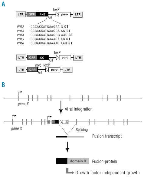
The open reading frame of a fusion between EGFP and the dimerization domain of ETV6 (PNT) was subcloned in the MSCV-puro vector. An artificial splice site (GTAAGT) was placed directly after the ETV6 part (Figure 1A). Three different constructs were generated to obtain 3 different phases at the end of the ETV6 ‘exon’, and two additional constructs with slightly different sequence at the end of the ‘exon’ were also constructed. One additional construct was generated in which the dimerization domain of ETV6 was replaced by the dimerization domain of EML1 (CC), and in another construct ETV6 was replaced by the myc-tag (Figure 1A). Viral vector production and transduction was performed as described before.8
Figure 1.
The retroviral insertion mutagenesis strategy. (A) Schematic representation of the different vectors used in this screen. (B) Generation of a fusion transcript as a consequence of retroviral insertion: the mechanism to induce activated kinases in this screening strategy.
After transduction, the retroviral vector will insert within the genome of the host cell and the dimerization domain will be expressed under control of the retroviral promotor (left LTR). When integrated into a gene, the splice donor site will enable splicing of the dimerization domain to a donor acceptor site of that gene, generating a fusion transcript with at the 5′ end the dimerization domain and at the 3′ end a part of the other gene (Figure 1B). A random insertion of the retrovirus throughout the genome of the Ba/F3 cells was aimed to identify proteins that could gain transforming capacity upon enforced dimerization. Two days after transduction of Ba/F3 cells with the retrovirus, the Ba/F3 cells were washed with PBS and plated in a 1.2% agar solution. After 2–3 weeks, IL3-independent Ba/F3 cell colonies became visible and were expanded individually for molecular analysis.
Cell culture
293T cells were grown in DMEM medium supplemented with 10% fetal bovine serum (FBS). Ba/F3 cells were grown in RPMI-1640 medium supplemented with 10% FBS and mouse IL3 (1 ng/mL). Growth curves were generated by plating Ba/F3 cells at 100,000 cells/mL one day after transduction in the absence of IL3. Growth was measured every two days until transformation was apparent. GFP-expression of the transduced Ba/F3 cells was measured on a flow cytometer (BD Biosciences) on day 0 and day 6 after IL3 withdrawal.
siRNA duplexes targeting ETV6 and GFP were purchased from Integrated DNA Technologies (sequences can be obtained upon request). One million cells were resuspended in 400 μL serum free medium and transferred to a 4mm cuvet (Biorad). siRNA duplex was added at a final concentration of 400 nM, mixed well and pulsed in a GenePulser Xcell™ (Biorad). Electroporation conditions were 300 V, 1000 μF and ∞ Ω. After electroporation, cells were transferred to 2 mL growth medium at 37°C. Viable cell numbers were determined at the day of electroporation and 48 h later using the Celltiter AQueousOne Solution (Promega).
Western Blotting
Western blot analysis was performed using NuPage 3 – 8% Tris-Acetate gels (Invitrogen). For this 2×10⁶ transformed Ba/F3 cells were used, lysed in cold lysis buffer containing 1% Triton X-100 and phosphatase inhibitors and reduced prior to loading. Antibodies used were anti-phosphotyrosine (4G10; Upstate Biotechnology), anti-GFP (Roche) and anti-mouse/anti-rabbit peroxidase-labeled antibodies (Amersham Biosciences).
Results and Discussion
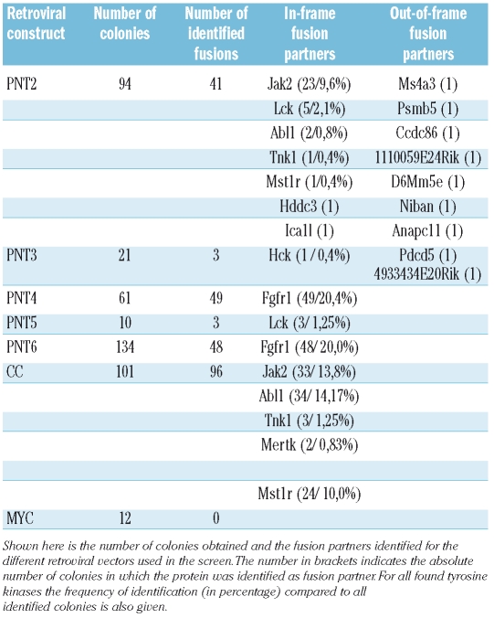
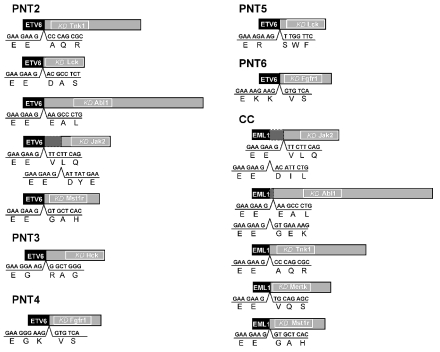
The produced retrovirus was used for different trans-ductions, generating in total 433 IL3-independent Ba/F3 cell colonies. Using 3′ RACE and subsequent sequence analysis of the PCR products, 240 fusion genes were identified that were expressed in the transformed Ba/F3 cells (Table 1). Of these, 231 contained fusions between ETV6 or EML1 and 8 different PTKs: Abl1, Fgfr1, Hck, Jak2, Lck, Mertk, Mst1r and Tnk1 (Figure 2, Table 1). For Jak2 and Abl1 two different breakpoints were identified in the Ba/F3 colonies (Figure 2). Since multiple colonies were picked-up with fusions involving Abl1, Jak2 and Fgfr1, we conclude that the screen was more or less saturating.
Table 1.
Results of the retroviral insertion mutagenesis screen.
Figure 2.
Schematic overview of identified fusion kinases. Summary of the different PTKs identified in the different screens, each based on the use of a specific retroviral construct. 3′-RACE followed by sequence analysis was used to determine the presence of a fusion gene involving the dimerization domain and to identify the partner gene. The ETV6 and EML1 dimerization domains are presented with the black box, the tyrosine kinase part present in the identified fusion and its kinase domain are shown with the gray and white boxes, respectively.
Only a limited amount of fusions with other, non-tyrosine kinase proteins, were identified, and in all cases these fusions were only identified once, suggesting they were most likely artifacts of the procedure. In addition, sequence analysis showed that most of them were out-of-frame fusions with ETV6. Although it remains possible that some of these fusions resulted in the activation or inactivation of genes leading to transformation of the Ba/F3 cells, we did not further investigate this because these events were not recurrently identified. In 193 colonies no fusion transcript was identified and in a fraction of these clones, we determined the genomic insertion site of the virus, indicating potential inactivated genes. Again, none of these insertion sites were identified more than once, suggesting that these events were unlikely to be the cause of Ba/F3 cell transformation.
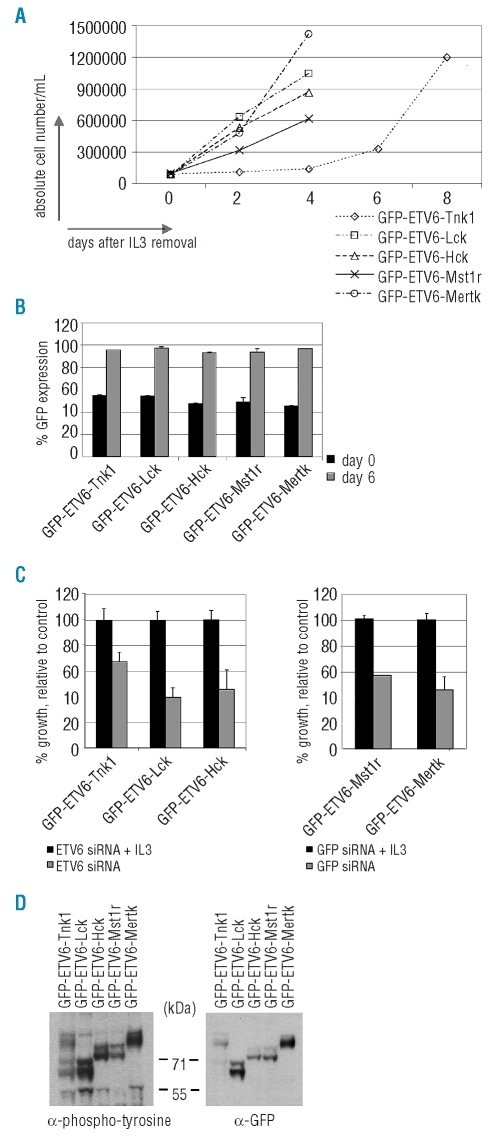
For the ETV6-kinase fusions, we confirmed that these fusions mediated the observed transformation of the Ba/F3 cells, by testing the transforming properties of ETV6-Tnk1, ETV6-Mertk, ETV6-Lck, ETV6-Hck and ETV6-Mst1r, in an independent experiment. The oncogenic properties of ABL1, JAK2 and FGFR1 have been well documented previously,1 and were not further evaluated in this study. Expression of ETV6-Tnk1, ETV6-Mertk, ETV6-Lck, ETV6-Hck and ETV6-Mst1r indeed transformed Ba/F3 cells, confirming the results of the screen. From Figure 3A it is evident that there is a slight difference in transforming properties of the different fusions with ETV6-Tnk1 clearly being the weakest oncogene. During transformation, the GFP positivity of the transduced Ba/F3 cells increased to over 90%, confirming the specific transformation of cells expressing the ETV6-kinase fusions (Figure 3B). To confirm that the Ba/F3 cells were transformed by the expression of the fusion constructs, we used siRNAs directed against GFP or ETV6 sequences to knock down the expression of the fusion kinases. For all Ba/F3 cells, we observed a decreased proliferation upon siRNA mediated knock down, clearly illustrating that the expression of the fusion kinases was the transforming factor (Figure 3C). By Western blot analysis we determined the phosphorylation status of the tyrosine kinases in these fusions. As shown in figure 3D, each of the tested fusion proteins (ETV6-Tnk1, ETV6-Mertk, ETV6-Lck, ETV6-Hck and ETV6-Mst1r) was phosphorylated on tyrosine residues, indicating autophosphorylation activity in the Ba/F3 cells.
Figure 3.
Transforming properties of the identified fusion kinases. To confirm the transforming properties of the identified fusion transcripts, the different fusions were generated and expressed in Ba/F3 cells. (A) The ability of each construct to induce transformation of Ba/F3 cells to IL3 independent proliferation was determined. (B) GFP-expression of the transduced Ba/F3 cells was measured on a flow cytometer on day 0 and day 6 after IL3 withdrawal. Transformed cells were confirmed to be GFP positive. (C) Using siRNA mediated knock down of ETV6 or GFP, a clear decrease in Ba/F3 cell proliferation is observed 48 h after electroporation. (D) Western blot analysis of whole cell lysates of the transduced Ba/F3 cells confirmed expression and phosphorylation of the fusion proteins.
In this study, 8 different PTKs were identified that upon activation by fusion with ETV6 can transform Ba/F3 cells. ABL1, FGFR1 and JAK2 are well known oncogenes, fused to a variety of partner genes including ETV6. For these proteins, it is already known that activation can be obtained by enforced homodimerization, and that this provides the proteins with transforming properties. For LCK, another oncogene identified in our screen, overexpression due to a chromosomal rearrangement has been described as a rare event in T-cell acute lymphoblastic leukemia (T-ALL), implicating LCK as an oncogene in the pathogenesis of T-ALL.9 Our data show that fusion of LCK to self-associating partner proteins can also lead to its activation.
In contrast to the well known oncogenic properties of ABL1, JAK2, FGFR1, and LCK, the role of MERTK, MST1R and HCK in the development of cancer is less clear. There is some evidence that expression of MERTK may be increased in T-ALL, and that ectopic MERTK expression can induce lymphomas in mice after long latency.10 Our data confirm that activation of Mertk can indeed stimulate proliferation and survival of hematopoietic cells. MST1R (RON), a PTK closely related to the proto-oncogene MET, was shown to be over-expressed in a variety of tumors, and also alternative splicing of the MST1R transcript may play a role in tumor development.11 It is becoming clear that MET and MST1R both play a major role in driving metastasis of tumor cells, and for example increased expression of MET and MST1R in breast carcinoma clearly correlates with reduced disease-free survival.12 It was previously shown that MST1R can be activated by missense mutations in the kinase domain or truncating mutations in its c-terminal region.13,14 Here we show that MST1R can also be activated by fusion to a homodimerization domain, and that this leads to the stimulation of proliferation and survival pathways. HCK belongs to the SRC kinase family and different reports suggest a role for HCK in the oncogenic signaling pathways downstream of BCR-ABL1.15
Finally, we also demonstrate that enforced dimerization of TNK1 can lead to growth factor independent growth of Ba/F3 cells. TNK1 is a member of the ACK family of non-RTKs and was originally cloned from CD34+/Lin−/CD38− hematopoietic progenitor cells.16 The signaling pathways induced by TNK1 remain largely unknown, although it was recently suggested that activation of TNK1 enables TNFα-induced apoptosis by selectively inhibiting TNFα-induced activation of NF-κB.17 Apart from this function, TNK1 was also shown to play a negative regulatory role in the Ras-Raf1-MAPK pathway.18 The identification of TNK1 in our retroviral insertion screen indicates that activation of TNK1 can play a dominant role in oncogenic transformation, and suggests that TNK1 is also capable of stimulating proliferation and survival pathways. Our attempt to identify randomly generated TNK1 point mutants able to transform Ba/F3 cells gave no results (data not shown).
In the Mitelman catalog, numerous cases are described with chromosomal aberrations in the region of the genes encoding LCK (1p34), MERTK (2q14), MST1R (3p21), HCK (20q11) and TNK1 (17p13). In acute myeloid leukemia (AML), deletions involving the region 3p21 or 20q11, and translocations involving 17p13 have been frequently reported. These findings may suggest the involvement of MST1R, HCK or TNK1 in AML. In contrast, no recurrent aberrations were identified for the regions 2q14 and 1p34. Similar results were obtained for acute lymphoblastic leukemia, although aberrations for the 3p21 region were more rare. The implication of one of these 5 tyrosine kinases in the reported aberrations remains to be determined. These data together with their reported expression in different malignancies and leukemic cell lines, and our own results suggest the potential involvement of these kinases in cancer pathogenesis and underscores the clinical relevance of our findings.
In a direct cloning strategy, different PTK kinase domains were fused to the ETV6 dimerization domain of the retroviral vector used for our insertion screen and their oncogenic potential was evaluated in the Ba/F3 cell line. For 19 of the 40 tested kinases, growth factor independent growth of Ba/F3 cells was observed, specifically related to constitutive kinase activity, suggesting many more PTKs have transforming potential upon enforced dimerization (data not shown). These results are in sharp contrast with the 8 PTKs identified in the unbiased retroviral insertional mutagenesis screen. Although repetitive identification of Abl1, Jak2 and Fgfr1 suggested a saturation of the screen, we were unable to pick-up the vast majority of PTKs with this strategy, including known oncogenic PTKs such as Kit, Flt3, Pdgfrα and Pdgfrβ. The reason why we did not obtain fusions with some of the known oncogenic PTKs, remains unknown, but is most likely due to the lack of integration of the virus into these genes.19
We describe here a novel retroviral insertion mutagenesis screen, which results in the generation of fusion genes consisting of a part of the viral vector and part of a cellular gene. We used the Ba/F3 cell line for our screen in order to identify fusion genes that could transform the cells to IL3 independent growth. Variant screens with different cells, different read-out or even different protein domains in the retroviral vector should also be possible. With our screen, we showed that MERTK, MST1R, HCK and TNK1 can be activated by fusion to the dimerization domain of ETV6, and that activation of these PTKs can stimulate proliferation and survival pathways.
Footnotes
Authorship and Disclosures
EL, HVM, and EB performed experiments and analyzed data; EL and JC designed the study and wrote the manuscript.
The authors reported no potential conflicts of interest.
Funding: E.L. is an Aspirant of the FWO-Vlaanderen. This work was supported by grants from the FWO-Vlaanderen, Belgian Federation against Cancer, European Research Council, and the K.U.Leuven.
References
- 1.Krause DS, Van Etten RA. Tyrosine kinases as targets for cancer therapy. N Engl J Med. 2005;353:172–87. doi: 10.1056/NEJMra044389. [DOI] [PubMed] [Google Scholar]
- 2.McWhirter JR, Galasso DL, Wang JY. A coiled-coil oligomerization domain of Bcr is essential for the transforming function of Bcr-Abl oncoproteins. Mol Cell Biol. 1993;13:7587–95. doi: 10.1128/mcb.13.12.7587. [DOI] [PMC free article] [PubMed] [Google Scholar]
- 3.Carroll M, Tomasson MH, Barker GF, Golub TR, Gilliland DG. The TEL/platelet-derived growth factor beta receptor (PDGF beta R) fusion in chronic myelomonocytic leukemia is a transforming protein that self-associates and activates PDGF beta R kinase-dependent signaling pathways. Proc Natl Acad Sci USA. 1996;93:14845–50. doi: 10.1073/pnas.93.25.14845. [DOI] [PMC free article] [PubMed] [Google Scholar]
- 4.Soda M, Choi YL, Enomoto M, Takada S, Yamashita Y, Ishikawa S, et al. Identification of the transforming EML4-ALK fusion gene in non-small-cell lung cancer. Nature. 2007;448:561–6. doi: 10.1038/nature05945. [DOI] [PubMed] [Google Scholar]
- 5.Smedley D, Demiroglu A, bdul-Rauf M, Heath C, Cooper C, Shipley J, et al. ZNF198-FGFR1 transforms Ba/F3 cells to growth factor independence and results in high level tyrosine phosphorylation of STATS 1 and 5. Neoplasia. 1999;1:349–55. doi: 10.1038/sj.neo.7900035. [DOI] [PMC free article] [PubMed] [Google Scholar]
- 6.Mandanas RA, Boswell HS, Lu L, Leibowitz D. BCR/ABL confers growth factor independence upon a murine myeloid cell line. Leukemia. 1992;6:796–800. [PubMed] [Google Scholar]
- 7.Cools J, DeAngelo DJ, Gotlib J, Stover EH, Legare RD, Cortes J, et al. A tyrosine kinase created by fusion of the PDGFRA and FIP1L1 genes as a therapeutic target of imatinib in idiopathic hypereosinophilic syndrome. N Engl J Med. 2003;348:1201–14. doi: 10.1056/NEJMoa025217. [DOI] [PubMed] [Google Scholar]
- 8.Schwaller J, Frantsve J, Aster J, Williams IR, Tomasson MH, Ross TS, et al. Transformation of hematopoietic cell lines to growth-factor independence and induction of a fatal myelo- and lymphoproliferative disease in mice by retrovirally transduced TEL/JAK2 fusion genes. EMBO J. 1998;17:5321–33. doi: 10.1093/emboj/17.18.5321. [DOI] [PMC free article] [PubMed] [Google Scholar]
- 9.Majolini MB, Boncristiano M, Baldari CT. Dysregulation of the protein tyrosine kinase LCK in lymphoproliferative disorders and in other neoplasias. Leuk Lymphoma. 1999;35:245–54. doi: 10.3109/10428199909145727. [DOI] [PubMed] [Google Scholar]
- 10.Keating AK, Salzberg DB, Sather S, Liang X, Nickoloff S, Anwar A, et al. Lymphoblastic leukemia/lymphoma in mice overexpressing the Mer (MerTK) receptor tyrosine kinase. Oncogene. 2006;25:6092–100. doi: 10.1038/sj.onc.1209633. [DOI] [PubMed] [Google Scholar]
- 11.Camp ER, Liu W, Fan F, Yang A, Somcio R, Ellis LM. RON, a tyrosine kinase receptor involved in tumor progression and metastasis. Ann Surg Oncol. 2005;12:273–81. doi: 10.1245/ASO.2005.08.013. [DOI] [PubMed] [Google Scholar]
- 12.Lee WY, Chen HH, Chow NH, Su WC, Lin PW, Guo HR. Prognostic significance of co-expression of RON and MET receptors in node-negative breast cancer patients. Clin Cancer Res. 2005;11:2222–8. doi: 10.1158/1078-0432.CCR-04-1761. [DOI] [PubMed] [Google Scholar]
- 13.Santoro MM, Penengo L, Minetto M, Orecchia S, Cilli M, Gaudino G. Point mutations in the tyrosine kinase domain release the oncogenic and metastatic potential of the Ron receptor. Oncogene. 1998;17:741–9. doi: 10.1038/sj.onc.1201994. [DOI] [PubMed] [Google Scholar]
- 14.Yokoyama N, Ischenko I, Hayman MJ, Miller WT. The C terminus of RON tyrosine kinase plays an autoinhibitory role. J Biol Chem. 2005;280:8893–900. doi: 10.1074/jbc.M412623200. [DOI] [PubMed] [Google Scholar]
- 15.Lionberger JM, Wilson MB, Smithgall TE. Transformation of myeloid leukemia cells to cytokine independence by Bcr-Abl is suppressed by kinase-defective Hck. J Biol Chem. 2000;275:18581–5. doi: 10.1074/jbc.C000126200. [DOI] [PubMed] [Google Scholar]
- 16.Hoehn GT, Stokland T, Amin S, Ramirez M, Hawkins AL, Griffin CA, et al. Tnk1: a novel intracellular tyrosine kinase gene isolated from human umbilical cord blood CD34+/Lin−/ Oncogene. 1996;12:903–13. [PubMed] [Google Scholar]
- 17.Azoitei N, Brey A, Busch T, Fulda S, Adler G, Seufferlein T. Thirty-eight-negative kinase 1 (TNK1) facilitates TNFα-induced apoptosis by blocking NF-κB activation. Oncogene. 2007;26:6536–45. doi: 10.1038/sj.onc.1210476. [DOI] [PubMed] [Google Scholar]
- 18.Hoare K, Hoare S, Smith OM, Kalmaz G, Small D, Stratford MW. Kos1, a nonreceptor tyrosine kinase that suppresses Ras signaling. Oncogene. 2003;22:3562–77. doi: 10.1038/sj.onc.1206480. [DOI] [PubMed] [Google Scholar]
- 19.Lewinski MK, Yamashita M, Emerman M, Ciuffi A, Marshall H, Crawford G, et al. Retroviral DNA integration: viral and cellular determinants of target-site selection. PLoS Pathog. 2006;2:e60. doi: 10.1371/journal.ppat.0020060. [DOI] [PMC free article] [PubMed] [Google Scholar]


