Abstract
Replication of alphaviruses strongly depends on the promoters located in the plus- and minus-strands of virus-specific RNAs. The most sophisticated promoter is encoded by the 5′ end of the viral genome. This RNA sequence is involved in initiation of translation of viral nsPs, and synthesis of both minus- and plus-strands of the viral genome. Part of the promoter, the 51-nt conserved sequence element (CSE), is located in the nsP1-coding sequence, and this limits the spectrum of possible mutations that can be performed. We designed a recombinant Venezuelan equine encephalitis virus genome, in which the promoter and nsP1-coding sequences are separated. This modification has allowed us to perform a wide variety of genetic manipulations, without affecting the amino-acid sequence of the nsPs, and to further investigate 51-nt CSE functioning. The results of this study suggest a direct interaction of the amino terminal domain of nsP2 with the 5′ end of the viral genome.
Keywords: RNA replication, 51-nt CSE, Venezuelan equine encephalitis virus
INTRODUCTION
The alphavirus genus of the Togaviridae family contains almost 30 members, some of which are important human and animal pathogens (Griffin, 2001; Strauss and Strauss, 1994). Alphaviruses are widely distributed on all continents except in the Arctic and Antarctic areas. They efficiently replicate both in mosquito vectors and vertebrate hosts. However, in mosquitoes they cause a persistent, lifelong infection that does not noticeably affect biological functions of the vectors, while in vertebrate hosts, the infection is acute and characterized by high-titer viremia, rash and fever, until the virus is cleared by the immune system. The New World alphaviruses can cause severe encephalitis in humans and animals that can result in death or neurological disorders (Dal Canto and Rabinowitz, 1981; Griffin, 2001; Johnston and Peters, 1996; Leon, 1975). These encephalitogenic alphaviruses include Venezuelan (VEEV), eastern (EEEV) and western equine encephalitis (WEEV) viruses and represent a serious public health threat in the US (Rico-Hesse et al., 1995; Weaver and Barrett, 2004; Weaver et al., 1994; Weaver et al., 1996). They continue to circulate in the Central, South and North Americas and cause severe, and sometimes fatal disease in humans and horses. During VEEV epizootics, equine mortality due to encephalitis can reach 83%, and in humans, while the overall mortality rate is below 1%, neurological disease including disorientation, ataxia, mental depression, and convulsions, can be detected in up to 14% of all infected individuals, especially children (Johnson and Martin, 1974). Sequelae of VEEV-related clinical encephalitis in humans and rats are also described (Garcia-Tamayo, Carreno, and Esparza, 1979; Leon, 1975). In spite of the continuous threat of VEEV epidemics, the biology of this virus and the mechanism of its replication are insufficiently understood.
The VEEV genome is represented by a ca. 11.5 kb-long, single-stranded RNA of positive polarity (Strauss, Rice, and Strauss, 1984), which mimics the structure of the cellular mRNAs, in that it contains a 5′ cap and poly(A)-tail at the 5′ and 3′ ends, respectively. The genome contains two polyprotein-coding sequences. The first, a 7,500 nt-long, 5′-terminal open reading frame (ORF) is translated into viral nonstructural proteins (nsP1–4) that form, together with the cellular proteins, the enzyme complex required for genome replication and transcription of the subgenomic RNA. The latter RNA encodes the second polyprotein that is co- and post-translationally processed into viral structural proteins (capsid, E2 and E1) that form infectious viral particles.
The replication of the alphavirus genome is a multi-step process that begins with synthesis of a full-length, minus-strand RNA intermediate that, in turn, serves as a template for synthesis of the plus-strand viral genomes and transcription of the subgenomic RNA. Synthesis of these RNA species is a highly synchronized process regulated by differential processing of the viral nsPs (Lemm and Rice, 1993; Lemm et al., 1994; Shirako and Strauss, 1994). At the early stages of viral replication, the ns polyprotein is partially processed by nsP2-associated protease into P123 and nsP4, an enzyme complex capable of minus-, but not plus-strand RNA synthesis. Then, after further processing of the polyproteins into nsP1+P23+nsP4, the intermediate polymerase functions in synthesis of both plus and minus genome-sized RNA, but appears to be unable to efficiently use the internal promoter for 26S mRNA synthesis. Finally, after complete P1234 processing to individual nsP1–4, the replication complex (RC) efficiently (Wang, Sawicki, and Sawicki, 1994) synthesizes the plus-strand viral genome and subgenomic RNA, but is no longer capable of minus-strand synthesis (Lemm et al., 1998; Lemm et al., 1994; Sawicki and Sawicki, 1987). Thus, the early and mature RCs most likely utilize different RNA promoters located in the plus- and minus-strands of virus-specific RNAs.
The critical element of the promoter for minus-strand RNA synthesis is a 19-nt-long, conserved sequence element (CSE) adjacent to the poly(A) and located at the 3′ end of the viral genome (Hardy, 2006; Hardy and Rice, 2005; Kuhn, Hong, and Strauss, 1990). The mutations in this sequence have a deleterious effect on alphavirus replication. However, it can be replaced by an artificial AU-rich sequence that might efficiently function in RNA synthesis (Raju et al., 1999). It was also demonstrated that the presence of poly(A) at the 3′-terminus and cap at the 5′ terminus strongly stimulate the RNA replication (Hardy and Rice, 2005). Moreover, the sequence of the 5′UTR determines the minus-strand RNA synthesis as well (Frolov, Hardy, and Rice, 2001; Gorchakov et al., 2004), and these facts strongly suggest the importance of the 5′-3′ end interaction in the RNA replication, which most likely proceeds through formation of the translation initiation complex. The subgenomic promoter required for transcription of the subgenomic RNA is well defined (Levis, Schlesinger, and Huang, 1990; Ou, Strauss, and Strauss, 1983; Wielgosz, Raju, and Huang, 2001) and represented by a 24-nt CSE located in the minus-strand genome intermediate. The latter CSE covers not only the nucleotides adjacent to the start of the subgenomic RNA, but the first nucleotides of the subgenomic RNA RNA as well. This promoter is recognized in vitro by the protein complex containing all of the viral nsPs (Li and Stollar, 2004).
The most sophisticated promoter of RNA synthesis is encoded by the 5′ end of the alphavirus genome. Its functioning was intensively studied in the context of Sindbis virus (SINV) (Fayzulin and Frolov, 2004; Gorchakov et al., 2004; Niesters and Strauss, 1990a; Niesters and Strauss, 1990b; Strauss and Strauss, 1994), but is still poorly understood. The 5′ terminus appears to contain two elements: the 5′-terminal sequence, encoded by the 5′UTR (that appears to represent a core promoter) and a 51-nt CSE, found in the nsP1-coding sequence that might be a replication enhancer, functioning in a virus- and cell-dependent manner (Fayzulin and Frolov, 2004; Niesters and Strauss, 1990b). The problem with investigation of the 5′ promoter lies in the involvement of its sequence in initiation of translation of viral nsPs, and synthesis of both minus- and plus-strands of the viral genome. In addition, part of the promoter, the 51-nt CSE is located in the nsP1-coding sequence, and this strongly limits a spectrum of mutations that can be done without affecting nsP1 functioning.
We considered these difficulties when we undertook the VEEV promoter investigation and designed a VEEV genome, in which the promoter sequence and the nsP1-coding sequence are separated. This modification allowed us to both perform a wide variety of genetic manipulations without affecting the amino acid sequence of the nsPs, and study the function of 51-nt CSE in virus replication. The results of this study suggest a direct interaction of the amino terminal domain of VEEV nsP2 with the 5′ end of the viral RNA and a possibility of applying a similar approach in studying the 5′-terminal promoter functioning in replication of other RNA-positive viruses.
RESULTS
Recombinant viruses with the 5′ promoter sequences located outside of the nsPs’ ORF
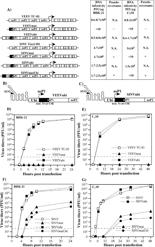
In our initial experiments, we attempted to develop an experimental system to study the effect of extended genetic manipulations in the 5′-terminal alphavirus genome elements without affecting the amino acid (a.a.) sequence of the nsPs. Thus, we separated the 5′ end-specific promoter and the nsP-coding sequence in the context of the VEEV TC-83 genome. To achieve this, we introduced 95 mutations into the first 300 nt of the nsP1-coding gene of VEEVmut (Fig. 1A), and these mutations did not change the encoded protein sequence. However, based on computer predictions, they destroyed the original secondary structure of the fragment, including the secondary structure of the 51-nt CSE. After transfection of the in vitro-synthesized RNA into BHK-21 cells, no cytopathic effect (CPE) was detected and no infectious virus was recovered even after following blind passages of the harvested media on the naïve BHK-21 cells (data not shown). This indicated that the introduced mutations and, most likely, the destabilization of the secondary structure made the genome RNA incapable of replication, and neither the reverting nor pseudoreverting mutations accumulated in the VEEVmut genome.
FIG. 1.
Replication of VEEV and SINV viruses with modified 5′ ends of the genome in BHK-21 and C710 cells. (A) Schematic representation of VEEV and SINV genomes with modified 5′ ends, and infectivities of the in vitro-synthesized viral RNAs in the infectious center assay. Solid boxes indicate the nsP1 sequence containing clustered silent mutations. Arrows indicate position of the subgenomic promoter. N.A. indicates “not applicable,” because of the wt phenotype of the designed mutants. (B and C) Schematic representation of the 5′ ends of VEEVubi and SINVubi genomes, respectively. Solid boxes indicate nsP1 sequence containing clustered silent mutations. Arrows indicate positions of the initiating AUG in the computer-predicted secondary structure and starting AUG of the nsP1–4 polyprotein. (D and E, F and G) Single-step viral growth curves after electroporation of 3 μg of in vitro-synthesized RNAs into BHK-21 or C710 cells. At the indicated times, the medium was replaced, and virus titers were determined in BHK-21 cells, as described in Materials and Methods. The dashed lines indicate the limit of detection.
In the next step, we cloned a 347-nt-long, 5′-terminal fragment of the VEEV TC-83 genome into the VEEVmut genome upstream from the initiating AUG (see Fig. 1A and B for details). In the VEEVubi, fusion of the cloned sequence and the nsP1 was performed through the ubiquitin (Ubi) gene to preserve synthesis of the following nsP1 protein in its natural form, including the first methionine. Thus, the ORF started from the AUG located downstream from the 5′UTR and continued through the cloned fragment of nsP1 and Ubi into the P1234 polyprotein.
The in vitro-synthesized recombinant viral genome had the same infectivity as did the in vitro-synthesized genome of VEEV TC-83 (Fig. 1A). The harvested virus formed plaques that were of the same size as those formed by VEEV TC-83 and replicated at essentially the same rate as did the latter wt virus in both BHK-21 and mosquito C710 cells (Figs. 1D and E). Taken together, these data indicated that by cloning a duplicate of the wt 5′ end, we restored the virus’ ability to efficiently replicate. The recombinant genome could be further used for extended deletions and insertions in the promoter region, including the 51-nt CSE without affecting the amino acid sequence of nsP1. Most importantly, we attained a possibility of deleting the predicted, promoter-specific stem-loops without causing deleterious changes in the secondary structure of the entire 5′ end. The only restriction that had to be met was conservation of the ORF during modifications: the translation had to start on the initiating AUG and continue into P1234 through the Ubi.
Interestingly, we were unable to prevent SINV replication by introducing a similar set of mutations into the beginning of the nsP1-coding sequence. In spite of the presence of 97 silent clustered mutations that completely destroyed the secondary structure of the 5′ end of SINV genome, the designed SINVmut (see Figs. 1A and B for details) was capable of replication in both BHK-21 and C710 cells albeit to dramatically lower titers (Figs. 1F and G). It formed heterogeneous plaques, suggesting an accumulation of the adaptive mutations that increased the replication rates. After inserting the wt 5′-terminal SINV genome sequence and Ubi gene upstream from the mutated nsP1, the recombinant virus (SINVubi) became indistinguishable from the wt SINV Toto1101, and, as expected, the insertion of the mutated nsP1 sequence containing the same 97 silent mutations did not increase the rate of virus (SINVmutUbi) replication (Figs. 1A, C, F and G). These data indicated that the nucleotide sequence located downstream from the 5′-terminal stem-loops 1 and 2 (and including the 51-nt CSE) plays a critical role in VEEV replication, but has a less significant role in replication of SINV. The latter virus is capable of replication even if this sequence contains clustered mutations that destroy the secondary structure of more than 300-nt-long 5′-terminal fragment of the viral genome. The residual activity of the 5′UTR-associated basal promoter appears to be sufficient for replication of the mutated virus at detectable levels.
The stem-loops in the VEEV 51-nt CSE have redundant functions in RNA replication
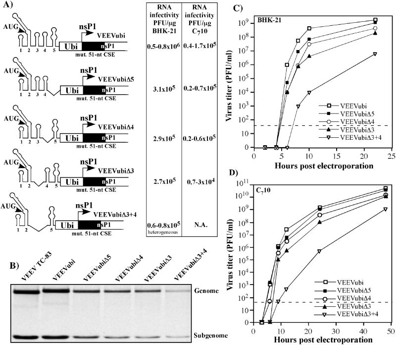
The development of VEEVubi opened an opportunity to perform extended mutagenesis of the 5′-terminal promoter elements in the viral genome by deleting the predicted particular structural elements without affecting the others. We provisionally divided the VEEV 5′ terminus into 5 elements that represented 1) the very 5′-terminal stem-loop (nt 4–26); 2) the large stem-loop containing the initiating AUG (nt 36–126); 3) and 4) two small stem-loops containing the 51-nt CSE (nt 133–158 and 162–184); and 5) the 162 nt-long sequence located between the 51-nt CSE and Ubi. All of the deletions were done according to this prediction. They were designed to save the ORF starting at nt 45, and the stem-loops 1 and 2 were not deleted in any constructs to conserve the efficiency of nsPs translation at the level of the wt virus genome (however, the translation efficiency was not experimentally evaluated).
The deletion of elements 3 or 4, or 5 did not have a deleterious effect on virus replication. In the infectious center assay, the in vitro-synthesized viral RNAs were as infectious as that of VEEVubi (Fig. 2A). However, there was a noticeable decrease in RNA replication (Fig. 2B) and slower rates of infectious virus release after transfection of both BHK-21 and C710 cells (Figs. 2C and D). Moreover, the deletion of stem-loop 3 had the strongest effect on virus production. At any time post transfection, titers of VEEVubiΔ3 were 10- to 100-fold lower than were those of VEEVubi. Thus, these three elements were involved in VEEV replication, but likely had redundant functions, and the presence of any two of them in the viral genome was sufficient for supporting efficient replication.
FIG. 2.
Replication of VEEVubi deletion mutants in BHK-21 and C710 cells. (A) Schematic representation of VEEVubi deletion mutants genomes and infectivities of the in vitro-synthesized viral RNAs in the infectious center assay. Solid boxes indicate the nsP1 sequence containing clustered silent mutations. N.A. indicates “not applicable,” because the mutant was incapable of forming plaques in C710 cells. (B) Synthesis of virus-specific RNAs in the transfected BHK-21 cells. At 2 h post transfection with 3 μg of in vitro-synthesized RNA, viral RNAs were metabolically labeled with [3H]uridine for 6h, as described in Materials and methods, and analyzed by agarose gel electrophoresis. Positions of the genomic and subgenomic RNAs are indicated. (C and D) Single-step viral growth curves after electroporation of 3 μg of in vitro-synthesized RNAs into BHK- 21 and C710 cells. At the indicated times, the medium was replaced and virus titers determined in BHK-21 cells, as described in Materials and Methods. The dashed lines indicate the limit of detection.
However, deletion of the entire 51-nt CSE represented by stem-loops 3 and 4 downregulated the genome RNA replication in BHK-21 cells more than 10-fold to a barely detectable level (Fig. 2B). The designed virus, VEEVubiΔ3+4, demonstrated a high rate of evolution and formed heterogeneous plaques immediately after transfection of RNA into BHK-21 and C710 cells. It was difficult to assess virus replication rates (Figs. 2C and D), because heterogeneity of plaques formed by viruses in the harvested samples suggested presence of multiple variants. In addition, VEEVubiΔ3+4 was incapable of causing any CPE in C710 cells at all. Based on the development of plaques that strongly differed in size, we assumed the growth curves demonstrated in Fig. 2C and D did not represent replication of the original construct, but more likely the replication of the arising pseudorevertants. The restoration of the 51-nt CSE was a highly unlikely event, and, therefore, we expected to the viruses to contain pseudoreverting mutations in the genome fragments other than the 5′ end, most likely in the nsP genes, whose products form the replication complex.
VEEVubiΔ3+4 accumulates adaptive mutations
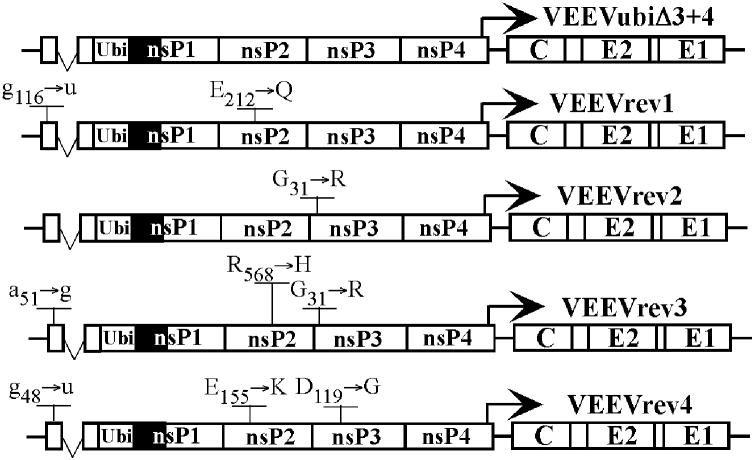
VEEVubiΔ3+4 harvested after transfection of BHK-21 cells was passaged twice in the same cells at an MOI of ~10 PFU/cell to increase the concentration of the efficiently replicating variants in the samples. To avoid isolation of the same variants, stocks from two different transfection experiments were passaged independently. Then large individual viral plaques were randomly selected and the entire ns polyprotein-coding sequence and the 3′ and 5′ ends of the genomes were sequenced. The putative adaptive mutations were identified in all of the isolates. They were found i) in stem-loop 2 downstream of the initiating AUG or in the initiating AUG, ii) in the nsP2 and iii) in the nsP3 (Fig. 3). It is highly unlikely that the small fragment of nsP1 encoded upstream of the Ubi is involved in the replication complex formation or functioning, and, therefore, the stem-loop 2-specific mutations are presented as nucleotide, but not as a.a. changes. These mutations affected the nucleotide sequence and the secondary structure of stem-loop 2 and, thus, could be involved in modulation of the promoter functioning (see following sections for details). Other mutations changed the amino acids in the nsP2 and nsP3, but their effect had to be evaluated in reverse genetics experiments.
FIG. 3.
Mutations found in the VEEVubiΔ3+4 plaque isolates that became capable of efficient replication in BHK-21 cells. The nucleotide changes in the stem-loop 2 are indicated by lower-case letters. The a.a. changes identified in nsP2 and nsP3 are indicated by upper-case letters.
The adaptive mutations have synergistic effect on viral RNA replication
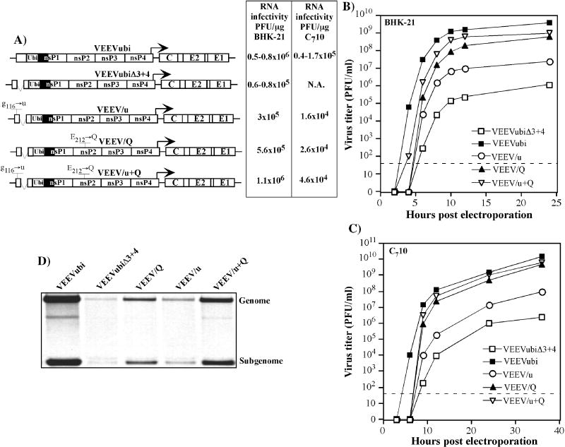
Sequencing of the genome of efficiently replicating VEEVrev1 revealed a g116→u mutation in the 5′ end and another mutation, E212→Q, in the nsP2. To understand the effect of these mutations on virus replication, they were transferred either separately or together into the VEEVubiΔ3+4 genome (Fig. 4A). The in vitro-synthesized RNAs were transfected into BHK-21 and C710 cells, and the RNA infectivity was assessed in the infectious center assay (Fig. 4A). We also examined virus replication in both transfected cell cultures and viral RNA replication in BHK-21 cells (Figs. 4B and C). The mutation in nsP2 had a strong stimulatory effect on virus growth. The effect of g116→u replacement was less obvious, but it also made titers of the released virus 20- to 100-fold higher, compared to the original VEEVubiΔ3+4 at any time post transfection. However, the results suggest that when combined, the effects of these two mutations are additive and, thus, likely affect different processes (Fig. 4D).
FIG. 4.
Replication of VEEVubiΔ3+4 variants, containing the mutations identified in the efficiently replicating VEEVrev1 pseudorevertant, in BHK-21 and C710 cells. (A) Schematic representation of VEEVubiΔ3+4 genomes with different mutations and infectivities of the in vitro-synthesized viral RNAs in the infectious center assay. Solid boxes indicate the nsP1 sequence containing clustered silent mutations. N.A. indicates “not applicable,” because the mutant was incapable of forming plaques in C710 cells. (B and C) Single-step virus growth curves after electroporation of 3 μg of in vitro- synthesized RNAs into BHK-21 or C710 cells. At the indicated times, the medium was replaced and virus titers were determined in BHK-21 cells, as described in Materials and Methods. The dashed lines indicate the limit of detection. (D) Synthesis of virus-specific RNAs in the transfected BHK-21 cells. At 2 h post transfection, viral RNAs were metabolically labeled with [3H]uridine for 6h, as described in Materials and Methods, and analyzed by agarose gel electrophoresis. Positions of the genomic and subgenomic RNAs are indicated.
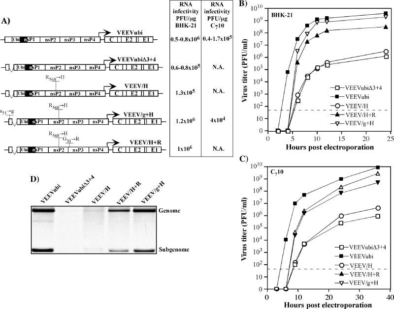
Other two pseudorevertants, VEEVrev2 and VEEVrev3, had a common mutation G31→R in nsP3 (Fig. 3), but in the VEEVrev2, this was the only mutation found, and VEEVrev3 developed two additional mutations a51→g in the 5′ terminus and R568→H in the nsP2. The effect of these mutations was tested by cloning their combinations into VEEVubiΔ3+4 (Fig. 5). In contrast to VEEVrev1, the mutation in nsP2 had no positive effect either on RNA or virus replication, indicating that this was likely a neutral change. However, both the 5′ end- and nsP3-specific mutations strongly enhanced the virus’ replication, because of a more efficient synthesis of viral genome and transcription of the subgenomic RNA. Taken together these data suggested that the negative effect of the deletion of the replication enhancer in the VEEV genome can be neutralized by compensatory mutations in the amino terminal domains of nsP2 or nsP3, and the mutations in the RNA stem located in the beginning of the ns ORF (stem-loop 2) can additionally stimulate virus replication. Moreover, the stimulatory effect of the mutations might be determined by different mechanisms.
FIG. 5.
Replication of VEEVubiΔ3+4 variants, containing the mutations identified in efficiently replicating VEEVrev2 and 3 pseudorevertants, in BHK-21 and C710 cells. (A) Schematic representation of VEEVubiΔ3+4 genomes with different mutations and infectivities of the in vitro-synthesized viral RNAs in the infectious center assay. Solid boxes indicate the nsP1 sequence containing clustered silent mutations. N.A. indicates “not applicable,” because the mutant was incapable of forming plaques in C710 cells. (B and C) Single-step virus growth curves after electroporation of 3 μg of in vitro- synthesized RNAs into BHK-21 or C710 cells. At the indicated times, the medium was replaced and virus titers were determined in BHK-21 cells, as described in Materials and Methods. The dashed lines indicate the detection limit. (D) Synthesis of virus-specific RNAs in the transfected BHK-21 cells. At 2 h post transfection, viral RNAs were metabolically labeled with [3H]uridine for 6h, as described in Materials and Methods, and analyzed by agarose gel electrophoresis. Positions of the genomic and subgenomic RNAs are indicated.
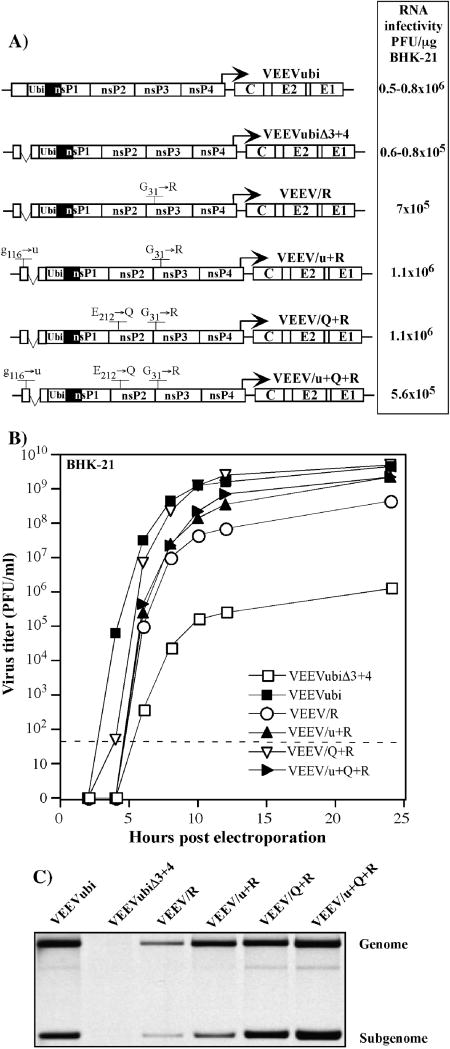
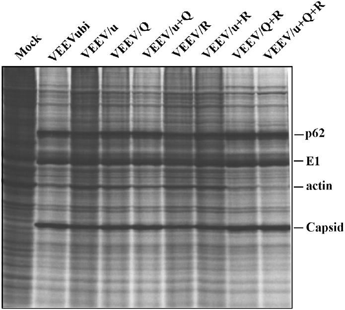
To further test this hypothesis, we cloned into the VEEVubiΔ3+4 genome combinations the adaptive mutations found in different plaque isolates (VEEVrev2 and VEEVrev4). Variants containing only nsP2- and nsP3-specific mutations (E212→Q and G31→R, respectively) or all of the mutations together (E212→Q, G31→R and g116→u) replicated in BHK-21 cells as efficiently as did the VEEVubi; however, a 1- to 2-h-long delay in virus release was still noticeable at the early times, within the first 4 h post transfection (Fig. 6A and B). However, the finding of the same infectivity for the in vitro-synthesized mutant and VEEVubi RNAs, as well as the homogeneity of plaque size in the in the infectious center assay indicate that further evolution either does not proceed or does not have a critical effect on virus replication. The cumulative effect of the mutations was readily detectable in the analysis of RNA and protein synthesis in the infected cells (Figs. 6C and 7). Combining the nsP2- and nsP3-specific mutations in the same genome increased viral RNA synthesis to the level that we detected for the VEEVubi. The VEEV/Q+R and VEEV/u+Q+R produced viral structural proteins and interfered with cellular protein synthesis as efficiently as did VEEVubi, having an intact 51-nt CSE. The inhibition of cellular translation was even higher than that detected in the cells infected with VEEV/u+Q or VEEV/R mutants. These results strongly indicated that the nsP2- and nsP3-specific mutations functioned independently and could synergistically stimulate RNA replication and, consequently, virus growth.
FIG. 6.
Replication of VEEVubiΔ3+4 variants, containing combinations of mutations identified in efficiently replicating pseudorevertants, in BHK-21 and C710 cells. (A) Schematic representation of VEEVubiΔ3+4 genomes with different mutations and infectivities of the in vitro-synthesized viral RNAs in the infectious center assay. Solid boxes indicate nsP1 sequence containing clustered silent mutations. (B) Single-step viral growth curves after electroporation of 3 μg of in vitro-synthesized RNAs into BHK-21 cells. At the indicated times, the medium was replaced and virus titers were determined in BHK-21 cells, as described in Materials and Methods. The dashed line indicates the detection limit. (C) Synthesis of virus-specific RNAs in the transfected BHK-21 cells. At 2 h post transfection, viral RNAs were metabolically labeled with [3H]uridine for 6h, as described in Materials and Methods, and analyzed by agarose gel electrophoresis. Positions of the genomic and subgenomic RNAs are indicated.
FIG. 7.
Protein synthesis in BHK-21 cells infected with different variants of VEEVubiΔ3+4. Cells were infected with the viruses indicated at an MOI of 10 PFU/cell. At 20 h post infection, they were labeled with [35S]methionine and analyzed in a sodium dodecyl sulfate-10% polyacrylamide gel as described in Materials and Methods. Positions of VEEV-specific structural proteins and actin are indicated.
The adaptive mutations have positive effect on replication of the VEEVubi
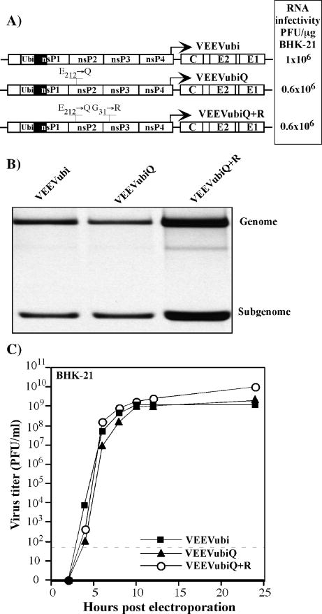
In the final experiments, we tested whether the identified adaptive mutations function only in the context of a deleted 51-nt CSE or if they have a stimulatory effect on replication of viruses having the intact CSE as well. Therefore, we cloned either E212→Q in nsP2 or both G31→R in nsP3 and E212→Q in nsP2 into the VEEVubi genome (Fig. 8A). After transfection of BHK-21 cells with the in vitro-synthesized RNA, we found the double mutant to cause faster CPE, to synthesize more virus-specific RNA (Fig. 8B) and, accordingly, to grow to almost 10-fold higher titers than the original VEEVubi (Fig. 8C). Thus, the mutations were capable of positively affecting ns proteins’ functions not only in the context of the 51-nt CSE deletion mutant, but also in the viruses having no changes in the 5′ promoter elements.
FIG. 8.
The effect of nsP2- and nsP3-specific mutations on replication of VEEVubi. (A) Schematic representation of VEEVubi genomes with different mutations and infectivities of the in vitro-synthesized viral RNAs in the infectious center assay. Solid boxes indicate the nsP1 sequence containing clustered silent mutations. (B) Synthesis of virus-specific RNAs in the transfected BHK-21 cells. At 2 h post transfection, viral RNAs were metabolically labeled with [3H]uridine for 6h, as described in Materials and Methods, and analyzed by agarose gel electrophoresis. Positions of the genomic and subgenomic RNAs are indicated. (C) Single-step virus growth curves after electroporation of 3 μg of in vitro-synthesized RNAs into BHK-21 cells. At the indicated times, the medium was replaced and virus titers were determined in BHK-21 cells, as described in Materials and Methods. The dashed line indicates the detection limit.
DISCUSSION
Replication of the VEEV genome is determined not only by functioning of the viral nsPs, but also by the promoters encoded by the 3′ and 5′ ends of the viral genome RNA. In spite of great efforts aimed at studying their organization and interaction with the cellular and virus-specific proteins, these promoters are yet insufficiently characterized. The 51-nt CSE is one of the most interesting elements of the RNA promoters. The RNA sequence, located between nt 132 and 185 in the VEEV genome, and corresponding amino acid sequence are conserved among different alphaviruses, This RNA fragment is predicted to form two short stem-loop structures in both positive and negative strands of the viral genome. (If the latter minus-strand RNA indeed exists in a free, but not in a double-stranded form.) Moreover, the rare nucleotide differences found in the 51-nt CSE of different alphaviruses are supplemented by compensatory mutations that preserve the secondary structure of the RNA stems, and this fact indirectly supports the existence of the stem-loops and their importance in RNA replication (Niesters and Strauss, 1990b). The 51-nt CSE is located in the nsP1-coding sequence, and this complicates reverse genetics experiments with this sequence. Only a limited number of manipulations can be performed without affecting the encoded protein sequence. For the most part, it is impossible to make any kind of deletions in the context of the infectious cDNA. They can certainly be done in the widely used defective alphavirus genomes (DI RNAs) (Frolov, Hardy, and Rice, 2001; Levis et al., 1986), for which the replicative enzymes are supplied in trans by helper virus or replicon, and, in this case, the effect of extended mutations on DI RNA replication can be investigated. However, the biological significance of such results is always questionable. Therefore, to introduce the deletions into a 51-nt CSE of VEEV genome, we made an attempt to separate the 5′ promoter and the nsP-coding sequence. To achieve this, we inactivated the original promoter by clustered silent mutations in the beginning of the nsP1-coding sequence and cloned the functional promoter upstream of this modified nsP1-coding gene. In this recombinant virus genome, the ORF started from the AUG in the cloned sequence and continued through this short fragment of nsP1 and Ubi into the ns polyprotein. The designed VEEVubi demonstrated growth characteristics that were very similar to those of the wt VEEV TC-83 (Fig. 1). The wt 51-nt CSE-containing fragment did not encode any protein that could function in RNA replication, and was further extensively modified in order to study functioning of the promoter elements in viral RNA replication. The same approach did not work so unambiguously with SINV. The 97 silent mutations in the nsP1 did not make this virus incapable of replication (Fig. 1). Therefore, it could be difficult to interpret the effect of adaptive mutations, and work with SINVubi was discontinued.
The studies of the 51-nt CSE deletion mutants suggested that each of stem-loops in the VEEV-specific CSE and the 162 nt-long sequence, located downstream from this element, could be deleted without strong negative effect on genome RNA replication. However, without both stem-loops, the residual activity of the promoter was insufficient for supporting productive infection. These results can be explained by the presence of more than one binding site for the proteins that are involved in RNA replication (Pardigon, Lenches, and Strauss, 1993; Pardigon and Strauss, 1992), and the experiments that followed indicated these proteins may be of viral origin.
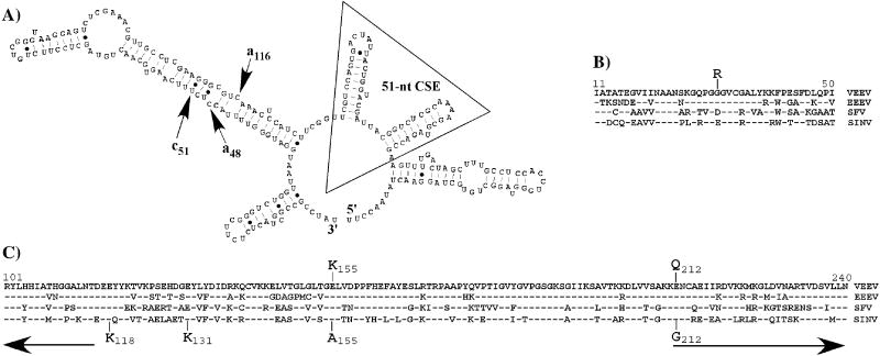
A low level of RNA replication created a productive background for selection of mutants demonstrating a higher replication efficiency. Such mutants appeared within the first passages of the viral pool harvested after transfection of in vitro-synthesized VEEVubiΔ3+4 genomes. We expected these viruses to contain adaptive mutations in the promoter sequences. To some extent, such was the case (Figs. 3 and 9A). In three plaque isolates, point mutations were found in the large stem-loop structure containing the initiating AUG, and no other changes in the cloned 5′-terminal fragment were discerned. These mutations stabilized computer-predicted RNA stem both in minus-strand (Fig. 9A) and plus-strand (data not shown) of the genome, and were localized in close proximity to each other, suggesting the importance of this stem-loop in VEEVubiΔ3+4 RNA replication. However, the tested mutations were insufficient for increasing replication to the levels found for the VEEVubi variant, which had an intact 51-nt CSE. Interestingly, the detected g48→u replacement even created a stop codon immediately downstream from the AUG, but the VEEVrev4 pseudorevertant was capable of efficient replication to a level similar to that of the VEEVubi (data not shown).
FIG. 9.
Sequence alignments and mutations found in VEEVrev1, VEEVrev2, VEEVrev3 and VEEVrev4 variants adapted for growth in BHK-21 cells. (A) Computer-predicted (m-fold) structure (Jaeger, Turner, and Zuker, 1989) of the 3′ end of the minus-strand of VEEV genome. Arrows indicate mutations found in VEEVrev1, VEEVrev3 and VEEVrev4. Triangle indicates the 51-nt CSE that is present in the minus strand of VEEVubi, but is deleted in VEEVubiΔ3+4. (B) Alignment of nsP3 residues 11 to 50 (VEEV numbering) for several alphaviruses. The mutation found in the VEEVrev2 and VEEVrev3 variants is indicated. (C) Alignment of nsP2 residues 101 to 240 (VEEV numbering). The mutations found in VEEVrev1 and VEEVrev4 and those previously detected in the genomes of SINV mutants adapted to growth in C710 cells after mutagenesis of 51-nt CSE (Fayzulin and Frolov, 2004) are indicated. Arrows indicate putative positions of the RNA helicase domain and the aminoterminal co-factor of nsP2-associated protease (Vasiljeva et al., 2003). VEEV, Venezuelan equine encephalitis virus (Kinney et al., 1989); EEEV, eastern equine encephalitis virus (Weaver et al., 1993); SFV, Semliki Forest virus (Takkinen, 1986); SINV, Sindbis virus (Strauss, Rice, and Strauss, 1984). Residues identical to those in the VEEV sequence are denoted by dashes.
The most important adaptive mutations that strongly increased the synthesis of virus-specific RNA and accelerated virus growth were detected, not in the 5′ end, but rather in the nsP2 and nsP3 genes. The nsP2-specific mutations E212→Q and E155→K (the effect of the latter mutation on RNA replication was not tested in the reverse genetics experiments) were in the sequence located between the predicted helicase domain and the amino terminal domain that was suggested as a co-factor of the nsP2-associated protease (Fig. 9C) (Vasiljeva et al., 2003). In our previous studies, we developed a SINV variant having clustered, silent mutations in the 5′-terminal sequence (which included the 51-nt CSE) that strongly affected the ability of virus to replicate in the mosquito cells (Fayzulin and Frolov, 2004). The SINV attained the mutations in the same sequence encoding a.a. 110–215 of nsP2 (Fig. 9C) to become capable of replication in these cells, and some of the mutations coincide with those found in the VEEV nsP2. This protein fragment demonstrates a moderate level of conservation between different alphaviruses (Fig. 9C), and its function is not as yet well defined. Our data suggest it may be either an independent domain involved in the binding of nsP2 (or the entire RC) to viral RNA or an integral part of the other amino terminal domains having NTPase, RNA helicase, and RNA triphosphatase activities (Gomez de Cedron et al., 1999; Rikkonen, Peranen, and Kaariainen, 1994; Vasiljeva et al., 2000).
In contrast to mutations in nsP2, the adaptive mutations accumulating in nsP3 in response to changes in the 5′-terminal promoter elements are not closely localized. The identified mutation in the VEEVrev2 and VEEVrev3 genomes was G31→R (Fig. 9B). Its stimulatory effect was confirmed in the reverse genetics experiments. Another mutation D119→G was not further studied, and, therefore, to date cannot be definitively considered adaptive. Previously, we demonstrated that SINV variants, which were incapable of replicating in C710 cells due to clustered mutations in the 5′ terminus, attained the mutation T286→I in nsP3 (Fayzulin and Frolov, 2004). However, the latter was incapable of enhancing RNA replication itself, had a stimulatory effect only when introduced together with E118→K in nsP2, and, thus, did not suggest an interaction of nsP3 with viral RNA. In contrast, the nsP3-specific, G31→R mutation in the VEEV genome was capable of strongly increasing viral and RNA replication when being introduced alone as well as in combination with other adaptive mutations. The G31→R is located in the so-called X domain of nsP3 that is conserved among alphaviruses and found in the rubella virus genome (Dominguez, Wang, and Frey, 1990; Strauss and Strauss, 1994), but its function has not yet been defined. However, the present data suggest possibility of its involvement in RNA recognition by alphavirus RC.
Taken together, the data from this study, combined with the previous results of the experiments with SINV (Fayzulin and Frolov, 2004; Frolov, Hardy, and Rice, 2001; Gorchakov et al., 2004; Niesters and Strauss, 1990b), suggest that the 51-nt CSE is a critical element of the promoter that functions as a replication enhancer, most likely, by positioning the RC for efficient recognition of the core promoter element. Based on the available data concerning VEEV RNA replication, it is difficult to propose the exact mechanism of its functioning, because this CSE might be required for plus- or minus- or both plus- and minus-strand RNA synthesis (Frolov, Hardy, and Rice, 2001; Gorchakov et al., 2004; Pardigon, Lenches, and Strauss, 1993; Pardigon and Strauss, 1992). Moreover, it is also possible that the changes in nsP2 and nsP3 function in the context of P123 rather than in the mature, completely processed RC.
The core promoter is located in the 5′UTR (and its complement in the 3′ terminus of the minus-strand intermediate), and its very terminal nucleotides, having high concentration of adenosines and uridines, are the most important for its activity in both plus- and minus-strand RNA synthesis (Frolov, Hardy, and Rice, 2001; Gorchakov et al., 2004). The deletion of the 51-nt CSE or introduction of the clustered silent mutations into its sequence strongly affects recognition of the core promoter by RC and makes the RNA replication inefficient in the cells of both vertebrate and invertebrate origin. Alphaviruses can adapt to the loss of 51-nt CSE by changing the RNA-binding sequence in the RC, which appears to be located within the amino terminal ~215 a. a. of nsP2. Most of the found mutations either decrease the number of the negatively charged amino acids and/or increase that of the positively charged ones. The mutations in the 5′ terminus of the genome and in the nsP3 can also have positive effects on replication of the 51-nt CSE mutants. This protein creates large, high-order structures in the infected cells (Frolova et al., 2006) and might be required either for changing the positioning of the nsP2 or cellular proteins on the RNA. Alternatively, it can directly interact with virus-specific RNAs. At present, because of insufficient data we cannot distinguish between these possibilities. Interestingly, the adaptive mutations in the VEEV nsPs have also a positive effect on replication of the virus with wt 51-nt CSE. It is highly unlikely that during its previous evolution, VEEV did not develop the nsPs and the cis-acting RNA elements supporting its efficient replication. An additional increase in RNA replication could be either disadvantageous for persistence of this virus in the mosquito vectors or might have a negative effect on replication of this virus in the vertebrate hosts due to a higher level of antiviral response caused by the increased production of dsRNA. However, this hypothesis needs further experimental support.
Such efficient adaptation of the alphaviruses to extended changes of the promoter sequences not only additionally supports the incredible plasticity of these viruses and their potential for evolution. It also suggests that development of attenuated alphaviruses and most likely other RNA-positive viruses for vaccine applications by mutating their promoter elements should be approached with caution, because of the rapid accumulation of the adaptive mutations in the nonstructural genes within a few rounds of infection.
MATERIALS AND METHODS
Cell cultures
BHK-21 cells were obtained from Dr. Paul Olivo (Washington University, St. Louis, MO). They were propagated in Alpha MEM (αMEM) supplemented with 10% fetal bovine serum (FBS) and vitamins. Mosquito C710 cells were obtained from Dr. Henry Huang (Washington University, St. Louis, MO). They were propagated in DMEM supplemented with 10% heat-inactivated FBS and 10% tryptose phosphate broth (TPB).
Plasmid constructs
The parental plasmid with VEEV TC-83 genome (Kinney et al., 1993) was obtained from Dr. Richard Kinney (CDC, Fort Collins, CO). The cDNA of the viral genome was cloned into a low copy number plasmid that was previously used for SINV genome (Rice et al., 1987) and the T7 promoter was replaced by a promoter of SP6 RNA polymerase. The parental pToto1101 plasmid encoding an infectious wt SINV genome was described elsewhere (Rice et al., 1987). All other plasmids encoding genetically modified VEEV and SINV genomes were constructed by standard PCR-based mutagenesis and cloning methods. All of the PCR fragments were sequenced to exclude the possibility of spontaneous mutations. The details of the designed constructs are described in the Results and figure legends. The details of the cloning procedures and sequences can be provided upon request.
RNA transcriptions
Plasmids were purified by centrifugation in CsCl gradients. Prior to transcription, the SINV and VEEV genome-coding plasmids were linearized by XhoI and MluI, respectively. RNAs were synthesized by SP6 RNA polymerase in the presence of cap analog (Rice et al., 1987). The yield and integrity of the transcripts were monitored by gel electrophoresis under non-denaturing conditions. Three-μg aliquots of the RNA transcripts from the reaction mixtures were directly used for electroporation (Liljeström et al., 1991).
RNA transfections and infectious center assay
In standard experiments, 3 μg of in vitro-synthesized, full-length RNA transcripts was used per electroporation. Ten-fold dilutions of electroporated BHK-21 or C710 cells were seeded in 6-well tissue culture plates containing naïve BHK-21 or C710 cells. After 2 h incubation at 37°C (for BHK- 21) or 30°C (for C710), BHK-21 cells were overlaid with 2 ml of 0.6% agarose (Invitrogen) containing MEM supplemented with 3% FBS, and C710 cells were overlaid with 2 ml of 0.6% tragacanth gum (ICN) containing MEM supplemented with 5% heat- inactivated FBS and 10% TPB. Plaques developed at 37°C (in BHK-21 cells) or 30°C (in C710 cells) and were stained with crystal violet after 2–2.5 days’ incubation. One-fifth of the electroporated cells was seeded into 35-mm dishes for radioactive labeling of RNAs or proteins. Another one-fifth was seeded into the same size dishes to evaluate virus replication, and the residual cells were used for generating viral stocks.
Selection of the pseudorevertants capable of efficient growth
The original stock of VEEVubiΔ3+4 was generated by transfection of BHK-21 cells. It was passaged twice in BHK-21 by infecting with 0.1 ml of the virus stock harvested after the previous passage. After the last passage, the individual large plaques were isolated from the agarose overlay, viruses were eluted in 1.5 ml of αMEM supplemented with 1% FBS for several hours, and 0.5 ml was used to infect 5×10⁵ BHK-21 cells in 35-mm dishes. Viruses were harvested after the development of a profound cytopathic effect, and RNA was isolated from the cells for sequencing of viral genomes. Four plaque-purified viruses were randomly selected from after passaging of viral stocks obtained after independent electroporations.
Sequencing of viral genomes
RNAs isolated from the infected cells and the pelleted viruses were used for RT-PCR and sequencing. Viruses were pelleted by ultracentrifugation at 52,000 rpm for 1 hour in a TLA-55 rotor at 4°C in an Optima MAX ultracentrifuge (Beckman Coulter). Pellets were suspended in 25 μl of PBS, and RNAs were isolated by TRIzol using the procedure recommended by the manufacturer (Invitrogen). Ten overlapping (approximately 1 kb-long) fragments representing the nonstructural genes and the 3′ end of the viral genome were synthesized using an RT-PCR procedure, purified by agarose gel electrophoresis and sequenced using the same primers as those applied for PCR. (Sequences of the primers can be provided upon request.) DNA fragments representing the 5′-terminal sequences were synthesized using a commercially available FirstChoice RLM-RACE Kit according to the procedure recommended by the manufacturer (Ambion). Fragments were purified by agarose gel electrophoresis and cloned into the plasmid pRS2. Multiple, independent clones were sequenced to determine possible variations in the 5′ ends of the genomes.
Virus growth analysis
To exclude the effect of accumulation of additional adaptive mutations, virus growth was analyzed directly after transfection of 3 μg of RNAs into C710 or BHK-21 cells. One-fifth of the electroporated cells was seeded into a 35-mm dish and incubated at 37°C (BHK-21 cells) or 30°C (C710 cells) for 1 hour. Then media was replaced by 1 ml of fresh media, and it continued to be replaced at indicated times post electroporation. Virus titers in the harvested samples were determined by a plaque assay on BHK-21 cells as described elsewhere (Lemm et al., 1990).
RNA analysis
One-fifth of the C710 cells electroporated with 3 μg of different RNAs were seeded into 35-mm dishes and incubated at 37°C. At 2 h post transfection virus-specific RNAs were labeled by replacing the media with αMEM supplemented with 10% FBS, [3H]uridine (20μCi/ml) and 1 μg/ml of Dactinomycin (ActD). RNAs were labeled for 6h at 37°C and isolated from the cells by TRIzol reagent, as recommended by the manufacturer (Invitrogen). Next, they were denatured with glyoxal in dimethyl sulfoxide and analyzed by agarose gel electrophoresis in the previously described conditions (Frolov, Hardy, and Rice, 2001).
Analysis of protein synthesis
BHK-21 cells were seeded into six-well Costar dishes at a concentration of 5×10⁵ cells/well. After 4h incubation at 37°C in 5% CO2, they were infected at an MOI of 10 PFU/cell in 200 μl of PBS supplemented with 1%FBS for 1 h at 37°C. The inoculum was then replaced by complete medium, and incubation continued at 37°C for 20 h. Then cells were washed three times with phosphate-buffered saline (PBS) and then incubated for 45 minutes at 37°C in 0.8 ml of DMEM medium lacking methionine, supplemented with 0.1% FBS and 20 μCi/ml of [35S]methionine. After this incubation, cells were scraped into PBS, pelleted by centrifugation and dissolved in standard gel-loading buffer. After electrophoresis, gels were dried and analyzed by autoradiography.
Acknowledgments
We wish to thank Mardelle Susman, technical editor, for critical reading and editing of the manuscript. This work was supported by Public Health Service grant AI050537.
Footnotes
Publisher's Disclaimer: This is a PDF file of an unedited manuscript that has been accepted for publication. As a service to our customers we are providing this early version of the manuscript. The manuscript will undergo copyediting, typesetting, and review of the resulting proof before it is published in its final citable form. Please note that during the production process errors may be discovered which could affect the content, and all legal disclaimers that apply to the journal pertain.
References
- Dal Canto MC, Rabinowitz SG. Central nervous system demyelination in Venezuelan equine encephalomyelitis infection. J Neurol Sci. 1981;49(3):397–418. doi: 10.1016/0022-510x(81)90030-7. [DOI] [PubMed] [Google Scholar]
- Dominguez G, Wang C, Frey TK. Sequence of the genome RNA of rubella virus: evidence for genetic rearrangement during togavirus evolution. Virol. 1990;177:225–238. doi: 10.1016/0042-6822(90)90476-8. [DOI] [PMC free article] [PubMed] [Google Scholar]
- Fayzulin R, Frolov I. Changes of the secondary structure of the 5′ end of the Sindbis virus genome inhibit virus growth in mosquito cells and lead to accumulation of adaptive mutations. J Virol. 2004;78(10):4953–64. doi: 10.1128/JVI.78.10.4953-4964.2004. [DOI] [PMC free article] [PubMed] [Google Scholar]
- Frolov I, Hardy R, Rice CM. Cis-acting RNA elements at the 5′ end of Sindbis virus genome RNA regulate minus- and plus-strand RNA synthesis. Rna. 2001;7(11):1638–51. doi: 10.1017/s135583820101010x. [DOI] [PMC free article] [PubMed] [Google Scholar]
- Frolova E, Gorchakov R, Garmashova N, Atasheva S, Vergara LA, Frolov I. Formation of nsP3-Specific Protein Complexes during Sindbis Virus Replication. J Virol. 2006;80(8):4122–34. doi: 10.1128/JVI.80.8.4122-4134.2006. [DOI] [PMC free article] [PubMed] [Google Scholar]
- Garcia-Tamayo J, Carreno G, Esparza J. Central nervous system alterations as sequelae of Venezuelan equine encephalitis virus infection in the rat. J Pathol. 1979;128(2):87–91. doi: 10.1002/path.1711280206. [DOI] [PubMed] [Google Scholar]
- Gomez de Cedron M, Ehsani N, Mikkola ML, Garcia JA, Kaariainen L. RNA helicase activity of Semliki Forest virus replicase protein NSP2. FEBS Lett. 1999;448(1):19–22. doi: 10.1016/s0014-5793(99)00321-x. [DOI] [PubMed] [Google Scholar]
- Gorchakov R, Hardy R, Rice CM, Frolov I. Selection of functional 5′ cis-acting elements promoting efficient sindbis virus genome replication. J Virol. 2004;78(1):61–75. doi: 10.1128/JVI.78.1.61-75.2004. [DOI] [PMC free article] [PubMed] [Google Scholar]
- Griffin DE. Alphaviruses. In: Knipe DM, Howley PM, editors. Fields Virology. 4. Lippincott, Williams and Wilkins; New York: 2001. pp. 917–962. [Google Scholar]
- Hardy RW. The role of the 3′ terminus of the Sindbis virus genome in minus-strand initiation site selection. Virology. 2006;345(2):520–31. doi: 10.1016/j.virol.2005.10.018. [DOI] [PubMed] [Google Scholar]
- Hardy RW, Rice CM. Requirements at the 3′ end of the sindbis virus genome for efficient synthesis of minus-strand RNA. J Virol. 2005;79(8):4630–9. doi: 10.1128/JVI.79.8.4630-4639.2005. [DOI] [PMC free article] [PubMed] [Google Scholar]
- Jaeger JA, Turner DH, Zuker M. Improved predictions of secondary structures for RNA. Proc Natl Acad Sci USA. 1989;86:7706–7710. doi: 10.1073/pnas.86.20.7706. [DOI] [PMC free article] [PubMed] [Google Scholar]
- Johnson KM, Martin DH. Venezuelan equine encephalitis. Adv Vet Sci Comp Med. 1974;18:79–116. [PubMed] [Google Scholar]
- Johnston RE, Peters CJ. Alphaviruses. In: Fields BN, Knipe DM, Howley PM, editors. Virology. 3. Lippincott-Raven; New York: 1996. pp. 843–898. [Google Scholar]
- Kinney RM, Chang GJ, Tsuchiya KR, Sneider JM, Roehrig JT, Woodward TM, Trent DW. Attenuation of Venezuelan equine encephalitis virus strain TC-83 is encoded by the 5′-noncoding region and the E2 envelope glycoprotein. J Virol. 1993;67(3):1269–77. doi: 10.1128/jvi.67.3.1269-1277.1993. [DOI] [PMC free article] [PubMed] [Google Scholar]
- Kinney RM, Johnson BJB, Welch JB, Tsuchiya KR, Trent DW. The full-length nucleotide sequences of the virulent Trinidad donkey strain of Venezuelan equine encephalitis virus and its attenuated vaccine derivative, strain TC-83. Virology. 1989;170:19–30. doi: 10.1016/0042-6822(89)90347-4. [DOI] [PubMed] [Google Scholar]
- Kuhn RJ, Hong Z, Strauss JH. Mutagenesis of 3′ nontranslated region of Sindbis virus RNA. J Virol. 1990;64:1465–1476. doi: 10.1128/jvi.64.4.1465-1476.1990. [DOI] [PMC free article] [PubMed] [Google Scholar]
- Lemm JA, Bergqvist A, Read CM, Rice CM. Template-dependent initiation of Sindbis virus replication in vitro. J Virol. 1998;72:6546–6553. doi: 10.1128/jvi.72.8.6546-6553.1998. [DOI] [PMC free article] [PubMed] [Google Scholar]
- Lemm JA, Durbin RK, Stollar V, Rice CM. Mutations which alter the level or structure of nsP4 can affect the efficiency of Sindbis virus replication in a host-dependent manner. J Virol. 1990;64:3001–3011. doi: 10.1128/jvi.64.6.3001-3011.1990. [DOI] [PMC free article] [PubMed] [Google Scholar]
- Lemm JA, Rice CM. Roles of nonstructural polyproteins and cleavage products in regulating Sindbis virus RNA replication and transcription. J Virol. 1993;67:1916–1926. doi: 10.1128/jvi.67.4.1916-1926.1993. [DOI] [PMC free article] [PubMed] [Google Scholar]
- Lemm JA, Rümenapf T, Strauss EG, Strauss JH, Rice CM. Polypeptide requirements for assembly of functional Sindbis virus replication complexes: A model for the temporal regulation of minus and plus-strand RNA synthesis. EMBO J. 1994;13:2925–2934. doi: 10.1002/j.1460-2075.1994.tb06587.x. [DOI] [PMC free article] [PubMed] [Google Scholar]
- Leon CA. Sequelae of Venezuelan equine encephalitis in humans: a four year follow-up. Int J Epidemiol. 1975;4(2):131–40. doi: 10.1093/ije/4.2.131. [DOI] [PubMed] [Google Scholar]
- Levis R, Schlesinger S, Huang HV. Promoter for Sindbis virus RNA-dependent subgenomic RNA transcription. J Virol. 1990;64:1726–1733. doi: 10.1128/jvi.64.4.1726-1733.1990. [DOI] [PMC free article] [PubMed] [Google Scholar]
- Levis R, Weiss BG, Tsiang M, Huang H, Schlesinger S. Deletion mapping of Sindbis virus DI RNAs derived from cDNAs defines the sequences essential for replication and packaging. Cell. 1986;44:137–145. doi: 10.1016/0092-8674(86)90492-7. [DOI] [PubMed] [Google Scholar]
- Li ML, Stollar V. Identification of the amino acid sequence in Sindbis virus nsP4 that binds to the promoter for the synthesis of the subgenomic RNA. Proc Natl Acad Sci U S A. 2004;101(25):9429–34. doi: 10.1073/pnas.0400995101. [DOI] [PMC free article] [PubMed] [Google Scholar]
- Liljeström P, Lusa S, Huylebroeck D, Garoff H. In vitro mutagenesis of a full-length cDNA clone of Semliki Forest virus: the small 6,000-molecular-weight membrane protein modulates virus release. J Virol. 1991;65:4107–4113. doi: 10.1128/jvi.65.8.4107-4113.1991. [DOI] [PMC free article] [PubMed] [Google Scholar]
- Niesters HGM, Strauss JH. Defined mutations in the 5′ nontranslated sequence of Sindbis virus RNA. J Virol. 1990a;64:4162–4168. doi: 10.1128/jvi.64.9.4162-4168.1990. [DOI] [PMC free article] [PubMed] [Google Scholar]
- Niesters HGM, Strauss JH. Mutagenesis of the conserved 51 nucleotide region of Sindbis virus. J Virol. 1990b;64:1639–1647. doi: 10.1128/jvi.64.4.1639-1647.1990. [DOI] [PMC free article] [PubMed] [Google Scholar]
- Ou JH, Strauss EG, Strauss JH. The 5′-terminal sequences of the genomic RNAs of several alphaviruses. J Mol Biol. 1983;168:1–15. doi: 10.1016/s0022-2836(83)80319-2. [DOI] [PubMed] [Google Scholar]
- Pardigon N, Lenches E, Strauss JH. Multiple binding sites for cellular proteins in the 3′ end of Sindbis alphavirus minus-sense RNA. J Virol. 1993;67(8):5003–11. doi: 10.1128/jvi.67.8.5003-5011.1993. [DOI] [PMC free article] [PubMed] [Google Scholar]
- Pardigon N, Strauss JH. Cellular proteins bind to the 3′ end of Sindbis virus minus-strand RNA. J Virol. 1992;66(2):1007–15. doi: 10.1128/jvi.66.2.1007-1015.1992. [DOI] [PMC free article] [PubMed] [Google Scholar]
- Raju R, Hajjou M, Hill KR, Botta V, Botta S. In vivo addition of poly(A) tail and AU-rich sequences to the 3′ terminus of the Sindbis virus RNA genome: a novel 3′-end repair pathway. J Virol. 1999;73(3):2410–9. doi: 10.1128/jvi.73.3.2410-2419.1999. [DOI] [PMC free article] [PubMed] [Google Scholar]
- Rice CM, Levis R, Strauss JH, Huang HV. Production of infectious RNA transcripts from Sindbis virus cDNA clones: Mapping of lethal mutations, rescue of a temperature-sensitive marker, and in vitro mutagenesis to generate defined mutants. J Virol. 1987;61(12):3809–3819. doi: 10.1128/jvi.61.12.3809-3819.1987. [DOI] [PMC free article] [PubMed] [Google Scholar]
- Rico-Hesse R, Weaver SC, de Siger J, Medina G, Salas RA. Emergence of a new epidemic/epizootic Venezuelan equine encephalitis virus in South America. Proc Natl Acad Sci USA. 1995;92:5278–5281. doi: 10.1073/pnas.92.12.5278. [DOI] [PMC free article] [PubMed] [Google Scholar]
- Rikkonen M, Peranen J, Kaariainen L. ATPase and GTPase activities associated with Semliki Forest virus nonstructural protein nsP2. J Virol. 1994;68(9):5804–5810. doi: 10.1128/jvi.68.9.5804-5810.1994. [DOI] [PMC free article] [PubMed] [Google Scholar]
- Sawicki DL, Sawicki SG. Alphavirus plus and minus strand RNA synthesis. In: Brinton M, Ruckert R, editors. Positive-strand RNA viruses. Alan R. Liss, Inc; New York: 1987. pp. 251–259. [Google Scholar]
- Shirako Y, Strauss JH. Regulation of Sindbis virus RNA replication: Uncleaved P123 and nsP4 function in minus strand RNA synthesis whereas cleaved products from P123 are required for efficient plus strand RNA synthesis. J Virol. 1994;185:1874–1885. doi: 10.1128/jvi.68.3.1874-1885.1994. [DOI] [PMC free article] [PubMed] [Google Scholar]
- Strauss EG, Rice CM, Strauss JH. Complete nucleotide sequence of the genomic RNA of Sindbis virus. Virology. 1984;133:92–110. doi: 10.1016/0042-6822(84)90428-8. [DOI] [PubMed] [Google Scholar]
- Strauss JH, Strauss EG. The alphaviruses: gene expression, replication, evolution. Microbiol Rev. 1994;58:491–562. doi: 10.1128/mr.58.3.491-562.1994. [DOI] [PMC free article] [PubMed] [Google Scholar]
- Takkinen K. Complete nucleotide sequence of the non-structural protein genes of Semliki Forest virus. Nucleic Acids Res. 1986;14:5667–5682. doi: 10.1093/nar/14.14.5667. [DOI] [PMC free article] [PubMed] [Google Scholar]
- Vasiljeva L, Merits A, Auvinen P, Kaariainen L. Identification of a novel function of the alphavirus capping apparatus. RNA 5′-triphosphatase activity of Nsp2. J Biol Chem. 2000;275(23):17281–7. doi: 10.1074/jbc.M910340199. [DOI] [PubMed] [Google Scholar]
- Vasiljeva L, Merits A, Golubtsov A, Sizemskaja V, Kaariainen L, Ahola T. Regulation of the sequential processing of Semliki Forest virus replicase polyprotein. J Biol Chem. 2003;278(43):41636–45. doi: 10.1074/jbc.M307481200. [DOI] [PubMed] [Google Scholar]
- Wang YF, Sawicki SG, Sawicki DL. Alphavirus nsP3 functions to form replication complexes transcribing negative-strand RNA. J Virol. 1994;68:6466–6475. doi: 10.1128/jvi.68.10.6466-6475.1994. [DOI] [PMC free article] [PubMed] [Google Scholar]
- Weaver SC, Barrett AD. Transmission cycles, host range, evolution and emergence of arboviral disease. Nat Rev Microbiol. 2004;2(10):789–801. doi: 10.1038/nrmicro1006. [DOI] [PMC free article] [PubMed] [Google Scholar]
- Weaver SC, Hagenbaugh A, Bellew LA, Gousset L, Mallampalli V, Holland JJ, Scott TW. Evolution of alphaviruses in the eastern equine encephalomyelitis complex. J Virol. 1994;68(1):158–69. doi: 10.1128/jvi.68.1.158-169.1994. [DOI] [PMC free article] [PubMed] [Google Scholar]
- Weaver SC, Hagenbaugh A, Bellew LA, Netesov SV, Volchkov VE, Chang GJ, Clarke DK, Gousset L, Scott TW, Trent DW, et al. A comparison of the nucleotide sequences of eastern and western equine encephalomyelitis viruses with those of other alphaviruses and related RNA viruses. Virology. 1993;197(1):375–90. doi: 10.1006/viro.1993.1599. [DOI] [PubMed] [Google Scholar]
- Weaver SC, Salas R, Rico-Hesse R, Ludwig GV, Oberste MS, Boshell J, Tesh RB. Re-emergence of epidemic Venezuelan equine encephalomyelitis in South America. VEE Study Group. Lancet. 1996;348(9025):436–40. doi: 10.1016/s0140-6736(96)02275-1. [DOI] [PubMed] [Google Scholar]
- Wielgosz MM, Raju R, Huang HV. Sequence requirements for Sindbis virus subgenomic mRNA promoter function in cultured cells. J Virol. 2001;75(8):3509–19. doi: 10.1128/JVI.75.8.3509-3519.2001. [DOI] [PMC free article] [PubMed] [Google Scholar]


