Biology of Reproduction logoLink to Biology of Reproduction
. 2010 Dec 22;84(2):400–408. doi: 10.1095/biolreprod.110.090530

ADDITIONS AND CORRECTIONS

PMCID: PMC3071269  PMID: 21312389

CORRECTION

Wei Zhou, Gensheng Wang, Christopher L. Small, Zhilin Liu, Connie C. Weng, Lizhong Yang, Michael D. Griswold, and Marvin L. Meistrich. Gene Expression Alterations by Conditional Knockout of Androgen Receptor in Adult Sertoli Cells of Utp14b jsd/jsd (jsd) Mice. Biol Reprod 2010; 83:759–766. DOI: 10.1095/biolreprod.110.085472

In the Abstract, an edit to the following sentence inadvertently changed the meaning: “ARregulated genes in Sertoli cells must therefore be involved in the regulation of spermatogonial differentiation, although there was no significant differentiation from spermatocytes in SCARKO-jsd mice.” The sentence should have read “…to spermatocytes in SCARKO-jsd mice.”

Also, in Table 1, there were two formatting errors. 1) “Tenomodulin (Tnmd)” was presented as a header rather than as an item under the header “Upregulated in SCARKO-jsd”; and 2) “Variable Result with different probes” was incorrectly presented as an item under “Tenomodulin (Tnmd)” when it should have been its own head.

The editors regret these errors. The complete paper has been reprinted below with corrections.

Biol Reprod. 2010 Jul 21;83(5):759–766.

Gene Expression Alterations by Conditional Knockout of Androgen Receptor in Adult Sertoli Cells of Utp14bjsd/jsd (jsd) Mice1

Wei Zhou 4,2, Gensheng Wang 4,3, Christopher L Small 5, Zhilin Liu 6, Connie C Weng 4, Lizhong Yang 5, Michael D Griswold 5, Marvin L Meistrich 4

Abstract

Spermatogenesis is dependent primarily on testosterone action on the Sertoli cells, but the molecular mechanisms have not been identified. Attempts to identify testosterone-regulated target genes in Sertoli cells have used microarray analysis of gene expression in mice lacking the androgen receptor (AR) in Sertoli cells (SCARKO) and wild-type mice, but the analyses have been complicated both by alteration of germ cell composition of the testis when pubertal or adult mice were used and by differences in Sertoli-cell gene expression from the expression in adults when prepubertal mice were used. To overcome these limitations and identify AR-regulated genes in adult Sertoli cells, we compared gene expression in adult jsd (Utp14bjsd/jsd, juvenile spermatogonial depletion) mouse testes and with that in SCARKO-jsd mouse testes, since their cellular compositions are essentially identical, consisting of only type A spermatogonia and somatic cells. Microarray analysis identified 157 genes as downregulated and 197 genes as upregulated in the SCARKO-jsd mice compared to jsd mice. Some of the AR-regulated genes identified in the previous studies, including Rhox5, Drd4, and Fhod3, were also AR regulated in the jsd testes, but others, such as proteases and components of junctional complexes, were not AR regulated in our model. Surprisingly, a set of germ cell–specific genes preferentially expressed in differentiated spermatogonia and meiotic cells, including Meig1, Sycp3, and Ddx4, were all upregulated about 2-fold in SCARKO-jsd testes. AR-regulated genes in Sertoli cells must therefore be involved in the regulation of spermatogonial differentiation, although there was no significant differentiation to spermatocytes in SCARKO-jsd mice. Further gene ontogeny analysis revealed sets of genes whose changes in expression may be involved in the dislocation of Sertoli cell nuclei in SCARKO-jsd testes.

androgen receptor, conditional knockout, meiosis, Sertoli cells, spermatogenesis, testis, testosterone

INTRODUCTION

Androgens are essential for normal spermatogenesis and fertility. Androgen regulation of spermatogenesis must occur through the Sertoli cell because selective knockout of the androgen receptor (AR) in these cells, using a floxed Ar-gene and a Sertoli cell–specific cre, impedes spermatogenesis [13]. Further studies indicated that the genomic activity of AR in Sertoli cells plays the essential role in spermatogenesis [4], but specific genes involved are not known. Studies have attempted to identify androgen-regulated genes affecting spermatogenesis using microarray analysis [reviewed in 5] on 10-day-old SCARKO (Sertoli-cell androgen receptor knockout) mice [6], 20-day-old testicular feminized (Tfm) mice [7], hypogonadal (hpg) mice briefly treated with testosterone [8], and AR elimination in Sertoli cells of adult mice that were already hypomorphic for AR function [9]. Whereas these studies have provided valuable information, they all have some limitations. Conclusions from studies employing immature animals may be limited to the effects of androgen on immature Sertoli cells, which differ from the effects in adults. Conclusions from studies in adult animals are complicated by alterations in germ cell compositions resulting from elimination of AR. Studies that were limited to the acute changes induced by the hormones may have missed some important functional changes that result from chronic treatment, such as indirect regulation of targets.

We used a recently constructed SCARKO model, based on mice homozygous for the juvenile spermatogonial depletion (jsd) mutation [10] in the Utp14b gene [11, 12] (referred to as jsd mice) to circumvent these problems. The Utp14b gene is primarily expressed in germ cells and only very weakly in Sertoli cells [13], and Sertoli cells in testis of jsd mice can support differentiation of transplanted normal spermatogonia [14]. The jsd mice become sterile after the first few waves of spermatogenesis [15] and the only germ cells remaining in most seminiferous tubules of adult jsd mice are undifferentiated type A spermatogonia [16]. It has been shown that suppression of testosterone by gonadotropin-releasing hormone analogues can reverse the block of spermatogonial differentiation [1719]. However, it appeared that Sertoli cell AR was not responsible for the spermatogonial block in the jsd mice, as SCARKO-jsd mice showed no histological evidence of recovery of spermatogenesis, although their testes did support spermatogonial differentiation when systemic levels of testosterone were suppressed [20]. Nevertheless, the absence of AR action on Sertoli cells of jsd mice resulted in significant alteration of the localization of their nuclei.

Because germ cell compositions of adult jsd and SCARKO-jsd mice are the same, they make an excellent model with which to identify the genes regulated by chronic deficiency of testosterone action in adult Sertoli cells by microarray analysis, without the complication introduced by changes in cellular components. In this report we identify changes in gene expression resulting from selective and prolonged elimination of AR in adult Sertoli cells of jsd mice.

MATERIALS AND METHODS

Animals

Transgenic mice with floxed-Ar (Arflox, official symbol Artm1Verh), on a 129/Sv genetic background, were obtained from the Catholic University of Leuven, Belgium [1]. Both males and females were crossed with mice carrying the jsd mutation on a C57BL/6 background, and the offspring were intercrossed for several generations to generate Arflox and jsd homozygous double mutant females (Arflox/flox, Utp14bjsd/jsd). Mice expressing cre recombinase under the control of the anti-Mullerian hormone (Amh) promoter (Amh-cre mice,cre+/−, official symbol Tg(Amh-cre)8815Reb, on a 129/Sv background were obtained from the University of Washington, Seattle [3]. Both males and females (cre+/) were crossed with mice carrying the jsd mutation on a C3H-B6–129 (HB129) mixed background [21] for one or more generations to generate double-mutant males that were heterozygous for both Amh-cre and jsd (cre+/−, Utp14b+/jsd). The Arflox/flox, Utp14bjsd/jsd females were then mated with cre+/, Utp14bjsd/+ males to produce the triple-mutant test model males (Arflox/Y,cre+/−, Utp14bjsd/jsd, SCARKO-jsd). The jsd mice used as controls carried Arflox. Genotyping of SCARKO-jsd and control jsd mice was described earlier [20]. Note that the same breeding protocol was used as in the earlier report [20], although some of the background strains were misidentified in that report. All animal studies were approved by the M.D. Anderson Cancer Center Institutional Animal Care and Use Committee.

Testicular Histology

After the mice were euthanized, testes were removed and fixed in Bouin's solution and embedded in methacrylate. The blocks were sectioned and stained with hematoxylin and periodic acid–Schiff reagent.

Stereology Analysis

Stereological analysis using the optical dissector approach [22] was performed on 25-μm thick sections with Stereo Investigator version 8.0 software (MicroBrightField, Inc., Williston, VT) under a stage-controlled microscope (Leica DMLB 100S, Leica Microsystems, Germany) using a 100× oil immersion lens. A 60 μm × 60 μm counting frame was positioned at multiple sampling sites across the section by the systematic random sampling method. The total sampling volume (Vs) for a given section was calculated as the product of counting frame area, number of sampling sites, and dissector height (15 μm). The total numbers of germ, Sertoli, interstitial, vascular smooth muscle, and peritubular myoid cells per testis (Nt) were then calculated as follows: Nt = Ns × (Vt/Vs), where Ns is the number of nuclei of the specific cells counted in the total sampling volume and Vt is the testis volume, which was determined by dividing the testis weight by the testicular tissue density of 1.05 g/cm3 [23].

RNA Preparation and Microarray Hybridization

Testis tissue samples were removed from three 12-wk-old control jsd and three 12-wk-old SCARKO-jsd mice and immediately placed in RNAlater (Qiagen, Valencia, CA) and stored at −20°C until total RNA was extracted using the RNeasy Mini kit (Qiagen). To normalize both microarray and qRT-PCR data on a per testis basis, we added 4 μl of a 1:10 000 dilution of B. subtilis RNA containing the dap gene with an artificial poly A tail (GeneChip Poly-A RNA Control Kit, Affymetrix, Santa Clara, CA) to each testis sample. We then extracted total RNA using the RNeasy Mini kit. RNA quality was determined by both electrophoresis and by 230-, 260-, and 280-nm absorption readings (NanoDrop, Wilmington, DE).

Ten micrograms of total RNA from each sample was labeled using a GeneChip One-Cycle Target Labeling Kit (Affymetrix) to produce labeled cRNA, which was then hybridized to the Affymextrix GeneChip Mouse Genome M430 2.0 array (Affymetrix). Production of cDNA and cRNA, hybridization to arrays, and evaluation of hybridization quality were completed by the Laboratory for Biotechnology and Bioanalysis I at Washington State University, Pullman, WA.

Microarray Analysis

Expression analysis was performed using the Robust Multi-array Averaging function [24] from the Affy package (v1.5.8) through the BioConductor software (http://www.bioconductor.org/ [25]). Then, genes differentially expressed between jsd and SCARKO-jsd mice were identified with Significance Analysis of Microarrays software [26]. The data were deposited into the National Center for Biotechnology Information Gene Expression Omnibus, with accession number GSE20918. Analysis of the levels of dap RNA in the normalized microarray data showed that there were equivalent levels of dap RNA in the jsd and the SCARKO-jsd samples (Supplemental Table S1, all Supplemental Data are available online at www.biolreprod.org). This result demonstrated that the relative values of gene expression obtained in the normalized microarray data were indeed equivalent to those that would be calculated on a per testis basis [27].

Real-time RT-PCR

Testis samples were collected from 12-wk-old jsd and SCARKO-jsd mice (n = 5 for jsd and n = 4 for SCARKO-jsd, which includes the samples used in the microarray), and total RNA was extracted using RNeasy Mini kit (Qiagen), with DNase I treatment to digest genomic DNA. Total RNA (3 μg) was used to generate the cDNA with a Transcriptor First Strand cDNA synthesis kit (Roche Applied Sciences, Indianapolis, IN), which was then used as the template for real-time RT-PCR. Quantitative real-time PCR was performed using the Rotor-Gene 3000 thermocycler (Corbett Research, Sydney, Australia) and SYBR Green (JumpStart Taq ready mix, Sigma, St. Louis, MO). The housekeeping gene, beta-actin, was used to normalize concentration values for each sample. The ratios of beta-actin to dap RNA were identical in the samples from the jsd and the SCARKO-jsd testes (Supplemental Table S1), demonstrating that values presented relative to beta-actin were equivalent to those on a per testis basis. The PCR reaction volume of 10 μl contained 5 μl of SYBR Green JumpStart Taq Mix, 4.8 μl of first-strand cDNA product (1:100 dilution), and 0.1 μl each of specific forward and reverse primers, with a final concentration of 0.1 μM (Sigma Genosys, Houston, TX). Cycling conditions were as follows: 2 min 95°C hold, followed by 40 cycles of 15 sec at 95°C, 1 min at 60°C, and 1 min at 72°C. Fluorescence was measured and acquired at 72°C. The primer sequences are listed in Supplemental Table S2. All samples were run in triplicate. Relative levels of gene expression were calculated using Rotor-Gene 6.0 software.

Gene Ontology Analysis

Genes that were differentially regulated in SCARKO-jsd mouse testis were analyzed by DAVID Bioinformatics Resources (http://david.abcc.ncifcrf.gov).

RESULTS

Stereological Analysis of Cellular Components

Both morphological and stereological analysis demonstrated that the cellular compositions of the SCARKO-jsd testes were essentially identical to those of jsd testes (Fig. 1). About 42% of the cells in the testes of these mice were Sertoli cells and only 6% were germ cells. Of the germ cells, 96% were premeiotic (spermatogonia or preleptotene spermatocytes) and there were only a few spermatocytes in meiotic prophase. This suggests that microarray analysis of differential gene expression between adult testes with and without active AR in the Sertoli cells would largely reflect changes in Sertoli cell gene expression, with minimal possibility of false positives due to changes in cellular composition.

FIG. 1.

FIG. 1.

Cellular composition and histology of the SCARKO-jsd mouse testis compared to that of jsd mouse testis. Numbers of somatic (A) and germ (B) cells of SCARKO-jsd and jsd mouse testis analyzed by stereology. Error bars represent mean ± SEM. We could not reliably distinguish preleptotene spermatocytes from B spermatogonia and so counted them with the spermatogonia. C) Cross-section of jsd mouse testis showing Sertoli cells and a few spermatogonia at the periphery of seminiferous tubules. D) Cross-section of SCARKO-jsd mouse testis showing disorganization of Sertoli cells within the tubules. Original magnification (C, D) ×200.

Microarray Analysis

The RNA samples from SCARKO-jsd and jsd mouse testes were analyzed by microarray. The results for all probes are presented in a searchable Excel file (Supplemental Table S3), which can be used for further analyses. By using ±1.8-fold as a cutoff and a false discovery rate of less than 10%, 157 genes (probes with unique UniGene identifiers) in the SCARKO-jsd mice were shown to be downregulated and 197 genes were upregulated compared to jsd mice (Supplemental Table S3). The 12 most strongly downregulated and 10 most strongly upregulated genes identified in SCARKO-jsd testis are listed in Table 1.

TABLE 1.

Genes most strongly downregulated and upregulated in SCARKO-jsd mouse testes vs. jsd testes.

graphic file with name bire-83-05-06-t01.jpg

Confirmation of Microarray Data

Real-time RT-PCR confirmed the gene expression changes for 21 of the 22 genes that appeared to be strongly regulated (Table 1). The only exception was probe set 1431417_at, which was one of five probes representing junction adhesion molecule 2 (Jam2). Since the other four microarray probe sets for Jam2 exhibited minimal changes between jsd and SCARKO-jsd testes (Supplemental Table S3), we conclude that the downregulation of Jam2 probe set 1431417_at in SCARKO-jsd testis was a false-positive result and should be discarded. This result also demonstrates that caution must be taken when multiple probe sets representing the same gene exhibit different expression patterns.

Ontology Analysis

Gene ontology studies revealed that, among the significantly regulated genes resulting from the absence of AR in Sertoli cells, there was an overrepresentation of genes involved in bone mineralization (Ank, Spp1, P2rx7, Ahsg, and Cd276) (P = 5 × 10⁵) and neuron differentiation (Ank3, Gas7, Tiam1, Olig1, Mtap1b, Ptprz1, Id4, Nrcam, Alcam, Bex1, Ret, Pigt, and Mcf2) (P = 0.002). Most surprisingly, the most significantly overrepresented category was meiosis, which included the genes Syce1, Meig1, Dazl, Sycp3, Stra8, and Piwil2 (P = 3 × 10⁵). Further examination of the microarray data and real-time RT-PCR (Fig. 2) revealed that Zbtb16 (formerly known as Plzf), a gene present only in undifferentiated A spermatogonia, was unchanged in SCARKO-jsd testes. Genes expressed in only A and B spermatogonia (Fthl17, Sohlh1), as well as those whose expression continued in later stages (Sycp3, Piwil2, Ddx4, and Stk31), were all upregulated 2- to 3-fold in SCARKO-jsd testis. Whereas Meig1, whose expression appeared to begin in B spermatogonia, was also upregulated in SCARKO-jsd testes, genes that were first expressed in spermatocytes in meiotic prophase, such as Ldhc and Adam2, were essentially absent in jsd testes and very weakly and variably expressed in SCARKO-jsd testes, and no statistically significant upregulation could be demonstrated. These observations indicate that the absence of AR in the Sertoli cells induced significant expression of genes associated with differentiated spermatogonia in jsd mouse testis.

FIG. 2.

FIG. 2.

Expression of germ cell–specific genes in jsd and SCARKO-jsd mouse testis. Cell types expressing the mRNA of the genes are indicated above arrows. Data are presented as fold induction compared to jsd testis. Error bars represent mean ± SEM. ab., scored as absent in microarray data.

DISCUSSION

In this study, we used the selective elimination of AR from Sertoli cells in adult Utp14b mutant (Utp14bjsd/jsd) mice as a unique model system for identifying the androgen-regulated genes in adult Sertoli cells that may be involved in the testosterone-dependent functions of these cells, such as the progression of germ cells through meiosis.

Whereas no system is perfect, our current system fulfills many criteria for an ideal system to uncover important hormonally regulated Sertoli cell genes (Table 2). First, it detects gene expression changes due to loss of androgen action, specifically on the Sertoli cells, whereas other models, such as hpg [8] and Tfm [7], involve reduced androgen action on multiple testicular cells. In the study using the Tfm model, this problem was partially overcome by employing methods to eliminate genes primarily expressed in germ or interstitial cells. Second, it involves the use of adult animals, which is preferable since Sertoli cells from immature mice may not express the genes necessary for the maturation of germ cells, as exemplified by the elimination of Sertoli-cell AR having no effect on germ cell populations in 10-day-old mice [6]. Third, it involves prolonged differential levels of androgen action between the test model and the control, since the SCARKO mice are deficient in AR in Sertoli cells from birth. Fourth, our model showed minimal differences in composition of germ cells from the control, thus eliminating the secondary effects of changing germ cell populations on the expression of genes by the Sertoli cells. Furthermore, the almost total absence of germ cells, which make up the bulk of the cells in a normal testis, increases the efficiency of detection of Sertoli cell genes in the jsd mice. However, the absence of germ cells may be a shortcoming of the model that could result in differences from the normal testis, since germ cells affect gene expression in Sertoli cells and induce stage-specific cyclical changes. Therefore, the Sertoli cells in jsd or SCARKO-jsd mice do experience some different physiological conditions from those in mice with a full complement of germ cells. Also, the current model and others previously reported [16] eliminate AR from Sertoli cells in the embryonic testis, which may alter Sertoli cell development during neonatal development and cause some gene expression changes in adult testes, which differ from those that would be observed by elimination of AR specifically in the adult testis. This can be improved in the future by using a conditional knockout model that will knockout AR in Sertoli cells when the animals have entered adulthood.

TABLE 2.

Comparisons of models used for microarray analysis of androgen regulated genes in Sertoli cells.

graphic file with name bire-83-05-06-t02.jpg

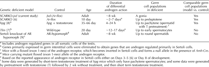
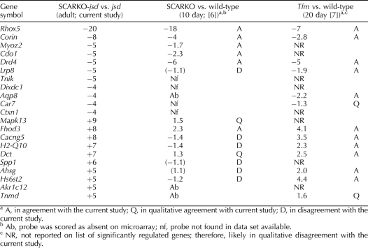
As reviewed above (Table 2), the various model systems used to identify androgen-regulated genes in Sertoli cells employ different conditions. We propose that the genes that are AR-regulated under multiple conditions are more likely to be directly regulated by AR and be the ones that are regulated by AR in the normal adult testis. Conversely, the ones that are regulated by AR only in one specific model system may be false positives, indirect effects, or in populations other than Sertoli cells, and they may not be regulated by AR in the Sertoli cells of the normal adult testis. In Table 3, we have compared the changes in expression of the highly regulated genes in SCARKO-jsd (Table 1) with the regulation of these genes in two other studies of androgen-regulated Sertoli-cell gene expression [6, 7]. Of the 21 genes, only three downregulated genes, Rhox5, Corin, and Drd4, and one upregulated gene, Fhod3, are qualitatively regulated in the same direction in all three systems. The androgen regulation of Rhox5 has been well studied, occurs in rat as well as mouse [28], and is a direct target of AR [29]. Using the PATSER tool (http://rsat.ulb.ac.be/rsat/) [30] with a cutoff of Weight Score 7 (P <10−4), we have identified at least one potential ARE binding site for all of the genes in Table 1 (from −5000 bp upstream of the transcription start site to the end of transcription) except for Akr1c12 (data not shown). An attempt to determine whether these genes were regulated similarly in the rat using the irradiated rat testis model [31] was not informative, as these genes either could not be found on the rat 230 2.0 expression array or their expression was too low to be detected on that array.

TABLE 3.

Comparison of regulation of genes strongly regulated by Sertoli-cell AR in adult jsd mice with the AR regulation of these genes in immature SCARKO and Tfm mice.

graphic file with name bire-83-05-06-t03.jpg

The present results can also be used to test the robustness of reports of effects of AR elimination on changes in expression of functional groups of genes, such as junctional complex proteins, proteases and protease inhibitors, proteins involved in vitamin A metabolism, and solute carriers [6, 7, 9, 32] in Sertoli cells (Supplemental Table S4). Some major proteins involved in the tight junctions between Sertoli cells [33], such as occludin (Ocln), gelsolin (Gsn), and claudin 11 (Cldn11), are not AR-regulated in the jsd model. Only alpha actinin 3 (Actn3) and thrombospondin 1 (Thsb1) showed consistent downregulation in several Sertoli AR knockout models. In addition, none of the proteases or protease inhibitors or genes regulating the vitamin A levels showed the same regulation by elimination of AR in the jsd model as in other models; only eppin (Spinlw1), which was downregulated 8-fold in 10-day-old SCARKO mice, and alcohol dehydrogenase (Adh1), which was upregulated 9-fold in 20-day-old Tfm mice, showed the same direction of regulation, but these changes were only 1.4- and 1.7-fold, respectively. Finally, none of the solute transport proteins, except the cationic amino acid transporter Slc7a4, were regulated by elimination of Sertoli-cell AR in the jsd model. These results suggest that caution should be exercised in making broad generalizations as to groups of genes regulated by AR in Sertoli cells, when only one model of a specific age and germ cell composition is used.

Since Rhox5 was strongly downregulated in the absence of AR in SCARKO-jsd testes, we investigated whether genes reported to be regulated by Rhox5 in other models would also be regulated by AR in adult jsd testes. However, of the 13 genes reported to be regulated by Rhox5 [34, 35], only Tmem176a showed a change in SCARKO-jsd testis in the same direction as predicted, but the change was very small (only −1.3-fold downregulation), and one gene, Ins2, which had been reported to be upregulated by Rhox5 [34], was actually upregulated 2.5-fold in SCARKO-jsd Sertoli cells, cells whose Rhox5 was dramatically reduced (Supplemental Table S3). Thus, these results suggest that the regulation of these genes by Rhox5 is highly dependent on the developmental stage and environment of the Sertoli cells and/or they are also regulated by AR-dependent mechanisms independently of Rhox5, which overcome the effects of the Rhox5 regulation. In either case, it does not appear that changes in expression of Rhox5-regulated genes are responsible for the dislocation of Sertoli cell nuclei in SCARKO-jsd mice.

To investigate whether the Sertoli cell AR-regulated genes identified in multiple model systems are important for spermatogenesis, we reviewed the effects of targeted disruption. Only homozygous mice with targeted disruption of Lrp8 [36] and Rhox5 [37], but not those with null mutation for Corin, Myoz2, Drd4, and Thbs1, have been reported to exhibit male infertility. However, in the case of Lrp8 null mice, spermatogenesis proceeds normally and the sperm defects develop in the epididymis; in Rhox5 null mice, germ cells pass through meiosis and the mice still retain some fertility. Since there is no evidence that any specific androgen-regulated gene is critical for the completion of spermatogenesis, it is likely that when AR is eliminated from Sertoli cells, spermatogenesis is disrupted as a result of multiple gene alterations.

The biological significance of the dramatic overrepresentation of a group of genes involved in bone mineralization among the Sertoli cell AR–regulated genes in jsd testes is not clear. It is tempting to speculate that since the Sertoli cells and bone cells are both involved in tissue remodeling processes, there is some overlap in gene expression. Three of the genes identified (Spp1, Ank, and Cd276) are preferentially expressed in the Sertoli cells, and hence alterations of their expression in testis could be directly affected by changes in the Sertoli cells. Also, since bone mineralization is stimulated by androgens, it would be expected that bone mineralization genes would be preferentially found among an androgen-regulated set. However, only one of the genes identified in the group (Spp1, also known as osteopontin) is known to be downregulated by androgen [38]. Alternatively, it is quite possible that, although these genes all share the “bone mineralization” functional category, it is their other functions, including cell adhesion, signal transduction, and cell differentiation and/or proliferation, that make them stand out in this analysis, and these functions explain the disrupted Sertoli cell nuclear localization and detachment from the basement membrane in the SCARKO-jsd testes [20].

The neuron differentiation genes may have been identified by ontology analysis due to the same principle. Their functions include cell adhesion (Alcam, Nrcam, Ret), signal transduction (Alcam, Ank3, Mcf2, Tiam1), cell differentiation and/or proliferation (Bex1, Gas7, Id4), and cytoskeleton formation (Mtap1b, Nrcam), which may have been responsible for the alteration of Sertoli cell nuclear localization in SCARKO-jsd mouse testis. It is also possible that the cell elongation and induction of asymmetry, which occur during both neuron differentiation and Sertoli cell maturation (Fig. 1C), involve the same cellular processes, which appear to be lost in the SCARKO-jsd testes.

Previous studies have shown that the selective elimination of AR from Sertoli cells disrupted the localization of Sertoli cell nuclei both in adult SCARKO mouse testes [32] and in adult SCARKO-jsd testes [20]. With only a few type A spermatogonia left in the seminiferous tubules, Sertoli cell nuclear localization was more drastically altered in SCARKO-jsd testis [20] than in the SCARKO testis. When we examined genes that were identified as significantly altered in expression in postnatal day 10.5 SCARKO mouse testis and were considered to play roles in Sertoli cell nucleus dislocation [32], including Vim, Lama5, Cldn11, Ocln, Gsn, Cacna1a, and Plat, we found them in SCARKO-jsd testis to be unchanged compared to jsd testis or not expressed at all (Supplemental Table S3). The lack of changes in expression of these genes in SCARKO-jsd mice indicates that they are not the main causes of Sertoli cell nuclear dislocation and their changes in the day-10.5 SCARKO mice could result from a difference between prepubertal and adult animals. Therefore, changes in expression of the bone mineralization (Ank, Spp1, P2rx7, Ahsg, and Cd276) and neuron differentiation (Ank3, Gas7, Tiam1, Olig1, Mtap1b, Ptprz1, Id4, Nrcam, Alcam, Bex1, Ret, Pigt, and Mcf2) genes identified in our array data provide reasonable explanations for the alteration in Sertoli cell nucleus localization. These genes make good candidates with which to further explore the detailed molecular mechanisms of AR regulation of Sertoli cell structure.

The upregulation of a group of genes that are expressed in differentiated germ cells in the SCARKO-jsd mice was surprising, since no morphological changes in germ cell populations were detected (Fig. 1) [20]. Microarray and real-time RT-PCR results demonstrated that the genes known to be expressed in differentiated A spermatogonia and B spermatogonia were increased 2–3 fold when the AR was eliminated from Sertoli cells, whereas an undifferentiated spermatogonial marker remained unchanged and spermatocyte makers were not significantly detectable. These results demonstrate that the presence of AR in Sertoli cells did contribute, to a small extent, to the inhibition of spermatogonial differentiation in jsd mice. One of the histological endpoints that showed no significant difference between SCARKO-jsd and jsd testes [20], the tubule differentiation index, which measures the percentage of tubules with cells at the B spermatogonial stage or later [39], had a large variance in that study so that a 2-fold change may not have been detected. Although there is no 2-fold increase in spermatogonial numbers in SCARKO-jsd testes (Fig. 1), we cannot rule out the possibility of a relative increase in the more differentiated spermatogonial stages in the SCARKO-jsd mice since all spermatogonia were counted together. In any case, the degree of spermatogonial differentiation induced by elimination of AR from the Sertoli cells of jsd mice is minor compared to that caused by total elimination of AR [19] or addition of AR antagonists [18]. This further demonstrates that testosterone acts on the AR in other somatic cell types to produce the factors that inhibit jsd spermatogonial differentiation.

In summary, this study, using a unique model of specific elimination of AR in adult Sertoli cells in a testis essentially lacking germ cells, has identified a group of genes that are consistently AR-regulated in Sertoli cells of different ages and environments and has demonstrated that other previously reported genes are AR-regulated in Sertoli cells only under specific conditions. We have also identified groups of genes common to other biological processes that are regulated by AR in adult Sertoli cells, which may provide insight into the cause of the nuclear dislocation of the Sertoli cells in SCARKO-jsd testis. Finally, the results demonstrate that AR action in Sertoli cells contributes to a small but significant degree to the inhibition of spermatogonial differentiation in jsd mutant mice.

Footnotes

1

Supported by National Institutes of Health (NIH) Research grants HD-40397 (to M.L.M.) and NIH Cancer Center Support grant CA 16672 to M.D. Anderson Cancer Center, HD-10808 (to M.D.G.) and HD-16229 (to Joanne Richards, Baylor College of Medicine, Houston, Texas).

REFERENCES

  1. De Gendt K, Swinnen JV, Saunders PT, Schoonjans L, Dewerchin M, Devos A, Tan K, Atanassova N, Claessens F, Lecureuil C, Heyns W, Carmeliet P, et al. A Sertoli cell-selective knockout of the androgen receptor causes spermatogenic arrest in meiosis. Proc Natl Acad Sci U S A 2004; 101: 1327 1332 [DOI] [PMC free article] [PubMed] [Google Scholar]
  2. Chang C, Chen YT, Yeh SD, Xu Q, Wang RS, Guillou F, Lardy H, Yeh S. Infertility with defective spermatogenesis and hypotestosteronemia in male mice lacking the androgen receptor in Sertoli cells. Proc Natl Acad Sci U S A 2004; 101: 6876 6881 [DOI] [PMC free article] [PubMed] [Google Scholar]
  3. Holdcraft RW, Braun RE. Androgen receptor function is required in Sertoli cells for the terminal differentiation of haploid spermatids. Development 2004; 131: 459 467 [DOI] [PubMed] [Google Scholar]
  4. Lim P, Robson M, Spaliviero J, McTavish KJ, Jimenez M, Zajac JD, Handelsman DJ, Allan CM. Sertoli cell androgen receptor DNA binding domain is essential for the completion of spermatogenesis. Endocrinology 2009; 150: 4755 4765 [DOI] [PubMed] [Google Scholar]
  5. Verhoeven G, Denolet E, Swinnen JV, Willems A, Claessens F, Saunders PTK, Sharpe RM, De Gendt K. Contribution of recent transgenic models and transcriptional profilling studies to our understanding of the mechanisms by which androgens control spermatogenesis. Immun Endocrinol Metab Agents Med Chem 2008; 8: 2 13 [Google Scholar]
  6. Denolet E, De Gendt K, Allemeersch J, Engelen K, Marchal K, Van Hummelen P, Tan KA, Sharpe RM, Saunders PT, Swinnen JV, Verhoeven G. The effect of a Sertoli cell-selective knockout of the androgen receptor on testicular gene expression in prepubertal mice. Molec Endocrinol 2006; 20: 321 334 [DOI] [PubMed] [Google Scholar]
  7. O'Shaughnessy PJ, Abel M, Charlton HM, Hu B, Johnston H, Baker PJ. Altered expression of genes involved in regulation of vitamin A metabolism, solute transportation, and cytoskeletal function in the androgen-insensitive Tfm mouse testis. Endocrinology 2007; 148: 2914 2924 [DOI] [PubMed] [Google Scholar]
  8. Sadate-Ngatchou PI, Pouchnik DJ, Griswold MD. Identification of testosterone-regulated genes in testes of hypogonadal mice using oligonucleotide microarray. Molec Endocrinol 2004; 18: 422 433 [DOI] [PubMed] [Google Scholar]
  9. Eacker SM, Shima JE, Connolly CM, Sharma M, Holdcraft RW, Griswold MD, Braun RE. Transcriptional profiling of androgen receptor (AR) mutants suggests instructive and permissive roles of AR signaling in germ cell development. Molec Endocrinol 2007; 21: 895 907 [DOI] [PubMed] [Google Scholar]
  10. Beamer WG, Cunliffe-Beamer TL, Shultz KL, Langley SH, Roderick TH. Juvenile spermatogonial depletion (jsd): a genetic defect of germ cell proliferations of male mice. Biol Reprod 1988; 38: 899 908 [DOI] [PubMed] [Google Scholar]
  11. Bradley J, Baltus A, Skaletsky H, Royce-Tolland M, Dewar K, Page DC. An X-to-autosome retrogene is required for spermatogenesis in mice. Nat Genet 2004; 36: 872 876 [DOI] [PubMed] [Google Scholar]
  12. Rohozinski J, Bishop C. The mouse juvenile spermatogonial depletion (jsd) phenotype is due to a mutation in the X-derived retrogene, mUtp14b. Proc Natl Acad Sci U S A 2004; 101: 11695 11700 [DOI] [PMC free article] [PubMed] [Google Scholar]
  13. Zhao M, Rohozinski J, Sharma M, Ju J, Braun RE, Bishop CE, Meistrich ML. Utp14b: a unique retrogene within a gene that has acquired multiple promoters and a specific function in spermatogenesis. Dev Biol 2007; 304: 848 859 [DOI] [PMC free article] [PubMed] [Google Scholar]
  14. Boettger-Tong HL, Johnston DS, Russell LD, Griswold MD, Bishop CE. Juvenile spermatogonial depletion (jsd) mutant seminiferous tubules are capable of supporting transplanted spermatogenesis. Biol Reprod 2000; 63: 1185 1191 [DOI] [PubMed] [Google Scholar]
  15. Kojima Y, Kominami K, Dohmae K, Nonomura N, Miki T, Okuyama A, Nishimune Y, Okabe M. Cessation of spermatogenesis in juvenile spermatogonial depletion (jsd/jsd) mice. Int J Urol 1997; 4: 500 507 [DOI] [PubMed] [Google Scholar]
  16. de Rooij DG, Okabe M, Nishimune Y. Arrest of spermatogonial differentiation in jsd/jsd, Sl17H/Sl17H, and cryptorchid mice. Biol Reprod 1999; 61: 842 847 [DOI] [PubMed] [Google Scholar]
  17. Tohda A, Matsumiya K, Tadokoro Y, Yomogida K, Miyagawa Y, Dohmae K, Okuyama A, Nishimune Y. Testosterone suppresses spermatogenesis in juvenile spermatogonial depletion (jsd) mice. Biol Reprod 2001; 65: 532 537 [DOI] [PubMed] [Google Scholar]
  18. Shetty G, Wilson G, Huhtaniemi I, Boettger-Tong H, Meistrich ML. Testosterone inhibits spermatogonial differentiation in juvenile spermatogonial depletion mice. Endocrinology 2001; 142: 2789 2795 [DOI] [PubMed] [Google Scholar]
  19. Shetty G, Weng CC, Porter KL, Zhang Z, Pakarinen P, Kumar TR, Meistrich ML. Spermatogonial differentiation in juvenile spermatogonial depletion (jsd) mice with androgen receptor or follicle stimulating hormone mutations. Endocrinology 2006; 147: 3563 3570 [DOI] [PubMed] [Google Scholar]
  20. Wang G, Weng CC, Shao SH, Zhou W, De Gendt K, Braun RE, Verhoeven G, Meistrich ML. Androgen receptor in Sertoli cells is not required for testosterone-induced suppression of spermatogenesis, but contributes to Sertoli cell organization in UTP14bisd mice. J Androl 2009; 30: 338 348 [DOI] [PMC free article] [PubMed] [Google Scholar]
  21. Bolden-Tiller OU, Chiarini-Garcia H, Poirier C, Alves-Feitas D, Weng CC, Shetty G, Meistrich ML. Genetic factors contributing to defective spermatogonial differentiation in juvenile spermatogonial depletion (Utp14b-jsd) mice. Biol Reprod 2007; 77: 237 246 [DOI] [PubMed] [Google Scholar]
  22. Wreford NG. Theory and practice of stereological techniques applied to the estimation of cell number and nuclear volume in the testis. Microsc Res Tech 1995; 32: 423 436 [DOI] [PubMed] [Google Scholar]
  23. Yang ZW, Wreford NG, de Kretser DM. A quantitative study of spermatogenesis in the developing rat testis. Biol Reprod 1990; 43: 629 635 [DOI] [PubMed] [Google Scholar]
  24. Irizarry RA, Hobbs B, Collin F, Beazer-Barclay YD, Antonellis KJ, Scherf U, Speed TP. Exploration, normalization, and summaries of high density oligonucleotide array probe level data. Biostatistics 2003; 4: 249 264 [DOI] [PubMed] [Google Scholar]
  25. Gentleman RC, Carey VJ, Bates DM, Bolstad B, Dettling M, Dudoit S, Ellis B, Gautier L, Ge Y, Gentry J, Hornik K, Hothorn T, et al. Bioconductor: open software development for computational biology and bioinformatics. Genome Biol 2004; 5: R80 [DOI] [PMC free article] [PubMed] [Google Scholar]
  26. Tusher VG, Tibshirani R, Chu G. Significance analysis of microarrays applied to the ionizing radiation response. Proc Natl Acad Sci U S A 2001; 98: 5116 5121 [DOI] [PMC free article] [PubMed] [Google Scholar]
  27. Baker PJ, O'Shaughnessy PJ. Expression of prostaglandin D synthetase during development in the mouse testis. Reproduction 2001; 122: 553 559 [DOI] [PubMed] [Google Scholar]
  28. Lindsey SJ, Wilkinson M. An androgen-regulated homebox gene expressed in rat testis and epididymis. Biol Reprod 1996; 55: 975 983 [DOI] [PubMed] [Google Scholar]
  29. Bhardwaj A, Rao MK, Kaur R, Buttigieg MR, Wilkinson MF. GATA factors and androgen receptors collaborate to transcriptionally activate the Rhox5 homeobox gene in Sertoli cells. Mol Cell Biol 2008; 28: 2138 2153 [DOI] [PMC free article] [PubMed] [Google Scholar]
  30. Hertz GZ, Stormo GD. Identifying DNA and protein patterns with statistically significant alignments of multiple sequences. Bioinformatics 1999; 15: 563 577 [DOI] [PubMed] [Google Scholar]
  31. Zhou W, Bolden-Tiller OU, Shetty G, Shao SH, Weng CC, Pakarinen P, Liu Z, Stivers DN, Meistrich ML. Changes in gene expression in somatic cells of rat testes resulting from hormonal modulation and radiation-induced germ cell depletion. Biol Reprod 2010; 82: 54 65 [DOI] [PMC free article] [PubMed] [Google Scholar]
  32. Wang RS, Yeh S, Chen LM, Lin HY, Zhang C, Ni J, Wu CC, di Sant'Agnese PA, deMesy-Bentley KL, Tzeng CR, Chang C. Androgen receptor in Sertoli cell is essential for germ cell nursery and junctional complex formation in mouse testes. Endocrinology 2006; 147: 5624 5633 [DOI] [PubMed] [Google Scholar]
  33. Mruk DD, Cheng CY. Sertoli-Sertoli and Sertoli-germ cell interactions and their significance in germ cell movement in the seminiferous epithelium during spermatogenesis. Endocr Rev 2004; 25: 747 806 [DOI] [PubMed] [Google Scholar]
  34. Hu Z, MacLean JA, Bhardwaj A, Wilkinson MF. Regulation and function of the Rhox5 homeobox gene. Ann N Y Acad Sci 2007; 1120: 72 83 [DOI] [PubMed] [Google Scholar]
  35. Hu Z, Dandekar D, O'Shaughnessy PJ, De Gendt K, Verhoeven G, Wilkinson MF. Androgen-induced Rhox homeobox genes modulate the expression of AR-regulated genes. Molec Endocrinol 2010; 24: 60 75 [DOI] [PMC free article] [PubMed] [Google Scholar]
  36. Andersen OM, Yeung CH, Vorum H, Wellner M, Andreassen TK, Erdmann B, Mueller EC, Herz J, Otto A, Cooper TG, Willnow TE. Essential role of the apolipoprotein E receptor-2 in sperm development. J Biol Chem 2003; 278: 23989 23995 [DOI] [PubMed] [Google Scholar]
  37. MacLean JA, II, Chen MA, Wayne CM, Bruce SR, Rao M, Meistrich ML, Macleod C, Wilkinson MF. Rhox: a new homeobox gene cluster. Cell 2005; 120: 369 382 [DOI] [PubMed] [Google Scholar]
  38. Seenundun S, Robaire B. Time-dependent rescue of gene expression by androgens in the mouse proximal caput epididymidis-1 cell line after androgen withdrawal. Endocrinology 2007; 148: 173 188 [DOI] [PubMed] [Google Scholar]
  39. Meistrich ML, van Beek MEAB. Spermatogonial stem cells: assessing their survival and ability to produce differentiated cells. Chapin RE, Heindel J. Methods in Toxicology, vol. 3A. New York: Academic Press; 1993: 106 123 [Google Scholar]

Articles from Biology of Reproduction are provided here courtesy of Oxford University Press

RESOURCES