

Abstract
The mechanical properties of DNA play a critical role in many biological functions. For example, DNA packing in viruses involves confining the viral genome in a volume (the viral capsid) with dimensions that are comparable to the DNA persistence length. Similarly, eukaryotic DNA is packed in DNA–protein complexes (nucleosomes), in which DNA is tightly bent around protein spools. DNA is also tightly bent by many proteins that regulate transcription, resulting in a variation in gene expression that is amenable to quantitative analysis. In these cases, DNA loops are formed with lengths that are comparable to or smaller than the DNA persistence length. The aim of this review is to describe the physical forces associated with tightly bent DNA in all of these settings and to explore the biological consequences of such bending, as increasingly accessible by single-molecule techniques.
Keywords: tightly bent DNA, DNA looping, viral DNA packing, nucleosome accessibility
TIGHTLY BENT DNA IS A FACT OF LIFE
In a decade whose most notable scientific achievement was the sequencing of the human genome, most discussions of DNA center on its information content. On the other hand, many of the mechanisms by which genetic information is stored and used involve deforming the DNA. Indeed, tightly bent DNA is a fact of life with biological consequences. Figure 1 shows three distinct examples of the way in which genomic DNA is subjected to tight bending. The aim of this review is to consider the physical cost and biological consequences of these different examples of DNA bending.
FIGURE 1.

Biological examples of tightly bent DNA. (A) Transcription factor mediated DNA looping, (B) DNA packing in the nucleosome, (C) DNA packing in bacterial viruses. (Courtesy: David Goodsell, Scripps Research Institute, La Jolla, CA).
The problems we consider can be divided into two broad classes that involve tightly bent DNA: (i) genomic packing and (ii) transcriptional regulation. Often, genomic packing involves bending DNA on scales that are small in comparison with the persistence length, which is the length scale above which DNA is typically bent by thermal fluctuations. Similarly, transcriptional regulatory architectures often involve the formation of DNA loops.
The persistence length of a polymer is defined as
| (1) |
where κ is the flexural rigidity of the filament,1 and kBT is the thermal energy scale, around 4 pN nm (or 0.6 kcal/mole). The idea of the persistence length is that it defines the scale over which a polymer remains roughly unbent in solution. At longer scales, thermal fluctuations result in spontaneous bending of the DNA. For DNA, the persistence length has a value of ~50 nm (~150 bp). Scales larger than the persistence length are typical of those that DNA assumes in most in vitro molecular biology experiments such as single-molecule DNA pulling experiments.2,3 DNA bending has been exhaustively studied in this regime. When DNA is bent on a scale shorter than ξp, we refer to it as tightly bent, implying that the energy cost to effect such bending is large compared to kBT. Interestingly, in many of the most important biological processes, DNA adopts tightly bent configurations.
A review of these topics is timely, since work over the last decade has illustrated the way in which the mechanical properties of DNA can be used as a tunable dial to elicit particular biological responses. For example, precise control of the level of gene expression can be achieved by small changes in the genomic positions of transcription factor binding sites that induce DNA looping. Similarly, the role of forces in the viral life cycle can be explored in DNA packing and ejection experiments by using DNA length as a tunable dial. One of the intriguing outcomes of this line of thought is that problems that appear only distantly related when viewed strictly from the biological perspective bring precisely the same issues into focus when viewed from a physical perspective.
The outline of the article is as follows. In the first section, we examine how tightly bent DNA plays a role in the lifestyle of bacterial viruses (bacteriophage). As a result of recent measurements of the forces that build up during DNA packing, there has been a surge of interest in the energetics of DNA packing and ejection. The second section describes another example of how genomic packing requires tightly bent DNA, but highlighting the role of bending of nucleosomal DNA in eukaryotes. The final section explores the connection between DNA mechanics and gene expression in systems that exploit DNA looping as part of their regulatory architecture. This section focuses on the difficulties in reconciling the in vitro and in vivo pictures of DNA mechanics. Space does not permit an in-depth discussion of the intriguing question of how DNA mechanics is compatible with tight bending, that is, how DNA artfully contrives to appear stiff at scales comparable to the persistence length and yet adopts a variety of tightly bent configurations in the presence of proteins as shown in Figure 1.4
Though we concentrate on three case studies that are at the center of our own research efforts (bacteriophage DNA packing, eukaryotic DNA packing, DNA looping in bacterial transcriptional regulation), tightly bent DNA is much more widespread.5 In that sense, this is a review of ideas on tightly bent DNA as illustrated by particular case studies, not a complete survey of the wide variety of different biological examples.
DNAVIRUSES
Many double-stranded DNA viruses have a capsid (the protein shell containing the genome) with typical dimensions of 30–100 nm. This capsid houses the entire viral genome, which is packaged during viral assembly. Since the genome typically has a length in excess of 10 μm, it must be tightly bent to fit into such a small protein capsid. The physical processes of genome packing and ejection in viruses raise a variety of interesting questions. How tightly is DNA bent within a virus, and what effect does this have on its lifecycle? How does DNA move from its tightly bent state within a capsid to being free within the cytoplasm of the infected cell?
The Structure of Viral DNA
In this section, we will focus on viruses that enclose DNA within icosahedral capsids, such as herpes simplex virus 1 (HSV-1) and bacteriophage λ, as well as nearly-icosahedral asymmetric capsids (such as T7). To get a sense of the degree of confinement, it is useful to compare the capsid dimensions, 30–100 nm in diameter,6 to the persistence length, ξp ≈ 50 nm. That is, the radius of the capsids is generally less than ξp. This means that even the outermost of the many loops of DNA within the capsid is bent at a radius smaller than ξp. Such a highly curved structure is unlikely for free DNA; even a loosely packed eukaryotic virus such as HPV-1 (diameter ≈ 60 nm7,8) contains its DNA in a volume thousands of times smaller than the space it would occupy if allowed to diffuse freely in solution.
Another measure of the DNA compaction is given by comparing the volume of the DNA to the volume of the capsid. For example, the length of the bacteriophage λ genome is 16 μm and it is stored in a 58 nm diameter spherical capsid.9 Taking DNA to be a cylinder 2 nm in diameter, the λ genome takes up a volume of roughly 50,000 nm3 which should be compared to 100,000 nm3, the approximate volume available within the capsid. This corresponds to a solution DNA density of about 500 mg/mL.
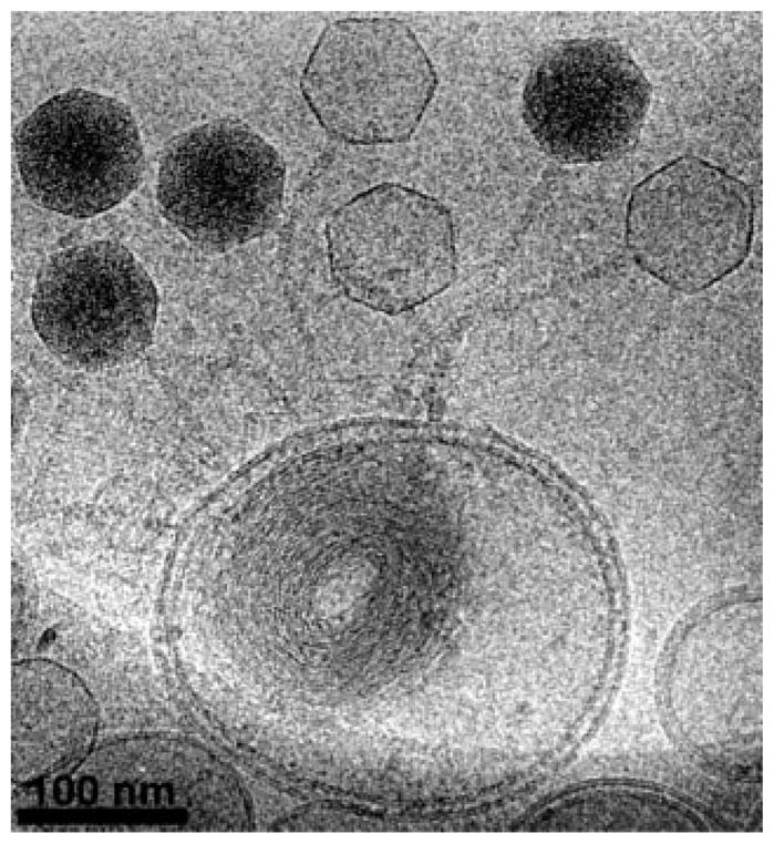
Early X-ray scattering experiments showed that DNA within bacteriophages is tightly packed into a nearly crystalline hexagonal array, forming the basis for models of the arrangement of DNA within the capsids.9 These were followed by cryo-electron microscopy measurements that used averaging of tens of images to reveal a picture of the many concentric rings of DNA within bacteriophage capsids.10 The clearest pictures of tightly-bent DNA in viruses come from recent asymmetric cryo-electron microscopy reconstructions. An example is shown in Figure 2. These studies combine data from thousands of particles to produce three-dimensional images of the capsid and genomic DNA, allowing the visualization of several layers of DNA loops within the capsid.11–13
FIGURE 2.

Images of packaged viral DNA. This figure shows two recent reconstructions using cryo electron microsopy of the packaged DNA. (A) Phage ε15 DNA from Jiang et al.11—reconstruction without symmetry (reprinted by permission from Macmillan Publishers Ltd). The size of the scale bar is 10 nm. (B) Phage P22 DNA, with the portal shown in red (courtesy: Gabriel Lander and Jack Johnson). This view is looking into the capsid at the portal (the entry site for DNA) and the green hoops reflect density corresponding to the packed DNA.
Models of Tightly-Packed Viral DNA
To gain intuition about the forces involved in DNA packing and ejection from viruses, many models of tightly bent DNA have been proposed.14–20 The force due to bending is small during the initial stages of packing. However, as more DNA is forced into the capsid, the DNA takes up increasing amounts of available space and loops must be produced at smaller radii, increasing the force. The resulting DNA structure, thought to involve concentric loops of DNA arranged at decreasing radii about a single axis, is referred to as an “inverse spool”. Alternative models of the packed DNA structure have also been proposed,21 but the asymmetric cryo-electron microscopy structures described above strongly support the inverse spool model. In one of the original models of the energetics of viral DNA packing,14 the DNA is assumed to be packed tightly into an inverse spool, with strands touching each other so that they are locally aligned on a square lattice, with an interstrand separation d = 2 nm. Applied to bacteriophage λ, this model predicts that the DNA loops in the center of the capsid have a radius as small as ~10 nm.
Because of their high negative charge, neighboring DNA loops do not touch each other, but are pushed apart by electrostatic and hydration forces.22–27 The radius of the innermost loop will therefore be determined by an equilibrium between bending forces and the DNA–DNA interactions. This effect was taken into account in subsequent models of DNA packing.16–18,28–30 These models are generally consistent with each other, but they focus on different kinds of predictions, such as the structure of the DNA, the forces and pressures involved in DNA confinement, and the effect of ions and DNA condensing agents. A recent advance is the construction of a model where all parameters were matched to conditions of an experiment on bacteriophage λ; the predictions could then be compared directly to experimental results without fitting, giving weight to the correctness of the model. According to this model, the inner loop will be at the extremely tight radius of ~3 nm.31
To further explore the predictions of these models, we can make a simple estimate of how much force is required to bend DNA to various amounts during the packaging of a bacteriophage λ capsid, which has a radius of about 29 nm. The energy to bend a DNA segment of length ΔL into an arc of circle of radius R is given by1
| (2) |
This implies that inserting a segment of DNA of length ΔL into the capsid, when it must be bent at this radius, will require a force of
| (3) |
If we use a radius R = 29 nm this results in a force of order 0.12 pN, a relatively small force compared to the maximum forces exerted by molecular motors, which are typically in the pN regime. The force required to bend the DNA increases as the radius of the bent DNA decreases. At R = 3 nm, a force of 12 pN is required. This is a high force that implies that a strong molecular motor is required simply to overcome the bending stiffness of DNA. The required force is supplied by packing motors, which consume ~1 ATP/2 bp and produce forces as high as 60 pN.32,33
It is important to keep in mind that the energy of compressed DNA within the capsid is stored in both bending and DNA–DNA interaction components: due to the force balance, the total predicted force is exactly twice what we calculated above for DNA bending alone. However, the outer strands of DNA are bent less severely than the inner strands, so that the total energy is stored primarily in the DNA–DNA interaction. In fact, in the absence of an energetic cost to DNA bending, the DNA–DNA repulsive interactions would expand the DNA crystal in the capsid, leading to large curvatures toward the capsid center. High forces would still be produced during packing due to the DNA–DNA interactions. It is the DNA–DNA interactions that are responsible for the extremely tight bending thought to exist at the center of a phage capsid. Many authors speculate that the DNA may actually be bent so tightly that it forms 180° kinks.9,21
The mathematical models described above are complemented by computer simulations that aim to show how the DNA arranges itself into a spool during packing34–38 or how it moves out of the capsid during ejection.39,40 Simulations present plausible scenarios for the details of packing and ejection, but their most interesting features are tied to assumptions that may or may not be correct in biological situations. For example, in the work by Spakowitz and Wang,38 the arrangement of the DNA depends highly on whether it is twisted during packing. Zandi et al.39 consider that the forces that pull DNA out from the capsid depend on the range and strength of attraction of DNA-binding particles (such as RNA polymerase) in the cytoplasm. This issue is elaborated on in a later theoretical study.41 In general, these kinds of assumptions present excellent targets for experimentalists desiring to improve our understanding of DNA mechanics in bacteriophages.
Measurements of the Packing and Ejection Processes
The forces and dynamics of bacteriophage packing and ejection are being studied with a variety of innovative experimental techniques. Since a single bacteriophage possesses a complex structure and follows a complex life cycle, averaging experimental data over particles will destroy critical information. A conceptually simple but experimentally demanding solution is to study single virus particles with microscopy and pN-scale force-probe technology, revealing information without averaging. A key experiment demonstrating the power of this approach is a study in which one end of the DNA of bacteriophage φ29 was held in an optical tweezer during genome packing.32 The optical tweezer experiment can be run without feedback, in which case the force generated by the packing motor reaches an equilibrium with the force applied by the tweezer, or with constant-force feedback, in which case there is a constant tension on the DNA during packing. What was seen in the no-feedback case is that the motor can produce a force around 57 pN before it stalls, making reverse slips more and more often as it approaches the stall point. Using the constant-force case, it was determined that an opposing force was building up in the capsid throughout packing, reaching a value of about 50 pN near 100% packing.
For studying the ejection process, corresponding single-molecule techniques are not practical, since it is difficult to push on a long piece of flexible DNA. However, osmotic pressure can be used to push on the DNA, freezing it in an equilibrium configuration where only a fraction of the DNA, from 0 to 100%, has been ejected. Though single particles are not observed with this technique, the osmotic suppression of ejection allows us effectively to take a snapshot of a single moment in the ejection process. A series of such experiments was done on λ phage, demonstrating forces as high as 10 pN (the force corresponding to 25 atm of external osmotic pressure).31,42–44 Since φ29 and λ are both packed to a similar DNA density, it is unclear whether the six fold difference in forces is caused by a difference between the phages or a difference in the experimental conditions. The experiments on φ29 and λ are all consistent with the models described above.
The dynamics of ejection, which is not accessible with osmotic techniques, has been most completely addressed in recent in vivo studies on phage T7 and φ29, where it was shown that DNA enters the cell over a period of 10–30 min.45,46 In this case, the study reveals the extent to which the force built up by DNA can drive the ejection. For φ29, it appears that force from within the capsid only drives the first part of ejection, after which an unknown cytoplasmic source of energy pulls the rest of the DNA into the cell. In the case of T7, force within the capsid does not have any apparent effect on the ejection process, and the entire DNA strand is translocated at a constant, relatively slow speed by RNA polymerase.47
The λ genome is known to completely enter the cell in less than 2 min, according to cyclization times and the dam-nuclease assay.48 However, no lower bound exists for this transfer time; we do not know how fast the λ genome can unwind from its spool. Quantitative data about λ ejection, combined with the equilibrium force measurements, could confirm or invalidate models of the DNA ejection process.
One interesting related experiment addressed the issue using lipid vesicles containing LamB (the receptor to which phage λ attaches and which induces ejection) and filled with ethidium bromide.49 When the DNA was ejected from λ particles into the vesicles the ethidium bromide binds to the entering DNA, causing an increase in fluorescence. The time-scale of ejection as determined by this experiment was ~30 s. However, only ~1000 molecules of ethidium bromide were present in each of the vesicles, so that the experiment was only capable of measuring the first few kbp of DNA entry. The vesicles themselves were ~100 nm in diameter, so that the DNA was entering a region where it would continue to be highly bent. It is also important to realize that this experiment measures the bulk fluorescence of the entire phage ejection reaction, rather than the fluorescence of individual phage genomes, so the observed fluorescent signal will be a combination (mathematically, a convolution) of the initiation process and the actual genome transfer. Recent single molecule experiments designed to address all of these issues show that the genome transfer is actually much faster than initiation, with a timescale of about 10 s.50
Figure 3 shows a beautiful experiment which illustrates phage that have ejected their genomes into a lipid bilayer vesicle in a way that is analogous to the experiment on ejection dynamics. One of the most interesting features of the ejected DNA which also bears on the issue of charge interactions is that the DNA within the vesicle is collapsed into a toroid. More generally, these in vitro experiments on DNA toroids may help shed light on the physical forces associated with tightly bent DNA.52,53
FIGURE 3.
Images of DNA ejected into a lipid bilayer vesicle. Empty capsids are distinguishable from their full counterparts because the full capsids are much darker (reprinted with permission from Elsevier).51
For the first time, recent in vitro studies of T5 have described the dynamics of DNA ejection at both the bulk54,55 and single particle56 levels. In T5, it appears that nicks present in the genome cause the ejection to halt temporarily at defined locations. The ejection proceeds between these halting points extremely quickly, within one frame of video: it is now clear that DNA can eject at a rate of at least 75 kbp/s, but again, no lower bound can be placed on the transfer time.
Future Work on DNA Bending in Viruses
We have seen that DNA is tightly bent within many viruses; in the bacteriophages, in particular, it may be bent nearly to the limit of DNA flexibility, with a radius of curvature of roughly 3 nm. A handful of experiments has been done to investigate how the DNA unpacks, and it appears that different phages follow very different ejection mechanisms, with some ejecting tens of kbp in a fraction of a second and others taking minutes to release their genomes. However, each phage has been studied with different techniques, so it is hard to make cross-species comparisons and we do not yet have a complete picture of the packing and ejection process for any phage. The versatility of bacteriophage λ suggests that a complete set of studies may soon be possible, using all of the in vivo and in vitro techniques described above. It will be particularly interesting to learn how the DNA is wound into the capsid, what parts spin or play during packing and ejection, and what kind of frictional forces result from the motion of the DNA.
Bacteriophages have long served as model systems for understanding a variety of processes in biology; by studying DNA bending in phages, we gain insight into the operation of similarly-constructed eukaryotic viruses as well as DNA packing and transport in general.
DNA PACKING IN EUKARYOTES
Like viruses, eukaryotic cells pack their genomes by tightly bending them. In these cells, chromosomal DNA is packed in a hierarchical structure. At the lowest level in the hierarchy (and our prime concern here), DNA is wrapped in 147 base pair segments roughly 1 ¾ times around a protein complex (the histone octamer) to form a structure known as the nucleosome as shown in Figure 4.
FIGURE 4.
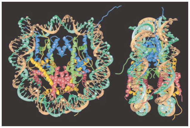
Structure of the nucleosome. Two orthogonal views of the nucleosome showing the wrapping of the DNA around an octameric histone protein core (reprinted by permission from Macmillan Publishers Ltd).57 The core histone proteins are colored yellow, red, blue and green for histone H2A, H2B, H3, and H4, respectively. There are two copies of each histone in the core histone octamer. The two strands of the double helix are colored cyan and brown. The diameter of the nucleosome is roughly 11 nm and its height is roughly 6 nm.
The nucleosomal packing motif is reiterated at short intervals along the entire length of the genomic DNA, with nucleosomes separated by short ≈ 10–50 bp-long stretches of unwrapped linker DNA. Thus, 75–90% of eukaryotic genomic DNA is wrapped in nucleosomes. The nucleosome structure itself has several particularly striking aspects. First, the DNA is exceptionally tightly bent compared to the intrinsic length scale over which DNA is flexible: the 147 bp DNA length corresponds to one persistence length, which is wrapped into loops of only ≈ 80 bp per superhelical turn. Second, the two adjacent gyres of wrapped DNA are packed extremely close together, and with their backbones in close apposition, suggesting the likelihood of strong electrostatic interactions between the two DNA gyres. Third, most of the surface of the wrapped DNA is occluded from interaction with other proteins: it is occluded on one face by close contact with the protein surface and on the side by the close proximity of the second super helical turn of the wrapped DNA. Since most of the genomic DNA is wrapped in nucleosomes, the preferred locations of nucleosomes may strongly impact the DNA accessibility and function of critical DNA regions.
The aim of the discussion here is to explore the consequences of the fact that the nucleosomal DNA is tightly bent on the scale of the persistence length. Despite the energetic cost of bending the DNA on these small scales, the favorable contacts between positively-charged residues on the histones and the negative charges on the DNA suffice to overcome this energetic penalty.58,59 Indeed, in some sense the question is not why do nucleosomes form, but rather, how do proteins that need to gain access to nucleosomal DNA do so?
Equilibrium and Dynamics of Nucleosomal DNA Accessibility
X-ray crystal structures of nucleosomes57,60 (see Figure 4) show the wrapped DNA to be largely inaccessible to the many protein complexes that must bind it for essential DNA transactions such as replication, transcription, recombination, and repair.61–65 However, as is often the case in biology, the structure appears to be tuned for marginal net stability, with the attractive interactions slightly exceeding the elastic cost of wrapping tightly around the positively-charged protein spools. Probabilities depend exponentially on the energetics, so the probabilities overwhelmingly favor the wrapped state; but because the energetics are marginal, there will nevertheless be frequent (if short lived) unwrapping events.66
To see how the relevant energies compete with each other, we resort to some simple estimates. The energy associated with bending the DNA can be estimated simply by invoking a version of Eq. (2) applicable to circular loops of radius R and given by
| (4) |
where ξp is the DNA persistence length. Here we use a flexural rigidity, κ = ξpkBT. To get an estimate of the energy scale, we note that the radius of curvature at the centerline of the DNA is roughly 4.5 nm, corresponding to an energy Eloop = 35 kBT. As noted above, the second key contribution to the energy comes from the interactions between the charges on the histones (positive charges) and the DNA (negative charges). Over the 1 ¾ times that the DNA wraps around the histone octamer, there are 14 distinct contacts each of which has a contact energy of roughly −6 kBT. These contacts between the protein core of the nucleosome and the wrapped DNA occur in patches, every DNA helical turn, when the minor groove (DNA backbone) wraps around and faces inward toward the protein core.57,60 The contact energy can also be modeled as a continuous adhesion energy Econtact = γadhL, where γadh is an energy/length with a value of roughly γadh ≈ −2.0kBT/nm, with the minus sign signaling that this is a favorable contact. These values can be obtained by fitting this simple model to measurements on the equilibrium accessibility of nucleosomes.67
One of the principal puzzles posed by the function of nucleosomes is how these structures are at once stable and yet accessible to DNA-binding proteins. Restriction enzymes experience the same accessibility obstacles for action on nucleosomal DNA as do eukaryotic protein complexes, and have been used to probe the equilibrium accessibility of the wrapped DNA.67 The basic idea behind these experiments is to measure the probability of restriction digestion as a function of burial depth of the restriction site of interest within the nucleosome. These studies reveal that stretches of the nucleosomal DNA located a short distance inside the nucleosome from one end act as though they are (unwrapped) naked DNA molecules a surprisingly large fraction (several percent) of the time, i.e., there is an equilibrium constant for dynamic unwrapping of the ends of the nucleosomal DNA on the order of 0.01–0.1. This equilibrium accessibility drops progressively with distance further inside the nucleosome, decreasing to 10−4 to 10−5 for sites located near the middle of the nucleosome.
These findings can be understood using the simple model described above based on the structure of the nucleosome. For simplicity, we assume that each contact patch contributes an equivalent net favorable free energy for DNA wrapping, and that access to sites further inside the nucleosome is achieved by starting at one end of the nucleosome, and unwrapping the DNA one helical turn at a time, breaking contact patches in succession, until enough DNA is unwrapped such that a given site is now accessible. Each broken contact costs a certain amount of net free energy, so access to sites further inside the nucleosome comes with a stepwise increasing cost in free energy, and a corresponding stepwise decrease in probability or equilibrium constant. In the continuum model described above, one assumes that the free energy cost is a continuous function of the degree of unwrapping. In particular, if the nucleosomal DNA is peeled off by an amount xe, then the free energy of the bound DNA is F(xe) = (γbend − γadh)(L − xe) (Figure 5 inset). Using this simple model of the energetics of nucleosomal DNA, the configurational equilibrium constant can be computed as
FIGURE 5.
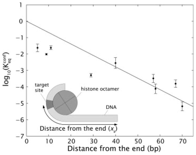
Configurational equilibrium constant. Measured values of equilibrium accessibility and corresponding results from the model of nucleosome energetics.67 The inset shows a schematic of the coordinate system used to define the burial depth of the binding site of interest.
| (5) |
where xre is the depth of the site of interest, L is the total length of wrapped DNA and γbend is the bending energy per unit length associated with the wrapped DNA. A fit of this model to the experimental data67 using γadh as the only free parameter is shown in Figure 5.
The experiments described above show nucleosomal DNA to be dynamically accessible, but leave open the question of the actual rates. Two new experiments show that nucleosomes spontaneously open to allow access to at least the first 20–30 bp on timescales as short as 250 ms.66 The dynamic accessibility implies that transcription factor binding sites, promoters, etc. that are buried in a nucleosome can remain active although at a significantly lower level than identical sequences that are unbound. A large number of nucleosome remodeling factors have been identified suggesting that cells may further increase the accessibility of buried sites by active mechanisms.68–71
Nucleosomes look like those imaged by X-ray crystallography for very short periods of time before spontaneously undergoing large scale opening conformational changes. However, these open states do not last long, typically only 10–50 ms, before the DNA spontaneously rewraps. A mean-first-passage-time calculation based on a continuum version of the nucleosome-DNA adhesion picture described above allows for parameter-free predictions of the opening and rewrapping rates as a function of distance.72 A different experiment probing unwrapping to sites further inside the nucleosome appears to concur at least qualitatively with these predictions, showing that the unwrapping to greater depth does occur on a significantly slower timescale.73
Sequence-Dependence of Nucleosome Formation and Accessibility
In our discussion of DNA packing in viruses, we showed how the length of the DNA molecule could be used as a tunable dial to alter the mechanical forces associated with the packaged DNA. DNA sequence is yet another way in which the energetics of tightly-bent DNA can be tuned and altered. The key point is that different sequences have different intrinsic bendability, and hence a quantitatively different tendency to form nucleosomes. In particular, the tight bending of DNA in nucleosomes causes them to prefer certain particular DNA sequences over others. DNA sequences exhibiting a greater than 5000-fold range of affinities for wrapping into nucleosomes are documented74,75; moreover, the range of affinities may be even greater, as the experiments used to measure the relative affinities may artificially underestimate the true range.
These sequence preferences are not due to particular favorable base-specific interactions,57 as would normally be the case for site-specific protein–DNA complexes. Rather, sequence-dependent nucleosome positioning represents an extreme case of indirect readout.76 In indirect readout, sequence preferences arise from the differing abilities of differing DNA sequences to adopt particular idiosyncratic conformations required by the proteins, which for the case of the nucleosome, is dominated by the extremely tight DNA bending required.
The most important DNA sequence motifs that confer high affinity binding to the nucleosome are AA, TT, or TA di-nucleotide steps (i.e., an A followed by another A, and so on), which recur every 10 bp, in phase with the DNA helical repeat, every time the DNA minor groove (phosphodiester backbone) rotates around to face inward toward the center of curvature of the nucleosomes protein core. A survey of high resolution X-ray crystallographic structures of DNA5 suggests that no dinucleotide steps favor such bending into the minor groove, but, evidently, these particular steps minimize the unfavorable energetic cost. There exist also weaker preferences for certain other steps, most notably GC (that is, G followed by C) to occur exactly out of phase with the AA/TT/TA steps, every time the minor groove faces outward.
Several lines of reasoning and, more importantly, direct experimental tests75,77 show that particular DNA sequences that are especially soft for bending (as opposed to intrinsically bent in the manner favored by the nucleosome) make particularly stable nucleosomes. The role of DNA bending in determining these sequence preferences is illustrated dramatically by comparing the free energy of cyclization (the cost to make a small loop in solution) with the free energy of nucleosome formation as shown in Figure 6. Nevertheless, the detailed molecular mechanics basis of all of these sequence preferences remains unknown and is an important topic for further research.
FIGURE 6.
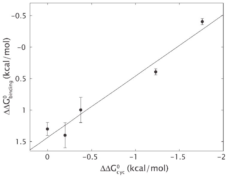
Free energy of cyclization and nucleosome formation. Difference free energies for wrapping of different 94 bp DNAs around the core histone H32H42 tetramer are plotted against the difference free energies of cyclization for these same DNAs.74 The line illustrates the least-squares fit to the data. The slope of the line is one, implying that the entirety of the difference in affinity for wrapping around histones can be explained by the difference in the ability to cyclize.
The biological significance of these nucleosomal DNA sequence preferences arises because they imply that nucleosomes are not distributed randomly along genomic DNA. Eukaryotic genomes utilize these sequence preferences together with the powerful force of steric hindrance—nucleosomes occupy space and cannot overlap—to encode an intrinsic nucleosome organization. The resulting in vivo distribution of nucleosome occupancies appears to facilitate many aspects of chromosome function, including transcription factor binding, transcription initiation, and even remodeling of the nucleosomes themselves.78
Interestingly, eubacterial genomes, which lack histones, nevertheless encode 11 bp-periodic distributions of AA/TT dinucleotides.79 These are not attributable only to protein coding requirements80,81 and instead suggest that prokaryotic genomes encode an intrinsic three-dimensional organization of their own chromosomes different from, but analogous in some ways to, the intrinsic nucleosome organization encoded in eukaryotic genomes.
TIGHTLY BENT DNA IN TRANSCRIPTIONAL REGULATION
Gene expression is subject to tight control and one of the most important mechanisms of regulation occurs at the level of transcription. Transcriptional regulation is carried out by a variety of DNA-binding proteins known as transcription factors. The two key case studies that led to the elucidation of the operon concept (the idea that there are genes that control other genes),82 namely the lac operon and the λ switch, both involve DNA looping.83,84 In these cases, the DNA binding proteins that mediate transcriptional control bind at two sites on the DNA simultaneously, looping the intervening DNA. Indeed, tightly bent DNA is a ubiquitous motif in both prokaryotic and eukaryotic transcriptional regulation. In Table I we highlight some of the best known examples of this regulatory architecture. In most cases, the relevant loops have lengths that are comparable to or smaller than the persistence length.
Table I.
DNA Looping in Prokaryotic and Eukaryotic Transcriptional Regulation
| Molecule or locus | Mode of action | Wild type loop lengths (bp) |
|---|---|---|
| Lac repressor83 | Repression | 92, 401 |
| AraC85 | Repression and activation | 210 |
| Gal repressor85 | Repression | 115 |
| Deo repressor85 | Repression | 270, 599, 869 |
| Nag repressor86 | Repression | 93 |
| NtrC87 | Activation | 110–140 |
| λ repressor84,88 | Repression and activation | ~2400 |
| XylR89 | Activation | ~150 |
| PapI87,90 | Activation | ~100 |
| β-globin locus91,92 | Activation | 40,000–60,000 |
| RXR93 | Activation | ~0–500 |
| SpGCF194 | Activation, domain intercommunication | ~0–2500 |
| HSTF95 | Activation | 23 |
| p5396 | Repression and activation | 50–3000 |
| Sp197–99 | Activation | ~1,800 |
| c-Myb and C/EBP100 | Activation | ~80 |
Loop lengths and mechanisms of action of some of the best known looping systems in bacteria and eukaryotes. Note that these loop lengths suggest tightly bent configurations since the in vitro measured persistence length is 150 bp.
Given that the persistence length is the scale over which DNA is stiff, it is surprising that short loops play such an important role in transcription. The implicit assumption that leads to that surprise is that the effective in vivo DNA flexibility is the same as that measured extensively for bare DNA in vitro. However, such in vitro measurements generally only probe length scales much longer than those relevant to the structures in Figure 1.4 To analyze the role of tightly bent DNA in transcriptional regulation we will focus on three physical mechanisms: (i) the in vivo bendability and twistability of DNA, (ii) the contribution from protein conformation, and (iii) the presence of a whole battery of nonspecific or nucleoid-associated DNA binding proteins, which play an active role in determining structural and dynamical properties of the bacterial chromosome.
Though there are a host of interesting examples of transcriptional regulation that involve DNA looping, we focus almost exclusively on the dissection of the role of looping in the lac operon. The lac operon refers to the genes responsible for lactose metabolism in bacteria.83 In particular, when faced with an absence of glucose and the presence of lactose, this operon will be “on” resulting in the production of β-galactosidase (and several other proteins as well), the enzyme responsible for the digestion of lactose. The challenge is to see how in vivo and in vitro experiments and modeling approaches can be used to tease out the mechanism and biological significance of DNA looping: why do genomes bother to loop?
We will focus on data in which the mechanical properties of DNA are used as an adjustable dial to tune a desired biological outcome during transcriptional regulation. In particular, we will address in vivo experiments like those performed by Müller et al.,101 Law et al.,102 and Becker et al.,103 where the level of repression is systematically measured as a function of the distance between two binding sites for Lac repressor (Figure 7A inset). For reviews on Lac repressor refer to Matthews and Nichols105 and to Lewis.106 In addition, we will examine corresponding in vitro measurements of the interaction between Lac repressor and its target DNA.
FIGURE 7.

In vivo DNA looping by Lac repressor and the in vitro challenge. (A) Data from Müller et al.101 showing repression as a function of distance between operators. (B) Change in looping free energy obtained from the Müller-Hill data (black) and theoretical prediction of the energy of cyclization of a DNA molecule based on the worm like chain model104 and assuming a volume for E. coli of Vcell ≈ 1 μm3 such that ΔFcyclization = −ln(Jcyclization Vcell). Note that the minima in the two curves do not coincide, suggesting that the effective looping free energy in vivo is not the same as the bare looping free energy deduced from in vitro cyclization measurements. In addition, there is an overall shift in the scales in the two cases. (Inset, B) Difference in the magnitude of the twist modulation between the looping energy obtained from the Becker et al.103 data and the theoretical cyclization energy based on harmonic deformations of the base steps.75
As shown in Table I there are many other interesting examples of DNA looping in transcriptional regulation. We focus on one such case study because in this case, there are a broad range of quantitative measurements that permit a careful comparison of results from both in vivo and in vitro experiments. These results may be used to form a coherent picture of looping in transcriptional regulation though current models fall short of a complete picture of these problems that leads to consistent, falsifiable experimental predictions. We view this as an opportunity to propose a set of careful quantitative and systematic experiments that will help decouple the contributions and importance of the different molecular players in this process.
In Vivo DNA Looping: Using Cells as Test Tubes
The most common and straightforward way of characterizing the action of some regulatory motif on gene expression is by measuring relative changes in the activity or concentration of the regulated protein product. The classic reporter has been β-galactosidase. The concentration of this gene product is characterized by measuring its activity in lysed cells using a colorimetric assay.107 The unequivocal signature for DNA looping since its discovery by Schleif and coworkers in the arabinose operon has been the modulation of gene expression as a function of the length of the DNA loop with a periodicity of roughly 11 bp corresponding to the effective in vivo helical pitch of DNA.108–110 This type of experiment shows how quantitative, single-molecule mechanical properties can be extracted from cells by looking at changes in the protein expression profile of an entire population of cells. It is remarkable that changes in DNA such as making the molecule a single base pair longer or shorter, can result in such clear macroscopic effects in an ensemble of cells. An example of this kind of data for the lac operon is shown in Figure 7A. In many ways, the remainder of this review centers on understanding the many distinctive features of this curve which has hidden within it several intriguing clues and puzzles concerning DNA mechanics.
Precise and rich data like those shown in Figure 7A present a variety of theoretical challenges. Thermodynamic models of transcriptional regulation111,112 have been used to extract the free energy of looping which is a measure of the cost of the looped configuration as a function of the distance between the operators.102,113–115 These models use equilibrium statistical mechanics to describe the probability of transcription as it is modulated by the presence of the repressor and its partner looped DNA.
One of the biggest challenges in modeling the Lac repressor-loop-mediated repression lies in the fact that the free energy of the looped configuration is determined by a variety of factors. In addition to the free energy of DNA looping itself, it is also necessary to consider the geometry and flexibility of the looping protein116,117 and the presence of nonspecific binding proteins such as HU, IHF, and H-NS in the background103 (for a review of the role of these proteins in the organization of bacterial chromatin refer to Luijsterburg et al.118). Nevertheless, it is still meaningful as a first approximation to compare the in vivo looping energy extracted from these experiments to the energy of cyclization of DNA circles defined in previous sections at the same length scales, where the additional subtleties of the in vivo experiment are not present. Such a comparison is shown in Figure 7B.
There are at least three striking features of the in vivo looping energy in comparison with its in vitro counterpart. First, the minimum in the looping free energy at 70 bp does not coincide with the expected cyclization minimum at around three persistence lengths. Second, at 70 bp, there is an overall offset between the in vitro and in vivo values and, finally, a difference in the amplitude of the twist modulation. All of these features suggest that it is easier for DNA to adopt tightly bent configurations in the in vivo setting than would be expected from our intuition based on studies of DNA mechanics in vitro. In the remainder of this section, we review some of the available evidence that sheds light on the origin of these differences between in vivo DNA looping and in vitro DNA cyclization.
The position of the minimum in the in vivo looping free energy shown in Figure 7B suggests that for these tightly bent configurations, DNA has a lower effective persistence length than the canonical value of ~150 bp. Interestingly, proteins that are expected to be more flexible than wild type Lac repressor such as AraC109,119–121 and Lac repressor mutants122 present a different shape in their gene expression curves and, consequently, in their looping energies. In both cases the looping energy does not display a minimum. Rather, it keeps decreasing as the interoperator distance gets shorter. Various computational studies have addressed the issue of protein flexibility.123–126 Even though the difference in the position of the minima can be accounted for, a smaller value of persistence length is still needed in order to fit the models to the available in vivo data.126
It can be argued that the main difference in the absolute value of the looping energy between cyclization and in vivo looping in transcriptional regulation can be accounted for by a difference in the definition of the standard states or zeros of free energy. For example, in the in vitro case, the reference state is defined as the uncyclized linear molecule in solution. On the other hand, in the more complex in vivo case, this reference state is not as clearly defined. In particular, in this case, even when not bound to specific operator sites, DNA is bound nonspecifically,127 presumably resulting in a host of different looped states. The set of all of these different looped states defines the reference state for the in vivo case. Additionally, the presence of negative supercoiling inside the cell128 and of nonspecific DNA binding proteins such as the histone-like HU103 have been shown to be factors that can modify the reference energy. Without knowledge of how this reference state is determined, no absolute comparison between invivo and in vitro data can be made.
The third key feature calling for attention is the unexpectedly small amplitude of the periodic modulation in the in vivo looping free energy. One explanation for this difference between the effective in vivo looping free energy and the cyclization free energy could be a higher DNA twistability of tightly bent DNA.75 So far, the available computational models have not been able to show how protein flexibility alone can account for this difference.126 Müller-Hill and coworkers proposed that such an apparent lower modulation could be explained if different loop species were present.129 These different species could correspond to different topoisomers,130 different orientations of the operators with respect to the symmetric binding heads or different conformations in Lac repressor,124,126,131 which are supported by in vitro evidence (see latter).
Understanding DNA looping in vivo in bacteria requires understanding the role of tightly bent DNA in these systems. However, the in vivo approach only yields a single quantity, namely, the looping free energy. The problem is that this quantity reflects not only the mechanical properties of DNA, but also the effect of protein flexibility and the effects of other proteins bound to the DNA. Addressing this problem from the in vitro perspective of biochemistry allows for a more controlled characterization of the effect of the different molecular players in this process. We conclude this section by reviewing some recent and classical in vitro studies of DNA looping by Lac repressor.
In Vitro DNA Looping: DNA Mechanics One Molecule at a Time
Complex cellular processes like those described above can be tackled in vitro using the tools of solution biochemistry and single molecule biophysics. Both of these approaches have been unleashed on the problem of DNA looping in the context of transcriptional regulation.
Bulk binding assays involving DNA-binding proteins such as Lac repressor and their DNA targets measure the affinity of these proteins for configurations with different looping lengths or degree of supercoiling, for example. Filter binding assays and electrophoretic mobility shift assays are two examples of these kinds of technique. In the gel-shift assay, the electrophoretic mobility of a given fragment of DNA is measured both in the absence and presence of the DNA-binding protein of interest. When the DNA-binding protein binds to the DNA fragment, it changes its motility in the gel and is detected as a new band. By tuning the concentration of the binding protein, as well as controlling variables dictating DNA mechanics (such as the looping length or the degree of supercoiling), it is possible to measure how these mechanical variables alter the binding probability.
In contrast to the in vivo observation, using the gel shift assay, Krämer et al. determined that the probability of looping decreases as the distance between operators on a linear DNA fragment decreases from 210 to 60 base pairs.132 This result agrees with the observations by Hsieh et al. using the filter binding assay, whose quantitative results are shown in Figure 8.133 This disagreement in the behavior of the looping free energy as the distance between operators decreases between the in vivo and in vitro experiments is a stark reminder of the challenge of reconciling the in vitro and in vivo pictures of DNA mechanics in general, and protein-mediated looping in specifically.
FIGURE 8.
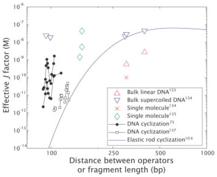
Effective J-factors for in vitro DNA looping. The graph is constructed by using a variety of different in vitro measurements to derive an effective looping J-factor, even in those cases where there was no direct measurement of J itself. The derived values were obtained from: (i) bulk linear DNA,133 (ii) bulk super-coiled DNA,134 (iii) single molecule measurements,135,136 (iv) DNA cyclization75,137 and the blue curve is a theoretical curve for cyclization corresponding to an extrapolation of the elastic rod model.104
Similar experiments have been used to characterize the role of supercoiling by using supercoiled plasmids.134,129,138 Interestingly, these experiments reveal an increase in the affinity of the Lac repressor to a single site showing that negative supercoiling favors binding. Most importantly though, a dramatic increase in the looping probability was observed. This increase in looping probability is revealed in changes in the protein–DNA complex dissociation times that varied from 2 h to more than 20 h.129 These experiments also suggested that the looping energy does not change much over distances between 100 and 500 bp for a negatively super-coiled template.134 However, this could not be confirmed because the distance between operators was not systematically varied. A decrease in the twist modulation was also observed, suggesting, as was mentioned in the previous section, that multiple topoisomers coexist for certain separations.129
These results have been supplemented with several other classes of experiments, some of which involve the direct observation of individual loops. Using microscopy techniques such as electron microscopy129,132 and atomic force microscopy,136 individual loops can be observed and key parameters such as the loop length can be measured. These experiments have been valuable not only in the context of Lac repressor, but also in identifying different looping motifs in complex cis-regulatory regions in eukaryotic systems.94
Another important class of experiments that have shed light on the mechanics of DNA looping in vitro are single-molecule measurements using the Tethered Particle Motion (TPM) method as shown in Figure 9.139 TPM was first used by Finzi and Gelles in the context of DNA looping to directly detect Lac repressor mediated loop formation and breakdown, and to measure the kinetics of such processes.140 In this method, a DNA molecule is tethered between a microscope slide and a microsphere, which is large enough to be imaged with conventional optical microscopy. The Brownian motion of the bead serves as a reporter of the underlying DNA dynamics. In particular, when the molecule is unlooped, the tether has its full length and the excursions of the bead are large. When the DNA is looped, the tether is shortened and the excursions are reduced.141–148 Thus, modulations in motion reflect conformational changes in the tethered molecule. This method has recently revealed136,149 the presence of two-looped states which is consistent with the presence of multiple configurations observed using FRET,150,151 electron microscopy studies152 and suggested by X-ray crystallography studies.116 All of these experiments suggest an important role for protein flexibility. A more sophisticated technique which has been successfully applied to Gal repressor is the magnetic tweezer assay.153 In this case, the tether can be stretched and twisted as the dynamics of looping and unlooping are followed leading to measurements of the underlying kinetics, thermodynamics, and supercoiling dependence.
FIGURE 9.
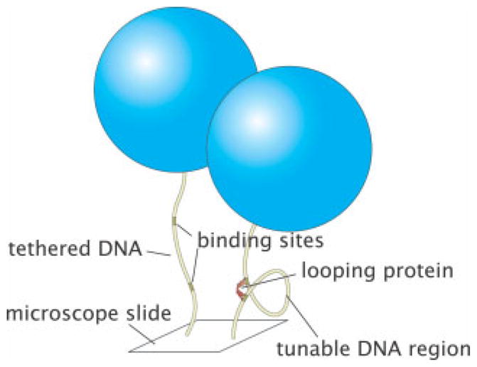
Illustration of TPM method. Schematics of both the unlooped and looped states which show how the effective tether length is a reporter of the state of looping. Typical tethers have a length of 1000 bp and typical bead sizes are 0.2–1.0 μm.
Even though Lac repressor can loop in the absence of any other DNA binding proteins, other systems such as GalR require the presence of the nonspecific DNA binding protein HU.153 HU has been proven to alter the effective flexibility of DNA.154 However, this issue has not been studied systematically in the context of DNA looping or in the presence of other nonspecific binding proteins such as H-NS and IHF.
In spite of more than two decades of investigation, there is still no comprehensive or quantitative link between in vivo and in vitro studies of looping and DNA conformation (Figures 7 and 8). For instance, although it is known qualitatively that nucleoid-associated-protein binding and supercoiling can both significantly enhance looping efficiency, we still do not know whether these mechanisms are sufficient alone or in tandem to explain the dependence of repression on inter-operator spacing observed in a host of biological systems. To get to the bottom of these questions will require further systematic and quantitative experiments. In particular, systematic experiments which vary specific experimental tuning parameters (operator distance, sequence, concentration of nucleoid-associated proteins) need to be performed.
Most of the in vivo data on DNA mechanics as revealed by transcriptional regulation suggests an increased DNA flexibility, signaling that there is more to the effective in vivo looping free energy than is offered by the wormlike chain model alone. Interestingly, recent in vitro experiments also suggest short-length scale anomalies in DNA mechanics,4,75,155,156 even though no consensus has been reached.133 To fully understand the role of tightly bent DNA in transcriptional regulation the contribution of the different molecular players (intrinsic DNA mechanics, architectural proteins, transcription factors, supercoiling) has to be decoupled.
CONCLUSION
We have argued that tightly bent DNA is a common feature in living organisms. The packing of genomic DNA in viruses, prokaryotes and eukaryotes involves both indirect (confinement by protein capsids) and direct (architectural proteins such as HU and histones) interactions between DNA and proteins which lead to highly deformed DNA configurations. Similarly, transcriptional regulation in prokaryotes and eukaryotes routinely requires the formation of DNA loops involving DNA segments that are shorter than the persistence length.
Interestingly, in all of the examples described in this review, the physical mechanisms associated with tightly bent DNA lead to biological consequences. For example, because of the energetic costs associated with genome confinement, bacteriophages have extremely strong molecular motors to pack their DNA. Eukaryotic DNA is packed in nucleosomes, requiring a bevy of proteins to rearrange nucleosomes them. In addition, nucleosomes preferentially bind to DNA sequences that are easy to bend. Combinatorial control in transcriptional regulation is often mediated by transcription factors that induce DNA looping. In each of these cases, there is a direct connection between the physical properties of DNA and its biological function.
These problems have been addressed by scores of researchers using a wide variety of different experimental and theoretical techniques. Interestingly, the flow of information and understanding works in two ways: fundamental studies of DNA mechanics in these various settings reveal new biology; and fundamental studies of the basic biology reveal striking new aspects of DNA mechanics. One of the surprising outcomes of work in this area has been the realization that DNA mechanics can play a significant role in dictating biological function. Further, it has become increasingly possible to dial in different DNA mechanical properties (using DNA sequence and length as tuning parameters) as a way of either controlling or exploring different biological processes.
One of the significant outstanding challenges is that our in vitro and in vivo pictures of the mechanical properties of DNA are inconsistent. These inconsistencies could only be appreciated when the problems were viewed quantitatively. The resolution of these outstanding issues will require systematic, quantitative experiments in both the in vitro and in vivo settings. As a result, there remain a wide variety of important unanswered questions concerning the mechanical behavior of tightly bent DNA and how it relates to biological function which will keep researchers from both the biological and physical sciences busy for a long time to come.
Acknowledgments
We are extremely grateful to a number of people who have given us both guidance and amusement in thinking about these problems (and some for commenting on the manuscript): Sankar Adhya, David Bensimon, Laura Finzi, Bill Gelbart, Jeff Gelles, Uli Gerland, Shura Grosberg, Jack Johnson, Jason Kahn, John Maddocks, Jim Maher, Ian Molineux, Wilma Olson, Prashant Purohit, Michael Rubenstein, Robert Schleif, Andy Spakowitz, Elizabeth Villa, and Nils Walter. RP, PG, LH, MI and PW acknowledge the support of the Keck Foundation, National Science Foundation grant Nos. CMS-0301657 and CMS-0404031, and the National Institutes of Health Director’s Pioneer Award grant No. DP1 OD000217. HG is grateful for support from both the NSF funded NIRT and the NIH Director’s Pioneer Award. JK acknowledges the support of NSF DMR-0403997 and is a Cottrell Scholar of Research Corporation. PN acknowledges NSF Grant DMR04–04674 and the NSF-funded NSEC on Molecular Function at the Nano/Bio Interface DMR04–25780. JW acknowledges support from NIH grants R01GM054692 and R01GM058617.
References
- 1.Boal DH. Mechanics of the Cell. Cambridge University Press; Cambridge, England: 2002. [Google Scholar]
- 2.Nelson P. Biological Physics: Energy, Information, Life. Freeman; New York: 2004. [Google Scholar]
- 3.McCauley MJ, Williams MC. 2007;85:154–168. doi: 10.1002/bip.20622. [DOI] [PubMed] [Google Scholar]
- 4.Wiggins PA, Heijde Tvd, Moreno-Herrero F, Spakowitz A, Phillips R, Widom J, Dekker C, Nelson PC. Nat Nanotechnol. 2006;1:137–141. doi: 10.1038/nnano.2006.63. [DOI] [PubMed] [Google Scholar]
- 5.Olson WK, Gorin AA, Lu XJ, Hock LM, Zhurkin VB. Proc Natl Acad Sci USA. 1998;95:11163–11168. doi: 10.1073/pnas.95.19.11163. [DOI] [PMC free article] [PubMed] [Google Scholar]
- 6.Baker TS, Olson NH, Fuller SD. Microbiol Mol Biol Rev. 1999;63:862–922. doi: 10.1128/mmbr.63.4.862-922.1999. [DOI] [PMC free article] [PubMed] [Google Scholar]
- 7.Baker TS, Newcomb WW, Olson NH, Cowsert LM, Olson C, Brown JC. Biophys J. 1991;60:1445–1456. doi: 10.1016/S0006-3495(91)82181-6. [DOI] [PMC free article] [PubMed] [Google Scholar]
- 8.Belnap DM, Olson NH, Cladel NM, Newcomb WW, Brown JC, Kreider JW, Christensen ND, Baker TS. J Mol Biol. 1996;259:249–263. doi: 10.1006/jmbi.1996.0317. [DOI] [PMC free article] [PubMed] [Google Scholar]
- 9.Earnshaw WC, Harrison SC. Nature. 1977;268:598–602. doi: 10.1038/268598a0. [DOI] [PubMed] [Google Scholar]
- 10.Cerritelli ME, Cheng N, Rosenberg AH, McPherson CE, Booy FP, Steven AC. Cell. 1997;91:271–280. doi: 10.1016/s0092-8674(00)80409-2. [DOI] [PubMed] [Google Scholar]
- 11.Jiang W, Chang J, Jakana J, Weigele P, King J, Chiu W. Nature. 2006;439:612–616. doi: 10.1038/nature04487. [DOI] [PMC free article] [PubMed] [Google Scholar]
- 12.Chang J, Weigele P, King J, Chiu W, Jiang W. Structure. 2006;14:1073–1082. doi: 10.1016/j.str.2006.05.007. [DOI] [PubMed] [Google Scholar]
- 13.Lander GC, Tang L, Casjens SR, Gilcrease EB, Prevelige P, Poliakov A, Potter CS, Carragher B, Johnson JE. Science. 2006;312:1791–1795. doi: 10.1126/science.1127981. [DOI] [PubMed] [Google Scholar]
- 14.Riemer SC, Bloomfield VA. Biopolymers. 1978;17:785–794. doi: 10.1002/bip.1978.360170317. [DOI] [PubMed] [Google Scholar]
- 15.Gabashvili IS, Grosberg AY. J Biomol Struct Dyn. 1992;9:911–920. doi: 10.1080/07391102.1992.10507966. [DOI] [PubMed] [Google Scholar]
- 16.Odijk T. Biophys J. 1998;75:1223–1227. doi: 10.1016/S0006-3495(98)74041-X. [DOI] [PMC free article] [PubMed] [Google Scholar]
- 17.Kindt J, Tzlil S, Ben-Shaul A, Gelbart WM. Proc Natl Acad Sci USA. 2001;98:13671–13674. doi: 10.1073/pnas.241486298. [DOI] [PMC free article] [PubMed] [Google Scholar]
- 18.Purohit PK, Kondev J, Phillips R. Proc Natl Acad Sci USA. 2003;100:3173–3178. doi: 10.1073/pnas.0737893100. [DOI] [PMC free article] [PubMed] [Google Scholar]
- 19.Klug WS, Ortiz M. J Mech Phys Solids. 2003;51:1815–1847. [Google Scholar]
- 20.Forrey C, Muthukumar M. Biophys J. 2006;91:25–41. doi: 10.1529/biophysj.105.073429. [DOI] [PMC free article] [PubMed] [Google Scholar]
- 21.Black LW. Annu Rev Microbiol. 1989;43:267–292. doi: 10.1146/annurev.mi.43.100189.001411. [DOI] [PubMed] [Google Scholar]
- 22.Rau DC, Lee B, Parsegian VA. Proc Natl Acad Sci USA. 1984;81:2621–2625. doi: 10.1073/pnas.81.9.2621. [DOI] [PMC free article] [PubMed] [Google Scholar]
- 23.Parsegian VA, Rand RP, Fuller NL, Rau DC. Methods Enzymol. 1986;127:400–416. doi: 10.1016/0076-6879(86)27032-9. [DOI] [PubMed] [Google Scholar]
- 24.Podgornik R, Strey HH, Rau DC, Parsegian VA. Biophys Chem. 1995;57:111–121. doi: 10.1016/0301-4622(95)00058-6. [DOI] [PubMed] [Google Scholar]
- 25.Lyubartsev AP, Nordenskiold L. J Phys Chem. 1995;99:10373–10382. [Google Scholar]
- 26.Strey HH, Parsegian VA, Podgornik R. Phys Rev Lett. 1997;78:895–898. doi: 10.1103/PhysRevLett.84.3105. [DOI] [PubMed] [Google Scholar]
- 27.Strey HH, Podgornik R, Rau DC, Parsegian VA. Curr Opin Struct Biol. 1998;8:309–313. doi: 10.1016/s0959-440x(98)80063-8. [DOI] [PubMed] [Google Scholar]
- 28.Tzlil S, Kindt JT, Gelbart WM, Ben-Shaul A. Biophys J. 2003;84:1616–1627. doi: 10.1016/S0006-3495(03)74971-6. [DOI] [PMC free article] [PubMed] [Google Scholar]
- 29.Odijk T. Philos Trans A Math Phys Eng Sci. 2004;362:1497–1517. doi: 10.1098/rsta.2004.1385. [DOI] [PubMed] [Google Scholar]
- 30.Purohit PK, Inamdar MM, Grayson PD, Squires TM, Kondev J, Phillips R. Biophys J. 2005;88:851–866. doi: 10.1529/biophysj.104.047134. [DOI] [PMC free article] [PubMed] [Google Scholar]
- 31.Grayson P, Evilevitch A, Inamdar MM, Purohit PK, Gelbart WM, Knobler CM, Phillips R. Virology. 2006;348:430–436. doi: 10.1016/j.virol.2006.01.003. [DOI] [PMC free article] [PubMed] [Google Scholar]
- 32.Smith DE, Tans SJ, Smith SB, Grimes S, Anderson DL, Bustamante C. Nature. 2001;413:748–752. doi: 10.1038/35099581. [DOI] [PubMed] [Google Scholar]
- 33.Chemla YR, Aathavan K, Michaelis J, Grimes S, Jardine PJ, Anderson DL, Bustamante C. Cell. 2005;122:683–692. doi: 10.1016/j.cell.2005.06.024. [DOI] [PubMed] [Google Scholar]
- 34.Arsuaga J, Tan RK, Vazquez M, Sumners DW, Harvey SC. Biophys Chem. 2002;101/102:475–484. doi: 10.1016/s0301-4622(02)00197-7. [DOI] [PubMed] [Google Scholar]
- 35.Marenduzzo D, Micheletti C. J Mol Biol. 2003;330:485–492. doi: 10.1016/s0022-2836(03)00584-9. [DOI] [PubMed] [Google Scholar]
- 36.LaMarque JC, Le TV, Harvey SC. Biopolymers. 2004;73:348–355. doi: 10.1002/bip.10529. [DOI] [PubMed] [Google Scholar]
- 37.Ali I, Marenduzzo D, Yeomans JM. J Chem Phys. 2004;121:8635–8641. doi: 10.1063/1.1798052. [DOI] [PubMed] [Google Scholar]
- 38.Spakowitz AJ, Wang ZG. Biophys J. 2005;88:3912–3923. doi: 10.1529/biophysj.104.052738. [DOI] [PMC free article] [PubMed] [Google Scholar]
- 39.Zandi R, Reguera D, Rudnick J, Gelbart WM. Proc Natl Acad Sci USA. 2003;100:8649–8653. doi: 10.1073/pnas.1533334100. [DOI] [PMC free article] [PubMed] [Google Scholar]
- 40.Ali I, Marenduzzo D, Yeomans JM. Phys Rev Lett. 2006;96:208102. doi: 10.1103/PhysRevLett.96.208102. [DOI] [PubMed] [Google Scholar]
- 41.Inamdar MM, Gelbart WM, Phillips R. Biophys J. 2006;91:411–420. doi: 10.1529/biophysj.105.070532. [DOI] [PMC free article] [PubMed] [Google Scholar]
- 42.Evilevitch A, Lavelle L, Knobler CM, Raspaud E, Gelbart WM. Proc Natl Acad Sci USA. 2003;100:9292–9295. doi: 10.1073/pnas.1233721100. [DOI] [PMC free article] [PubMed] [Google Scholar]
- 43.Evilevitch A, Castelnovo M, Knobler CM, Gelbart WM. J Phys Chem B. 2004;108:6838–6843. [Google Scholar]
- 44.Evilevitch A, Gober JW, Phillips M, Knobler CM, Gelbart WM. Biophys J. 2005;88:751–756. doi: 10.1529/biophysj.104.045088. [DOI] [PMC free article] [PubMed] [Google Scholar]
- 45.Kemp P, Gupta M, Molineux IJ. Mol Microbiol. 2004;53:1251–1265. doi: 10.1111/j.1365-2958.2004.04204.x. [DOI] [PubMed] [Google Scholar]
- 46.Gonzalez-Huici V, Salas M, Hermoso JM. Mol Microbiol. 2004;52:529–540. doi: 10.1111/j.1365-2958.2004.03993.x. [DOI] [PubMed] [Google Scholar]
- 47.Molineux IJ. Mol Microbiol. 2001;40:1–8. doi: 10.1046/j.1365-2958.2001.02357.x. [DOI] [PubMed] [Google Scholar]
- 48.Garcia LR, Molineux IJ. J Bacteriol. 1995;177:4066–4076. doi: 10.1128/jb.177.14.4066-4076.1995. [DOI] [PMC free article] [PubMed] [Google Scholar]
- 49.Novick SL, Baldeschwieler JD. Biochemistry. 1988;27:7919–7924. doi: 10.1021/bi00420a050. [DOI] [PubMed] [Google Scholar]
- 50.Grayson P, Han L, Winther T, Phillips R. in preparation. [Google Scholar]
- 51.Bohm J, Lambert O, Frangakis AS, Letellier L, Baumeister W, Rigaud JL. Curr Biol. 2001;11:1168–1175. doi: 10.1016/s0960-9822(01)00349-9. [DOI] [PubMed] [Google Scholar]
- 52.Hud NV, Downing KH. Proc Natl Acad Sci USA. 2001;98:14925–14930. doi: 10.1073/pnas.261560398. [DOI] [PMC free article] [PubMed] [Google Scholar]
- 53.Conwell CC, Sarkar TL, James Mahe I, Maher LJ, III, Hud NV. in preparation. [Google Scholar]
- 54.de Frutos M, Letellier L, Raspaud E. Biophys J. 2005;88:1364–1370. doi: 10.1529/biophysj.104.048785. [DOI] [PMC free article] [PubMed] [Google Scholar]
- 55.de Frutos M, Brasiles S, Tavares P, Raspaud E. Eur Phys J E Soft Matter. 2005;17:429–434. doi: 10.1140/epje/i2005-10019-5. [DOI] [PubMed] [Google Scholar]
- 56.Mangenot S, Hochrein M, Radler J, Letellier L. Curr Biol. 2005;15:430–435. doi: 10.1016/j.cub.2004.12.080. [DOI] [PubMed] [Google Scholar]
- 57.Luger K, Mader AW, Richmond RK, Sargent DF, Richmond TJ. Nature. 1997;389:251–260. doi: 10.1038/38444. [DOI] [PubMed] [Google Scholar]
- 58.Schiessel H. J Phys Condens Matter. 2003;15:R699–R774. [Google Scholar]
- 59.Schiessel H. Eur Phys J E Soft Matter. 2006;19:251–262. doi: 10.1140/epje/i2005-10049-y. [DOI] [PubMed] [Google Scholar]
- 60.Davey CA, Sargent DF, Luger K, Maeder AW, Richmond TJ. J Mol Biol. 2002;319:1097–1113. doi: 10.1016/S0022-2836(02)00386-8. [DOI] [PubMed] [Google Scholar]
- 61.Felsenfeld G. Cell. 1996;86:13–19. doi: 10.1016/s0092-8674(00)80073-2. [DOI] [PubMed] [Google Scholar]
- 62.Kornberg RD, Lorch Y. Curr Opin Genet Dev. 1999;9:148–151. doi: 10.1016/S0959-437X(99)80022-7. [DOI] [PubMed] [Google Scholar]
- 63.Kornberg RD, Lorch Y. Cell. 1999;98:285–294. doi: 10.1016/s0092-8674(00)81958-3. [DOI] [PubMed] [Google Scholar]
- 64.Ahmad K, Henikoff S. Cell. 2002;111:281–284. doi: 10.1016/s0092-8674(02)01081-4. [DOI] [PubMed] [Google Scholar]
- 65.Felsenfeld G, Groudine M. Nature. 2003;421:448–453. doi: 10.1038/nature01411. [DOI] [PubMed] [Google Scholar]
- 66.Li G, Levitus M, Bustamante C, Widom J. Nat Struct Mol Biol. 2005;12:46–53. doi: 10.1038/nsmb869. [DOI] [PubMed] [Google Scholar]
- 67.Polach KJ, Widom J. J Mol Biol. 1995;254:130–149. doi: 10.1006/jmbi.1995.0606. [DOI] [PubMed] [Google Scholar]
- 68.Cairns BR. Curr Opin Genet Dev. 2005;15:185–190. doi: 10.1016/j.gde.2005.01.003. [DOI] [PubMed] [Google Scholar]
- 69.Saha A, Wittmeyer J, Cairns BR. Nat Rev Mol Cell Biol. 2006;7:437–447. doi: 10.1038/nrm1945. [DOI] [PubMed] [Google Scholar]
- 70.Varga-Weisz PD, Becker PB. Curr Opin Genet Dev. 2006;16:151–156. doi: 10.1016/j.gde.2006.02.006. [DOI] [PubMed] [Google Scholar]
- 71.Stockdale C, Bruno M, Ferreira H, Garcia-Wilson E, Wiechens N, Engeholm M, Flaus A, Owen-Hughes T. Biochem Soc Symp. 2006;73:109–119. doi: 10.1042/bss0730109. [DOI] [PubMed] [Google Scholar]
- 72.Inamdar MM, Rimachala T, Garcia H, Kondev JE, Widom J, Phillips R. in press. [Google Scholar]
- 73.Tomschik M, Zheng H, van Holde K, Zlatanova J, Leuba SH. Proc Natl Acad Sci USA. 2005;102:3278–3283. doi: 10.1073/pnas.0500189102. [DOI] [PMC free article] [PubMed] [Google Scholar]
- 74.Cloutier TE, Widom J. Mol Cell. 2004;14:355–362. doi: 10.1016/s1097-2765(04)00210-2. [DOI] [PubMed] [Google Scholar]
- 75.Cloutier TE, Widom J. Proc Natl Acad Sci USA. 2005;102:3645–3650. doi: 10.1073/pnas.0409059102. [DOI] [PMC free article] [PubMed] [Google Scholar]
- 76.Gromiha MM, Siebers JG, Selvaraj S, Kono H, Sarai A. J Mol Biol. 2004;337:285–294. doi: 10.1016/j.jmb.2004.01.033. [DOI] [PubMed] [Google Scholar]
- 77.Roychoudhury M, Sitlani A, Lapham J, Crothers DM. Proc Natl Acad Sci USA. 2000;97:13608–13613. doi: 10.1073/pnas.250476297. [DOI] [PMC free article] [PubMed] [Google Scholar]
- 78.Segal E, Fondufe-Mittendorf Y, Chen L, Thastrom A, Field Y, Moore IK, Wang JP, Widom J. Nature. 2006;442:772–778. doi: 10.1038/nature04979. [DOI] [PMC free article] [PubMed] [Google Scholar]
- 79.Widom J. J Mol Biol. 1996;259:579–588. doi: 10.1006/jmbi.1996.0341. [DOI] [PubMed] [Google Scholar]
- 80.Herzel H, Weiss O, Trifonov EN. Bioinformatics. 1999;15:187–193. doi: 10.1093/bioinformatics/15.3.187. [DOI] [PubMed] [Google Scholar]
- 81.Cohanim AB, Kashi Y, Trifonov EN. J Biomol Struct Dyn. 2006;23:559–566. doi: 10.1080/07391102.2006.10507081. [DOI] [PubMed] [Google Scholar]
- 82.Beckwith J. Escherichia coli and Salmonella. In: Neidhardt FC, Ingraham JL, Magasanik B, Low KB, Schaechter M, Umbarger HE, editors. Cellular and Molecular Biology. ASM; Washington, DC: 1996. pp. 1227–1231. [Google Scholar]
- 83.Müller-Hill B. The Lac Operon: A Short History of a Genetic Paradigm. Walter de Gruyter; Berlin: 1996. [Google Scholar]
- 84.Ptashne M. A Genetic Switch: Phage Lambda Revisited. Cold Spring Harbor Laboratory Press; Cold Spring Harbor, New York: 2004. [Google Scholar]
- 85.Schleif R. Annu Rev Biochem. 1992;61:199–223. doi: 10.1146/annurev.bi.61.070192.001215. [DOI] [PubMed] [Google Scholar]
- 86.Plumbridge J, Kolb A. Nucleic Acids Res. 1998;26:1254–1260. doi: 10.1093/nar/26.5.1254. [DOI] [PMC free article] [PubMed] [Google Scholar]
- 87.Matthews KS. Microbiol Rev. 1992;56:123–136. doi: 10.1128/mr.56.1.123-136.1992. [DOI] [PMC free article] [PubMed] [Google Scholar]
- 88.Dodd IB, Shearwin KE, Perkins AJ, Burr T, Hochschild A, Egan JB. Genes Dev. 2004;18:344–354. doi: 10.1101/gad.1167904. [DOI] [PMC free article] [PubMed] [Google Scholar]
- 89.Inouye S, Gomada M, Sangodkar UM, Nakazawa A, Nakazawa T. J Mol Biol. 1990;216:251–260. doi: 10.1016/S0022-2836(05)80317-1. [DOI] [PubMed] [Google Scholar]
- 90.Nou X, Braaten B, Kaltenbach L, Low DA. EMBO J. 1995;14:5785–5797. doi: 10.1002/j.1460-2075.1995.tb00267.x. [DOI] [PMC free article] [PubMed] [Google Scholar]
- 91.Tolhuis B, Palstra RJ, Splinter E, Grosveld F, de Laat W. Mol Cell. 2002;10:1453–1465. doi: 10.1016/s1097-2765(02)00781-5. [DOI] [PubMed] [Google Scholar]
- 92.Carter D, Chakalova L, Osborne CS, Dai YF, Fraser P. Nat Genet. 2002;32:623–626. doi: 10.1038/ng1051. [DOI] [PubMed] [Google Scholar]
- 93.Yasmin R, Yeung KT, Chung RH, Gaczynska ME, Osmulski PA, Noy N. J Mol Biol. 2004;343:327–338. doi: 10.1016/j.jmb.2004.08.070. [DOI] [PubMed] [Google Scholar]
- 94.Zeller RW, Griffith JD, Moore JG, Kirchhamer CV, Britten RJ, Davidson EH. Proc Natl Acad Sci USA. 1995;92:2989–2993. doi: 10.1073/pnas.92.7.2989. [DOI] [PMC free article] [PubMed] [Google Scholar]
- 95.Cohen RS, Meselson M. Nature. 1988;332:856–858. doi: 10.1038/332856a0. [DOI] [PubMed] [Google Scholar]
- 96.Stenger JE, Tegtmeyer P, Mayr GA, Reed M, Wang Y, Wang P, Hough PV, Mastrangelo IA. EMBO J. 1994;13:6011–6020. doi: 10.1002/j.1460-2075.1994.tb06947.x. [DOI] [PMC free article] [PubMed] [Google Scholar]
- 97.Su W, Jackson S, Tjian R, Echols H. Genes Dev. 1991;5:820–826. doi: 10.1101/gad.5.5.820. [DOI] [PubMed] [Google Scholar]
- 98.Mastrangelo IA, Courey AJ, Wall JS, Jackson SP, Hough PV. Proc Natl Acad Sci USA. 1991;88:5670–5674. doi: 10.1073/pnas.88.13.5670. [DOI] [PMC free article] [PubMed] [Google Scholar]
- 99.Courey AJ, Holtzman DA, Jackson SP, Tjian R. Cell. 1989;59:827–836. doi: 10.1016/0092-8674(89)90606-5. [DOI] [PubMed] [Google Scholar]
- 100.Ogata K, Sato K, Tahirov TH. Curr Opin Struct Biol. 2003;13:40–48. doi: 10.1016/s0959-440x(03)00012-5. [DOI] [PubMed] [Google Scholar]
- 101.Müller J, Oehler S, Müller-Hill B. J Mol Biol. 1996;257:21–29. doi: 10.1006/jmbi.1996.0143. [DOI] [PubMed] [Google Scholar]
- 102.Law SM, Bellomy GR, Schlax PJ, Record MT., Jr J Mol Biol. 1993;230:161–173. doi: 10.1006/jmbi.1993.1133. [DOI] [PubMed] [Google Scholar]
- 103.Becker NA, Kahn JD, Maher LJ., III J Mol Biol. 2005;349:716–730. doi: 10.1016/j.jmb.2005.04.035. [DOI] [PubMed] [Google Scholar]
- 104.Yamakawa H. Helical Wormlike Chains in Polymer Solutions. Springer; Berlin: 1997. [Google Scholar]
- 105.Matthews KS, Nichols JC. Prog Nucleic Acid Res Mol Biol. 1998;58:127–164. doi: 10.1016/s0079-6603(08)60035-5. [DOI] [PubMed] [Google Scholar]
- 106.Lewis M. C R Biol. 2005;328:521–548. doi: 10.1016/j.crvi.2005.04.004. [DOI] [PubMed] [Google Scholar]
- 107.Miller JH. Experiments in Molecular Genetics. Cold Spring Harbor Laboratory Press; Cold Spring Harbor, New York: 1972. [Google Scholar]
- 108.Dunn TM, Hahn S, Ogden S, Schleif RF. Proc Natl Acad Sci USA. 1984;81:5017–5020. doi: 10.1073/pnas.81.16.5017. [DOI] [PMC free article] [PubMed] [Google Scholar]
- 109.Lee DH, Schleif RF. Proc Natl Acad Sci USA. 1989;86:476–480. doi: 10.1073/pnas.86.2.476. [DOI] [PMC free article] [PubMed] [Google Scholar]
- 110.Bloomfield VA, Crothers DM, Tinoco I. Nucleic Acids: Structures, Properties, and Functions. University Science Books; Sausalito, California: 1999. [Google Scholar]
- 111.Buchler NE, Gerland U, Hwa T. Proc Natl Acad Sci USA. 2003;100:5136–5141. doi: 10.1073/pnas.0930314100. [DOI] [PMC free article] [PubMed] [Google Scholar]
- 112.Bintu L, Buchler NE, Garcia HG, Gerland U, Hwa T, Kondev J, Phillips R. Curr Opin Genet Dev. 2005;15:116–124. doi: 10.1016/j.gde.2005.02.007. [DOI] [PMC free article] [PubMed] [Google Scholar]
- 113.Bintu L, Buchler NE, Garcia HG, Gerland U, Hwa T, Kondev J, Kuhlman T, Phillips R. Curr Opin Genet Dev. 2005;15:125–135. doi: 10.1016/j.gde.2005.02.006. [DOI] [PMC free article] [PubMed] [Google Scholar]
- 114.Vilar JM, Saiz L. Curr Opin Genet Dev. 2005;15:136–144. doi: 10.1016/j.gde.2005.02.005. [DOI] [PubMed] [Google Scholar]
- 115.Saiz L, Rubi JM, Vilar JM. Proc Natl Acad Sci USA. 2005;102:17642–17645. doi: 10.1073/pnas.0505693102. [DOI] [PMC free article] [PubMed] [Google Scholar]
- 116.Friedman AM, Fischmann TO, Steitz TA. Science. 1995;268:1721–1727. doi: 10.1126/science.7792597. [DOI] [PubMed] [Google Scholar]
- 117.Allemand JF, Cocco S, Douarche N, Lia G. Eur Phys J E Soft Matter. 2006;19:293–302. doi: 10.1140/epje/i2005-10073-y. [DOI] [PubMed] [Google Scholar]
- 118.Luijsterburg MS, Noom MC, Wuite GJ, Dame RT. J Struct Biol. 2006;156:262–272. doi: 10.1016/j.jsb.2006.05.006. [DOI] [PubMed] [Google Scholar]
- 119.Carra JH, Schleif RF. EMBO J. 1993;12:35–44. doi: 10.1002/j.1460-2075.1993.tb05629.x. [DOI] [PMC free article] [PubMed] [Google Scholar]
- 120.Eustance RJ, Bustos SA, Schleif RF. J Mol Biol. 1994;242:330–338. doi: 10.1006/jmbi.1994.1584. [DOI] [PubMed] [Google Scholar]
- 121.Schleif R. personal communication. 2006.
- 122.Müller J, Barker A, Oehler S, Muller-Hill B. J Mol Biol. 1998;284:851–857. doi: 10.1006/jmbi.1998.2253. [DOI] [PubMed] [Google Scholar]
- 123.Villa E, Balaeff A, Schulten K. Proc Natl Acad Sci USA. 2005;102:6783–6788. doi: 10.1073/pnas.0409387102. [DOI] [PMC free article] [PubMed] [Google Scholar]
- 124.Swigon D, Coleman BD, Olson WK. Proc Natl Acad Sci USA. 2006;103:9879–9884. doi: 10.1073/pnas.0603557103. [DOI] [PMC free article] [PubMed] [Google Scholar]
- 125.Zhang Y, McEwen AE, Crothers DM, Levene SD. Biophys J. 2006;90:1903–1912. doi: 10.1529/biophysj.105.070490. [DOI] [PMC free article] [PubMed] [Google Scholar]
- 126.Zhang Y, McEwen AE, Crothers DM, Levene SD. PloS one. doi: 10.1371/journal.pone.0000136. in press. [DOI] [PMC free article] [PubMed] [Google Scholar]
- 127.Kao-Huang Y, Revzin A, Butler AP, O’Conner P, Noble DW, von Hippel PH. Proc Natl Acad Sci USA. 1977;74:4228–4232. doi: 10.1073/pnas.74.10.4228. [DOI] [PMC free article] [PubMed] [Google Scholar]
- 128.Vologodskii A, Cozzarelli NR. Biophys J. 1996;70:2548–2556. doi: 10.1016/S0006-3495(96)79826-0. [DOI] [PMC free article] [PubMed] [Google Scholar]
- 129.Krämer H, Amouyal M, Nordheim A, Müller-Hill B. EMBO J. 1988;7:547–556. doi: 10.1002/j.1460-2075.1988.tb02844.x. [DOI] [PMC free article] [PubMed] [Google Scholar]
- 130.Purohit PK, Nelson PC. Phys Rev E Stat Nonlin Soft Matter Phys. 2006;74:2006. doi: 10.1103/PhysRevE.74.061907. [DOI] [PubMed] [Google Scholar]
- 131.Semsey S, Virnik K, Adhya S. Trends Biochem Sci. 2005;30:334–341. doi: 10.1016/j.tibs.2005.04.009. [DOI] [PubMed] [Google Scholar]
- 132.Krämer H, Niemoller M, Amouyal M, Revet B, von Wilcken-Bergmann B, Müller-Hill B. EMBO J. 1987;6:1481–1491. doi: 10.1002/j.1460-2075.1987.tb02390.x. [DOI] [PMC free article] [PubMed] [Google Scholar]
- 133.Hsieh WT, Whitson PA, Matthews KS, Wells RD. J Biol Chem. 1987;262:14583–14591. [PubMed] [Google Scholar]
- 134.Whitson PA, Hsieh WT, Wells RD, Matthews KS. J Biol Chem. 1987;262:14592–14599. [PubMed] [Google Scholar]
- 135.Vanzi F, Broggio C, Sacconi L, Pavone FS. Nucleic Acids Res. 2006;34:3409–3420. doi: 10.1093/nar/gkl393. [DOI] [PMC free article] [PubMed] [Google Scholar]
- 136.Wong OK, Guthold M, Erie DA, Gelles J. in preparation. [Google Scholar]
- 137.Du Q, Smith C, Shiffeldrim N, Vologodskaia M, Vologodskii A. Proc Natl Acad Sci USA. 2005;15:5397–5402. doi: 10.1073/pnas.0500983102. [DOI] [PMC free article] [PubMed] [Google Scholar]
- 138.Borowiec JA, Zhang L, Sasse-Dwight S, Gralla JD. J Mol Biol. 1987;196:101–111. doi: 10.1016/0022-2836(87)90513-4. [DOI] [PubMed] [Google Scholar]
- 139.Schafer DA, Gelles J, Sheetz MP, Landick R. Nature. 1991;352:444–448. doi: 10.1038/352444a0. [DOI] [PubMed] [Google Scholar]
- 140.Finzi L, Gelles J. Science. 1995;267:378–380. doi: 10.1126/science.7824935. [DOI] [PubMed] [Google Scholar]
- 141.Yin H, Landick R, Gelles J. Biophys J. 1994;67:2468–2478. doi: 10.1016/S0006-3495(94)80735-0. [DOI] [PMC free article] [PubMed] [Google Scholar]
- 142.Tolic-Norrelykke SF, Engh AM, Landick R, Gelles J. J Biol Chem. 2004;279:3292–3299. doi: 10.1074/jbc.M310290200. [DOI] [PubMed] [Google Scholar]
- 143.Blumberg S, Gajraj A, Pennington MW, Meiners JC. Biophys J. 2005;89:1272–1281. doi: 10.1529/biophysj.105.061242. [DOI] [PMC free article] [PubMed] [Google Scholar]
- 144.Pouget N, Dennis C, Turlan C, Grigoriev M, Chandler M, Salome L. Nucleic Acids Res. 2004;32:e73. doi: 10.1093/nar/gnh073. [DOI] [PMC free article] [PubMed] [Google Scholar]
- 145.Segall DE, Nelson PC, Phillips R. Phys Rev Lett. 2006;96:088306. doi: 10.1103/PhysRevLett.96.088306. [DOI] [PMC free article] [PubMed] [Google Scholar]
- 146.Nelson PC, Zurla C, Brogioli D, Beausang JF, Finzi L, Dunlap D. J Phys Chem B Condens Matter Mater Surf Interfaces Biophys. 2006;110:17260–17267. doi: 10.1021/jp0630673. [DOI] [PubMed] [Google Scholar]
- 147.Zurla C, Franzini A, Galli G, Dunlap DD, Lewis DEA, Adhya S, Finzi L. J Phys Condens Matter. 2006;18:S225–S234. [Google Scholar]
- 148.van den Broek B, Vanzi F, Normanno D, Pavone FS, Wuite GJ. Nucleic Acids Res. 2006;34:167–174. doi: 10.1093/nar/gkj432. [DOI] [PMC free article] [PubMed] [Google Scholar]
- 149.Han L, Blumberg S, Phillips R. unpublished. [Google Scholar]
- 150.Edelman LM, Cheong R, Kahn JD. Biophys J. 2003;84:1131–1145. doi: 10.1016/S0006-3495(03)74929-7. [DOI] [PMC free article] [PubMed] [Google Scholar]
- 151.Morgan MA, Okamoto K, Kahn JD, English DS. Biophys J. 2005;89:2588–2596. doi: 10.1529/biophysj.105.067728. [DOI] [PMC free article] [PubMed] [Google Scholar]
- 152.Ruben GC, Roos TB. Microsc Res Tech. 1997;36:400–416. doi: 10.1002/(SICI)1097-0029(19970301)36:5<400::AID-JEMT10>3.0.CO;2-W. [DOI] [PubMed] [Google Scholar]
- 153.Lia G, Bensimon D, Croquette V, Allemand JF, Dunlap D, Lewis DE, Adhya S, Finzi L. Proc Natl Acad Sci USA. 2003;100:11373–11377. doi: 10.1073/pnas.2034851100. [DOI] [PMC free article] [PubMed] [Google Scholar]
- 154.van Noort J, Verbrugge S, Goosen N, Dekker C, Dame RT. Proc Natl Acad Sci USA. 2004;101:6969–6974. doi: 10.1073/pnas.0308230101. [DOI] [PMC free article] [PubMed] [Google Scholar]
- 155.Shroff H, Reinhard BM, Siu M, Agarwal H, Spakowitz A, Liphardt J. Nano Lett. 2005;5:1509–1514. doi: 10.1021/nl050875h. [DOI] [PubMed] [Google Scholar]
- 156.Wiggins PA, Nelson PC. Phys Rev E Stat Nonlin Soft Matter Phys. 2006;73:031906. doi: 10.1103/PhysRevE.73.031906. [DOI] [PubMed] [Google Scholar]