

Abstract
Acyl lipids in Arabidopsis and all other plants have a myriad of diverse functions. These include providing the core diffusion barrier of the membranes that separates cells and subcellular organelles. This function alone involves more than 10 membrane lipid classes, including the phospholipids, galactolipids, and sphingolipids, and within each class the variations in acyl chain composition expand the number of structures to several hundred possible molecular species. Acyl lipids in the form of triacylglycerol account for 35% of the weight of Arabidopsis seeds and represent their major form of carbon and energy storage. A layer of cutin and cuticular waxes that restricts the loss of water and provides protection from invasions by pathogens and other stresses covers the entire aerial surface of Arabidopsis. Similar functions are provided by suberin and its associated waxes that are localized in roots, seed coats, and abscission zones and are produced in response to wounding. This chapter focuses on the metabolic pathways that are associated with the biosynthesis and degradation of the acyl lipids mentioned above. These pathways, enzymes, and genes are also presented in detail in an associated website (ARALIP: http://aralip.plantbiology.msu.edu/). Protocols and methods used for analysis of Arabidopsis lipids are provided. Finally, a detailed summary of the composition of Arabidopsis lipids is provided in three figures and 15 tables.
CONTENTS
1. INTRODUCTION.................................................................... 2
-
2.1. Fatty Acid Synthesis and Export...................................... 3
2.2. Plastid Glycerolipid Synthesis......................................... 5
2.3. Eukaryotic Phospholipid Synthesis.................................. 7
2.4. Sphingolipid Synthesis.................................................. 10
2.5. Mitochondrial Lipid Synthesis........................................ 12
2.6. Triacylglycerol Synthesis................................................ 15
2.7. Lipid Trafficking.............................................................. 18
2.8. Cuticular Waxes............................................................. 19
2.9. Biosynthesis of Cutin Polyesters.................................... 22
2.10. Biosynthesis of Suberin Polyesters............................. 24
2.11. Triacylglycerol Lipases................................................. 26
2.12. Fatty Acid β-Oxidation................................................. 29
-
3.1. Lipid Extraction Methods and Separation.................... 30
3.2. Determination of Total Fatty Acid Profiles.................... 32
3.3. Glycerolipid Analysis Methods..................................... 33
3.4. Seed Oil Quantification................................................ 35
3.7. Sphingolipid Analyses................................................. 38
3.8. Lipid Polyester Analysis............................................... 39
3.9. Analysis of Cuticular Waxes........................................ 41
3.10. Lipidomics.................................................................... 41
3.11. Strategies for Imaging in Plant Lipid Biology............... 43
-
4. SUMMARY OF ARABIDOPSIS LIPID COMPOSITION ..... 46
Tables................................................................................... 46
Figure................................................................................... 56
REFERENCES.......................................................................... 57
REFERENCES.
1.INTRODUCTION
The reactions of Arabidopsis acyl-lipid metabolism require at least 120 enzymatic reactions and more than 600 genes to encode the proteins and regulatory factors involved. These pathways can be grouped in many ways, but in this chapter we have organized them into 12 sections based on the types of lipids produced and their subcellular localization. To cover such a broad scope of biochemical pathways, structures, and functions is difficult for most researchers, who specialize in one or a few of the pathways or functions. Therefore, we decided to select a larger group of experts who could provide the detailed knowledge and the time needed to identify as many as possible of the Arabidopsis enzymes and genes that are known or suspected to participate in Arabidopsis acyl-lipid metabolism. The names and contact information of each contributor are provided with the sections they wrote so that others can contact the appropriate expert with corrections, updates, or questions. To better organize all these data, we also decided to link this chapter to a web-based community resource that could provide even more detailed information than possible in a chapter of The Arabidopsis Book. This website (ARALIP), http://aralip.plantbiology.msu.edu/, has evolved from the site developed in 2003 and described by Beisson et al. (2003), which in turn evolved from Mekhedov et al. (2000). Basil Shorrosh1 created the new site, the pathway figures, and the underlying relational database so that they could be updated easily to reflect new information. A key feature of the ARALIP website is that each of the figures that describe the pathways includes hyperlinks for all reactions and proteins involved in the pathways. These hyperlinks are activated by clicking on any of the red letters in the figure and will lead to a page of information on the genes that encode the proteins, rich annotations provided by the authors of this chapter, key references, known mutants, links to expression and coexpression data, and other information.
When the 2003 database was published (Beisson et al., 2003), only ∼15% of the 600 genes cataloged had functions that were confirmed by heterologous expression, mutant analysis, or similar strong evidence. The other 85% were identified as only “putative” based on sequence similarity to well-characterized genes from plants, animals, or microbes. Over the past 10 years, much progress has been made! In our current catalog, almost 40% of the genes are in the category of “function indicated/confirmed by mutant, heterologous expression, etc.” Approximately 20% of the genes in our catalog are represented by defined and characterized mutants.
We had three other goals in the production of this chapter. First, we asked authors of each pathway section to end with a list of major unanswered questions for their topic. We hope these will help focus work in the future. Second, in 11 additional sections, we include descriptions of methods and protocols for Arabidopsis lipid analysis. To our knowledge, no similar resource has previously been available for Arabidopsis lipid research. This will provide an especially important guide for researchers who have not worked previously on lipids and may help standardize procedures for our field. Third, we have provided a summary of lipid composition of Arabidopsis that provides easy access to data that are often difficult to find. Fifteen tables and three figures provide detailed data on the composition of membrane, storage, and surface lipids of Arabidopsis, including compositions at the organ, tissue, and subcellular levels.
We do not include in this chapter the very important roles of acyl lipids in signaling because this would involve more than 50 additional enzymes and hundreds of genes. We hope other authors will take up the challenge to include a chapter on Arabidopsis lipid signaling in The Arabidopsis Book.
A Brief Synopsis and History of Arabidopsis Acyl-Lipid Metabolism
Perhaps the best overview of plant acyl-lipid synthesis is provided in the textbook chapter by Somerville et al. (2000), which is expected to be updated in 2013. Other more specialized reviews can be found in the Reference section of this chapter, where they are designated with the term “Review” after the reference.
Based in large part on elegant radiolabeling studies of peas and spinach, Roughan and Slack (1982) of New Zealand first proposed that there are two distinct pathways for membrane synthesis in higher plants and named these the “prokaryotic pathway” and the “eukaryotic pathway.” The prokaryotic pathway refers to the synthesis of lipids within the plastid. The eukaryotic pathway refers to the sequence of reactions involved in synthesis of lipids in the endoplasmic reticulum (ER), transfer of some lipids between the ER and the plastid, and further modification of the lipids within the plastid. Glycerolipids synthesized by the prokaryotic pathway can be distinguished by the presence of 16:0 at the sn-2 position of the glycerol backbone, whereas eukaryotic lipids have predominantly 18 carbon unsaturated fatty acids at sn-2 and 16:0 is found at sn-1. A radiolabeling study by Browse et al. in 1986b allowed an estimate of acyl chain fluxes through the two pathways in Arabidopsis leaves. Approximately 40% of fatty acids (FAs) synthesized in chloroplasts enter the prokaryotic pathway, whereas 60% are exported to enter the eukaryotic pathway. About half of these exported FAs return to the plastid after they are desaturated in the ER and then support galactolipid synthesis for the thylakoid membranes. Thus, trafficking of lipids between chloroplasts and the ER and back is a major activity of leaf cells and an active area of research (Benning, 2009). An abbreviated scheme showing these fluxes and the mutants known at that time is shown in Figure 1 of Browse and Somerville (1991) and at http://ars.els-cdn.com/content/image/1-s2.0-S0163782701000273-gr1.gif
Less well understood are the reactions of mitochondrial lipid metabolism, and these deserve more attention, particularly considering the recent evidence of a key role of mitochondria in major pathways of biosynthesis of ER lipids in yeast (Riekhof et al., 2007) and triacylglycerol (TAG) in animals (Hammond et al., 2002; Linden et al., 2006).
Sphingolipids are critical components and one of the few complex lipids in which disrupted synthesis results in lethality (Dietrich et al., 2008). Sphingolipid synthesis has been difficult to study because of the more complex techniques needed for extraction and analysis. Fortunately, major advances have occurred in the past 5 to 10 years, including identification of genes for most of the pathway members, in most cases by homology to other organisms followed by reverse genetics.
Progress on cutin and suberin biosynthesis also was slow because of the complex nature of the structures involved and difficult analytical procedures. Early progress followed the forward genetic identification of several cuticle mutants with altered morphology. Between 2004 and 2012 the number of genes with experimental evidence for function and assignable to lipid polyester biosynthesis increased from 3 to 32, an indication of the recent rapid progress in this area. Likewise, many new genes are now assignable to the production and control of surface lipids (see Section 2.8).
Lipid degradation has received less attention than lipid biosynthesis. The genes for fatty acid β-oxidation are mostly known, but the pathways have primarily been studied during seed germination and reserve mobilization. The fact that the expression of these genes occurs in all cell types and at levels often similar to the expression of biosynthetic genes is puzzling, considering that fatty acid degradation occurs at only ∼2% of the rate of synthesis in leaves (Bao et al., 2000; Bonaventure et al., 2004a; Yang and Ohlrogge, 2009). Another major enigma of any survey of genes involved in plant lipid metabolism is that there are almost as many genes that are apparently involved in lipid turnover as in lipid biosynthesis. This includes more than 200 genes annotated as lipases or acyl-hydrolases or involved in β-oxidation. However, only a small proportion of these genes have been characterized experimentally, and therefore their further exploration may be a path toward new insights.
2. SUMMARY AND PERSPECTIVES ON MAJOR PATHWAYS OF ACYL-LIPID METABOLISM IN ARABIDOPSIS
2.1. Fatty Acid Synthesis and Export (Figure 1) (Sébastien Baud2)
Unlike in other eukaryotes, plant de novo fatty acid synthesis does not occur in the cytosol but in the plastid. This biosynthetic pathway of prokaryotic type is not restricted to specific tissues or organs but found in every cell of the plant. Since no transport of acetyl-coenzyme A (CoA) between subcellular compartments could be demonstrated in plant cells, plastidial acetyl-CoA is probably the unique building block used for fatty acid production. Measurements carried out in spinach and pea leaves have shown that the concentration of this two-carbon molecule in chloroplasts is low (sufficient to supply the needs of fatty acid synthesis for only a few seconds; Post-Beittenmiller et al., 1992) but fairly constant (Ohlrogge and Browse, 1995). In Arabidopsis, the most straightforward pathway that rapidly generates acetyl-CoA to maintain the pool is through the action of the plastidial pyruvate dehydrogenase complex (PDHC, Figure 1A). The PDHC is a large multienzyme structure catalyzing the oxidative decarboxylation of pyruvate to produce acetyl-CoA, CO2, and NADH (Johnston et al., 1997). The PDHC contains three components: E1 (pyruvate dehydrogenase, PDH, composed of E1β; and E1β subunits), E2 (dihydrolipoyl acyltransferase, DHLAT), and E3 (dihydrolipoamide dehydrogenase, LPD). The E2 protein is covalently bound via an amide linkage to lipoic acid (6,8-thioctic acid or 1,2-dithiolane3-pentanoic acid), a sulfur-containing coenzyme that is required for the catalytic activity of E1 (Lin et al., 2003). The attached lipoyl moiety functions as a carrier of reaction intermediates among the active sites of the components of the complex. E3, which belongs to a large family of flavoprotein oxidoreductases, completes the catalytic cycle by reoxidizing the lipoamide cofactor (Drea et al., 2001). Lipoic acid is synthesized from octanoic acid (see below) by the addition of two sulfur atoms into the octanoyl group bound to acyl carrier protein (ACP). This reaction is catalyzed by lipoic acid synthase (LS; Yasuno and Wada, 2002). A lipoyltransferase (LT) then transfers the lipoyl group from lipoyl-ACP to apoproteins such as E2 (Wada et al., 2001 b). A PDHC bypass pathway exists in Arabidopsis and other plants that results in the activation of free acetate into acetyl-CoA by plastidial acetyl-CoA synthetase (ACS; Lin and Oliver, 2008). This bypass might have a role in the detoxification of ethanol, acetaldehyde, and/or acetate in vegetative organs. However, acetyl-CoA made from acetate by ACS is probably not a major substrate for bulk fatty acid biosynthesis (Bao et al., 2000; Oliver et al., 2009).
Figure 1.

Fatty Acid Synthesis and Export.
(A) De Novo Fatty Acid Synthesis in Plastids of Arabidopsis thaliana. The plastidial pyruvate dehydrogenase complex generates acetyl-coenzyme A that is used as a building block for fatty acid production. Fatty acids are grown by sequential condensation of two-carbon units by enzymes of the fatty acid synthase complex. During each cycle, four reactions occur: condensation, reduction, dehydration, and reduction. Acyl carrier protein is a cofactor in all reactions. Synthesis of a C16 fatty acid requires that the cycle be repeated seven times. During the first turn of the cycle, the condensation reaction is catalyzed by ketoacyl-ACP synthase (KAS) III. For the next six turns of the cycle, the condensation reaction is catalyzed by isoform I of KAS. Finally, KAS II is used during the conversion of 16:0 to 18:0.
Abbreviations: ACC, acetyl-CoA carboxylase; ACP, acyl carrier protein; BC, biotin carboxylase; BCCP, biotin carboxyl carrier protein; CT, carboxyltransferase; DHLAT, dihydrolipoamide acetyltransferase; ENR, enoyl-ACP reductase; HACPS, holo-ACP synthase; HAD, hydroxyacyl-ACP dehydrase; KAR, ketoacyl-ACP reductase; KAS, ketoacyl-ACP synthase; LPD, dihydrolipoamide dehydrogenase; LS, lipoate synthase; LT, lipoyltransferase; MCMT, malonyl-CoA: ACP malonyltransferase; PDH, pyruvate dehydrogenase; PDHC, pyruvate dehydrogenase complex.
For additional details on genes involved in these reactions, please see http://aralip.plantbiology.msu.edu/pathways/fatty_acid_synthesis.
continued.

(B) Fatty Acid Elongation, Desaturation, and Export From Plastid.
C16:0 fatty acids produced by the pathways shown in Figure 1A can enter three possible reactions. First, they can be elongated by an additional cycle of fatty acid synthesis. In these cases, KAS II is used during the conversion of 16:0 to 18:0. Alternatively, C16:0 can enter the prokaryotic glycerolipid pathway as shown in Figure 2. Finally, 16:0-ACP can be hydrolyzed by FATB thioesterase to release free fatty acids that are exported from the plastid. Most 18:0-ACP produced by elongation is desaturated by the stearoyl-ACP desaturase.The resulting 18:1-ACP can either enter the prokaryotic glycerolipid pathway (Figure 2) or be hydrolyzed by FATA for export from the plastid.
Abbreviations: ABCAT, ABC acyl transporter; ACBP, acyl-CoA binding protein; ACP, acyl carrier protein; FAS, fatty acid synthase; FATA (B), fatty acyl thioesterase A (B); KAS, ketoacyl-ACP synthase; LACS, long-chain acylCoA synthetase; SAD, stearoyl-ACP desaturase.
For additional details on genes involved in these reactions, please see http://aralip.plantbiology.msu.edu/pathways/fatty_acid_elongation_desaturation_export_from_plastid.
The first committed step in fatty acid synthesis is the formation of malonyl-CoA from acetyl-CoA and bicarbonate by acetyl-CoA carboxylase (ACC; Konishi et al., 1996). This ATP-dependent reaction takes place in two steps, which are catalyzed on two physically and kinetically distinct catalytic sites of a multisubunit heteromeric enzyme complex of prokaryotic type (Harwood, 1996). In the first step, catalyzed by the biotin carboxylase (BC) domain of ACC, CO2 from bicarbonate is transferred to a biotin prosthetic group attached to a conserved lysine residue of biotin carboxyl carrier protein (BCCP, second domain of ACC). In the second reaction, catalyzed by the carboxyltransferase (CT) domain of ACC, the carboxyl group from carboxy-biotin is transferred to acetylCoA to yield malonyl-CoA. Interestingly, the CT domain of ACC is composed of associated nonidentical α-CT and β-CT subunits, the second one being plastome encoded. This is the only component of plant lipid metabolism known to be encoded by the plastid genome (Ohlrogge and Browse, 1995). Assembly of a complete ACC consequently requires coordination of cytosolic and plastid production of subunits. To date, little is known about this coordination (Ohlrogge and Jaworski, 1997). Before entering the fatty acid synthesis pathway, the malonyl group of malonyl-CoA produced by ACC has to be transferred from CoA to ACR This transfer is catalyzed by a malonyl-CoA:acyl carrier protein malonyltransferase (MCMT).
The production of 16- or 18-carbon fatty acid is performed by fatty acid synthase (FAS), an easily dissociable multisubunit complex consisting of monofunctional enzymes (Brown et al., 2006). Acetyl-CoA is used as the starting unit, and malonyl-ACP provides two-carbon units at each step of elongation. The malonyl-thioester enters into a series of condensation reactions with acetyl-CoA, then acyl-ACP acceptors. These reactions are catalyzed by condensing enzymes called 3-ketoacyl-ACP synthases (KAS) and result in the formation of a carbon-carbon bond and in the release of one molecule of CO2 from the malonyl-ACP. Three KAS isoforms have been identified that are required to produce an 18-carbon fatty acid. The initial condensation reaction of acetyl-CoA and malonyl-ACP is catalyzed by KAS isoform III (KASIII), yielding a four-carbon product (3-ketobutyrl-ACP). Subsequent condensations (up to 16:0-ACP) require a second enzyme, namely KASI, whereas the final elongation of the 16-carbon palmitoyl-ACP to the 18-carbon stearoyl-ACP is catalyzed by a third condensing enzyme, KASII (Pidkowich et al., 2007; Figure 1B). In addition to the condensing reaction, the successive addition of two-carbon units to the growing fatty acyl chain requires the participation of two reductases and a dehydrase. The 3-ketoacyl-ACP is first reduced by a 3-ketoacyl-ACP reductase (KAR), which uses NADPH as the electron donor; 3-hydroxyacyl-ACP is then subjected to dehydration by the enzyme hydroxyacyl-ACP dehydratase (HAD), and the enoyl-ACP thus obtained is finally reduced by the enzyme enoyl-ACP reductase (ENR), which uses NADH or NADPH to form a saturated fatty acid (Mou et al., 2000). Whereas some 16:0ACP is released from the FAS machinery, molecules elongated to 18:0-ACP are efficiently desaturated by a stromal Δ9 stearoylACP desaturase (SAD). Long-chain acyl groups are then hydrolyzed by acyl-ACP thioesterases that release fatty acids. These fatty acids are ultimately activated to CoA esters by a long-chain acyl-CoA synthetase (LACS) and exported to the endoplasmic reticulum or possibly enter PC at the plastid envelope by the action of lysophosphatidylcholine acyltransferase (LPCAT; Kjellberg et al., 2000; Bates et al., 2007).
The use of DNA microarrays has provided detailed expression patterns for genes involved in plant metabolic processes like fatty acid biosynthesis (Schmid et al., 2005). These data indicate that a number of genes encoding core fatty acid synthesis enzymes are likely to be coregulated at the transcriptional level (Mentzen et al., 2008). For instance, these genes are induced in a coordinated manner at the onset of seed maturation in embryonic tissues storing oil to high levels (Girke et al., 2000; Ruuska et al., 2002; Baud and Lepiniec, 2009). Transcription factors or proteins regulating mRNA turnover can control these changes in mRNA levels. So far, a single transcription factor has been isolated that directly controls the transcriptional activation of the fatty acid biosynthetic pathway: it is called WRINKLED1 (WRI1) and belongs to the APETALA2-ethylene responsive element-binding protein (AP2-EREBP) family (Cernac and Benning, 2004; Baud et al., 2007; Maeo et al., 2009). Beyond transcriptional regulations, control of fatty acid biosynthesis also relies on optimization of enzyme activity (Buckhout and Thimm, 2003). For instance, experimental data obtained with spinach leaves or tobacco suspension cells have pointed out a modulation of ACC activity by light/dark and feedback regulation by exogenous fatty acid supply (PostBeittenmiller et al., 1991 ; Shintani and Ohlrogge, 1995). Recently, the PII/AtGLB1 protein was shown to interact with BCCP subunits of heteromeric HtACCase in a 2-oxoglutarate-dependent manner and to control ACCase activity by reducing the Vmax of the enzyme (Feria Bourrellier et al., 2010).
Major unanswered questions:
Where exactly in the stroma are the enzymes involved in fatty acid biosynthesis localized? Does any substrate channeling occur between the different complexes involved in the pathway?
Are all the genes involved in de novo fatty acid biosynthesis controlled by the same transcriptional regulatory complex? What are the components of this complex?
What are the posttranscriptional and metabolic controls regulating fatty acid biosynthesis? What are the cross talks between metabolic, transcriptional, and posttranscriptional regulatory networks?
2.2. Plastid Glycerolipid Synthesis (Figure 2 and Figure 3) (Mats X. Andersson3 and Amélie A. Kelly4)
Figure 3.

Eukaryotic Galactolipid and Sulfolipid Synthesis.
The eukaryotic galactolipid and sulfolipid pathway differs from its prokaryotic version with regard to the DAG backbone, which is derived from eukaryotic PC and is characterized by a C18 acyl chain in the sn-2 position as well as C16:0 at the sn-1 position in some cases. The exact route and identity of the transported lipid moiety or moieties from the ER is still unknown, yet several possibilities have been discussed: DAG precursors are transported into the plastid involving TGD1, a permease-like protein of the inner chloroplast envelope. Other possibilities involve transport of PC to the chloroplast, where it is then either dephosphorylated by a nonspecific phospholipase C (nsPLC) to DAG or first hydrolyzed to PA by phospholipase D (PLDζ) and then dephosphorylated to DAG by PA phosphatase (PP). Instead of a direct transport, lysophosphatidylcholine (LPC), generated by the phospholipase A2 (PLA2), has also been suggested as a possible intermediate for PC transport into the chloroplast, where acylation by the lysophosphatidylcholine acyltransferase (LPCAT) would revert it to PC. Starting from DAG, the prokaryotic and eukaryotic pathways share the same activities, but they are sometimes encoded by an extra/different set of genes (see text). Note that when a C16:0 acyl chain is present at the sn-1 position, it is not desaturated.
Abbreviations: DAG, diacylglycerol; FAD, fatty acid desaturase; LPCAT, lysophosphatidylcholine acyltransferase; nsPLC, nonspecific phospholipase C; PC, phospholipid choline; PLDζ, phospholipase D; PA, phosphatidic acid; PP, PA phosphatase; PP, phosphatidate phosphatase; PLA2, phospholipase A2; TGD1, permease-like protein of inner chloroplast envelope.
For additional details on genes involved in these reactions, please see http://aralip.plantbiology.msu.edu/pathways/eukaryotic_galactolipid_sulfolipid_synthesis
The photosynthetic membranes of higher plant chloroplasts consist of four main classes of glycerolipids: mono- (MGDG) and digalactosyldiacylglycerol (DGDG), the phospholipid phosphatidylglycerol (PG), and the sulfolipid sulfoquinovosyldiacylglycerol (SQDG). The thylakoid membrane is more or less exclusively composed of these lipids. The inner envelope is similar in lipid composition to the thylakoid, although it harbors significantly fewer membrane-spanning proteins. The outer envelope membrane contains a higher proportion of typical eukaryotic lipids such as the phospholipid phosphatidylcholine (PC). Chloroplast galactolipids contain a large proportion of trienoic fatty acids (Moreau et al., 1998; Andersson and Dörmann, 2008). The functional roles of the thylakoid lipids also go beyond their purely structural function (Dörmann and Benning, 2002). Specific lipids are deeply embedded into the photosynthetic complexes (Jordan et al., 2001 ; Loll et al., 2005, 2007), and fatty acids derived from plastid lipids function as precursors for potent signaling molecules (Feussner and Wasternack, 2002). Much of what is known about chloroplast lipid biosynthesis relies on biochemical studies on isolated chloroplast fractions, and most of the pathways were quite well established by the early 1990s. The advent of molecular genetics saw the cloning and identification of most major enzymes in Arabidopsis.
Most of the different membrane lipids in the chloroplast are assembled in the envelope membranes. The diacylglycerol backbones for chloroplast lipid synthesis are derived from two different pathways, the ER-localized eukaryotic pathway and the innerenvelope-localized prokaryotic pathway (Ohlrogge and Browse, 1995). These are easily distinguished on the basis of the fatty acid specificity of the sn-2 acyltransferases. The ER-localized enzyme has a high specificity for C18 fatty acids, whereas the plastid-localized enzyme has a strong preference for C16 fatty acids. Thus, a C16 fatty acid on the sn-2 position is a signature for plastidial origin of a diacylglycerol backbone. All plants rely on the plastid pathway for assembly of thylakoid PG, but some plants, like Arabidopsis, also use the plastidial pathway for synthesis of the plastid galactolipids.Thus, Arabidopsis chloroplast galactolipids contain a high proportion of 16:3 fatty acids. Arabidopsis is referred to as a 16:3 plant, whereas other plants not using the plastidial pathway for plastid galactolipid synthesis are referred to as 18:3 plants. To feed the eukaryotic galactolipid synthesis, diacylglycerol (DAG) backbones derived from ER-localized lipid biosynthesis are transported by a still unknown mechanism to the chloroplast envelope (Moreau et al., 1998; Andersson and Dörmann, 2008). The exact identity of the transported lipid has been a matter of debate; however, as a minimum requirement there has to be a transfer mechanism for PC, as this phospholipid is synthesized in the ER but also present in the outer chloroplast envelope. The exact transport mechanism is not well understood, although much recent progress has been made (Benning, 2008, 2009; see Section 2.7).
The prokaryotic diacylglycerol backbones are assembled by the two inner-envelope-localized proteins acyltransferase 1 (ATS1) (Kunst et al., 1988; C.C. Xu et al., 2006) and ATS2 (Kim et al., 2004; Bin et al., 2004). Loss of ATS2 activity is embryo lethal, whereas loss of ATS1-activity seems to be less serious. The phosphatidic acid (PA) produced in the inner envelope can be directly used for PG synthesis in the inner envelope. This requires the three enzymes CDP-DAG synthase, PG-phosphate synthase, and PG-phosphate phosphatase (Andrews and Mudd, 1985). Of these three, the identity of the Arabidopsis gene encoding only the PG-phosphate synthase is known to date (Muller and Frentzen, 2001; Babiychuk et al., 2003). PA not channeled into PG synthesis is dephosphorylated to DAG by an inner-envelopelocalized PA-phosphatase (Ohlrogge and Browse, 1995). Again, the identity of this enzyme remains elusive, although a family of bacterial-derived PA-phosphatases (PPs) localized in the envelope was recently demonstrated (Nakamura et al., 2007).
The galactolipids are synthesized in the envelope by two different galactosyltransferase activities, each transferring a galactose moiety from UDP-Gal to the head group of DAG or MGDG (Kelly and Dörmann, 2004; Andersson and Dörmann, 2008). The anomeric configuration of the resulting galactolipids is always a β-glycosidic linkage to the first sugar and an α-glycosidic linkage to the second. Additionally, in isolated chloroplasts (Wintermans et al., 1981; Heemskerk et al., 1988; Kelly et al., 2003) and certain mutants (C.C Xu et al. 2003; Awai et al., 2006; B.B. Lu et al., 2007; C.C Xu et al., 2008) there is also a processive galactosyl transferase activity, resulting in all β di, tri- and tetragalactosyl diacylglycerol. Before the cloning of DGD1 and 2, this was thought to be the major pathway in DGDG synthesis. The gene encoding the processive galactosyl transferase in Arabidopsis was recently identified as SENSITIVE TO FREEZING 2 (SFR2) gene (Moellering, Muthan et al. 2010). Apparently this alternative galactolipid synthase produce oligogalactolipids which might protect membranes from freezing injury. The acidic sulfolipid SQDG can to a certain degree compensate for loss of PG, and this probably has a role under phosphate efficiency (Yu and Benning, 2003). SQDG is assembled in the chloroplast envelope in much the same way as MGDG (Benning, 2008). Sulfoquinovosyl is transferred from a UDP conjugate onto the head group of DAG. UDP-sulfoquinovose is assembled in the plastid stroma from sulfite and UDP-glucose, which in turn is synthesized by the recently discovered UDP-glucose pyrophos-phorylase 3 (Okazaki et al., 2009). Acyl lipids synthesized in the plastid envelope are subject to further desaturation by envelope or thylakoid-bound desaturases (Shanklin and Cahoon, 1998). These are responsible for the typical plastid lipid fatty acid desaturation signature, including 16:3 and 16:1Δ3, which are generally considered as exclusively plastidial. All the genes encoding chloroplastlocalized lipid desaturases (FAD5, 6, 7and 8) have been identified or cloned, along with the recent identification of FAD4, which introduces the trans-3 double bond in palmitic acid in plastid PG (Gao et al., 2009). Two other “FAD4-like” genes are in the Arabidopsis genome, but their function has not yet been identified. In addition, several “FAD5-like” desaturases, also previously referred to as “acyl-CoA desaturase-like,” are of uncertain function and subcellular location (Heilmann et al., 2004).
Figure 2.

Prokaryotic Galactolipid, Sulfolipid, and Phospholipid Synthesis.
(A) The pool of diacylglycerol (DAG) backbones for the so-called prokaryotic lipid synthesis is generated exclusively and entirely inside the plastid: Dihydroxyacetonephosphate (DHAP) is reduced to glycerol 3-phosphate (G3P), which is then first acylated at the sn-1 position with an activated fatty acid (18:1 ACP) by glycerol-3-phosphate acyltransferase (GPAT) to lysophosphatidic acid (LPA). LPA in turn is then acylated at the sn-2 position with 16:0 by the lysophosphatidic acid acyltransferase (LPAAT) to phosphatidic acid (PA). The specificity of this particular acyltransferase for C16 fatty acids distinguishes the prokaryotic from the eukaryotic lipids. PA is either dephosphorylated by PA phosphatase (PP) to DAG, which serves as precursor for galactolipid and sulfolipid biosynthesis (see diagram B), or a CDP-DAG synthase (CDP-DAGS) uses PA to synthesize activated CDP-DAG, which—together with G3P—is required for phosphatidylglycerol phosphate (PGP) synthesis, the precursor of phosphatidylglycerol (PG).
Abbreviations: CDP-DAGS, CDP-DAG synthase; DAG, diacylglycerol; DHAP, dihydroxyacetonephosphate; G3P, glycerol 3-phosphate; GPAT, glycerol-3-phosphate acyltransferase; LPA, lysophosphatidic acid; LPAAT, lysophosphatidic acid acyltransferase; PA, phosphatidic acid; PP, PA phosphatase; PGP, phosphatidylglycerol phosphate; PG, phosphatidylglycerol.
For additional details on genes involved in these reactions, please see http://aralip.plantbiology.msu.edu/pathways/prokaryotic_galactolipid_sulfolipid_phospholipid_synthesis
continued.

(B) PGP is dephosphorylated by the PGP phosphatase (PGPP) to phosphatidylglycerol (PG), which is subjected to several desaturation steps. The relevant fatty acid desaturases (FADs) insert cis (or transΔ3 for 16:0 at the sn-2 position of PG) double bonds at specific sites in the acyl groups at the sn-1 or sn-2 position. The monogalactosyldiacylglycerol transferase (MGDGS) transfers a galactose moiety from UDP-galactose to DAG, thus generating monogalactosyldiacylglycerol. A small proportion of this MGDG is subsequently glycosylated by the also UDP-galactose-dependent digalactoslydiacylglycerol synthase (DGDGS) to digalactosyldiacylglycerol (DGDG) carrying two galactose molecules in its headgroup. Both MGDG and DGDG acyl chains are also characterized by a high degree of desaturation introduced by the various FAD (fatty acid desaturase) enzymes. The first step of sulfoquinovosyldiacylglycerol (SQDG) synthesis is performed by the UDP-glucose pyrophosphorylase (UGP), which catalyzes the formation of UDP-glucose from glucose-1-phosphate and UTP (UGP3Glc1P). UDP-Sulfoquinovose Synthase (SQS) then condenses UDP-Glucose with sulfite to generate UDP 6-sulfoquinovosyl, which is then transferred by the sulfolipid synthase (SLS) on to DAG to serve as sugar donor for the headgroup. Again, the acyl chains in SQDG are desaturated by the various FAD enzymes.
Abbreviations: FAD, fatty acid desaturases; MGDGS, monogalactosyldiacylglycerol transferase; PG, phosphatidylglycerol; PGPP, PGP phosphatase; SLS, sulfolipid synthase SQDG, sulfoquinovosyldiacylglycerol; SQS, UDP-sulfoquinovose synthase.
For additional details on genes involved in these reactions, please see http://aralip.plantbiology.msu.edu/pathways/prokaryotic_galactolipid_sulfolipid_phospholipid_synthesis_2
In contrast to MGDG (a non-bilayer-prone lipid), DGDG is a bilayer-forming lipid like most phospholipids. DGDG can therefore act as surrogate lipid to ensure membrane homeostasis during phosphate-limited growth. During these conditions DGDG is also exported from the chloroplast and replaces phospholipids in several other organelles and membranes (Härtel et al., 2000) such as the plasma membrane (Andersson et al., 2003, 2005), tonoplast (Andersson et al., 2005), and mitochondria (Jouhet et al., 2004). Galactolipid synthesis for extraplastidial membranes and in several other nongreen tissues is mediated by an additional set of galactolipid synthases, MGD2 and 3 (Awai et al., 2001 ; Kobayashi et al., 2004, 2009) and DGD2 (Kelly and Dörmann, 2002; Klaus et al., 2002). The synthesis of exported DGDG likely takes place in the outer envelope, and the exported DGDG has a lipid species composition resembling that of extraplastidial phospholipids (16:0 at the sn-1 position and 18:2 at the sn-2 position, Härtel et al., 2000; Kelly et al., 2003).Two PPs from the eukaryotic phospholipid metabolism have been identified recently, and it has been suggested that they are involved in generating DAG for eukaryotic galactolipid synthesis during phosphate-limited growth (Nakamura et al., 2009).
Major unanswered questions:
1. What are the molecular details of the ER-to-plastid lipid transport, and what is the main precursor for eukaryotic DAG in the plastid?
2. What is the function of the FAD4-like and FAD5-like sequences in the Arabidopsis genome?
3. How is plastid lipid synthesis regulated?
4. How is transport of membrane lipids inside the plastid mediated, and what is the molecular function of vesicle-induced protein in plastids 1 (VIPPI, see Section 2.7)?
2.3. Eukaryotic Phospholipid Synthesis (Figure 4) (lkuo Nishida5 and Philip D. Bates6)
Figure 4.

Eukaryotic Phospholipid Synthesis and Editing.
G3P produced by GPDH is converted to PA by sequential acylation reactions regulated by GPAT and LPAAT. PA is converted to CDP-DAG, from which Pl is produced by PIS. Alternatively, the PA is hydrolyzed to DAG by PP. DAGs are combined with CDP-choline and CDP-ethanolamine to produce PC and PE, respectively; the enzyme responsible for these reactions (AAPTs) may have dual substrate specificity. Phosphoethanolamine produced by EK is an important intermediate for PE and PC biosyntheses. Cytosolic CKs also provide phosphocholine for PC biosynthesis from free choline, which may be recovered from PC by PLDs or derived from other tissues by phloem translocation. Phosphoethanlamine is converted to PE via CDP-ethanolamine, whereas the same substrate is methylated to phosphocholine by PEAMT and then converted to PC via CDP-choline. PE is a substrate for PS biosynthesis by BE-PSS, whereas PS is converted to PE by PSD. PC is the major substrate for desaturation and acyl editing. Acyl editing involves a dynamic exchange of fatty acids predominatly between the sn-2 (but also sn-1 ) position of PC and acyl-CoA pools, which may be regulated by PLA2 and LPCAT. PLMT catalyzes a putative pathway to PC via methylation of methyl-PE. In addition to the above pathways, the conversion of PGP to PG by PGP phosphatase (PGPP) is not shown.
Abbreviations: AAPT, aminoalchoholphosphotransferase; BE-PSS, base-exchange-type phosphatydylserine synthase; CCT, CTP:phosphorylcholine cytidylyltransferase; CDP-DAGS, CDP-diacylglycerol synthetase; CK, choline kinase; DAG, diacylglycerol; DAG-CPT, CDP-choline:diacylglycerol cholinephosphotransferase; DAG-EPT, CDP-ethanolamine:diacylglycerol cholinephosphotransferase; DHAP: dihydroxyacetone phosphate; EK, ethanolamine kinase; FAD2, oleate desaturase; FAD3, linoleate desaturase; G3P, glycerol 3-phosphate; GIc 6-P, glucose 6-phosphate; GPAT, glycerol-3-phosphate acyltransferase; GPDH, glycerol-phosphate dehydrogenase; lno 3-P, inositol 3-phosphate; LACS, long chain acyl-CoA synthetase; LPA, lysophosphatidic acid; LPAAT, lysophosphatidic acid acyltransferase; LPC, lysophosphatidylcholine; LPCAT: lysophosphatidylcholine acyltransferase; LPL, lysophospholipid; lysophospholipid acyltransferase; MIPS, myo-inositol-3-phosphate synthase; PA, phasphatidic acid; PC, phosphatidylcholine; PE, phosphatidylethanolamine; PEAMT, phosphoethanolamine N-methyltransferase; PECT, CTP:phosphorylethanolamine cytidyltransferase; PG, phosphatidylglycerol; PGP, phosphatidyglycerophosphate; PGPS, phosphatidylglycerophosphate synthase; Pl, phosphatidylinositol; PIS, phosphatidylinositol synthase; PLA2, PLMT, N-methylphospholipid methyltransferase; phospholipase A2 (Cytosolic); PLD, phospholipase D; PP, phosphatidate phosphatase; PS, phosphatidylserine; PSD, phosphatidylserine decarboxylase.
For additional details on genes involved in these reactions, please see http://aralip.plantbiology.msu.edu/pathways/eukaryotic_phospholipid_synthesis_editing
2.3.1. Eukaryotic lipid molecular species
The ER is the major site for phospholipid biosynthesis. PA, the common precursor to phospholipids, is synthesized via serial reactions catalyzed by acyl-CoA:glycerol-3-phosphate acyltransferase (GPAT) and acyl-CoA:lysophosphatidic acid acyltransferase (LPAAT). PAs that originate from the ER pathway exclusively contain C18 fatty acids in the sn-2 position (eukaryotic molecular species), whereas PAs synthesized in plastids exclusively contain C16 fatty acids in the sn-2 position (prokaryotic molecular species).
2.3.2. Enzymes required for phospholipid biosyntheses
Eight plant-specific and membrane-bound GPAT family members GPAT1 to GPAT7 (Zheng et al., 2003) and GPAT8 (Beisson et al., 2007) - were originally considered as candidates for the first reaction of membrane glycerolipid assembly. However, GPAT1 to GPAT3 have putative mitochondrial targeting signals, but only GPAT1 is shown to be targeted to mitochondria and exhibit GPAT activity (Zheng et al., 2003). GPAT4 to GPAT7 also exhibit GPAT activity (Zheng et al., 2003). However, gpat5 is altered in suberin and not membrane lipids (Beisson et al., 2007), whereas gpat4 gpat8 mutants show defects in cutin biosynthesis (Y.H. Li et al., 2007a). Recent results have also identified GPAT6 as involved in cutin biosynthesis in flowers (Li-Beisson et al., 2009). It now appears this family may be primarily involved in the synthesis of extracellular lipids. The GPAT(s) that initiate the eukaryotic phospholipid biosynthetic pathway remains elusive but may include “GPAT9,” a member of the membrane bound O-acyl transferase (MBOAT) family and a homolog of animal GPATs (Gidda et al., 2009). ER-localized LPAATs are homologs of yeast SLC1 (Nagiec et al., 1993). Arabidopsis LPAAT2 is a ubiquitous ER-localized LPAAT, whereas LPAAT3 is predominantly expressed in pollen (Kim et al., 2005). The identity of LPAAT4 and LPAAT5 as LPAATs remains to be elucidated.
DAG is the substrate for PC and phosphatidylethanolamine (PE) biosynthesis via the CDP-choline (CDP-Cho) and CDPethanolamine (CDP-Etn) pathways, respectively. DAG is produced by phosphatidate phosphatases (PPs). Yeast has Mg2+dependent soluble PPs (Carman, 1997); one such gene, PAH1, commits PA → DAG conversion for TAG biosynthesis (Han et al., 2006). Arabidopsis contains two orthologs of yeast PAH1 (AtPAHI and AtPAH2), which have recently been shown to be involved in the phospholipase D-mediated pathway to produce DAG from ER phospholipids for eukaryotic galactolipid synthesis in the plastid. The double knockout of AtPAHI and AtPAH2 is partially impaired in the turnover of ER phospholipids during times of phosphate stress (Nakamura et al., 2009) and is slightly reduced in total seed FA content (Eastmond et al. 2010). Yeast also contains Mg2+-independent membrane-bound phospholipid phosphatases (PLPs), which convert PA and diacylglycerol pyrophosphate (DGPP) to DAG (Carman 1997). AtLPP1–AtLPP3 (Pierrugues et al., 2001) and AtLPP4 (Katagiri et al., 2005) appear to be regulators of PA and/or DGPP signaling rather than of lipid biosynthesis.
Eukaryotes synthesize PE via the CDP-ethanolamine pathway and/or the phosphatidylserine (PS) decarboxylation pathway. In Arabidopsis, PS decarboxylase 1 (PSD1) is localized in mitochondria, whereas PSD2 and PSD3 are localized in endomembranes (Nerlich et al., 2007). The CDP-ethanolamine pathway includes serial reactions catalyzed by ethanolamine kinase (EK), CTP:phosphorylethanolamine cytidyltransferase (PECT), and CDP-ethanolamine:DAG ethanolaminephosphotransferase (EPT). Arabidopsis contains a single gene for a putative EK (At2g26830; Tasseva et al., 2004). EKs purified from other plants have been shown to be specific for Etn (Macher and Mudd, 1976; Wharfe and Harwood, 1979). Arabidopsis PECT1 is localized in the outer layer of mitochondria, and the embryonic lethality of the null mutant pect1-6 suggests that Arabidopsis synthesizes PE via the CDP-ethanolamine pathway (Mizoi et al., 2006).
In eukaryotes, PC is synthesized via the CDP-choline pathway and/or PE methylation pathway. No homolog is found in Arabidopsis for a novel PC synthase found in some bacteria (Sohlenkamp et al., 2000; López-Lara and Geiger, 2001). The CDP-choline pathway includes serial reactions catalyzed by choline kinase (CK), CTP:phosphorylcholine cytidyltransferase (CCT), and CDP-choline:DAG cholinephosphotransferase (CPT). Arabidopsis contains three genes for CK—CK1 (At1g71697), At1g74320, and At4g09760 (Tasseva et al., 2004). The homologous genes from soybean have been shown to strictly utilize choline (Monks et al., 1996). CK(At4g09760) responds relatively strongly to salt stresses (Tasseva et al., 2004). Arabidopsis contains the two CCT genes CCT1 and CCT2 (lnatsugi et al., 2002). The knockout mutants cct1 and cct2 grow indistinguishably from the wild type (WT), indicating that either of the isogenes is sufficient for PC biosynthesis at ambient temperature (lnatsugi et al., 2009).
The PE methylation pathway to PC biosynthesis includes PE methylase and N-methylphospholipid methyltransferase (PLMT). Arabidopsis has no homolog for PE methylase. AtPLMT methylates monomethyl- and dimethyl-PE, as revealed by yeast mutant complementation (Keogh et al., 2009). The knockout mutant plmt accumulates monomethyl-PE with no effect on PC levels, suggesting a bypass role of PLMT in PC biosynthesis. Arabidopsis may synthesize CDP-monomethylethanolamine by CCTs and/or PECT1.
In yeast and mammals, CPT and EPT are distinct enzymes. In plants, however, aminoalchoholphosphotransferases (AAPT) play a dual role for CPT and EPT (Dewey et al., 1994). Arabidopsis and Chinese cabbage (Brassica campestris) contain AAPTI and AAPT2 (Min et al., 1997; Goode et al., 1999; Choi et al., 2000), and Brassica napus AAPT1 utilizes both CDP-Cho and CDP-Etn with some preference for CDP-Cho (Qi et al., 2003). AAPT2 may also show dual substrate specificity, although it remains unclear if AAPT2 shows some preference toward CDP-Etn. Because AAPTs are ER-localized enzymes and PECT1 is associated with mitochondria, coordination between ER and mitochondria may exist in Arabidopsis for PE biosynthesis via the CDP-Etn pathway.
2.3.3. Biosynthesis of acidic phospholipids
CDP-DAG synthase (CDP-DAGS) catalyzes CTP + PA→ CDPDAG + PPi (Kopka et al., 1997). In eukaryotes, CDP-DAG serves as a substrate for phosphatidylinositol (PI), PG, and PS biosyntheses. Arabidopsis, however, does not contain CDP-DAGdependent PS synthase. In Escherichia coli and yeast, PS is exclusively synthesized by CDP-DAG-dependent PS synthase. In mammals, PS is synthesized by base-exchange-type PS synthase (BE-PSS): PSS1 catalyzes PC + serine (Ser) → Cho + PS, whereas PSS2 catalyzes PE + Ser → Etn + PS (Kuge and Nishijima, 2003). Arabidopsis has an ortholog of BE-PSS (AtPSS1); experiments using a recombinant AtPSS1 expressed in E. coli suggested that PE may serve as a substrate for PS biosynthesis in Arabidopsis (Yamaoka et al., 2011).
PI is synthesized from CDP-DAG and myo-inositol (Ino). Two types of PI synthase (PIS), designated PIS1 and PIS2, have been identified (Xue at al., 2000; Löfke et al., 2008). Both isozymes are localized in ER and Golgi membranes (Löfke et al., 2008). PIS1 expressed in E. coli catalyzes the reversible reaction CMP+ PI -→ CDP-DAG + Ino (Justin et al., 2002). The catalytic activity requires Mg2+ (Xue et al., 2000) or Mn2+ (Justin et al., 2002). PIS2 prefers unsaturated CDP-DAG molecular species, whereas PSI1 prefers saturated CDP-DAG molecular species (Löfke et al., 2008). PIS1 overexpression increases PI molecular species with saturated fatty acids as well as PE and DAG, whereas PIS2 overexpression increases PI and phosphoinositides, both of which contain unsaturated fatty acids (Löfke et al., 2008).
PG synthesis proceeds in two steps: phosphatidylglycerol phosphate (PGP) synthase (PGPS) catalyzes CDP-DAG + glycerol-3-phosphate (G3P) → PGP + CMP, and PGP phosphatase (PGPP) catalyzes dephosphorylation of PGP to produce PG. PGPS1 and PGPS2 are responsible for PG biosynthesis in Arabidopsis (Müller and Frentzen, 2001 ; Hagio et al., 2002; C.C. Xu et al., 2002;); PGPS1 shows dual localization in plastids and mitochondria (Babiychuk et al., 2003), whereas PGPS2 is targeted to ER in yeast cells (Müller and Frentzen, 2001).
2.3.4. Fatty acid desaturation and acyl editing
Acyl groups esterified to PC are the site of extraplastidic FA desaturation (Sperling et al., 1993). The FAD2 (Okuley et al., 1994) and FAD3 (Browse et al., 1993) enzymes convert PC-bound oleate to linoleate and then linolenate, respectively. However, the GPAT and LPAAT reactions of phospholipid synthesis (or triacyglycerol synthesis) utilize a mixed pool of acyl-CoA substrates (16:0, 18:1-3, etc.) that in many tissues is produced mostly from a PC acyl editing cycle. The PC acyl editing cycle involves rapid deacylation of PC, generating lyso-PC and releasing the FA or acyl-CoA to the mixed acyl-CoA pool. Reacylation of lyso-PC with a different acyl-CoA from the mixed pool completes the cycle. Acyl editing, also termed remodeling, is defined as any process that exchanges acyl groups between polar lipids (mostly different PC molecular species) but that does not by itself result in the net synthesis of the polar lipids. Since the acyl editing cycle does not result in net synthesis of glycerolipids, the total flux is not constrained by the rate of FA synthesis or G3P acylation. The total rate of PC acyl editing has been estimated to be 4x and 20x the rate of FA synthesis in developing seeds and leaves, respectively (Bates et al., 2007, 2009). Newly synthesized FA exported from the plastid (16:0, 18:1) enter the mixed pool of acyl-CoA involved in acyl editing and because of the high acyl editing flux are more rapidly incorporated into PC than esterified to G3P by the GPAT and LPAAT reactions of de novo glycerolipid synthesis (Bates et al., 2007, 2009). The integration of FA synthesis and PC acyl editing limits accumulation of relatively saturated membrane lipid molecular species (e.g., 16:0/18:1 and 18:1/18:1), which may affect membrane fluidity, especially at cold temperatures (Tasseva et al., 2004). Acyl editing may proceed by CoA:PC acyl exchange, producing lyso-PC and acyl-CoA (Stymne and Stobart, 1984), or by phospholipase cleavage of FA from the sn-1 or sn-2 position of PC (Chen et al., 2011), generating lyso-PC and an FA that is reesterified to CoA by LACS. Completion of the acyl editing cycle involves re-esterification of lyso-PC by LPCAT at the sn-1 or sn-2 position (Sperling and Heinz, 1993). In developing Arabidopsis seeds LPCAT1 and LPCAT2 are responsible for the sn-2 acylation of lyso-PC within the acyl editing cycle (Stahl et al., 2009, Bates et al., 2012, Wang et al., 2012). Acyl-CoA binding proteins which bind both acyl-CoA and PC may also influence the acyl exchange reactions involved in acyl editing (Xiao and Chye 2011, Yurchenko et al. 2009). PC acyl editing has been demonstrated through in vivo radiolabeling experiments in Arabidopsis developing seed and cell suspension cultures (Bates et al., 2012, Tjellström et al., 2012), expanding pea leaves (Bates et al., 2007), mature B. napus leaves (Williams et al., 2000), developing safflower and sunflower cotyledons (Griffiths et al., 1988), and developing soybean embryos (Bates et al. 2009).
2.3.5. Soluble substrates for phospholipid biosynthesis
G3P is synthesized either from reduction of dihydroxyacetone phosphate (DHAP) by G3P dehydrogenases (GPDH; At2g40690 and At2g41540) or by phosphorylation of glycerol by glycerol kinase (GKI; At1g80460). Ethanolamine is produced from L-serine by serine decarboxylase (SDC, Rontein et al., 2001 ). Phosphorylcholine is produced by phosphorylethanlamine N-methyltransferase (PEAMT; Mou et al., 2002). A silencing line for PEAMT, which contains ∼64% of the WT choline levels, shows temperature-sensitive male sterility and salt hypersensitivity (Mou et al., 2002). Another peamt mutant called xipotl develops unusual roots with disturbed epidermal integrity (Cruz-Ramírez et al., 2004). D-myo-Inositol-3-phosphate synthase (MIPS) catalyzes the conversion of glucose 6-phosphate to myo-inositol 3-phosphate, which is the rate-limiting step of myo-inositol biosynthesis. Three MIPS genes (MPS1, At4g39800; MIPS2, At2g22240; MIPS3, At5g10170) have been identified: mips1 mips2 mips3 mutants cause embryo Iethality, whereas mips1 mips3 or mips1 mips2+/- mutants have abnormal embryos and resemble auxin mutants (Luo etal. 2011).
Major unanswered questions:
How are the genes and enzymes involved in phospholipid biosynthesis regulated during membrane biogenesis of plants?
What are the roles of phospholipid biosynthetic genes in lipid signaling?
What is the mechanism of acyl editing (transacylase or lipase mediated), and which genes are responsible for acyl editing?
Is the same acyl editing pathway in glycerolipid biosynthesis involved in the remodeling of phospholipid acyl groups due to stress conditions (e.g., cold) or to FA damage (e.g., oxidation)?
2.4. Sphingolipid Synthesis (Figure 5) (Jonathan E. Markham7)
Until recently, the synthesis of plant sphingolipids had not been studied at the genetic level or in any appreciable detail in one plant species, with most research focusing on the structural identification of glucosylceramides (GlcCer; lmai et al., 1995, 2000; Sullards et al., 2000) or the characterization of enzyme activities (Lynch, 2000) from a wide variety of species. However, upon completion of the Arabidopsis genome, Dunn and coworkers (2004) identified many open-reading frames with homology to the known genes of sphingolipid metabolism in yeast. Since then, reverse genetics and yeast complementation have been used to characterize and identify many genes and mutants of the sphingolipid biosynthetic pathway in Arabidopsis (Chen et al., 2006, 2008; Tsegaye et al., 2007; Dietrich et al., 2008;Wang et al., 2008; Michaelson et al., 2009).
Sphingolipid biosynthesis in Arabidopsis begins in the ER with the condensation of serine and palmitoyl-CoA to form 3-ketosphinganine, which is then reduced to form the long-chain base sphinganine (d18:0). This is a committed step in sphingolipid biosynthesis, yet little is known about its regulation. A small, activating subunit, TSC3p, is known in yeast (Gable et al., 2000), and a similar small subunit has recently been identified in mammals (Han et al., 2009), but characterization of a similar subunit from plants awaits more research.
Long-chain bases (LCBs) can undergo several modifications in plants, such as 4-hydroxylation, 4-desaturation, and 8-desaturation, but it is not always clear what the substrates are for the enzymes performing these modifications, and hence the stage at which they occur in the pathway is not obvious. In Arabidopsis, at least 4-hydroxylation appears to precede the synthesis of ceramide as knockout of the two enzymes responsible for 4-hydroxylation, SBH1 and 2, causes a drastic increase in the synthesis of sphingolipids containing palmitic acid (M. Chen et al., 2008). A competing reaction, 4-desaturation, introduces trans double bonds but is largely absent from Arabidopsis (Michaelson et al., 2009), but in tomato the Δ4-unsaturated LCB is as abundant as the 4-hydroxy LCB (Markham et al., 2006). Interestingly, both in tomato and Arabidopsis, almost all non-4-hydroxy LCB ends up in GlcCer, suggesting that 4-hydroxylation or desaturation is a branch point in the sphingolipid biosynthetic pathway. The Δ8 desaturation in plants typically occurs in either cis or trans configuration and may affect the ability of plants to withstand abiotic stresses such as cold and metal ions (Chen et al., 2012; Ryan et al., 2007).
Further evidence for the branching of sphingolipid biosynthesis comes from the distribution of fatty acids in sphingolipids. Sphingolipids may contain either very long chain fatty acid (VLCFA) (mostly 24 carbons) or palmitic acid, but the fatty acid content of GlcCer and glycosylinositolphosphoryl-ceramide (GIPC) are quite different. In general, GlcCer is enriched in (2-hydroxy) palmitic acid and low in VLCFA, while GIPC is enriched in VLCFA and low in (2-hydroxy) palmitic acid (Sullards et al., 2000; Markham and Jaworski, 2007).Together, these data point to a bifurcation of sphingolipid biosynthesis at the stage of ceramide biosynthesis, in which one ceramide synthase (CS1, encoded by LOH1 and LOH3) combines 4-hydroxy LCBs with VLCFA to produce ceramides for GIPC biosynthesis and another, CS2 (encoded by LOH2), combines non-4-hydroxy LCBs with palmitic acid to produce ceramides for GlcCer biosynthesis. Significantly, sphingolipids containing VLCFA are essential for Arabidopsis and the transport of specific, polarly-localized, plasma-membrane proteins, while ceramides containing C16fatty acids appear to be dispensable, suggesting an as yet unidentified role for C16-containing sphingolipids in plant biology (Markham et al, 2011).
Although all plants contain monohexosylceramides, there is some diversity with respect to the structure of the GIPC headgroup. Certain species, such as tobacco and soybean, make very complex headgroup structures with up to six glycosyl groups (Hsieh et al., 1978; Kaul and Lester, 1978), whereas Arabidopsis synthesizes a single GIPC structure consisting of three glycosyl groups (Markham et al., 2006). The enzymes responsible for the synthesis of the complex GIPC structures have yet to be identified, as has the functional significance of such complex headgroup structures.
Figure 5.

Sphingolipid Biosynthesis in Arabidopsis.
(A) Biosynthesis of Sphingobases and Ceramides in the ER. Palmitoyl-CoA for the serine-palmitoyl transferase (SPT) reaction and ceramide synthesis is captured by SPT at the cytosolic face of the ER membrane. Very long chain fatty acids (VLCFA) also generated by elongation at the cytosolic face of the ER are incorporated into ceramides by ceramide synthase (CS). Phosphorylation of sphingobases by long-chain base kinase (LCBK) may facilitate their increased solubilization in the cytosol.
Abbreviations: DSD, dihydrosphinganine Δ4-desaturase; FA2H, fatty acid 2-hydroxylase; KSR, 3-ketosphinganine reductase; LCBPP, long-chain base phosphate phosphatase; SBH, sphingoid base hydroxylase; SLD, sphingolipid Δ8-desaturase; STP, sphingosine transfer protein.
For additional details on genes involved in these reactions, please see http://aralip.plantbiology.msu.edu/pathways/sphingolipid_biosynthesis
continued.

(B) Fate of Ceramide Within the Cell.
Synthesis of ceramide in the ER results in two distinct pools of ceramide. One pool is glycosylated by glucosylceramide synthase (GCS). The other pool is transported to the Golgi apparatus, where it received a phosphorylinositol headgroup from phosphatidylinositol through the action of inositolphosphorylceramide synthase (IPCS). Both complex sphingolipids end up in the plasma membrane where they are eventually turned over by hydrolysis.
Abbreviations: CERK, ceramide kinase; CES, ceramidase; DPL1, dihydrosphingosine phosphate lyase; GCG, glucosylceramide glucosidase; GIPCS, glycosylinositolphosphoylceramide synthase; LCBK, long-chain base kinase; PLC, phospholipase C; STP, sphingosine transfer protein.
For additional details on genes involved in these reactions, please see http://aralip.plantbiology.msu.edu/pathways/sphingolipid_biosynthesis_2
Turnover and breakdown of sphingolipids is also a poorly understood area, and neither the GlcCer glucosidase (GCG) nor the GIPC phospholipase C have been identified. Assuming that these enzymes work in a manner analgous to sphingomyelinase in animals, they may generate free ceramide in the plasma membrane where a recently identified ceramide kinase may generate ceramide-1 -phosphate that is somehow involved in regulating the intercellular level of free ceramide and programmed cell death (PCD; Liang et al., 2003). Ceramide generated by the turnover of complex SL is presumably then hydrolyzed to free fatty acid and LCB. Free LCB is broken down at the ER by phosphorylation and hydrolysis to ethanolamine and hexadecenal (Tsegaye et al., 2007). Interestingly, disruption of the enzyme responsible for this reaction in Arabidopsis, long-chain base phosphate (LCBP) lyase, led only to the accumulation of t18:1 -P, suggesting that other LCBPs are processed by the LCBP phosphatase even though the LCBP lyase is capable of hydrolyzing all LCP phosphates.
Overall, this suggests a complex picture of sphingolipid metabolism that we have only just begun to decipher. Given the link between sphingolipids and induction of PCD (Brodersen et al., 2002; Liang et al., 2003; Townley et al., 2005), a complex organization might be predicted. Due to the immature nature of research into plant sphingolipids, many problems remain unresolved, including the identification of several enzymes of sphingolipid biosynthesis, the absolute structures of many plant sphingolipids, and the mechanism of transport of sphingolipid metabolites within the cell.
Major unanswered questions:
1. What functions of cell biology are facilitated by sphingolipids? How do the different structures of sphingolipids contribute to these functions?
2. How are sphingolipid synthesis and metabolism regulated and coordinated with other cellular processes and metabolic pathways (e.g., sterol biosynthesis)?
3. How is sphingolipid metabolism organized within the cell to allow for the generation of distinct ceramide and LCB(P) pools?
4. How do sphingolipid metabolites regulate programmed cell death?
2.5. Mitochondrial Lipid Synthesis (Figure 6) (Hajime Wada,* Kenta Katayama,8 and Katherine M. Schmid18)
Mitochondria consist of an outer membrane and an inner membrane that surrounds the matrix and forms the cristae (Logan, 2006). The major components of the mitochondrial membranes are glycerolipids and proteins. The acyl groups in the glycerolipids originate mainly from fatty acids synthesized in plastids. However, mitochondria possess their own FAS, which differs from that of plastids (Wada et al., 1997; Gueguen et al., 2000). Plant mitochondria, except those from the Poaceae (Focke et al., 2003; Heazlewood et al., 2003), lack acetyl-CoA carboxylase and require malonate for fatty acid synthesis (Wada et al., 1997; Gueguen et al., 2000). Malonate transported from the cytosol to mitochondria can be converted into malonyl-CoA by malonylCoA synthetase and then into malonyl acyl carrier protein (malonyl-ACP) by malonyl-CoA:ACP transacylase or malonyl-ACP synthase (Gueguen et al., 2000; Chen et al., 2011). The synthesized malonyl-ACP is used as the primer and the acyl donor. The initial condensation of malonyl-ACP with acetyl-ACP, which is synthesized by the decarboxylation of malonyl-ACP, is catalyzed by 3-ketoacyl-ACP synthase (KAS, Yasuno et al., 2004). The subsequent steps in fatty acid synthesis are catalyzed by 3-ketoacyl-ACP reductase, 3-hydroxyacyl-ACP dehydrase, and enoyl-ACP reductase. The mitochondrial KAS catalyzes not only the initial condensation but also subsequent condensation steps. The genes for mitochondrial ACP (At2g44620 and At1g65290) and KAS (At2g04540) have been identified in Arabidopsis (Shintani and Ohlrogge, 1994; Yasuno et al., 2004; Meyer et al., 2007), while those of the other components of mitochondrial FAS have not been identified. Experiments with isolated mitochondria showed that mitochondria effectively synthesize octanoyl-ACP from exogenously supplied malonate (Wada et al., 1997; Gueguen et al., 2000). Octanoyl-ACP synthesized in mitochondria is used for the biosynthesis of lipoic acid (Wada et al., 1997; Gueguen et al., 2000). Lipoic acid is an essential sulfur-containing cofactor that is covalently bound via an amide bond to the ε-amino group of a specific lysine residue of the H protein of the glycine decarboxylase complex and the E2 subunits of pyruvate dehydrogenase, α-ketoglutarate dehydrogenase, and branched chain α-ketoacid dehydrogenase complexes (Kim and Oliver, 1990; Macherel et al., 1990; Perham, 1991). The octanoyl group in octanoyl-ACP synthesized by mitochondrial FAS is transferred to the lysine residue of H protein and the E2 subunits by lipoyl (octanoyl) transferase (Wada et al., 2001a, b).Then the transferred octanoyl group is converted into a lipoyl group by lipoic acid synthase (Yasuno and Wada, 1998).
Figure 6.

Mitochondrial Fatty Acid Synthesis.
(A) Mitochondrial Fatty Acid and Lipoic Acid Synthesis.
In mitochondria, fatty acids are synthesized by type II fatty acid synthase located in the matrix using malonate as a precursor. Octanoic acid, which is a major fatty acid synthesized in mitochondria, is used for biosynthesis of lipoic acid that is bound to the H protein of the GDC complex and the E2 subunits of PDH, KGDH, and BCKADH complexes as a cofactor.
Abbreviations: BCKADH, branched chain α-keto acid dehydrogenase; ENR, enoyl-ACP reductase; GDC, glycine decarboxylase; HAD, 3-hydroxyacyl-ACP dehydrase; HACPS, holo-ACP synthase; KAR, 3-ketoacylACP reductase; KAS, 3-ketoacyl-ACP synthase; KGDH, α-ketoglutarate dehydrogenase; LS, lipoic acid synthase; LT, lipoyltransferase; MAS, Malonyl-ACP synthetase; MCAMT, malonyl-CoA: ACP malonyltransferase; MCS, malonyl-CoA synthase; PDH, pyruvate dehydrogenase.
For additional details on genes involved in these reactions, please see http://aralip.plantbiology.msu.edu/pathways/mitochondrial_fatty_acid_lipoic_acid_synthesis
continued.

(B) Arabidopsis mitochondria can channel 3-hydroxy-14:0-ACP into lipopolysaccharide via a pathway homologous to the E. coli Lipid A pathway at least as far as Lipid IVA . Lipid IVA is further metabolized by a homolog to LpxK, which in E. coli incorporates 3-deoxy-D-manno-octulosonic acid (Kdo) moieties, but products of this enzyme have not yet been detected in Arabidopsis.
Abbreviations: LpxA, UDP-N-acetylglucosamine acyltransferase; LpxB, Lipid-A-disaccharide synthase; LpxC, UDP-3-O-acyl N-acetylglucosamine deacetylase; LpxD, UDP-3-0-(3-hydroxymyristoyl)glucosamine N-acyltransferase; LpxH, UDP-2,3-diacylglucosamine pyrophosphatase; LpxK, tetraacyldisaccharide 4'-kinase; KdtA, 3-deoxy-D-manno-octulosonic acid (Kdo) transferase.
For additional details on genes involved in these reactions, please see http://aralip.plantbiology.msu.edu/pathways/mitochondrial_lipopolysaccharide_synthesis
continued.

(C) Mitochondrial Phospholipid Synthesis.
Although mitochondrial membranes contain phosphatidylcholine (PC), phosphatidylethanolamine (PE), phosphatidylinositol (Pl), and cardiolipin (CL) as major phospholipids, PC, PE, and Pl are mainly synthesized in the ER and transported to mitochondria. Phosphatidylserine (PS) synthesized in the ER is also transported to mitochondria and is used for biosynthesis of PE by PS decarboxylase. It has been suggested that CL is synthesized de novo in mitochondria from glycerol 3-phosphate and acyl-ACP. However, a part of PG and CDP-DAG synthesized in the ER might be transported to mitochondria and used for biosynthesis of CL.
Abbreviations: ALCAT, acyl-CoA:monolysocardiolipin acyltransferase; CDP-DAG, CDP-diacylglycerol; CDP-DAGS, CDP-diacylglycerol synthase; CLD, cardiolipin deacylase; CLS, cardiolipin synthase; DAG3P, diacylglycerol 3-phosphate; G3P, glycerol-3-phosphate; GPAT, glycerol-3-phosphate acyltransferase; LPAAT, lysophosphatidic acid acyltransferase; PGP, phosphatidylglycerolphosphate; PGPP, phosphatidylglycerol-phosphate phosphatase; PGPS, phosphatidylglycerol-phosphate synthase; PSD, phosphatidylserine decarboxylase; TAZ, Taffazzin (cardiolipin transacylase); VLCFA, very long chain fatty acid.
For additional details on genes involved in these reactions, please see http://aralip.plantbiology.msu.edu/pathways/mitochondrial_phospholipid_synthesis
Recently, Arabidopsis mitochondria have been shown to express six genes homologous to those encoding enzymes of the lipid A pathway in E. coli (Li et al., 2011). Lipid X (2,3-di-3-dihydroxymyristoylglucosamine 1-phosphate) is detectable in wild type mitochondria, and can be reduced by knocking out genes for either of the acyltransferases involved in its synthesis, UDPN-acetylglucosamine acyltransferase (AtLpxA) and UDP-3-0-(3hydroxymyristoyl)glucosamine N-acyltransferase (AtLpxD2). An RNAi construct designed to down-regulate expression of all five deacetylase (AtLpxC) homologs with potential to convert AtLpxA product to AtLpxD substrate likewise decreases lipid X. Only the enzyme catalyzing removal of the UMP moiety to form mature lipid X, encoded by LpxH in bacteria, remains unidentified in Arabidopsis. Mutant analysis also supports continuation of the lipid A pathway beyond lipid X, with AtLpxB catalyzing formation of tetraacyl disaccharide phosphate, and AtLpxK adding a second phosphate to produce lipid IVA. In bacteria, lipid IVA is glycosylated by KdtA, 3-deoxy-D-manno-2-octulosonic acid (Kdo) transferase. Although AtKdtA is clearly active, since a knock-out accumulates Lipid IVA (Li et al., 2011), and Kdo is a critical component of plant rhamnogalactan (Séveno et al., 2010), to date no Kdo derivatives of lipid IVA have been identified in Arabidopsis. Unlike Kdo mutations affecting rhamnogalactan synthesis (Séveno et al., 2010), neither atkdta nor other lipid A pathway mutants have obvious phenotypes (Li et al., 2011), leaving both the role of the pathway and its ultimate product(s) obscure.
Mitochondrial membranes contain phosphatidylcholine, phosphatidylethanolamine, phosphatidylinositol, phosphatidylglycerol, and cardiolipin (CL) as the major glycerolipids (Caiveau et al., 2001 ; Jouhet et al., 2004). CL is a unique glycerolipid with a tetraacyl structure that is found only in mitochondrial membranes. Under phosphate-limited conditions, the degradation of PC and PE is induced, and they are replaced by digalactocyldiacylglycerol, which is transported from plastids (Jouhet et al., 2004). Although mitochondrial membranes contain PC, PE, and Pl, these are synthesized mainly in the ER and transported to the mitochondria, presumably via the mitochondria-associated membrane (MAM) domains of the ER (Kornmann et al., 2009). Phosphatidylserine synthesized in the ER is also transported to mitochondria and is used for the biosynthesis of PE by PS decarboxylase (Rontein et al., 2003; Nerlich et al., 2007).
The de novo biosynthesis of CL in mitochondria was investigated using isolated mitochondria, and the authors suggested that mitochondria are capable of synthesizing CL (Frentzen and Griebau, 1994; Griebau and Frentzen, 1994). In the first reaction in the biosynthesis of CL, glycerol-3-phosphate acyltransferase transfers an acyl group from acyl-ACP to the sn-1 position of glycerol-3-phosphate to generate lysophosphatidic acid (LPA). Then LPA is further acylated by LPAAT, which transfers an acyl group from acyl-ACP to the sn-2 position of LPA to generate phosphatidic acid. The PA synthesized by this two-step acylation is converted into CDP-diacylglycerol by CDP-DAG synthase, which transfers the CMP moiety from CTP to PA. The synthesized CDP-DAG reacts with glycerol 3-phosphate to produce PG phosphate and CMP in a reaction catalyzed by PGP synthase. The resulting PGP is converted into PG by dephosphorylation catalyzed by PGP phosphatase. In the final step in the biosynthesis of CL, CL synthase transfers a phosphatidyl group from CDP-DAG to PG to produce CL.
As described above, six enzymes are required for the biosynthesis of CL. However, the genes for only four enzymes have been identified. A soluble mitochondrial LPAAT (At4g24160) may provide the necessary PA (Ghosh et al., 2009). Arabidopsis has five CDS genes encoding one or more CDP-DAG synthase isoforms that can be imported by yeast mitochondria, although their distribution in planta remains uncertain. Cardiolipin production is maintained in cds4/cds5 mutants with seriously compromised plastidial PG synthesis, but the potential for further CDP-DAG synthase redundancy in the mitochondria makes it difficult to exclude dual locations for CD4 or CD5 isoforms in wild type plants (Haselier et al., 2010). PGP synthases are encoded by two genes in Arabidopsis: PGP1 and PGP2 (Müller and Frentzen, 2001; Hagio et al., 2002; C.C. Xu et al., 2002; Babiychuk et al., 2003). PGP1 (At2g39290) encodes a PGP synthase that is targeted to both mitochondria and plastids, whereas PGP2 (At3g55030) encodes the microsomal isozyme (Müller and Frentzen, 2001; Babiychuk et al., 2003). CL synthase is encoded by a single gene (At4g04870) in Arabidopsis (Katayama et al., 2004; Nowicki et al., 2005). The genes for the other enzymes have not been identified; consequently, it has not been confirmed whether a de novo biosynthetic pathway for CL is present in mitochondria, and the possibility that PG or CDPDAG synthesized in the ER is transported to mitochondria and used for the biosynthesis of CL cannot be excluded (Babiychuk et al., 2003). If this is the case, the de novo biosynthesis of CL from G3P observed in isolated mitochondria might result from contamination with ER, especially with the MAM domains of the ER. Moreover, it is possible that the biosynthetic pathway for CL differs among tissues. Molecular species of CL differ appreciably from those of PG, although CL is synthesized from PG and CDP-DAG. The substrate specificities of CL synthase cannot explain the typical molecular species of CL (Frentzen and Griebau, 1994; Nowicki et al., 2005). Therefore, it is likely that systems for remodeling CL exist in plant mitochondria, as in those of other eukaryotes (Schlame, 2008).
Major unanswered questions:
1. Are mitochondria really able to synthesize CL from G3P?
2. How are glycerolipids transported from the ER to mitochondria? Are they transported through the MAM domains of the ER?
3. What is the function of the lipid A pathway in plants?
2.6. Triacylglycerol Synthesis (Figure 7) (Timothy P. Durrett9)
Figure 7.

Triacylglycerol Synthesis in Arabidopsis.
Newly synthesized acyl-CoAs (shown in green) are exported from the plastid to the endoplasmic reticulum (ER) membrane, where they join a larger acyl-CoA pool. A series of reactions adds the fatty acids to a glycerol backbone to form triacylglycerol (TAG). TAG molecules coalesce to form oil droplets that bud out of the ER membrane. Additional reactions integrate the synthesis of TAG with that of phosphatidylcholine (PC), an important membrane lipid. Fatty acids can be further desaturated when they are part of the PC pool.
Abbreviations: CCT, choline-phosphate cytidylyltransferase; CK, choline kinase; DAG, diacylglycerol; DAG-CPT, diacylglycerol cholinephosphotransferase; DAGTA, diacylglycerol transacylase; DGAT, acyl-CoA: diacylglycerol acyltransferase; DHAP, dihydroxyacetone phosphate; FAD2, oleate desaturase; FAD3, linoleate desaturase; G3P, glycerol-3-phosphate; GPAT, glycerol-3-phosphate acyltransferase; GPDH, glycerol-3-phosphate dehydrogenase; LPA, 2-lysophosphatidic acid; LPAAT, 2-lysophosphatidic acid acyltransferase; LPC, 2-lysophosphatidylcholine; LPCAT, 2-lysophosphatidylcholine acyltransferase; MAG, monoacylglycerol; MAGAT, monoacylglycerol acyltransferase; PA, phosphatidic acid; PDAT, phospholipid:diacylglycerol acyltransferase; PDCT, phosphatidylcholine:diacylglycerol cholinephosphotransferase; PLA2, phospholipase A2; PP, phosphatidate phosphatase.
For additional details on genes involved in these reactions, please see http://aralip.plantbiology.msu.edu/pathways/triacylglycerol_biosynthesis
Because of their highly reduced state, triacylglycerols (TAG) represent a compact molecule for energy and carbon storage in organisms. Thus, these neutral lipids represent a major component of seed oil in Arabidopsis. In addition to seeds, other tissues such as senescing leaves, floret tapetosomes, and pollen grains also accumulate TAGs (Kaup et al., 2002; Kim et al., 2002).
TAG biosynthesis occurs at the ER and probably also involves reactions at the oil body (Huang, 1992) . In its simplest form, the pathway consists of the sequential acylation and subsequent dephosphorylation of glycerol-3-phosphate (G3P), which is formed by the reduction of dihydroxyacetonephosphate. This pathway is often referred to as the Kennedy pathway or the glycerol phosphate pathway; most of its early steps are common to the synthesis of membrane lipids.
The first acylation of G3P at the sn-1 position is catalyzed by glycerol-3-phosphate acyltransferase (GPAT; EC 2.3.1.15). Initial attempts to isolate the genes encoding this enzymatic activity based on similarity to yeast GPAT and other acyltransferases led to the discovery of an eight-member gene family in Arabidopsis (Zheng et al., 2003; Beisson et al., 2007). However, further characterization of this family suggested that at least several of these GPATs play a role in the production of cutin and suberin instead of in seed oil synthesis in Arabidopsis (Beisson et al., 2007; Y.H. Li et al., 2007a, 2007b; Li-Beisson et al., 2009). Therefore, the GPAT important for TAG and membrane glycerolipid synthesis remains to be identified. However, the closest Arabidopsis homolog (At5g60620) of the recently discovered microsomal GPAT important for TAG production in mice and humans (Cao et al., 2006) provides an obvious candidate for further study.
Similar to the situation with Arabidopsis GPAT activity, the Arabidopsis 2-lysophosphatidic acid acyltransferase (LPAAT; EC 2.3.1.51) responsible for the second acylation during TAG synthesis remains to be definitely identified. Five Arabidopsis LPAAT genes have been identified based on sequence similarity to characterized LPAATs from other organisms (Kim and Huang, 2004); the LPAAT activity of the enzymes encoded by two of these genes, AtLPAAT2 and AtLPAAT3, has been confirmed (Kim et al., 2005). The most highly expressed member of the family, LPAAT2, is necessary for female gametophyte development; thus, homozygous mutants abort during seed development. In this case the haploid dies and the homozygous mutant is never created. This lethality of the Ipat2 mutation has prevented the confirmation of the role of this enzyme in TAG synthesis and illustrates one of the difficulties in studying a pathway shared with the synthesis of membrane lipids.
After the second acylation, the dephosphorylation of the resultant PA is catalyzed by phospatidate phosphatase (PP; EC 3.1.3.4) to form DAG. In other eukaryotic systems, two classes of PP enzymes have been identified as Mg2+ dependent and Mg2+ independent (reviewed in Carman and Han, 2006). The latter class, also referred to as PP2 enzymes, is involved in lipid signaling. In contrast, the former class (referred to as PP1 enzymes) appear to play a role in the synthesis of lipids. Yeast pahlΔA mutants with reduced PP1 activity accumulate PA and contain reduced amounts of DAG and TAG (Han et al., 2006). The two close Arabidopsis homologues of PAH1 (AtPAH1, At3g09560; AtPAH2, At5g42870) possess Mg2+dependent PP activity when expressed in yeast (Nakamura et al., 2009) and both are strongly expressed in developing seeds (Eastmond, 2010). However, Atpah1 Atpah2-1 mutant seeds possess only a slight reduction in TAG content (Eastmond, 2010). One explanation for this lack of phenotype is that the activity of other PP enzymes is increased to compensate for these mutations.
DAG represents an important branch point between storage and membrane lipid synthesis. The final acylation reaction, converting DAG to TAG, is therefore unique to the TAG biosynthetic pathway. At least three mechanisms differing in their acyl donor sources have been identified as contributing to this step.
In the first of these reactions, catalyzed by diacylglycerol acyltransferase (DGAT; EC 2.3.1.20) enzymes, DAG is acylated on the sn-3 position using a fatty acyl-CoA molecule. Pioneering work has identified two different classes of DGAT enzymes (Cases et al., 1998, 2001; Lardizabal et al., 2001), orthologs of which have been isolated in various plants. DGAT1 and DGAT2 enzymes are unrelated, differing not only in their sequence and membrane topology, but also in terms of their substrate discrimination. Additionally, DGAT1 and DGAT2 from tung tree (Vernicia fordii) appear to localize to different subdomains of the ER (Shockey et al., 2006). While Arabidopsis possesses both DGAT1 and DGAT2 orthologs, so far only DGAT1 has been shown to play a role in seed oil accumulation in Arabidopsis. For example, mutations in the DGAT1 gene led to reduced TAG content (Katavic et al., 1995; Routaboul et al., 1999; Zou et al., 1999). In contrast, the role of DGAT2, if any, in Arabidopsis TAG synthesis remains to be confirmed. While DGAT2 possesses very weak activity in vitro (Lardizabal et al., 2001), unlike DGAT1, expression in yeast fails to complement a mutant defective in its ability to synthesize TAG, and T-DNA insertions in the gene locus have no apparent phenotype (Zhang et al., 2009). This apparent lack of involvement of AtDGAT2 in TAG production is surprising given the importance of DGAT2 orthologs in other organisms (reviewed in Yen et al., 2008). A third, soluble class of DGAT enzyme has been reported in peanut (Saha et al., 2006), but similar activity has yet to be identified in other systems.
DAG can also be acylated using PC as the acyl donor. This reaction is catalyzed by a phospholipid:diacylglycerol acyltransferase (PDAT; EC 2.3.1.43). PDAT activity has been detected in yeast and developing oil seeds (Dahlqvist et al., 2000) and the gene encoding such activity identified in Arabidopsis (Stahl et al., 2004). T-DNA insertion mutants of PDAT1 lacked a distinct phenotype (Mhaske et al., 2005). However, double mutants of PDAT1 and DGAT1 are lethal, and RNAi suppression of either gene in a mutant background lacking the other gene results in severe defects in pollen and seed development, including greatly reduced oil bodies and oil content (Zhang et al., 2009). Thus, the absence of DGAT1 is evidently compensated by PDAT1, explaining the relatively minor reduction in oil content in dgat1 mutants. The synthesis of TAG by PDAT1 is dependent on lysophosphatidylcholine acyltransferase (LPCAT; EC 2.3.1.23) activity for the regeneration of PC from lyso-PC. Thus the seeds of dgatl Ipcat2 double mutants contain reduced oil content relative to both wild-type and dgat1 seed (Xu et al, 2012).
Work with developing safflower seeds has suggested the existence of diacylglycerol:diacylglycerol transacylase activity, providing a third possible reaction mechanism to acylate DAG to form TAG (Stobart et al., 1997). However, the identity of enzymes catalyzing such activity in Arabidopsis has yet to be identified.
In addition to the Kennedy pathway, other reactions are important for TAG synthesis in plants. In this regard, PC clearly functions as a key intermediate. It has long been known that additional desaturation of fatty acids occurs when they are esterified to PC (reviewed in Ohlrogge and Jaworski, 1997). A phosphatidylcholine: diacylglycerol cholinephosphotransferase (PDCT), encoded by the ROD1 gene, catalyzes the transfer of the phosphocholine headgroup from PC to DAG and is thus important for the flux of more desaturated fatty acids into DAG and subsequently into TAG (C. Lu et al., 2009). Additionally, work with developing soybean embryos has demonstrated that about 60% of newly synthesized acyl chains are incorporated directly into the sn-2 position of PC through an acyl-editing mechanism rather than through the sequential acylation of G3P (Bates et al., 2009).
Once synthesized, TAG molecules coalesce to form structures referred to as oil bodies or lipid droplets. These organelles consist of a TAG core surrounded by a phospholipid monolayer decorated with a number of different proteins. The most abundant of these are the oleosins, but others such as caleosins and steroleosins are also present (Jolivet et al., 2004). Oleosins contain a hydrophic oil body-binding domain flanked by two amphipathic domains. Mutant analysis has confirmed that oleosins determine the size of oil bodies and thus facilitate mobilization of the TAG storage reserves during seed germination by maximizing the surface-to-volume ratio of the oil bodies (Siloto et al., 2006; Shimada et al., 2008). Caleosins also appear to play a role in TAG mobilization during germination, possibly by facilitating interactions with vacuoles (Poxleitner et al., 2006). Steroleosins, in addition to an oil body-anchoring domain, possess a sterol-binding dehydrogenase that might play a role in signal transduction (L.J. Lin et al., 2002).
Numerous transcription factors are involved in a complex and hierarchical system integrating TAG production with other aspects of seed and embryo development (Santos-Mendoza et al., 2008; Suzuki and McCarty, 2008). The transcription factor WRINKLED1 functions downstream in this regulatory cascade (Cernac and Benning, 2004; Baud et al., 2007; Mu et al., 2008). However, putative targets of WRl1 include genes important for glycolysis, fatty acid synthesis, and the biosynthesis of biotin and lipoic acid, but not those required for TAG assembly (Ruuska et al., 2002; Baud et al., 2007). Presumably, therefore, additional factors that regulate the transcription of the genes necessary for TAG synthesis in Arabidopsis remain to be discovered.
As mentioned previously, TAGs also accumulate in plant tissues outside of seeds. A role for these TAGs remains to be elucidated, but involvement in membrane lipid remodeling has been proposed. For example, senescing Arabidopsis leaves synthesize TAGs enriched in the fatty acids hexadecatrienoate (16:3) and linolenate (18:3) usually abundant in thylakoid galactolipids (Kaup et al., 2002). It is thought that this accumulation of TAG might serve to temporarily sequester the fatty acids derived from the breakdown of senescing thylakoid membranes. DGAT1 appears to play an important role in this process: Increased DGAT1 transcript and protein levels have been detected in senescing Arabidopsis leaves (Kaup et al., 2002), and mutations in DGAT1 reduce the accumulation of TAGs in senescing leaves (Slocombe et al., 2009).
Major unanswered questions:
1. What are the specific identities of remaining enzymes involved? Only DGAT1, PDAT1, LPCAT2, and ROD1 (PDCT) have been confirmed by mutant analysis to be involved in vivo in TAG biosynthesis.
2. How is TAG synthesis regulated? While a picture of the regulatory network controlling TAG synthesis is starting to emerge, much remains unknown. For example, little is known about posttranslational control. In this regard, recent work has suggested that the activity of DGAT1 from nasturtium (Tropaeolum majus) might be regulated by the phosphorylation state of the protein (J. Xu et al., 2008).
3. What enzymes catalyze the acyl editing reactions controlling acyl flux into TAGs in Arabidopsis?
2.7. Lipid Trafficking (Figure 8) (Changcheng Xu10)
Figure 8.

Summary of Intracellular Lipid Transport Processes in Arabidopsis.
Process 1a. A vesicular mechanism is proposed for trafficking of membrane proteins and certain lipids between cellular organelles in the secretory pathways.
Process 1b. The vesicular transfer of lipids from the inner chloroplast envelope to the thylakoids.
Process 2. Polar lipid flipping across the ER and outer and inner chloroplast envelope membranes.
Process 3. Lipid transfer through membrane contact sites between ER and plastids, mitochondria, plasma membrane, or vacuoles.
Abbreviations: ALA1, aminophospholipid ATPase; TGD1, permease-like protein of inner chloroplast envelope; TGD2, phosphatidic acid-binding protein; TGD3, ATPase; TGD4, protein involved in lipid transport; VIPP1, vesicle-inducing protein in plastids.
For additional details on genes involved in these reactions, please see http://aralip.plantbiology.msu.edu/pathways/lipid_trafficking
The biosynthesis of lipids often involves spatially separated enzymatic reactions and occurs in subcellular compartments different from their target destinations. Therefore, the intracellular trafficking of lipids is essential for membrane proliferation, organelle biogenesis and cell growth.
In higher plants, the quantitatively largest flux of lipids is between the two major sites of glycerolipid assembly, namely the ER and the plastid (Somerville and Browse, 1996; Benning et al., 2006). Almost all the acyl chains that form the core of the plant membranes are first produced by fatty acid synthase in the plastid. In most plants these acyl chains are then exported to the ER, where they become esterified to glycerol, are desaturated while they are part of phosphatidylcholine, and then are returned to the plastid. The exact mechanisms for the export and return of acyl chains are still uncertain, although much has been learned. The export of newly synthesized fatty acids from plastids across the chloroplast envelope membranes is known to involve a free fatty acid intermediate and probably is a channeled or facilitated process rather than free diffusion because only a tiny pool of free fatty acid is ever detected (Koo et al., 2004). An acyl-CoA synthetase on the envelope membrane is believed to quickly convert the exported fatty acid to a thioester form that is then a substrate for acyltransferases. In Synechocystis, an acyl activating enzyme has recently been implicated in transmembrane transport of fatty acids by vectorial acylation (von Berlepsch et al., 2012). A similar mechanism may be involved in fatty acid export from the chloroplast of higher plants. Transfer of acyl groups to the ER may occur via diffusion of the acyl-CoAs; however, recent evidence suggests this initial acyl transfer reaction involves acylation of lysophosphatidylcholine, and it might occur at the chloroplast envelope (Bates et al., 2007; Tjellstrom et al., 2012).
The plastid and ER compartments co-operate in the synthesis of thylakoid lipids in the plastid and the majority of extraplastidic phospholipids in the ER and other extracellular membranes. Although mitochondria also harbor enzymes for phospholipid biosynthesis, recent genetic and biochemical evidence suggests that the biogenesis of mitochondrial membranes depends to a large extent on the import of various phospholipids from the ER (Babiychuk et al., 2003; Nerlich et al., 2007). Other intracellular membranes have a limited or no capacity to synthesize their own membrane lipids (Bishop and Bell, 1988) and therefore rely primarily on import from the ER to generate their own full complement of lipids.
Although most phospholipids are synthesized in the ER, the membranes of intracellular organelles differ in their lipid compositions. Furthermore, for some membranes, different lipid species are distributed asymmetrically between the two leaflets of the lipid bilayer. A notable example is PC, which is restricted to the cytosolic leaflet of the outer envelope bilayer of chloroplasts (Dorne et al., 1985). In addition, the asymmetric syntheses of phospholipids in the ER (Bell et al., 1981) and galactolipids in the envelope membranes of plastids (Benning, 2008), with the active sites of enzymes restricted to one leaflet, necessitate the existence of efficient transport mechanisms to maintain the bilayer structure of the membrane. Thus, a key challenge in the cell biology of lipids is understanding how newly synthesized lipids are transported and sorted into various intracellular membrane systems and how these processes are regulated to ensure correct assembly for normal cellular function.
Most of our knowledge about biochemical processes and molecular mechanisms underlying intracellular lipid transport comes from studies in yeast and mammalian cells, and our understanding of how lipids are moved and sorted in plant model systems is rather limited. Conceptually, the mechanisms of lipid transport can be broadly classified as vesicular and nonvesicular. The latter encompasses intermembrane lipid movement and intracellular lipid transport.
Vesicular transport plays a central role in the trafficking of membrane proteins and certain lipids between organelles of the secretory pathways via the budding and fusion of membrane vesicles (van Meer et al., 2008; Figure 8, Process 1a). This mechanism was long thought to mediate the trafficking of lipids from the inner plastid envelope to thylakoid membranes (Figure 8, Process 1b) based on ultrastructural studies (Carde et al., 1982) and experiments using classical inhibitors of vesicular transport (Westphal et al., 2001b). A protein component potentially involved in this trafficking mechanism, vesicle-induced protein in plastids, was identified in cyanobateria, Chlamydomonas and Arabidopsis (Westphal et al., 2001a; Kroll et al., 2001). Disruption of the VIPP1 locus in Arabidopsis abolished thylakoid biogenesis and the ability to generate vesicles in chloroplasts incubated at low temperature. Recent studies suggest a role of VIPP1 in the structural organization of thylakoid membrane core complexes (Nordhues et al., 2012). In contrast to the situation inside the plastid, much of the available data suggest that the transport of phospholipids between organelles follows some specialized and poorly defined routes independent of vesicular trafficking.
The movement of polar lipids between the two membrane leaflets does not happen spontaneously but requires catalysis by lipid transporter or flippase proteins (Figure 8, Process 2). Compelling evidence exists for the involvement of ATP-dependent flippases in asymmetric distribution of lipids across the bilayer in yeast and mammalian cells. These types of flippases include ATP binding cassette (ABC) transporters and P-type ATPases that use ATP hydrolysis to move specific lipids against a concentration gradient. One member of a gene family of P-type ATPases in Arabidopsis has been implicated in generating membrane lipid asymmetry and contributing to cold tolerance (Gomes et al., 2000). Plant ABC lipid transporters involved in cutin and wax secretion in the plant epidermis have been described (see Samuels et al., 2008). An Arabidopsis ABC lipid transporter consisting of the TGD1, 2, and 3 proteins was identified recently (Benning, 2008, 2009; Roston et al., 2012)). This protein complex is localized in the inner chloroplast envelope membrane and is proposed to mediate the transfer of phosphatidate across this membrane. Inactivation of this transporter blocks the lipid trafficking between the ER and the plastid. In contrast to these energy-dependent flippases, biogenic membranes are equipped with ATP-independent flippases that facilitate a passive equilibration of lipids between the two membrane halves, but the molecular identity of such flippases remains largely unclear.
Another mechanism proposed to be involved in intracellular lipid transport is lipid transfer through membrane contact sites (Levine and Loewen, 2006; Jouhet et al., 2007; Benning, 2008; Figure 8, Process 3). In plants, close interactions by membrane contacts or a continuum between ER and other subcellular compartments including plastids, mitochondria, plasma membrane, nuclear envelope, and vacuoles were observed in many early studies (see Staehelin et al., 1997). The presence of strong physical interactions between the ER and the plastid were demonstrated in vivo (Andersson et al., 2007). However, the exact roles of membrane contact sites in lipid transport still need to be demonstrated in plants, and the structural components of such a transport system are not clear. A novel Arabidopsis protein, TGD4, was identified by a genetic approach (Xu et al., 2008) and inactivation of TGD4 locus blocks lipid transfer from the ER to plastids. Recent biochemical data indicate that TGD4 is phosphatidate binding protein residing in the outer chloroplast envelope membrane (Wang et al., 2012). It remains to be seen, however, whether TGD4 is functionally associated with membrane contact sites in lipid trafficking.
Arabidopsis PAH1/2, two Mg2+dependent cytosolic phosphatidic acid phosphohydrolases were suggested to play a role in the eukaryotic pathway of thylakoid lipid biosynthesis and lipid remodeling by supplying diacylglycerol (Nakamura et al., 2009). Recent results, however, show that PAH1/2 function redundantly in repressing phospholipid biosynthesis at the ER (Eastmond et al., 2010). The double knockout of PAH1/2 leads to a 1.8-fold increase in phospholipid content and aberrant ER membrane expansion with no major effect on thylakoid lipid content and fatty acid composition.
Besides the potential lipid transport mechanisms described above, some classes of lipids particularly lysophospholipids can be rapidly translocated between membranes by spontaneous diffusion without the aid of proteins and special carriers. For example, lyso-phosphatidylcholine (LPC) has been shown to be translocated from the ER to chloroplasts by a partition process (Bessoule et al. 1995) and the import of LPC from the ER into chloroplasts has been proposed as a possible source of eukaryotic thylakoid lipids (Mongrand et al. 2000).
Plant lipid transfer proteins (LTPs) are small abundant proteins that were originally assumed to be involved in intracellular lipid transport based on their ability to transfer lipids in vitro between membranes. However, these proteins have secretory peptides and are known to be extracellular.There is now almost no reason to believe they are involved in intracellular lipid trafficking, but they may be involved in movement of surface lipids such as waxes or cutin precursors through the cell wall.
Major unanswered questions:
1. What genes and proteins are involved in the export of fatty acids from plastids?
2. To what extent are vesicular trafficking versus membrane contact sites responsible for movement of lipids to various organelles and plasma membrane?
2.8. Cuticular Waxes (Figure 9) (Owen Rowland11 and David Bird12)
Figure 9.

Fatty Acid Elongation and Cuticular Wax Biosynthesis
Pathways for the formation in epidermal cells of very long chain fatty acyl-CoAs (VLCFAs) and cuticular waxes. Analogous FA elongation pathways occur in all cells to produce VLCFA for sphingolipids, and in seeds for elongation of 18:1 to 20:1 and 2:1. Saturated C16 and C18 fatty acids, produced in the plastid (FAS), are esterifed to coenzyme A (CoA) by LACS. 16:0- and 18:0-CoA esters are elongated by reiteratively adding a two-carbon unit to the carboxy terminal to generate VLCFA wax precursors between 20 and 34 carbons in length. The endoplasmic reticulum (ER)-associated fatty acid elongase consists of four enzymatic activities (KCS, KCR, HACD, and ECR) that work sequentially to elongate the fully saturated acyl-CoA chains. Once the VLCFAs are synthesized, they are converted to cuticular waxes by the coordinated activities of a number of enzymes. A proportion of the elongated acyl-CoAs are hydrolyzed by a thioesterase to release free fatty acids. Conversely, free VLCFAs can be reactivated into CoA esters by LACS1. However, most of the elongated fatty acyl-CoAs enter one of two wax biosynthetic pathways: an alkane-forming pathway (also known as the decarbonylation pathway) that produces aldehydes, alkanes, secondary alcohols, and ketones, and a primary alcohol forming pathway (also known as the acyl reduction pathway) that produces primary alcohols and wax esters. The waxes are transported to the plasma membrane (PM) by an unknown mechanism, and transport across the PM is facilitated by ABC transporters (CER5 and ABCG). Lipid transfer proteins (LTPs) may be involved in transport of waxes across the cell wall in order to reach the final destination at the cuticle. The elongase, decarbonylation, and acyl reduction pathways are localized to the ER or ER membrane, however the location of the LACS enzymes has yet to be established.
Abbreviations: ABCG, ATP-binding cassette transporter G subfamily; ACC, acetyl-CoA carboxylase (Cytosolic; Homomeric); ACT, acyl-CoA thioesterase; ADC, aldehyde decarbonylase; AIcFAR, alcohol-forming fatty acyl-CoA reductase; AIdFAR, aldehyde-forming fatty acyl-CoA reductase; ATP-CL-α(β), ATP citrate lyase α(β) subunit; CER5, ECERIFERUM 5; ECR, enoyl-CoA reductase; HACD, hydroxyacyl-CoA dehydrase; KCS, ketoacyl-CoA synthase; KCR, ketoacyl-CoA reductase; LACS, long-chain acyl-CoA synthetase; LTP, lipid transfer protein; LTPG, GPl-anchored lipid transfer protein; MAH, midchain alkane hydroxylase; MAO, midchain alkanol oxidase; VLCFA, very long-chain fatty acid; WSD, bifunctional wax ester synthase/diacylglycerol acyltransferase.
For additional details on genes involved in these reactions, please see http://aralip.plantbiology.msu.edu/pathways/fatty_acid_elongation_wax_biosynthesis
The aerial surfaces of plants are coated with a protective cuticle that comprises primarily cutin and waxes. Cutin forms a polyester matrix overlaying the cell wall and serves as the main structural component of the cuticle (see Section 2.9). Intracuticular waxes are embedded in the cutin matrix, and epicuticular waxes, often in the form of crystals, cover the outer surface.
Molecular genetic studies in Arabidopsis have identified many genes involved in cuticular wax synthesis and deposition (Samuels et al., 2008). Forward genetic studies identified wax-deficient mutant loci, often termed eceriferum mutants, by their glossy stems. Additional candidate genes for wax biosynthesis were revealed in a DNA microarray study that identified Arabidopsis genes upregulated in the epidermis (Suh et al., 2005). This facilitated the identification of loci that affect wax load and/or composition but that, when mutated, do not have a visible phenotype (Greer et al., 2007; F. Li et al., 2008; DeBono et al., 2009; Lee et al., 2009).
The first step in wax biosynthesis is an elongation cycle converting C16:0 and C18:0 fatty acyl-CoAs, produced in the plastid (see Section 2.1), to generate VLCFA wax precursors between 20 and 34 carbons in length. Malonyl-CoA is the carbon donor in this cycle and is produced by cytosolic ACC1, which adds carbon dioxide to acetyl-CoA (Baud et al., 2003; Lü et al., 2011).
Fatty acid elongation is catalyzed by an ER-associated, multienzyme complex known as fatty acid elongase (Joubes et al., 2008). Each elongation cycle involves four successive reactions. The first step involves condensation of malonyl-CoA with an acyl-CoA catalyzed by a β-ketoacyl-CoA synthase (KCS). The β-ketoacyl-CoA is then reduced by a β-ketoacyl-CoA reductase (KCR). The resulting β-hydroxyacyl-CoA then undergoes dehydration by a β-hydroxyacyl-CoA dehydratase (HCD). In the final step, the enoyl-CoA is reduced to an acyl-CoA by enoyl-CoA reductase (ECR). This cycle results in an acyl chain extended by two carbons, and the cycle can be repeated. PAS1, an immunophilin protein, interacts with the elongase complex and is required for VLCFA synthesis, possibly acting as a scaffold (Roudier et al., 2011). The KCS enzyme determines the substrate and tissue specificity of fatty acid elongation (Millar and Kunst, 1997). Five KCSs have thus far been associated with cuticle formation: CER6(CUT1), KCS1, KCS2(DAISY), KCS20, and FDH (Millar et al., 1999; Todd et al., 1999; Yephremov et al., 1999; Fiebig et al., 2000; Pruitt et al., 2000; Lee et al., 2009; Voisin et al., 2009), although only CER6 has been shown to be specific for cuticular wax synthesis. The Arabidopsis genes encoding KCR, HCD, and ECR have been identified, but mutations in these genes are pleiotropic, affecting, for example, sphingolipids and seed triacylglycerols in addition to cuticular wax (Zheng et al., 2005; Bach et al., 2008; Beaudoin et al., 2009).
Once saturated VLCFA-CoA chains are synthesized in the ER, they are converted to cuticular waxes by the coordinated activities of additional enzymes. A proportion of the VLCFA-CoAs are hydrolyzed by a putative thioesterase to release free fatty acids destined for the cuticle (Lu et al., 2009). Arabidopsis CER8/ LACS1 is a VLCFA-CoA synthetase that reactivates VLCFAs for further modification by the wax pathway (Lu et al., 2009; Weng et al., 2010). The balance of thioesterase and synthetase activities may be an important control point in wax biosynthesis. Most of the epidermal VLCFA-CoAs enter one of two ER-localized pathways: an acyl reduction pathway that produces primary alcohols and wax esters, and an alkane-forming pathway (also known as the decarbonylation pathway) that produces aldehydes, alkanes, secondary alcohols, and ketones.
The enzymes of the acyl reduction pathway are relatively well characterized. Arabidopsis CER4 is the fatty acyl-CoA reductase that generates the vast majority of cuticular C24:0-C30:0 primary alcohols in stem and leaf cuticles (Rowland etal., 2006). In stems, a proportion of these fatty alcohols enter a condensation reaction with mainly C16:0 acyl-CoAs to form wax esters (Lai et al., 2007), which is catalyzed by a bifunctional wax synthase/acylCoA:diacylglycerol acyltransferase (WS/DGAT) enzyme called WSD1 (F. Li et al., 2008).
Products of the alkane-forming pathway represent the majority of stem (84%) and leaf (60%) wax loads of Arabidopsis plants. A midchain alkane hydroxylase 1 (MAH1) hydroxylates odd-chain alkanes to secondary alcohols and likely performs a second hydroxylation to yield ketones (Greer et al., 2007). The beginning of the pathway, however, remains unclear. Two sequential steps are proposed to form odd-chain alkanes: the production of fatty aldehydes from fatty acyl-CoA precursors by an aldehyde-forming fatty acyl-CoA reductase (Vioque and Kolattukudy 1997), followed by decarbonylation by an aldehyde decarbonylase (Cheesbrough and Kolattukudy, 1984). However, these enzymes have not been identified. CER1 has been proposed to code for an aldehyde decarbonylase and in support of this overexpression of CER1 results in a dramatic increase in alkanes (Bourdenx et al., 2011). Arabidopsis cer3 mutants have significantly lower stem wax levels of aldehydes, alkanes, secondary alcohols, and ketones and higher levels of C30 primary alcohols, indicating a key role in the alkane-forming pathway. The predicted CER3/WAX2/YRE/FLP1 protein is related to CER1, and both contain a di-iron-binding motif characteristic of a class of membrane desaturases (Shanklin et al., 1994), but demonstration of their biochemical activities is currently lacking (Chen et al., 2003; Rowland et al., 2007).
Once synthesized, there are three possible routes for trafficking from the ER to the plasma membrane (PM): vesicular trafficking, transport by cytosolic carrier proteins, or direct transfer through membrane contact sites (ER-PM junctions). Cuticular lipids may preferentially associate with lipid rafts microdomains enriched in sphingolipids and sterols to allow for targeted secretion to the PM Acyl-CoA binding proteins (ACBPs) might act as carrier proteins for cuticular lipids (Leung et al., 2006; Xiao and Chye, 2009). However, the role, if any, of ACBPs in cuticular lipid trafficking is not known. Finally, contact sites between the plasma membrane and endoplasmic reticulum have been suggested to function as lipid transport sites, directly translocating the lipids between two closely associated membranes (Levine, 2004; see Section 2.7.). Movement of cuticular waxes from the PM to the apoplast involves at least two ABC transporters of the ABCG subfamily. Disruption of either or both ABCG12/CER5 or ABCG11/ COF1/DSO results in a 50% reduction of surface wax. Moreover, ABCG11 and ABCG12 heterodimerize and ABCG11 is required for ABCG12 trafficking to the PM, suggesting(Pighin et al., 2004; Bird et al., 2007; Panikashvili et al., 2007; Ukitsu et al., 2007; Luo etal., 2007). This suggests that ABCG11 and 12 interact to transport waxes (McFarlane et al., 2010). Since the abcg11 abcg12 double mutant still has -50% surface wax, additional transporters must be involved in wax secretion (Bird et al., 2007). ABCG11 is likely involved in the transport of cutin precursors as well, since the abcg11 mutation results in epidermal fusions and a 30% reduction in cutin monomers (Bird et al., 2007; Panikashvili et al., 2007; Ukitsu et al., 2007). Lipid transfer proteins have been suggested to be involved in the transport of cuticular lipids from the PM through the cell wall and to the cuticle (Somerville et al., 2000). One member of this family, LTPG, appears to be involved in wax transport and is localized to the exterior face of the PM (DeBono et al., 2009; Lee et al., 2009). However, LTPG is a glycosylphosphatidylinositol (GPI)-anchored, LTP and it is unclear if this protein moves across the cell wall with wax cargo after cleavage of the GPI anchor or has a different function in wax export.
Cuticular wax biosynthesis is controlled, at least in part, at the level of transcription. Overexpression of AP2/EREBP-type transcription factors SHN1/WIN1, SHN2, or SHN3 upregulates many wax biosynthetic genes and causes significant increases in all cuticular wax components (Aharoni et al., 2004; Broun et al., 2004). The MYB-type transcription factors MYB30, MYB41, and MYB96 directly or indirectly modulate genes involved in fatty acid elongation and cuticle metabolism (Cominelli et al., 2008; Raffaele et al., 2008, Seo et al., 2011). Wax accumulation in Arabidopsis is also controlled by the mRNA stability of a repressor of transcription that controls the expression of CER3 (Hooker et al., 2007). Furthermore, drought or high-salt treatments stimulate cuticular lipid accumulation in Arabidopsis with a correlative increase in cuticular lipid-related gene transcripts (Kosma et al., 2009). Overall, though, the regulatory mechanisms by which cuticular wax formation is developmentally regulated or in response to environmental cues (e.g., drought) are currently unknown.
Major unanswered questions:
1. How are alkanes synthesized? What enzymes are involved?
2. How are waxes transported from the endoplasmic reticulum to the plasma membrane and then across the cell wall to the surface?
3. What are the regulatory mechanisms controlling cuticular wax biosynthesis?
2.9. Biosynthesis of Cutin Polyesters (Figure 10) (Fred Beisson and Yonghua Li-Beisson13)
The cuticle of land plants is a noncellular hydrophobic layer that covers and seals the epidermis of most aerial organs (e.g., leaves, stems, floral organs, fruits). It is composed of an insoluble polymer matrix (cutin) embedded and covered with a complex mixture of hydrophobic molecules (waxes, see Section 2.8 ). One of the largest biological interfaces in nature, the cutin-based cuticle is a major barrier against water loss and the first physical barrier encountered by most phytopathogens (Jetter et al., 2006).
Because of its polymeric and insoluble character, cutin has been much less studied than its associated waxes (Kolattukudy, 2001a; Nawrath, 2006; Pollard et al., 2008; Beisson et al. 2012; Serra et al., 2012). The cutin polymer is known to be composed of mostly C16 and C18 fatty acid derivatives (such as hydroxy-fatty acids and fatty diacids) that are esterified to each other and to glycerol, hence the term polyester. Cutin monomer composition can be determined by delipidation of isolated cuticles or whole organs, chemical cleavage of ester bonds, monomer extraction in organic phase, and gas chromatography mass spectometry (GC-MS) analysis [see Section 3.8]. In many species, including Arabidopsis, a residue with a high aliphatic content called cutan often remains after delipidation and depolymerization of isolated cuticles. Cutan could be a polymer distinct from cutin or might correspond to a fraction of the cutin polymer in which monomers are linked by ether or C-C bonds in addition to ester bonds.
While Arabidopsis stems and leaves have an atypical cutin composition rich in unsaturated diacids (Bonaventure et al., 2004b), Arabidopsis flowers are rich in the typical cutin monomer 10,16-dihydroxypalmitate (Beisson et al., 2007). Early insights into the enzymes and reactions involved in lipid polyester biosynthesis came mostly from the work of Kolattukudy's group in the 1970s and 1980s (Kolattukudy, 2001a), as well as biochemical studies of P450 fatty acyl oxidases (Pinot and Beisson, 2011). Major progress in cutin research has long been hampered by the fact that Arabidopsis cutin mutants are not easily identified.
In the past 10 years, significant progress has been made in our understanding of cutin synthesis and secretion in Arabidopsis. This includes the isolation by genetic screens of mutants affected in cuticle (permeability, pathogen resistance, or epidermal fusions), the development of methods to analyze and quantify lipid polyesters in Arabidopsis (Bonaventure et al., 2004b; Franke et al., 2005; Molina et al., 2006), the identification of candidate genes for biosynthesis of cutin and associated waxes by transcriptomic studies (Suh et al., 2005), and the generation and characterization of Arabidopsis cutin mutants by reverse genetics (reviewed in Beisson et al., 2012).
Figure 10.

Proposed Pathways for the Synthesis of the Cutin Polyester.
(A) This biosynthetic scheme represents one possible scenario for the synthesis of cutin building blocks (possibly acylglycerols and free fatty acids) and their assembly. It is assumed that the sequential order of reaction on acyl chains is activation to coenzyme-A by LACS, oxidation by P450s, and esterification to glycerol-3-phosphate by GPATs.Abbreviations: EH, epoxide hydrolase; FAH, Fatty acyl ω-hydroxylase; FAIH, Fatty acyl in-chain hydroxylase; FT, Feruloyl Transferase; G3P, glycerol-3-phosphate; GPAT, glycerol 3-phosphate acyltransferase; HFADH, ù-hydroxy fatty acyl dehydrogenase; HFAE, ω-hydroxy fatty acyl epoxygenase; LACS, long-chain acyl-coA synthetase; LTP, lipid transfer proteins; OFADH, ω-oxo fatty acyl dehydrogenase; PS, polyester synthase.
For additional details on genes involved in these reactions, please see http://aralip.plantbiology.msu.edu/pathways/cutin_synthesis_transport
continued.

(B) The site of polymerization is now known as extracellular, but, the nature of the molecules transported across the cytoplasm and cell wall remain unknown (adapted from Pollard et al., 2008).
Abbreviations: EH, epoxide hydrolase; FAH, Fatty acyl ω-hydroxylase; FAIH, Fatty acyl in-chain hydroxylase; FT, Feruloyl Transferase; G3P, glycerol-3-phosphate; GPAT, glycerol 3-phosphate acyltransferase; HFADH, ù-hydroxy fatty acyl dehydrogenase; HFAE, ω-hydroxy fatty acyl epoxygenase; LACS, long-chain acyl-coA synthetase; LTP, lipid transfer proteins; OFADH, ω-oxo fatty acyl dehydrogenase; PS, polyester synthase.
For additional details on genes involved in these reactions, please see http://aralip.plantbiology.msu.edu/pathways/cutin_synthesis_transport_2
Genetic and biochemical studies have led to the identification of several enzymes required for the synthesis of cutin: fatty acyl ω-oxidases (cytochrome P450 - CYP86 family; Benveniste et al., 1998; Wellesen et al., 2001 ; Xiao et al., 2004; Y.H. Li et al., 2007a; Rupasinghe et al., 2007; Molina et al., 2008), a fatty acyl in-chain hydroxylase (the cytochrome P450 CYP77A6; Li-Beisson et al., 2009), acyl-activating enzymes (LACS family; Schnurr et al., 2004; Bessire et al., 2007; Lü et al. 2009; Weng et al. 2010), sn-2 glycerol3-phosphate acyltransferases (GPAT family; Y.H. Li et al., 2007a; Li-Beisson et al., 2009; Yang et al., 2010; X.C Li et al., 2012; Yang et al., 2012) and two members of the BAHD superfamily, one with acyltransferase activity (Defective in Cuticular Ridges, DCR; Panikashvili et al., 2009; Rani et al., 2010) and the other with feruloyl transferase activity (Deficient in Cutin Ferulate, DCF; Rautengarten et al., 2012). Two other Arabidopsis proteins with no demonstrated biochemical activity have been tentatively mapped to reactions of cutin biosynthesis: HOTHEAD (HTH; Kurdyukov et al., 2006b) to diacid formation and BODYGUARD (BDG; Kurdyukov et al., 2006a) to polymer cross-linking. A few of these proteins have been localized at the subcellular level: CYP86A1 is in the ER (Höfer et al., 2008), DCR is mostly localized to the cytosol (Panikashvili et al., 2009), DCF is in the cytosol (Rautengarten et al., 2012) and BDG localizes to the cell wall (Kurdyukov et al., 2006a).
The exact in vivo substrates and sequential order of reactions are still uncertain for some enzymes in the pathway. In the case of the common cutin monomer 10,16-dihydroxypalmitate, genetic and biochemical evidence demonstrates that the in-chain hydroxylase CYP77A6 acts after the ω-hydroxylase CYP86A4 (Li-Beisson et al., 2009). In addition, major new insights in the reactions of the pathway have arisen from the detailed enzymological characterization of the Arabidopsis GPAT acyltransferase family (8 proteins). Results of in vitro assays using recombinant GPATs and acyl-CoA substrates (unsubstituted or ω-oxidized) strongly support the view that ω-oxidation of acyl chains precedes transfer to glycerol backbone (Yang et al. 2010; Yang et al. 2012). Furthermore, it has been demonstrated that GPAT4, GPAT8 and GPAT6 esterify acyl groups predominantly to the sn-2 position of glycerol and possess a phosphatase activity. Such a bifunctional activity has not previously been described in any organism and may be specific to the biosynthesis of extracellular glycerolipid polymers. GPAT5 and GPAT1 are sn-2 acyltransferase but do not appear to have phosphatase activity (Yang et al., 2010; Yang et al 2012). The sn-2 GPAT activity has been assigned a new EC number (2.3.1.198). Phylogenese analyses indicate that the GPAT4/6/8 clade arose with the appearance of bryophytes, whereas the phosphatase-minus GPAT 5/7 and 1/2/3 clades appeared later in tracheophytes.Transfer to the sn-2 position may help the cell of land plants to bifurcate the typical membrane/storage glycerolipids from extracellular acylglycerol polyesters.
Despite these major advances, several enzymes of the pathway including epoxygenases and epoxide hydrolases still need to be identified. In addition, the polyester synthase, which is the enzyme(s) catalyzing the esterification of hydroxyacids to each other and to diacids (i.e., the elongation of polyester chains, a key step of polyester assembly) is not known in Arabidopsis The fact that the overexpression of the acyltransferase GPAT5 results in the accumulation of 2-monoacylglycerols and free fatty acids in the cuticle (Y.H. Li et al., 2007b) supports the view that these compounds are important intermediates in polyester biosynthesis and that polyester chain elongation is extracellular. Although the GDSL hydrolase/lipase/esterase superfamily had long been a strong candidate for the Arabidopsis polyester synthase(s) based on preferential expression of some GDSL-encoding genes in the stem epidermis (Suh et al. 2005), coregulation with known cutin biosynthesis genes (Kannangara et al., 2007) and altered cuticle in knock-down mutants (Shi et al., 2011), no study had demonstrated their role as enzymes catalyzing the polymerization of oxidized fatty acids. Recent results in tomato now provide strong evidence that a GDSL hydrolase is essential for cutin deposition in the fruit cuticle (Girard et al., 2012; Yeats et al., 2012) and is able to catalyze in vitro acyltransfer reactions (Yeats et al., 2012). These reactions use 2-mono(dihydroxypalmitoyl)glycerol as substrate and result in the formation of oligomers with up to seven acyl groups. Accumulation of 2-mono(dihydroxypalmitoyl)glycerol in the cuticle of the mutant lends further support to the idea that in the tomato fruit, this GDSL member catalyzes polyester chain elongation in the apoplast using 2-monoacylglycerols as substrates (Yeats et al., 2012). An important task in Arabidopsis will be to show that some of the >100 GDSL gene superfamily members play a similar role and to determine whether multiple GDSL hydrolases act together at different stages of polyester elongation or if enzymes from other families are also involved (Beisson and Ohlrogge, 2012).
One of the greatest challenges of cutin research is to understand how hydrophobic precursors of a lipid polymer can be efficiently transported through aqueous compartments (cytosol, cell wall) to be secreted at the epidermal surface. This path represents a major carbon flux at the level of epidermal cells because it has been estimated that the flux of acyl chains exported to the cell wall (cutin and waxes) can represent up to 60% of the total flux of fatty acid synthesis in epidermal cells of rapidly elongating stems (Suh et al., 2005). Transport of cutin precursors through the plasma membrane likely involves ABC transporters. Several members of this superfamily are known to be required for cutin accumulation (Bird et al., 2007; Luo et al., 2007; Panikashvili et al., 2007; Ukitsu et al., 2007; Panikashvili et al., 2011 ; Bessire et al., 2011), but whether they actually transport 2-monoacylglycerols for extracellular polymerization remains to be demonstrated. The transporter WBC11/ABCG11 required for cutin and wax deposition forms homodimers and flexible dimer partnerships with other half transporters like ABCG12, which is essential for wax export (Mc Farlane et al., 2010).
Several proteins involved in the regulation of the cutin biosynthesis pathway have now been identified in Arabidopsis (Kannangara et al., 2007; Voisin et al. 2009; Shi et al. 2011; Wu et al., 2011; Lü et al., 2012). The most studied is WIN1/SHN1, a transcription factor belonging to a clade of the AP2-domain/ethylene response element binding protein superfamily, which has been shown to bind to the LACS2 promotor (Kannangara et al., 2007). WIN1 over-expression activates the expression of other genes known to be involved in cutin synthesis such as GPAT4 and CYP86A4. It was further shown that in flowers SHN2 and SHN3 act redundantly with SHN1 to modulate cell elongation, adhesion and separation and pattern the distinctive nanoridges of epidermal surfaces (Shi et al., 2011), which are known to be made of cutin (Li-Beisson et al., 2009; Panikashvili et al., 2009).
Finally, it should be noted that the similar hydroxyacid and fatty acid-based chemical composition of cutin and the aliphatic part of suberin (see Section 2.10) is reflected in the fact that some enzymes of biosynthesis are encoded by the same gene families (e.g., GPAT acy!transferases and CYP86A fatty acid oxidases). The similarity of the cutin and suberin biosynthetic machineries (enzymes and transport mechanisms) is further substantiated by the fact that the ectopic coexpression in Arabidopsis of suberinrelated proteins (GPAT5 and CYP86A1, GPAT5 and CYP86B1) results in the production of suberin-like monomers in the cutin layer of stems (Y.H. Li et al., 2007a; Molina et al., 2009). Studies on the biosynthesis of one polymer are thus likely to give insights into that of the other.
Major unanswered questions:
1. Which Arabidopsis GDSL lipases are involved in fatty acid polymerization and are they redundant or do they play distinct roles?
2. What is the role of the BAHD acyltransferase DCR?
3. How do the presumably insoluble precursors or partially assembled cutin structures exit the cell and travel through the cell wall to reach the surface of epidermal cells?
4. Is cutin insoluble due to its large size or cross-links to the cell wall or to other factors?
5. What is the structural and metabolic relationship between cutin and cutan?
2.10. Biosynthesis of Suberin Polyesters (Figure 11) (Isabel Molina14 and Rochus B. Franke15)
Suberin is a complex hydrophobic polymer. Unlike cutin, which coats the outermost cell surface, suberin is deposited close to the plasma membrane in the cell wall of specific tissues. It functions as a barrier against uncontrolled water loss, ion movement and microbial aggression (Ranathunge et al., 2011). Suberin is typically found in the periderm of secondary stems and roots and in the hypodermis and endodermis of primary roots Ranathunge et al., 2011;Beisson et al., 2012). Suberin depositions have also been detected in floral abscission zones (Franke et al., 2009) and seed coats (Espelie et al., 1980; Molina et al., 2006, 2008). Furthermore, suberization is locally induced upon wounding and in response to environmental stressors (Kolattukudy, 2001b).
Suberin is generally described as a glycerol-based, aliphatic polyester associated with cross-linked polyaromatics and embedded waxes (Bernards, 2002; Graça and Santos, 2007). Here, we limit our scope to the biosynthesis of the suberin polyester. Monomers released by transesterification of Arabidopsis suberin are mainly ω-oxygenated and partially unsaturated fatty acid derivatives, namely ω-hydroxyacids and α,ω-dicaboxylic acids (α,ω-DCAs), plus carboxylic acids, primary alcohols, and ferulate. While root suberin is dominated by C16, C18:1, and C22 monomers (Franke et al., 2005), the predominant chain lengths in the seed coat are C22 and C24 (Molina et al., 2006; Table 10). Glycerol, which forms mono- and diesters with ω-hydroxyacids and α,ω-DCAs in suberized bark periderms (Graça and Pereira, 2000; Graça and Santos, 2006a, 2006b), was also identified in depolymerization products of Arabidopsis suberin. Furthermore, monoacylglycerols (MAGs) have been found in extracts of periderm-containing Arabidopsis roots (Li et al., 2007b; Table 11). These soluble components have been named and proposed to function as root waxes, although may also represent biosynthetic intermediates (Li et al., 2007b). In structural models of potato and cork suberin, ester-bound hydroxycinnamates, mainly ferulate, are proposed to link polyaliphatics to polyaromatics and cell wall carbohydrates (Bernards, 2002; Graça and Santos, 2007).
Table 10.
Suberin Monomer Composition in Seed Coats and Roots
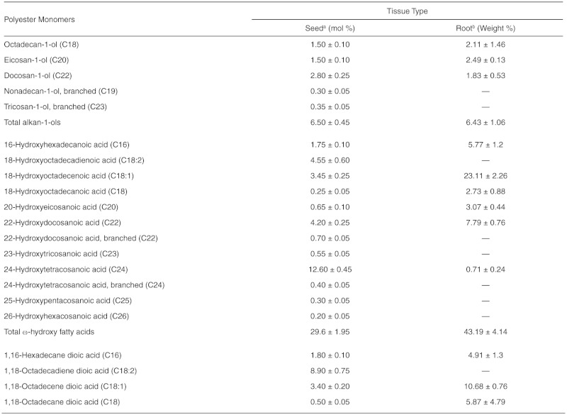
continued.
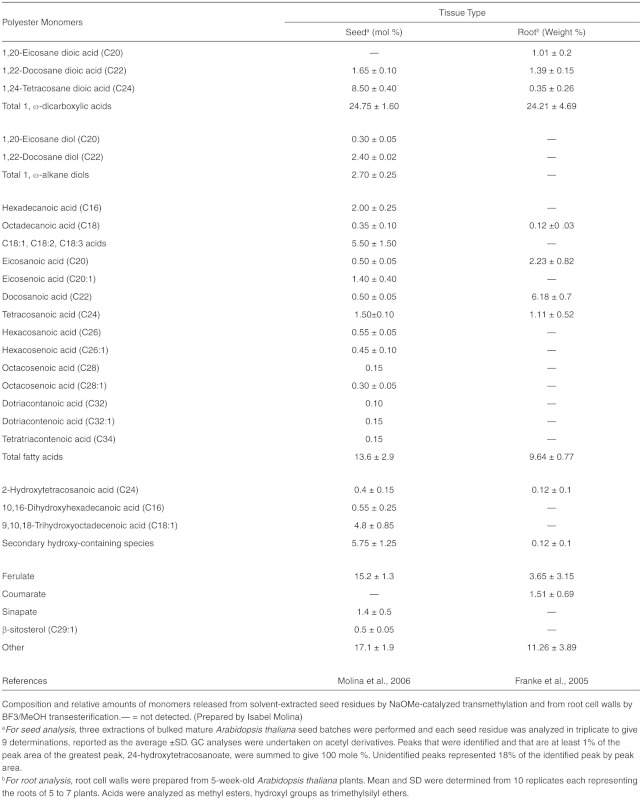
Table 11.
Composition of Arabidopsis Root Waxes
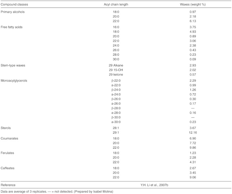
Figure 11.

Proposed Pathways for the Synthesis of Suberin Aliphatic Polyester.
This biosynthetic scheme represents one possible scenario for the synthesis of suberin building blocks (i.e. acyl glycerols), where we assume that acyl oxidation occurs after elongation and before esterification to glycerol (or G3P). So far, the order of such reactions remains unclear. Likewise, subcellular compartments where these pathways may take place are unknown, but fatty acid-modification and acyl glycerol synthesis steps are believed to take place in the endoplasmic reticulum (ER) membranes. Transport mechanisms required to transfer monomers or oligomers/polymers to the site of polyester assembly remain hypothetical (Beisson et al., 2012). One ABC transporter has been associated with both cutin and suberin synthesis (ABCG11; Panikashvili et al., 2010); a membrane-bound LTP (LTPG1) have been associated with cuticular wax synthesis (DeBono et al., 2009), and homologous proteins could be involved in suberin assembly. Polyester synthase(s), required to link monomers to produce high molecular weight suberin polyesters, as well as polymerization site(s) remain unknown. However, two extracellular proteins, BDG (Kurdyukov et al., 2006) and CD1 (Yeats et al., 2012), belonging to the αβ-hydrolase superfamily and the GDSL lipase/hydrolase family respectively, have been proposed as a cutin synthases.
(A) Synthesis of aliphatic suberin monomers.
Abbreviations: ABC, ATP binding cassette transporter; FAR, alcohol-forming fatty acyl-CoA reductase; ASFT, aliphatic suberin feruloyl transferase; BDG, BODYGUARD; CD1, cutin deficient 1; FAH, Fatty acyl ω-hydroxylase; GDSL, family of serine lipase/esterase enzymes; G3P, glycerol-3-phosphate; sn-2 GPAT, glycerol 3-phosphate acyltransferase; HFADH, ω-hydroxy fatty acyl dehydrogenase; LACS, long chain acylcoA synthase; LTP, lipid transport protein; LTPG, glycosylphosphatidylinositol (GPI)-anchored protein; OFADH, ω-oxo fatty acyl dehydrogenase; ω-hydroxy-FA, ω-hydroxy fatty acid.
(B) Synthesis of aromatic suberin monomers for the aliphatic domain.
Abbreviations: 4CL, 4-coumarate ligase; ASFT, aliphatic suberin feruloyl transferase; C3H, Coumarate 3-hydroxylase; C4H, Cinnamate 4-hydroxylase; CCoAOMT, caffeoyl coA O-methyltransferase; MAG, monoacylglycerol.
(C) Potential transport mechanisms involved in the assembly of suberin polyesters.
Abbreviations: ABC, ATP binding cassette transporter; ABCG1/DSO, a member of the ABCG subgroup of the ABC transport family; BDG, BODYGUARD; CCoAOMT, caffeoyl coA O-methyltransferase; GDSL, family of serine lipase/ esterase enzymes; LTP, lipid transport protein; LTPG, glycosylphosphatidylinositol (GPI)-anchored protein; PS, polyester synthase.
For additional details on genes involved in these reactions, please see http://aralip.plantbiology.msu.edu/pathways/suberin_synthesis_transport
continued.

(B) Synthesis of aromatic suberin monomers for the aliphatic domain.
Abbreviations: 4CL, 4-coumarate ligase; ASFT, aliphatic suberin feruloyl transferase; C3H, Coumarate 3-hydroxylase; C4H, Cinnamate 4-hydroxylase; CCoAOMT, caffeoyl coA O-methyltransferase; MAG, monoacylglycerol.
For additional details on genes involved in these reactions, please see http://aralip.plantbiology.msu.edu/pathways/suberin_synthesis_transport_2
continued.

(C) Potential transport mechanisms involved in the assembly of suberin polyesters.
Abbreviations: ABC, ATP binding cassette transporter; BDG, BODYGUARD; DCR, DEFECTIVE IN CUTICULAR RIDGES; CCoAOMT, caffeoyl coA O-methyltransferase; FAH, Fatty acyl ω-hydroxylase; G3P, glycerol LTP, lipid transport protein; PS, polyester synthase
For additional details on genes involved in these reactions, please see http://aralip.plantbiology.msu.edu/pathways/suberin_synthesis_transport_3
In the late 1970s, the characterization of fatty acid ω-hydroxylation and ω-hydroxyacid oxidation activities from cutinizing and suberizing tissues provided first insigths into the biosynthesis and structure of suberin (Agrawal and Kolattukudy 1977, 1978; Soliday and Kolattukudy, 1977). Further progress of biochemical approaches was probably limited because suberin catalysts are associated with membrane systems and/or enzyme complexes. With the molecular genetic tools available for Arabidopsis and the development of quantitative methods for the chemical analysis of Arabidopsis suberin (Franke et al., 2005; Molina et al., 2006), molecular genetic approaches were initiated. As no suberin-targeted forward genetic screen has yet been developed, bioinformatics-guided reverse genetics has emerged as a successful strategy to identify suberin-involved genes, and significant progress has been made in the past few years.
The C16 and C18 acyl precursors for suberin aliphatic biosynthesis are provided by plastidial FA biosynthesis and/or acyl editing of membranes (Bates et al., 2007). These fatty acids undergo acyl-oxidation, acyl-reduction and acyl-transfer reactions to produce suberin building blocks (i.e., acyl glycerols, alkyl ferulates). Alternatively, fatty acyl-CoAs are elongated by enzymes of the fatty acid elongation (FAE) complex to produce C20-C24 acyl-CoAs, which are also oxygenated and transferred to glycerol-3-phosphate (G3P).The biosynthetic scheme shown in Figure 11 represents one possible scenario where we assume that acyl oxidation occurs after elongation and before esterification to glycerol; this order of reactions is supported in part by biochemical assays with recombinant GPAT enzymes (section 2.9; Yang et al., 2010; 2012). A detailed characterization of the involved catalysts and their substrates may provide arguments for this or alternative order of reactions (Beisson et al., 2012).
Despite differences in composition and localization, the synthesis of cutin and aliphatic suberin precursors involve proteins of the same enzyme families, namely the CYP86 subfamily of P450 monooxygenases, acyl-CoA synthetases of the LACS family, and acyltransferases of the GPAT family (Figure 10). Compositional analysis of suberin from insertion mutants uncovered chain-length-specific changes demonstrating that the fatty acid ω-hydroxylases CYP86A1 (Li et al., 2007a; Höfer et al., 2008) and CYP86B1 (Compagnon et al., 2009; Molina et al., 2009) are required for the biosynthesis of C16-C18 and C22-C24 ω-oxygenated fatty acids in suberin, respectively. Whether these oxidases are multifunctional and can catalyze all ω-oxidation steps to form α,ω-DCAs, as shown for CYP94 members (Kandel et al., 2006, 2007; Pinot and Beisson, 2011 ), is still unknown. The alternative is a two-step oxidation by dehydrogenases including proteins related to the cutin-involved HTH (Kurdyukov et al., 2006a). Reverse genetics approaches also demonstrated that the Fatty Acid Elongation (FAE) complex components KCS2 and KCS20 (Franke et al., 2009; Lee et al., 2009) and the sn-2 G3P acyltransferase GPAT5 (Beisson et al., 2007) are involved in the biosynthesis and transfer of C20-C24 monomers, respectively. Similarly, mutants of the endodermis-expressed fatty acyl-CoA reductases FAR5, FAR4 and FAR1 presented chain-length specific reductions in C18, C20 and C22 suberin alcohols (Domergue et al., 2010). These phenotypes correlated with the, partially overlapping, chain length specificities determined for the recombinant FARs. The moderate chemical phenotypes observed in most of the studied suberin mutants suggest the involvement of partially redundant enzymes of the CYP, GPAT, FAR and KCS families. Other catalysts such as LACS, HTH-like ω-hydroxyacid dehydrogenases, fatty acid desaturases (FAD), can be deduced by analogy to cutin biosynthesis (Figure 10). In fact, chemical analyses of loss-of-function mutants of the cutin-related LACS2 (Molina, unpublished data) and GPAT4 (Li-Beisson, unpublished data) genes indicate additional roles in suberin formation. Mutant analysis also allowed the discovery of an acyltransferase of the BAHD family that functions as an aliphatic suberin feruloyl transferase (ASFT; Molina et al., 2009, Gou et al., 2009). Heterologous protein expression experiments confirmed that ASFT catalyzes the acyl transferfrom feruloyl-CoA to ω-hydroxyfatty acids and fatty alcohols (Molina et al., 2009). In addition, chemical phenotypes in suberin mutants may also facilitate our understanding of suberin structure. Indeed, the compositions of both asft and cyp86b1 knockout (KO) mutants question two aspects of the current models for suberin, namely esterified ferulate as a linker between the aliphatic and aromatic domains and the existence of an extended aliphatic polyester (Molina et al., 2009). Other reactions presented in Figure 11 are still unknown.
Ectopic expression of fluorescent protein fusions demonstrated that key enzymes in suberin aliphatic biosynthesis, CYP86A1 (Höfer et al., 2008) and CYP86B1 (Compagnon et al., 2009), are associated with the ER. Membrane targeting predictions for GPAT5, GPAT4, and HTH (Schwacke et al., 2003) and subcellular localization of FAE components tagged with green fluorescent proteins (GFP; Kunst and Samuels, 2003; Zheng et al., 2005; Bach et al., 2008) imply that the bulk of suberin monomer biosynthesis takes place at the ER. Furthermore, P450 monooxygenases in ferulic acid formation are also ER-membrane associated (Boerjan et al., 2003). Thus, most of the reactions shown in Figure 11 are predicted to take place on the ER membranes.
Completely unknown are the intermediates being transported and the reactions required for export, interlinkage of precursors, and macromolecular assembly that finally give rise to the highly organized lamellar structure of suberin depositions observed by transmission electron microscopy (TEM; Graça and Santos, 2007, Ranathunge et al., 2011). Exported and/or polymerized suberin building blocks might be free suberin-monomeric fatty acid derivatives, preformed oligomeric esters or MAGs (Pollard et al., 2008). Regarding the latter it is interesting to note that the biochemical characterization of the GPAT family revealed that — in contrast to cutin related GPATs, which synthesize sn-2 MAGs — the recombinant GPAT5 uniquely produces sn-2 acyl-LPAs (Yang et al., 2010) that might represent suberin-specific metabolites. Peridermal transcriptome (Soler et al., 2007) and transcript coexpression analyses suggest a function for plasma membrane-localized ABC transporters and LTPs in apoplastic suberin deposition. Mutant analysis indicated a role of the ABCG11 in suberin formation (Panikashvili et al., 2010), whereas LTPs have only been demonstrated to be required for cutin deposition so far (DeBono et al., 2009, Lee et al., 2009). Alternatively, exocytotic export mechanisms involving secretory vesicles or oleophilic bodies could facilitate transport of suberin intermediates. Because fungal lipases can polymerize suberin monomers in vitro (Olsson et al., 2007), candidate(s) for an unknown polyester synthase include homologues of cutin related extracellular BDG-like esterases of the α,ω-fold hydrolase family and GDSL lipases (Yeats et al., 2012). Interestingly, mutations in the functionally unknown ENHANCED SUBERIN gene (ESB1) led to a twofold increase in aliphatic root suberin (Baxter et al., 2009). ESB1 has similarity with dirigent proteins, which are proposed to be involved in the stereochemistry and apoplastic arrangement of aromatics prior to polymerization (Davin and Lewis, 2005).
The restriction of suberin depositions to specific, often monocellular tissue layers, the localized deposition in certain cell wall domains (e.g., Casparian bands), and the induction upon environmental stresses indicate that suberin biosynthesis must be tightly regulated at the tissue and cellular level. Additional regulatory complexity is given by the coordination of acyl and phenylpropanoid metabolism. To date, no regulators in suberin biosynthesis have been identified.
Major unanswered questions:
1. What are the in vivo substrates for suberin aliphatic oxidation enzymes? Are they free or conjugated (CoA, MAG) acyl donors?
2. How is the temporal and spatial control of suberization regulated?
3. How is the organized lamellar structure of suberin depositions, as observed by TEM, generated at the molecular level?
4. What is the function of aromatics in suberin structure and cross-linking?
5. What is the stoichiometric ratio of aliphatics to glycerol in the polyester?
2.11.Triacylglycerol Lipases (Figure 12)(Vincent Arondel* and Rémi Zallot16)
Figure 12.

Triacylglycerol and Fatty Acid Degradation.
Triacylglycerols are first degraded by cellular lipases to release free fatty acids from glycerol. Then reactions involved in the β-oxidation of straight-chain fatty acids occur in the peroxisome (adapted from Goepfert and Poirier, 2007). “2x cycles” indicates that the molecule undergoes two successive rounds through the β-oxidation cycle. CTS is an ABC transporter involved in the import of β-oxidation substrates to the peroxisome.
(A) Examples of the involvement of auxiliary activities in the degradation of oleic acid (C18:1Δ9c/s).
(B) The core β-oxidation cycle.
(C) Example of the involvement of the monofunctional type 2 enoyl-CoA hydratase (ECH2) in the degradation of fatty acids with a cis double bond on an even-numbered carbon (C18:1Δ6cis, petroselinic acid).
Abbreviations: ACT, acyl-CoA thioesterase; ACX, acyl-CoA oxidase; DCH, D3,5,D2,4-dienoyl-CoA isomerase; lsom, D3,D2-enoyl-CoA isomerase; KAT, 3-ketothiolase; LACS, long chain acyl-CoA synthetase; MAGL, monoacylglycerol lipase; MFP, multifunctional protein containing a 2E-enoyl-CoA hydratase (ECH) and a 3S-hydroxyacyl-CoA dehydrogenase (HACDH); Red, 2,4-dienoyl-CoA reductase; TAGL, triacylglycerol lipase.
For additional details on genes involved in these reactions, please see http://aralip.plantbiology.msu.edu/pathways/triacylglycerol_fatty_acid_degradation
2.11.1. Background
Triacylglycerol lipases are enzymes that hydrolyze long-chain, insoluble TAGs. Their biochemical properties and structure-function relationships have been extensively studied in mammals, fungi, and bacteria (Woolley et al., 1994; Schmid and Verger, 1998). Their most important features are summarized in this section.
All possess the same fold, called α/β hydrolase fold (Ollis et al., 1992). This fold is not specific for TAG lipases, as other carboxylesterases unable to hydrolyze lipids share the same fold. The active site is made of a catalytic triad (ser, asp/glu, his). Despite this structural homology, several families of lipases have been described. Their sequences can diverge widely, and only a loose consensus around the catalytic serine (Prosite PS00120) can be noted. Not all known TAG lipases contain this consensus, while a few non-lipolytic esterases do. Therefore, it is not possible to predict a TAG lipase based on sequence information only. Recently, TAG lipases responsible for the mobilization of intracellular stores in yeast, mammals, insects, and plants have been identified (Zechner et al., 2009). They differ from previously characterized TAG lipases: They resemble calcium-independent phospholipase A2 (iPLA2), they contain a so-called patatin domain, and their active site is made of a catalytic dyad.
TAG lipases can be capable of hydrolyzing other substrates. For instance, most of them can hydrolyze DAG and, more rarely, MAG. In addition, some of them can hydrolyze phospholipids at the sn-1 position, cutin polymer, polyhydroxyalkanoates, steryl esters, and CoA esters. It is likely that widening the range of substrates used to characterize TAG lipases would show several other possible substrates. Actually, lipases used in bioconversions can act on numerous substrates (Schmid and Verger, 1998). Therefore, it is not because an enzyme hydrolyzes TAGs that it acts necessarily as a TAG lipase in vivo. Actually, there are about 50 to 70 putative TAG lipases (based on sequence similarity) in Arabidopsis; it is unlikely that all these enzymes are physiological TAG lipases (aside from the fact that these might be non-lipolytic esterases). For instance, the ‘Defective In Anther Dehiscence1’ (DAD1) (lshiguro et al., 2001 ) encodes a lipase that hydrolyzes both TAGs and glycerolipids; it is likely that only the last function is physiologically relevant (i.e., release of linolenic acid for jasmonate synthesis; Acosta and Farmer, 2010); the activity of recombinant DAD1 on TAGs represents 6% than that on phosphatidylcholine. Furthermore, many enzymes have been sometimes hastily classified as lipases based on partial biochemical characterization. This is the case for the so-called GDSL family of lipases (Akoh et al., 2004). Because insoluble substrates such as TAGs are not convenient to handle, numerous enzymatic studies have been carried out using soluble chromogenic substrates (Huang, 1993; Beisson et al., 2000) such as short-chain esters of paranitrophenol (e.g., para-nitrophenyl acetate or butyrate). Unfortunately, these substrates can be hydrolyzed by esterases that are unable to hydrolyze true lipids. Only the GDSL lipase purified from sunflower has been quantitatively characterized (Teissere et al., 1995) with regard to true lipase activity: The specific activity on long-chain TAGs (15 nmol fatty acids min-1 mg-1) is five orders of magnitude below the one of pure human pancreatic lipase (3000 µmol min-1 mg-1) and 3000 times less than pure recombinant SDP1. Recently, a pure GDSL lipase from B. napus was found to hydrolyze sinapoyl-choline (Clauss et al., 2008) with a specific activity of 10 µmol min-1 mg-1. Therefore, it is clear that GDSL “lipases” are esterases. Whether some of them can indeed hydrolyze lipids and can be physiological lipolytic enzymes cannot be excluded, but this remains to be firmly demonstrated.
2.11.2. TAG lipases in Arabidopsis
Biochemical data are available on several TAG lipases from higher plants, most of them obtained from non-pure fractions (Huang, 1993; Mukherjee, 1994). One of the most studied enzymes is castor bean acid lipase, cloned in 2004 (Eastmond, 2004). Only seed and seedling lipases have been studied, except for lattices and the fruit of oil palm. However, TAGs are likely to be present in trace amounts in all tissues (W.L. Lin and Oliver, 2008) and can be abundant in other tissues/organelles than seed oil bodies.
In Arabidopsis, four possible lipases have been cloned and the corresponding recombinant enzymes characterized. AtLIPI (El-Kouhen et al., 2005) resembles lysosomal acid lipase. It hydrolyzes TAGs with a specific activity estimated at 45 µmol min-1 mg-1. It appears to hydrolyze neither phospho- and galactolipids nor cholesteryloleate. The knockout mutant does not show any obvious phenotype and is not impaired in post-germinative fat storage breakdown. SAG 101 (He and Gan, 2002) is a lipase-like protein that plays a role in leaf senescence: Senescence is delayed in antisense plants while over-expression of the gene leads to premature senescence. SAG 101 recombinant protein exhibits a weak TAG lipase activity (about 4 nmol min-1 mg-1). At2g31690 codes for a protein that resembles fungal lipases (Padham et al., 2007). It locates to the plastid and is most abundant in 6-week-old leaves. Confocal microscopy studies suggest that neutral lipids are more abundant in antisense plants. Ultrastructure of plastids is modified when compared to WT and the antisense plants show delayed senescence. A purified recombinant protein hydrolyzes TAGs with a low specific activity (about 10 nmol min-1 mg-1). Actually, it appears to be much more active on galactolipids than on TAGs (Seo et al., 2009). At4g24140 shares sequence homology to CGI-58, a known cofactor to the mammalian lipase responsible for intracellularTAG breakdown (Zechner et al., 2009). Its recombinant gene product shows low acyltransferase, phospholipase, and TAG lipase activities. (Ghosh et al., 2009). The mutant shows a 10-fold TAG increase in leaf tissue when compared to wild type (James et al., 2010). However, At4g24140 does not seem to be involved in TAG hydrolysis of young seedlings (Kelly et al., 2011 ). Sugar dependant 1 (SDP1) is a lipase involved in fat storage breakdown during post germinative growth (Eastmond, 2006); it is discussed below.
2.11.3. TAG lipases involved in Arabidopsis fat storage breakdown
An ethyl methanesulfonate (EMS) mutant screen was performed to select seedlings with impaired growth on minimal media that could be rescued by transfer to a sucrose-containing medium. Among the mutants isolated, sdp1 was found to be affected in a gene that codes for a lipase that controls fat storage breakdown (Eastmond, 2006). The gene was identified by positional cloning and characterized in depth. The evidence presented is fairly strong: Only 20% of the mutant oil is hydrolyzed in 5-dayold seedlings (vs. 98% in WT). Electron microscopy data show that lipid bodies are fairly abundant in mutant 5-day-old seedlings, while they are totally absent from WT control. While lipase activity from a mutant crude extract is reduced by about 20%, the lipase activity specifically bound to oil bodies fell by 75% when compared to WT. An SDP1-GFP fusion protein associates to oil-bodies hemi-membrane in vivo. Recombinant SDP1 protein hydrolyzes triolein with a reasonably high specific activity (50 µmol min-1 mg-1), which is probably underestimated due to experimental conditions (limiting substrate).
SDP1 sequence relates more to iPLA2 than to most TAG lipases. It contains a patatin domain and probably hydrolyzes its substrate through a catalytic dyad. Arabidopsis contains another related gene called SDP1-like (At3g57140). Five-day-old seedlings from a double SDP1-SDP1-like knockout contain exactly the same amount of TAGs as at day 0, strongly suggesting that SDP1-like is responsible forthe weak hydrolysis of TAGs detected in the SDP1 mutant (Quettier and Eastmond, 2009).
Major unanswered questions:
-
1. How are TAG lipases regulated?
The lipolysis step is under regulation as the oil is not degraded in β-oxidation mutants (Graham, 2008). This is likely untrue; high oil is due to stimulation of TAG synthesis. Because SDP1 transcript is predominantly detected in maturating seeds and levels are much lower during postgerminative growth, it is likely that regulation occurs at a post-transcriptional level. A few hints have been suggested (Quettier and Eastmond, 2009): A mutant affected in caleosin, an oil body protein, exhibits delayed TAG breakdown (Poxleitner et al., 2006).
-
2. How are DAG and MAG hydrolyzed during postgerminative growth?
SDP1 recombinant lipase shows no activity on MAG and little activity on DAGs (Eastmond, 2006). This suggests the possible involvement of other lipases capable of hydrolyzing these substrates. There are no obvious candidates, as Atlipl does not hydrolyze MAGs, either. In mammals, MAG lipases have been described (Karlsson et al., 1997). There are about 15 proteins in Arabidopsis that resemble this enzyme (BLAST scores from 75 to 123). Again, as for TAG lipases, similarity of sequence does not necessarily mean identical function.
-
3. Are there other physiological TAG lipases?
The studies on TAGs have been focused mostly on seed oil bodies of oleaginous plants. However, TAGs can be found in pollen grain, tapetum cells (Hsieh and Huang, 2004), and leaves treated with ozone or in the process of senescence. Therefore, it is likely that other physiological TAG lipases exist; At4g24140 might be a candidate (James et al., 2010).
2.12. Fatty Acid β-Oxidation (Figure 12) (Ian A. Graham17)
2.12.1. Context and location
The fatty acid β-oxidation spiral is a ubiquitous process that breaks down fatty acids derived primarily from either membrane lipid turnover or the mobilization of triacylglycerol storage reserves. In higher plants and most fungi, straight-chain fatty acid catabolism via the β-oxidation spiral is located in the peroxisomes, whereas breakdown of branched-chain amino acids via the β-oxidation spiral is distributed between the mitochondria and peroxisomes (reviewed in Graham and Eastmond, 2002; Penfield et al., 2006).
Fatty acids released by lipolysis of TAG or membrane lipids are transported across the single membrane of the peroxisome by an ABC transporter protein identified in three different genetic screens in Arabidopsis and independently named as PEROXISOMAL ABC TRANSPORTER 1 (PXA1; Zolman et al., 2001), PEROXISOME DEFICIENT 3 (PED3; Hayashi et al., 2002), and COMATOSE (CTS; Footitt et al., 2002). In Arabidopsis, two peroxisomal LACS enzymes act in the same pathway as the ABC transporter and are essential for the uptake of fatty acids, which raises still unanswered questions regarding the mechanism of import (Fulda et al., 2004; Penfield et al., 2006).
2.12.2. Core activities of the β-oxidation spiral
The esterification of fatty acids to acyl-CoAs by the LACS enzymes results in their activation for oxidative attack at the C-3 or β-carbon position. In each round of the β-oxidation spiral, acetyl-CoA (C2) is cleaved from acyl-CoA (Cn), and the remaining acyl-CoA (Cn-2) re-enters the β-oxidation spiral to repeat the process (Figure 12). This core pathway requires the enzymes acyl-CoA oxidase (ACX), multifunctional protein (MFP), and 3-ketoacyl-CoA thiolase (KAT) to catalyze oxidation, hydration and dehydrogenation, and thiolytic cleavage, respectively, of acyl-CoA. The complete degradation of long-chain acyl-CoAs to C2 acetyl units requires the enzymes responsible for catalyzing each step to accept substrates with diminishing carbon chain length with each passage through the β-oxidation spiral. The core group of β-oxidation enzymes has evolved two alternate strategies to cope with this range of substrates: either multiple isoforms with different chain-length specificities or enzymes with broad substrate specificity (low carbon chain length substrate selectivity).
2.12.3. Acyl-CoA oxidases
The ACXs (EC 1.3.3.6) catalyze the first step of peroxisomal β-oxidation of acyl-CoA to 2-trans-enoyl-CoA. Six ACX genes have been identified in Arabidopsis (Adham et al., 2005; Graham, 2008), and of these ACX1 (C12:0 to C16:0), ACX2 (C14:0 to C20:0), ACX3 (C8:0 to C14:0), and ACX4 (C4:0 to C8:0) have been characterized biochemically and shown to encode proteins with overlapping but distinct substrate specificities (Hooks et al., 1999; Eastmond et al., 2000; Froman et al., 2000; Hayashi et al., 1999). The ACX1, ACX2, ACX3, and ACX4 genes are ail upregulated coordinately during Arabidopsis seed germination and early postgerminative growth (Rylott et al., 2001 ), which correlates with the period of most rapid breakdown of fatty acids derived from storage TAG. Compromised seed germination and seedling establishment are observed in Arabidopsis double mutants disrupted in both ACX1 and ACX2, with seedling establishment but not seed germination being rescued by exogenous sucrose (PinfieldWells et al., 2005).
2.12.4. Multifunctional protein
MFP contains two of the core β-oxidation pathway reactions, 2-frans-enoyl-CoA hydratase (EC 4.2.1.17) and L-3-hydroxyacylCoA dehydrogenase (EC1.1.1.35), as well as additional auxiliary activities necessary for the β-oxidation of some unsaturated FAs (reviewed in Graham, 2008; Poirier et al., 2006). Arabidopsis contains two isoforms of MFP, the first of which was characterized genetically as abnormal inflorescence meristeml (aim1) since it exhibits aberrant vegetative and reproductive developmenta unique phenotype among the β-oxidation mutants, and one for which we still do not have a mechanistic explanation (Richmond and Bleecker, 1999; Baker et al., 2006). The second isoform, MFP2, is strongly induced during postgerminative seedling growth (Eastmond and Graham, 2000).The mfp2 mutant requires an exogenous supply of sucrose for seedling establishment and is compromised in storage oil breakdown, but not as severely as the acx1 acx2 double mutant or the kat2 mutant (Rylott et al., 2006). Interestingly, the MFP2 2-trans-enoyl-CoA hydratase is only active against long-chain (C18:0) substrates, whereas the L3-hydroxyacyl-CoA dehydrogenase is active on C6:0, C12:0, and C18:0 substrates (Rylott et al., 2006).
2.12.5. 3-Ketoacyl-CoA thiolase
In the β-oxidation spiral the KAT enzyme (EC 2.3.1.16) catalyzes the final thiolytic cleavage of 3-ketoacyl-CoA to acyl-CoA and acetyl-CoA. The Arabidopsis genome contains three loci that encode KAT enzymes, annotated as KAT1, KAT2, and KAT5 (At1g04710, At2g33150, and At5g48880, respectively), with KAT5 producing two polypeptides, KAT5.1 and KAT5.2, apparently as a consequence of alternate RNA splicing (Germain et al., 2001 ; Carrie et al., 2007). KAT2 is the only one of the three KAT genes expressed at significant levels during seed germination in Arabidopsis. kat2 mutants are blocked in storage oil breakdown and are dependent on exogenous sucrose for seedling establishment (Germain et al., 2001). Extensive substrate specificity experiments have not been performed for the KAT proteins due to the difficulty of synthesizing long-chain substrate.
2.12.6. Auxiliary activities of the β-oxidation spiral
Many fatty acids, particularly those found in seed storage oils, have unsaturated bonds in the cis-configuration at even-numbered positions or unsaturated bonds at odd-numbered positions on the carbon chain. Both of these arrangements result in metabolic blocks for the β-oxidation pathway if only the core set of enzymes is considered.
β-oxidation of unsaturated FAs with a cis double bond on an even-numbered carbon eventually produces the intermediate 2-trans, 4-cis-dienoyl-CoA (Figure 12). Two alternative pathways were proposed for the continued β-oxidation of this intermediate (Schulz and Kunau, 1987). In the hydratase/epimerase pathway, 3R-Hydroxyacyl-CoA is converted to 3S-Hydroxyacyl-CoA either by an epimerase activity or an auxiliary enzyme, enoyl-CoA hydratase2 (ECH2/Hydratase) (Goepfert et al., 2006), and the resulting 2-frans-enoyl-CoA then re-enters the normal set of core reactions of β-oxidation.
Our understanding of the complete breakdown of fatty acids with cis double bonds at odd-numbered carbons such as C18:1 D9 cis (oleic acid) has been improved by the functional characterization of the D3,5, D2,4-dienoyl-CoA isomerase (DCl/lsomerase) and the D3, D2,-enoyl-CoA isomerases from Arabidopsis (Goepfert et al., 2005,2008).
2.12.7. Additional roles for the fatty acid β-oxidation spiral
Arabidopsis mutants disrupted in the core steps in the β-oxidation spiral have revealed additional roles beyond the catabolism of straight-chain fatty acids, including the conversion of indole butyric acid to the phytohormone indole acetic acid (IAA; Zolman et al., 2000) and the production of the fatty acid signalling molecule jasmonic acid (Theodoulou et al., 2005). A comprehensive analysis of the biosynthesis and roles of jasmonates in Arabidopsis is provided in another chapter of The Arabidopsis Book (Acosta and Farmer, 2010). A major challenge now is to understand how the flux of these different metabolites through the β-oxidation spiral is regulated, particularly since they exist at very different concentrations in the cell, with straight fatty acid metabolites being at much higher levels than substrates and intermediates giving rise to signalling molecules.
Major unanswered questions:
1. What is the exact mechanism by which fatty acids are imported and activated through the combined action of the peroxisomal ABC transporter and LACS enzymes?
2. What regulates the flux through the shared β-oxidation spiral of substrates ranging from relatively abundant straight-chain fatty acids (derived from membrane lipids and storage TAG) to much less abundant precursors for signalling molecules (such as jasmonic acid and indole acetic acid)?
3. Why does oil body TAG persist in mutants disrupted in peroxisomal fatty acid β-oxidation?
4. What is the mechanism for the coordinated regulation of expression of genes encoding enzymes of the core reactions of β-oxidation during postgerminative seedling growth?
5. What is the functional significance of genes encoding proteins with various “auxiliary” β-oxidation activities, particularly with respect to their in vivo substrates?
3. METHODS AND PROTOCOLS FOR ARABIDOPSIS LIPID ANALYSES
3.1. Lipid Extraction Methods and Separation (Katherine M.Schmid18)
The following sections summarize methods that have been applied specifically to analysis of lipids from Arabidopsis and plant systems with similar challenges. Many other protocols are available, and readers may be especially interested in The Lipid Library, available online at http://lipidlibrary.aocs.org.
Although recent advances in nuclear magnetic resonance (NMR)-based techniques have made quantitation of oil in intact Arabidopsis seeds feasible (Jako et al., 2001; Colnago et al., 2007), most lipid analyses require extraction of the desired fractions into organic solvents. Since polarities and solubilities of lipids differ radically, methods of extraction will vary with the goals of the researcher. In all cases, oxidation of unsaturated fatty acids should be minimized by drying and storing samples under nitrogen, including antioxidants such as 25 mg L-1 butylated hydroxytoluene (BHT), and avoiding diethyl ether and unstabilized chloroform. Sample containers and equipment should be chosen to prevent introduction of contaminating plasticizers and lubricants (Christie, 1993). Thus, glass or Teflon containers rinsed with high purity solvents should be used rather than plastics, Teflon tape and solvent-washed aluminum foil should replace parafilm and plastic wraps, and stopcock grease and homogenizers with exposed lubricated bearings must be avoided.
If native lipid species are to be characterized, it is critical to immediately denature lipases, esterases, and oxidases, which may even be stimulated by some solvents or by freezing and persist at subzero temperatures (Christie, 1993). Therefore, even quickly frozen tissues will undergo degradation when stored in the freezer. Wounding quickly and strongly activates conversion of Arabidopsis leaf fatty acids to oxophytodienoic acids (Buseman et al., 2006). Immediate brief treatment of plant tissue with boiling isopropanol to inactivate enzymes (Kates and Eberhardt, 1957) is the typical remedy and is highly recommended. To prevent transesterification artifacts, extracts should not be stored in solvents containing primary alcohols such as methanol (Christie, 1993).
Arabidopsis lipids have most often been extracted with chloroform:methanol (2:1; Folch et al., 1957) or (1:2; Bligh and Dyer, 1959) (v/v). The latter permits lower solvent-to-sample ratios, since additional methanol increases the water content at which phase separation and concomitant loss of extraction efficiency occur beyond the 6.54% (w/v) limit of chloroform:methanol (2:1; Schmid 1973). Bligh and Dyer (1959) stipulated three subsequent chloroform washes, bringing the combined extracts to chloroform:methanol (2:1). Both protocols call for induction of phase separation once extraction is complete. Use of a salt solution such as 0.88% (w/v) potassium chloride (KCl) rather than pure water to achieve optimal chloroform:methanol:water (8:4:3 v/v/v) helps keep most acidic lipids protonated, so that they partition into the lower lipid phase (chloroform:methanol:water 86:14:1) rather than being lost with polar contaminants to the upper phase (3:48:47; Folch et al., 1957). Additional washes of the lower phase with salt solution reduce lipid yield compared to washes with salt solution:methanol (1:1 v/v; Christie, 1993). For a variant of the chloroform:methanol method suggested by the Kansas City Lipidomics Research Center (http://www.k-state.edu/lipid/lipidomics/leaf-extraction.html), see the protocol below. A method employing chloroform:methanol:formic acid (1:1:0.1) for extraction, followed immediately by a wash containing 1 M KCl and 0.2M H3PO4 to promote phase separation and lipolytic enzyme inactivation respectively, has also been applied to Arabidopsis (Zbierzak et al., 2011).
Given the toxicity of chloroform and the advantages of boiling isopropanol pretreatment, some Arabidopsis researchers have adopted extraction with hexane:isopropanol (3:2) as proposed by Hara and Radin (1978). The initial hexane:isopropanol solution yields a relatively uncontaminated extract that can be used for gas chromatography (GC) or thin-layer chromatography (TLC) analysis without partitioning or washing. However, to remove nonlipids, the extract may be partitioned into an upper hexane phase by addition of aqueous sodium sulfate (e.g., ½ volume 6.5% [w/v] Na2SO4; (Y.H. Li et al., 2006). Back extraction of the lower phase with hexane:isopropanol 7:2 is necessary to avoid loss of polar lipids. For a sample protocol, see below. Extraction of lipids with methanol:tert-butyl-ether:water (1:3:1) has been recommended if quantitative recovery of proteins and starch from the extract is desirable (Giavalisco et al., 2011).
Protocols for general extraction of Arabidopsis lipids.
For minimum oxidation, all solvents should contain 0.01% BHT.
Samples should be placed in glass tubes with Teflon-lined screw caps and extracted immediately after harvesting.
Isopropanol should be preheated prior to step 1 for effective killing of phospholipase D.
Appropriate internal standard(s) may be added in step 1.
After extraction is complete, lipid extracts may be concentrated under a stream of nitrogen for TLC and preparation of derivatives.
Choroform/Methanol method
For soft tissue samples up to 0.5 g fresh weight (approx. 30 mg dry weight)
Cover sample with 3 mL preheated isopropanol. Cap and heat at 75°C for 15 min. Cool to room temperature.
Add 1.5 mL chloroform and 0.6 mL water.
Incubate with shaking for 1 hr.
After transferring lipid extract to a fresh tube, reextract tissue with 4 mL chloroform:methanol (2:1 v/v).
Repeat step 4 until tissue is white.
To combined lipid extracts from steps 4 and 5, add 1 mL 1M KCl. Vortex, centrifuge, and dicard upper phase.
To lower phase from step 6, add 2 mL water. Vortex, centrifuge, and discard upper phase.
Hexane/lsopropanol method
For up to 1 g fresh weight
Add 8 volumes isopropanol (v/w) to sample. Cap and heat at 80°C for 5 min. Cool to room temperature.
Add 12 v/w of hexane.
Homogenize (mortar and pestle, polytron, or other suitable equipment).
Briefly centrifuge to facilitate separation of lipid extract from tissue.
Transfer the upper phase (hexane:isopropanol-containing lipids) to another tube.
For complete recovery, re-extract the pellet with hexane:isopropanol (7:2) and combine extracts with upper phase from step 5.
If less contamination of the extract is required, partition the hexane:isopropanol with Na2SO4 as described by Hara and Radin (1978) or Y.H. Li et al. (2006).
Modified Bligh and Dyer (1959) protocol for lipid extraction from Arabidopsis leaves.
Put frozen plant material (snap-frozen in liquid nitrogen and stored in –80°C, put back in liquid nitrogen prior to extraction) in a glass homogenizer
Add 3.75 mL MeOH: CHCl3 (2:1, v/v).
Add 1mL 1mM EDTA in 0.15M HAc (acetic acid).
Homogenize carefully.
Transfer to a fresh glass tube with screw cap (cap with teflon rubber inside).
Rinse homogenizer with 1.25 mL CHCl3; transfer to the glass tube.
To the glass tube, add 1.25 mL 0.88% (w/v) KCl.
Vortex carefully.
Centrifuge appr. 3000 rpm for 2 min.
Transfer lipid (CHCl3-) phase (lower phase) with a Pasteur pipette to a fresh glass tube, store in freezer–20°C
Unfortunately, few direct comparisons of laboratory scale extractions of plant material are available. Fishwick and Wright (1977) found that the procedure of Bligh and Dyer (1959) gave better lipid yields than that of Folch et al. (1957) for spinach leaf, tomato fruit, and potato tuber. A small-scale study by Khor and Chan (1985) suggested that the hexane:isopropanol procedure of Hara and Radin (1978), including a 6.5% Na2SO4 wash, extracted neutral lipids from soybean seeds but left behind about ⅘ of phospholipid extractable with chloroform/methanol (2:1). Schäfer (1998), however, obtained better extraction from a mixture of wheat, soybean meal, and barley with hexane:isopropanol (3:2) than with chloroform:methanol (2:1).
In addition to the general techniques above, many specialized applications are available. High-throughput techniques such as screening of total fatty acid profiles by simultaneous extraction and transmethylation of tissue samples in hot acidic methanol (Browse et al., 1986a) have been popular in the Arabidopsis community. For complete transesterification of Arabidopsis seeds or other tissues high in triacylglycerols, a cosolvent such as toluene should be included (Y.H. Li et al., 2006).
Finally, some lipids are poorly represented in extracts prepared by standard methods. Table 1 provides references to approaches for lipid classes requiring special attention.
Table 1.
Extraction Methods for Acyl Lipids Not Amenable to Standard Chloroform/Methanol Extraction Techniques
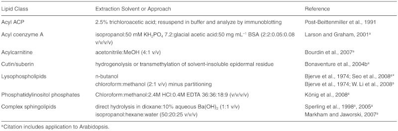
While high resolution direct-infusion mass spectrometry has made the analysis of complex lipid mixtures feasible (Horn & Chapman, 2012), laboratory scale analyses typically require prior separation of lipid classes. Techniques for fractionating lipid extracts include TLC, high-performance liquid chromatography (HPLC), and column chromatography (Christie, 2003). Often a gross separation into nonpolar lipids, glycolipids, and phospholipids by silicic acid column chromatography (Rouser et al., 1967) precedes further analysis.
3.2. Determination of Total Fatty Acid Profiles (Martine Miquel19)
In plants, fatty acids are mainly present as esters linked to glycerol, sterols, or waxes (long-chain alcohols) or as amides to sphingolipids, while free (unesterified) fatty acids are minor constituents. The total fatty acid profile of plant tissues other than seeds (see Section 3.4) or from lipid extracts can be determined by direct transesterification followed by analysis by GC or GC-MS. This process applies to the most commonly found fatty acids ranging from 14 to 24 carbon straight chains with zero to three double bonds. Fatty acids are identified by comparison of retention times (and also split patterns) to standards. With addition of an internal standard such as heptadecanoic acid (C17:0), which is normally not present in the lipid extracts, the quantity of each fatty acid in the sample analyzed can be determined. A typical GC chromatogram of Arabidopsis leaf is provided in Figure 13.
Figure 13.

Separation of the Methyl Ester Derivatives of Fatty Acids From Arabidopsis Leaf.A capillary fused silica column coated with (50% Cyanopropyl)-methylpolysiloxane (DB-23; J&W Scientific) was temperature-programmed from 140°C to 260°C at 10°C min-1 with helium as carrier gas. One µL of sample was injected in a 270°C inlet with a 30:1 split ratio. Methyl ester derivatives were detected using a FID at 270°C. (Courtesy of Dr. Imad Ajjawi, Michigan State University, Michigan)
For fresh tissue, for example, from leaf or root, the following acid-based procedure (Browse et al., 1986a) is widely used. This procedure allows methylation of both free fatty acids and transmethylation of O-acyl lipids. O-acyl lipids and free fatty acids in lipid extracts can be transesterified/methylated using the same protocol. For particular cases (short-chain and unusual fatty acids, free fatty acids, sphingolipids and N-acyl lipids, derivatization for GC-MS), protocols are available (Christie, 1993, 2003), as well as beginner's guides on methylation of fatty acids (http://lipidlibrary.aocs.org/topics/ester_93/index.htm) and mass spectrometry of fatty acids (http://lipidlibrary.aocs.org/ms/masspec.html).
A direct acid-catalyzed transmethylation protocol.
Place up to 50 mg tissue in a Teflon-lined screw capped glass tube.
Add 1 mL of 2.5 % H2SO4 (v/v) in methanol (freshly prepared).
Heat at 80°C for 1 h.
Cool to room temperature.
Add 500 µL of pentane followed by 1.5 mL 0.9% NaCl (w/v) to extract fatty acid methyl esters (FAME).
Shake vigorously and then briefly centrifuge to facilitate phase separation.
Transfer some of the upper phase (pentane-containing FAME) to an injection vial. Concentrate with stream of nitrogen if necessary for GC sensitivity.
Run GC with a flame ionization detector (FID) on a polar column such as Econo-Cap™ EC™-WAX capillary column (15 m long, 0.53 mm i. d., 1.20 µm film, Alltech Associates, Deerfield, III.).
Typical GC conditions—split or splitless mode injection, injector, and flame ionization detector temperature, 250°C; oven temperature program–160°C for 1 min, 40°C min-1 to 190°C, 4°C min-1 to 230°C, holding this temperature for 4 min.
Notes:
This method works well for Arabidopsis leaves and also roots, although duration of the transmethylation step for the latter should be increased to 1.5 h.
Hexane and heptane have been used traditionally for extraction of FAMEs. However, due to known long-term toxicity of hexane, one may consider using pentane, which is less toxic. Should hexane or heptane be used, care should be taken.
Care should be taken when applying nitrogen stream to evaporate the solvent, as a considerable amount of C14 (even C16) FAMEs can be lost if the sample is heated or the nitrogen flow too vigorous.
3.3. Glycerolipid Analysis Methods (Philip D. Bates20)
Glycerolipid analysis involves separation of individual polar and neutral glycerolipid classes. Separation may be followed by quantitation of acyl groups within the class, stereospecific analysis to determine composition of acyl groups at each position of the glycerol backbone, or characterization of molecular species with specific combinations of acyl groups esterified to a single glycerol backbone.
Accurate analysis of glycerolipids requires care in lipid extraction to minimize oxidation, lipolysis, or transesterification [see Section 3.1]. Although initial separation of the extract may be performed by HPLC (Beermann et al., 2003), the ease of use and low cost of TLC have made it a dominant technique for more than 60 years. Once separated, individual lipid classes can be quantified and fatty acid composition determined by direct conversion to fatty acid methyl esters and GC (Wu et al., 1994), or the glycerolipids may be collected for other analytical methods. Many different TLC separation and analysis methods are available for lipids and have been reviewed in depth (Christie, 2003, http://lipidlibrary.aocs.org).
3.3.1. Separation of glycerolipid classes by TLC
TLC plates are typically made of glass and coated with silica gel. Differences in gel composition and binders may affect lipid migration and downstream analysis (Sowa and Subbaiah, 2004). Comigration of lipid class standards should be used for identification of unknown compounds. Commercial TLC plates can be used directly; however, sometimes it is beneficial to heat TLC plates to ∼11O°C to drive off any moisture, especially in areas of high humidity. The ability of TLC plates to separate plant lipids can be enhanced by impregnating the plates with salts or compounds that will interact with the lipids. For instance, the silver ions of AgNO3 will slow the migration of double bond-containing lipids, allowing separation of molecular species (Christie, 2003; Bates et al., 2009). To prevent oxidation of fatty acids, both spotting of samples on plates and drying of samples eluted from plates are performed under nitrogen, and 0.01% BHT may be added to samples, TLC solvents, and detection sprays.
Several reagents are available for detection of lipids on TLC plates. Lipids containing double bonds can be stained with iodine vapor by placing a TLC plate for 15 to 60 min together with iodine crystals in a TLC tank. The iodine staining is mostly reversible, but because iodine can destroy double bonds, very light staining is advised if further analysis is required. Alternatively, general lipids can be sprayed lightly with 0.005% primulin in 80% acetone and lipids visualized under UV light. This sensitive, nondestructive stain will not interfere with most downstream analyses. Other nondestructive sprays for lipid detection under UV include 0.01% Rhodamine 6G (w/v) in water and 0.1 % (w/v) 2′,7′-dichlorofluorescein in 95% methanol. Both solvents and sprays should be handled in a fume hood.
Figure 14 illustrates separation of the major lipid classes found in Arabidopsis and other plants on Partisil K6 silica gel 60 Å TLC plates (Whatman, Maidstone, U.K.). Neutral lipids are separated on TLC plates developed once in hexane/diethyl ether/acetic acid (70/30/1, v/v/v) (Figure 14A). Waxes and sterol esters migrate the most quickly, followed by triacylglycerols, free fatty acids, diacylglycerols, and monoacylglycerols, while polar lipids remain at the origin. For polar lipid separation, TLC plates are dipped in a solution of 0.15 M (NH4J2SO4 and allowed to air dry. Just before use, the plates are heated to 110° C for at least 3 h. After lipid application, the plates are developed once or twice in acetone:toluene:H20 (91/30/8, v/v/v) (Khan and Williams, 1977). Typical migration order of the major plant membrane lipids is as follows: total neutral lipids, monogalactosyldiacylglycerol, phosphatidylglycerol, sulfoquinovosyldiacylglycerol, digalactosyldiacylglycerol, phosphatidyinositol and phosphatidylserine, phosphatidylethanolamine, and phosphatidylcholine. See Figure 14B for separation of polar lipids after metabolic radiolabeling of soybean embryos or Härtel et al. (2000) for separation of Arabidopsis leaf lipids.
Figure 14.

TLC Image Examples and Flow Chart for Analysis of Radiolabeled Glycerolipids From [14C]acetate and [14C]glycerol Labeled Soybean Embryos.
Examples of the major TLC systems used during analysis of radiolabeled glycerolipid classes, molecular species, and FA composition. Molecular species (e.g. SSS, SM, etc.) are represented as a combination of two or three FA, total saturates, S; monoenes (18:1), M; dienes (18:2), D; trienes (18:3), T.
(A) Neutral lipid class TLC, pictured 6 min [14C]acetate labeling.
(B) Polar lipid class TLC, pictured 6 min [14C]acetate labeling.
(C) TAG molecular species AgNO3-TLC, pictured 30 min [14C]glycerol labeling.
(D) Molecular species separation of PC (after phospholipase C digestion) and DAG as 3-acetyl-1,2-diacyl-glycerols, pictured 6 min [14C]acetate labeled PC.
(E) FAME AgNO3-TLC, pictured FAMEs from different 6 min [14C]acetate labeled PC molecular species separated in D. See (Bates et al., 2009) for labeling experiment details and TLC protocols.
Abbreviations: DAG, diacylglycerol; FFA, free fatty acid; PG, phosphaglycerol; Pl, phosphainostol; PE, phosphoethanolamine; PC, phosphacholine; PLC, phosphalipase C; PS, phosphaserine; PUFA, polyunsaturated fatty acid; TAG, triacylglycerol;
3.3.2. Stereochemical analysis of glycerolipids
The acyltransferases of glycerolipid synthesis have different specificities for acyl groups and thus produce lipids with different acyl compositions at each position of the glycerol backbone. Stereospecific phospholipase A2 will cleave acyl groups from the sn-2 position of phospholipids. The free fatty acid and lyso-lipid products can be separated for analysis by TLC Stereospecific analysis of glycolipids can be done with the TAG lipase from Rhizopus arrhizus that will cleave at the sn-1 position. There is no enzyme that differentiates the sn-1 and sn-3 positions of TAG, and thus a regiochemical analysis of sn-2 versus sn-⅓ is commonly employed with TAG lipase. Complete stereochemical analyses of TAG can be done through multistep procedures that involve partial degradation of TAG, generating a mixture of 1,2-DAG and 2,3-DAG. The mixed DAGs are either separated by chiral chromatography or are chemically converted to phospholipids for stereospecific PLA2 digestion of sn-1,2 species as for natural phospholipids above. Protocols can be found in Christie (2003).
3.3.3. FAME and molecular species separations
GC and mass spectrometry are the preferred methods of measurement for lipid-derived FAME and individual lipid molecular species, respectively. For detailed coverage of molecular species analysis by mass spectrometry, see Section 3.10. However, TLC separations can be very useful in circumstances such as analysis of radiolabeled lipids from metabolic labeling experiments. For example, molecular species of triacylglycerols and acetylated diacylglycerols may be separated based on their number of double bonds by TLC on AgNO3-impregnated TLC plates developed with a series of chloroform/methanol mixtures (Bates et al., 2009; Figure 14C and Figure 14D). FAME may be separated based on the number of double bonds with AgNO3-TLC (Figure 14E) or based on both number of double bonds and fatty acid chain length with reverse phase TLC plates (Christie, 2003; Marquardt and Wilson, 1998).
3.4. Seed Oil Quantification (Yonghua Li-Beisson21)
Arabidopsis stores over 35% oil in its seeds as energy and carbon reserves. A range of methods has been applied to quantify oil amount in Arabidopsis seeds (Table 2). These methods differ in sample size, sensitivity, instrument required, and information provided. Functional genetic screens have increasingly been used as a way to identify genes/proteins involved in storage oil metabolism. To facilitate mutant identification, a reliable and medium- to highthroughput oil quantification method is desirable.
Table 2.
Comparison of Methods Reported for Quantifying Oil Content of Arabidopsis Seed
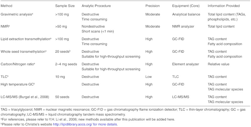
Arabidopsis seeds have a thin seed coat and are tiny (∼20 µg per seeds). Each seed has 5 to 8 µµg of fatty acids, and over 90% of these fatty acids are stored as triacylglycerols. It has been shown that total TAGs can be quantified based on fatty acid composition compared to internal standard (Y.H. Li et al., 2006). A direct whole seed transmethylation protocol is described below. It has several advantages: (1) It bypasses oil extraction; (2) it requires a GC-FID, which is found in most lipid labs, and GC-FID-based methods offer very high sensitivity for quantifying acyl chains; (3) it provides not only total TAG content but also fatty acid composition; and (4) it is suitable for medium- to high-throughput screening and can be used in functional genetic screens. A typical GC chromatogram of the FAME profile for seeds is shown in Figure 15.
Figure 15.

GC-FlD Analysis of Fatty Acid Methyl Esters Derived From Neutral Lipids Isolated From Arabidopsis Col-0 Seed.
Neutral lipids were isolated from wild type Col-0 seed and transmethylated and the subsequent fatty acid methyl esters separated on a J+W DB-23 (50% cyanopropyl) methylpolysiloxane 30 m column and detected using a flame ionization detector. The column temperature was initially held at 150 °C for 3 min, then increased to 240 °C at rate of 10 °C min-1, and then held at 240°C for 10 min. Tripentadecanoin (15:0) and triheptadecanoin (C17:0) were used as seed lipid extraction and transmethylation internal standards, respectively. IS = internal standard (Courtesy of Dr. Timothy Durrett, Michigan State University)
Notes:
This method works well for Arabidopsis seeds because almost all of the fatty acids are esterified to form TAGs, and the tiny seeds can be extracted without first grinding. For larger seeds (such as seeds of Brassica napus) or in cases where significant amounts of fatty acids are stored in other forms, TAGs might need to be isolated.
Before applying this method to other systems, total oil content and fatty acid distribution need to be verified by other methods.
Often researchers use as little as 1 or 2 seeds for the analysis. From our experience, this is sufficient to screen a large number of lines aiming to find changes in fatty acid profile or production of an unusual fatty acid. For quantification purpose, this usually gives very low value. Variations are likely due to seed-to-seed variation and inaccuracy in seed weight determinations.
Toluene is added as a cosolvent since neutral lipids such as TAGs and wax esters do not dissolve well in methanol alone.
Care should be taken when applying nitrogen stream to evaporate the solvent as a considerable amount of C14 (even C16) FAME can be lost if the nitrogen flow is too vigorous.
This method gives information on FAME content and composition; therefore, when reporting oil content, calculation is required: percent oil by weight = 100 (4 total mol FAME/3) + total g FAME)/g tissue, where 4 is the Mr. difference between TAG and three moles of FAME.
A whole seed acid-catalyzed transmethylation protocol.
Count 20 seeds and add them to a Teflon-lined screw-capped glass tube.
Add 1 mL 5% H2SO4 (v/v) in methanol (freshly prepared), 50 µg BHT, 20 µg C17:0 TAG (triheptadecanoin) as internal standard, and 300 µL of toluene as cosolvent.
Vortex vigorously for 30 s.
Heat at 85° to 90°C for 1.5 h.
Cool to room temperature.
Add 1.5 mL 0.9% NaCl (w/v) and add 1 mL of hexane to extract FAME.
Mix well (vortex) and then centrifuge briefly to facilitate phase separation.
Transfer the upper organic phase (hexane-containing FAME) to a new tube.
Evaporate the extracts under a stream of nitrogen.
Redissolve in 50 µL of hexane (vortex well).
Run GC with a flame ionization detector on a polar column-like DB23 (30 m by 0.25 mm i.d., 0.25 µm film; J&W Scientific, Folsom, CA).
The GC conditions were as follows: split mode injection (1:40); injector and flame ionization detector temperature, 260°C; oven temperature program 150°C for 3 min, then increasing at 10°C min–1 to 240°C and holding this temperature for 5 min.
3.5. TAG Analysis by Liquid Chromatography Mass Spectrometry (Tony Larson22)
The yield and fatty acid composition of seed triacylglycerols reflects the combined activities of fatty acid synthesis, desaturation, elongation, and transferase reactions, the latter of which are reversible. TAG analysis is therefore an important tool in evaluating how the process of acyl chain assembly into storage lipids is controlled, which may be especially important in metabolically engineered oil synthesis (Burgal et al., 2008). Three components of TAG analysis are potentially useful measures: (1) absolute quantity (i.e., yield), (2) relative TAG molecular species distribution, and (3) acyl position-specific information for a given TAG species. In the outlined method, absolute quantification is difficult as MS-based methods return biased responses depending on acyl chain length and degree of unsaturation (X.W. Li and Evans, 2005); instead, GC-FID-based FAME analyses of TAG derivatives is recommended. However, seed oil TAGs from Arabidopsis, comprising mixed acyl chains in the narrow 18–20 carbon number and 1–3 double bond range, generally provide a proxy of FAME-calculated yield within an error of ±10%. Less than half of the 80 to 120 resolvable Arabidopsis TAG molecular species are chromatographically separated; the remainder is resolved in the MS dimension by nominal mass. Constituent acyl species are then empirically assigned by reconciling the MS2 neutral-loss DAG fragments with the parent ammoniated molecular ion. In some cases, sn-2 position can be assigned due to the theoretically favorable loss of acyl chains from sn-1,3 positions (represented as more intense sn-1,2 and sn-2,3 DAG fragments in MS2 spectra). However, full positional assignments are not possible with MS techniques alone; prior stereospecific digestion techniques are required and are not covered here.
TAGs are extracted by grinding 20 to 200 Arabidopsis seeds in a 1.5 mL microfuge tube with 10 µL 1,1,113C-triolein (0.5–5 mg mL-1 in chloroform; internal standard) + 400 µL hexane/isopropanol (3:2, v/v), snap-freezing in liquid nitrogen, incubating at 4°C for 60 min, and centrifuging at 14,000 RPM for 5 min, and the supernatant is transferred to a fresh tube. The pellet is washed 3 times with 100 µL hexane:isopropanol and the supernatants pooled, combined with 350 µL 6.7% sodium sulphate (w/v), vortexed and centrifuged for 30 s at 14,000 RPM, and the supernatant dried in vacuo in an HPLC vial. The lipid residue is reconstituted in 100 µL chloroform and 10 µL injected on an LCQ-MS (Thermo Finnigan) equipped with a C30 HPLC column (YMC, 250 × 4.6 mm, 5 µm particle size) held at 30°C. A ternary separation gradient is used at 1 mL min-1 with solvents containing 0.2% formic acid (v/v): Solvent A is 20 mM ammonium formate in 80% (v/v) methanol, B is methanol, and C is tetrahydrofuran. Gradient is 0 to 5 min isocratic 5% A, 95% B; 5 to 45 min to 5% A, 35% B, 60% C, then isocratic 45–50 min; 10 min re-equilibration time between injections. The column eluent fed unsplit into an atmospheric pressure chemical ionization (APCI) source: vaporizer temperature 350°C; sheath gas (N2) flow 60 units, aux gas flow 60 units; source current 5 µA; capillary voltage 32 V; capillary temperature 150°C. Fullscan MS data are collected over the range 450 to 1500 m/z, and MS2 fragmentation data are collected in data-dependent mode at 60% normalized collision energy and an isolation width of 4 m/z.
A representative total ion current trace for Col-0 seed extract is shown in Figure 16A, with the identification process shown for a dominant TAG species outlined in Figure 16B.
Figure 16.

TAG Profile of Col-0 Dry Seed.
The total ion current (A) is used for the data-dependent selection of parent ions (inset in (B)), which are subjected to MS2 fragmentation analysis. Up to three daughter diacylglycerol (DAG) fragments are generated per triacylglycerol (TAG); in this example, two are generated because there are only two unique constituent fatty acids. The neutral losses for the observed DAG fragments (after correction for adducts) can be used for a constrained calculation of fatty acid identity (B). The correct stoichometric combination of these fatty acids to give the parent ion is used to calculate the TAG molecular formula.
3.6. Acyl-CoA Analysis by High-Performance Liquid Chromatography (Tony Larson23)
Long-chain (C16–18) and very long chain acyl-CoAs (C20–C24) are cytosolic intermediates for glycerolipid synthesis, and their accumulation would suggest a bottleneck or limiting step. Additionally, these acyl-CoAs (and also short-, medium-, and branched-chain variants) are measureable intermediates during peroxisomal β-oxidation in all tissues, especially those tissues undergoing rapid catabolism (e.g., seedlings, senescing leaves). CoA species also include the rapidly turned-over ubiquitous intermediates, acetyl and malonyl CoAs, which, although occasionally seen, are troublesome to recover quantitatively from extracts using the method described below.
The method given is an HPLC-reversed-phase-based separation with an integral washing step to remove interfering components. It is based on established techniques for acyl-CoA extraction from biological samples (Mancha et al., 1975), with extraction optimized to maximize recovery from plant tissues and detection specificity and sensitivity increased to cope with the low inherent concentration of acyl-CoAs in complex plant tissue matrices (Larson and Graham, 2001). Sample extraction combines prepurification with conversion to stable etheno fluorescent derivatives that can be readily separated by HPLC and sensitively and quantitatively detected. The acyl chain moiety of novel acyl-CoAs can be structurally determined in preconcentrated extracts using liquid chromatography tandem mass spectrometry (LC-MS/MS) techniques (Ishizaki et al., 2005). Acyl-CoA standards for verification of unknowns can be purchased or synthesized using enzymatic techniques for long or very long chains (Taylor et al., 1990) or chemical synthesis (Kawaguchi et al., 1981) for short or medium chains.
Fresh or frozen tissue samples (2–20 mg) are transferred to a microfuge tube and internal standards (10 µL each of 0.2 µM isovaleroyl and heptadecanoyl CoAs) added, followed by 200 µL freshly made ice-cold extraction buffer (200:200:5:8 [v/v] isopropanol:50mM phosphate buffer pH 7.2: acetic acid: 50 mg mL-1 BSA). It is important not to exceed 20 mg plant material, or recoveries will be greatly compromised. The samples are ground and lipids removed by 3 × 200 µL washes with water-saturated petroleum ether. Saturated ammonium sulphate (5 µL) is added to each sample, followed by 600 µL 2:1 [v/v] methanol:chloroform. Samples are vortexed and left at room temperature (RT) to precipitate for 20 min before centrifuging at 14,000 RPM for 2 min. The supernatant is transferred to HPLC vials and dried in vacuo. Derivatizing reagent (0.5 M chloroacetaldehyde, 0.5% [v/v] SDS, 150 mM citrate buffer pH 4.0; stored at RT for up to 3 months) is added to each vial (40 µL), and the sealed vials are heated at 85°C for 20 min. The derivatized samples (stable for at least a week at RT) are injected (20 µL) for HPLC analysis. The HPLC is equipped with a Luna C18(2) column (Phenomenex, 150 × 2.0 mm, 5 µm particle size) held at 40deg;C. A quaternary separation gradient is used with solvents: A, 1% acetic acid; B, 90% acetonitrile 1% acetic acid; C, 0.25% triethylamine; D, 90% acetonitrile. The run gradient is as follows: 0–5 min, 0.75 mL min-1 A:B (90:10) – A:B (20:80); 5–5.1 min, A:B (20:80) – A:C(20:80); 5.1–7 min, A:C (20:80) – C:D(97:3); 7–10 min, C:D(97:3) – C:D(95:5); 10–10.1 min, flow rate reduced to 0.2 mL min-1; 10.1–50 min, C:D (95:5) – C:D (55:45); 50–50.1 min, C:D(55:45) – D; 50.1–52 min, D; 52–52.1 min, flow rate increased to 0.2 mL min-1; 52.1–57 min D; 57–57.1 min, D — A:B (90:10); 57.1–60 min, A: B (10:90). The eluent is sent to a fluorescent detector with excitation set to 230 nm and emission to 420 nm. Peak area is directly proportional to molar quantities, and concentrations can be determined by reference to the internal standards. An example trace for Col-0 seedling extracts is shown in Figure 17.
Figure 17.

High-Performance Liquid Chromatography Trace of Acyl-CoA From a Col-0 Seedling Extract.The extract was made as described from 30 seedlings (grown on media plates) harvested at 5 days after imbibition. This developmental stage encompasses both rapid storage lipid breakdown and de novo lipid biosynthesis associated with seedling establishment. Peak areas and heights are directly proportional to the absolute amounts of the indicated acyl-CoAs. lS = internal standard.
3.7. Sphingolipid Analyses (Jonathan E. Markham24)
Sphingolipids have unique chemistry that has both advantages and drawbacks for the lipid biochemist. Due to their unique longchain base component, total sphingolipid content can be quantified from intact, dry tissue by hydrolysis, derivitization, HPLC separation, and fluorescence detection (Markham et al., 2006). A similar technique can be used to release the 2-hydroxy fatty acids (which are almost exclusively found in sphingolipids) to obtain information about the sphingolipid LCB and fatty acid content, although information about which LCBs are found in different classes of sphingolipid and with what fatty acids is not obtained.
Analysis of intact lipids is more challenging due to the highly glycosylated nature of the complex sphingolipids, which means they are largely insoluble in pure solvent and hence not easily extracted by the usual methods to extract lipids (Markham et al., 2006). For this reason, many analyses of sphingolipids from plants have focused on simpler sphingolipids such as ceramide or glucosylceramide ( Ohnishi and Fujino, 1981; Imai et al., 1995, 2000). Analysis of complex glycosphingolipids requires more extensive purification and techniques of carbohydrate analysis to uncover the absolute structure of the complex headgroup (Kaul and Lester, 1975; Markham et al., 2006). Intact sphingolipids from Arabidopsis can be extracted and quantified by LC-MS/MS, albeit with some limitations (Markham and Jaworski, 2007), but nonetheless this remains the only viable option for extensive analysis of intact sphingolipids from Arabidopsis.
This protocol is very simple and a quick way to check for changes in sphingolipid metabolism in Arabidopsis due to genetic mutation or environmental challenge. It is robust, very sensitive, quantitative, and free from most artifacts generated by other hydrolysis conditions. In the absence of a HPLC fitted with a fluorimeter, LCBs can be analyzed by making dinitrophenyl derivatives of the LCBs and analyzing by HPLC with a UV detector (Sperling et al., 1998), converting the LCBs to aldehydes and analysis by GC-FID/GC-MS (Bonaventure et al., 2003), or making N-acetyl, O-trimethylsilanederivatives and also detecting by GC-FID/GC-MS.
Protocol: Hydrolysis and quantification of LCBs from sphingolipids of Arabidopsis.
Materials: Internal standard D-erytho-C20-sphingosine (d20:1, Matreya) dissolved in methanol at 0.1 nmol µL-1; 10% Ba(OH)2 (dissolve 10 g of Ba(OH)2.8H20 [Sigma] in 96 mL of water with heating and stirring, warm before use); dioxane (HPLC grade), 2% Ammonium sulphate, OPA reagent (dissolve 5mg of O-phthaldialdehyde in 100 µL of methanol, add 5 µL of mercaptoethanol, 6.6 mL of water and 3.3 mL of 3% Boric acid pH 10.5); OPA diluent (combine 10 mL of water, 50 µL of 1M KHPO4 pH7 and 60 mL of methanol).
Freeze dry sample. Grind tissue to powder. a. Alternatively, dry a lipid extract under nitrogen and proceed to step 3.
Weigh approximately 10 mg of tissue into a screw-cap glass tube and record weight.
Add 10 µL of d20:1 standard, 1 mL of dioxane, and 1 mL of 10% Ba(OH)2.
Screw cap onto tube as tightly as possible.
Place tube in 110°C heat block. Hydrolysis reaction is essentially complete after 8 h.
Add 2 mL 2% ammonium sulfate and 2 mL diethylether. Vortex.
Spin at 500 g for 10 min. Remove upper phase to a new tube and dry under nitrogen.
Add 100 µL of methanol and 50 µL of OPA reagent. Allow to derivatize at room temperature for 20 min.
Add 350 µL of OPA diluent.
Spin at 500 g for 10 min. Transfer sample to labeled autosampler vial and seal.
Run on HPLC as soon as possible. Store samples in dark at 4°C if there is any delay.
Quantify the resulting peaks by comparison to the internal standard.
Note: HPLC conditions: Column—Agilent XDB-C18 4.6 × 250mm with guard column; Buffer A 5 mM K2HPO4 pH 7; Buffer B Methanol Flow Rate 1.5 mL min-1
HPLC gradient.
Fluorescence detector: Excitation 340 nm Detection 455 nm
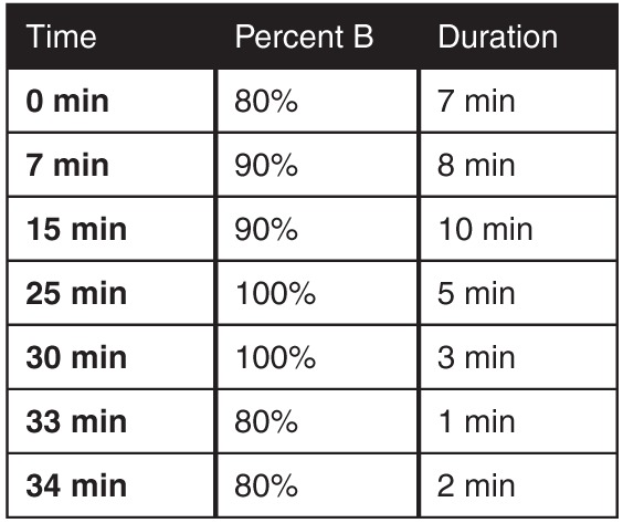
Notes:
The major peak in Arabidopsis is t18:1Δ8E that elutes around 17–18 min. Artifacts generated by the hydrolysis include the partial hydrolysis of the glycosidic bond resulting in Glc-t18:1 peaks and the formation of 1,4-anhydro derivatives from the phosphorylated sphingolipids.
3.8. Lipid Polyester Analysis (Fred Beisson25)
A protocol for the routine quantitative analysis of ester-linked monomers of Arabidopsis cutin and suberin is described below. Major steps are delipidation of tissues, chemical depolymerization of the residue, and extraction of released monomers in an organic phase and analysis of monomers by gas chromatography after derivatization of their hydroxyl groups. Various protocols using different delipidation, depolymerization, monomer extraction, and derivatization methods have been described (Bonaventure et al., 2004b; Franke et al., 2005; Molina et al., 2006). The protocol reported here is mostly adapted from the Bonaventure and Molina references. It can be performed on all Arabidopsis organs and uses minimal amounts of biological material and solvent.
I. Delipidation of tissues.
All solvent extraction steps include 0.01% (w/v) butylated hydroxytoluene (BHT) added from a 5% (w/v) stock solution in methanol. Unless indicated otherwise, each extraction is performed at room temperature by vortexing for 1 h (use a multitube vortexer, glass tubes with Teflon-lined screw caps, and 4 to 10 mL of solvent).
Fill preweighed tubes with isopropanol and heat up (85°C). Immerse Arabidopsis tissues in boiling isopropanol (10 min at 85°C). Leaves need to be cut in small pieces, and stems need to be cut in 1–2 cm bits and longitudinally in two halves. Examples of amounts of starting material per tube: 20–100 mg mature seeds, 100–300 mg fresh weight (fw) leaves or stems, 10–20 flowers, 50–200 mg fw secondary roots.
Cool down and extract by vortexing for 1 h. Alternatively, shake on a rocking agitator or a rotating wheel for at least 2 h and up to overnight.
Discard solvent. Most chlorophyll should have been extracted at this step. Extract again with isopropanol.
Discard solvent and extract with chloroform/methanol (2:1, v/v).
Discard solvent and extract with chloroform/methanol (1:2, v/v).
Discard solvent and extract with methanol.
Discard solvent and dry under a gentle stream of nitrogen gas. Dry in vacuum dessicator under reduced pressure until constant weight is achieved (at least 24 h). It is very important to remove as much water as possible.
II. Depolymerization of residue: Base- or acid-catalyzed transmethylation.
Weigh tubes to determine amount of dry residue after delipidation (there should be 10–50 mg depending on tissue). Transmethylations can be done by either acid or base catalysis.
Base catalysis
In each tube, add 2 ml of freshly made reaction medium and 5–10 pg of methyl heptadecanoate and ω-pentadecalactone as internal standards. To make 20 mL of reaction medium: In 12 mL methanol, add 3 mL methyl acetate and 5 ml 25% sodium methoxide in methanol (Sigma).
Heat the mixture at 60°C for 2 h.
Cool, add 4 ml dichloromethane, 0.5 mL glacial acetic acid to neutralize to pH 4–5, and 1 mL of buffer 0.9% NaCl (w/v) Tris 100 mM pH 8.0. Shake well and phase separate by centrifugation for 2 min at 1500 g. Check pH of upper phase with pH indicator paper.
Collect the lower organic phase, wash it with 2 mL of buffer, and dry over anhydrous sodium sulfate. Evaporate to dryness under a gentle stream of nitrogen gas (heat up tubes at 35°C max).
Acid catalysis
In each tube, add 2 mL freshly made reaction medium, 0.2 mL toluene as a cosolvent, and 5–10 µg of methyl heptadecanoate and ω-pentadecalactone as internal standards. To make 20 mL of reaction medium: In 19 mL methanol, add 1 mL concentrated sulfuric acid. Alternatively, 3N methanolic hydrochloride can be used.
Heat the mixture at 80deg;C for 2 h.
Cool. Add 4 mL dichloromethane and 1 mL of buffer 0.9% NaCl (w/v) Tris 100 mM pH 8.0. Shake well and phase separate by centrifugation for 2 min at 1500 g.
Collect the lower organic phase, wash it with 2 mL of buffer, and dry it over anhydrous sodium sulphate. Evaporate to dryness under a gentle stream of nitrogen gas (heat up tubes at 35°C max).
III. Derivatization of monomers and GC-(MS) analysis.
5. Hydroxyl residues can be acetylated or sylilated.
6. Acetylation: Add 100 µL of anhydrous pyridine and 100 µL of acetic anhydride. Heat up at 60°C for 2 h. Evaporate solvent under nitrogen stream.
7. Sylilation: Add 20 µL of anhydrous pyridine and 20 µL of BSTFA [N,O-bis(trimethylsilyl)-trifluoroacetamide]. Vortex for 10 s and heat up at 70°C for 60 min (or 110°C for 10 min).
8. Redissolve monomers in 30–200 µL of heptane:toluene (1:1, v/v) and run samples on GC-(MS). GC-FID conditions: HP-5 capillary column (30 m, 0.32 mm ID, 0.25 µm film thickness) with helium carrier gas at 2 ml min–1 and oven temperature programmed from 140°C to 310°C at 3°C min–1) and then held for 10 min at 310°C. Samples are injected in split mode (30:1 ratio, 310°C injector temperature) and peaks quantified on the basis of their FID ion current. For GC-MS, the same column is used with He carrier gas at 2 mL min–1 and oven temperature programmed from 110°C to 300°C at 10°C min–1. Splitless injection is used and the mass spectrometer run in scan mode over 40–500 amu (electron impact ionization) with peaks quantified on the basis of their total ion current.
Additional tips and cautionary notes
Grinding tissues. For seeds, it is necessary to grind material in liquid nitrogen with mortar and pestle before solvent extractions (Molina et al., 2006). To ensure optimal delipidation of seeds, additional extraction/centrifugation steps should be performed after the final methanol step: methanol (30 min), water (30 min), 2 M NaCl (1 h), water (30 min), methanol (30 min), chloroform/methanol 1:2 (v/v) (1h to overnight), chloroform/methanol 2:1 (v/v) (1h to overnight), and methanol (1h). To improve delipidation steps, tissues such as stems and leaves can also be ground using a Polytron (following immersion in hot isopropanol and cooling down). Recovery of ground material after each extraction can be performed using centrifugation or filtration through several layers of filter paper. One will have to be careful to minimize loss of material at each extraction step, however. Grinding and centrifugation or filtration is thus easier for experiments aiming at quantifying polyester monomer on a dry residue weight basis than those requiring quantification on a leaf/stem surface area basis.
Quantification. To express polyester loads as micrograms per unit surface area, scan stems and leaves after harvest. To express polyester loads in ng per seed, weigh an aliquot of 200 seeds to estimate seed number.
Depolymerization. The acid-catalyzed depolymerization procedure is a bit faster because it does not require acidification prior to monomer extraction. One has to be aware that acid catalysis will release substantial amounts of 2-OH fatty acids that are not O-acylated to polyesters and might come from residual sphingolipids (Molina et al., 2006).
Monomer extraction. To extract fatty acid methyl esters, it is important to use dichloromethane (and not hexane) because polyhydroxy fatty acids are very poorly extracted in hexane.
Derivatization. Acetylation is less sensitive to water than sylilation. However, it is recommended to perform both methods because a few monomers that do not separate well with one derivatization will separate better with the other one. Ideally, this should be done on two samples of the same monomer extract. Alternatively, the acetyl or trimethylsilyl group from an already derivatized sample can be derivatized again with the other agent by doing a short acid-catalyzed transmethylation (5 min), re-extracting, drying, and derivatizing.
Drying organic phase. The drying organic phase can also be achieved by using 2,2-dimethoxypropane (DMP). This is done by reducing the volume of the organic phase to ∼1 mL by evaporating with N2, adding 1.5–2 volumes of DMP, incubating at 50 60°C for ∼ 5–15 min, and evaporating under nitrogen gas to complete dryness (Kosma et al., 2009).
3.9. Analysis of Cuticular Waxes (Owen Rowland26)
Cuticular wax is the mixture of compounds removed from plant surfaces by brief immersion in an organic solvent of low polarity. The resulting extract (wax) is typically a mixture of saturated hydrocarbon backbones that may carry an oxygen-containing functional group (e.g., mixture of alkanes, aldehydes, primary and secondary alcohols, ketones, and alkyl esters). Each lipid class is present as a homologous series (e.g., C24:0, C26:0, C28:0, and C30:0 primary alcohols), or one chain length may predominate. In addition to straight-chain aliphatics, cuticular wax may also contain secondary metabolites such as triterpenoids and phenylpropanoids. A detailed discussion of the composition of plant cuticular waxes and methods used for chemical analysis can be found in Jetter et al. (2006).
3.9.1. Wax extraction
After 4 to 7 weeks of plant growth, aerial organs (e.g., 5 cm length of stem or 3–4 rosette leaves) are submerged twice for 30 s each in chloroform at room temperature. Fresh, healthy tissue samples should be used that are free of surface lesions to prevent contamination with internal lipids. A volume required to cover the tissues completely is sufficient. A 13 × 100 mm glass tube holding 10 mL of chloroform is convenient for dipping stems. n-Hexane can also be used as the solvent for extraction, but it has a very low polarity, and larger volumes may be required to exhaustively extract the more polar wax constituents. It is important that all vessels are prerinsed thoroughly with solvent to prevent contamination of samples. An internal standard to determine wax quantities is added immediately before or after dipping of organs in solvent. In each tube, 1 µg of n-Tetracosane (C24 alkane) is typically added as the internal standard because it is chemically similar to the common wax constituents but runs at a distinct retention time. The samples are then completely evaporated under a gentle stream of nitrogen and derivatized with bis-N,O-(trimethylsilyl)trifluoroacetamide (BSTFA) to transform all hydroxyl- and carboxyl-containing compounds into the corresponding trimethylsilyl derivatives. Derivatization conditions vary, but heating samples resuspended in 50 µL of BSTFA with 1% trimethylchlorosilane (available in 1 mL ampules from Pierce or Sigma-Aldrich) at 80°C for 60 min or 10 µL of BSTFA mixed with 10 µL of pyridine at 70°C for 60 min are typical (Rowland et al., 2006; Greer et al., 2007).
3.9.2. Gas chromatography
Derivatized samples are injected onto a capillary GC column with helium or hydrogen as carrier gas. A typical column for wax analysis: 15–30 meter, 0.32 mm i.d., df= 1 µm HP-1 column (Agilent, or equivalent column from another supplier). A typical GC method: oven temperature set at 50°C for 2 min, raised by 4O°C min-1 to 200°C, held for 2 min at 200°C, raised by 3°C min-1 to 320°C, and held for 30 min at 320°C (Wen and Jetter, 2009). After separation by GC, quantitative analysis of individual wax components is usually done with a FID, as it is highly sensitive and has a broad range of proportionality. Absolute values in units of wax mass per surface area are determined by comparison with the known internal standard and measured surface areas. The surface areas of leaves can be conveniently measured using microscope imaging software (e.g., Zeiss Axiovision) and stem surface areas either by microscope imaging or by using a caliper. Values are sometimes reported as units of wax mass per dry or fresh tissue weight, but surface area is more typical and generally preferred. Identification of individual wax components is done by GC-MS in comparison with published MS libraries or authentic standards (many wax constituents are represented in MS libraries).
3.9.3. Thin layer chromatography
TLC is a convenient and rapid way to analyze general alterations of wax compound classes between WT, mutant, and transgenic plants (Greer et al., 2007). Total wax mixtures, extracted as above, of approximately 2 mg are readily separated on silica gel with a mobile phase of CHCl3:ethanol 99:1. The separated fractions are sprayed with 0.01% primuline in acetone:H2O (4:1) and then visualized under UV light. Individual compound classes can then be scraped from the TLC plate, eluted with CHCl3, filtered, concentrated in a stream of N2, and then analyzed by GC-MS to identify all homologues and/ or isomers of, for example, alkyl esters, secondary alcohols, and ketones (Rowland et al., 2006; Wen and Jetter, 2009).
3.10. Lipidomics (Ruth Welti27)
Lipidomics typically describes the use of electrospray ionization (ESI) triple quadrupole mass spectrometry (MS/MS) to profile lipid molecular species. Quantitative information on numerous individual lipid species is acquired directly from organic extracts [see Section 3.1] of plant material, typically without chemical modification. Lipidomics is rapid in comparison to “traditional” lipid analysis and requires relatively small amounts of material (i.e., 0.1 mg of leaf dry weight). Comparison of the lipid profiles of WT plants with those of plants that have been subjected to forward- or reverse-genetic manipulation, in parallel with developmental and physiological phenotyping, can aid in characterization of the roles of the manipulated genes and enzymes (e.g., Welti et al., 2002; Nandi et al., 2003; Cruz-Ramirez, 2006; Devaiah et al., 2006; M.Y. Li et al., 2006b; Welti et al., 2007; Chen et al., 2008; W. Li et al., 2008; Maeda et al., 2008). Association of lipid and genetic alterations can provide clues as to the physiological substrates and products of the altered gene products (enzymes; e.g., Welti et al., 2002).
Lipid extracts can be introduced directly to a mass spectrometer (direct-infusion ESI-MS/MS) or through a liquid chromatography column (LC-MS/MS). Thus far, phospholipids and galactolipids in Arabidopsis have been analyzed primarily by direct-infusion ESI-MS/MS, sphingolipids [see Section 3.7]and acyl-CoAs [see Section 3.6] have been analyzed primarily by LC ESI-MS/MS (Larson and Graham, 2001; Markham and Jaworski, 2007), and triacylglycerols [see Section 3.5] have been analyzed by several mass-spectrometry based approaches. Direct-infusion ESI-MS/MS for analysis of complex lipids is described in this section.
The direct-infusion ESI-MS/MS approach most applied to plant polar lipids (Welti and Wang, 2004) utilizes a series of “precursor” and “neutral loss” scans (based on Brügger et al., 1997). This method takes advantage of the formation of common fragments from related complex lipids upon collision-induced dissociation (CID) in a triple quadrupole mass spectrometer. Among polar lipids, the CID fragment is typically a head group fragment common to all members of a lipid class. For example, phosphatidylcholine molecular species, which vary in fatty acid composition, produce a common phosphocholine fragment. If the common fragment is charged, a scan for the precursors of the fragment (a precursor scan) yields a spectrum (plot of signal vs. m/z or mass/charge ratio, where z is typically = 1) in which there are signals at m/z corresponding to the masses of intact lipid molecular species ions containing the fragment (Welti and Wang, 2004). If the common fragment is uncharged, then a neutral loss scan provides the spectrum of the molecular species that contain the fragment. A complete lipid profile is obtained by sequentially carrying out characteristic precursor and neutral loss scans for each lipid group or class. Essentially, these scans allow one to look at the molecular species within one class or group of lipids at a time, while the extract is continuously infused into the mass spectrometer. Scans for analysis of many complex plant lipid classes are shown in Table 3.
Table 3.
Precursor and neutral loss scans utilizing characteristic fragments generated by electrospray ionization for analysis of polar complex lipids from Arabidopsis
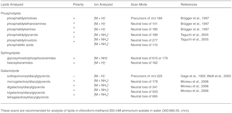
The precursor and neutral loss scans thus provide, for each lipid molecular species, the mass and signal of the intact ion, along with the mass of one molecular fragment that allows the lipid to be classified into a class or group. Given the classification, the mass can be interpreted as the total number of carbons and double bonds in the component acyl (or sphingosine/fatty amide) chains. To obtain more complete characterization of the complex lipid molecule, further analysis, such as mass spectral product ion analysis to identify individual fatty acyl components, can be performed. Devaiah et al. (2006) utilized product ion analysis to characterize many Arabidopsis glycerolipid molecular species in terms of fatty acyl composition.
For each detected lipid molecular species, the signal size allows quantification. To achieve accurate quantification by the direct-infusion ESI-MS/MS approach, a large number of internal standard compounds, optimally at least two non-naturally-occurring compounds for each class or group, are required (e.g., Welti et al., 2002; Devaiah et al., 2006). Alternatively, relative quantification among samples can be achieved by comparing mass spectral responses to an arbitrary standard compound detected with the same polarity (i.e., positive or negative mode) and scanning mode (i.e., precursor or neutral loss scanning) as the lipids of interest (e.g., detection of oxylipin-containing complex lipids in Maeda et al., 2008).
Challenges remaining in lipidomics include:
Standardizing lipidomics methodology and making the technology widely accessible.
Establishment of analyses for additional groups of lipids.
Development of a standardized and available data processing system for interpreting mass spectral data to produce lipid profiles.
Development of a web-accessible lipid profile database that facilitates integration with genomic, gene expression, proteomic, and other metabolomic data.
3.11. Strategies for Imaging in Plant Lipid Biology (Allan DeBono and Lacey Samuels*28)
3.11.1. Background
The goal of the microscopy of lipids is to visualize these hydrophobic compounds in their cellular context, with minimal rearrangements. When planning a microscopy approach, it is useful to think about what level of detailed cell structure is required. For high-resolution information on cellular organelles, transmission electron microscopy must be used, but TEM requires that cells be fixed and cut into thin sections, presenting a static view. Scanning electron microscopy (SEM) is useful for directly visualizing detailed surface structures such as the cuticle. For dynamic processes in live cells, light microscopy is the only choice as live cells fare poorly in the high-vacuum conditions of conventional electron microscopes. However, the resolution of the light microscope is limiting, so organelle identification is often done by correlating “puncta” with markers of known subcellular compartments.
In fluorescence microscopy, a sample containing fluorescent molecules, such as a dye or a fluorescent protein, is excited with energy of a given wavelength from a light source (e.g., a mercury lamp). Lower energy, longer wavelength light is emitted, collected through specific filters, and detected by eye or with a camera. Confocal laser scanning microscopy (CLSM) follows the same excitation and emission principles of fluorescence microscopy but provides improved imaging due to removal of out-of-focus fluorescence. The source of excitation is a laser that scans across the sample. The emission is collected through a pinhole of adjustable size that filters out-of-focus light. This allows for shallow depth of focus, which is exploited to generate a series of optical sections in the z-axis, which can be reassembled into a three-dimensional data set (Nikon Microscopy U website.)
In the study of plant lipids, CLSM is used both to detect fluorochrome dyes and to localize enzymes and other gene products related to lipid metabolism in the cell using fluorescent protein fusions. Storage lipids, such as triacylglycerols in oil bodies, can be imaged in live cells using Nile Red staining and fluorescence microscopy (e.g., Schmidt and Herman, 2008; Quettier and Eastmond, 2009). The plasma membrane can be stained using the amphipathic fluorescent styryl dye, FM4-64, as it partitions into the plasma membrane of live cells (Boite et al., 2004). Endocytosis of the plasma membrane then can be followed over time as the bilayer is internalized and recycled (Zheng et al., 2005; Dettmer et al., 2006; DeBono et al., 2009). FM1-43, a closely related styryl dye, has also been used to label the plasma membrane, endocytic pathway, and even secretory vesicles (Okamoto et al., 2008; Bove et al., 2008).
The following list of protocols is not comprehensive; rather, it represents the experimental approaches that we have found most robust and reliable. For specialized applications, classical histochemistry stains or other fluorescent probes may be more appropriate, and a search of the literature to find how others have approached imaging lipids in the same system is always the best preliminary step.
3.11.2. Protocols
3.11.2.1. Nile red, a general lipophilic stain. Nile Red is a polycyclic lipid stain that fluoresces intensely in a hydrophobic environment but not in aqueous media (Fowler and Greenspan, 1985). Nile Red has sensitivity to the hydrophobic environment, exhibiting red emission in the presence of polar lipids to more yellow emission in the presence of esterified cholesterol and triacylglycerols (Diaz et al., 2008). Nile Red has been used to stain sites of lipid accumulation in plants (Pighin et al., 2004; Schmidt and Herman, 2008; Dietrich et al., 2009) and alterations of surface lipids (Y.H. Li et al., 2007a; Figure 18).
Figure 18.
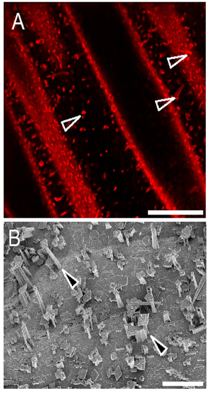
Wax Crystals of Arabidopsis Stems Viewed by Confocal Microscopy and Cryo-Scanning Electron Microscopy.
(A) Nile Red and confocal microscopy of the Arabidopsis stem epidermis surface with wax crystal structures.
(B) CryoSEM of Arabidopsis stem epidermis surface with wax crystal structures (closed arrowhead).Bars = 8 µm (A) and 5 µm (B).
Protocol:
A stock solution of Nile Red, also called Nile Blue A Oxazone (Sigma #72485), is dissolved in 100% DMSO to 1 mg/mL and should be stored protected from light at –2O°C.
Working solutions range from 1–5 µg mL-1, diluted with water or buffer.
For tissues with cuticle, cut small segments to allow the stain to penetrate. A leaf disc or 5 mm longitudinal segment from a stem or seedling root will provide abundant imaging material. For elongated cell types, avoid transverse sections to prevent cell rupture and think about the cell geometry if the tissue is dissected. The tissue can be rinsed to remove excess dye.
Generally, Nile Red is excited with 488 nm or 543 nm laser lines and collected with a 560–615 nm filter. For special applications, Nile Red yellow emission, for nonpolar lipids, can be observed with 460 nm excitation and 535 nm emission; the red emission of Nile Red, for polar lipids, can be observed with 540 nm excitation and a 590 nm long pass emission filter (Diaz et al., 2008).
3.11.2.2. FM4-64 staining of the plasma membrane and the endocytic pathway. FM4-64, which fluoresces in the red range, is a useful counterstain to demonstrate plasma membrane localization in plants expressing green or yellow fluorescent proteins (GFP, YFP). FM4-64 has several properties that have contributed to its widespread use: It is not toxic to cells at the working concentrations; it fluoresces intensely only in a lipidic environment or when bound in membranes, reducing background; and it is soluble in water (http://www.invitrogen.com; Boite et al., 2004). FM1-43 is the green fluorescent equivalent of the red FM4-64 (both spectra can be viewed at the Invitrogen Spectral Viewer).
Protocol:
Stock solutions of FM4-64 and FM1-43 (Invitrogen #T-3166 and #T-3163) are prepared at 10 mM in 100% DMSO and stored in aliquots, protected from light, at –20°C
Typical applications of FM4-64 use a working concentration of 4–10 µM for roots (Dettmer et al., 2006), leaves (Zheng et al., 2005), and stems (DeBono et al., 2009). Typical working concentrations of FM1-43 are 160 nM to 2 µM for pollen (Bove et al., 2008; Sousa et al., 2008) and roots (Okamoto et al., 2008).
Dissect tissue to allow dye penetration but minimize cell damage (Figure 19) and immerse in dye at room temperature.
The plasma membranes of cells from Arabidopsis stems and leaves can be observed after 10 min, while roots require shorter incubation times (less than 60 sec). Endocytosis of FM4-64 typically occurs in 20–30 minutes (Figure 20). Timing must be determined empirically for each tissue.
FM4-64 can be excited with the 488 nm laser lines and detected with a 560 nm long pass filter. Alternatively, the 543 or 561 nm laser lines can be used for excitation and emission detected with a 584–664 nm band pass filter. Although this means collecting on the shoulder of the FM4-64 emission peak, with the intensity of FM4-64 label, it is often adequate and can be used to limit chloroplast autofluorescence. FM143 can also be excited with the 488 nm laser line and detected with a 500–600 nm filter.
Figure 19.
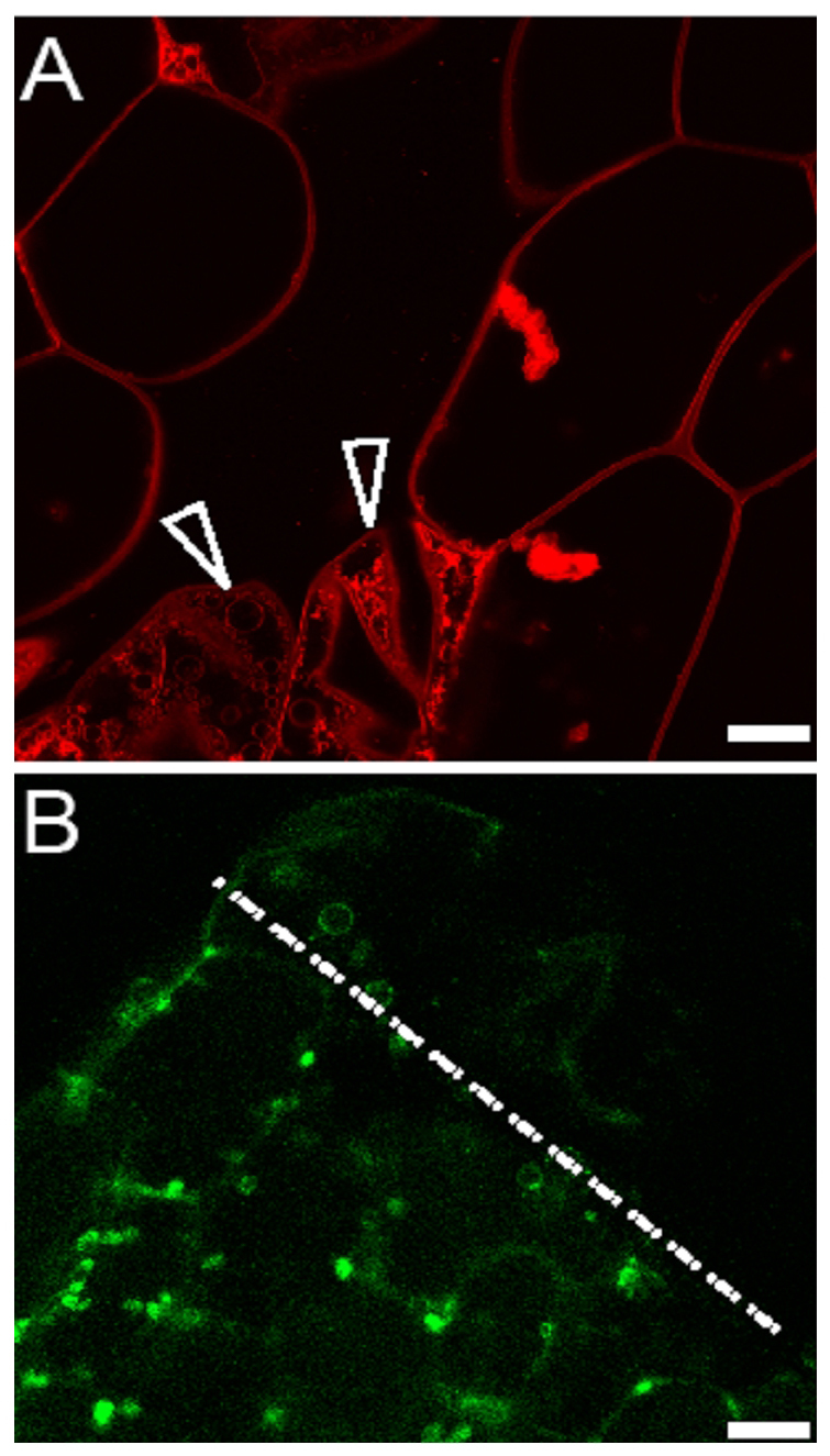
Arabidopsis Hypocotyl Cells Damaged During Handling Prior to Live Imaging.
(A) Hypocotyl cells stained with FM4-64 are vesiculated after crushing (arrowheads).
(B) Yellow fluorescent protein tagged glycosylphosphatidyl-inositol (GPI)anchored lipid transfer protein (YFP-LTPG) is plasma membrane localized in undamaged cells. In contrast, due to damage during sample dissection, cells along the incision (dotted line) contain numerous large vesicles. Bars = 14 µm.
Figure 20.
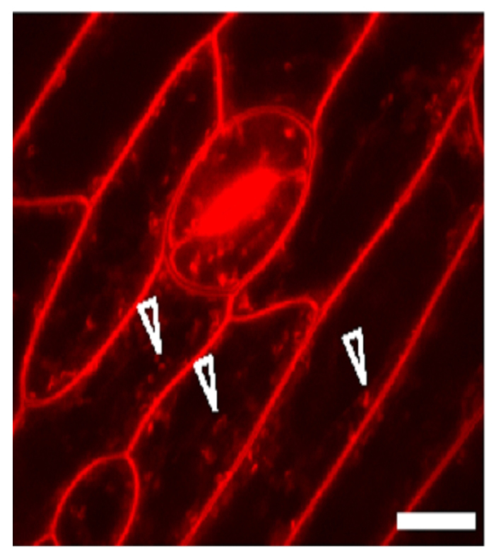
Arabidopsis Stem Epidermal Cells Stained With FM4-64.
Prolonged exposure to FM4-64 stains endocytic compartments (arrowheads) in addition to the plasma membrane. Bar = 9 µm.
3.11.2.3. Scanning electron microscopy. Since the surface structures of the cuticle are sensitive to fixatives and dehydrating agents (Reed, 1982), the undisturbed cuticle is best viewed without conventional SEM preparation (Neinhuis and Barthlott, 1997). Samples can be air dried directly on the SEM stub (Jackson, 2002) or frozen and viewed with cryo-SEM (Pighin et al., 2004; Radboud University Nijmegen).
3.11.2.4. Transmission electron microscopy. TEM allows high-resolution imaging of lipidic cell structure, including membranes and oil bodies. However, lipids are poorly crosslinked by the first aldehyde fixation step in conventional chemical electron microscopy sample preparation (Hopwood, 1972). This can cause membrane and organelle rearrangements, so cryofixation, such as high-pressure freezing and freeze substitution, is recommended for all lipid-rich systems (Bird et al., 2007; Schmidt and Herman, 2008). However, even in cryofixed cells, lipids are probably extracted during room temperature embedding. For example, we have observed that the ultrastructure of the cuticle of Arabidopsis stems was identical in samples that had been dipped in hexane to remove soluble waxes compared to controls without dipping (Samuels lab, unpublished data). This suggests that the cuticle viewed in the TEM is primarily cutin and cutan components. With these caveats in mind, TEM can still provide useful information about membrane structure, oil body size and distribution, and cutin organization.
Chemical Fixation Protocol:
Dissect plant part of interest into pieces no bigger than 2–3 mm. Immersion of the sample in fixative (2% glutaraldehyde in 50 mM PIPES buffer, pH 7.0) at room temperature for 2 h is usually adequate.
Wash 3 × in 50 mM PIPES buffer.
Postfix in 2% OsO4 In 50 mM PIPES buffer for 1 h. Samples will turn black.
Dehydrate in an ethanol series, 20 min for each step (30% ethanol, 50% ethanol, 70% ethanol, 95% ethanol, 100% ethanol twice).
Mix the liquid plastic resin (Spurr's epoxy resin) with the ethanol to gradually infiltrate the resin into the cells. Infiltrate using resin mixed with solvent of increasing concentration series (10% resin, 25% resin, 50% resin, 75% resin, 100% resin). For each mix, let cells incubate for 2 h minimum, overnight maximum.
Put samples into BEEM® polyethylene embedding capsules capsules with fresh resin and bake overnight at 60°C.
Cut into 0.5 µm sections and stain with 1 % toluidine blue in 1 % sodium borate to check the morphology of fixed cells with light microscopy. Choose the best blocks and cut 70 nm sections, stain with 2% aqueous uranyl acetate 15 min and Reynold's lead citrate for 5 min, then view with a transmission electron microscope.
Cryofixation Protocol: For detailed information on sample preparation, download the Practical Methods Manual to High-Pressure Freezing (HPF) by Mary Morphew from the Boulder Lab for 3-D Electron Microscopy of Cells.
Prepare HPF according to manufacturer's directions and do test runs.
With extra fine razor blades, cut small blocks of tissue under a pool of extracellular cryoprotectant, such as sucrose at a nonplasmolyzing concentration. Transfer cells to sample carriers and freeze without delay. The sample preparation and handling is critical; if samples are anoxic, crushed, or air-dried (mild drying leads to plasmolysis), then the best freezing will be for naught.
Following freezing, transfer samples to cryovials with freeze substitution medium (2% OsO4 in anhydrous acetone with 8% dimethoxypropane).
Substitute at –80°C for 3 to 5 days using an automated freeze substitution system or an acetone/dry ice slush in a styrofoam chest, which equilibrates at –80°C.
Gradually warm samples to room temperature, ramping the temperature up over an 8-hour period, allowing the OsO4 to react with the stabilized cell structures.
Rinse in clean acetone several times and remove the HPF carriers.
Slowly infiltrate with Spurr's resin. Add 1 drop of resin to 1 mL of acetone and mix well, then incubate the samples with the resin and acetone mixture for 5–10 min. Continue to increase the amount of resin by 1 drop/mL until you have added up to 10%–25% resin, then leave overnight.
Incubate in fresh 25% resin, 2–3 h; 50% resin, 2–-3 h; 75% resin, 2–3 h, 100% resin 2–3 h twice or overnight with an extra 1 h change in the morning.
Incubate at 60°C to polymerize the resin.
Sectioning, staining, and TEM then follow the conventional protocol above.
3.11.3. Comments
The light and electron microscopy techniques above remain popular approaches, but often simple, classical lipid stains, which are used with bright field microscopy, are the most appropriate technique to identify lipid-rich structures such as the cuticle, suberized endodermis, or periderm (Brundrett et al., 1991; Shen et al., 2003; Y.H. Li et al., 2007b). For practical methods in classical histochemical techniques, see Harris et al. (1994).
Despite the problem that structures in the submicrometer range are difficult to resolve, light microscopy continues to be an important tool for studying plant lipids, especially in conjunction with molecular biology and mutant analyses. The localization of proteins of interest using fluorescent protein fusions provides useful information but requires careful experimental design.
Before beginning a molecular biology protocol to fuse a gene of interest to GFP, the following considerations can make the difference between success and disaster. First, consider predicted targeting sites and topology for the protein, and plan the site of fusion to minimize the probability that the fluorescent protein tag will be cleaved off the mature protein during targeting. Check the autofluorescence of the tissue where the protein will be expressed, and choose a protein that does not fluoresce in that range. Finally, your work will have greater credibility if you can demonstrate that the GFP-fusion protein is functional in vivo by complementing a mutant phenotype. GFP research has evolved from the older and now obsolete variants of green fluorescent protein to fluorescent proteins with increased brightness, pH stability, and monomerization (e.g., eYFP to CitrineYFP and mRFP to mCherry; Shaner et al., 2007). If your protein of interest is secreted to the acidic cell wall or vacuole, then older GFP variants will be quenched by the low pH. New variants of YFP, Venus, and Citrine have improved pH and chloride sensitivity (Griesbeck et al., 2001; Nagai et al., 2002). After selection of transgenic lines, it is important to screen 10 to 12 lines and select those with appropriate fluorescence levels.
Although live plant cell imaging preparation is relatively straightforward, there are simple provisions that can be made to improve image quality. In addition to differences in anatomical structure, airspaces contribute to the differences in uptake of dyes between leaves and stems versus roots. To overcome the air spaces and the cuticle in such tissues, we have found that a brief centrifugation (30 s) at 500g after incubation in a given dye will improve staining, reduce the time required before imaging, and most importantly permit the tissue to be excited using lower, less damaging laser intensity.
The appropriate amount of laser power is the absolute minimum that can excite the fluor without generating autofluorescence in an untreated, nonfluorescent protein control. Too much laser intensity is toxic and will alter membrane morphology and/or cause vesiculation.
4. SUMMARY OF ARABIDOPSIS LIPID COMPOSITION
This section summarizes the acyl lipid composition for various tissues and organs of wild type Arabidopsis. Data presented in this chapter were collected from Col-0 ecotype unless otherwise noted. The goal of this section is to provide a quick and easy access to summary on acyl lipid content and composition, which sometimes can be difficult to find. It is composed of 15 tables and 3 figures as outlined below:
Tables included:
- Table 4.
Fatty Acid Composition of Arabidopsis Tissues
- Table 5.
Molecular Species Composition for 52 TAGs From Dry Seeds
- Table 6.
Glycerolipid Composition of Arabidopsis Tissues
- Table 7.
Fatty Acid Composition of Individual Leaf Glycerolipids From Arabidopsis
- Table 8.
Load of Cuticular Wax Compound Classes in Stems and Rosette Leaves of Arabidopsis
- Table 9.
Total Load of Suberin and Suberin-Associated Waxes in Arabidopsis Seeds and Roots
- Table 10.
Suberin Monomer Composition in Seed Coats and Roots
- Table 11.
Composition of Arabidopsis Root Waxes
- Table 12.
Composition of Arabidopsis Seed Waxes
- Table 13.
Cutin Monomer Composition in Arabidopsis Tissues
- Table 14.
Fatty Acid Composition of Glycerophospholipids in Mitochondrial Membranes of Arabidopsis
- Table 15.
Lipid Composition of Mitochondria Isolated From Arabidopsis
- Table 16.
Acyl-CoA Composition of Arabidopsis Leaf Tissues
- Table 17.
Sphingolipid Composition of Arabidopsis Tissues
- Table 18.
Stereospecific Analysis of Arabidopsis Seed Triacylglycerols
Table 5.
Molecular Species Composition for 52 TAGs From Dry Seeds
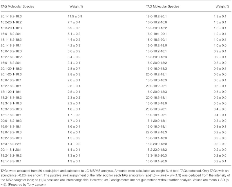
Table 6.
Glycerolipid Composition of Arabidopsis Tissues
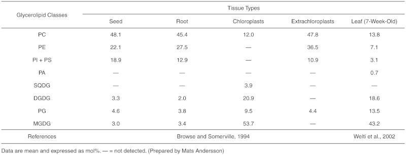
Table 7.
Fatty Acid Composition of Individual Leaf Glycerolipids From Arabidopsis
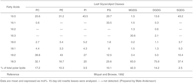
Table 8.
Load of Cuticular Wax Compound Classes in Stems and Rosette Leaves of Arabidopsis
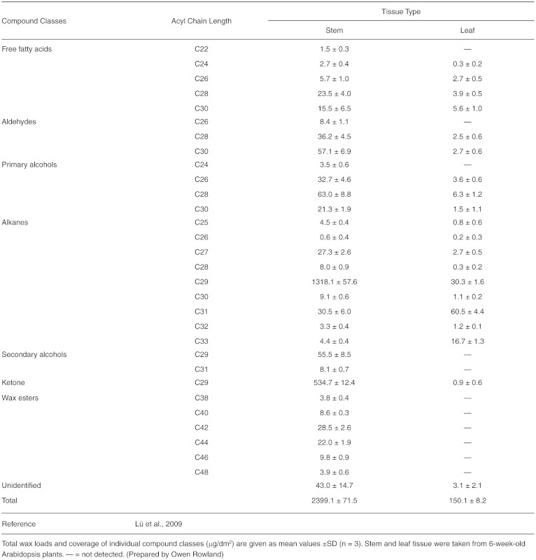
Table 9.
Total Load of Suberin and Suberin-Associated Waxes in Arabidopsis Seeds and Roots
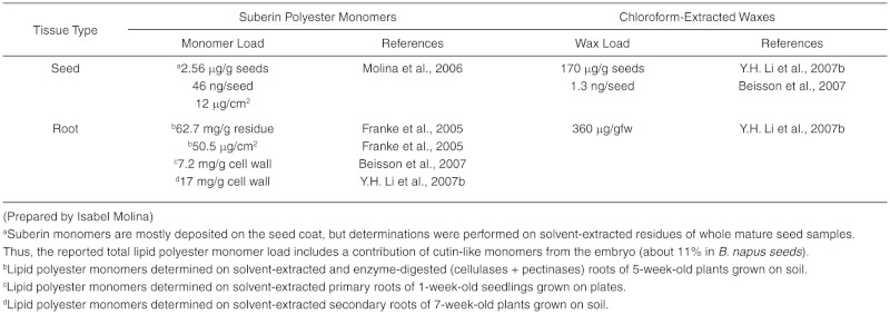
Table 12.
Composition of Arabidopsis Seed Waxes
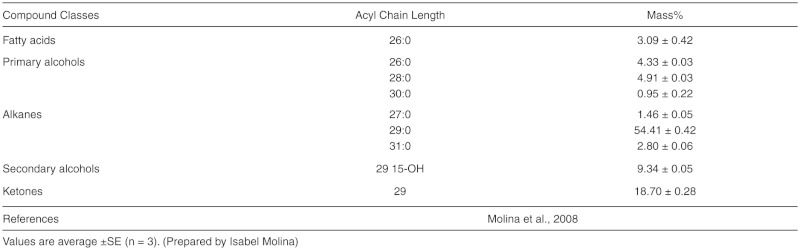
Table 13.
Cutin Monomer Composition in Arabidopsis Tissues
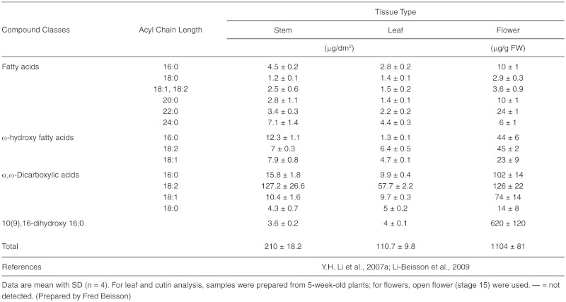
Table 14.
Fatty Acid Composition of Glycerophospholipids in Mitochondrial Membranes of Arabidopsis
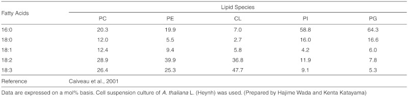
Table 15.
Lipid Composition of Mitochondria Isolated from Arabidopsis
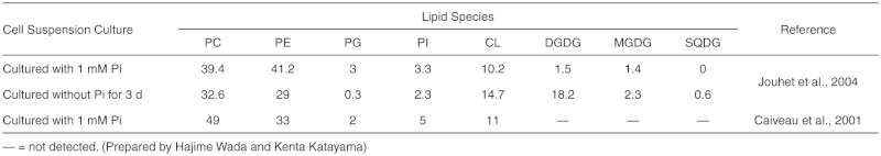
Table 16.
Acyl-CoA Composition of Arabidopsis Leaf Tissues
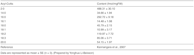
Table 17.
Sphingolipid Composition of Arabidopsis Tissues
The sphingolipid content of Arabidopsis varies based on the method used to determine composition and the tissue from which the sphingolipids are extracted. Data are provided for the two main methods of analysis, hydrolysis and measurement of the long-chain base (LCB) component and liquid chromatography tandem mass spectrometry (LC-MS/MS) of the intact sphingolipids. All data are from Arabidopsis leaf tissue at 5–-6 weeks of age. (Prepared by Jonathan E. Markham)
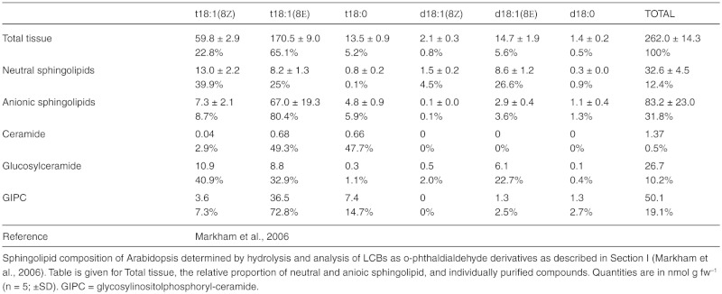
Table 17.2.
Sphingolipid Composition Determined by LC-MS/MS
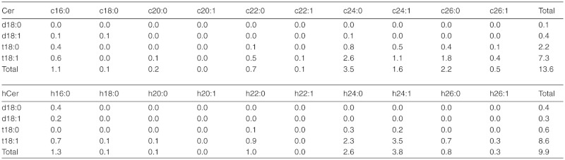
continued.
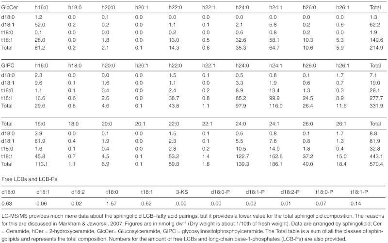
Table 18.
Stereospecific Analysis of Arabidopsis Seed Triacylglycerols
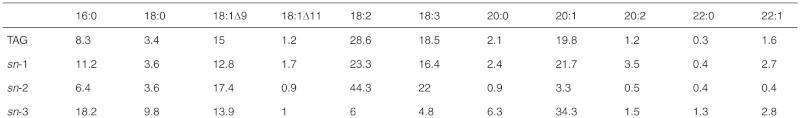
Table 18.2.
Occurrence of Each Fatty Acid Across All Three sn-Positions
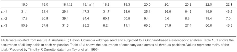
Table 4.
Fatty Acid Composition of Arabidopsis Tissues
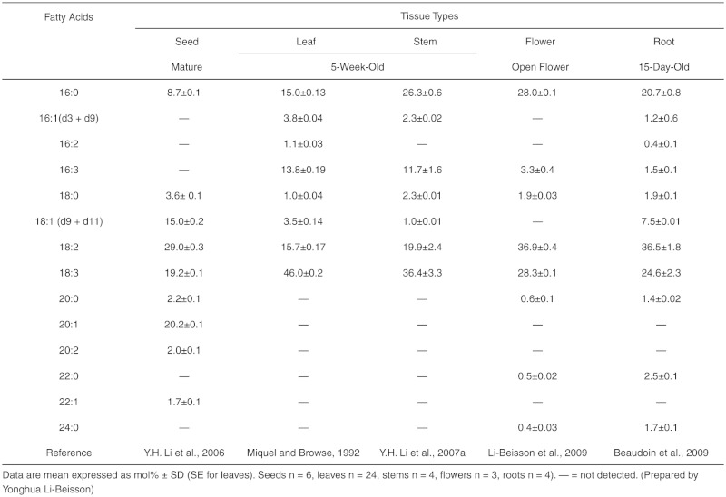
Figures included:
- Figure 21.
Relative Distribution of Lipids and Other Components of Arabidopsis Leaf.
- Figure 22.
Relative Distribution of Lipids and Other Components of Arabidopsis Seeds.
- Figure 23.
Distribution of Extracellular Lipids in Mature Seeds
Figure 21.

Relative Distribution of Lipids and Other Components of Arabidopsis Leaf.
Data adapted from Browse and Somerville (1994). (Prepared by John Ohlrogge)
Figure 22.

Relative Distribution of Lipids and Other Components of Arabidopsis Seeds.
Relative contribution of storage lipids and proteins were obtained from Y.H. Li et al., 2006. Percentage of membrane glycerolipids relative to total lipids is from Ohlrogge and Browse, 1995. Content of surface lipids is from Molina et al. (2006) and Beisson et al. (2007). (Prepared by Isabel Molina)
Figure 23.

Distribution of Extracellular Lipids in Mature Seeds.
Values of total polyester monomers and distribution between seed coat and embryo (inferred from B. napus data) are from Molina et al. (2006). Distribution of polyester monomers in inner integument (ii) and outer integument (oi) summarize results from Molina et al. (2008). Surface wax load was reported by Beisson et al. (2007). (Prepared by Isabel Molina)
Footnotes
Citation: Yonghua Li-Beisson, Basil Shorrosh, Fred Beisson, Mats X. Andersson, Vincent Arondel, Philip D. Bates, Sébastien Baud, David Bird, Allan DeBono, Timothy P. Durrett, Rochus B. Franke, Ian A. Graham, Kenta Katayama, Amélie A. Kelly, Tony Larson, Jonathan E. Markham, Martine Miquel, Isabel Molina, Ikuo Nishida, Owen Rowland, Lacey Samuels, Katherine M. Schmid, Hajime Wada, Ruth Welti, Changcheng Xu, Rémi Zallot, and John Ohlrogge. (2013) Acyl-Lipid Metabolism. The Arabidopsis Book 9:e0161. doi:10.1199/tab.0161
elocation-id: e0161
First published on January 28, 2013: e0161. doi: 10.1199/tab.0161
This chapter is an updated version of a chapter originally published on June 11, 2010, e0133. doi: 10.1199/tab.0133
1 [wire] stone, 145 E. Mountain Avenue, Fort Collins, CO 80525. basil.shorrosh@wirestone.com
2 Institut Jean-Pierre Bourgin, INRA-AgroParisTech, 78026 Versailles Cedex, France. Email: sebastien.baud@versailles.inra.fr
3 Department of Plant- and Environmental Sciences, University of Gothenburg, Box 461, SE-405 30 Göteborg, Sweden. Email: mats.andersson@ dpes.gu.se
4 Warwick HRI, University of Warwick, Wellesbourne, Warwickshire, CV35 9EF, UK. Email: a.a.b.kelly@warwick.ac.uk
5 Division of Life Science, Graduate School of Science and Engineering, Saitama University, Shimo-Okubo 255, Sakura-ku, Saitama, 338-8570, Japan. Email: nishida@molbiol.saitama-u.ac.jp
6 Institute of Biological Chemistry, Washington State University, Pullman, WA. Email: phil_bates@wsu.edu
7 Department of Biochemistry, University of Nebraska-Lincoln, NE, 68588. Email: jemarkham@unl.edu
8 Department of Life Sciences, Graduate School of Arts and Sciences, University of Tokyo, Komaba 3-8-1, Meguro-ku, Tokyo 153-8902, Japan. *Email: hwada@ bio.cu-tokyo.ac.jp
9 Department of Plant Biology, Michgan State University, East Lansing, Ml 48824. Email: tdurrett@msu.edu
10 Biology Department, Brookhaven National Laboratory, Upton, NY 11973-5000. Email: cxu@bnl.gov
11 Department of Biology and Institute of Biochemistry, Carleton University, Ottawa, ON K1S 5B6, Canada. Email: owen_rowland@carleton.ca
12 Department of Biology, Mount Royal University, Calgary T3E 6K6, Canada. Email: dbird@mtroyal.ca
13 Department of Plant Biology and Environmental Microbiology, CEA/ CNRS/Aix-Marseille University, Cadarache, France. Email: frederic.beisson@cea.fr oryonghua.li@cea.fr
14 Department of Biology, Algoma University, Sault Ste Marie, Ontario, Canada. E-mail: isabel.molina@algomau.ca
15 Institut für Zelluläre and Molekulare Botanik, Universität Bonn, Bonn, Germany. Email: rochus.franke@uni-bonn.de
16 Laboratoire de Biogenèse Membranaire, CNRS/University of Bordeaux, CNRS UMR5200, Bordeaux, France. *Email: vincent.arondel@ubordeaux2.fr
17 Centre for Novel Agricultural Products, Department of Biology, University of York, Heslington.York, YO10 5YW, UK. Email: iagl@york.ac.uk
18 Department of Biological Sciences, Butler University, 4600 Sunset Avenue, Indianapolis, IN 46208. Email: kschmid@butler.edu
19 Institut Jean-Pierre Bourgin, INRA-AgroParisTech, 78026 Versailles Cedex, France. Email: Martine.Miquel@versailles.inra.fr
20 Institute of Biological Chemistry, Washington State University, Pullman, WA 99164. Email: phil_bates@wsu.edu
21 Department of Plant Biology and Environmental Microbiology, CEA/CNRS/Aix-Marseille University, Cadarache, France. Email: yonghua.li@cea.fr
22 Centre for Novel Agricultural Products, Department of Biology, University of York, Heslington, York, YO10 5YW, UK. Email: trl1@york.ac.uk
23 Centre for Novel Agricultural Products, Department of Biology, University of York, Heslington, York, YO10 5YW, UK. Email: trl1@york.ac.uk
24 Donald Danforth Plant Science Center, St. Louis, MO 63132. Email: jmarkham@danforthcenter.org
25 Department of Environmental Plant Biology and Environmental Microbiology, CEA/CNRS/Aix-Marseille University, 13108 Cadarache, France. Email: frederic.beisson@cea.fr
26 Department of Biology and Institute of Biochemistry, Carleton University, Ottawa, ON K1S 5B6, Canada. Email: owen_rowland@carleton.ca
27 Kansas Lipidomics Research Center, Division of Biology, Kansas State University, Manhattan, KS 66506. Email: welti@ksu.edu
28 Department of Botany, University of British Columbia, Vancouver, BC, Canada V6T 1Z4. *Email: lsamuels@interchange.ubc.ca
References
- Aarts M.G.M., Keijzer C.J., Stiekema W.J., Pereira A. Molecular characterization of the CER1 gene of arabidopsis involved in epicuticular wax biosynthesis and pollen fertility. Plant Cell. 1995;7:2115–2127. doi: 10.1105/tpc.7.12.2115. [DOI] [PMC free article] [PubMed] [Google Scholar]
- Acosta I.F., Farmer E.E. The Arabidopsis Book. Rockville, MD: American Society of Plant Biologists; 2010. Jasmonates. [DOI] [PMC free article] [PubMed] [Google Scholar]
- Adham A.R., Zolman B.K., Millius A., Bartel B. Mutations in Arabidopsis acyl-CoA oxidase genes reveal distinct and overlapping roles in beta-oxidation. Plant J. 2005;41:859–874. doi: 10.1111/j.1365-313X.2005.02343.x. [DOI] [PubMed] [Google Scholar]
- Agrawal V.P., Kolattukudy P.E. Biochemistry of suberization: Omega-hydroxyacid oxidation in enzyme preparations from suberizing potato tuber disks. Plant Physiol. 1977;59:667–672. doi: 10.1104/pp.59.4.667. [DOI] [PMC free article] [PubMed] [Google Scholar]
- Agrawal V.P., Kolattukudy P.E. Purification and characterization of a wound-induced omega-hydroxyfatty acid:NADP oxidoreductase from potato tuber disks (Solanum tuberosum L.). Arch. Biochem. Biophys. 1978;191:452–465. doi: 10.1016/0003-9861(78)90384-3. [DOI] [PubMed] [Google Scholar]
- Aharoni A., Dixit S., Jetter R., Thoenes E., van Arkel G., Pereira A. The SHINE clade of AP2 domain transcription factors activates wax biosynthesis, alters cuticle properties, and confers drought tolerance when overexpressed in Arabidopsis. Plant Cell. 2004;16:2463–2480. doi: 10.1105/tpc.104.022897. [DOI] [PMC free article] [PubMed] [Google Scholar]
- Akoh C.C., Lee G.C., Liaw Y.C, Huang T.H., Shaw J.F. GDSL family of serine esterases/lipases. Prog. Lipid Res. 2004;43:534–552. doi: 10.1016/j.plipres.2004.09.002. REVIEW. [DOI] [PubMed] [Google Scholar]
- Andersson M.X., Dörmann P. Chloroplast membrane lipid biosynthesis and transport. In: Sandelius A., Aronsson H., editors. The Chloroplast: Interactions With the Environment. Berlin: Springer; 2008. pp. 125–159. REVIEW. [Google Scholar]
- Andersson M.X., Goksor M., Sandelius A.S. Optical manipulation reveals strong attracting forces at membrane contact sites between endoplasmic reticulum and chloroplasts. J. Biol. Chem. 2007;282:1170–1174. doi: 10.1074/jbc.M608124200. [DOI] [PubMed] [Google Scholar]
- Andersson M.X., Larsson K.E., Tjellstrom H., Liljenberg C., Sandelius A.S. The plasma membrane and the tonoplast as major targets for phospholipid-to-glycolipid replacement and stimulation of phospholipases in the plasma membrane. J. Biol. Chem. 2005;280:27578–27586. doi: 10.1074/jbc.M503273200. [DOI] [PubMed] [Google Scholar]
- Andersson M.X., Stridh M.H., Larsson K.E., Lijenberg C., Sandelius A.S. Phosphate-deficient oat replaces a major portion of the plasma membrane phospholipids with the galactolipid digalactosyldiacylglycerol. FEBS Lett. 2003;537:128–132. doi: 10.1016/s0014-5793(03)00109-1. [DOI] [PubMed] [Google Scholar]
- Andrews J., Mudd J.B. Phosphatidylglycerol synthesis in pea chloroplasts: Pathway and localization. Plant Physiol. 1985;79:259–265. doi: 10.1104/pp.79.1.259. [DOI] [PMC free article] [PubMed] [Google Scholar]
- Awai K., Marechal E., Block M.A., Brun D., Masuda T, Shimada H., Takamiya K., Ohta H., Joyard J. Two types of MGDG synthase genes, found widely in both 16:3 and 18:3 plants, differentially mediate galactolipid syntheses in photosynthetic and nonphotosynthetic tissues in Arabidopsis thaliana. Proc. Natl. Acad. Sci. USA. 2001;98:10960–10965. doi: 10.1073/pnas.181331498. [DOI] [PMC free article] [PubMed] [Google Scholar]
- Awai K., Xu C.C., Tamot B., Benning C. A phosphatidic acid-binding protein of the chloroplast inner envelope membrane involved in lipid trafficking. Proc. Natl. Acad. Sci. USA. 2006;103:10817–10822. doi: 10.1073/pnas.0602754103. [DOI] [PMC free article] [PubMed] [Google Scholar]
- Babiychuk E., Müller F., Eubel H., Braun H.P., Frentzen M., Kushnir S. Arabidopsis phosphatidylglycerophosphate synthase 1 is essential for chloroplast differentiation, but is dispensable for mitochondrial function. Plant J. 2003;33:899–909. doi: 10.1046/j.1365-313x.2003.01680.x. [DOI] [PubMed] [Google Scholar]
- Bach L., Michaelson L.V., Haslam R., Bellec Y., Gissot L., Marion J., Da Costa M., Boutin J.P., Miquel M., Tellier F., Domergue F., Markham J.E., Beaudoin F., Napier J.A., Faure J.D. The very-long-chain hydroxy fatty acyl-CoA dehydratase PASTICCI-NO2 is essential and limiting for plant development. Proc. Natl. Acad. Sci. USA. 2008;105:14727–14731. doi: 10.1073/pnas.0805089105. [DOI] [PMC free article] [PubMed] [Google Scholar]
- Baker A., Graham I.A., Holdsworth M., Smith S.M., Theodoulou F.L. Chewing the fat: Beta-oxidation in signalling and development. Trends Plant Sci. 2006;11:124–132. doi: 10.1016/j.tplants.2006.01.005. REVIEW. [DOI] [PubMed] [Google Scholar]
- Bao X., Focke M., Pollard M., Ohlrogge J. Understanding in vivo carbon precursor supply for fatty acid synthesis in leaf tissue. Plant J. 2000;22:39–50. doi: 10.1046/j.1365-313x.2000.00712.x. [DOI] [PubMed] [Google Scholar]
- Bates P.D., Browse J. The pathway of triacylglycerol synthesis through phosphatidylcholine in Arabidopsis produces a bottleneck for the accumulation of unusual fatty acids in transgenic seeds. Plant J. 2011;68:387–399. doi: 10.1111/j.1365-313X.2011.04693.x. [DOI] [PubMed] [Google Scholar]
- Bates P.D., Durrett T.P., Ohlrogge J.B., Pollard M. Analysis of acyl fluxes through multiple pathways of triacylglycerol synthesis in developing soybean embryos. Plant Physiol. 2009;150:55–72. doi: 10.1104/pp.109.137737. [DOI] [PMC free article] [PubMed] [Google Scholar]
- Bates P.D., Ohlrogge J.B., Pollard M. Incorporation of newly synthesized fatty acids into cytosolic glycerolipids in pea leaves occurs via acyl editing. J. Biol. Chem. 2007;282:31206–31216. doi: 10.1074/jbc.M705447200. [DOI] [PubMed] [Google Scholar]
- Baud S., Lepiniec L. Regulation of de novo fatty acid synthesis in maturing oilseeds of Arabidopsis. Plant Physiol, and Biochem. 2009;47:448–455. doi: 10.1016/j.plaphy.2008.12.006. [DOI] [PubMed] [Google Scholar]
- Baud S., Guyon V., Kronenberger J., Wuilleme S., Miquel M., Caboche M., Lepiniec L., Rochat C. Multifunctional acetyl-CoA carboxylase 1 is essential for very long chain fatty acid elongation and embryo development in Arabidopsis. Plant J. 2003;33:75–86. doi: 10.1046/j.1365-313x.2003.016010.x. [DOI] [PubMed] [Google Scholar]
- Baud S., Santos-Mendoza M., To A., Harscoet E., Lepiniec L., Dubreucq B. WRINKLED1 specifies the regulatory action of LEAFY COTYLEDON2 towards fatty acid metabolism during seed maturation in Arabidopsis. Plant J. 2007;50:825–838. doi: 10.1111/j.1365-313X.2007.03092.x. [DOI] [PubMed] [Google Scholar]
- Baxter I., Hosmani P.S., Rus A., Lahner B., Borevitz J.O., Muthukumar B., Mickelbart M.V., Schreiber L., Franke R.B., Salt D.E. Root suberin forms an extracellular barrier that affects water relations and mineral nutrition in Arabidopsis. PLoS Genet. 2009;5:e1000492. doi: 10.1371/journal.pgen.1000492. [DOI] [PMC free article] [PubMed] [Google Scholar]
- Baxter I., Hosmani P.S., Rus A., Lahner B., Borevitz J.O., Muthukumar B., Mickelbart M.V., Schreiber L., Franke R.B., Salt D.E. Root suberin forms an extracellular barrier that affects water relations and mineral nutrition in Arabidopsis. PLoS genetics. 2009;5:e1000492. doi: 10.1371/journal.pgen.1000492. [DOI] [PMC free article] [PubMed] [Google Scholar]
- Beaudoin F., Wu X.Z., Li F.L., Haslam R.P., Markham J.E., Zheng H.Q., Napier J.A., Kunst L. Functional characterization of the Arabidopsis beta-ketoacyl-coenzyme A reductase candidates of the fatty acid elongase. Plant Physiol. 2009;150:1174–1191. doi: 10.1104/pp.109.137497. [DOI] [PMC free article] [PubMed] [Google Scholar]
- Beermann C., Green A., Mobius M., Schmitt J.J., Boehm G. Lipid class separation by HPLC combined with GC FA analysis: Comparison of seed lipid compositions from different Brassica napus L. varieties. J. Am. Oil Chem. Soc. 2003;80:747–753. [Google Scholar]
- Beisson R, Koo A.J., Ruuska S., Schwender J., Pollard M., Thelen J.J., Paddock T., Salas J.J., Savage L., Milcamps A., Mhaske V.B., Cho Y., Ohlrogge J.B. Arabidopsis genes involved in acyl lipid metabolism: A 2003 census of the candidates, a study of the distribution of expressed sequence tags in organs, and a web-based database. Plant Physiol. 2003;132:681–697. doi: 10.1104/pp.103.022988. [DOI] [PMC free article] [PubMed] [Google Scholar]
- Beisson F., Li Y.H., Bonaventure G., Pollard M., Ohlrogge J.B. The acyltransferase GPAT5 is required for the synthesis of suberin in seed coat and root of Arabidopsis. Plant Cell. 2007;19:351–368. doi: 10.1105/tpc.106.048033. [DOI] [PMC free article] [PubMed] [Google Scholar]
- Beisson F., Li-Beisson Y., Pollard M. Solving the puzzles of cutin and suberin polymer biosynthesis. Curr. Opin. Plant Biol. 2012a;15:329–337. doi: 10.1016/j.pbi.2012.03.003. [DOI] [PubMed] [Google Scholar]
- Beisson F., Ohlrogge J. Plants: Knitting a polyester skin. Nat. Chem. Biol. 2012b;8:603–604. doi: 10.1038/nchembio.1004. [DOI] [PubMed] [Google Scholar]
- Beisson F., Tiss A., Riviere C, Verger R. Methods for lipase detection and assay: A critical REVIEW. Eur. J. Lipid Sci. Technol. 2000;102:133–153. REVIEW. [Google Scholar]
- Bell R.M., Ballas L.M., Coleman R.A. Lipid topogenesis. J. Lipid Res. 1981;22:391–403. [PubMed] [Google Scholar]
- Benning C. A role for lipid trafficking in chloroplast biogenesis. Prog. Lipid Res. 2008;47:381–389. doi: 10.1016/j.plipres.2008.04.001. REVIEW. [DOI] [PubMed] [Google Scholar]
- Benning C. Mechanisms of lipid transport involved in organelle biogenesis in plant cells. Annu. Rev. Cell Dev. Biol. 2009;25:71–91. doi: 10.1146/annurev.cellbio.042308.113414. REVIEW. [DOI] [PubMed] [Google Scholar]
- Benning C., Xu CC, Awai K. Non-vesicular and vesicular lipid trafficking involving plastids. Curr. Opin. Plant Biol. 2006;9:241–247. doi: 10.1016/j.pbi.2006.03.012. REVIEW. [DOI] [PubMed] [Google Scholar]
- Benveniste I., Tijet N., Adas F., Philipps G., Salaun J.P., Durst F. CYP86A1 from Arabidopsis thaliana encodes a cytochrome P450-dependent fatty acid omega-hydroxylase. Biochem. Biophys. Res. Commun. 1998;243:688–693. doi: 10.1006/bbrc.1998.8156. [DOI] [PubMed] [Google Scholar]
- Bernards M.A. Demystifying suberin. Can. J. Bot. 2002;80:227–240. REVIEW. [Google Scholar]
- Bessire M., Borel S., Fabre G., Carraca L., Efremova N., Yephremov A., Cao Y., Jetter R., Jacquat A.C., Metraux J.P., Nawrath C. A member of the PLEIOTROPIC DRUG RESISTANCE family of ATP binding cassette transporters is required for the formation of a functional cuticle in Arabidopsis. Plant Cell. 2011;23:1958–1970. doi: 10.1105/tpc.111.083121. [DOI] [PMC free article] [PubMed] [Google Scholar]
- Bessire M., Chassot C., Jacquat A.C., Humphry M., Borel S., Petetot J.M.C., Metraux J.P., Nawrath C. A permeable cuticle in Arabidopsis leads to a strong resistance to Botrytis cinerea. EMBO J. 2007;26:2158–2168. doi: 10.1038/sj.emboj.7601658. [DOI] [PMC free article] [PubMed] [Google Scholar]
- Bessoule J.J., Testet E., Cassagne C. Synthesis of phosphatidylcholine in the chloroplast envelope after import of lysophosphatidylcholine from endoplasmic reticulum membranes. Eur. J. Biochem. 1995;228:490–497. [PubMed] [Google Scholar]
- Bin Y., Wakao S., Fan J.L., Benning C. Loss of plastidic lysophosphatidic acid acyltransferase causes embryo-lethality in Arabidopsis. Plant Cell Physiol. 2004;45:503–510. doi: 10.1093/pcp/pch064. [DOI] [PubMed] [Google Scholar]
- Bird D., Beisson F., Brigham A., Shin J., Greer S., Jetter R., Kunst L., Wu X.W., Yephremov A., Samuels L. Characterization of Arabidopsis ABCG11/WBC11, an ATP binding cassette (ABC) transporter that is required for cuticular lipid secretion. Plant J. 2007;52:485–498. doi: 10.1111/j.1365-313X.2007.03252.x. [DOI] [PubMed] [Google Scholar]
- Bird D.A. The role of ABC transporters in cuticular lipid secretion. Plant Sci. 2008;174:563–569. REVIEW. [Google Scholar]
- Bishop W.R., Bell R.M. Assembly of phospholipids into cellular membranes: Biosynthesis, transmembrane movement and intracellular translocation. Annu. Rev. Cell Biol. 1988;4:579–610. doi: 10.1146/annurev.cb.04.110188.003051. REVIEW. [DOI] [PubMed] [Google Scholar]
- Bjerve K.S., Daae L.N.W, Bremer J. The selective loss of lysophospholipids in some commonly used lipid-extraction procedures. Anal. Biochem. 1974;58:238–245. doi: 10.1016/0003-2697(74)90463-1. [DOI] [PubMed] [Google Scholar]
- Bligh E.G., Dyer W.J. A rapid method of total lipid extraction and purification. Can. J. Biochem. Physiol. 1959;37:911–917. doi: 10.1139/o59-099. [DOI] [PubMed] [Google Scholar]
- Boerjan W., Ralph J., Baucher M. Lignin biosynthesis. Annual REVIEW of Plant Biology. 2003;54:519–546. doi: 10.1146/annurev.arplant.54.031902.134938. REVIEW. [DOI] [PubMed] [Google Scholar]
- Bolte S., Talbot C., Boutte Y., Catrice O., Read N.D., Satiat-Jeunemaitre B. FM-dyes as experimental probes for dissecting vesicle trafficking in living plant cells. J. Microsc. (Oxford, U.K.) 2004;214:159–173. doi: 10.1111/j.0022-2720.2004.01348.x. [DOI] [PubMed] [Google Scholar]
- Bonaventure B., Salas J.J., Pollard M.R., Ohlrogge J.B. Disruption of the FATB gene in Arabidopsis demonstrates an essential role of saturated fatty acids in plant growth. Plant Cell. 2003;15:1020–1033. doi: 10.1105/tpc.008946. [DOI] [PMC free article] [PubMed] [Google Scholar]
- Bonaventure G., Bao X., Ohlrogge J., Pollard M. Metabolic responses to the reduction in palmitate caused by disruption of the FATB gene in Arabidopsis. Plant Physiol. 2004a;135:1269–1279. doi: 10.1104/pp.104.043372. [DOI] [PMC free article] [PubMed] [Google Scholar]
- Bonaventure G., Beisson F., Ohlrogge J., Pollard M. Analysis of the aliphatic monomer composition of polyesters associated with Arabidopsis epidermis: Occurrence of octadeca-cis-6, cis9-diene-1,18-dioate as the major component. Plant J. 2004b;40:920–930. doi: 10.1111/j.1365-313X.2004.02258.x. [DOI] [PubMed] [Google Scholar]
- Bourdenx B., Bernard A., Domergue F., Pascal S., Leger A., Roby D., Pervent M., Vile D., Haslam R.P., Napier J.A., Lessire R., Joubes J. Overexpression of Arabidopsis ECERIFERUM1 promotes wax very-long-chain alkane biosynthesis and influences plant response to biotic and abiotic stresses. Plant Physiol. 2011;156:29–45. doi: 10.1104/pp.111.172320. [DOI] [PMC free article] [PubMed] [Google Scholar]
- Bourdin B., Adenier H., Perrin,Y Carnitine is associated with fatty acid metabolism in plants. Plant Physiol. Biochem. 2007;45:926–931. doi: 10.1016/j.plaphy.2007.09.009. [DOI] [PubMed] [Google Scholar]
- Bove J., Vaillancourt B., Kroeger J., Hepler P.K., Wiseman P.W., Geitmann A. Magnitude and direction of vesicle dynamics in growing pollen tubes using spatiotemporal image correlation spectroscopy and fluorescence recovery after photobleaching. Plant Physiol. 2008;147:1646–1658. doi: 10.1104/pp.108.120212. [DOI] [PMC free article] [PubMed] [Google Scholar]
- Brodersen P., Petersen M., Pike H.M., Olszak B., Skov S., Odum N., Jorgensen L.B., Brown R.E., Mundy J. Knockout of Arabidopsis ACCELERATED-CELL-DEATH11 encoding a sphingosine transfer protein causes activation of programmed cell death and defense. Genes Dev. 2002;16:490–502. doi: 10.1101/gad.218202. [DOI] [PMC free article] [PubMed] [Google Scholar]
- Broun P., Poindexter P., Osborne E., Jiang C.Z., Riechmann J.L. WIN1, a transcriptional activator of epidermal wax accumulation in Arabidopsis. Proc. Natl. Acad. Sci. USA. 2004;101:4706–4711. doi: 10.1073/pnas.0305574101. [DOI] [PMC free article] [PubMed] [Google Scholar]
- Brown A.P., Affleck V., Fawcett T., Slabas A.R. Tandem affinity purification tagging of fatty acid biosynthetic enzymes in Synechocystis sp PCC6803 and Arabidopsis thaliana. J. Exp. Bot. 2006;57:1563–1571. doi: 10.1093/jxb/erj150. [DOI] [PubMed] [Google Scholar]
- Browse J., Somerville C. Glycerolipid synthesis Biochemistry and regulation. Annu. Rev. Plant Physiol. 1991;42:467–506. REVIEW. [Google Scholar]
- Browse J., Somerville C.R. Glycerolipids. In: Meyerowitz E.M., Somerville C.R., editors. Arabidopsis. Plainview, NY: Cold Spring Harbor Laboratory Press: 1994. pp. 881–936. REVIEW. [Google Scholar]
- Browse J., McConn M., James D., Jr, Miquel M. Mutants of Arabidopsis deficient in the synthesis of alpha-linolenate: Biochemical and genetic characterization of the endoplasmic reticulum linoleoyl desaturase. J. Biol. Chem. 1993;268:16345–16351. [PubMed] [Google Scholar]
- Browse J., McCourt P.J., Somerville C.R. Fatty acid composition of leaf lipids determined after combined digestion and fatty acid methyl ester formation from fresh tissue. Anal. Biochem. 1986a;152:141–145. doi: 10.1016/0003-2697(86)90132-6. [DOI] [PubMed] [Google Scholar]
- Browse J., Warwick N., Somerville C.R., Slack C.R. Fluxes through the prokaryotic and eukaryotic pathways of lipid synthesis in the “16:3” plant Arabidopsis thaliana. Biochem. J. 1986b;235:25–31. doi: 10.1042/bj2350025. [DOI] [PMC free article] [PubMed] [Google Scholar]
- Brügger B., Erben G., Sandhoff R., Wieland F.T., Lehmann W.D. Quantitative analysis of biological membrane lipids at the low picomole level by nano-electrospray ionization tandem mass spectrometry. Proc. Natl. Acad. Sci. USA. 1997;94:2339–2344. doi: 10.1073/pnas.94.6.2339. [DOI] [PMC free article] [PubMed] [Google Scholar]
- Brundrett M.C, Kendrick B., Peterson C.A. Efficient lipid staining in plant material with sudan red 7B or fluorol [correction of fluoral] yellow 088 in polyethylene glycol-glycerol. Biotech. Histochem. 1991;66:111–116. doi: 10.3109/10520299109110562. [DOI] [PubMed] [Google Scholar]
- Buckhout T.J., Thimm O. Insights into metabolism obtained from microarray analysis. Curr. Opin. Plant Biol. 2003;6:288–296. doi: 10.1016/s1369-5266(03)00040-2. REVIEW. [DOI] [PubMed] [Google Scholar]
- Burgal J., Shockey J., Lu C.F., Dyer J., Larson T., Graham I., Browse J. Metabolic engineering of hydroxy fatty acid production in plants: RcDGAT2 drives dramatic increases in ricinoleate levels in seed oil. Plant Biotechnol. J. 2008;6:819–831. doi: 10.1111/j.1467-7652.2008.00361.x. [DOI] [PMC free article] [PubMed] [Google Scholar]
- Buseman C.M., Tamura P., Sparks A.A., Baughman E.J., Maatta S., Zhao J., Roth M.R., Esch S.W., Shah J., Williams T.D., Welti R. Wounding stimulates the accumulation of glycerolipids containing oxophytodienoic acid and dinor-oxophytodienoic acid in Arabidopsis leaves. Plant Physiology. 2006;142:28–39. doi: 10.1104/pp.106.082115. [DOI] [PMC free article] [PubMed] [Google Scholar]
- Caiveau O., Fortune D., Cantrel C., Zachowski A., Moreau F. Consequences of ω-6-oleate desaturase deficiency on lipid dynamics and functional properties of mitochondrial membranes of Arabidopsis thaliana. J. Biol. Chem. 2001;276:5788–5794. doi: 10.1074/jbc.M006231200. [DOI] [PubMed] [Google Scholar]
- Cao J.S., Li J.L., Li D.M., Tobin J.F., Gimeno R.E. Molecular identification of microsomal acyl-CoA:glycerol-3-phosphate acyltransferase, a key enzyme in de novo triacylglycerol synthesis. Proc. Natl. Acad. Sci. USA. 2006;103:19695–19700. doi: 10.1073/pnas.0609140103. [DOI] [PMC free article] [PubMed] [Google Scholar]
- Carde J.P., Joyard J., Douce R. Electron-microscopic studies of envelope membranes from spinach plastids. Biol. Cell. 1982;44:315–324. [Google Scholar]
- Carman G.M. Phosphatidate phosphatases and diacylglycerol pyrophosphate phosphatases in Saccharomyces cerevisiae and Escherichia coli. Biochim. Biophys. Acta, Lipids Lipid Metab. 1997;1348:45–55. doi: 10.1016/s0005-2760(97)00095-7. [DOI] [PubMed] [Google Scholar]
- Carman G.M., Han G.S. Roles of phosphatidate phosphatase enzymes in lipid metabolism. Trends Biochem. Scis. 2006;31:694–699. doi: 10.1016/j.tibs.2006.10.003. REVIEW. [DOI] [PMC free article] [PubMed] [Google Scholar]
- Carrie C., Murcha M.W., Millar A.H., Smith S.M., Whelan J. Nine 3-ketoacyl-CoA thiolases (KATs) and acetoacetyl-CoA thiolases (ACATs) encoded by five genes in Arabidopsis thaliana are targeted either to peroxisomes or cytosol but not to mitochondria. Plant Mol. Biol. 2007;63:97–108. doi: 10.1007/s11103-006-9075-1. [DOI] [PubMed] [Google Scholar]
- Cases S., Smith S.J., Zheng Y.W., Myers H.M., Lear S.R., Sande E., Novak S., Collins C., Welch C.B., Lusis A.J., Erickson S.K., Farese R.V. Identification of a gene encoding an acyl CoA:diacylglycerol acyltransferase, a key enzyme in triacylglycerol synthesis. Proc. Natl. Acad. Sci. USA. 1998;95:13018–13023. doi: 10.1073/pnas.95.22.13018. [DOI] [PMC free article] [PubMed] [Google Scholar]
- Cases S., Stone S.J., Zhou P., Yen E., Tow B., Lardizabal K.D., Voelker T., Farese R.V. Cloning of DGAT2, a second mammalian diacylglycerol acyltransferase, and related family members. J. Biol. Chem. 2001;276:38870–38876. doi: 10.1074/jbc.M106219200. [DOI] [PubMed] [Google Scholar]
- Cernac A., Benning C. WRINKLED1 encodes an AP2/ EREB domain protein involved in the control of storage compound biosynthesis in Arabidopsis. Plant J. 2004;40:575–585. doi: 10.1111/j.1365-313X.2004.02235.x. [DOI] [PubMed] [Google Scholar]
- Cheesbrough T.M., Kolattukudy P.E. Alkane biosynthesis by decarbonylation of aldehydes catalyzed by a particulate preparation from Pisum sativum. Proc. Natl. Acad. Sci. USA. 1984;81:6613–6617. doi: 10.1073/pnas.81.21.6613. [DOI] [PMC free article] [PubMed] [Google Scholar]
- Chen H., Kim H.U., Weng H., Browse J. Malonyl-CoA synthetase, encoded by ACYL ACTIVATING ENZYME13, is essential for growth and development of Arabidopsis. Plant Cell. 2011;23:2247–2262. doi: 10.1105/tpc.111.086140. [DOI] [PMC free article] [PubMed] [Google Scholar]
- Chen M., Han G.S., Dietrich C.R., Dunn T.M., Cahoon E.B. The essential nature of sphingolipids in plants as revealed by the functional identification and characterization of the Arabidopsis LCB1 subunit of serine palmitoyltransferase. Plant Cell. 2006;18:3576–3593. doi: 10.1105/tpc.105.040774. [DOI] [PMC free article] [PubMed] [Google Scholar]
- Chen M., Markham J.E., Dietrich C.R., Jaworski J.G., Cahoon E.B. Sphingolipid long-chain base hydroxylation is important for growth and regulation of sphingolipid content and composition in Arabidopsis. Plant Cell. 2008;20:1862–1878. doi: 10.1105/tpc.107.057851. [DOI] [PMC free article] [PubMed] [Google Scholar]
- Chen X.B., Goodwin S.M., Boroff V.L., Liu X.L., Jenks M.A. Cloning and characterization of the WAX2 gene of Arabidopsis involved in cuticle membrane and wax production. Plant Cell. 2003;15:1170–1185. doi: 10.1105/tpc.010926. [DOI] [PMC free article] [PubMed] [Google Scholar]
- Choi Y.H., Lee J.K., Lee C.H., Cho S.H. cDNA cloning and expression of an aminoalcoholphosphotransferase isoform in Chinese cabbage. Plant Cell Physiol. 2000;41:1080–1084. doi: 10.1093/pcp/pcd030. [DOI] [PubMed] [Google Scholar]
- Christie W.W. Preparation of lipid extracts from tissues. In: Christie W.W., editor. Advances in Lipid Methodology. Dundee, Scotland: Oily Press; 1993. pp. 195–213. reprinted http://lipidlibrary.aocs.org/topics/extract2/file.pdf. REVIEW. [Google Scholar]
- Christie W.W. Lipid Analysis: Isolation, Separation, Identification and Structural Analysis of Lipids. 3rd ed. Bridgwater, England: Oily Press; 2003. REVIEW. [Google Scholar]
- Clauss K., Baumert A., Nimtz M., Milkowski C, Strack D. Role of a GDSL lipase-like protein as sinapine esterase in Brassicaceae. Plant J. 2008;53:802–813. doi: 10.1111/j.1365-313X.2007.03374.x. [DOI] [PubMed] [Google Scholar]
- Colnago L.A., Engelsberg M., Souza A.A., Barbosa L.L. High-throughput, non-destructive determination of oil content in intact seeds by continuous wave-free precession NMR. Anal. Chem. 2007;79:1271–1274. doi: 10.1021/ac062091+. [DOI] [PubMed] [Google Scholar]
- Cominelli E., Sala T., Calvi D., Gusmaroli G., Tonelli C. Over-expression of the Arabidopsis AtMYB41 gene alters cell expansion and leaf surface permeability. Plant J. 2008;53:53–64. doi: 10.1111/j.1365-313X.2007.03310.x. [DOI] [PubMed] [Google Scholar]
- Compagnon V., Diehl P., Benveniste I., Meyer D., Schaller H., Schreiber L., Franke R., Pinot F. CYP86B1 is required for very long chain omega-hydroxyacid and alpha,omega-dicarboxylic acid synthesis in root and seed suberin polyester. Plant Physiol. 2009;150:1831–1843. doi: 10.1104/pp.109.141408. [DOI] [PMC free article] [PubMed] [Google Scholar]
- Cruz-Ramírez A., Lopez-Bucio J., Ramirez-Pimentel G., Zurita-Silva A., Sanchez-Calderon L., Ramirez-Chavez E., Gonzalez-Ortega E., Herrera-Estrella L. The xipot1 mutant of Arabidopsis reveals a critical role for phospholipid metabolism in root system development and epidermal cell integrity. Plant Cell. 2004;16:2020–2034. doi: 10.1105/tpc.103.018648. [DOI] [PMC free article] [PubMed] [Google Scholar]
- Cruz-Ramírez A., Oropeza-Aburto A., Razo-Hernandez F., Ramirez-Chavez E., Herrera-Estrella L. Phospholipase DZ2 plays an important role in extraplastidic galactolipid biosynthesis and phosphate recycling in Arabidopsis roots. Proc. Natl. Acad. Sci. USA. 2006;103:6765–6770. doi: 10.1073/pnas.0600863103. [DOI] [PMC free article] [PubMed] [Google Scholar]
- Dahlqvist A., Stahl U., Lenman M., Banas A., Lee M., Sandager L., Ronne H., Stymne H. Phospholipid:diacylglycerol acyltransferase: An enzyme that catalyzes the acyl-CoA-independent formation of triacylglycerol in yeast and plants. Proc. Natl. Acad. Sci. USA. 2000;97:6487–6492. doi: 10.1073/pnas.120067297. [DOI] [PMC free article] [PubMed] [Google Scholar]
- Davin L.B., Lewis N.G. Dirigent phenoxy radical coupling: Advances and challenges. Curr. Opin. Biotechnol. 2005;16:398–406. doi: 10.1016/j.copbio.2005.06.010. REVIEW. [DOI] [PubMed] [Google Scholar]
- DeBono A., Yeats T.H., Rose J.K.C, Bird D., Jetter R., Kunst L., Samuelsa L. Arabidopsis LTPG is a glycosylphosphatidylinositol-anchored lipid transfer protein required for export of lipids to the plant surface. Plant Cell. 2009;21:1230–1238. doi: 10.1105/tpc.108.064451. [DOI] [PMC free article] [PubMed] [Google Scholar]
- Dettmer J., Hong-Hermesdorf A., Stierhof Y.D., Schumacher K. Vacuolar H+-ATPase activity is required for Endocytic and secretory trafficking in Arabidopsis. Plant Cell. 2006;18:715–730. doi: 10.1105/tpc.105.037978. [DOI] [PMC free article] [PubMed] [Google Scholar]
- Devaiah S.P., Roth M.R., Baughman E., Li M.Y., Tamura P., Jeannotte R., Welti R., Wang X.M. Quantitative profiling of polar glycerolipid species from organs of wild-type Arabidopsis and a PHOSPHOLIPASE D alpha 1 knockout mutant. Phytochemistry. 2006;67:1907–1924. doi: 10.1016/j.phytochem.2006.06.005. [DOI] [PubMed] [Google Scholar]
- Dewey R.E., Wilson R.F., Novitzky W.P., Goode J.H. The AAPT1 gene of soybean complements a cholinephosphotransferasedeficient mutant of yeast. Plant Cell. 1994;6:1495–1507. doi: 10.1105/tpc.6.10.1495. [DOI] [PMC free article] [PubMed] [Google Scholar]
- Diaz G., Melis M., Batetta B., Angius R, Falchi A.M. Hydrophobic characterization of intracellular lipids in situ by Nile Red red/ yellow emission ratio. Micron. 2008;39:819–824. doi: 10.1016/j.micron.2008.01.001. [DOI] [PubMed] [Google Scholar]
- Dietrich CR., Han G., Chen M., Berg R.H., Dunn T.M., Cahoon E.B. Loss-of-function mutations and inducible RNAi suppression of Arabidopsis LCB2 genes reveal the critical role of sphingolipids in gametophytic and sporophytic cell viability. Plant J. 2008;54:284–298. doi: 10.1111/j.1365-313X.2008.03420.x. [DOI] [PubMed] [Google Scholar]
- Dietrich D., Schmuths H., Lousa C.D., Baldwin J.M., Baldwin S.A., Baker A., Theodoulou F.L., Holdsworth M.J. Mutations in the Arabidopsis peroxisomal ABC transporter COMATOSE allow differentiation between multiple functions in planta: Insights from an allelic series. Mol. Biol. Cell. 2009;20:530–543. doi: 10.1091/mbc.E08-07-0745. [DOI] [PMC free article] [PubMed] [Google Scholar]
- Domergue F., Vishwanath S.J., Joubes J., Ono J., Lee J.A., Bourdon M., Alhattab R., Lowe C., Pascal S., Lessire R., Rowland O. Three Arabidopsis fatty acyl-coenzyme A reductases, FAR1, FAR4, and FAR5, generate primary fatty alcohols associated with suberin deposition. Plant Physiol. 2010;153:1539–1554. doi: 10.1104/pp.110.158238. [DOI] [PMC free article] [PubMed] [Google Scholar]
- Dörmann P., Benning C. Galactolipids rule in seed plants. Trends Plant Sci. 2002;7:112–118. doi: 10.1016/s1360-1385(01)02216-6. REVIEW. [DOI] [PubMed] [Google Scholar]
- Dorne A.J., Joyard J., Block M.A., Douce R. Localization of phosphatidylcholine in outer envelope membrane of spinach chloroplasts. J. Cell Biol. 1985;100:1690–1697. doi: 10.1083/jcb.100.5.1690. [DOI] [PMC free article] [PubMed] [Google Scholar]
- Drea S.C., Mould R.M., Hibberd J.M., Gray J.C, Kavanagh T.A. Tissue-specific and developmental-specific expression of an Arabidopsis thaliana gene encoding the lipoamide dehydrogenase component of the plastid pyruvate dehydrogenase complex. Plant Mol. Biol. 2001;46:705–715. doi: 10.1023/a:1011612921144. [DOI] [PubMed] [Google Scholar]
- Dunn T.M., Lynch D.V., Michaelson L.V., Napier J.A. A post-genomic approach to understanding sphingolipid metabolism in Arabidopsis thaliana. Ann. Bot. 2004;93:483–497. doi: 10.1093/aob/mch071. REVIEW. [DOI] [PMC free article] [PubMed] [Google Scholar]
- Eastmond P.J. Cloning and characterization of the acid lipase from castor beans. J. Biol. Chem. 2004;279:45540–45545. doi: 10.1074/jbc.M408686200. [DOI] [PubMed] [Google Scholar]
- Eastmond P.J. SUGAR-DEPENDENT1 encodes a patatin domain triacylglycerol lipase that initiates storage oil breakdown in germinating Arabidopsis seeds. Plant Cell. 2006;18:665–675. doi: 10.1105/tpc.105.040543. [DOI] [PMC free article] [PubMed] [Google Scholar]
- Eastmond P.J., Graham I.A. The multifunctional protein AtMFP2 is co-ordinately expressed with other genes of fatty acid betaoxidation during seed germination in Arabidopsis thaliana (L.) Heynh. Biochem. Soc. Trans. 2000a;28:95–99. doi: 10.1042/bst0280095. [DOI] [PubMed] [Google Scholar]
- Eastmond P.J., Hooks M.A., Williams D., Lange P., Bechtold N., Sarrobert C., Nussaume L., Graham I.A. Promoter trapping of a novel medium-chain acyl-CoA oxidase, which is induced transcriptionally during Arabidopsis seed germination. J. Biol. Chem. 2000b;275:34375–34381. doi: 10.1074/jbc.M004945200. [DOI] [PubMed] [Google Scholar]
- Eastmond P.J., Quettier A.L., Kroon J.T., Craddock C, Adams N., Slabas A.R. Phosphatidic acid phosphohydrolase 1 and 2 regulate phospholipid synthesis at the endoplasmic reticulum in Arabidopsis. Plant Cell. 2010;22:2796–2811. doi: 10.1105/tpc.109.071423. [DOI] [PMC free article] [PubMed] [Google Scholar]
- El-Kouhen K., Blangy S., Ortiz E., Gardies A.-M., Ferte N., Arondel V. Identification and characterization of a triacylglycerol lipase in Arabidopsis homologous to mammalian acid lipases. FEBS Lett. 2005;579:6067–6073. doi: 10.1016/j.febslet.2005.09.072. [DOI] [PubMed] [Google Scholar]
- Espelie K.E., Davis R.W., Kolattukudy P.E. Composition, ultrastructure and function of the cutin-containing and suberin-containing layers in the leaf, fruit peel, juice-sac and inner seed coat of grapefruit (citrus-paradisi macfed). Planta. 1980;149:498–511. doi: 10.1007/BF00385755. [DOI] [PubMed] [Google Scholar]
- Fang X., Zhang J., Xu H., Zhang C., Du Y., Shi X., Chen D., Sun J., Jin Q., Lan X., Chen H. Polymorphisms of diacylglycerol acyltransferase 2 gene and their relationship with growth traits in goats. Mol. Bio. Rep. 2012;39:1801–1807. doi: 10.1007/s11033-011-0921-0. [DOI] [PubMed] [Google Scholar]
- Feria Bourrellier A.B., Valot B., Guillot A., Ambard-Bretteville F., Vidal J., Hodges M. Chloroplast acetyl-CoA carboxylase activity is 2-oxoglutarate-regulated by interaction of PII with the biotin carboxyl carrier subunit Proc. Natl. Acad. Sci. USA. 2010;107:502–507. doi: 10.1073/pnas.0910097107. [DOI] [PMC free article] [PubMed] [Google Scholar]
- Feussner I., Wasternack C. The lipoxygenase pathway. Annu. Rev. Plant Biol. 2002;53:275–297. doi: 10.1146/annurev.arplant.53.100301.135248. REVIEW. [DOI] [PubMed] [Google Scholar]
- Fiebig A., Mayfield J.A., Miley N.L., Chau S., Fischer R.L., Preuss D. Alterations in CER6, a gene identical to CUT1, differentially affect long-chain lipid content on the surface of pollen and stems. Plant Cell. 2000;12:2001–2008. doi: 10.1105/tpc.12.10.2001. [DOI] [PMC free article] [PubMed] [Google Scholar]
- Fishwick M.J., Wright A.J. Comparison of methods for extraction of plant lipids. Phytochemistry. 1977;16:1507–1510. [Google Scholar]
- Focke M., Gieringer E., Schwan S., Jänsch L., Binder S., Braun H.P. Fatty acid biosynthesis in mitochondria of grasses: Malonyl-coenzyme A is generated by a mitochondrial-localized acetyl-coenzyme A carboxylase. Plant Physiol. 2003;133:875–884. doi: 10.1104/pp.103.027375. [DOI] [PMC free article] [PubMed] [Google Scholar]
- Folch J., Lees M., Sloane Stanley G.H. A simple method for the isolation and purification of total lipides from animal tissues. J. Biol. Chem. 1957;226:497–509. [PubMed] [Google Scholar]
- Footitt S., Slocombe S.P., Larner V., Kurup S., Wu Y.S., Larson T., Graham I., Baker A., Holdsworth M. Control of germination and lipid mobilization by COMATOSE. the Arabidopsis homologue of human ALDR EMBO J. 2002;21:2912–2922. doi: 10.1093/emboj/cdf300. [DOI] [PMC free article] [PubMed] [Google Scholar]
- Fowler S.D., Greenspan P. Application of Nile red, a fluorescent hydrophobic probe, for the detection of neutral lipid deposits in tissue sections: Comparison with oil red O. J. Histochem. Cytochem. 1985;33:833–836. doi: 10.1177/33.8.4020099. [DOI] [PubMed] [Google Scholar]
- Franke R., Schreiber L. Suberin-A biopolyester forming apoplastic plant interfaces. Curr. Opin. Plant Biol. 2007;10:252–259. doi: 10.1016/j.pbi.2007.04.004. REVIEW. [DOI] [PubMed] [Google Scholar]
- Franke R., Briesen I., Wojciechowski T., Faust A., Yephremov A., Nawrath C., Schreiber L. Apoplastic polyesters in Arabidopsis surface tissues-A typical suberin and a particular cutin. Phytochemistry. 2005;66:2643–2658. doi: 10.1016/j.phytochem.2005.09.027. [DOI] [PubMed] [Google Scholar]
- Franke R., Hofer R., Briesen I., Emsermann M., Efremova N., Yephremov A., Schreiber L. The DAISY gene from Arabidopsis encodes a fatty acid elongase condensing enzyme involved in the biosynthesis of aliphatic suberin in roots and the chalaza-micropyle region of seeds. Plant J. 2009;57:80–95. doi: 10.1111/j.1365-313X.2008.03674.x. [DOI] [PubMed] [Google Scholar]
- Frentzen M., Griebau R. Biosynthesis of cardiolipin in plant mitochondria. Plant Physiol. 1994;106:1527–1532. doi: 10.1104/pp.106.4.1527. [DOI] [PMC free article] [PubMed] [Google Scholar]
- Froman B.E., Edwards P.C., Bursch A.G., Dehesh K. ACX3, a novel medium-chain acyl-coenzyme A oxidase from Arabidopsis. Plant Physiol. 2000;123:733–741. doi: 10.1104/pp.123.2.733. [DOI] [PMC free article] [PubMed] [Google Scholar]
- Fulda M., Schnurr J., Abbadi A., Heinz E., Browse J. Peroxisomal acyl-CoA synthetase activity is essential for seedling development in Arabidopsis thaliana. Plant Cell. 2004;16:394–405. doi: 10.1105/tpc.019646. [DOI] [PMC free article] [PubMed] [Google Scholar]
- Gable K., Slife H., Bacikova D., Monaghan E., Dunn T.M. Tsc3p is an 80-amino acid protein associated with serine palmitoyltransferase and required for optimal enzyme activity. J. Biol. Chem. 2000;275:7597–7603. doi: 10.1074/jbc.275.11.7597. [DOI] [PubMed] [Google Scholar]
- Gage D.A., Huang Z.H., Benning C. Comparison of sulfoquinovosyl diacylglycerol from spinach and the purple bacterium Rhodobacter spaeroides by fast atom bombardment tandem mass spectrometry. Lipids. 1992;27:632–636. doi: 10.1007/BF02536123. [DOI] [PubMed] [Google Scholar]
- Gao J., Ajjawi I., Manoli A., Sawin A., Xu C., Froehlich J.E., Last R.L., Benning C. FATTY ACID DESATURASE4 of Arabidopsis encodes a protein distinct from characterized fatty acid desaturases. Plant J. 2009;60:832–839. doi: 10.1111/j.1365-313X.2009.04001.x. [DOI] [PubMed] [Google Scholar]
- Germain V., Rylott E.L., Larson T.R., Sherson S.M., Bechtold N., Carde J.P., Bryce J.H., Graham I.A., Smith S.M. Requirement for 3-ketoacyl-CoA thiolase-2 in peroxisome development, fatty acid beta-oxidation and breakdown of triacylglycerol in lipid bodies of Arabidopsis seedlings. Plant J. 2001;28:1–12. doi: 10.1046/j.1365-313x.2001.01095.x. [DOI] [PubMed] [Google Scholar]
- Ghosh A.K., Chauhan N., Rajakumari S., Daum G., Rajasekharan R. At4g24160, a soluble acyl-coenzyme A-dependent lysophosphatidic acid acyltransferase. Plant Physiol. 2009;151:869–881. doi: 10.1104/pp.109.144261. [DOI] [PMC free article] [PubMed] [Google Scholar]
- Giavalisco P., Li Y., Matthes A., Eckhardt A., Hubberten H.M., Hesse H., Segu S., Hummel J., Köhl K., Willmitzer L. Elemental formula annotation of polar and lipophilic metabolites using (13) C, (15) N and (34) S isotope labelling, in combination with high-resolution mass spectrometry. Plant J. 2011;68:364–376. doi: 10.1111/j.1365-313X.2011.04682.x. [DOI] [PubMed] [Google Scholar]
- Gidda S.K., Shockey J.M., Rothstein S.J., Dyer J.M., Mullen R.T. Arabidopsis thaliana GPAT8 and GPAT9 are localized to the ER and possess distinct ER retrieval signals: Functional divergence of the dilysine ER retrieval motif in plant cells. Plant Physiol. Biochem. 2009;47:867–879. doi: 10.1016/j.plaphy.2009.05.008. [DOI] [PubMed] [Google Scholar]
- Girard A.L., Mounet F., Lemaire-Chamley M., Gaillard C, Elmorjani K., Vivancos J., Runavot J.L., Quemener B., Petit J., Germain V., Rothan C, Marion D., Bakan B. Tomato GDSL1 ls Required for Cutin Deposition in the Fruit Cuticle. Plant Cell. 2012;24:31193134. doi: 10.1105/tpc.112.101055. [DOI] [PMC free article] [PubMed] [Google Scholar]
- Girke T., Todd J., Ruuska S., White J., Benning C, Ohlrogge J. Microarray analysis of developing Arabidopsis seeds. Plant Physiol. 2000;124:1570–1581. doi: 10.1104/pp.124.4.1570. [DOI] [PMC free article] [PubMed] [Google Scholar]
- Goepfert S., Poirier Y. Beta-oxidation in fatty acid degradation and beyond. Curr.Opin. Plant Biol. 2007;10:245–251. doi: 10.1016/j.pbi.2007.04.007. REVIEW. [DOI] [PubMed] [Google Scholar]
- Goepfert S., Hiltunen J.K., Poirier Y. Identification and functional characterization of a monofunctional peroxisomal enoyl-CoA hydratase 2 that participates in the degradation of even cis-unsaturated fatty acids in Arabidopsis thaliana. J. Biol. Chem. 2006;281:35894–35903. doi: 10.1074/jbc.M606383200. [DOI] [PubMed] [Google Scholar]
- Goepfert S., Vidoudez C., Rezzonico E., Hiltunen J.K., Poirier Y. Molecular identification and characterization of the Arabidopsis Delta(3,5),Delta (2,4)-dienoyl-coenzyme A isomerase, a peroxisomal enzyme participating in the beta-oxidation cycle of unsaturated fatty acids. Plant Physiol. 2005;138:1947–1956. doi: 10.1104/pp.105.064311. [DOI] [PMC free article] [PubMed] [Google Scholar]
- Goepfert S., Vidoudez C., Tellgren-Roth C., Delessert S., Hiltunen J.K., Poirier Y. Peroxisomal Delta(3),Delta(2)-enoyl CoA isomerases and evolution of cytosolic paralogues in embryophytes. Plant J. 2008;56:728–742. doi: 10.1111/j.1365-313X.2008.03635.x. [DOI] [PubMed] [Google Scholar]
- Gomes E., Jakobsen M.K., Axelsen K.B., Geisler M., Palmgren M.G. Chilling tolerance in arabidopsis involves ALA1, a member of a new family of putative aminophospholipid translocases. Plant Cell. 2000;12:2441–2453. [PMC free article] [PubMed] [Google Scholar]
- Goode J.H., Dewey R.E. Characterization of aminoalcoholphosphotransferases from Arabidopsis thaliana and soybean. Plant Physiol. Biochem. 1999;37:445–457. [Google Scholar]
- Graça J., Pereira H. Suberin structure in potato periderm: Glycerol, long-chain monomers, and glyceryl and feruloyl dimers. J. Agric. Food Chem. 2000;48:5476–5483. doi: 10.1021/jf0006123. [DOI] [PubMed] [Google Scholar]
- Graça J., Santos S. Glycerol-derived ester oligomers from cork suberin. Chem. Phys. Lipids. 2006a;144:96–107. doi: 10.1016/j.chemphyslip.2006.08.001. [DOI] [PubMed] [Google Scholar]
- Graça J., Santos S. Linear aliphatic dimeric esters from cork suberin. Biomacromolecules. 2006b;7:2003–2010. doi: 10.1021/bm060174u. [DOI] [PubMed] [Google Scholar]
- Graça J., Santos S. Suberin: A biopolyester of plants' skin. Macromol. Biosci. 2007;7:128–135. doi: 10.1002/mabi.200600218. REVIEW. [DOI] [PubMed] [Google Scholar]
- Graham I.A. Seed storage oil mobilization. Ann. Rev. Plant Biol. 2008;59:115–142. doi: 10.1146/annurev.arplant.59.032607.092938. REVIEW. [DOI] [PubMed] [Google Scholar]
- Graham I.A., Eastmond P.J. Pathways of straight and branched chain fatty acid catabolism in higher plants. Prog. Lipid Res. 2002;41:156–181. doi: 10.1016/s0163-7827(01)00022-4. REVIEW. [DOI] [PubMed] [Google Scholar]
- Greer S., Wen M., Bird D., Wu X.M., Samuels L., Kunst L., Jetter R. The cytochrome p450 enzyme CYP96A15 is the midchain alkane hydroxylase responsible for formation of secondary alcohols and ketones in stem cuticular wax of arabidopsis. Plant Physiol. 2007;145:653–667. doi: 10.1104/pp.107.107300. [DOI] [PMC free article] [PubMed] [Google Scholar]
- Griebau R., Frentzen M. Biosynthesis of phosphatidylglycerol in isolated mitochondria of etiolated mung bean (Vigna radiata L.) seedlings. Plant Physiol. 1994;105:1269–1274. doi: 10.1104/pp.105.4.1269. [DOI] [PMC free article] [PubMed] [Google Scholar]
- Griesbeck O., Baird G.S., Campbell R.E., Zacharias D.A., Tsien R.Y. Reducing the environmental sensitivity of yellow fluorescent protein Mechanism and applications. J. Biol. Chem. 2001;276:29188–29194. doi: 10.1074/jbc.M102815200. [DOI] [PubMed] [Google Scholar]
- Gueguen V., Macherel D., Jaquinod M., Douce R., Bourguignon J. Fatty acid and lipoic acid biosynthesis in higher plant mitochondria. J. Biol. Chem. 2000;275:5016–5025. doi: 10.1074/jbc.275.7.5016. [DOI] [PubMed] [Google Scholar]
- Hagio M., Sakurai I., Sato S., Kato T., Tabata S., Wada H. Phosphatidylglycerol is essential for the development of thylakoid membranes in Arabidopsis thaliana. Plant Cell Physiol. 2002;43:1456–1464. doi: 10.1093/pcp/pcf185. [DOI] [PubMed] [Google Scholar]
- Hall T.C, Bilku D.K., Al-Leswas D., Horst C., Dennison A.R. Biomarkers for the differentiation of sepsis and SIRS: the need for the standardisation of diagnostic studies. Ir J Med Sci. 2011;180:793–798. doi: 10.1007/s11845-011-0741-1. [DOI] [PubMed] [Google Scholar]
- Hammond L.E., Gallagher P.A., Wang S., Hiller S., Kluckman K.D., Posey-Marcos E.L., Maeda N., Coleman R.A. Mitochondrial glycerol-3-phosphate acyltransferase-deficient mice have reduced weight and liver triacylglycerol content and altered glycerolipid fatty acid composition. Mol. Cell Biol. 2002;22:8204–8214. doi: 10.1128/MCB.22.23.8204-8214.2002. [DOI] [PMC free article] [PubMed] [Google Scholar]
- Han G., Gupta S.D., Gable K., Niranjanakumari S., Moitra P., Eichler F., Brown R.H., Harmon J.M., Dunn T.M. Identification of small subunits of mammalian serine palmitoyltransferase that confer distinct acyl-CoA substrate specificities. Proc. Natl. Acad. Sci. USA. 2009;106:8186–8191. doi: 10.1073/pnas.0811269106. [DOI] [PMC free article] [PubMed] [Google Scholar]
- Han G.S., Wu W.I., Carman G.M. The Saccharomyces cerevisiae lipin homolog is a Mg2+-dependent phosphatidate phosphatase enzyme. J. Biol. Chem. 2006;281:9210–9218. doi: 10.1074/jbc.M600425200. [DOI] [PMC free article] [PubMed] [Google Scholar]
- Hara A., Radin N.S. Lipid extraction of tissues with a low-toxicity solvent. Anal. Biochem. 1978;90:420–426. doi: 10.1016/0003-2697(78)90046-5. [DOI] [PubMed] [Google Scholar]
- Harris N., Spence J., Oparka K.J. General and enzyme histochemistry. In: Harris N., Oparka K. J., editors. Plant Cell Biology: A Practical Approach. Oxford University Press; 1994. pp. 51–68. [Google Scholar]
- Härtel H., Dormann P., Benning C. DGDI-independent biosynthesis of extraplastidic galactolipids after phosphate deprivation in Arabidopsis. Proc. Natl. Acad. Sci. USA. 2000;97:10649–10654. doi: 10.1073/pnas.180320497. [DOI] [PMC free article] [PubMed] [Google Scholar]
- Harwood J.L. Recent advances in the biosynthesis of plant fatty acids. Biochim. Biophys. Acta, Lipids Lipid Metab. 1996;1301:7–56. doi: 10.1016/0005-2760(95)00242-1. REVIEW. [DOI] [PubMed] [Google Scholar]
- Haselier A., Akbari H., Weth A., Baumgartner W., Frentzen M. Two closely related genes of Arabidopsis encode plastidial cytidinediphosphate diacylglycerol synthases essential for photoautotrophic growth. Plant Physiol. 2010;153:1372–1384. doi: 10.1104/pp.110.156422. [DOI] [PMC free article] [PubMed] [Google Scholar]
- Hayashi H., De Bellis L., Ciurli A., Kondo M., Hayashi M., Nishimura R. A novel acyl-CoA oxidase that can oxidize short-chain acyl-CoA in plant peroxisomes. J. Biol. Chem. 1999;274:12715–12721. doi: 10.1074/jbc.274.18.12715. [DOI] [PubMed] [Google Scholar]
- Hayashi M., Nito K., Takei-Hoshi R., Yagi M., Kondo M., Suenaga A., Yamaya T., Nishimura M. Ped3p is a peroxisomal ATPbinding cassette transporter that might supply substrates for fatty acid beta-oxidation. Plant Cell Physiol. 2002;43:1–11. doi: 10.1093/pcp/pcf023. [DOI] [PubMed] [Google Scholar]
- He Y.H., Gan S.S. A gene encoding an acyl hydrolase is involved in leaf senescence in Arabidopsis. Plant Cell. 2002;14:805–815. doi: 10.1105/tpc.010422. [DOI] [PMC free article] [PubMed] [Google Scholar]
- Heazlewood J.L., Howell K.A., Whelan J., Millar A.H. Towards an analysis of the rice mitochondrial proteome. Plant Physiol. 2003;132:230–242. doi: 10.1104/pp.102.018986. [DOI] [PMC free article] [PubMed] [Google Scholar]
- Heemskerk J.W., Bogemann G., Helsper J.P., Wintermans J.F. Synthesis of mono- and digalactosyldiacylglycerol in isolated spinach chloroplasts. Plant Physiol. 1988;86:971–977. doi: 10.1104/pp.86.3.971. [DOI] [PMC free article] [PubMed] [Google Scholar]
- Heilmann I., Pidkowich M.S., Girke T., Shanklin J. Switching desaturase enzyme specificity by alternate subcellular targeting. Proc. Natl. Acad. Sci. USA. 2004;101:10266–10271. doi: 10.1073/pnas.0402200101. [DOI] [PMC free article] [PubMed] [Google Scholar]
- Höfer R., Briesen I., Beck M., Pinot, R, Schreiber L., Franke R. The Arabidopsis cytochrome P450 CYP86A1 encodes a fatty acid omega-hydroxylase involved in suberin monomer biosynthesis. J. Exp. Bot. 2008;59:2347–2360. doi: 10.1093/jxb/ern101. [DOI] [PMC free article] [PubMed] [Google Scholar]
- Hooker T.S., Lam P., Zheng H.Q., Kunst L. A core subunit of the RNA-processing/degrading exosome specifically influences cuticular wax biosynthesis in Arabidopsis. Plant Cell. 2007;19:904–913. doi: 10.1105/tpc.106.049304. [DOI] [PMC free article] [PubMed] [Google Scholar]
- Hooks M.A., Kellas F., Graham I.A. Long-chain acyl-CoA oxidases of Arabidopsis. Plant J. 1999;20:1–13. doi: 10.1046/j.1365-313x.1999.00559.x. [DOI] [PubMed] [Google Scholar]
- Hopwood D. Theoretical and practical aspects of glutaraldehyde fixation. Histochem. J. 1972;4:267–303. doi: 10.1007/BF01005005. [DOI] [PubMed] [Google Scholar]
- Horn P.J., Chapman K.D. Lipidomics in tissues, cells and subcellular compartments. Plant J. 2012;70:69–80. doi: 10.1111/j.1365-313X.2011.04868.x. [DOI] [PubMed] [Google Scholar]
- Hsieh K., Huang A.H.C. Endoplasmic reticulum, oleosins, and oils in seeds and tapetum cells. Plant Physiol. 2004;136:3427–3434. doi: 10.1104/pp.104.051060. [DOI] [PMC free article] [PubMed] [Google Scholar]
- Hsieh T.C, Kaul K., Laine R.A., Lester R.L. Structure of a major glycophosphoceramide from tobacco leaves, PSL-I: 2-deoxy-2-acetamido-D-glucopyranosyl(alpha1 leads to 4)-D-glucuronopyranosyl(alpha1 leads to 2)myoinositol-1-0-phosphoceramide. Biochemistry. 1978;17:3575–3581. doi: 10.1021/bi00610a024. [DOI] [PubMed] [Google Scholar]
- Huang A. Lipid Metabolism in Plants. Boca Raton, FL: CRC Press; 1993. Lipases. pp. 473–503. REVIEW. [Google Scholar]
- Huang A.H.C. Oil bodies and oleosins in seeds. Annu. Rev. Plant Phys. 1992;43:177–200. [Google Scholar]
- Imai H., Morimoto Y., Tamura K. Sphingoid base composition of monoglucosylceramide in Brassicaceae. J. Plant Physiol. 2000;157:453–456. [Google Scholar]
- Imai H., Ohnishi M., Kinoshita M., Kojima M., lto S. Structure and distribution of cerebroside containing unsaturated hydroxy fatty-acids in plant-leaves. Biosci. Biotechnol. Biochem. 1995;59:1309–1313. [Google Scholar]
- lnatsugi R., Kawai H., Yamaoka Y., Yu Y., Sekiguchi A., Nakamura M., Nishida I. Isozyme-specific modes of activation of CTP:phosphorylcholine cytidylyltransferase in Arabidopsis thaliana at low temperature. Plant Cell Physio. 2009;50:1727–1735. doi: 10.1093/pcp/pcp115. [DOI] [PubMed] [Google Scholar]
- lnatsugi R., Nakamura M., Nishida I. Phosphatidylcholine biosynthesis at low temperature: Differential expression of CTP:phosphorylcholine cytidylyltransferase isogenes in Arabidopsis thaliana. Plant Cell Physiol. 2002;43:1342–1350. doi: 10.1093/pcp/pcf169. [DOI] [PubMed] [Google Scholar]
- Ishiguro S., Kawai-Oda A., Ueda J., Nishida I., Okada K. The DEFECTIVE IN ANTHER DEHISCENCE1 gene encodes a novel phospholipase A1 catalyzing the initial step of jasmonic acid biosynthesis, which synchronizes pollen maturation, anther dehiscence, and flower opening in Arabidopsis. Plant Cell. 2001;13:2191–2209. doi: 10.1105/tpc.010192. [DOI] [PMC free article] [PubMed] [Google Scholar]
- Ishizaki K., Larson T.R., Schauer N., Fernie A.R., Graham I.A., Leaver C.J. The critical role of Arabidopsis electron-transfer flavoprotein: Ubiquinone oxidoreductase during dark-induced starvation. Plant Cell. 2005;17:2587–2600. doi: 10.1105/tpc.105.035162. [DOI] [PMC free article] [PubMed] [Google Scholar]
- Jackson D. Imaging of fresh Arabidopsis tissue in the SEM. In: Weigel D., Glazebrook J., editors. Arabidopsis: A Laboratory Manual. Cold Spring Harbor, NY: Cold Spring Harbor Laboratory Press; 2002. [Google Scholar]
- Jako C., Kumar A., Wei Y.D., Zou J.T., Barton D.L., Giblin E.M., Covello P.S., Taylor D.C. Seed-specific over-expression of an Arabidopsis cDNA encoding a diacylglycerol acyltransferase enhances seed oil content and seed weight. Plant Physiol. 2001;126:861–874. doi: 10.1104/pp.126.2.861. [DOI] [PMC free article] [PubMed] [Google Scholar]
- Jenks M.A., Tuttle H.A., Eigenbrode S.D., Feldmann K.A. Leaf epicuticular waxes of the eceriferum mutants in Arabidopsis. Plant Physiol. 1995;108:369–377. doi: 10.1104/pp.108.1.369. [DOI] [PMC free article] [PubMed] [Google Scholar]
- Jetter R., Kunst L., Samuels A.L. 2006. Composition of plant cuticular waxes. Biology of the Plant Cuticle Riederer M., Muller C. Oxford, England; and Ames, IA: Blackwell; 145– 175 REVIEW [Google Scholar]
- Johnston M.L., Luethy M.H., Miernyk J.A., Randall D.D. Cloning and molecular analyses of the Arabidopsis thaliana plastid pyruvate dehydrogenase subunits. Biochim. Biophys. Acta, Bioenerg. 1997;1321:200–206. doi: 10.1016/s0005-2728(97)00059-5. [DOI] [PubMed] [Google Scholar]
- Jolivet P., Roux E., d'Andrea S., Davanture M., Negroni L., Zivy M., Chardot T. Protein composition of oil bodies in Arabidopsis thaliana ecotype WS. Plant Physiol. Biochem. 2004;42:501–509. doi: 10.1016/j.plaphy.2004.04.006. [DOI] [PubMed] [Google Scholar]
- Jordan P., Fromme P., Witt H.T., Klukas O., Saenger W., Krauss N. Three-dimensional structure of cyanobacterial photosystem l at 2.5 angstrom resolution. Nature. 2001;411:909–917. doi: 10.1038/35082000. [DOI] [PubMed] [Google Scholar]
- Joubes J., Raffaele S., Bourdenx B., Garcia, C, Laroche-Traineau J., Moreau P., Domergue R, Lessire R. The VLCFA elongase gene family in Arabidopsis thaliana: Phylogenetic analysis, 3D modelling and expression profiling. Plant Mol. Biol. 2008;67:547–566. doi: 10.1007/s11103-008-9339-z. [DOI] [PubMed] [Google Scholar]
- Jouhet J., Marechal E., Block M.A. Glycerolipid transfer for the building of membranes in plant cells. Prog. Lipid Res. 2007;46:37–55. doi: 10.1016/j.plipres.2006.06.002. REVIEW. [DOI] [PubMed] [Google Scholar]
- Jouhet J., Marechal E., Baldan B., Bligny R., Joyard J., Block M.A. Phosphate deprivation induces transfer of DGDG galactolipid from chloroplast to mitochondria. J. Cell Biol. 2004;167:863–874. doi: 10.1083/jcb.200407022. [DOI] [PMC free article] [PubMed] [Google Scholar]
- Justin A.M., Kader J.C, Collin S. Phosphatidylinositol synthesis and exchange of the inositol head are catalysed by the single phosphatidylinositol synthase 1 from Arabidopsis. Eur. J. Biochem. 2002;269:2347–2352. doi: 10.1046/j.1432-1033.2002.02893.x. [DOI] [PubMed] [Google Scholar]
- Kandel S., Sauveplane V., Compagnon V., Franke R., Millet Y., Schreiber L., Werck-Reichhart D., Pinot F. Characterization of a methyl jasmonate and wounding-responsive cytochrome P450 of Arabidopsis thaliana catalyzing dicarboxylic fatty acid formation in vitro. FEBS J. 2007;274:5116–5127. doi: 10.1111/j.1742-4658.2007.06032.x. [DOI] [PubMed] [Google Scholar]
- Kandel S., Sauveplane V., Olry A., Diss L., Benveniste I., Pinot F. Cytochrome P450-dependent fatty acids hydroxylases in plants. Phytochem. Rev. 2006;5:359–372. [Google Scholar]
- Kannangara R., Branigan C., Liu Y., Penfield T., Rao V., Mouille G., Hofte H., Pauly M., Riechmann J.L., Broun P. The transcription factor WIN1/SHN1 regulates cutin biosynthesis in Arabidopsis thaliana. Plant Cell. 2007;19:1278–1294. doi: 10.1105/tpc.106.047076. [DOI] [PMC free article] [PubMed] [Google Scholar]
- Karlsson M., Contreras J.A., Hellman U., Tornqvist H., Holm C. cDNA cloning, tissue distribution, and identification of the catalytic triad of monoglyceride lipase-Evolutionary relationship to esterases, lysophospholipases, and haloperoxidases. J. Biol. Chem. 1997;272:27218–27223. doi: 10.1074/jbc.272.43.27218. [DOI] [PubMed] [Google Scholar]
- Katagiri.T. lshiyama K., Kato T., Tabata S., Kobayashi M., Shinozaki K. An important role of phosphatidic acid in ABA signaling during germination in Arabidopsis thaliana. Plant J. 2005;43:107–117. doi: 10.1111/j.1365-313X.2005.02431.x. [DOI] [PubMed] [Google Scholar]
- Katavic V., Reed D.W., Taylor D.C, Giblin E.M., Barton D.L., Zou J., Mackenzie S.L., Covello P.S., Kunst L. Alteration of seed fatty acid composition by an ethyl methanesulfonate-induced mutation in Arabidopsis thaliana affecting diacylglycerol acyltransferase activity. Plant Physiol. 1995;108:399–409. doi: 10.1104/pp.108.1.399. [DOI] [PMC free article] [PubMed] [Google Scholar]
- Katayama K., Sakurai I., Wada H. Identification of an Arabidopsis thaliana gene for cardiolipin synthase located in mitochondria. FEBS Lett. 2004;577:193–198. doi: 10.1016/j.febslet.2004.10.009. [DOI] [PubMed] [Google Scholar]
- Kates M., Eberhardt F.M. Isolation and fractionation of leaf phosphatides. Can. J. Bot. 1957;35:895–905. [Google Scholar]
- Kaul K., Lester R.L. Characterization of inositol-containing phosphosphingolipids from tobacco leaves: Isolation and identification of two novel, major lipids: N-acetylglucosamidoglucuronidoinositol phosphorylceramide and glucosamidoglucuronidoinositol phosphorylceramide. Plant Physiol. 1975;55:120–129. doi: 10.1104/pp.55.1.120. [DOI] [PMC free article] [PubMed] [Google Scholar]
- Kaul K., Lester R.L. Isolation of 6 novel phosphoinositol-containing sphingolipids from tobacco-leaves. Biochemistry. 1978;17:3569–3575. doi: 10.1021/bi00610a023. [DOI] [PubMed] [Google Scholar]
- Kaup M.T., Froese C.D., Thompson J.E. A role for diacylglycerol acyltransferase during leaf senescence. Plant Physiol. 2002;129:1616–1626. doi: 10.1104/pp.003087. [DOI] [PMC free article] [PubMed] [Google Scholar]
- Kawaguchi A., Yoshimura T., Okuda S. A new method for the preparation of acyl-CoA thioesters. J. Biochem. 1981;89:337–339. doi: 10.1093/oxfordjournals.jbchem.a133207. [DOI] [PubMed] [Google Scholar]
- Kelly A.A., Dörmann P. DGD2, an Arabidopsis gene encoding a UDP-galactose-dependent digalactosyldiacylglycerol synthase is expressed during growth under phosphate-limiting conditions. J. Biol. Chem. 2002;277:1166–1173. doi: 10.1074/jbc.M110066200. [DOI] [PubMed] [Google Scholar]
- Kelly A.A., Dörmann P. Green light for galactolipid trafficking. Curr. Opin. Plant Biol. 2004;7:262–269. doi: 10.1016/j.pbi.2004.03.009. REVIEW. [DOI] [PubMed] [Google Scholar]
- Kelly A.A., Froehlich J.E., Dörmann P. Disruption of the two digalactosyldiacylglycerol synthase genes DGD1 and DGD2 in Arabidopsis reveals the existence of an additional enzyme of galactolipid synthesis. Plant Cell. 2003;15:2694–2706. doi: 10.1105/tpc.016675. [DOI] [PMC free article] [PubMed] [Google Scholar]
- Keogh M.R., Courtney P.D., Kinney A.J., Dewey R.E. Functional characterization of phospholipid N-methyltransferases from Arabidopsis and soybean. J. Biol. Chem. 2009;284:15439–15447. doi: 10.1074/jbc.M109.005991. [DOI] [PMC free article] [PubMed] [Google Scholar]
- Khan M.U., Williams J.P. Improved thin-layer Chromatographic method for the separation of major phospholipids and glycolipids from plant lipid extracts and phosphatidyl glycerol and bis(monoacylglyceryl) phosphate from animal lipid extracts. J. Chromatogr. 1977;140:178–185. doi: 10.1016/s0021-9673(00)88412-5. [DOI] [PubMed] [Google Scholar]
- Khor H.T., Chan S.L. Comparative studies of three solvent mixtures for the extraction of soybean lipids. J. Am. Oil Chem. Soc. 1985;62:98–99. [Google Scholar]
- Kim H.U., Huang A.H.C. Plastid lysophosphatidyl acyltransferase is essential for embryo development in arabidopsis. Plant Physiol. 2004;134:1206–1216. doi: 10.1104/pp.103.035832. [DOI] [PMC free article] [PubMed] [Google Scholar]
- Kim H.U., Hsieh K., Ratnayake C, Huang A.H.C. A novel group of oleosins is present inside the pollen of Arabidopsis. J. Biol. Chem. 2002;277:22677–22684. doi: 10.1074/jbc.M109298200. [DOI] [PubMed] [Google Scholar]
- Kim H.U., Li Y.B., Huang A.H.C. Ubiquitous and endoplasmic reticulum-located lysophosphatidyl acyltransferase, LPAT2, is essential for female but not male gametophyte development in Arabidopsis. Plant Cell. 2005;17:1073–1089. doi: 10.1105/tpc.104.030403. [DOI] [PMC free article] [PubMed] [Google Scholar]
- Kim Y., Oliver D.J. Molecular cloning, transcriptional characterization, and sequencing of cDNA encoding the H-protein of the mitochondrial glycine decarboxylase complex in peas. J. Biol. Chem. 1990;265:848–853. [PubMed] [Google Scholar]
- Kjellberg J.M., Trimborn M., Andersson M., Sandelius A.S. Acyl-CoA dependent acylation of phospholipids in the chloroplast envelope. Biochim. Biophys. Acta. 2000;1485:100–110. doi: 10.1016/s1388-1981(00)00040-8. [DOI] [PubMed] [Google Scholar]
- Klaus D., Hartel H., Fitzpatrick L.M., Froehlich J.E., Hubert J., Benning C, Dormann P. Digalactosyldiacylglycerol synthesis in chloroplasts of the Arabidopsis dgd1 mutant. Plant Physiol. 2002;128:885–895. doi: 10.1104/pp.010780. [DOI] [PMC free article] [PubMed] [Google Scholar]
- Kobayashi K., Awai K., Nakamura M., Nagatani A., Masuda T., Ohta H. Type-B monogalactosyldiacylglycerol synthases are involved in phosphate starvation-induced lipid remodeling, and are crucial for low-phosphate adaptation. Plant J. 2009;57:322–331. doi: 10.1111/j.1365-313X.2008.03692.x. [DOI] [PubMed] [Google Scholar]
- Kobayashi K., Awai K., Takamiya K., Ohta H. Arabidopsis type B monogalactosyldiacylglycerol synthase genes are expressed during pollen tube growth and induced by phosphate starvation. Plant Physiol. 2004;134:640–648. doi: 10.1104/pp.103.032656. [DOI] [PMC free article] [PubMed] [Google Scholar]
- Kolattukudy P.E. a). Polyesters in higher plants. In: Babel W., Steinbuschel A., editors. Advances in Biochemical Engineering/Biotechnology, Vol. 71, Biopolyesters. Berlin: Springer; 2001. pp. 1–49. REVIEW. [DOI] [PubMed] [Google Scholar]
- Kolattukudy P.E. Suberin from plants. In: Doi Y., Steinbüchel A., editors. Biopolymers, Vol. 3A, Polyesters I Biological Systems and Biotechnological Production. Weinheim, Germany; Chichester, England: Wiley-VCH; 2001b. pp. 41–68. REVIEW. [Google Scholar]
- König S., Hoffmann M., Mosblech A., Heilmann I. Determination of content and fatty acid composition of unlabeled phosphoinositide species by thin-layer chromatography and gas chromatography. Anal. Biochem. 2008;378:197–201. doi: 10.1016/j.ab.2008.03.052. [DOI] [PubMed] [Google Scholar]
- Konishi T., Shinohara K., Yamada K., Sasaki Y. AcetylCoA carboxylase in higher plants: Most plants other than gramineae have both the prokaryotic and the eukaryotic forms of this enzyme. Plant Cell Physiol. 1996;37:117–122. doi: 10.1093/oxfordjournals.pcp.a028920. [DOI] [PubMed] [Google Scholar]
- Koo A.J.K., Ohlrogge J.B., Pollard M. On the export of fatty acids from the chloroplast. J. Biol. Chem. 2004;279:16101–16110. doi: 10.1074/jbc.M311305200. [DOI] [PubMed] [Google Scholar]
- Kopka J., Ludewig M., MullerRober B. Complementary DNAs encoding eukaryotic-type cytidine-5′-diphosphate-diacylglycerol synthases of two plant species. Plant Physiol. 1997;113:997–1002. doi: 10.1104/pp.113.3.997. [DOI] [PMC free article] [PubMed] [Google Scholar]
- Kornmann B., Currie E., Collins S.R., Schuldiner M., Nunnari J., Weissman J.S., Walter P. An ER-mitochondria tethering complex revealed by a synthetic biology screen. Science. 2009;325:477–481. doi: 10.1126/science.1175088. [DOI] [PMC free article] [PubMed] [Google Scholar]
- Kosma D.K., Bourdenx B., Bernard A., Parsons E.P., Lu S., Joubes J., Jenks M.A. The impact of water deficiency on leaf cuticle lipids of Arabidopsis. Plant Physiol. 2009;151:1918–1929. doi: 10.1104/pp.109.141911. [DOI] [PMC free article] [PubMed] [Google Scholar]
- Kroll D., Meierhoff K., Bechtold N., Kinoshita M., Westphal S., Vothknecht U.C., Soll J., Westhoff P. ). VIPP1, a nuclear gene of Arabidopsis thaliana essential for thylakoid membrane formation. Proc. Natl. Acad. Sci. USA. 2001;98:4238–4242. doi: 10.1073/pnas.061500998. [DOI] [PMC free article] [PubMed] [Google Scholar]
- Kuge O., Nishijima M. Biosynthetic regulation and intracellular transport of phosphatidylserine in mammalian cells. J. Biochem. 2003;133:397–403. doi: 10.1093/jb/mvg052. [DOI] [PubMed] [Google Scholar]
- Kunst L., Samuels A.L. Biosynthesis and secretion of plant cuticular wax. Prog. Lipid Res. 2003;42:51–80. doi: 10.1016/s0163-7827(02)00045-0. REVIEW. [DOI] [PubMed] [Google Scholar]
- Kunst L., Browse J., Somerville C. Altered regulation of lipid biosynthesis in a mutant of Arabidopsis deficient in chloroplast glycerol-3-phosphate acyltransferase activity. Proc. Natl. Acad. Sci. USA. 1988;85:4143–4147. doi: 10.1073/pnas.85.12.4143. [DOI] [PMC free article] [PubMed] [Google Scholar]
- Kurdyukov S., Faust A., Nawrath C., Bar S., Voisin D., Efremova N., Franke R., Schreiber L., Saedler H., Metraux J.P., Yephremov A. The epidermis-specific extracellular BODYGUARD controls cuticle development and morphogenesis in Arabidopsis. Plant Cell. 2006a;18:321–339. doi: 10.1105/tpc.105.036079. [DOI] [PMC free article] [PubMed] [Google Scholar]
- Kurdyukov S., Faust A., Trenkamp S., Bar S., Franke R., Efremova N., Tietjen K., Schreiber L., Saedler H., Yephremov A. Genetic and biochemical evidence for involvement of HOTHEAD in the biosynthesis of long-chain alpha-.omega-dicarboxylic fatty acids and formation of extracellular matrix. Planta. 2006b;224:315–329. doi: 10.1007/s00425-005-0215-7. [DOI] [PubMed] [Google Scholar]
- Lai C., Kunst L., Jetter R. Composition of alkyl esters in the cuticular wax on inflorescence stems of Arabidopsis thaliana cer mutants. Plant J. 2007;50:189–196. doi: 10.1111/j.1365-313X.2007.03054.x. [DOI] [PubMed] [Google Scholar]
- Lardizabal K.D., Mai J.T., Wagner N.W., Wyrick A., Voelker T., Hawkins D.J. DGAT2 is a new diacylglycerol acyltransferase gene family Purification, cloning, and expression in insect cells of two polypeptides from Mortierella ramanniana with diacylglycerol acyltransferase activity. J. Biol. Chem. 2001;276:38862–38869. doi: 10.1074/jbc.M106168200. [DOI] [PubMed] [Google Scholar]
- Larson T.R., Graham I.A. A novel technique for the sensitive quantification of acyl CoA esters from plant tissues. Plant J. 2001;25:115–125. doi: 10.1046/j.1365-313x.2001.00929.x. [DOI] [PubMed] [Google Scholar]
- Lee S.B., Go Y.S., Bae H.J., Park J.H., Cho S.H., Cho H.J., Lee D.S., Park O.K., Hwang I., Suh M.C. Disruption of glycosylphosphatidylinositol-anchored lipid transfer protein gene altered cuticular lipid composition, increased plastoglobules, and enhanced susceptibility to infection by the fungal pathogen Alternaria brassicicola. Plant Physiol. 2009;150:42–54. doi: 10.1104/pp.109.137745. [DOI] [PMC free article] [PubMed] [Google Scholar]
- Lee S.B., Jung S.J., Go Y.S., Kim H.U., Kim J.K., Cho H.J., Park O.K., Suh M.C. Two Arabidopsis 3-ketoacyl CoA synthase genes, KCS20 and KCS2/DAISY, are functionally redundant in cuticular wax and root suberin biosynthesis, but differentially controlled by osmotic stress. Plant J. 2009;60:462–475. doi: 10.1111/j.1365-313X.2009.03973.x. [DOI] [PubMed] [Google Scholar]
- Leung K.C., Li H.Y., Xiao S., Tse M.H., Chye M.L. Arabidopsis ACBP3 is an extracellularly targeted acyl-CoA-binding protein. Planta. 2006;223:871–881. doi: 10.1007/s00425-005-0139-2. [DOI] [PubMed] [Google Scholar]
- Levine T. Short-range intracellular trafficking of small molecules across endoplasmic reticulum junctions. Trends Cell Biol. 2004;14:483–490. doi: 10.1016/j.tcb.2004.07.017. REVIEW. [DOI] [PubMed] [Google Scholar]
- Levine T., Loewen C. Inter-organelle membrane contact sites: Through a glass, darkly. Curr. Opin. Cell Biol. 2006;18:371–378. doi: 10.1016/j.ceb.2006.06.011. REVIEW. [DOI] [PubMed] [Google Scholar]
- Li C., Guan Z., Liu D., Raetz C.R. Pathway for lipid A biosynthesis in Arabidopsis thaliana resembling that of Escherichia coli Proc. Natl. Acad. Sci. USA. 2011;108:11387–11392. doi: 10.1073/pnas.1108840108. [DOI] [PMC free article] [PubMed] [Google Scholar]
- Li R, Wu X., Lam P., Bird D., Zheng H., Samuels L., Jetter R., Kunst L. Identification of the wax ester synthase/acyl-coenzyme A:diacylglycerol acyltransferase WSD1 required for stem wax ester biosynthesis in Arabidopsis. Plant Physiol. 2008;148:97–107. doi: 10.1104/pp.108.123471. [DOI] [PMC free article] [PubMed] [Google Scholar]
- Li M.Y., Welti R., Wang X.M. Quantitative profiling of Arabidopsis polar glycerolipids in response to phosphorus starvation: Roles of phospholipases D zeta 1 and D zeta 2 in phosphatidylcholine hydrolysis and digalactosyldiacylglycerol accumulation in phosphorusstarved plants. Plant Physiol. 2006;142:750–761. doi: 10.1104/pp.106.085647. [DOI] [PMC free article] [PubMed] [Google Scholar]
- Li W., Wang R., Li M., Li L., Wang C, Welti R., Wang X. Differential degradation of extraplastidic and plastidic lipids during freezing and post-freezing recovery in Arabidopsis thaliana. J. Biol. Chem. 2008;283:461–468. doi: 10.1074/jbc.M706692200. [DOI] [PubMed] [Google Scholar]
- Li X.C, Zhu J., Yang J., Zhang G.R., Xing W.F., Zhang S., Yang Z.N. Glycerol-3-phosphate acyltransferase 6 (GPAT6) is important for tapetum development in Arabidopsis and plays multiple roles in plant fertility. Mol Plant. 2012;5:131–142. doi: 10.1093/mp/ssr057. [DOI] [PubMed] [Google Scholar]
- Li X.W., Evans J.J. Examining the collision-induced decomposition spectra of ammoniated triglycerides as a function of fatty acid chain length and degree of unsaturation: I. The OXO/YOY series. Rapid Commun. Mass Spectrom. 2005;19:2528–2538. doi: 10.1002/rcm.2087. [DOI] [PubMed] [Google Scholar]
- Li Y.H., Beisson F., Koo A.J.K., Molina I., Pollard M., Ohlrogge J. Identification of acyltransferases required for cutin biosynthesis and production of cutin with suberin-like monomers. Proc. Natl. Acad. Sci. USA. 2007a;104:18339–18344. doi: 10.1073/pnas.0706984104. [DOI] [PMC free article] [PubMed] [Google Scholar]
- Li Y.H., Beisson F., Ohlrogge J., Pollard M. Monoacylglycerols are components of root waxes and can be produced in the aerial cuticle by ectopic expression of a suberin-associated acyltransferase. Plant Physiol. 2007b;144:1267–1277. doi: 10.1104/pp.107.099432. [DOI] [PMC free article] [PubMed] [Google Scholar]
- Li Y.H., Beisson F., Pollard M., Ohlrogge J. Oil content of Arabidopsis seeds: The influence of seed anatomy, light and plant-toplant variation. Phytochemistry. 2006;67:904–915. doi: 10.1016/j.phytochem.2006.02.015. [DOI] [PubMed] [Google Scholar]
- Liang H., Yao N., Song L.T., Luo S., Lu H., Greenberg L.T. Ceramides modulate programmed cell death in plants. Genes Dev. 2003;17:2636–2641. doi: 10.1101/gad.1140503. [DOI] [PMC free article] [PubMed] [Google Scholar]
- Li-Beisson Y., Pollard M., Sauveplane V., Pinot F., Ohlrogge J., Beisson F. Nanoridges that characterize the surface morphology of flowers require the synthesis of cutin polyester. Proc. Natl. Acad. Sci. USA. 2009;106:22008–22013. doi: 10.1073/pnas.0909090106. [DOI] [PMC free article] [PubMed] [Google Scholar]
- Lin L.J., Tai S.S.K., Peng C.C, Tzen J.T.C. Steroleosin, a sterol-binding dehydrogenase in seed oil bodies. Plant Physiol. 2002;128:1200–1211. doi: 10.1104/pp.010982. [DOI] [PMC free article] [PubMed] [Google Scholar]
- Lin M., Oliver D.J. The role of acetyl-coenzyme a synthetase in Arabidopsis. Plant Physiol. 2008;147:1822–1829. doi: 10.1104/pp.108.121269. [DOI] [PMC free article] [PubMed] [Google Scholar]
- Lin M., Behal R., Oliver D.J. Disruption of plE2, the gene for the E2 subunit of the plastid pyruvate dehydrogenase complex, in Arabidopsis causes an early embryo lethal phenotype. Plant Mol. Biol. 2003;52:865–872. doi: 10.1023/a:1025076805902. [DOI] [PubMed] [Google Scholar]
- Lin W.L., Oliver D.J. Role of triacylglycerols in leaves. Plant Sci. 2008;175:233–237. [Google Scholar]
- Linden D., William-Olsson L., Ahnmark A., Ekroos K., Hallberg C., Sjogren H.P., Becker B., Svensson L., Clapham J.C., Oscarsson J., Schreyer S. Liver-directed overexpression of mitochondrial glycerol-3-phosphate acyltransferase results in hepatic steatosis, increased triacylglycerol secretion and reduced fatty acid oxidation. FASEB J. 2006;20:434–443. doi: 10.1096/fj.05-4568com. [DOI] [PubMed] [Google Scholar]
- Löfke C, Ischebeck T., Konig S., Freitag S., Heiliviann I. Alternative metabolic fates of phosphatidylinositol produced by phosphatidylinositol synthase isoforms in Arabidopsis thaliana. Biochem. J. 2008;413:115–124. doi: 10.1042/BJ20071371. [DOI] [PubMed] [Google Scholar]
- Logan D.C. The mitochondrial compartment. J. Exp. Bot. 2006;57:1225–1243. doi: 10.1093/jxb/erj151. REVIEW. [DOI] [PubMed] [Google Scholar]
- Loll B., Kern J., Saenger W., Zouni A., Biesiadka J. Towards complete cofactor arrangement in the 3.0 angstrom resolution structure of photosystem II. Nature. 2005;438:1040–1044. doi: 10.1038/nature04224. [DOI] [PubMed] [Google Scholar]
- Loll B., Kern J., Saenger W., Zouni A., Biesiadka J. Lipids in photosystem II: Interactions with protein and cofactors. Acta, Bioenerg. 2007;1767:509–519. doi: 10.1016/j.bbabio.2006.12.009. [DOI] [PubMed] [Google Scholar]
- López-Lara I.M., Geiger O. Novel pathway for phosphatidylcholine biosynthesis in bacteria associated with eukaryotes. J. Biotechnol. 2001;91:211–221. doi: 10.1016/s0168-1656(01)00331-5. [DOI] [PubMed] [Google Scholar]
- Lu B.B., Xu C.C, Awai K., Jones A.D., Benning C. A small ATPase protein of Arabidopsis, TGD3, involved in chloroplast lipid import. J. Biol. Chem. 2007;282:35945–35953. doi: 10.1074/jbc.M704063200. [DOI] [PubMed] [Google Scholar]
- Lu C., Xin Z., Ren Z., Miquel M., Browse J. An enzyme regulating triacylglycerol composition is encoded by the ROD1 gene of Arabidopsis. Proc. Natl. Acad. Sci. USA. 2009;106:18837–18842. doi: 10.1073/pnas.0908848106. [DOI] [PMC free article] [PubMed] [Google Scholar]
- Lu S., Zhao H., Des Marais D.L., Parsons E.P., Wen X., Xu X., Bangarusamy D.K., Wang G., Rowland O., Juenger T., Bressan R.A., Jenks M.A. Arabidopsis ECERIFERUM9 involvement in cuticle formation and maintenance of plant water status. Plant Physiol. 2012;159:930–944. doi: 10.1104/pp.112.198697. [DOI] [PMC free article] [PubMed] [Google Scholar]
- Lu S., Zhao H., Parsons E.P., Xu C., Kosma D.K., Xu X., Chao D., Lohrey G., Bangarusamy D.K., Wang G., Bressan R.A., Jenks M.A. The glossyhead1 allele of ACC1 reveals a principal role for multidomain acetyl-coenzyme A carboxylase in the biosynthesis of cuticular waxes by Arabidopsis. Plant Physiol. 2011;157:1079–1092. doi: 10.1104/pp.111.185132. [DOI] [PMC free article] [PubMed] [Google Scholar]
- Luo B., Xue X.Y., Hu W.L., Wang L.J., Chen X.Y. An ABC transporter gene of Arabidopsis thaliana, AtWBC11, is involved in cuticle development and prevention of organ fusion. Plant Cell Physiol. 2007;48:1790–1802. doi: 10.1093/pcp/pcm152. [DOI] [PubMed] [Google Scholar]
- Luo Y., Qin G., Zhang J., Liang Y., Song Y., Zhao M., Tsuge T., Aoyama T., Liu J., Gu H., Qu L.J. D-myo-inositol-3-phosphate affects phosphatidylinositol-mediated endomembrane function in Arabidopsis and is essential for auxin-regulated embryogenesis. Plant Cell. 2011;23:1352–1372. doi: 10.1105/tpc.111.083337. [DOI] [PMC free article] [PubMed] [Google Scholar]
- Lynch D.V. Enzymes of sphingolipid metabolism in plants. Methods Enzymol. 311. 2000:130–149. doi: 10.1016/s0076-6879(00)11074-2. [DOI] [PubMed] [Google Scholar]
- Lu S.Y., Song T., Kosma D.K., Parsons E.P., Rowland O., Jenks M.A. Arabidopsis CER8 encodes LONG-CHAIN ACYL-COA SYNTHETASE 1 (LACS1) that has overlapping functions with LACS2 in plant wax and cutin synthesis. Plant J. 2009;59:553–564. doi: 10.1111/j.1365-313X.2009.03892.x. [DOI] [PubMed] [Google Scholar]
- Macher B.A., Mudd J.B. Partial purification and properties of ethanolamine kinase from spinach leaf. Arch. Biochem. Biophys. 1976;177:24–30. doi: 10.1016/0003-9861(76)90411-2. [DOI] [PubMed] [Google Scholar]
- Macherel D., Lebrun M., Gagnon J., Neuburger M., Douce R. cDNA cloning, primary structure and gene expression for Hprotein, a component of the glycine-cleavage system (glycine decarboxylase) of pea (Pisum sativum) leaf mitochondria. Biochem. J. 1990;268:783–789. doi: 10.1042/bj2680783. [DOI] [PMC free article] [PubMed] [Google Scholar]
- Maeda H., Sage T.L., Isaac G., Welti R., DellaPenna D. Tocopherols modulate extraplastidic polyunsaturated fatty acid metabolism in Arabidopsis at low temperature. Plant Cell. 2008;20:452–470. doi: 10.1105/tpc.107.054718. [DOI] [PMC free article] [PubMed] [Google Scholar]
- Maeo K., Tokuda T., Ayame A., Mitsui N., Kawai T., Tsukagoshi H., Ishiguro S., Nakamura K. An AP2-type transcription factor, WRINKLED1, of Arabidopsis thaliana binds to the AW-box sequence conserved among proximal upstream regions of genes involved in fatty acid synthesis. Plant J. 2009;60:476–487. doi: 10.1111/j.1365-313X.2009.03967.x. [DOI] [PubMed] [Google Scholar]
- Mancha M., Stokes G.B., Stumpf P.K. Fat metabolism in higher plants: The determination of acyl-acyl carrier protein and acyl coenzyme A in a complex lipid mixture 1,2. Anal. Biochem. 1975;68:600–608. doi: 10.1016/0003-2697(75)90655-7. [DOI] [PubMed] [Google Scholar]
- Markham J.E., Jaworski J.G. Rapid measurement of sphingolipids from Arabidopsis thaliana by reversed-phase high-performance liquid chromatography coupled to electrospray ionization tandem mass spectrometry. Rapid Commun. Mass Spectrom. 2007;21:1304–1314. doi: 10.1002/rcm.2962. [DOI] [PubMed] [Google Scholar]
- Markham J.E., Li J., Cahoon E.B., Jaworski J.G. Separation and identification of major plant sphingolipid classes from leaves. J. Biol. Chem. 2006;281:22684–22694. doi: 10.1074/jbc.M604050200. [DOI] [PubMed] [Google Scholar]
- Markham J.E., Molino D., Gissot L., Bellec Y., Hematy K., Marion J., Belcram K., Palauqui J.C, Satiat-Jeunemaitre B., Faure J.D. Sphingolipids containing very-long-chain fatty acids define a secretory pathway for specific polar plasma membrane protein targeting in Arabidopsis. Plant Cell. 2011;23:2362–2378. doi: 10.1105/tpc.110.080473. [DOI] [PMC free article] [PubMed] [Google Scholar]
- Marquardt T.C., Wilson R.F. An improved reversed-phase thin-layer chromatography method for separation of fatty acid methyl esters. J. Am. Oil Chem. Soc. 1998;75:1889–1892. [Google Scholar]
- McFarlane H.E., Shin J.J., Bird D.A., Samuels A.L. Arabidopsis ABCG transporters, which are required for export of diverse cuticular lipids, dimerize in different combinations. Plant Cell. 2010;22:3066–3075. doi: 10.1105/tpc.110.077974. [DOI] [PMC free article] [PubMed] [Google Scholar]
- Mekhedov S., de llarduya O.M., Ohlrogge J. Toward a functional catalog of the plant genome: A survey of genes for lipid biosynthesis. Plant Physiol. 2000;122:389–402. doi: 10.1104/pp.122.2.389. [DOI] [PMC free article] [PubMed] [Google Scholar]
- Mentzen W.I., Peng J.L., Ransom N., Nikolau B.J., Wurtele E.S. Articulation of three core metabolic processes in Arabidopsis: Fatty acid biosynthesis, leucine catabolism and starch metabolism. BMC Plant Biol. 2008:8. doi: 10.1186/1471-2229-8-76. [DOI] [PMC free article] [PubMed] [Google Scholar]
- Meyer E.H., Heazlewood J.L., Millar A.H. Mitochondrial acyl carrier proteins in Arabidopsis thaliana are predominantly soluble matrix proteins and none can be confirmed as subunits of respiratory Complex I. Plant Mol. Biol. 2007;64:319–327. doi: 10.1007/s11103-007-9156-9. [DOI] [PubMed] [Google Scholar]
- Mhaske V., Beldjilali K., Ohlrogge J., Pollard M. Isolation and characterization of an Arabidopsis thaliana knockout line for phospholipid:diacylglycerol transacylase gene (At5g13640). Plant Physiol. Biochem. 2005;43:413–417. doi: 10.1016/j.plaphy.2005.01.013. [DOI] [PubMed] [Google Scholar]
- Michaelson L.V., Zauner S., Markham J.E., Haslam R.P., Desikan R., Mugford S., Albrecht S., Warnecke D., Sperling P., Heinz E., Napier J.A. Functional characterization of a higher plant sphingolipid Delta4-desaturase: Defining the role of sphingosine and sphingosine-1-phosphate in Arabidopsis. Plant Physiol. 2009;149:487–498. doi: 10.1104/pp.108.129411. [DOI] [PMC free article] [PubMed] [Google Scholar]
- Millar A.A., Kunst L. Very-long-chain fatty acid biosynthesis is controlled through the expression and specificity of the condensing enzyme. Plant J. 1997;12:121–131. doi: 10.1046/j.1365-313x.1997.12010121.x. [DOI] [PubMed] [Google Scholar]
- Millar A.A., Clemens S., Zachgo S., Giblin E.M., Taylor D.C, Kunst L. CUT1, an Arabidopsis gene required for cuticular wax biosynthesis and pollen fertility, encodes a very-long-chain fatty acid condensing enzyme. Plant Cell. 1999;11:825–838. doi: 10.1105/tpc.11.5.825. [DOI] [PMC free article] [PubMed] [Google Scholar]
- Min K.M., Bae Y.G., Lee J.S., Choi Y.H., Cha Y.R., Cho S.H. Cloning of an aminoalcoholphosphotransferase cDNA from Chinese cabbage roots. J. Plant Biol. 1997;40:234–239. [Google Scholar]
- Miquel M., Browse J. Arabidopsis mutants deficient in polyunsaturated fatty acid synthesis: Biochemical and genetic characterization of a plant oleoyl-phosphatidylcholine desaturase. J. Biol. Chem. 1992;267:1502–1509. [PubMed] [Google Scholar]
- Mizoi J., Nakamura M., Nishida I. Defects in CTP:PHOSPHORYLETHANOLAMINE CYTIDYLYLTRANSFERASE affect embryonic and postembryonic development in Arabidopsis. Plant Cell. 2006;18:3370–3385. doi: 10.1105/tpc.106.040840. [DOI] [PMC free article] [PubMed] [Google Scholar]
- Moellering E. R., Muthan B., Benning C. Freezing tolerance in plants requires lipid remodeling at the outer chloroplast membrane. Science. 2010;330:226–228. doi: 10.1126/science.1191803. [DOI] [PubMed] [Google Scholar]
- Molina I., Bonaventure G., Ohlrogge J., Pollard M. The lipid polyester composition of Arabidopsis thaliana and Brassica napus seeds. Phytochemistry. 2006;67:2597–2610. doi: 10.1016/j.phytochem.2006.09.011. [DOI] [PubMed] [Google Scholar]
- Molina I., Li-Beisson Y., Beisson R, Ohlrogge J.B., Pollard M. Identification of an Arabidopsis feruloyl-coenzyme a transferase required for suberin synthesis. Plant Physiol. 2009;151:1317–1328. doi: 10.1104/pp.109.144907. [DOI] [PMC free article] [PubMed] [Google Scholar]
- Molina I., Ohlrogge J.B., Pollard M. Deposition and localization of lipid polyester in developing seeds of Brassica napus and Arabidopsis thaliana. Plant J. 2008;55:437–449. doi: 10.1111/j.1365-313X.2007.03348.x. [DOI] [PubMed] [Google Scholar]
- Mongrand S., Cassagne C, Bessoule J.J. Import of lysophosphatidylcholine into chloroplasts likely at the origin of eukaryotic plastidial lipids. Plant Physiol. 2000;122:845–852. doi: 10.1104/pp.122.3.845. [DOI] [PMC free article] [PubMed] [Google Scholar]
- Monks D.E., Goode J.H., Dewey R.E. Characterization of soybean choline kinase cDNAs and their expression in yeast and Escherichia coli. Plant Physiol. 1996;110:1197–1205. doi: 10.1104/pp.110.4.1197. [DOI] [PMC free article] [PubMed] [Google Scholar]
- Moreau P., Bessoule J.J., Mongrand S., Testet E., Vincent P., Cassagne C. Lipid trafficking in plant cells. Prog. Lipid Res. 1998;37:371–391. doi: 10.1016/s0163-7827(98)00016-2. REVIEW. [DOI] [PubMed] [Google Scholar]
- Moreau R.A., Doehlert D.C, Welti R., Isaac G., Roth M., Tamura P., Nunez A. The identification of mono-, di-, tri-, and tetragalactosyl-diacylglycerols and their natural estolides in oat kernels. Lipids. 2008;43:533–548. doi: 10.1007/s11745-008-3181-6. [DOI] [PMC free article] [PubMed] [Google Scholar]
- Mou Z.L., He Y.K., Dai Y., Liu X.F., Li J.Y. Deficiency in fatty acid synthase leads to premature cell death and dramatic alterations in plant morphology. Plant Cell. 2000;12:405–417. doi: 10.1105/tpc.12.3.405. [DOI] [PMC free article] [PubMed] [Google Scholar]
- Mou Z.L., Wang X.Q., Fu Z.M., Dai Y., Han C., Ouyang J., Bao R, Hu Y.X., Li J.Y. Silencing of phosphoethanolamine N-methyl-transferase results in temperature-sensitive male sterility and salt hypersensitivity in Arabidopsis. Plant Cell. 2002;14:2031–2043. doi: 10.1105/tpc.001701. [DOI] [PMC free article] [PubMed] [Google Scholar]
- Mu J., Tan H., Zheng Q., Fu R, Liang Y., Zhang J., Yang X., Wang T., Chong K., Wang X., Zuo J. LEAFY COTYLEDON1 is a key regulator of fatty acid biosynthesis in Arabidopsis. Plant Physiol. 2008;148:1042–1054. doi: 10.1104/pp.108.126342. [DOI] [PMC free article] [PubMed] [Google Scholar]
- Mukherjee K.D. Plant lipases and their application in lipid bio-transformations. Prog. Lipid Res. 1994;33:165–174. doi: 10.1016/0163-7827(94)90019-1. REVIEW. [DOI] [PubMed] [Google Scholar]
- Müller R, Frentzen M. Phosphatidylglycerophosphate synthases from Arabidopsis thaliana. FEBS Lett. 2001;509:298–302. doi: 10.1016/s0014-5793(01)03163-5. [DOI] [PubMed] [Google Scholar]
- Nagai,T. Ibata K., Park E.S., Kubota M., Mikoshiba K., Miyawaki A. A variant of yellow fluorescent protein with fast and efficient maturation for cell-biological applications. Nature Biotechnol. 2002;20:87–90. doi: 10.1038/nbt0102-87. [DOI] [PubMed] [Google Scholar]
- Nagiec M. M., Wells G. B., Lester R. L., Dickson R.C. A suppressor gene that enables Saccharomyces cerevisiae to grow without making sphingolipids encodes a protein that resembles an Escherichia coli fatty acyltransferase. J. Biol. Chem. 1993;268:22156–22163. [PubMed] [Google Scholar]
- Nakamura Y., Koizumi R., Shui G., Shimojima M., Wenk M.R., Ito T., Ohta H. Arabidopsis lipins mediate eukaryotic pathway of lipid metabolism and cope critically with phosphate starvation. Proc. Natl. Acad. Sci. USA. 2009;106:20978–20983. doi: 10.1073/pnas.0907173106. [DOI] [PMC free article] [PubMed] [Google Scholar]
- Nakamura Y., Tsuchiya M., Ohta H. Plastidic phosphatidic acid phosphatases identified in a distinct subfamily of lipid phosphate phosphatases with prokaryotic origin. J. Biol. Chem. 2007;282:29013–29021. doi: 10.1074/jbc.M704385200. [DOI] [PubMed] [Google Scholar]
- Nandi A., Krothapaili K., Buseman C.M., Li M.Y., Welti R., Enyedi A., Shah J. Arabidopsis sfd mutants affect plastidic lipid composition and suppress dwarfing, cell death, and the enhanced disease resistance phenotypes resulting from the deficiency of a fatty acid desaturase. Plant Cell. 2003;15:2383–2398. doi: 10.1105/tpc.015529. [DOI] [PMC free article] [PubMed] [Google Scholar]
- Nawrath C. Unraveling the complex network of cuticular structure and function. Curr. Opin. Plant Biol. 2006;9:281–287. doi: 10.1016/j.pbi.2006.03.001. REVIEW. [DOI] [PubMed] [Google Scholar]
- Neinhuis C, Barthlott W. Characterization and distribution of water-repellent, self-cleaning plant surfaces. Ann. Bot. 1997;79:667–677. REVIEW. [Google Scholar]
- Nerlich A., von Orlow M., Rontein D., Hanson A.D., Dörmann P. Deficiency in phosphatidylserine decarboxylase activity in the psdl psd2 psd3 triple mutant of Arabidopsis affects phosphatidylethanolamine accumulation in mitochondria. Plant Physiol. 2007;144:904–914. doi: 10.1104/pp.107.095414. [DOI] [PMC free article] [PubMed] [Google Scholar]
- Nowicki M., Müller R, Frentzen M. Cardiolipin synthase of Arabidopsis thaliana. FEBS Lett. 2005;579:2161–2165. doi: 10.1016/j.febslet.2005.03.007. [DOI] [PubMed] [Google Scholar]
- Ohlrogge J., Browse J. Lipid biosynthesis. Plant Cell. 1995;7:957–970. doi: 10.1105/tpc.7.7.957. REVIEW. [DOI] [PMC free article] [PubMed] [Google Scholar]
- Ohlrogge J.B., Jaworski J.G. Regulation of fatty acid synthesis. Annu. Rev. Plant Phys. 1997;48:109–136. doi: 10.1146/annurev.arplant.48.1.109. REVIEW. [DOI] [PubMed] [Google Scholar]
- Ohnishi M., Fujino Y. Chemical-composition of ceramide and cerebroside in azuki bean-seeds. Agric. Biol. Chem. 1981;45:1283–1284. [Google Scholar]
- Okamoto,T. Tsurumi S., Shibasaki K., Obana Y., Takaji H., Oono Y., Rahman A. Genetic dissection of hormonal responses in the roots of Arabidopsis grown under continuous mechanical impedance. Plant Physiol. 2008;146:1651–1662. doi: 10.1104/pp.107.115519. [DOI] [PMC free article] [PubMed] [Google Scholar]
- Okazaki Y., Shimojima M., Sawada Y., Toyooka K., Narisawa T., Mochida K., Tanaka H., Matsuda R, Hirai A., Hirai M.Y., Ohta H., Saito K. A chloroplastic UDP-Glucose pyrophosphorylase from Arabidopsis is the committed enzyme for the first step of sulfolipid biosynthesis. Plant Cell. 2009;21:892–909. doi: 10.1105/tpc.108.063925. [DOI] [PMC free article] [PubMed] [Google Scholar]
- Okuley J., Lightner J., Feldmann K., Yadav N., Lark E., Browse J. Arabidopsis FAD2 gene encodes the enzyme that is essential for polyunsaturated lipid synthesis. Plant Cell. 1994;6:147–158. doi: 10.1105/tpc.6.1.147. [DOI] [PMC free article] [PubMed] [Google Scholar]
- Oliver D.J., Nikolau B.J., Wurtele E.S. Acetyl-CoA-life at the metabolic nexus. Plant Sci. 2009;176:597–601. [Google Scholar]
- Ollis D.L., Cheah E., Cygler M., Dijkstra B., Frolow F., Franken S.M., Harel M., Remington S.J., Silman I., Schrag J., J.L Sussman, K.H Verschueren, Goldman A. The alpha/beta hydrolase fold. Protein Eng. 1992;5:197–211. doi: 10.1093/protein/5.3.197. REVIEW. [DOI] [PubMed] [Google Scholar]
- Olsson A., Lindstrom M., Iversen T. Lipase-catalyzed synthesis of an epoxy-functionalized polyester from the suberin monomer cis-9,10-epoxy-18-hydroxyoctadecanoic acid. Biomacromolecules. 2007;8:757–760. doi: 10.1021/bm060965w. [DOI] [PubMed] [Google Scholar]
- Padham A.K., Hopkins M.T., Wang T.W., McNamara L.M., Lo M., Richardson L.G.L., Smith M.D., Taylor C.A., Thompson J.E. Characterization of a plastid triacylglycerol lipase from arabidopsis. Plant Physiol. 2007;143:1372–1384. doi: 10.1104/pp.106.090811. [DOI] [PMC free article] [PubMed] [Google Scholar]
- Panikashvili D., Savaldi-Goldstein S., Mandel T., Yifhar T., Franke R.B., Hofer R., Schreiber L., Chory J., Aharoni A. The Arabidopsis DESPERADO/AtWBC11 transporter is required for cutin and wax secretion. Plant Physiol. 2007;145:1345–1360. doi: 10.1104/pp.107.105676. [DOI] [PMC free article] [PubMed] [Google Scholar]
- Panikashvili D., Shi J.X., Bocobza S., Franke R.B., Schreiber L., Aharoni A. The Arabidopsis DSO/ABCG11 transporter affects cutin metabolism in reproductive organs and suberin in roots. Mol Plant. 2010;3:563–575. doi: 10.1093/mp/ssp103. [DOI] [PubMed] [Google Scholar]
- Panikashvili D., Shi J.X., Schreiber L., Aharoni A. The Arabidopsis DCR encoding a soluble BAHD acyltransferase is required for cutin polyester formation and seed hydration properties. Plant Physiol. 2009;151:1773–1789. doi: 10.1104/pp.109.143388. [DOI] [PMC free article] [PubMed] [Google Scholar]
- Panikashvili D., Shi J.X., Schreiber L., Aharoni A. The Arabidopsis ABCG13 transporter is required for flower cuticle secretion and patterning of the petal epidermis. New Phytol. 2011. [DOI] [PubMed]
- Penfield S., Pinfield-Wells H.M., Graham I.A. Storage reserve mobilization and seedling establishment in Arabidopsis. In: Rockville MD, editor. The Arabidopsis Book. American Society of Plant Biologists; 2006. REVIEW. [DOI] [PMC free article] [PubMed] [Google Scholar]
- Perham R.N. Domains, motifs, and linkers in 2-oxo acid dehydrogenase multienzyme complexes: A paradigm in the design of a multifunctional protein. Biochemistry. 1991;30:8501–8512. doi: 10.1021/bi00099a001. [DOI] [PubMed] [Google Scholar]
- Pidkowich M.S., Nguyen H.T., Heilmann I., Ischebeck T., Shanklin J. Modulating seed beta-ketoacyl-acyl carrier protein synthase II level converts the composition of a temperate seed oil to that of a palm-like tropical oil. Proc. Natl. Acad. Sci. USA. 2007;104:4742–4747. doi: 10.1073/pnas.0611141104. [DOI] [PMC free article] [PubMed] [Google Scholar]
- Pierrugues O., Brutesco C., Oshiro J., Gouy M., Deveaux Y., Carman G.M., Thuriaux P., Kazmaier M. Lipid phosphate phosphatases in Arabidopsis Regulation of the AtLPPI gene in response to stress. J. Biol. Chem. 2001;276:20300–20308. doi: 10.1074/jbc.M009726200. [DOI] [PubMed] [Google Scholar]
- Pighin J.A., Zheng H.Q., Balakshin L.J., Goodman I.P., Western T.L., Jetter R., Kunst L., Samuels A.L. Plant cuticular lipid export requires an ABC transporter. Science. 2004;306:702–704. doi: 10.1126/science.1102331. [DOI] [PubMed] [Google Scholar]
- Pinfield-Wells H., Rylott E.L., Gilday A.D., Graham S., Job K., Larson T.R., Graham I.A. Sucrose rescues seedling establishment but not germination of Arabidopsis mutants disrupted in peroxisomal fatty acid catabolism. Plant J. 2005;43:861–872. doi: 10.1111/j.1365-313X.2005.02498.x. [DOI] [PubMed] [Google Scholar]
- Pinot R, Beisson F. Cytochrome P450 metabolizing fatty acids in plants: characterization and physiological roles. FEBS J. 2011;278:195–205. doi: 10.1111/j.1742-4658.2010.07948.x. [DOI] [PubMed] [Google Scholar]
- Poirier Y., Antonenkov V.D., Glumoff T., Hiltunen J.K. Peroxisomal beta-oxidation A metabolic pathway with multiple functions. Biochim. Biophys. Acta, Mol. Cell Res. 2006;1763:1413–1426. doi: 10.1016/j.bbamcr.2006.08.034. [DOI] [PubMed] [Google Scholar]
- Pollard M., Beisson F., Li Y.H., Ohlrogge J.B. Building lipid barriers: Biosynthesis of cutin and suberin. Trends Plant Sci. 2008;13:236–246. doi: 10.1016/j.tplants.2008.03.003. REVIEW. [DOI] [PubMed] [Google Scholar]
- Post-Beittenmiller D., Jaworski J.G., Ohlrogge J.B. In vivo pools of free and acylated acyl carrier proteins in spinach: Evidence for sites of regulation of fatty acid biosynthesis. J. Biol. Chem. 1991;266:1858–1865. [PubMed] [Google Scholar]
- Post-Beittenmiller D., Roughan G., Ohlrogge J.B. Regulation of plant fatty acid biosynthesis: Analysis of acyl-coenzyme A and acyl-acyl carrier protein substrate pools in spinach and pea chloroplasts. Plant Physiol. 1992;100:923–930. doi: 10.1104/pp.100.2.923. [DOI] [PMC free article] [PubMed] [Google Scholar]
- Poxleitner M., Rogers S.W., Samuels A.L., Browse J., Rogers J.C. A role for caleosin in degradation of oil-body storage lipid during seed germination. Plant J. 2006;47:917–933. doi: 10.1111/j.1365-313X.2006.02845.x. [DOI] [PubMed] [Google Scholar]
- Pruitt R.E., Vielle-Calzada J.P., Ploense S.E., Grossniklaus U., Lolle S.J. FIDDLEHEAD, a gene required to suppress epidermal cell interactions in Arabidopsis, encodes a putative lipid biosynthetic enzyme. Proc. Natl. Acad. Sci. USA. 2000;97:13117–1316. doi: 10.1073/pnas.97.3.1311. [DOI] [PMC free article] [PubMed] [Google Scholar]
- Qi Q.G., Huang Y.F., Cutler A.J., Abrams S.R., Taylor D.C. Molecular and biochemical characterization of an aminoalcoholphosphotransferase (AAPT1) from Brassica napus: Effects of low temperature and abscisic acid treatments on AAPT expression in Arabidopsis plants and effects of over-expression of BnAAPTI in transgenic Arabidopsis. Planta. 2003;217:547–558. doi: 10.1007/s00425-003-1031-6. [DOI] [PubMed] [Google Scholar]
- Quettier A.L., Eastmond P.J. Storage oil hydrolysis during early seedling growth. Plant Physiol. Biochem. 2009;47:485–490. doi: 10.1016/j.plaphy.2008.12.005. [DOI] [PubMed] [Google Scholar]
- Raffaele S., Vailleau F., Leger A., Joubes J., Blee E., Domergue R, Mongrand S., Roby D. A MYB transcription factor regulates fatty acid biosynthesis for the activation of the hypersensitive cell death in Arabidopsis. Biology of Plant—Microbe Interactions, Vol. 6. Proceedings of the 13th International Congress on Molecular Plant—Microbe Interactions, Sorrento, Italy, 21–27 July 2007, article. 2008;80 [Google Scholar]
- Ranathunge K., Schreiber L., Franke R. Suberin research in the genomics era--new interest for an old polymer. Plant Sci. 2011;180:399–413. doi: 10.1016/j.plantsci.2010.11.003. [DOI] [PubMed] [Google Scholar]
- Rani S.H., Krishna T.H., Saha S., Negi A.S., Rajasekharan R. Defective in cuticular ridges (DCR) of Arabidopsis thaliana, a gene associated with surface cutin formation, encodes a soluble diacylglycerol acyltransferase. J. Biol. Chem. 2010;285:38337–38347. doi: 10.1074/jbc.M110.133116. [DOI] [PMC free article] [PubMed] [Google Scholar]
- Rautengarten C., Ebert B., Ouellet M., Nafisi M., Baidoo E.E., Benke P., Stranne M., Mukhopadhyay A., Keasling J.D., Sakuragi Y., Scheller H.V. Arabidopsis Deficient in Cutin Ferulate encodes a transferase required for feruloylation of omega-hydroxy fatty acids in cutin polyester. Plant Physiol. 2012;158:654–665. doi: 10.1104/pp.111.187187. [DOI] [PMC free article] [PubMed] [Google Scholar]
- Reed D.W. Wax alteration and extraction during electron microscopy preparation of leaf cuticles. In: Cutler D.F, Alvin K.L., Price C.E., editors. The Plant Cuticle. London; New York: Academic Press; 1982. pp. 181–195. [Google Scholar]
- Richmond T.A., Bleecker A.B. A defect in beta-oxidation causes abnormal inflorescence development in Arabidopsis. Plant Cell. 1999;11:1911–1923. doi: 10.1105/tpc.11.10.1911. [DOI] [PMC free article] [PubMed] [Google Scholar]
- Riekhof W.R., Wu J., Jones J.L., Voelker D.R. Identification and characterization of the major lysophosphatidylethanolamine acyltransferase in Saccharomyces cerevisiae. J. Biol. Chem. 2007;282:28344–28352. doi: 10.1074/jbc.M705256200. [DOI] [PubMed] [Google Scholar]
- Rontein D., Nishida L., Tashiro G., Yoshioka K., Wu W.I., Voelker D.R., Basset G., Hanson A.D. Plants synthesize ethanolamine by direct decarboxylation of serine using a pyridoxal phosphate enzyme. J. Biol. Chem. 2001;276:35523–35529. doi: 10.1074/jbc.M106038200. [DOI] [PubMed] [Google Scholar]
- Rontein D., Wu W.I., Voelker D.R., Hanson A.D. Mitochondrial phosphatidylserine decarboxylase from higher plants: Functional complementation in yeast, localization in plants, and overexpression in Arabidopsis. Plant Physiol. 2003;132:1678–1687. doi: 10.1104/pp.103.023242. [DOI] [PMC free article] [PubMed] [Google Scholar]
- Roston R.L., Gao J., Murcha M.W., Whelan J., Benning C. TGD1, -2, and -3 proteins involved in lipid trafficking form ATPbinding cassette (ABC) transporter with multiple substrate-binding proteins. J. Biol. Chem. 2012;287:21406–21415. doi: 10.1074/jbc.M112.370213. [DOI] [PMC free article] [PubMed] [Google Scholar]
- Roudier F., Gissot L., Beaudoin F., Haslam R., Michaelson L., Marion J., Molino D., Lima A., Bach L., Morin H., Tellier R, Palauqui J.C, Bellec Y., Renne C., Miquel M., Dacosta M., Vignard J., Rochat C., Markham J.E., Moreau P., Napier J., Faure J.D. Very-long-chain fatty acids are involved in polar auxin transport and developmental patterning in Arabidopsis. Plant Cell. 2010;22:364–375. doi: 10.1105/tpc.109.071209. [DOI] [PMC free article] [PubMed] [Google Scholar]
- Roughan P.G., Slack C.R. Cellular-Organization of Glycerolipid Metabolism. Annu Rev Plant Phys. 1982;33:97–132. REVIEW. [Google Scholar]
- Rouser G., Kritchevsky G., Simon G., Nelson G.J. Quantitative analysis of brain and spinach leaf lipids employing silicic acid column chromatography and acetone for elution of glycolipids. Lipids. 1967;2:37–40. doi: 10.1007/BF02531998. [DOI] [PubMed] [Google Scholar]
- Routaboul J.M., Benning C., Bechtold N., Caboche M., Lepiniec L. The TAG1 locus of Arabidopsis encodes for a diacylglycerol acyltransferase. Plant Physiol. Biochem. 1999;37:831–840. doi: 10.1016/s0981-9428(99)00115-1. [DOI] [PubMed] [Google Scholar]
- Rowland O., Lee R., Franke R., Schreiber L., Kunst L. The CER3 wax biosynthetic gene from Arabidopsis thaliana is allelic to WAX2/YRE/FLP1. FEBS Lett. 2007;581:3538–3544. doi: 10.1016/j.febslet.2007.06.065. [DOI] [PubMed] [Google Scholar]
- Rowland O., Zheng H.Q., Hepworth S.R., Lam P., Jetter R., Kunst L. CER4 encodes an alcohol-forming fatty acyl-coenzyme A reductase involved in cuticular wax production in Arabidopsis. Plant Physiol. 2006;142:866–877. doi: 10.1104/pp.106.086785. [DOI] [PMC free article] [PubMed] [Google Scholar]
- Rupasinghe S.G., Duan H., Schuler M.A. Molecular definitions of fatty acid hydroxylases in Arabidopsis thaliana. Proteins: Struct., Funct., Bioinf. 2007;68:279–293. doi: 10.1002/prot.21335. [DOI] [PubMed] [Google Scholar]
- Ruuska S.A., Girke T., Benning C., Ohlrogge J.B. Contrapuntal networks of gene expression during Arabidopsis seed filling. Plant Cell. 2002;14:1191–1206. doi: 10.1105/tpc.000877. [DOI] [PMC free article] [PubMed] [Google Scholar]
- Ryan P.R., Liu Q., Sperling P., Dong B., Franke S., Delhaize E. A higher plant delta8 sphingolipid desaturase with a preference for (Z)-isomer formation confers aluminum tolerance to yeast and plants. Plant Physiol. 2007;144:1968–1977. doi: 10.1104/pp.107.100446. [DOI] [PMC free article] [PubMed] [Google Scholar]
- Rylott E.L., Eastmond P.J., Gilday A.D., Slocombe S.P., Larson T.R., Baker A., Graham I.A. The Arabidopsis thaliana multifunctional protein gene (MFP2) of peroxisomal beta-oxidation is essential for seedling establishment. Plant J. 2006;45:930–941. doi: 10.1111/j.1365-313X.2005.02650.x. [DOI] [PubMed] [Google Scholar]
- Rylott E.L., Hooks M.A., Graham I.A. Co-ordinate regulation of genes involved in storage lipid mobilization in Arabidopsis thaliana. Biochem. Soc. Trans. 2001;29:283–287. doi: 10.1042/0300-5127:0290283. [DOI] [PubMed] [Google Scholar]
- Saha S., Enugutti B., Rajakumari S., Rajasekharan R. Cytosolic triacylglycerol biosynthetic pathway in oilseeds: Molecular cloning and expression of peanut cytosolic diacylglycerol acyltransferase. Plant Physiol. 2006;141:1533–1543. doi: 10.1104/pp.106.082198. [DOI] [PMC free article] [PubMed] [Google Scholar]
- Samuels L., Kunst L., Jetter R. Sealing plant surfaces: Cuticular wax formation by epidermal cells. Annu. Rev. Plant Biol. 2008;59:683–707. doi: 10.1146/annurev.arplant.59.103006.093219. REVIEW. [DOI] [PubMed] [Google Scholar]
- Santos-Mendoza M., Dubreucq B., Baud S., Parcy R, Caboche M., Lepiniec L. Deciphering gene regulatory networks that control seed development and maturation in Arabidopsis. Plant J. 2008;54:608–620. doi: 10.1111/j.1365-313X.2008.03461.x. [DOI] [PubMed] [Google Scholar]
- Schäfer K. Accelerated solvent extraction of lipids for determining the fatty acid composition of biological material. Anal. Chim. Acta. 1999;358:69–77. [Google Scholar]
- Schlame M. Thematic REVIEW series: Glycerolipids—Cardiolipin synthesis for the assembly of bacterial and mitochondrial membranes. J. Lipid Res. 2008;49:1607–1620. doi: 10.1194/jlr.R700018-JLR200. REVIEW. [DOI] [PMC free article] [PubMed] [Google Scholar]
- Schmid M., Davison T.S., Henz S.R., Pape U.J., Demar M., Vingron M., Scholkopf B., Weigel D., Lohmann J.U. A gene expression map of Arabidopsis thaliana development. Nat. Genet. 2005;37:501–506. doi: 10.1038/ng1543. [DOI] [PubMed] [Google Scholar]
- Schmid P. Extraction and purification of lipids: 2. Why is chloroform-methanol such a good lipid solvent? Physiol. Chem. Phys. 1973;5:141–150. [Google Scholar]
- Schmid R.D., Verger R. Lipases: Interfacial enzymes with attractive applications. Angew. Chem., Int. Ed. 1998;37:1609–1633. doi: 10.1002/(SICI)1521-3773(19980703)37:12<1608::AID-ANIE1608>3.0.CO;2-V. REVIEW. [DOI] [PubMed] [Google Scholar]
- Schmidt M.A., Herman E.M. Suppression of soybean oleosin produces micro-oil bodies that aggregate into oil body/ER complexes. Mol. Plant. 2008;1:910–924. doi: 10.1093/mp/ssn049. [DOI] [PubMed] [Google Scholar]
- Schnurr J., Shockey J., Browse J. The acyl-CoA synthetase encoded by LACS2 is essential for normal cuticle development in Arabidopsis. Plant Cell. 2004;16:629–642. doi: 10.1105/tpc.017608. [DOI] [PMC free article] [PubMed] [Google Scholar]
- Schulz H., Kunau W.H. Beta-oxidation of unsaturated fattyacids—A revised pathway. Trends Biochem. Sci. 1987;12:403–406. REVIEW. [Google Scholar]
- Schwacke R., Schneider A., van der Graaff E., Fischer K., Catoni E., Desimone M., Frommer W.B., Flugge U.I., Kunze R. ARAMEMNON, a novel database for Arabidopsis integral membrane proteins. Plant Physiol. 2003;131:16–26. doi: 10.1104/pp.011577. [DOI] [PMC free article] [PubMed] [Google Scholar]
- Seo P.J., Lee S.B., Suh M.C, Park M.J., Go Y.S., Park C.M. ). The MYB96 transcription factor regulates cuticular wax biosynthesis under drought conditions in Arabidopsis. Plant Cell. 2011;23:1138–1152. doi: 10.1105/tpc.111.083485. [DOI] [PMC free article] [PubMed] [Google Scholar]
- Seo Y., Kim E., Kim J., Kim W. Enzymatic characterization of class I DAD1-like acylhydrolase members targeted to chloroplast in Arabidopsis. FEBS Lett. 2009;583:2301–2307. doi: 10.1016/j.febslet.2009.06.021. [DOI] [PubMed] [Google Scholar]
- Seo Y.S., Kim E.Y., Mang H.G., Kim W.T. Heterologous expression, and biochemical and cellular characterization of CaPLA1 encoding a hot pepper phospholipase A1 homolog. Plant J. 2008;53:895–908. doi: 10.1111/j.1365-313X.2007.03380.x. [DOI] [PubMed] [Google Scholar]
- Serra O., Chatterjee S., Huang W., Stark R.E. Mini-REVIEW: What nuclear magnetic resonance can tell us about protective tissues. Plant Sci. 2012;195:120–124. doi: 10.1016/j.plantsci.2012.06.013. [DOI] [PMC free article] [PubMed] [Google Scholar]
- Séveno M., Séveno-Carpentier E., Voxeur A., Menu-Bouaouiche L., Rihouey C, Delmas F., Chevalier C., Driouich A., Lerouge P. Characterization of a putative 3-deoxy-D-manno-2-octulosonic acid (Kdo) transferase gene from Arabidopsis thaliana. Glycobiology. 2010;20:617–628. doi: 10.1093/glycob/cwq011. [DOI] [PubMed] [Google Scholar]
- Shaner N.C, Patterson G.H., Davidson M.W. Advances in fluorescent protein technology. J. Cell Sci. 2007;120:4247–4260. doi: 10.1242/jcs.005801. REVIEW. [DOI] [PubMed] [Google Scholar]
- Shanklin J., Cahoon E.B. Desaturation and related modifications of fatty acids 1. Annu. Rev. Plant Physiol. Plant Mol. Biol. 1998;49:611–641. doi: 10.1146/annurev.arplant.49.1.611. REVIEW. [DOI] [PubMed] [Google Scholar]
- Shanklin J., Whittle E., Fox B.G. Eight histidine residues are catalytically essential in a membrane-associated iron enzyme, stearoyl-CoA desaturase, and are conserved in alkane hydroxylase and xylene monooxygenase. Biochemistry. 1994;33:12787–12794. doi: 10.1021/bi00209a009. [DOI] [PubMed] [Google Scholar]
- Shen B., Li C.J., Min Z., Meeley R.B., Tarczynski M.C., Olsen O.A. sal1 determines the number of aleurone cell layers in maize endosperm and encodes a class E vacuolar sorting protein. Proc. Natl. Acad. Sci. USA. 2003;100:6552–6557. doi: 10.1073/pnas.0732023100. [DOI] [PMC free article] [PubMed] [Google Scholar]
- Shi J.X., Malitsky S., De Oliveira S., Branigan C., Franke R.B., Schreiber L., Aharoni A. SHINE transcription factors act redundantly to pattern the archetypal surface of Arabidopsis flower organs. PLoS Genetics. 2011;7:e1001388. doi: 10.1371/journal.pgen.1001388. [DOI] [PMC free article] [PubMed] [Google Scholar]
- Shimada T.L., Shimada T., Takahashi H., Fukao Y., Hara-Nishimura I. A novel role for oleosins in freezing tolerance of oilseeds in Arabidopsis thaliana. Plant J. 2008;55:798–809. doi: 10.1111/j.1365-313X.2008.03553.x. [DOI] [PubMed] [Google Scholar]
- Shintani D.K., Ohlrogge J.B. The characterization of a mitochondrial acyl carrier protein isoform isolated from Arabidopsis thaliana. Plant Physiol. 1994;104:1221–1229. doi: 10.1104/pp.104.4.1221. [DOI] [PMC free article] [PubMed] [Google Scholar]
- Shintani D.K., Ohlrogge J.B. Feedback inhibition of fatty acid sythesis in tobacco suspension cells. Plant J. 1995;7:577–587. [Google Scholar]
- Shockey J.M., Gidda S.K., Chapital D.C., Kuan J.C., Dhanoa P.K., Bland J.M., Rothstein S.J., Mullen R.T., Dyer J.M. Tung tree DGAT1 and DGAT2 have nonredundant functions in triacylglycerol biosynthesis and are localized to different subdomains of the endoplasmic reticulum. Plant Cell. 2006;18:2294–2313. doi: 10.1105/tpc.106.043695. [DOI] [PMC free article] [PubMed] [Google Scholar]
- Siloto R.M.P., Findlay K., Lopez-Villalobos A., Yeung E.C, Nykiforuk C.L., Moloney M.M. The accumulation of oleosins determines the size of seed oilbodies in Arabidopsis. Plant Cell. 2006;18:1961–1974. doi: 10.1105/tpc.106.041269. [DOI] [PMC free article] [PubMed] [Google Scholar]
- Slocombe S.P., Cornah J., Pinfield-Wells H., Soady K., Zhang Q., Gilday A., Dyer J.M., Graham I.A. Oil accumulation in leaves directed by modification of fatty acid breakdown and lipid synthesis pathways. Plant Biotechnol. J. 2009;7:694–703. doi: 10.1111/j.1467-7652.2009.00435.x. [DOI] [PubMed] [Google Scholar]
- Sohlenkamp C., de Rudder K.E.E., Rohrs V., Lopez-Lara I.M., Geiger O. Cloning and characterization of the gene for phosphatidylcholine synthase. J. Biol. Chem. 2000;275:18919–18925. doi: 10.1074/jbc.M000844200. [DOI] [PubMed] [Google Scholar]
- Soler M., Serra O., Molinas M., Huguet G., Fluch S., Figueras M. A genomic approach to suberin biosynthesis and cork differentiation. Plant Physiol. 2007;144:419–431. doi: 10.1104/pp.106.094227. [DOI] [PMC free article] [PubMed] [Google Scholar]
- Soliday C.L., Kolattukudy P.E. Biosynthesis of cutin omega-hydroxylation of fatty acids by a microsomal preparation from germinating Vicia faba. Plant Physiol. 1977;59:1116–1121. doi: 10.1104/pp.59.6.1116. [DOI] [PMC free article] [PubMed] [Google Scholar]
- Somerville C., Browse J. Dissecting desaturation: Plants prove advantageous. Trends Cell Biol. 1996;6:148–153. doi: 10.1016/0962-8924(96)10002-7. REVIEW. [DOI] [PubMed] [Google Scholar]
- Somerville CR., Browse J., Jaworski J., Ohlrogge J. Lipids. In: Buchanan B.B., Gruissem W., Jones R.L., editors. Biochemistry and Molecular Biology of Plants. Rockville, MD: American Society of Plant Physiologists; 2000. pp. 456–526. Chap 10. REVIEW. [Google Scholar]
- Sousa E., Kost B., Malho R. Arabidopsis phosphatidylinositol-4-monophosphate 5-kinase 4 regulates pollen tube growth and polarity by modulating membrane recycling. Plant Cell. 2008;20:3050–3064. doi: 10.1105/tpc.108.058826. [DOI] [PMC free article] [PubMed] [Google Scholar]
- Sowa J.M., Subbaiah P.V. Variable recoveries of fatty acids following the separation of lipids on commercial silica gel TLC plates—Selective loss of unsaturated fatty acids on certain brands of plates. J. Chromatogr., B: Anal.Technol. Biomed. Life Sci. 2004;813:159–166. doi: 10.1016/j.jchromb.2004.09.026. [DOI] [PubMed] [Google Scholar]
- Sperling P., Heinz E. Isomeric sn-1-octadecenyl and sn2-octadecenyl analogs of lysophosphatidylcholine as substrates for acylation and desaturation by plant microsomal-membranes. European Journal of Biochemistry. 1993;213:965–971. doi: 10.1111/j.1432-1033.1993.tb17841.x. [DOI] [PubMed] [Google Scholar]
- Sperling P., Franke S., Luthje S., Heinz E. Are glucocerebrosides the predominant sphingolipids in plant plasma membranes? Plant Physiol. Biochem. 2005;43:1031–1038. doi: 10.1016/j.plaphy.2005.10.004. [DOI] [PubMed] [Google Scholar]
- Sperling P., Linscheid M., Stocker S., Muhlbach H.P., Heinz E. In vivo desaturation of cis-delta 9-monounsaturated to cis-delta 9,12-diunsaturated alkenylether glycerolipids. J. Biol. Chem. 1993;268:26935–26940. [PubMed] [Google Scholar]
- Sperling P., Zähringer U., Heinz E. A sphingolipid desaturase from higher plants-Identification of a new cytochrome b5 fusion protein. J. Biol. Chem. 1998;273:28590–28596. doi: 10.1074/jbc.273.44.28590. [DOI] [PubMed] [Google Scholar]
- Staehelin L.A. The plant ER: A dynamic organelle composed of a large number of discrete functional domains. Plant J. 1997;11:1151–1165. doi: 10.1046/j.1365-313x.1997.11061151.x. REVIEW. [DOI] [PubMed] [Google Scholar]
- Stahl U., Carlsson A.S., Lenman M., Dahlqvist A., Huang B.Q., Banas W., Banas A., Stymne S. Cloning and functional characterization of a Phospholipid:Diacylglycerol acyltransferase from Arabidopsis. Plant Physiol. 2004;135:1324–1335. doi: 10.1104/pp.104.044354. [DOI] [PMC free article] [PubMed] [Google Scholar]
- Stahl U., Stalberg K., Stymne S., Ronne H. A family of eukaryotic lysophospholipid acyltransferases with broad specificity. FEBS Lett. 2008;582:305–309. doi: 10.1016/j.febslet.2007.12.020. [DOI] [PubMed] [Google Scholar]
- Stalberg K., Stahl U., Stymne S., Ohlrogge J. Characterization of two Arabidopsis thaliana acyltransferases with preference for lysophosphatidylethanolamine. BMC Plant Biol. 2009;9:60. doi: 10.1186/1471-2229-9-60. [DOI] [PMC free article] [PubMed] [Google Scholar]
- Stobart K., Mancha M., Lenman M., Dahlqvist A., Stymne S. Triacylglycerols are synthesised and utilized by transacylation reactions in microsomal preparations of developing safflower (Carthamus tinctorius L) seeds. Planta. 1997;203:58–66. [Google Scholar]
- Stymne S., Stobart A.K. Evidence for the reversibility of the acyl-CoA:lysophosphatidylcholine acyltransferase in microsomal preparations from developing safflower (Carthamus tinctorius L.) cotyledons and rat liver. Biochem. J. 1984;223:305–314. doi: 10.1042/bj2230305. [DOI] [PMC free article] [PubMed] [Google Scholar]
- Suh M.C., Samuels A.L., Jetter R., Kunst L., Pollard M., Ohlrogge J., Beisson F. Cuticular lipid composition, surface structure, and gene expression in Arabidopsis stem epidermis. Plant Physiol. 2005;139:1649–1665. doi: 10.1104/pp.105.070805. [DOI] [PMC free article] [PubMed] [Google Scholar]
- Sullards M.C, Lynch D.V., Merrill A.H., Adams J. Structure determination of soybean and wheat glucosylceramides by tandem mass spectrometry. J. Mass Spectrom. 2000;35:347–353. doi: 10.1002/(SICI)1096-9888(200003)35:3<347::AID-JMS941>3.0.CO;2-3. [DOI] [PubMed] [Google Scholar]
- Suzuki M., McCarty D.R. Functional symmetry of the B3 network controlling seed development. Curr. Opin. Plant Biol. 2008;11:548–553. doi: 10.1016/j.pbi.2008.06.015. [DOI] [PubMed] [Google Scholar]
- Taguchi R., Houjou T., Nakanishi H., Yamazaki T., Ishida M., Imagawa M., Shimizu T. Focused lipidomics by tandem mass spectrometry. J. Chromatogr., B: Anal. Technol. Biomed. Life Sci. 2005;823:26–36. doi: 10.1016/j.jchromb.2005.06.005. [DOI] [PubMed] [Google Scholar]
- Tasseva G., Richard L., Zachowski A. Regulation of phosphatidylcholine biosynthesis under salt stress involves choline kinases in Arabidopsis thaliana. FEBS Lett. 2004;566:115–120. doi: 10.1016/j.febslet.2004.04.015. [DOI] [PubMed] [Google Scholar]
- Taylor D.C, Giblin E.M., Reed D.W., Hogge L.R., Olson D.J., Mackenzie S.L. Stereospecific analysis and mass-spectrometery of triacylglycerol from Arabidopsis seeds. J. Am. Oil Chem. Soc. 1995;72:305–308. [Google Scholar]
- Taylor D.C, Weber N., Hogge L.R., Underhill E.W. A simple enzymatic method for the preparation of radiolabeled erucoyl-CoA and other long-chain fatty acyl-CoAs and their characterization by mass spectrometry. Anal. Biochem. 1990;184:311–316. doi: 10.1016/0003-2697(90)90686-4. [DOI] [PubMed] [Google Scholar]
- Teissere M., Borel M., Caillol B., Nari J., Gardies A.M., Noat G. Purification and characterization of a fatty acyl-ester hydrolase from post-germinated sunflower seeds. Biochim. Biophys. Acta. 1995;1255:105–112. doi: 10.1016/0005-2760(94)00222-k. [DOI] [PubMed] [Google Scholar]
- Theodoulou F.L., Job K., Slocombe S.P., Footitt S., Holdsworth M., Baker A., Larson T.R., Graham I.A. Jasmonoic acid levels are reduced in COMATOSE ATP-binding cassette transporter mutants: Implications for transport of jasmonate precursors into peroxisomes. Plant Physiol. 2005;137:835–840. doi: 10.1104/pp.105.059352. [DOI] [PMC free article] [PubMed] [Google Scholar]
- Tjellstrom H., Yang Z., Allen D.K., Ohlrogge J.B. Rapid kinetic labeling of Arabidopsis cell suspension cultures: implications for models of lipid export from plastids. Plant Physiol. 2012;158:601–611. doi: 10.1104/pp.111.186122. [DOI] [PMC free article] [PubMed] [Google Scholar]
- Todd J., Post-Beittenmiller D., Jaworski J.G. KCS1 encodes a fatty acid elongase 3-ketoacyl-CoA synthase affecting wax biosynthesis in Arabidopsis thaliana. Plant J. 1999;17:119–130. doi: 10.1046/j.1365-313x.1999.00352.x. [DOI] [PubMed] [Google Scholar]
- Townley H.E., McDonald K., Jenkins G.I., Knight M.R., Leaver C.J. Ceramides induce programmed cell death in Arabidopsis cells in a calcium-dependent manner. Biol. Chem. 2005;386:161–166. doi: 10.1515/BC.2005.020. [DOI] [PubMed] [Google Scholar]
- Tsegaye Y., Richardson C.G., Bravo J.E., Mulcahy B.J., Lynch D.V., Markham J.E., Jaworski J.G., Chen M., Cahoon E.B., Dunn T.M. Arabidopsis mutants lacking long chain base phosphate lyase are fumonisin-sensitive and accumulate trihydroxy-18:1 long chain base phosphate. J. Biol. Chem. 2007;282:28195–28206. doi: 10.1074/jbc.M705074200. [DOI] [PubMed] [Google Scholar]
- Ukitsu H., Kuromori T., Toyooka K., Goto Y., Matsuoka K., Sakuradani E., Shimizu S., Kamiya A., Imura Y., Yuguchi M., Wada T., Hirayama T., Shinozaki K. Cytological and biochemical analysis of COF1, an Arabidopsis mutant of an ABC transporter gene. Plant Cell Physiol. 2007;48:1524–1533. doi: 10.1093/pcp/pcm139. [DOI] [PubMed] [Google Scholar]
- van Meer G., Voelker D.R., Feigenson G.W. Membrane lipids: Where they are and how they behave. Nat. Rev. Mol. Cell Biol. 2008;9:112–124. doi: 10.1038/nrm2330. REVIEW. [DOI] [PMC free article] [PubMed] [Google Scholar]
- Verrier P.J., Bird D., Burla B., Dassa E., Forestier C, Geisler M., Klein M., U. Kolukisaoglu., Lee Y., Martinoia E., Murphy A., Rea P.A., Samuels L., Schulz B., Spalding E.J., Yazaki K., Theodoulou RL. Plant ABC proteins--a unified nomenclature and updated inventory. Trends Plant Sci. 2008;13:151–159. doi: 10.1016/j.tplants.2008.02.001. [DOI] [PubMed] [Google Scholar]
- Villena J.F., Dominguez E., Stewart D., Heredia A. Characterization and biosynthesis of non-degradable polymers in plant cuticles. Planta. 1999;208:181–187. doi: 10.1007/s004250050548. [DOI] [PubMed] [Google Scholar]
- Vioque J., Kolattukudy P.E. Resolution and purification of an aldehyde-generating and an alcohol-generating fatty acyl-CoA reductase from pea leaves (Pisum sativum L). Arch. Biochem. and Biophys. 1997;340:64–72. doi: 10.1006/abbi.1997.9932. [DOI] [PubMed] [Google Scholar]
- Voisin D., Nawrath C., Kurdyukov S., Franke R.B., Reina-Pinto J.J., Efremova N., Will L, Schreiber L., Yephremov A. Dissection of the complex phenotype in cuticular mutants of Arabidopsis reveals a role of SERRATE as a mediator. PLoS Genet. 2009;5:e1000703. doi: 10.1371/journal.pgen.1000703. [DOI] [PMC free article] [PubMed] [Google Scholar]
- von Berlepsch S., Kunz H.H., Brodesser S., Fink P., Marin K., Flugge U.I, Gierth M. The acyl-acyl carrier protein synthetase from Synechocystis sp. PCC 6803 mediates fatty acid import. Plant Physiol. 2012;159:606–617. doi: 10.1104/pp.112.195263. [DOI] [PMC free article] [PubMed] [Google Scholar]
- Wada H., Shintani D., Ohlrogge J. Why do mitochondria synthesize fatty acids? Evidence for involvement in lipoic acid production. Proc. Natl. Acad. Sci. USA. 1997;94:1591–1596. doi: 10.1073/pnas.94.4.1591. [DOI] [PMC free article] [PubMed] [Google Scholar]
- Wada M., Yasuno R., Wada H. Identification of an Arabidopsis cDNA encoding a lipoyltransferase located in plastids. FEBS Lett. 2001b;506:286–290. doi: 10.1016/s0014-5793(01)02932-5. [DOI] [PubMed] [Google Scholar]
- Wada M., Yasuno R., Jordan S.W., Cronan J.E., Wada H. Lipoic acid metabolism in Arabidopsis thaliana: Cloning and characterization of a cDNA encoding lipoyltransferase. Plant Cell Physiol. 2001a;42:650–656. doi: 10.1093/pcp/pce081. [DOI] [PubMed] [Google Scholar]
- Wang W.M., Yang X.H., Tangchaiburana S., Ndeh R., Markham J.E., Tsegaye Y., Dunn T.M., Wang G.L., Bellizzi M., Parsons J.R, Morrissey D., Bravo J.E., al et. An inositolphosphorylceramide synthase is involved in regulation of plant programmed cell death associated with defense in arabidopsis. Plant Cell. 2008;20:3163–3179. doi: 10.1105/tpc.108.060053. [DOI] [PMC free article] [PubMed] [Google Scholar]
- Wang Z., Xu C., Benning C. TGD4 involved in endoplasmic reticulum-to-chloroplast lipid trafficking is a phosphatidic acid binding protein. Plant J. 2012;70:614–623. doi: 10.1111/j.1365-313X.2012.04900.x. [DOI] [PubMed] [Google Scholar]
- Wellesen K., Durst R, Pinot F., Benveniste I., Nettesheim K., Wisman E., Steiner-Lange S., Saedler H., Yephremov A. Functional analysis of the LACERATA gene of Arabidopsis provides evidence for different robes of fatty acid omega-hydroxylation in development. Proc. Natl. Acad. Sci. USA. 2001;98:9694–9699. doi: 10.1073/pnas.171285998. [DOI] [PMC free article] [PubMed] [Google Scholar]
- Welti R., Wang X.M. Lipid species profiling: A high-throughput approach to identify lipid compositional changes and determine the function of genes involved in lipid metabolism and signaling. Curr. Opin. Plant Biol. 2004;7:337–344. doi: 10.1016/j.pbi.2004.03.011. REVIEW. [DOI] [PubMed] [Google Scholar]
- Welti R., Li W.Q., Li M.Y., Sang Y.M., Biesiada H., Zhou H.E., Rajashekar C.B., Williams T.D., Wang X.M. Profiling membrane lipids in plant stress responses Role of phospholipase D alpha in freezing-induced lipid changes in Arabidopsis. J. Biol. Chem. 2002;277:31994–32002. doi: 10.1074/jbc.M205375200. [DOI] [PubMed] [Google Scholar]
- Welti R., Shah J., Li W.Q., Li M.Y., Chen J.P., Burke J.J., Fauconnier M.L., Chapman K., Chye M.L., Wang X.M. Plant lipidomics: Discerning biological function by profiling plant complex lipids using mass spectrometry. Front. Biosci. 2007;12:2494–2506. doi: 10.2741/2250. REVIEW. [DOI] [PubMed] [Google Scholar]
- Welti R., Wang X.M., Williams T.D. Electrospray ionization tandem mass spectrometry scan modes for plant chloroplast lipids. Anal. Biochem. 2003;314:149–152. doi: 10.1016/s0003-2697(02)00623-1. [DOI] [PubMed] [Google Scholar]
- Wen M., Jetter R. Composition of secondary alcohols, ketones, alkanediols, and ketols in Arabidopsis thaliana cuticular waxes. J. Exp. Bot. 2009;60:1811–1821. doi: 10.1093/jxb/erp061. [DOI] [PMC free article] [PubMed] [Google Scholar]
- Weng H., Molina I., Shockey J., Browse J. Organ fusion and defective cuticle function in a lacs1 lacs2 double mutant of Arabidopsis. Planta. 2010;231:1089–1100. doi: 10.1007/s00425-010-1110-4. [DOI] [PubMed] [Google Scholar]
- Westphal S., Heins L., Soll J., Vothknecht U.C. Vippl deletion mutant of Synechocystis: A connection between bacterial phage shock and thylakoid biogenesis? Proc. Natl. Acad. Sci. USA. 2001a;98:4243–4248. doi: 10.1073/pnas.061501198. [DOI] [PMC free article] [PubMed] [Google Scholar]
- Westphal S., Soil J., Vothknecht U.C. A vesicle transport system inside chloroplasts. FEBS Lett. 2001b;506:257–261. doi: 10.1016/s0014-5793(01)02931-3. [DOI] [PubMed] [Google Scholar]
- Wharfe J., Harwood J.L. Lipid metabolism in germinating seeds: Purification of ethanolamine kinase from soya bean. Biochim. Biophys. Acta. 1979;575:102–111. doi: 10.1016/0005-2760(79)90135-8. [DOI] [PubMed] [Google Scholar]
- Williams J.P., Imperial V., Khan M.U., Hodson J.N. The role of phosphatidylcholine in fatty acid exchange and desaturation in Brassica napus L. leaves. Biochem. J. 2000;349:127–133. doi: 10.1042/0264-6021:3490127. [DOI] [PMC free article] [PubMed] [Google Scholar]
- Wintermans J.F., Van Besouw A., Bogemann G. Galactolipid formation in chloroplast envelopes: II. Isolation-induced changes in galactolipid composition. Biochim. Biophys. Acta. 1981;663:99–107. doi: 10.1016/0005-2760(81)90197-1. [DOI] [PubMed] [Google Scholar]
- Woolley P., Petersen S.B., Nordisk Industrifond . Lipases: Their Structure, Biochemistry, and Application. Cambridge, England; New York: Cambridge University Press; 1994. REVIEW. [Google Scholar]
- Wu J.R., James D.W., Dooner H.K., Browse J. A mutant of arabidopsis deficient in the elongation of palmitic acid. Plant Physiol. 1994;106:143–150. doi: 10.1104/pp.106.1.143. [DOI] [PMC free article] [PubMed] [Google Scholar]
- Wu R., Li S., He S., Wassmann R, Yu C, Qin G., Schreiber L., Qu L. J., Gu H. CFL1, a WW domain protein, regulates cuticle development by modulating the function of HDG1, a class IV homeodomain transcription factor, in rice and Arabidopsis. Plant Cell. 2011;23:3392–3411. doi: 10.1105/tpc.111.088625. [DOI] [PMC free article] [PubMed] [Google Scholar]
- Xiao F.M., Goodwin S.M., Xiao Y.M., Sun Z.Y., Baker D., Tang X.Y., Jenks M.A., Zhou J.M. Arabidopsis CYP86A2 represses Pseudomonas syringae type III genes and is required for cuticle development. EMBO J. 2004;23:2903–2913. doi: 10.1038/sj.emboj.7600290. [DOI] [PMC free article] [PubMed] [Google Scholar]
- Xiao S., Chye M.L. An Arabidopsis family of six acyl-CoA-binding proteins has three cytosolic members. Plant Physiol. Biochem. 2009;47:479–484. doi: 10.1016/j.plaphy.2008.12.002. [DOI] [PubMed] [Google Scholar]
- Xiao S., Chye M.L. New roles for acyl-CoA-binding proteins (ACBPs) in plant development, stress responses and lipid metabolism. Prog. Lipid Res. 2011;50:141–151. doi: 10.1016/j.plipres.2010.11.002. [DOI] [PubMed] [Google Scholar]
- Xu C.C., Fan J.L., Cornish A.J., Benning C. Lipid trafficking between the endoplasmic reticulum and the plastid in Arabidopsis requires the extraplastidic TGD4 protein. Plant Cell. 2008;20:2190–2204. doi: 10.1105/tpc.108.061176. [DOI] [PMC free article] [PubMed] [Google Scholar]
- Xu CC, Fan J.L., Riekhof W., Froehlich J.E., Benning C. A permease-like protein involved in ER to thylakoid lipid transfer in Arabidopsis. EMBO J. 2003;22:2370–2379. doi: 10.1093/emboj/cdg234. [DOI] [PMC free article] [PubMed] [Google Scholar]
- Xu C.C, Härtel H., Wada H., Hagio M., Yu B., Eakin C., Benning C. The pgp1 mutant locus of Arabidopsis encodes a phosphatidylglycerolphosphate synthase with impaired activity. Plant Physiol. 2002;129:594–604. doi: 10.1104/pp.002725. [DOI] [PMC free article] [PubMed] [Google Scholar]
- Xu C.C, Yu B., Cornish A.J., Froehlich J.E., Benning C. Phosphatidylglycerol biosynthesis in chloroplasts of Arabidopsis mutants deficient in acyl-ACP glycerol-3-phosphate acyltransferase. Plant J. 2006;47:296–309. doi: 10.1111/j.1365-313X.2006.02790.x. [DOI] [PubMed] [Google Scholar]
- Xu J., Carlsson A.S., Francis T., Zhang M., Hoffman T., Giblin M.E., Taylor D.C. Triacylglycerol synthesis by PDAT1 in the absence of DGAT1 activity is dependent on re-acylation of LPC by LPCAT2. BMC Plant Biol. 2012;12:4. doi: 10.1186/1471-2229-12-4. [DOI] [PMC free article] [PubMed] [Google Scholar]
- Xu J., Francis T., Mietkiewska E., Giblin E.M., Barton D.L., Zhang Y., Zhang M., Taylor D.C. Cloning and characterization of an acyl-CoA-dependent diacylglycerol acyltransferase 1 (DGAT1) gene from Tropaeolum majus, and a study of the functional motifs of the DGAT protein using site-directed mutagenesis to modify enzyme activity and oil content. Plant Biotechnol. J. 2008;6:799–818. doi: 10.1111/j.1467-7652.2008.00358.x. [DOI] [PubMed] [Google Scholar]
- Xue H.W., Hosaka K., Plesch G., Mueller-Roeber B. Cloning of Arabidopsis thaliana phosphatidylinositol synthase and functional expression in the yeast pis mutant. Plant Mol. Biol. 2000;42:757–764. doi: 10.1023/a:1006308909105. [DOI] [PubMed] [Google Scholar]
- Yamaoka Y., Yu Y., Mizoi J., Fujiki Y., Saito K., Nishijima M., Lee Y., Nishida I. PHOSPHATIDYLSERINE SYNTHASE1 is required for microspore development in Arabidopsis thaliana. Plant J. 2011;67:648–661. doi: 10.1111/j.1365-313X.2011.04624.x. [DOI] [PubMed] [Google Scholar]
- Yang W., Pollard M., Li-Beisson Y., Beisson R, Feig M., Ohlrogge J. A distinct type of glycerol-3-phosphate acyltransferase with sn-2 preference and phosphatase activity producing 2-monoacylglycerol. Proc. Natl. Acad. Sci. USA. 2010;107:12040–12045. doi: 10.1073/pnas.0914149107. [DOI] [PMC free article] [PubMed] [Google Scholar]
- Yang W., Simpson J.P., Li-Beisson Y., Beisson F., Pollard M., Ohlrogge J.B. A land-plant-specific glycerol-3-phosphate acyltransferase family in Arabidopsis: substrate specificity, sn-2 preference, and evolution. Plant Physiol. 2012;160:638–652. doi: 10.1104/pp.112.201996. [DOI] [PMC free article] [PubMed] [Google Scholar]
- Yang Z.L., Ohlrogge J.B. Turnover of fatty acids during natural senescence of Arabidopsis, Brachypodium, and Switchgrass and in Arabidopsis beta-oxidation mutants. Plant Physiol. 2009;150:1981–1989. doi: 10.1104/pp.109.140491. [DOI] [PMC free article] [PubMed] [Google Scholar]
- Yasuno R., Wada H. Biosynthesis of lipoic acid in Arabidopsis: Cloning and characterization of the cDNA for lipoic acid synthase. Plant Physiol. 1998;118:935–943. doi: 10.1104/pp.118.3.935. [DOI] [PMC free article] [PubMed] [Google Scholar]
- Yasuno R., Wada H. The biosynthetic pathway for lipoic acid is present in plastids and mitochondria in Arabidopsis thaliana. FEBS Lett. 2002;517:110–114. doi: 10.1016/s0014-5793(02)02589-9. [DOI] [PubMed] [Google Scholar]
- Yasuno R., von Wettstein-Knowles P., Wada H. Identification and molecular characterization of the β-ketoacyl-[acyl carrier protein] synthase component of the Arabidopsis mitochondrial fatty acid synthase. J. Biol. Chem. 2004;279:8242–8251. doi: 10.1074/jbc.M308894200. [DOI] [PubMed] [Google Scholar]
- Yeats T.H., Martin L.B., Viart H.M., Isaacson T., He Y., Zhao L., Matas A.J., Buda G.J., Domozych D.S., Clausen M.H., Rose J.K. The identification of cutin synthase: formation of the plant polyester cutin. Nat. Chem. Biol. 2012;8:609–611. doi: 10.1038/nchembio.960. [DOI] [PMC free article] [PubMed] [Google Scholar]
- Yen C.L.E., Stone S.J., Koliwad S., Harris C., Farese R.V. DGAT enzymes and triacylglycerol biosynthesis. J. Lipid Res. 2008;49:2283–2301. doi: 10.1194/jlr.R800018-JLR200. [DOI] [PMC free article] [PubMed] [Google Scholar]
- Yephremov A., Wisman E., Huijser P., Huijser C., Wellesen K., Saedler H. Characterization of the FIDDLEHEAD gene of Arabidopsis reveals a link between adhesion response and cell differentiation in the epidermis. Plant Cell. 1999;11:2187–2201. doi: 10.1105/tpc.11.11.2187. [DOI] [PMC free article] [PubMed] [Google Scholar]
- Yu B., Benning C. Anionic lipids are required for chloroplast structure and function in Arabidopsis. Plant J. 2003;36:762–770. doi: 10.1046/j.1365-313x.2003.01918.x. [DOI] [PubMed] [Google Scholar]
- Yurchenko O.P., Nykiforuk C.L., Moloney M.M., Stahl U., Banas A., Stymne S., Weselake R.J. A 10-kDa acyl-CoA-binding protein (ACBP) from Brassica napus enhances acyl exchange between acyl-CoA and phosphatidylcholine. Plpant Biotechnol. J. 2009;7:602–610. doi: 10.1111/j.1467-7652.2009.00427.x. [DOI] [PubMed] [Google Scholar]
- Zbierzak A.M., Dörmann P., Holzl G. Analysis of lipid content and quality in Arabidopsis plastids. Methods Mol. Biol. 2011;775:411–426. doi: 10.1007/978-1-61779-237-3_22. [DOI] [PubMed] [Google Scholar]
- Zechner R., Kienesberger P.C., Haemmerle G., Zimmermann R., Lass A. Adipose triglyceride lipase and the lipolytic catabolism of cellular fat stores. J. Lipid Res. 2009;50:3–21. doi: 10.1194/jlr.R800031-JLR200. REVIEW. [DOI] [PubMed] [Google Scholar]
- Zhang M., Fan J., Taylor D.C, Ohlrogge J.B. DGAT1 and PDAT1 acyltransferases have overlapping functions in Arabidopsis triacylglycerol biosynthesis and are essential for normal pollen and seed development. Plant Cell. 2009;21:3885–3901. doi: 10.1105/tpc.109.071795. [DOI] [PMC free article] [PubMed] [Google Scholar]
- Zheng H.Q., Rowland O., Kunst L. Disruptions of the Arabidopsis enoyl-CoA reductase gene reveal an essential role for very-long-chain fatty acid synthesis in cell expansion during plant morphogenesis. Plant Cell. 2005;17:1467–1481. doi: 10.1105/tpc.104.030155. [DOI] [PMC free article] [PubMed] [Google Scholar]
- Zheng Z.F., Zou J.T. The initial step of the glycerolipid pathway Identification of glycerol 3-phosphate/dihydroxyacetone phosphate dual substrate acyltransferases in Saccharomyces cerevisiae. J. Biol. Chem. 2001;276:41710–41716. doi: 10.1074/jbc.M104749200. [DOI] [PubMed] [Google Scholar]
- Zheng Z.F., Xia Q., Dauk M., Shen W.Y., Selvaraj G., Zou J.T. Arabidopsis AtGPAT1, a member of the membrane-bound glycerol-3-phosphate acyltransferase gene family, is essential for tapetum differentiation and male fertility. Plant Cell. 2003;15:1872–1887. doi: 10.1105/tpc.012427. [DOI] [PMC free article] [PubMed] [Google Scholar]
- Zolman B.K., Silva I.D., Bartel B. The Arabidopsis pxa1 mutant is defective in an ATP-binding cassette transporter-like protein required for peroxisomal fatty acid beta-oxidation. Plant Physiol. 2001;127:1266–1278. [PMC free article] [PubMed] [Google Scholar]
- Zolman B.K., Yoder A., Bartel B. Genetic analysis of indole-3-butyric acid responses in Arabidopsis thaliana reveals four mutant classes. Genetics. 2000;156:1323–1337. doi: 10.1093/genetics/156.3.1323. [DOI] [PMC free article] [PubMed] [Google Scholar]
- Zou J.T., Wei Y.D., Jako C., Kumar A., Selvaraj G., Taylor D.C. The Arabidopsis thaliana TAG1 mutant has a mutation in a diacylglycerol acyltransferase gene. Plant J. 1999;19:645–653. doi: 10.1046/j.1365-313x.1999.00555.x. [DOI] [PubMed] [Google Scholar]