Abstract
The genus Malassezia contains three member species: Malassezia furfur and Malassezia sympodialis, both obligatory lipophilic, skin flora yeasts of humans, and Malassezia pachydermatis, a nonobligatory lipophilic, skin flora yeast of other warm-blooded animals. Several characteristics suggest the basidiomycetous nature of these yeasts, although a perfect stage has not been identified. Classically, these organisms are associated with superficial infections of the skin and associated structures, including pityriasis versicolor and folliculitis. Recently, however, they have been reported as agents of more invasive human diseases including deep-line catheter-associated sepsis. The latter infection occurs in patients, primarily infants, receiving parenteral nutrition (including lipid emulsions) through the catheter. The lipids presumably provide growth factors required for replication of the organisms. It is unclear how deep-line catheters become colonized with Malassezia spp. Skin colonization with M. furfur is common in infants hospitalized in neonatal intensive care units, whereas colonization of newborns hospitalized in well-baby nurseries and of older infants is rarely observed. Catheter colonization, which may occur without overt clinical symptoms, probably occurs secondary to skin colonization, with the organism gaining access either via the catheter insertion site on the skin or through the external catheter hub (connecting port). There is little information on the colonization of hospitalized patients by M. sympodialis or M. pachydermatis. Diagnosis of superficial infections is best made by microscopic examination of skin scrapings following KOH, calcofluor white, or histologic staining. Treatment of these infections involves the use of topical or oral antifungal agents, and it may be prolonged. Diagnosis of Malassezia catheter-associated sepsis requires detection of the organism in whole blood smears or in buffy coat smears of blood drawn through the infected catheter or isolation of the organism from catheter or peripheral blood or the catheter tip. Culture of M. furfur from blood is best achieved with Isolator tubes and plating onto a solid medium supplemented with a lipid source. Appropriate treatment of patients requires removal of the infected catheter with or without temporary stoppage of lipid emulsions; administration of antifungal therapeutic agents does not appear to be necessary. Because many patients who develop Malassezia catheter-associated sepsis have severe underlying illnesses, caution must be exercised in attributing all clinical deterioration to Malassezia infection. Our better understanding of how these organisms cause disease awaits the development of a useful typing scheme for epidemiologic studies and further studies on microbial virulence factors and the role of the immune response in pathogenesis.
Full text
PDF
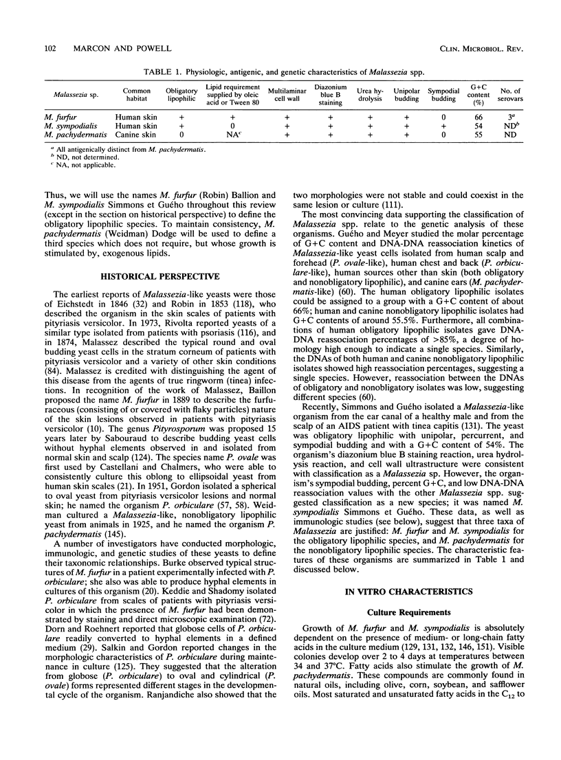
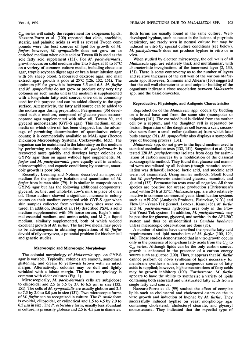
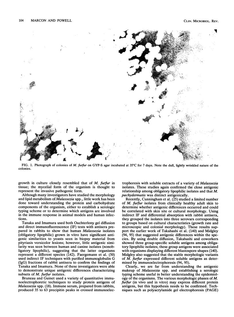
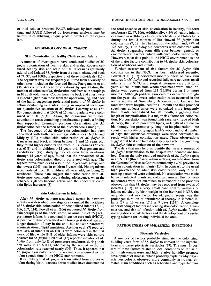
Images in this article
Selected References
These references are in PubMed. This may not be the complete list of references from this article.
- Abou-Gabal M., Chastain C. B., Hogle R. M. Pityrosporum pachydermatis "canis" as a major cause of otitis externa in dogs. Mykosen. 1979 Jun;22(6):192–199. doi: 10.1111/j.1439-0507.1979.tb01741.x. [DOI] [PubMed] [Google Scholar]
- Abou-Gabal M., Fagerland J. A. Electron microscopy of Pityrosporum canis "pachydermatis". Mykosen. 1979 Mar;22(3):85–90. doi: 10.1111/j.1439-0507.1979.tb01719.x. [DOI] [PubMed] [Google Scholar]
- Abraham Z., Berderly A., Lefler E. Pityrosporum orbiculare in children. Mykosen. 1987 Dec;30(12):581–583. doi: 10.1111/j.1439-0507.1987.tb04378.x. [DOI] [PubMed] [Google Scholar]
- Allen H. B., Charles C. R., Johnson B. L. Hyperpigmented tinea versicolor. Arch Dermatol. 1976 Aug;112(8):1110–1112. [PubMed] [Google Scholar]
- Alpert G., Bell L. M., Campos J. M. Malassezia furfur fungemia in infancy. Clin Pediatr (Phila) 1987 Oct;26(10):528–531. doi: 10.1177/000992288702601007. [DOI] [PubMed] [Google Scholar]
- Arnow P. M., Kushner R. Malassezia furfur catheter infection cured with antibiotic lock therapy. Am J Med. 1991 Jan;90(1):128–130. doi: 10.1016/0002-9343(91)90518-3. [DOI] [PubMed] [Google Scholar]
- Aschner J. L., Punsalang A., Jr, Maniscalco W. M., Menegus M. A. Percutaneous central venous catheter colonization with Malassezia furfur: incidence and clinical significance. Pediatrics. 1987 Oct;80(4):535–539. [PubMed] [Google Scholar]
- Azimi P. H., Levernier K., Lefrak L. M., Petru A. M., Barrett T., Schenck H., Sandhu A. S., Duritz G., Valesco M. Malassezia furfur: a cause of occlusion of percutaneous central venous catheters in infants in the intensive care nursery. Pediatr Infect Dis J. 1988 Feb;7(2):100–103. [PubMed] [Google Scholar]
- BURKE R. C. Tinea versicolor: susceptibility factors and experimental infection in human beings. J Invest Dermatol. 1961 May;36:389–402. doi: 10.1038/jid.1961.60. [DOI] [PubMed] [Google Scholar]
- Belew P. W., Rosenberg E. W., Jennings B. R. Activation of the alternative pathway of complement by Malassezia ovalis (Pityrosporum ovale). Mycopathologia. 1980 Mar 31;70(3):187–191. doi: 10.1007/BF00443030. [DOI] [PubMed] [Google Scholar]
- Bell L. M., Alpert G., Slight P. H., Campos J. M. Malassezia furfur skin colonization in infancy. Infect Control Hosp Epidemiol. 1988 Apr;9(4):151–153. doi: 10.1086/645819. [DOI] [PubMed] [Google Scholar]
- Bertini B., Kuttin E. S., Beemer A. M. Cytopathology of nipple discharge due to Pityrosporum orbiculare and cocci in an elderly woman. Acta Cytol. 1975 Jan-Feb;19(1):38–42. [PubMed] [Google Scholar]
- Broberg A., Faergemann J. Infantile seborrhoeic dermatitis and Pityrosporum ovale. Br J Dermatol. 1989 Mar;120(3):359–362. doi: 10.1111/j.1365-2133.1989.tb04160.x. [DOI] [PubMed] [Google Scholar]
- Brooks R., Brown L. Systemic infection with Malassezia furfur in an adult receiving long-term hyperalimentation therapy. J Infect Dis. 1987 Aug;156(2):410–411. doi: 10.1093/infdis/156.2.410. [DOI] [PubMed] [Google Scholar]
- Bruneau S. M., Guinet R. M. Quantitative immunoelectrophoretic study of genus Pytyrosporum Sabouraud. Mykosen. 1984 Mar;27(3):123–136. doi: 10.1111/j.1439-0507.1984.tb02010.x. [DOI] [PubMed] [Google Scholar]
- Bufill J. A., Lum L. G., Caya J. G., Chitambar C. R., Ritch P. S., Anderson T., Ash R. C. Pityrosporum folliculitis after bone marrow transplantation. Clinical observations in five patients. Ann Intern Med. 1988 Apr;108(4):560–563. doi: 10.7326/0003-4819-108-4-560. [DOI] [PubMed] [Google Scholar]
- Bäck O., Faergemann J., Hörnqvist R. Pityrosporum folliculitis: a common disease of the young and middle-aged. J Am Acad Dermatol. 1985 Jan;12(1 Pt 1):56–61. doi: 10.1016/s0190-9622(85)70009-6. [DOI] [PubMed] [Google Scholar]
- Charles C. R., Sire D. J., Johnson B. L., Beidler J. G. Hypopigmentation in tinea versicolor: a histochemical and electronmicroscopic study. Int J Dermatol. 1973 Jan-Feb;12(1):48–58. doi: 10.1111/j.1365-4362.1973.tb00212.x. [DOI] [PubMed] [Google Scholar]
- Cleri D. J., Corrado M. L., Seligman S. J. Quantitative culture of intravenous catheters and other intravascular inserts. J Infect Dis. 1980 Jun;141(6):781–786. doi: 10.1093/infdis/141.6.781. [DOI] [PubMed] [Google Scholar]
- Cunningham A. C., Leeming J. P., Ingham E., Gowland G. Differentiation of three serovars of Malassezia furfur. J Appl Bacteriol. 1990 May;68(5):439–446. doi: 10.1111/j.1365-2672.1990.tb02894.x. [DOI] [PubMed] [Google Scholar]
- DaMert G. J., Kirkpatrick C. H., Sohnle P. G. Comparison of antibody responses in chronic mucocutaneous candidiasis and tinea versicolor. Int Arch Allergy Appl Immunol. 1980;63(1):97–104. doi: 10.1159/000232614. [DOI] [PubMed] [Google Scholar]
- Dankner W. M., Spector S. A., Fierer J., Davis C. E. Malassezia fungemia in neonates and adults: complication of hyperalimentation. Rev Infect Dis. 1987 Jul-Aug;9(4):743–753. doi: 10.1093/clinids/9.4.743. [DOI] [PubMed] [Google Scholar]
- Di Silverio A., Mosca M., Brandozzi G., Gatti M. Pityriasis versicolor in the aged: a clinical investigation and epidemiological survey in 190 elderly hospitalized patients. Mycopathologia. 1989 Mar;105(3):187–190. doi: 10.1007/BF00437253. [DOI] [PubMed] [Google Scholar]
- Dorn M., Roehnert K. Dimorphism of Pityrosporum orbiculare in a defined culture medium. J Invest Dermatol. 1977 Aug;69(2):244–248. doi: 10.1111/1523-1747.ep12506384. [DOI] [PubMed] [Google Scholar]
- Drouhet E., Dompmartin D., Papachristou-Moraiti A., Ravisse P. Dermatite expérimentale à Pityrosporum ovale et (ou) Pityrosporum orbiculare chez le cobaye et la souris. Sabouraudia. 1980 Jun;18(2):149–156. [PubMed] [Google Scholar]
- Eisenstat B. A., Wormser G. P. Seborrheic dermatitis and butterfly rash in AIDS. N Engl J Med. 1984 Jul 19;311(3):189–189. doi: 10.1056/NEJM198407193110312. [DOI] [PubMed] [Google Scholar]
- Faergemann J., Aly R., Maibach H. I. Adherence of Pityrosporum orbiculare to human stratum corneum cells. Arch Dermatol Res. 1983;275(4):246–250. doi: 10.1007/BF00416670. [DOI] [PubMed] [Google Scholar]
- Faergemann J., Aly R., Maibach H. I. Growth and filament production of Pityrosporum orbiculare and P. ovale on human stratum corneum in vitro. Acta Derm Venereol. 1983;63(5):388–392. [PubMed] [Google Scholar]
- Faergemann J., Aly R., Maibach H. I. Quantitative variations in distribution of Pityrosporum orbiculare on clinically normal skin. Acta Derm Venereol. 1983;63(4):346–348. [PubMed] [Google Scholar]
- Faergemann J., Aly R., Wilson D. R., Maibach H. I. Skin occlusion: effect on Pityrosporum orbiculare, skin PCO2, pH, transepidermal water loss, and water content. Arch Dermatol Res. 1983;275(6):383–387. doi: 10.1007/BF00417338. [DOI] [PubMed] [Google Scholar]
- Faergemann J. Antibodies to Pityrosporum orbiculare in patients with tinea versicolor and controls of various ages. J Invest Dermatol. 1983 Feb;80(2):133–135. doi: 10.1111/1523-1747.ep12532935. [DOI] [PubMed] [Google Scholar]
- Faergemann J., Bernander S. Micro-aerophilic and anaerobic growth of Pityrosporum species. Sabouraudia. 1981 Jun;19(2):117–121. [PubMed] [Google Scholar]
- Faergemann J., Bernander S. The activity in vitro of five different antimycotics against Pityrosporum orbiculare. Acta Derm Venereol. 1979;59(6):521–524. doi: 10.2340/0001555559521524. [DOI] [PubMed] [Google Scholar]
- Faergemann J. Epidemiology and ecology of pityriasis versicolor. Curr Top Med Mycol. 1989;3:153–167. doi: 10.1007/978-1-4612-3624-5_7. [DOI] [PubMed] [Google Scholar]
- Faergemann J. Experimental tinea versicolor in rabbits and humans with Pityrosporum orbiculare. J Invest Dermatol. 1979 Jun;72(6):326–329. doi: 10.1111/1523-1747.ep12531766. [DOI] [PubMed] [Google Scholar]
- Faergemann J., Fredriksson T. Age incidence of Pityrosporum orbiculare on human skin. Acta Derm Venereol. 1980;60(6):531–533. [PubMed] [Google Scholar]
- Faergemann J., Fredriksson T. Experimental infections in rabbits and humans with Pityrosporum orbiculare and P. ovale. J Invest Dermatol. 1981 Sep;77(3):314–318. doi: 10.1111/1523-1747.ep12482488. [DOI] [PubMed] [Google Scholar]
- Faergemann J. In vitro and in vivo activities of ketoconazole and itraconazole against Pityrosporum orbiculare. Antimicrob Agents Chemother. 1984 Nov;26(5):773–774. doi: 10.1128/aac.26.5.773. [DOI] [PMC free article] [PubMed] [Google Scholar]
- Faergemann J., Maibach H. I. The Pityrosporon yeasts. Their role as pathogens. Int J Dermatol. 1984 Sep;23(7):463–465. doi: 10.1111/ijd.1984.23.7.463. [DOI] [PubMed] [Google Scholar]
- Faergemann J. Quantitative culture of Pityrosporon orbiculare. Int J Dermatol. 1984 Jun;23(5):330–333. doi: 10.1111/j.1365-4362.1984.tb04062.x. [DOI] [PubMed] [Google Scholar]
- Faergemann J. The use of contact plates for quantitative culture of Pityrosporum orbiculare. Mykosen. 1987 Jul;30(7):298-300, 303-4. [PubMed] [Google Scholar]
- Faergemann J., Tjernlund U., Scheynius A., Bernander S. Antigenic similarities and differences in genus Pityrosporum. J Invest Dermatol. 1982 Jan;78(1):28–31. doi: 10.1111/1523-1747.ep12497862. [DOI] [PubMed] [Google Scholar]
- Faergemann J. Treatment of seborrhoeic dermatitis of the scalp with ketoconazole shampoo. A double-blind study. Acta Derm Venereol. 1990;70(2):171–172. [PubMed] [Google Scholar]
- Ford G. P., Farr P. M., Ive F. A., Shuster S. The response of seborrhoeic dermatitis to ketoconazole. Br J Dermatol. 1984 Nov;111(5):603–607. doi: 10.1111/j.1365-2133.1984.tb06631.x. [DOI] [PubMed] [Google Scholar]
- Ford G. P., Ive F. A., Midgley G. Pityrosporum folliculitis and ketoconazole. Br J Dermatol. 1982 Dec;107(6):691–695. doi: 10.1111/j.1365-2133.1982.tb00530.x. [DOI] [PubMed] [Google Scholar]
- Ford G. Pityrosporon folliculitis. Int J Dermatol. 1984 Jun;23(5):320–321. doi: 10.1111/j.1365-4362.1984.tb04060.x. [DOI] [PubMed] [Google Scholar]
- Furukawa F., Danno K., Imamura S., Soh Y. Histological and serological studies of Pityrosporum orbiculare in cases of pityriasis versicolor. J Dermatol. 1981 Feb;8(1):27–30. doi: 10.1111/j.1346-8138.1981.tb02008.x. [DOI] [PubMed] [Google Scholar]
- GORDON M. A. Lipophilic yeastlike organisms associated with tinea versicolor. J Invest Dermatol. 1951 Nov;17(5):267–272. doi: 10.1038/jid.1951.93. [DOI] [PubMed] [Google Scholar]
- Garcia C. R., Johnston B. L., Corvi G., Walker L. J., George W. L. Intravenous catheter-associated Malassezia furfur fungemia. Am J Med. 1987 Oct;83(4):790–792. doi: 10.1016/0002-9343(87)90917-x. [DOI] [PubMed] [Google Scholar]
- Gidding H., Hawes L., Dwyer B. The isolation of Malassezia furfur from an episode of peritonitis. Med J Aust. 1989 Nov 20;151(10):603–603. doi: 10.5694/j.1326-5377.1989.tb101300.x. [DOI] [PubMed] [Google Scholar]
- Groisser D., Bottone E. J., Lebwohl M. Association of Pityrosporum orbiculare (Malassezia furfur) with seborrheic dermatitis in patients with acquired immunodeficiency syndrome (AIDS). J Am Acad Dermatol. 1989 May;20(5 Pt 1):770–773. doi: 10.1016/s0190-9622(89)70088-8. [DOI] [PubMed] [Google Scholar]
- Gueho E., Simmons R. B., Pruitt W. R., Meyer S. A., Ahearn D. G. Association of Malassezia pachydermatis with systemic infections of humans. J Clin Microbiol. 1987 Sep;25(9):1789–1790. doi: 10.1128/jcm.25.9.1789-1790.1987. [DOI] [PMC free article] [PubMed] [Google Scholar]
- Guého E., Meyer S. A. A reevaluation of the genus Malassezia by means of genome comparison. Antonie Van Leeuwenhoek. 1989 Mar;55(3):245–251. doi: 10.1007/BF00393853. [DOI] [PubMed] [Google Scholar]
- Hassall E., Ulich T., Ament M. E. Pulmonary embolus and Malassezia pulmonary infection related to urokinase therapy. J Pediatr. 1983 May;102(5):722–725. doi: 10.1016/s0022-3476(83)80244-3. [DOI] [PubMed] [Google Scholar]
- Heng M. C., Henderson C. L., Barker D. C., Haberfelde G. Correlation of Pityosporum ovale density with clinical severity of seborrheic dermatitis as assessed by a simplified technique. J Am Acad Dermatol. 1990 Jul;23(1):82–86. doi: 10.1016/0190-9622(90)70191-j. [DOI] [PubMed] [Google Scholar]
- Heymann W. R., Wolf D. J. Malassezia (Pityrosporon) folliculitis occurring during pregnancy. Int J Dermatol. 1986 Jan-Feb;25(1):49–51. doi: 10.1111/j.1365-4362.1986.tb03403.x. [DOI] [PubMed] [Google Scholar]
- Hill M. K., Goodfield M. J., Rodgers F. G., Crowley J. L., Saihan E. M. Skin surface electron microscopy in Pityrosporum folliculitis. The role of follicular occlusion in disease and the response to oral ketoconazole. Arch Dermatol. 1990 Aug;126(8):1071–1074. doi: 10.1001/archderm.126.8.1071. [DOI] [PubMed] [Google Scholar]
- Håkansson C., Faergemann J., Löwhagen G. B. Studies on the lipophilic yeast Pityrosporum ovale in HIV-seropositive and HIV-seronegative homosexual men. Acta Derm Venereol. 1988;68(5):422–426. [PubMed] [Google Scholar]
- Imwidthaya S., Thianprasit M. Malassezia furfur in dermatological patients and clinically normal persons. J Med Assoc Thai. 1988 Mar;71(3):145–149. [PubMed] [Google Scholar]
- Johansson S., Faergemann J. Enzyme-linked immunosorbent assay (ELISA) for detection of antibodies against Pityrosporum orbiculare. J Med Vet Mycol. 1990;28(3):257–260. doi: 10.1080/02681219080000321. [DOI] [PubMed] [Google Scholar]
- Keddie F., Shadomy S. Etiological significance of Pityrosporum orbiculare in tinea versicolor. Sabouraudia. 1963 Oct;3(1):21–25. [PubMed] [Google Scholar]
- Kikuchi I., Ogata K., Inoue S. Pityrosporum infection in an infant with lesions resembling erythema annulare centrifugum. Arch Dermatol. 1984 Mar;120(3):380–382. [PubMed] [Google Scholar]
- Klotz S. A., Drutz D. J., Huppert M., Johnson J. E. Pityrosporum folliculitis. Its potential for confusion with skin lesions of systemic candidiasis. Arch Intern Med. 1982 Nov;142(12):2126–2129. [PubMed] [Google Scholar]
- Klotz S. A. Malassezia furfur. Infect Dis Clin North Am. 1989 Mar;3(1):53–64. [PubMed] [Google Scholar]
- Larocco M., Dorenbaum A., Robinson A., Pickering L. K. Recovery of Malassezia pachydermatis from eight infants in a neonatal intensive care nursery: clinical and laboratory features. Pediatr Infect Dis J. 1988 Jun;7(6):398–401. doi: 10.1097/00006454-198806000-00006. [DOI] [PubMed] [Google Scholar]
- Leeming J. P., Holland K. T., Cunliffe W. J. The microbial ecology of pilosebaceous units isolated from human skin. J Gen Microbiol. 1984 Apr;130(4):803–807. doi: 10.1099/00221287-130-4-803. [DOI] [PubMed] [Google Scholar]
- Leeming J. P., Notman F. H., Holland K. T. The distribution and ecology of Malassezia furfur and cutaneous bacteria on human skin. J Appl Bacteriol. 1989 Jul;67(1):47–52. doi: 10.1111/j.1365-2672.1989.tb04953.x. [DOI] [PubMed] [Google Scholar]
- Leeming J. P., Notman F. H. Improved methods for isolation and enumeration of Malassezia furfur from human skin. J Clin Microbiol. 1987 Oct;25(10):2017–2019. doi: 10.1128/jcm.25.10.2017-2019.1987. [DOI] [PMC free article] [PubMed] [Google Scholar]
- Liñares J., Sitges-Serra A., Garau J., Pérez J. L., Martín R. Pathogenesis of catheter sepsis: a prospective study with quantitative and semiquantitative cultures of catheter hub and segments. J Clin Microbiol. 1985 Mar;21(3):357–360. doi: 10.1128/jcm.21.3.357-360.1985. [DOI] [PMC free article] [PubMed] [Google Scholar]
- Long J. G., Keyserling H. L. Catheter-related infection in infants due to an unusual lipophilic yeast--Malassezia furfur. Pediatrics. 1985 Dec;76(6):896–900. [PubMed] [Google Scholar]
- Maki D. G., Weise C. E., Sarafin H. W. A semiquantitative culture method for identifying intravenous-catheter-related infection. N Engl J Med. 1977 Jun 9;296(23):1305–1309. doi: 10.1056/NEJM197706092962301. [DOI] [PubMed] [Google Scholar]
- Marcon M. J., Durrell D. E., Powell D. A., Buesching W. J. In vitro activity of systemic antifungal agents against Malassezia furfur. Antimicrob Agents Chemother. 1987 Jun;31(6):951–953. doi: 10.1128/aac.31.6.951. [DOI] [PMC free article] [PubMed] [Google Scholar]
- Marcon M. J., Powell D. A., Durrell D. E. Methods for optimal recovery of Malassezia furfur from blood culture. J Clin Microbiol. 1986 Nov;24(5):696–700. doi: 10.1128/jcm.24.5.696-700.1986. [DOI] [PMC free article] [PubMed] [Google Scholar]
- Marcon M. J., Powell D. A. Epidemiology, diagnosis, and management of Malassezia furfur systemic infection. Diagn Microbiol Infect Dis. 1987 Jul;7(3):161–175. doi: 10.1016/0732-8893(87)90001-0. [DOI] [PubMed] [Google Scholar]
- Mathes B. M., Douglass M. C. Seborrheic dermatitis in patients with acquired immunodeficiency syndrome. J Am Acad Dermatol. 1985 Dec;13(6):947–951. doi: 10.1016/s0190-9622(85)70243-5. [DOI] [PubMed] [Google Scholar]
- Mickelsen P. A., Viano-Paulson M. C., Stevens D. A., Diaz P. S. Clinical and microbiological features of infection with Malassezia pachydermatis in high-risk infants. J Infect Dis. 1988 Jun;157(6):1163–1168. doi: 10.1093/infdis/157.6.1163. [DOI] [PubMed] [Google Scholar]
- Middleton C., Lowenthal R. M. Malassezia furfur fungemia as a treatable cause of obscure fever in a leukemia patient receiving parenteral nutrition. Aust N Z J Med. 1987 Dec;17(6):603–604. doi: 10.1111/j.1445-5994.1987.tb01270.x. [DOI] [PubMed] [Google Scholar]
- Midgley G. The diversity of Pityrosporum (Malassezia) yeasts in vivo and in vitro. Mycopathologia. 1989 Jun;106(3):143–153. doi: 10.1007/BF00443055. [DOI] [PubMed] [Google Scholar]
- Nanda A., Kaur S., Bhakoo O. N., Kaur I., Vaishnavi C. Pityriasis (tinea) versicolor in infancy. Pediatr Dermatol. 1988 Nov;5(4):260–262. doi: 10.1111/j.1525-1470.1988.tb00900.x. [DOI] [PubMed] [Google Scholar]
- Nazzaro-Porro M., Passi S. Identification of tyrosinase inhibitors in cultures of Pityrosporum. J Invest Dermatol. 1978 Sep;71(3):205–208. doi: 10.1111/1523-1747.ep12547184. [DOI] [PubMed] [Google Scholar]
- Noble W. C., Midgley G. Scalp carriage of Pityrosporum species: the effect of physiological maturity, sex and race. Sabouraudia. 1978 Sep;16(3):229–232. doi: 10.1080/00362177885380311. [DOI] [PubMed] [Google Scholar]
- Nordvall S. L., Johansson S. IgE antibodies to Pityrosporum orbiculare in children with atopic diseases. Acta Paediatr Scand. 1990 Mar;79(3):343–348. doi: 10.1111/j.1651-2227.1990.tb11467.x. [DOI] [PubMed] [Google Scholar]
- Oberle A. D., Fowler M., Grafton W. D. Pityrosporum isolate from the upper respiratory tract. Am J Clin Pathol. 1981 Jul;76(1):112–116. doi: 10.1093/ajcp/76.1.112. [DOI] [PubMed] [Google Scholar]
- Porro M. N., Passi S., Caprill F., Nazzaro P., Morpurgo G. Growth requirements and lipid metabolism of Pityrosporum orbiculare. J Invest Dermatol. 1976 Mar;66(3):178–182. doi: 10.1111/1523-1747.ep12481919. [DOI] [PubMed] [Google Scholar]
- Porro M. N., Passi S., Caprilli F., Mercantini R. Induction of hyphae in cultures of Pityrosporum by cholesterol and cholesterol esters. J Invest Dermatol. 1977 Dec;69(6):531–534. doi: 10.1111/1523-1747.ep12687967. [DOI] [PubMed] [Google Scholar]
- Potter B. S., Burgoon C. F., Jr, Johnson W. C. Pityrosporum folliculitis. Report of seven cases and review of the Pityrosporum organism relative to cutaneous disease. Arch Dermatol. 1973 Mar;107(3):388–391. doi: 10.1001/archderm.107.3.388. [DOI] [PubMed] [Google Scholar]
- Powell D. A., Aungst J., Snedden S., Hansen N., Brady M. Broviac catheter-related Malassezia furfur sepsis in five infants receiving intravenous fat emulsions. J Pediatr. 1984 Dec;105(6):987–990. doi: 10.1016/s0022-3476(84)80096-7. [DOI] [PubMed] [Google Scholar]
- Powell D. A., Hayes J., Durrell D. E., Miller M., Marcon M. J. Malassezia furfur skin colonization of infants hospitalized in intensive care units. J Pediatr. 1987 Aug;111(2):217–220. doi: 10.1016/s0022-3476(87)80070-7. [DOI] [PubMed] [Google Scholar]
- Powell D. A., Marcon M. J., Durrell D. E., Pfister R. M. Scanning electron microscopy of Malassezia furfur attachment to Broviac catheters. Hum Pathol. 1987 Jul;18(7):740–745. doi: 10.1016/s0046-8177(87)80246-0. [DOI] [PubMed] [Google Scholar]
- Powell D. A., Marcon M. J. Failure to eradicate Malassezia furfur broviac catheter infection with antifungal therapy. Pediatr Infect Dis J. 1987 Jun;6(6):579–580. doi: 10.1097/00006454-198706000-00022. [DOI] [PubMed] [Google Scholar]
- Prober C. G., Ein S. H. Systemic tinea versicolor, or how far can furfur go? Pediatr Infect Dis. 1984 Nov-Dec;3(6):592–592. doi: 10.1097/00006454-198411000-00022. [DOI] [PubMed] [Google Scholar]
- Redline R. W., Dahms B. B. Malassezia pulmonary vasculitis in an infant on long-term Intralipid therapy. N Engl J Med. 1981 Dec 3;305(23):1395–1398. doi: 10.1056/NEJM198112033052307. [DOI] [PubMed] [Google Scholar]
- Redline R. W., Redline S. S., Boxerbaum B., Dahms B. B. Systemic Malassezia furfur infections in patients receiving intralipid therapy. Hum Pathol. 1985 Aug;16(8):815–822. doi: 10.1016/s0046-8177(85)80253-7. [DOI] [PubMed] [Google Scholar]
- Richet H. M., McNeil M. M., Edwards M. C., Jarvis W. R. Cluster of Malassezia furfur pulmonary infections in infants in a neonatal intensive-care unit. J Clin Microbiol. 1989 Jun;27(6):1197–1200. doi: 10.1128/jcm.27.6.1197-1200.1989. [DOI] [PMC free article] [PubMed] [Google Scholar]
- Roberts S. O. Pityrosporum orbiculare: incidence and distribution on clinically normal skin. Br J Dermatol. 1969 Apr;81(4):264–269. doi: 10.1111/j.1365-2133.1969.tb13978.x. [DOI] [PubMed] [Google Scholar]
- Rokugo M., Tagami H., Usuba Y., Tomita Y. Contact sensitivity to Pityrosporum ovale in patients with atopic dermatitis. Arch Dermatol. 1990 May;126(5):627–632. [PubMed] [Google Scholar]
- Romano A., Segal E., Blumenthal M. Canaliculitis with isolation of Pityrosporum pachydermatis. Br J Ophthalmol. 1978 Oct;62(10):732–734. doi: 10.1136/bjo.62.10.732. [DOI] [PMC free article] [PubMed] [Google Scholar]
- Rosenberg E. W., Belew P. W. Improvement of psoriasis of the scalp with ketoconazole. Arch Dermatol. 1982 Jun;118(6):370–371. [PubMed] [Google Scholar]
- Rotrosen D., Calderone R. A., Edwards J. E., Jr Adherence of Candida species to host tissues and plastic surfaces. Rev Infect Dis. 1986 Jan-Feb;8(1):73–85. doi: 10.1093/clinids/8.1.73. [DOI] [PubMed] [Google Scholar]
- Ruiz-Maldonado R., López-Matínez R., Pérez Chavarría E. L., Rocio Castañn L., Tamayo L. Pityrosporum ovale in infantile seborrheic dermatitis. Pediatr Dermatol. 1989 Mar;6(1):16–20. doi: 10.1111/j.1525-1470.1989.tb00260.x. [DOI] [PubMed] [Google Scholar]
- SHIFRINE M., MARR A. G. THE REQUIREMENT OF FATTY ACIDS BY PITYROSPORUM OVALE. J Gen Microbiol. 1963 Aug;32:263–270. doi: 10.1099/00221287-32-2-263. [DOI] [PubMed] [Google Scholar]
- Salkin I. F., Gordon M. A. Polymorphism of Malassezia furfur. Can J Microbiol. 1977 Apr;23(4):471–475. doi: 10.1139/m77-069. [DOI] [PubMed] [Google Scholar]
- Sanguinetti V., Tampieri M. P., Morganti L. A survey of 120 isolates of Malassezia (Pityrosporum) pachydermatis. Preliminary study. Mycopathologia. 1984 Mar 15;85(1-2):93–95. doi: 10.1007/BF00436708. [DOI] [PubMed] [Google Scholar]
- Scheynius A., Faergemann J., Forsum U., Sjöberg O. Phenotypic characterization in situ of inflammatory cells in pityriasis (tinea) versicolor. Acta Derm Venereol. 1984;64(6):473–479. [PubMed] [Google Scholar]
- Shek Y. H., Tucker M. C., Viciana A. L., Manz H. J., Connor D. H. Malassezia furfur--disseminated infection in premature infants. Am J Clin Pathol. 1989 Nov;92(5):595–603. doi: 10.1093/ajcp/92.5.595. [DOI] [PubMed] [Google Scholar]
- Sohnle P. G., Collins-Lech C. Activation of complement by Pityrosporum orbiculare. J Invest Dermatol. 1983 Feb;80(2):93–97. doi: 10.1111/1523-1747.ep12531644. [DOI] [PubMed] [Google Scholar]
- Sohnle P. G., Collins-Lech C. Analysis of the lymphocyte transformation response to Pityrosporum orbiculare in patients with tinea versicolor. Clin Exp Immunol. 1982 Sep;49(3):559–564. [PMC free article] [PubMed] [Google Scholar]
- Sohnle P. G., Collins-Lech C. Cell-mediated immunity to Pityrosporum orbiculare in tinea versicolor. J Clin Invest. 1978 Jul;62(1):45–53. doi: 10.1172/JCI109112. [DOI] [PMC free article] [PubMed] [Google Scholar]
- Sohnle P. G., Collins-Lech C., Huhta K. E. Class-specific antibodies in young and aged humans against organisms producing superficial fungal infections. Br J Dermatol. 1983 Jan;108(1):69–76. doi: 10.1111/j.1365-2133.1983.tb04580.x. [DOI] [PubMed] [Google Scholar]
- Surmont I., Gavilanes A., Vandepitte J., Devlieger H., Eggermont E. Malassezia furfur fungaemia in infants receiving intravenous lipid emulsions. A rarity or just underestimated? Eur J Pediatr. 1989 Feb;148(5):435–438. doi: 10.1007/BF00595906. [DOI] [PubMed] [Google Scholar]
- Svejgaard E., Faergeman J., Jemec G., Kieffer M., Ottevanger V. Recent investigations on the relationship between fungal skin diseases and atopic dermatitis. Acta Derm Venereol Suppl (Stockh) 1989;144:140–142. doi: 10.2340/00015555144140142. [DOI] [PubMed] [Google Scholar]
- Takeo K., Nakai E. Mode of cell growth of Malassezia (Pityrosporum) as revealed by using plasma membrane configurations as natural markers. Can J Microbiol. 1986 May;32(5):389–394. doi: 10.1139/m86-074. [DOI] [PubMed] [Google Scholar]
- Tanaka M., Imamura S. Immunological studies on Pityrosporum genus and Malassezia furfur. J Invest Dermatol. 1979 Nov;73(5):321–324. doi: 10.1111/1523-1747.ep12549718. [DOI] [PubMed] [Google Scholar]
- Wallace M., Bagnall H., Glen D., Averill S. Isolation of lipophilic yeast in "sterile" peritonitis. Lancet. 1979 Nov 3;2(8149):956–956. doi: 10.1016/s0140-6736(79)92647-3. [DOI] [PMC free article] [PubMed] [Google Scholar]
- Wikler J. R., de Haan P., Nieboer C. The 'tape-method': a new and simple method for quantitative culture of Pityrosporum yeasts. Acta Derm Venereol. 1988;68(5):445–449. [PubMed] [Google Scholar]
- Wilde P. F., Stewart P. S. A study of the fatty acid metabolism of the yeast Pityrosporum ovale. Biochem J. 1968 Jun;108(2):225–231. doi: 10.1042/bj1080225. [DOI] [PMC free article] [PubMed] [Google Scholar]
- Wolter J. R. Pityrosporum species associated with dacryoliths in obstructive dacryocystitis. Am J Ophthalmol. 1977 Dec;84(6):806–809. doi: 10.1016/0002-9394(77)90501-3. [DOI] [PubMed] [Google Scholar]
- Wu Y. C., Chen K. T. Humoral immunity in patients with tinea versicolor. J Dermatol. 1985 Apr;12(2):161–166. doi: 10.1111/j.1346-8138.1985.tb01554.x. [DOI] [PubMed] [Google Scholar]
- Wurtz R. M., Knospe W. N. Malassezia furfur fungemia in a patient without the usual risk factors. Ann Intern Med. 1988 Sep 1;109(5):432–433. doi: 10.7326/0003-4819-109-5-432. [DOI] [PubMed] [Google Scholar]
- Yohn J. J., Lucas J., Camisa C. Malassezia folliculitis in immunocompromised patients. Cutis. 1985 Jun;35(6):536–538. [PubMed] [Google Scholar]


