Abstract
Statistically significant charge clusters (basic, acidic, or of mixed charge) in tertiary protein structures are identified by new methods from a large representative collection of protein structures. About 10% of protein structures show at least one charge cluster, mostly of mixed type involving about equally anionic and cationic residues. Positive charge clusters are very rare. Negative (or histidine-acidic) charge clusters often coordinate calcium, or magnesium or zinc ions [e.g., thermolysin (PDB code: 3tln), mannose-binding protein (2msb), aminopeptidase (1amp)]. Mixed-charge clusters are prominent at interchain contacts where they stabilize quaternary protein formation [e.g., glutathione S-transferase (2gst), catalase (8act), and fructose-1,6-bisphosphate aldolase (1fba)]. They are also involved in protein-protein interaction and in substrate binding. For example, the mixed-charge cluster of aspartate carbamoyl-transferase (8atc) envelops the aspartate carbonyl substrate in a flexible manner (alternating tense and relaxed states) where charge associations can vary from weak to strong. Other proteins with charge clusters include the P450 cytochrome family (BM-3, Terp, Cam), several flavocytochromes, neuraminidase, hemagglutinin, the photosynthetic reaction center, and annexin. In each case in Table 2 we discuss the possible role of the charge clusters with respect to protein structure and function.
Full text
PDF
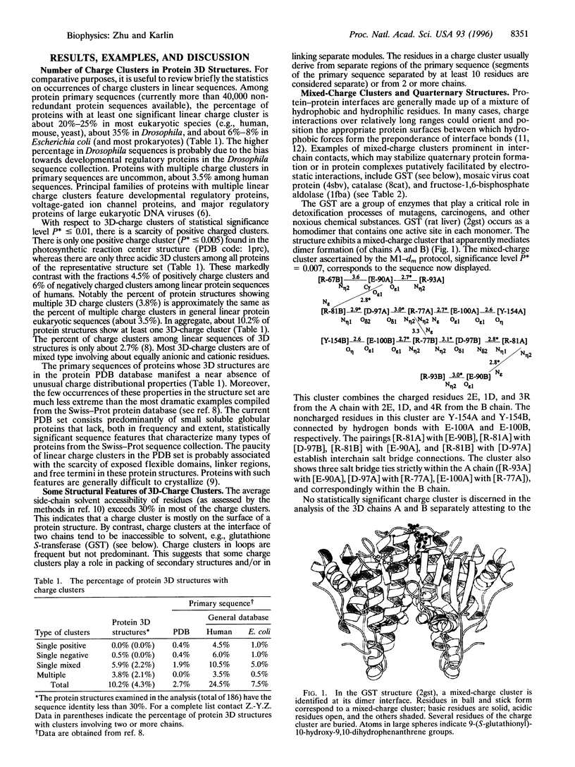
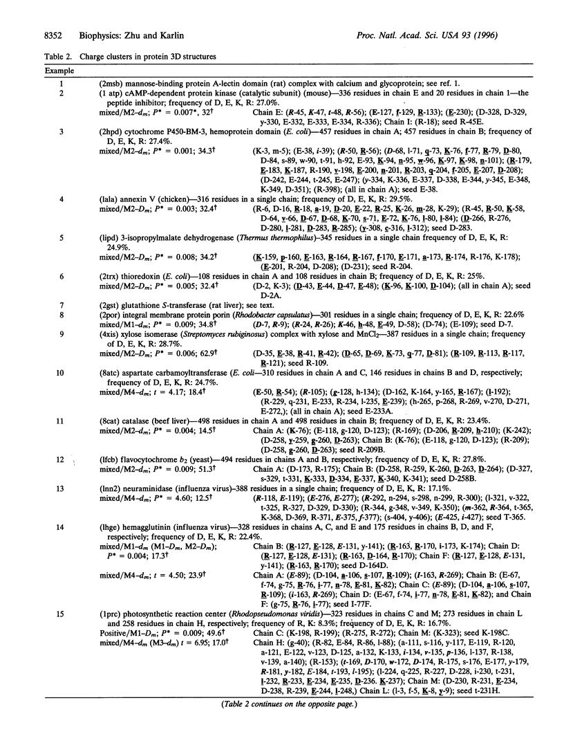
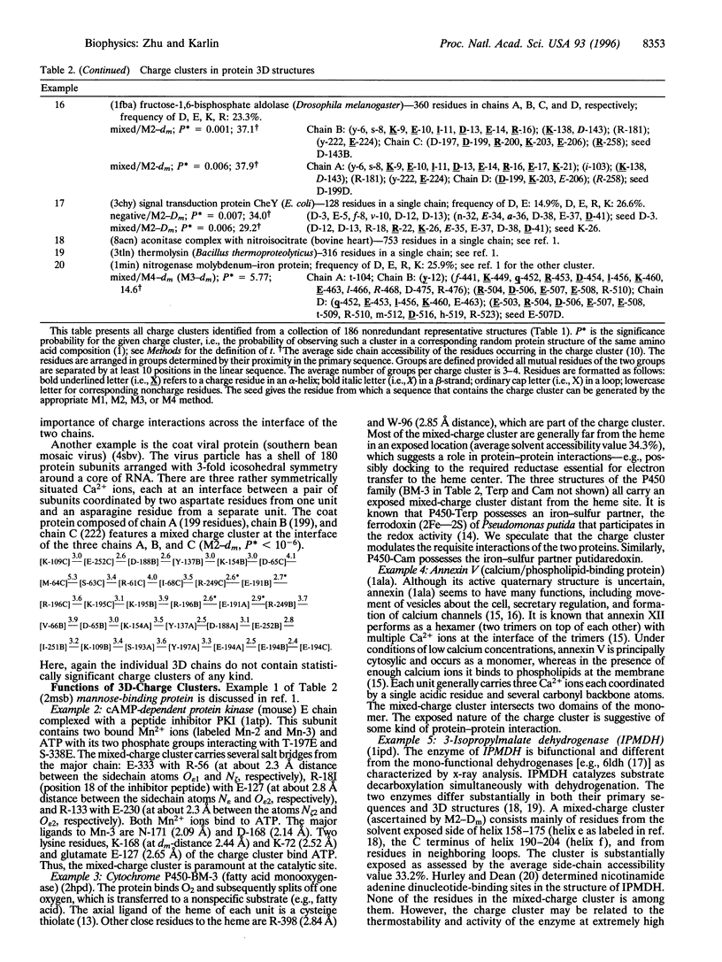
Images in this article
Selected References
These references are in PubMed. This may not be the complete list of references from this article.
- Baumann U., Wu S., Flaherty K. M., McKay D. B. Three-dimensional structure of the alkaline protease of Pseudomonas aeruginosa: a two-domain protein with a calcium binding parallel beta roll motif. EMBO J. 1993 Sep;12(9):3357–3364. doi: 10.1002/j.1460-2075.1993.tb06009.x. [DOI] [PMC free article] [PubMed] [Google Scholar]
- Brocchieri L., Karlin S. How are close residues of protein structures distributed in primary sequence? Proc Natl Acad Sci U S A. 1995 Dec 19;92(26):12136–12140. doi: 10.1073/pnas.92.26.12136. [DOI] [PMC free article] [PubMed] [Google Scholar]
- Burmeister W. P., Ruigrok R. W., Cusack S. The 2.2 A resolution crystal structure of influenza B neuraminidase and its complex with sialic acid. EMBO J. 1992 Jan;11(1):49–56. doi: 10.1002/j.1460-2075.1992.tb05026.x. [DOI] [PMC free article] [PubMed] [Google Scholar]
- Clackson T., Wells J. A. A hot spot of binding energy in a hormone-receptor interface. Science. 1995 Jan 20;267(5196):383–386. doi: 10.1126/science.7529940. [DOI] [PubMed] [Google Scholar]
- Colman P. M. Influenza virus neuraminidase: structure, antibodies, and inhibitors. Protein Sci. 1994 Oct;3(10):1687–1696. doi: 10.1002/pro.5560031007. [DOI] [PMC free article] [PubMed] [Google Scholar]
- Hasemann C. A., Ravichandran K. G., Peterson J. A., Deisenhofer J. Crystal structure and refinement of cytochrome P450terp at 2.3 A resolution. J Mol Biol. 1994 Mar 4;236(4):1169–1185. doi: 10.1016/0022-2836(94)90019-1. [DOI] [PubMed] [Google Scholar]
- Hester G., Brenner-Holzach O., Rossi F. A., Struck-Donatz M., Winterhalter K. H., Smit J. D., Piontek K. The crystal structure of fructose-1,6-bisphosphate aldolase from Drosophila melanogaster at 2.5 A resolution. FEBS Lett. 1991 Nov 4;292(1-2):237–242. doi: 10.1016/0014-5793(91)80875-4. [DOI] [PubMed] [Google Scholar]
- Honig B., Nicholls A. Classical electrostatics in biology and chemistry. Science. 1995 May 26;268(5214):1144–1149. doi: 10.1126/science.7761829. [DOI] [PubMed] [Google Scholar]
- Hope I. A., Mahadevan S., Struhl K. Structural and functional characterization of the short acidic transcriptional activation region of yeast GCN4 protein. Nature. 1988 Jun 16;333(6174):635–640. doi: 10.1038/333635a0. [DOI] [PubMed] [Google Scholar]
- Hurley J. H., Dean A. M., Sohl J. L., Koshland D. E., Jr, Stroud R. M. Regulation of an enzyme by phosphorylation at the active site. Science. 1990 Aug 31;249(4972):1012–1016. doi: 10.1126/science.2204109. [DOI] [PubMed] [Google Scholar]
- Hurley J. H., Dean A. M. Structure of 3-isopropylmalate dehydrogenase in complex with NAD+: ligand-induced loop closing and mechanism for cofactor specificity. Structure. 1994 Nov 15;2(11):1007–1016. doi: 10.1016/s0969-2126(94)00104-9. [DOI] [PubMed] [Google Scholar]
- Imada K., Sato M., Tanaka N., Katsube Y., Matsuura Y., Oshima T. Three-dimensional structure of a highly thermostable enzyme, 3-isopropylmalate dehydrogenase of Thermus thermophilus at 2.2 A resolution. J Mol Biol. 1991 Dec 5;222(3):725–738. doi: 10.1016/0022-2836(91)90508-4. [DOI] [PubMed] [Google Scholar]
- Karlin S., Blaisdell B. E., Brendel V. Identification of significant sequence patterns in proteins. Methods Enzymol. 1990;183:388–402. doi: 10.1016/0076-6879(90)83026-6. [DOI] [PubMed] [Google Scholar]
- Karlin S., Brocchieri L. Evolutionary conservation of RecA genes in relation to protein structure and function. J Bacteriol. 1996 Apr;178(7):1881–1894. doi: 10.1128/jb.178.7.1881-1894.1996. [DOI] [PMC free article] [PubMed] [Google Scholar]
- Karlin S. Statistical significance of sequence patterns in proteins. Curr Opin Struct Biol. 1995 Jun;5(3):360–371. doi: 10.1016/0959-440x(95)80098-0. [DOI] [PubMed] [Google Scholar]
- Karlin S., Zhu Z. Y. Characterizations of diverse residue clusters in protein three-dimensional structures. Proc Natl Acad Sci U S A. 1996 Aug 6;93(16):8344–8349. doi: 10.1073/pnas.93.16.8344. [DOI] [PMC free article] [PubMed] [Google Scholar]
- Kirino H., Aoki M., Aoshima M., Hayashi Y., Ohba M., Yamagishi A., Wakagi T., Oshima T. Hydrophobic interaction at the subunit interface contributes to the thermostability of 3-isopropylmalate dehydrogenase from an extreme thermophile, Thermus thermophilus. Eur J Biochem. 1994 Feb 15;220(1):275–281. doi: 10.1111/j.1432-1033.1994.tb18623.x. [DOI] [PubMed] [Google Scholar]
- Luecke H., Chang B. T., Mailliard W. S., Schlaepfer D. D., Haigler H. T. Crystal structure of the annexin XII hexamer and implications for bilayer insertion. Nature. 1995 Nov 30;378(6556):512–515. doi: 10.1038/378512a0. [DOI] [PubMed] [Google Scholar]
- Moss S. E. Ion channels. Annexins taken to task. Nature. 1995 Nov 30;378(6556):446–447. doi: 10.1038/378446a0. [DOI] [PubMed] [Google Scholar]
- Perutz M. F. Allosteric enzymes. Control by phosphorylation. Nature. 1988 Nov 17;336(6196):202–203. doi: 10.1038/336202a0. [DOI] [PubMed] [Google Scholar]
- Perutz M. F. Electrostatic effects in proteins. Science. 1978 Sep 29;201(4362):1187–1191. doi: 10.1126/science.694508. [DOI] [PubMed] [Google Scholar]
- Quiocho F. A. Carbohydrate-binding proteins: tertiary structures and protein-sugar interactions. Annu Rev Biochem. 1986;55:287–315. doi: 10.1146/annurev.bi.55.070186.001443. [DOI] [PubMed] [Google Scholar]
- Ravichandran K. G., Boddupalli S. S., Hasermann C. A., Peterson J. A., Deisenhofer J. Crystal structure of hemoprotein domain of P450BM-3, a prototype for microsomal P450's. Science. 1993 Aug 6;261(5122):731–736. doi: 10.1126/science.8342039. [DOI] [PubMed] [Google Scholar]
- Richardson W. D., Roberts B. L., Smith A. E. Nuclear location signals in polyoma virus large-T. Cell. 1986 Jan 17;44(1):77–85. doi: 10.1016/0092-8674(86)90486-1. [DOI] [PubMed] [Google Scholar]
- Richmond T. J., Richards F. M. Packing of alpha-helices: geometrical constraints and contact areas. J Mol Biol. 1978 Mar 15;119(4):537–555. doi: 10.1016/0022-2836(78)90201-2. [DOI] [PubMed] [Google Scholar]
- Sauter N. K., Glick G. D., Crowther R. L., Park S. J., Eisen M. B., Skehel J. J., Knowles J. R., Wiley D. C. Crystallographic detection of a second ligand binding site in influenza virus hemagglutinin. Proc Natl Acad Sci U S A. 1992 Jan 1;89(1):324–328. doi: 10.1073/pnas.89.1.324. [DOI] [PMC free article] [PubMed] [Google Scholar]
- Sauter N. K., Hanson J. E., Glick G. D., Brown J. H., Crowther R. L., Park S. J., Skehel J. J., Wiley D. C. Binding of influenza virus hemagglutinin to analogs of its cell-surface receptor, sialic acid: analysis by proton nuclear magnetic resonance spectroscopy and X-ray crystallography. Biochemistry. 1992 Oct 13;31(40):9609–9621. doi: 10.1021/bi00155a013. [DOI] [PubMed] [Google Scholar]
- Sygusch J., Beaudry D., Allaire M. Molecular architecture of rabbit skeletal muscle aldolase at 2.7-A resolution. Proc Natl Acad Sci U S A. 1987 Nov;84(22):7846–7850. doi: 10.1073/pnas.84.22.7846. [DOI] [PMC free article] [PubMed] [Google Scholar]
- Varghese J. N., McKimm-Breschkin J. L., Caldwell J. B., Kortt A. A., Colman P. M. The structure of the complex between influenza virus neuraminidase and sialic acid, the viral receptor. Proteins. 1992 Nov;14(3):327–332. doi: 10.1002/prot.340140302. [DOI] [PubMed] [Google Scholar]
- Wells J. A. Systematic mutational analyses of protein-protein interfaces. Methods Enzymol. 1991;202:390–411. doi: 10.1016/0076-6879(91)02020-a. [DOI] [PubMed] [Google Scholar]
- Xia Z. X., Mathews F. S. Molecular structure of flavocytochrome b2 at 2.4 A resolution. J Mol Biol. 1990 Apr 20;212(4):837–863. doi: 10.1016/0022-2836(90)90240-M. [DOI] [PubMed] [Google Scholar]
- Yutani K., Ogasahara K., Tsujita T., Sugino Y. Dependence of conformational stability on hydrophobicity of the amino acid residue in a series of variant proteins substituted at a unique position of tryptophan synthase alpha subunit. Proc Natl Acad Sci U S A. 1987 Jul;84(13):4441–4444. doi: 10.1073/pnas.84.13.4441. [DOI] [PMC free article] [PubMed] [Google Scholar]


