Abstract
Species are a fundamental unit of biodiversity, yet can be challenging to delimit objectively. This is particularly true of species complexes characterized by high levels of population genetic structure, hybridization between genetic groups, isolation by distance, and limited phenotypic variation. Previous work on the Cumberland Plateau Salamander, Plethodon kentucki, suggested that it might constitute a species complex despite occupying a relatively small geographic range. To examine this hypothesis, we sampled 135 individuals from 43 populations, and used four mitochondrial loci and five nuclear loci (5693 base pairs) to quantify phylogeographic structure and probe for cryptic species diversity. Rates of evolution for each locus were inferred using the multidistribute package, and time calibrated gene trees and species trees were inferred using BEAST 2 and *BEAST 2, respectively. Because the parameter space relevant for species delimitation is large and complex, and all methods make simplifying assumptions that may lead them to fail, we conducted an array of analyses. Our assumption was that strongly supported species would be congruent across methods. Putative species were first delimited using a Bayesian implementation of the GMYC model (bGMYC), Geneland, and Brownie. We then validated these species using the genealogical sorting index and BPP. We found substantial phylogeographic diversity using mtDNA, including four divergent clades and an inferred common ancestor at 14.9 myr (95% HPD: 10.8–19.7 myr). By contrast, this diversity was not corroborated by nuclear sequence data, which exhibited low levels of variation and weak phylogeographic structure. Species trees estimated a far younger root than did the mtDNA data, closer to 1.0 myr old. Mutually exclusive putative species were identified by the different approaches. Possible causes of data set discordance, and the problem of species delimitation in complexes with high levels of population structure and introgressive hybridization, are discussed.
Introduction
As species are fundamental units in ecology, biodiversity, conservation, and evolutionary biology, accurate species delimitation is of critical importance. Nonetheless, the diagnosis of species has a contentious history, with many biologists advocating alternative species concepts and conflicting taxonomies [1–5]. Work on species delimitation has historically focused on morphology and patterns of reproductive isolation [6–8], but molecular techniques are increasingly used to clarify species boundaries [9–13]. Using molecular markers, many morphologically cryptic taxa have been identified, and deep divergences among allopatric components quantified [14–17]. While species delimitation remains a challenging, philosophically rich topic [3,5], increasing numbers of biologists—especially systematists—are conceptualizing species as segments of independently evolving metapopulation-level evolutionary lineages, a perspective known as the general lineage species concept [4,18,19]. In particular, this perspective has been widely applied to molecular systematic investigations of complexes characterized by allopatry, parapatry, and morphological stasis [3,20–22]. In conjunction with advances in developing a unified concept of species [23], multispecies coalescent models, which lie at the interface of modern population genetic and phylogenetic methods, are revamping the science of species delimitation [24–26]. The development of these models was spurred by the observation that genealogies estimated from different genes can be discordant simply because the coalescence of genealogical lineages is a stochastic process. Unlike concatenated phylogenetic analyses, which assume a single tree underlies all loci, the multispecies coalescent accounts for gene tree conflict by modeling coalescent stochasticity [24,27]. Thus, evolutionary lineages can be diagnosed, and a reliable estimate of a species tree made, in the absence of monophyly, or when the information content of many loci is weak [28,29]. On the other hand, serious complications such as introgressive hybridization, high levels of population structure, isolation by distance, and phylogenetic estimation error present analytical challenges for genetic data, and if not accounted for can mislead inferences [25,30,31].
Species limits are notoriously difficult to identify in plethodontid salamanders, which can exhibit a high degree of phenotypic and ecological conservatism [32–35], yet commonly harbor extraordinary levels of genetic variation, including isolation by distance and deep population structure [9,36–41]. Objectively defining and delimiting plethodontid species is therefore a challenging task [3,42–44]. Nonetheless, largely as a result of studies using allozymes, the number of species in the genus Plethodon has increased from 16 in 1962 [45] to 55 today (AmphibiaWeb: http://amphibiaweb.org), including numerous cryptic and allopatric species [9,37,46]. Species complexes in Plethodon are thus eminent examples of a non-adaptive radiation, whereby an ancestral source taxon disintegrates into a complex of isolated lineages [46–49].
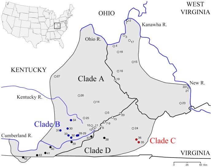
The Cumberland Plateau Salamander, P. kentucki, is an example of a species of Plethodon that harbors a high degree of genetic structure across its range. The species was originally described by Mittleman in 1951 [50]. However, it was not found to be morphologically distinct [51] and the name was long regarded as a junior synonym of P. glutinosus. In 1983, the species was rediscovered by Highton and MacGregor [52] while surveying patterns of genetic (allozyme) variation in the Plethodon glutinosus complex [9]. Geographic surveys showed that P. glutinosus and P. kentucki co-occur over most of the range of P. kentucki, though the range of P. kentucki is relatively restricted, including western Kentucky, southwestern West Virginia (south of the New and Kanawha Rivers), western Virginia, and a small section of northern Tennessee (Fig 1). Both species possess a black ground color overlain with white spots. Living specimens can be identified by subtle differences in P. kentucki, such as a lighter chin, a smaller number of dorsal spots, and a distinctive shape of the mental gland. However, the differences are quantitative, and within populations ranges of phenotypic variation between the two species can overlap.
Fig 1. Map of the range of Plethodon kentucki.
Sample localities are numbered, and match Fig 3 and S1 Appendix. Symbols identify the mtDNA clade of individuals in that population (Fig 3).
Despite the restricted range of P. kentucki, Highton and MacGregor [52] documented substantial amounts of genetic diversity. An electrophoretic analysis of 22 presumptive genetic loci showed that Nei’s [53] genetic distances were as high as 0.43 among geographically widespread samples. This is interesting because Highton [43] has argued that DN ≥ 0.15 commonly separates distinct species, while Wake and Schneider [44], counter to Highton, cite P. kentucki as an example of a single species with high levels of genetic differentiation. Here, we revisit patterns of genetic variation within P. kentucki using multilocus sequence data.
Species formation is a time-extended process, potentially including incomplete lineage sorting, introgressive hybridization, the anastomosis of formerly isolated lineages, and the development of complex patterns of population structure among incompletely separated lineages [3]. Thus, the parameter space relevant for species delimitation is large and complex. By contrast, all methods of species delimitation make a number of simplifying assumptions that may lead them to fail when faced with real world data sets [31,54]. In this paper we apply an array of approaches to species delimitation within P. kentucki, with the assumption that strongly supported species, at least, will we be recovered by alternative methods [11,31]. In this respect the approach is conservative. Incongruence among methods can be due to differences in their power to detect cryptic genetic lineages, or can be artifacts resulting from the violations of assumptions. We used three approaches to delimiting putative species. First, for mtDNA sequence data we employed a Bayesian implementation of the GMYC model [55]. Next, we used Brownie and Geneland [56–59] to delimit species using our nuclear data. Putative species were then validated using the genealogical sorting index, or gsi [60], and the program Bayesian Phylogenetics and Phylogeography (BPP). This latter approach uses the multispecies coalescent to analyze DNA sequence data, and can accommodate incomplete lineage sorting and uncertainty in the topology of the species tree [24,61,62]. We found that different methods were wildly inconsistent and advanced mutually exclusive taxonomies. We propose that this is a consequence of introgressive hybridization, isolation by distance, and high levels of population structure, and that P. kentucki may represent a particularly challenging real world scenario for modern species delimitation methods.
Materials and Methods
Natural history
The Cumberland Plateau Salamander, Plethodon kentucki, is a Woodland salamander in the family Plethodontidae. Unlike many amphibians, Woodland salamanders have no aquatic larval stage, do not migrate, and are completely terrestrial. Territoriality and home range size have not been studied in P. kentucki, but in Plethodon in general home ranges are small, on the order of a few square meters [63–65]. Small home ranges, territoriality, and limited mobility promote the accumulation of genetic differences among populations, including high levels of phylogeographic structure [40,41,66,67]. The systematic history of P. kentucki is reviewed in detail by Highton and MacGregor [52].
Sampling and laboratory techniques
Blood samples and tail tips were collected from 135 individuals from 43 populations of P. kentucki (Fig 1). Blood samples, designated with RH numbers in S1 Appendix, were collected from euthanized specimens in the early 1980s, prior to IACUC [52]. Animals were sacrificed by immersion in a solution of chlorotone before blood was collected. In addition, more recently about 3 mm of tail tip were collected from live specimens under Ohio University IACUC 12-L-050; these are designated with SRK numbers in S1 Appendix. Permits were obtained for our field efforts from the Virginia Department of Game and Inland Fisheries (015603, 048037), the West Virginia Division of Natural Resources (2013.115), and the Kentucky Department of Fish and Wildlife (SC1311198). Sampling was oriented toward geographic coverage and describing the limits of haplotype lineages. Total genomic DNA was extracted using Qiagen DNeasy Blood and Tissue Kits (Qiagen Corp., Valencia, CA). A total of 5693 base pairs (bp) of DNA were sequenced. Mitochondrial DNA sequence data were collected from most of the cytochrome b gene (Cyt-b; 1105 bp), the complete NADH dehydrogenase 2 gene (ND2; 1041 bp), the complete tRNAtrp locus (66 bp), and a portion of tRNAala (33 bp). ND2 was sequenced in two overlapping parts. MtDNA sequence data were collected from all but one individual (RH62903, S1 Appendix), but we have mtDNA data from two other individuals from that population (population 22). DNA sequence data were collected for five nuclear loci: the nuclear exon recombination activating gene 1 (RAG-1; 1152 bp), and the nuclear introns interleukin enhancer binding factor 3 (ILF3; 251bp), myosin light chain 2 mRNA (MLC2A; 416 bp), glyceraldehyde-3-phosphate dehydrogenase (GAPD; 686 bp), and β-fibrinogen intron 7 (BFI; 943 bp). BFI was amplified using a two-step protocol in which an initial long segment was first amplified from genomic DNA, then the product of this reaction was used as template in a subsequent PCR reaction, as described by [68]. This is the first study to use this gene in a plethodontid salamander. To our data set we added previously published sequence data from two individuals: ND2 for one individual from [69], and sequence data for one individual for Cyt-b, RAG-1, ILF3, MLC2A, and GAPD from [49,70]. Primers for all loci are provided in Table 1. Nuclear sequence data were collected from a subset of individuals, which varied by locus (Table 2). Details regarding the sampling of populations and loci, geographic coordinates, and GenBank accession numbers are presented in S1 Appendix.
Table 1. Primer sequences for all loci used in the study.
| Locus | Primer Name | Nucleotide Sequence (5'– 3') | Reference |
|---|---|---|---|
| Cyt-b | Pglut-F1b | GGTCTGAAAAACCAATGTTGTATTC | [71] |
| PThr-R2b | GCCCCCAATTTTGGYTTACAAG | [71] | |
| ND2 + | ND2-L4437-F2 | AAGCTTTCGGGCCCATACC | [72] |
| tRNAala + | ND2-RNEW1DEG | ATCCYAGGTGRGCGATGGAGG | This study |
| tRNAtrp | ND2-L5195-F3 | TGACAAAANCTNGCCCC | [69] |
| ND2-Ra | GTCTTGCAAGTTCGAGTCAGA | [73] | |
| ND2-5200 | CCTTGCCCTCTCATCCAAATCAGC | This study | |
| KND2-R2 | AAAGTGTTTGAGTTGCATTCA | [74] | |
| RAG-1 | Rag-1b-F | CTGTCTGGTCTGGTGCAGTCG | This study |
| Rag-1a-R | ATTCCCTTCACTCGCCCAAGC | This study | |
| BFI | BFI-EF1 | GGAGANAACAGNACNATGACAATNCAC | [68] |
| BFI-ER1 | ATCTNCCATTAGGNTTGGCTGCATGGC | [68] | |
| GAPD | GAPD-F | ACCTTTAATGCGGGTGCTGGCATTGC | [70] |
| GAPD-R | CATCAAGTCCACAACACGGTTGCTGTA | [70] | |
| ILF3 | ILF3-F | GATTTCAATCCATTTGCTCTTGC | [70] |
| ILF3-R | AGGATAAGCCCACCGTTACACTATT | [70] | |
| MLC2A | McL2a-F5 | TCCAATGTCTTTGCCATGTTCG | This study |
| MCL2a-R2 | AGTCATCCTTGTCTTTGGCTCC | This study |
Table 2. Sampling, genetic diversity, and models of evolution.
For the models of evolution, "All data" refers to the inclusion of every DNA sequence, whereas the “Complete data set" refers to the data set with no missing data (68 individuals). Numbers in parentheses refer to codon positions.
| Locus | Length1 | No. Indiv.2 | No. pops | P | Unique haplo-types | h | π | κ | Model of evolution: All data | Model of evolution: Complete data set |
|---|---|---|---|---|---|---|---|---|---|---|
| mtDNA | 5693 | –– | –– | –– | –– | –– | –– | –– | ND2(1), Cyt b(1), tRNAs: GTR + I + Γ; ND2(2), Cyt b(2): TN93 + I; Cytb(3): TN93; ND2(3): GTR + Γ | ND2(1), Cyt b(1), tRNAs: GTR + I; Cyt b(2): HKY + I; Cyt b(3): TN93; ND2(2): TN93 + I; ND2(3): GTR + Γ |
| Cyt b | 1105(973) | 133(129) | 43(43) | 154 | 39 | 0.96 | 0.037 | 35.7 | (1)GTR + I, (2)HKY + I, (3)TN93 | GTR + I |
| ND2 | 1041(977) | 85(77) | 43(43) | 169 | 43 | 0.98 | 0.043 | 41.6 | (1)GTR + I, (2)TN93 + I, (3)GTR + Γ | TN93 + Γ + I |
| tRNAtrp | 66(64) | 79(76) | 42(42) | 4 | 5 | 0.35 | 0.007 | 0.5 | HKY | HKY |
| tRNAala | 33(33) | 73(72) | 42(42) | 10 | 5 | 0.59 | 0.101 | 3.3 | GTR + I | GTR + I |
| RAG-1 | 1152(1145) | 74 | 42 | 30 | 30 | 0.67 | 0.001 | 1.6 | HKY + I | HKY + I |
| BFI | 943(898) | 75 | 42 | 46 | 37 | 0.64 | 0.001 | 1.2 | HKY + I | HKY + I |
| GAPD | 686(383) | 77(74) | 42 | 20 | 18 | 0.86 | 0.008 | 3.1 | HKY + Γ | HKY + Γ |
| MLC2A | 416 | 80(79) | 43(43) | 23 | 23 | 0.75 | 0.005 | 1.6 | JC + Γ | JC + Γ |
| ILF3 | 251 | 73(69) | 42(41) | 10 | 10 | 0.66 | 0.005 | 1.1 | JC | JC |
Diversity indices: P, the number of polymorphic sites; Unique haplotypes: the number of unique haplotypes in the sample; haplotype diversity, h, the probability that two randomly selected haplotypes are different from each other; nucleotide diversity, π, the average number of nucleotide differences per site between two sequences; sequence diversity, κ, the average number of nucleotide differences between paired sequences [78].
1. Sequence length. Numbers in parentheses are the length of the sequence used to calculate diversity indices after omitted all sites with missing data.
2. Number of individuals. Numbers in parentheses are after excluding short sequences, which were not used to calculate diversity indices because they exclude sites with missing data.
Most samples were sequenced in both the forward and reverse direction. The exception is ND2, which was only sequenced in the forward direction. Electropherograms for all sequences were viewed using the program Geneious v6.1 (Biomatters, Ltd., San Francisco, CA), and ambiguous base calls were manually corrected. The phase of heterozygous genotypes was estimated using PHASE v2.1.1 [75]. We ran PHASE for 1000 iterations, with a thinning interval of two steps and a burn-in of 100 iterations. PCR products exhibiting length heterogeneity due to the presence of indels were phased using Champuru v.1.0 [76,77]. In a few instances, sequences that could not be resolved using Champuru were cloned using the Invitrogen TOPO-TA Cloning Kit (Invitrogen, Carlsbad, CA). Four separate colonies were sequenced from each clone, and in all cases the heterozygotes were resolved. We tested for intragenic recombination using the difference in sum-of-squares (DSS) test implemented in TOPALi [78], including a 10 base pair increment, a window size of 100, and 500 parametric bootstraps. Recombination was not detected at any locus.
We used genetic diversity indices to compare patterns of genetic differentiation among mtDNA clades. Diversity indices included polymorphism (P), the number of segregating sites (S), haplotype diversity (h), sequence diversity (κ), and nucleotide diversity (π) [79]. Calculations were carried out in DNAsp5.10.1 [80].
Rates of Evolution
To obtain a time calibrated phylogeny it is necessary to either date nodes or provide an estimate of the rate of evolution. Currently it is not possible to date any of the nodes within P. kentucki, as there are no fossils or dated biogeographic events [81]. Thus, we estimated rates of evolution using Bayesian relaxed-clock dating with PAML 4.1 [82] and the set of programs in the Multidistribute package [83–85]. For each locus, baseml was used to estimate parameters under the F84+Γ model of nucleotide substitution, and paml2modelinf was used to transform output from baseml into a format appropriate for downstream analyses. The program estbranches was used to obtain maximum likelihood estimates of branch lengths and the variance-covariance matrix of those estimates. Finally, multidivtime was used to approximate the posterior distributions of substitution rates for each locus. These programs were run in Unix and through the R package LOGOPUS [86].
We estimated rates of evolution for Cyt-b, ND2, tRNAtrp and tRNAtrp, RAG-1, MLC2A, and GAPD using the phylogeny of Pyron and Wiens [87] trimmed to include only the genus Plethodon. The GenBank accession numbers and phylogenies used to estimate rates of evolution are provided in S2 Appendix. Species in the subgenus Hightonia (the clade of Plethodon restricted to the western US) served as outgroup taxa [88]. The species P. glutinosus, P. shermani, and P. aureolus were excluded because some analyses suggested they may be paraphyletic [70,89]. Clade age calibrations were taken from Wiens et al. [49], which were derived from three possible crown group ages for the family Plethodontidae of 50, 66, and 85 myr. We ran analyses using all three age estimates (Table 3). Priors for the mean (standard deviation) of the ingroup root age were 18.96 (1.17), 25.12 (1.46), and 32.25 (1.97) myr for crown group ages of 50, 66, and 85 myr, respectively. Time calibrated nodes included the cinereus group, glutinosus group, welleri-wehrlei group, and ouachitae group (see [89] for a discussion of group memberships), with upper and lower bounds set at two standard deviations from the mean. Two loci were analyzed separately. For ILF3, we designated the cinereus group as the outgroup [70,87,89] due to a lack of sequence data from the subgenus Hightonia. Ingroup root ages were estimated at 15, 20, and 25 myr (SD = 3 myr) for the crown group ages of 50, 66, and 85 myr, respectively (estimated from Figure 5 and Table 3 in [49]), and dated nodes included the glutinosus group, the welleri-wehrlei group, and the ouachitae group. For BFI, few sequences were available outside of P. kentucki. Thus, we estimated the rate of evolution using a three-taxon statement, with P. wehrlei as the outgroup and P. glutinous + P. kentucki as the ingroup. The ingroup age was set at 9.79 (SD = 0.3) myr [49]. Because this estimate of molecular evolution was derived from limited sampling, we also estimated the rate of evolution for BFI using a phylogeny and sequence data from the family Salamandridae, as described in S2 Appendix.
Table 3. Estimates of median rates of evolution.
Three estimates of the age of the crown group of plethodontids were used [71]. 95% confidence intervals are in parentheses. For clarity, all values are multiplied by 100, and thus are the estimated percentage change per million years.
| 50 Myr | 66 Myr | 85 Myr | |
|---|---|---|---|
| Cyt-b | 0.659 | 0.623 | 0.496 |
| (0.444, 0.912) | (0.424, 0.868) | (0.337, 0.696) | |
| ND2 | 0.889 | 0.844 | 0.665 |
| (0.629, 1.167) | (0.611, 1.088) | (0.483, 0.862) | |
| tRNAs | 0.149 | 0.127 | 0.099 |
| (0.016, 0.358) | (0.010, 0.324) | (0.007, 0.270) | |
| MLC2A | 0.083 | 0.074 | 0.061 |
| (0.012, 0.213) | (0.009, 0.195) | (0.007, 0.157) | |
| GAPD | 0.077 | 0.072 | 0.058 |
| (0.023, 0.164) | (0.023, 0.153) | (0.017, 0.126) | |
| RAG-1 | 0.029 | 0.026 | 0.023 |
| (0.011, 0.051) | (0.011, 0.046) | (0.009, 0.037) | |
| ILF3 | 0.102 | 0.074 | 0.043 |
| (0.022, 0.225) | (0.012, 0.173) | (0.005, 0.117) | |
| BFI | —— | 0.036 | —— |
| (0.006, 0.081) |
Phylogenetic Analyses
Gene trees were inferred using Bayesian phylogenetic analysis. We first analyzed all the data for each gene separately, without removing identical haplotypes [90], and including both alleles for nuclear loci. Because some methods do not accommodate missing data, and to facilitate comparison among loci, we also assembled a “complete data set” in which every OTU included data from every locus. We allowed missing data in either Cyt-b or ND2 (but not both) because these loci constitute a non-recombining unit. The complete data set included 68 individuals from 42 populations from throughout the range of the P. kentucki. For most populations, two individuals were included; only populations 1 and 18 are not represented in the complete data set (S1 Appendix).
For nuclear loci, models of evolution were assessed using jModeltest 2.1.5 [91], with the best model selected using AICc (Table 2). Models of evolution and the partitioning scheme for the concatenated mtDNA data were determined using PartitionFinder v1.1.1 [92] (Table 2). For all gene trees, we used a constant population size coalescent tree prior and a strict clock model. Clock models included a lognormal distribution with means and 95% confidence intervals that matched our multidivtime analyses (Table 3). In our preliminary analyses, the default priors (gamma) for rate.CG and rate.GT, though themselves well sampled (Effective Sample Sizes [ESS] > 200), resulted in very low ESS values for the prior and posterior distributions, even with long MCMC runs. The use of a lognormal prior on rate.CG and rate.GT increased the ESS values for the prior and posterior distributions to >6500. SRK conducted >200 separate runs in BEAST 2 before figuring this out.
Tree models were linked across partitions for mtDNA. For gene tree analyses, the length of the Markov chain Monte Carlo (MCMC) run was set to 50 million generations with parameters sampled every 5000 generations and a burn-in of 25%. All ESS values in all runs were >200. The Maximum Clade Credibility (MCC) tree was chosen using TreeAnnotator 2.1.2 [93].
To account for incomplete lineage sorting and provide input trees for downstream analyses, we performed species tree analyses using the multispecies coalescent model implemented in *BEAST 2 [28,94]. We defined species based on the bGMYC and Geneland results (see below). Analyses in *BEAST were set up as described above, except we used a Yule model for the tree prior and ran analyses for 500 million generations.
Finally, a maximum likelihood (ML) analysis of the concatenated data was conducted using RAxML v.8.1 [95]. We analyzed two data sets, one the nuclear DNA only (3438 bp), and one with nuclear DNA and mtDNA combined (5683 bp). Four individuals of P. glutinosus were used as the outgroup (S1 Appendix). For both data sets, we conducted 200 heuristic searches to obtain the ML tree, and 1000 rapid bootstrap pseudoreplicates to assess nodal support. The GTRGAMMA model was used, and protein coding loci were partitioned by codon.
Delimiting putative species
bGMYC
We first delimited putative species using a version of the general mixed Yule-coalescent (GMYC) [96]. Given a gene tree, this model infers on a phylogeny the transition from population-level (coalescent) processes to species-level (Yule model) processes. We used a Bayesian extension of this model, called the bGMYC, that accounts for uncertainty in gene trees by sampling over a posterior distribution of sampled trees [55]. The GMYC model is advantageous for single-locus datasets, and when the majority of phylogenetic signal is found in mtDNA, as in our data (see below). bGMYC analyses were run in the eponymous R package ‘bGMYC’ [55]. For the analyses, we randomly subsampled 1000 trees from the posterior distribution of our BEAST 2 analysis of the mtDNA data set. The following run options were used: MCMC = 100,000, burn-in = 50,000, thinning = 200, default scale parameters, default values on the Yule and coalescent rate change priors, and upper and lower bounds on the threshold parameter of 1 and 136, respectively, where 136 was the number of tips in our mtDNA tree. The starting number of species was set to 68, midway between the minimum and maximum number of species.
Geneland
To gain insight into population structure within P. kentucki using our nuclear loci, and for comparison with the bGMYC results, we used Geneland v4.0.5 [56–58]. This spatial clustering program estimates the number of populations by finding the number of groups that maximizes Hardy—Weinberg equilibrium within loci while minimizing linkage disequilibrium between loci. In addition, Geneland accounts for the spatial structure of samples when sampling coordinates are provided. Because missing data can bias the results, we used our complete data set, which was a genotype matrix of 68 diploid individuals (136 alleles) at five nuclear loci. We used the uncorrelated allele frequencies spatial model, as the correlated allele model is best used when differentiation is subtle, and model assumptions, such as no isolation by distance, are met [97]. The number of populations ranged from 1–30, and the MCMC was run for 20 million iterations, with sampling every 1000 steps and the first 20% of steps discarded as burn-in. All runs were replicated 10 times.
Brownie
Brownie identifies species limits by maximizing incongruence between gene trees within species, while minimizing incongruence between species. The logic is that within a species gene tree topologies will be random draws from a coalescent process, whereas between species gene trees will often show the same or similar topology. For input, we first randomly sampled one allele from each individual [11,98]. To examine the impact of this random sampling on inference, we analyzed four different samples of alleles. Two of the data sets, which we call data sets 1 and 2, did not include any shared alleles for heterozygous genotypes (e.g. if allele A at a locus was included in one dataset, allele B was used in the other). Data sets 3 and 4 were completely random relative to the other data sets. To make these data sets, we inferred calibrated gene trees in BEAST 2 using all alleles, as described above, and then pruned tips from these trees. We adopted this approach because we assume the accuracy of gene tree inference is increased through the inclusion of all available data. We also conducted analyses on these data sets with mtDNA included, and on the diploid data set without mtDNA. Heuristic searches in Brownie were run using default settings, except that all possible taxon reassignments on leaf splits were explored (Subsample = 1), and the minimum number of samples per species (MinSamp) was set to 2. We conducted 500 independent runs of each data set, and saved the complete set of recovered species trees and species delimitations.
Validation of putative species
The validity of delimitations inferred using the bGMYC and Geneland was tested using two approaches. First, we tested delimitations against a null hypothesis of no divergence using the genealogical sorting index (gsi), which quantifies the degree of exclusive ancestry of labeled groups on a rooted genealogy [60]. Populations in the process of diverging, or that split relatively recently, will usually display mismatches between gene trees and the species tree. However, over time the units are expected to transition from polyphyly to paraphyly to monophyly [99,100]. The time frame of this transition is dependent on the rate of genetic drift, and will vary among neutral loci because lineage sorting is a stochastic process. Relative to nuclear loci, mtDNA is expected to achieve monophyly quicker, on average, because it is haploid and maternally inherited, which results in a lower effective population size and a correspondingly high rate of genetic drift [101,102]. Values for the gsi range from 0 to 1, where 0 indicates the absence of exclusive ancestry and 1 indicates monophyly [60]. We calculated gsi values for each locus, as well as for an ensemble gsi (egsi), using the Genealogical Sorting Index web server (http://www.molecularevolution.org/software/phylogenetics/gsi). The null hypothesis of no divergence was evaluated using 10,000 permutations. As uneven sample sizes among groups can shift P-values downward for smaller groups, significance was inferred at P < 0.01 [60,98,103].
In addition to the gsi, the program Bayesian Phylogenetics and Phylogeography (BPP) v3.1 was used to evaluate species delimitations [26]. This program uses the multispecies coalescent to compare species delimitation models while simultaneously inferring a species tree that accounts for incomplete lineage sorting [26,104,105]. Consequently, BPP does not require reciprocal monophyly in gene trees to identify evolutionary lineages. In contrast with earlier versions of BPP, version 3 is not reliant on a fixed guide tree, but rather employs branch swapping with nearest neighbor interchange to alter the guide topology and account for phylogenetic uncertainty. Our analyses included all five nuclear loci, with a single allele sampled at random per individual (data sets 1–4, as described above), with and without mtDNA included. After several exploratory analyses, population size parameters (θ) were assigned the gamma prior G(2, 500), and the divergence time at the root of the species tree (τ) was assigned the gamma prior G(2, 4000); all other divergence time parameters were assigned the Dirichlet prior [104]. We used algorithm 0 with a fine-tune parameter (ε) of 10. Each species delimitation model was assigned equal prior probability. For the MCMC, after a burn-in of 5000 generations, samples were collected every two generations until 20,000 samples were obtained (45,000 generations total). Each analysis was run 2–5 times to confirm consistency among runs.
Results
Rates of molecular evolution
As expected, our mtDNA loci showed higher levels of variation than did our nuclear loci (Table 2). For example, sequence diversity (κ) for Cyt-b and ND2 was 35.7 and 41.6, respectively, but was 3.1 or less for the five nuclear loci. Mitochondrial tRNAs exhibited lower levels of variation than Cyt-b and ND2.
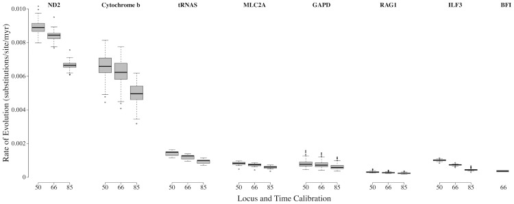
Median estimated rates of evolution for each gene are shown in Table 3 and Fig 2. Three different estimates are shown, representing three different calibration dates. For the 66 myr calibration, rates of evolution for the mitochondrial loci Cyt-b and ND2 were 0.623%/myr and 0.844%/myr, respectively. The tRNAs had a lower rate of 0.127%/myr. Rates of evolution for nuclear loci were roughly an order of magnitude slower than for Cyt-b and ND2, and ranged from a low of 0.026%/myr (RAG-1) to a high of 0.074%/myr (MLC2A and ILF3). When BFI was calibrated using Plethodon, the estimated rate of evolution was 0.036%/myr, which is an intermediate rate among the nuclear loci. By contrast, when BFI was dated using salamandrids, the estimated rates of evolution were higher than those estimated for our other nuclear loci (details in S2 Appendix). For our analyses, we used the Plethodon calibration.
Fig 2. Box plot depicting the estimated rates of evolution of each study locus.
Each box plot contains the estimates of evolutionary rate at each node and tip of the tree. The bottom and top of the box delimit the first and third quartiles, respectively, and whiskers extend to a maximum of 1.5 times the interquartile range. Asterisks indicate outlier points.
Phylogenetic inference: gene trees and concatenated analyses
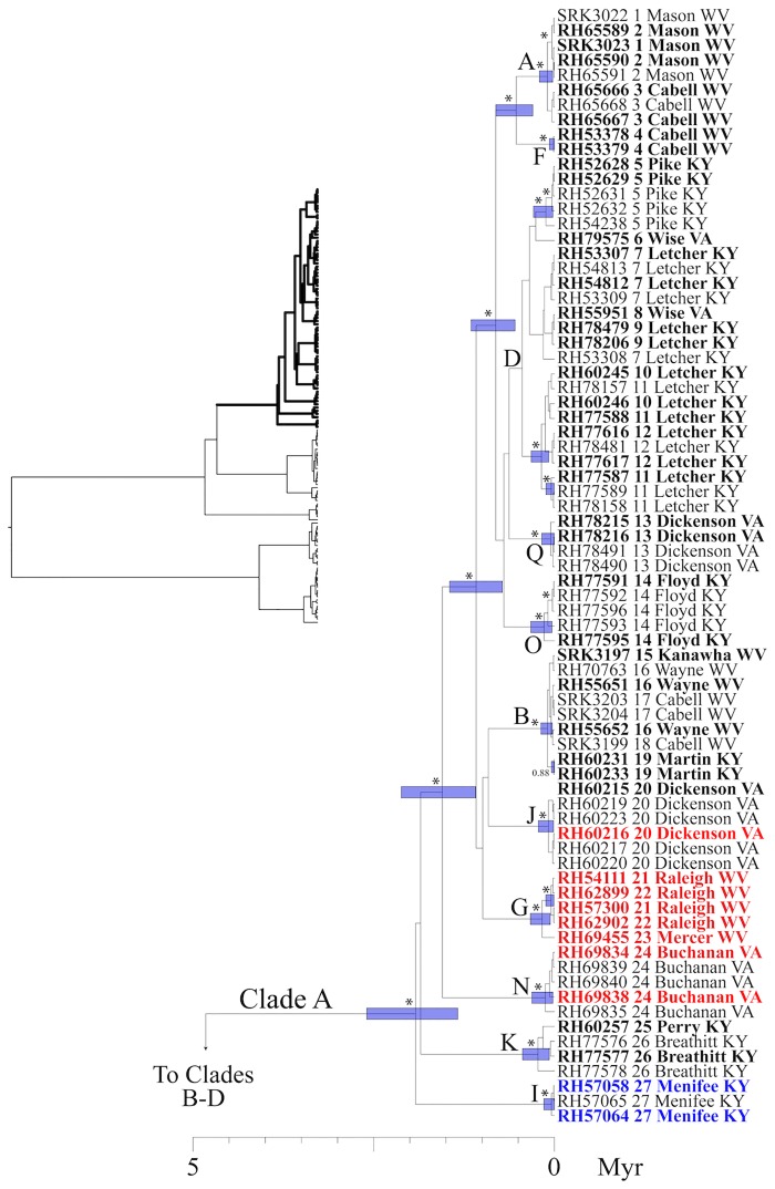
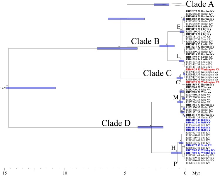
Our analyses of run diagnostics in TRACER suggested that MCMC stationarity was reached in all Bayesian phylogenetic analyses (e.g., all ESS > 200). In our mtDNA gene tree, four primary clades were recovered (Figs 3 and 4). The first split separates Clades A-C from Clade D, and was estimated to have occurred ~14.9 mya (95% HPD: 10.8–19.7 myr). Clade A occupies the broadest distribution of any clade, and is found at the northern and eastern limits of the range, from central Kentucky to eastern Virginia and western West Virginia, west of the New/Kanawha River. This clade harbors the most phylogeographic structure, with a number of subclades of largely unresolved affinity to one another. Clade B (populations 28–34) includes a geographically cohesive group of populations in SE Kentucky, north of the Cumberland River and south of the Kentucky River. Haplotypes from Clade C were found in two populations (35, 36), both of which are restricted to the south. Finally, Clade D occupies a restricted geographic range at the southwest limit of the distribution of P. kentucki, south of the Cumberland River (Fig 1). It is composed of two subclades, one to the east (populations 37–39), and one to the west (populations 40–43).
Fig 3. Bayesian maximum clade credibility tree.
This phylogeny was inferred using concatenated mtDNA data (Cyt-b, ND2, tRNAtrp, tRNAala). Taxon labels include specimen identification number, the population numbers from Fig 1, and county plus state information. Numbers adjacent to nodes are posterior probabilities (pp), and asterisks identify nodes with pp ≥ 0.95. Bars indicate 95% confidence intervals (CI) for dates of nodes. For visual clarity, many pp values and CI bars were removed near the tips of the tree. Specimens in the "complete" data set, which includes five nuclear loci in addition to mtDNA, are highlighted in bold. The bGMYC analysis delimited either 17 putative species, or two putative species, depending on the probability threshold employed (see text). The 17 putative species are identified using the letters (A-Q) adjacent to nodes; the two putative species are represented by Clade A, and Clades B-C. Finally, three putative species delimited in the Geneland analysis are highlighted using colored text that is either black (species A), red (species B), or blue (species C). Clade A is here illustrated. The entire phylogeny is illustrated in the upper left, with Clade A illustrated using bold lines. See Fig 4 for Clades B-D.
Fig 4. Bayesian maximum clade credibility tree.
As with Fig 3, but showing relationships within Clades B-D.
Phylogenies for our five nuclear loci, inferred using the complete data set (diploid, no missing loci), are presented in S3 Appendix. In contrast with our mtDNA phylogeny, relationships were poorly resolved. Recovered clades did not circumscribe geographically cohesive groups, and are not similar to any mtDNA clade. For example, in ILF3 four clades had at least moderate support, but none were geographically cohesive or reminiscent of any mtDNA clade. The same is true of RAG-1, BFI, GAPD, and MLC2A: all included supported clades, but these were composed of a mix of alleles from distant geographic localities. Moreover, none of the nuclear loci recovered a clade that was shared with another locus.
The results of maximum likelihood phylogenetic analyses of the concatenated data are presented in S4 Appendix. When the nuclear data alone were analyzed, no statistically supported clades (bs > 70%) were recovered within P. kentucki (Figure H in S4 Appendix). The three groups recovered in the Geneland analysis of the nuclear data were not covered as reciprocally monophyletic clades, and the clades that were recovered did not form geographically cohesive groups of populations. In addition, one individual of P. glutinosus (RH70700) was recovered within P. kentucki. When mtDNA was added to the nuclear data, the resulting phylogenetic inference largely reflected the mtDNA tree (c.f., Figure I in S4 Appendix; Figs 3 and 4), with individual RH70770 recovered as a member of a monophyletic outgroup. The discordant results with respect to individual RH70770 suggest that there is hybridization between P. kentucki and P. glutinosus, which was also documented by Highton and MacGregor [52] using allozymes.
Initial delimitation of putative species
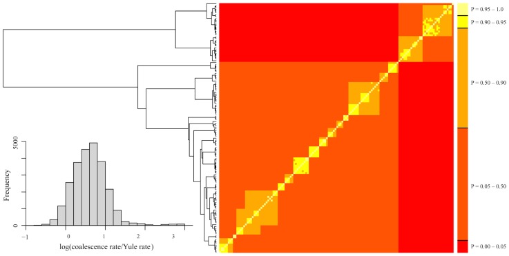
To probe our data for discrete evolutionary lineages, we used the bGMYC, Geneland, and Brownie. The bGMYC results are summarized in Fig 5. The colored matrix compares individuals, with colors corresponding to the posterior probability they are conspecific. To delimit species, it is necessary to specify a probability threshold above which individuals will be considered heterospecific. If we adopt a threshold of P = 0.95, two species are identified that correspond with Clade A-C vs. Clade D in our mtDNA phylogeny (Figs 3 and 4). If we use the posterior mean of the analysis as the probability threshold (P = 0.5), 17 species are identified. These largely correspond with statistically supported tip clades in our mtDNA phylogeny, and form geographically cohesive groups of populations (Figs 1, 3 and 4). The exception is species "P," which includes a single individual from population 40, even though four other individuals from population 40 were assigned to species "H". We lack nuclear data for species "P," and thus our validation of the 17 species delimitation (see below) included only 16 species.
Fig 5. bGMYC analyses.
To the left is the maximum clade credibility tree from BEAST 2 (Figs 3 and 4). The table is a sequence-by-sequence matrix, with cells colored by the posterior probability that the corresponding sequences are conspecific. Off-diagonal colors indicate uncertainty due to uncertainty in topology.
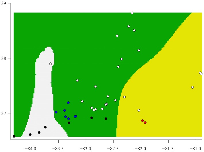
For comparison with the bGMYC, we explored patterns in our nuclear data using Geneland. Replicates of the Geneland analysis supported recognition of three populations, including one to the east, one formed by all the central sampling localities, and one that includes three southern localities in combination with the northwestern-most sample (Fig 6). None of these groups corresponds with a mtDNA clade (Fig 3).
Fig 6. Geneland results, with grouping inferred from nuclear loci.
The three colors correspond to the three groups inferred by Geneland. The dots are the collecting localities (see Fig 1), and are colored by clade: white = Clade A; blue = Clade B; red = Clade C; black = Clade D. The histogram shows the log of the ratio of the estimates rates of coalescence and the estimated Yule rates. Values above zero indicate the estimated rate of coalescence is higher.
Finally, using Brownie we attempted to delimit species using four haploid nuclear data sets, each of which was a random sample of the larger diploid data (see Material and Methods). These were all tested with and without the addition of mtDNA. A detailed presentation of the results can be found in S5 Appendix. In brief, Brownie did not reliably delimit species. Vast differences were recovered when different data sets were used, from one to 5018 species, even though these data sets were randomly sampled from the same larger diploid data set. An analysis of the diploid data gave results that differed from all of the haploid data sets. In these analyses, the delimited species did not form contiguous geographic groups, and typically did not include all the individuals from single populations. This was true whether or not mtDNA was included. Given the inconsistency of the findings, we did not attempt to validate these putative species (see below).
Phylogenetic inference: species trees
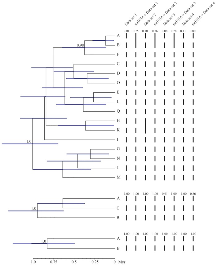
We used the results of our bGMYC (2 and 17 species) and Geneland (3 species) analyses to define a priori species for species tree analyses in *BEAST 2 [94]. All runs in *BEAST 2 resulted in thorough sampling of the posterior distributions (all ESS > 200). For all three phylogenies, the basal split in the tree was supported (pp = 1.00), but otherwise the trees were poorly resolved (pp < 0.95) (Fig 7). In the analysis including 16 species, only the clade including species A, B, and F was supported (pp = 0.98). All three analyses estimated the age of the MRCA at about 1 myr (range of HPDs: 0.50–1.38 myr).
Fig 7. Maximum clade credibility trees from *BEAST 2, with species delimitations from BPP.
Numbers at tree nodes are posterior probabilities; numbers <0.95 have been omitted. Bars at nodes represent the 95% highest posterior density for the inferred ages of nodes. To the right, bars connect putative species that were combined in the BPP analyses. (A) 16 species as delimited by bGMYC, using mtDNA data; (B) 3 species as delimited by Geneland, using the nuclear data; (4) 2 species as delimited by bGMYC, using mtDNA data. See also Fig 3 for species delimitations.
Validation of putative species
We tested for exclusive ancestry using the gsi with our four haploid nuclear data sets and a diploid data set. For the bGMYC delimitation of two species, no level of exclusive ancestry was detected using BFI, MLC2A, or RAG1 (S6 Appendix). GAPD and ILF3 recovered either species A, species B, or both, depending on the data set. However, the ensemble gsi (egsi), which considers all loci, detected species A and B in every data set except data set 3, which supported neither. The egsi values ranged from 0.14–0.24.
For data sets 1–4 and the bGMYC delimitation of 17 species, species A-C, F, I-O, and Q did not show a pattern of exclusive ancestry that was consistently different from zero at any locus. In some cases the egsi was significant, but this varied by data set. Species D, E, and H more consistently exhibited significant levels of exclusive ancestry, with egsi values that ranged between 0.20–0.25. Species G was the most consistently supported, with relatively high egsi values (range: 0.27–0.48). In general, the diploid data exhibited higher levels of exclusive ancestry for more loci than did the haploid data sets, with 11 of 16 egsi values significant (range: 0.18–0.36).
For the three species delimited using Geneland, the gsi provided consistent support relative to the bGMYC results. However, the same nuclear loci were used to delimit populations in Geneland, so one would expect the gsi to perform relatively well. Only BFI did not exhibit any patterns of exclusive ancestry. GAPD provided the strongest support, with significant gsi values for all three species in all data sets. Perhaps the patterns in GAPD contributed disproportionately to the population groupings recovered by Geneland. Overall, the egsi values, while significant, were not high (range: 0.20–0.34). When mtDNA was included, no pattern of exclusive ancestry was detected in any run. This may not be surprising, as the species delimited by Geneland do not correspond with the mtDNA phylogeny.
Finally, we used the program BPP to test the species delimited by the bGMYC (2 and 17 species) and Geneland analyses (3 species). Multiple runs of BPP produced consistent results, indicating that the MCMC chains were well mixed. For the delimitations, all four data sets were analyzed with and without mtDNA. Rannala and Yang [105] have suggested that different putative species only be considered distinct if their posterior probability exceeds a threshold such as 95% or even 99%. For the analysis of two species, the delimitation with the highest posterior probability (pp) always supported both species with pp > 0.95 (Fig 7). Similarly, with 3 species the delimitation with the highest pp always supported three species, with pp > 0.95 in 6 of 8 analyses (Fig 7). Note that these two delimitations (2 vs. 3 species) are composed of sets of OTUs that are mutually exclusive (Fig 3).
We had nuclear data for 16 of the 17 putative species delimited by the bGMYC (see above). Using the nuclear data alone, the delimitation with the highest posterior probability included either 16 (data sets 1, 3, 4) or 14 species (data set 2) (Fig 6). However, all of these delimitations had low posterior probabilities (range: <0.01 to 0.11), and no species had a pp of ≥0.95 in more than one data set. When mtDNA was included, the delimitation with the highest posterior probability included 15 or 16 species; in three of the four data sets, species A and B were combined into a single species. Posterior probabilities were substantially higher when mtDNA was included (range: 0.75–0.80), but none were ≥ 0.95.
Discussion
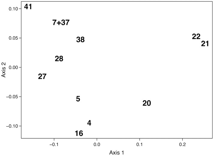
Delimiting species when morphology is highly conserved has long challenged systematists. We studied patterns of genetic variation in the Cumberland Plateau Salamander, P. kentucki, which is a cryptic species with respect to P. glutinosus. Prior research using allozymes [52] found that P. glutinosus exhibits relatively little genetic variation where it co-occurs with P. kentucki, whereas P. kentucki possesses striking levels of variation. Despite the high level of genetic differentiation, however, populations of P. kentucki are not easily sorted into distinct, geographically cohesive groups. For example, Fig 8 presents a multidimensional scaling (MDS) analysis of Nei’s genetic distances, which was made using the allozyme data in [52]. When inter-population variation is a function of geographic distance alone, an MDS of the first two dimensions produces a clustering pattern akin to a geographical map of the populations [36,39,106,107]. In Fig 8, the populations are widely spaced and do not for distinct clusters. The exception is populations 21–22, but these populations are also the most geographically isolated (Fig 1). The high levels of genetic variation and complex population genetic structure in the allozyme data suggested to us that P. kentucki was in need of further phylogeographic evaluation.
Fig 8. Multidimensional scaling of Nei's genetic distances.
Genetic data from Highton and MacGregor (1983). Populations numbers match Fig 1.
Phylogeographic differentiation
We sampled specimens from throughout the range of P. kentucki, and obtained sequence data from nine loci, including four mtDNA loci and five nuclear loci. According to mtDNA variation, P. kentucki is old and harbors a large amount of genetic structure, even for a relatively dispersal-limited amphibian with a small range [64,108,109]. Using Bayesian phylogenetic analyses, we recovered four divergent mitochondrial clades (Clades A-D). The basal split, which separates populations in the southwest (Clade D) from the other populations (Clades A-C), was dated at ~14.9 myr (95% HPD: 10.8–19.7 myr). Clade D is separated from Clades A-C by the upper reaches of the Cumberland River, suggesting the river could be a barrier to dispersal, though here it is not as so large as it is to the west. Similarly, north of the Cumberland, Clades A and C are separated by the upper reaches of the Kentucky River. Clades A-C have a common ancestor inferred to have existed 5.5 mya (95% HPD: 3.4–6.4 mya). Together, these clades occupy parts of the Cumberland Plateau and Valley and Ridge physiographic provinces, but the clade distributions do not follow the boundary between these provinces.
In contrast with mtDNA, nuclear loci exhibited low levels of divergence and limited phylogeographic structure (Table 2). Shared polymorphisms among populations in the nuclear data were found over broad spatial scales, a pattern that can result from retained ancestral polymorphism or introgressive hybridization [110]. Individual gene trees included few supported nodes, and did not identify geographically cohesive groups (S3 Appendix). In sum, the nuclear loci in this study provided low levels phylogeographic variation, in striking contrast with mtDNA. All of the nuclear loci used in our study, except for BFI, were used by Fisher-Reid and Wiens [70] in a phylogenetic analysis of relationships within Plethodon, with some success. We thus reasoned that these loci had a good chance of diagnosing clearly demarcated species within P. kentucki.
The low variation and lack of monophyly in our nuclear data are not necessarily fatal for species delimitation and species tree inference, as the multispecies coalescent accounts for stochasticity in the coalescent process [25,111–113]. Introgressive hybridization, however, is not modeled by most methods, and can be problematic [114–117]. One important consequence of introgression is species tree "compression" [115], whereby divergence times are severely underestimated. This occurs because the multispecies coalescent assumes all gene tree discordance is a consequence of incomplete lineage sorting, which requires speciation events to follow coalescent events [117,118]. In P. kentucki, we may have observed species tree compression as all three species trees estimated the root of the phylogeny at around 1.0 mya (Fig 6), whereas our mtDNA estimate was 14.9 mya (Fig 3). An alternative explanation is that P. kentucki is not deeply differentiated, but rather mtDNA is maintaining a signal reflective of ancient divergence events not recorded in the nuclear genome. This interpretation, however, conflicts with published allozyme data, which revealed high levels of diversity [52].
Species delimitation and validation
The problem of reconstructing species boundaries from genetic data is demanding. Molecular approaches to species delimitation, which hold great promise for diagnosing independent metapopulation-level evolutionary lineages, have been undergoing rapid development for the last 10–15 years [5]. This has yielded a wide array of methods, many of which incorporate advances in coalescent theory and the multispecies coalescent. Nonetheless, species formation is not so tidy as the word "speciation" implies, but is a time-extended process with complex dynamics through space and time [3]. Accordingly, the parameter space relevant for species delimitation is extraordinarily complex. By contrast, all methods of species delimitation make a number of simplifying assumptions that may cause them to fail under some real world circumstances [31,54]. In P. kentucki, we adopted a two-step approach to species delimitation: first, we delimited putative species using the bGMYC (mtDNA), Geneland (nuclear DNA), and Brownie (nuclear DNA and mtDNA); second, we validated these putative species using the gsi and BPP. A priori, we assumed (conservatively) that strongly supported species would be recovered by diverse methods [11,31]. Because our mtDNA phylogeny was much more resolved than any of our nuclear trees, we first used the bGMYC. When we used a probability of conspecificity threshold of P = 0.95, two species that corresponded with Clades A-C and Clade D on our mtDNA tree were identified (Figs 3 and 4). For this delimitation, ensemble gsi (egsi) values ranged from 0.19–0.23, and BPP strongly supported the existence of both species. When we explored a threshold of P = 0.5, 17 species were delimited, 16 of which corresponded with geographically cohesive groups of populations. However, support for these putative species was weak. Egsi results were inconsistent (S6 Appendix), and BPP did not strongly support any delimitation or any single species.
For comparison with our bGMYC results, we used Geneland to identify population clusters in the nuclear data. Three groups were recovered, none of which matched any of the mtDNA clades (Figs 3, 4 and 6). Thus, the putative species identified using the bGMYC and Geneland are mutually exclusive. Ensemble gsi values calculated from the nuclear data supported the exclusivity of these species (though there is an element of circularity, as the same nuclear data were used to define the putative species in Geneland). In BPP, the delimitation with the highest posterior probability included all three species in all analyses.
Finally, we analyzed our data using a nonparametric method that recovers species boundaries by minimizing interspecific congruence while maximizing intraspecific incongruence [59], as implemented in the program Brownie. Brownie produced erratic results, with the number of delimited species ranging from 1 to 5018. Moreover, the delimited species did not form contiguous geographic groups, and did not include all the individuals from single populations, whether or not mtDNA was included. In our analyses, we used four haploid data sets each randomly drawn from a diploid data set, and each data set produced different results. This suggests caution is warranted when a single random sample of alleles is used in Brownie and other programs [11,98,119].
Taxonomic Implications
While recent advances in the use of genetic data to diagnose cryptic evolutionary lineages are truly exciting, they are also under active development [5,118,120–122]. In particular, most methods do not account for gene flow, isolation by distance, and population fragmentation [28,31,118,121,123], all of which are common in many natural systems. High levels of population structure, and perhaps hybridization with P. glutinosus, characterizes P. kentucki. Thus, P. kentucki is likely a difficult test case for species delimitation methods. Wakeley [124] has described the genealogical pattern resulting from a fragmented metapopulation as possessing two phases: the scattering phase, which is relatively short and characterized by rapid coalescence within demes, and the collecting phase, in which each deme is its own lineage. Relative to the scattering phase, coalescence between demes is a time-extended process. Consequently, population structure will produce clustering patterns similar to those expected under the GMYC because lineages residing in the same deme coalesce more rapidly on average than those in different demes. The GMYC, and perhaps Brownie, risk diagnosing the scattering phase as the coalescent process and the collecting phase as the Yule process [125–127]. For the bGMYC to be effective, the rate of branching for the coalescent process should be much higher than the rate of branching under a Yule process. When this is not true, the model is in an area of parameter space that may not provide reliable results. We evaluated this assumption of the bGMYC by examining the distribution of the log of the ratio of the coalescence rate to the Yule rate (Fig 5). The mass of the distribution is between zero and one, suggesting that the rate of coalescence is higher than Yule rate, but not appreciably so. In addition, several of the estimates are negative, which occurs when the estimated coalescence rate is lower than the estimated Yule rate. Overall, these results indicate that the bGMYC may not be effective at identifying species boundaries in our data. In addition, a recent study by Dellicour et al. [54] suggests that GMYC models suffer especially poor performance in data sets comprised of 1–2 species, which could be the situation in P. kentucki.
In this study we validated putative species using the multispecies coalescent in BBP. The advantages of this program are that it takes sequence data (not gene trees) as input, accommodates uncertainties in the topologies and branch lengths of inferred gene trees, and accounts for ancestral polymorphism and incomplete lineage sorting. On the downside, it assumes neutral, clock-like evolution at each locus and a simple mutation model[26], and thus may not produce accurate representations of gene tree posteriors when divergence levels are high, as with our mtDNA data. BPP also assumes no gene flow between populations, though simulations suggest that low levels of gene flow may not be problematic [128]. Comparison of our mtDNA gene tree and our species trees suggests that introgressive hybridization with P. glutinosus may have impacted the species tree in P. kentucki. Whether gene flow among putative lineages of P. kentucki was sufficient to confound inference in BPP awaits further study.
Given the high levels of population structure, and the specter of introgressive hybridization, P. kentucki may be a worst-case scenario for many recently developed methods. In this study we obtained supported delimitations of two and three putative species that were mutually exclusive. This highlights the critical importance of identifying sets of putative species, as an incorrect delimitation can receive strong statistical validation. Our two species delimitation would seem reasonable if the three species delimitation had not been tested. As a partial solution to discordant results, some researchers advocate that molecular taxonomy simultaneously adopt several approaches [11,31], as we've done here. Given the conflicting results reported in this study, we do not currently advocate taxonomic changes. A weakness in our study is that the five nuclear loci employed all exhibited very low levels of variation, a surprise given the high levels of allozyme differentiation previously recorded [52] and the high levels of mtDNA variation we documented. Future work will revisit the problem of geographically structured genetic variation and species delimitation in P. kentucki using next generation sequence data.
Supporting Information
Table A.
(PDF)
Methods, Table B, Figures A, B.
(PDF)
Figures C-G.
(PDF)
Figures H-I.
(PDF)
Results, Tables C-H, Figures J-K.
(PDF)
Table I.
(PDF)
Acknowledgments
We thank Ohio University for all means of support, and Kaylee Soellner for her assistance in the DNA lab. John MacGregor kindly provided many samples from Kentucky. Maggie Hantak and Thomas Radomski provided useful comments on drafts of the manuscript.
Data Availability
Sequence data generated in the present study has been deposited in Genbank. Details regarding the sampling of populations and loci, geographic coordinates, and GenBank accession numbers are presented in the S1 Appendix.
Funding Statement
Funding was provided to SRK by Ohio University.
References
- 1.Sites JW, Marshall JC. Delimiting species: a renaissance issue in systematic biology. Trends Ecol Evol. 2003; 18: 462–470. 10.1016/S0169-5347(03)00184-8 [DOI] [Google Scholar]
- 2.Sites JW, Marshall JC. Operational criteria for delimiting species. Ann Rev Ecol Evol Syst. 2004; 35: 199–227. 10.1146/annurev.ecolsys.35.112202.130128 [DOI] [Google Scholar]
- 3.Kuchta S, Wake DB. Wherefore and wither the ring species? Copeia 2016; In press. [Google Scholar]
- 4.de Queiroz K. Species concepts and species delimitation. Syst Biol. 2007; 56: 879–886. 10.1080/10635150701701083 [DOI] [PubMed] [Google Scholar]
- 5.Camargo A, Sites JW. Species delimitation: a decade after the renaissance In: Pavlinov IY, editor. The Species Problem: Ongoing Issues. InTech—Open Access publisher, Rijeka, Croatia; 2013. 10.5772/52664 [DOI] [Google Scholar]
- 6.Mayr E. Systematics and the Origin of Species. Columbia University Press, New York; 1942. [Google Scholar]
- 7.Coyne JA, Orr HA. Speciation. Sinauer Associates, Sunderland, MA; 2004. [Google Scholar]
- 8.Nosil P. Ecological Speciation. Oxford University Press, Oxford; 2012. [Google Scholar]
- 9.Highton R. Biochemical evolution in the slimy salamanders of the Plethodon glutinosus complex in the Eastern United States. Illinois Biological Monographs; 1989. pp. 1–78. [Google Scholar]
- 10.Kuchta SR. Contact zones and species limits: hybridization between lineages of the California Newt, Taricha torosa, in the southern Sierra Nevada. Herpetologica 2007; 63: 332–350. 10.1655/0018-0831(2007)63[332:CZASLH]2.0.CO;2 [DOI] [Google Scholar]
- 11.Satler JD, Carstens BC, Hedin M. Multilocus species delimitation in a complex of morphologically conserved trapdoor spiders (Mygalomorphae, Antrodiaetidae, Aliatypus). Syst Biol. 2013; 62: 805–823. 10.1093/sysbio/syt041 [DOI] [PubMed] [Google Scholar]
- 12.Leaché AD, Fujita MK, Minin VN, Bouckaert RR. Species delimitation using genome-wide SNP data. Syst Biol. 2014; 63: 534–542. 10.1093/sysbio/syu018 [DOI] [PMC free article] [PubMed] [Google Scholar]
- 13.Myers EA, Rodríguez-Robles JA, Denardo DF, Staub RE, Stropoli A, Ruane S, Burbrink FT. Multilocus phylogeographic assessment of the California Mountain Kingsnake (Lampropeltis zonata) suggests alternative patterns of diversification for the California Floristic Province. Mol Ecol. 2013; 22: 5418–5429. 10.1111/mec.12478 [DOI] [PubMed] [Google Scholar]
- 14.Good DA, Wake DB. Geographic variation and speciation in the torrent salamanders of the genus Rhyacotriton (Caudata: Rhyacotritonidae). University of California Publications in Zoology. 1992;126: 1–91. [Google Scholar]
- 15.Highton R. Detecting cryptic species using allozyme data In: Bruce RC, Jaeger RG, Houck LD, editors. The Biology of Plethodontid Salamanders. Klewer Academic/Plenum Publishers; 2000. pp. 215–241. [Google Scholar]
- 16.Gottscho AD, Marks SB, Jennings WB. Speciation, population structure, and demographic history of the Mojave Fringe-toed Lizard (Uma scoparia), a species of conservation concern. Ecol Evol. 2014; 4: 2546–2562. 10.1002/ece3.1111 [DOI] [PMC free article] [PubMed] [Google Scholar]
- 17.Reilly SB, Wake DB. Cryptic diversity and biogeographical patterns within the black salamander (Aneides flavipunctatus) complex. J Biogeogr. 2015; 42: 280–291. 10.1111/jbi.12413 [DOI] [Google Scholar]
- 18.de Queiroz K. The general lineage concept of species and the defining properties of the species category In: Wilson RA, editor. Species: new interdisciplinary essays. MIT Press, Cambridge, MA; 1999. pp. 49–89. [Google Scholar]
- 19.de Queiroz K. The general lineage concept of species, species criteria, and the process of speciation: a conceptual unification and terminological recommendations In: Howard DJ, Berlocher SH, editors. Endless Forms: Species and Speciation. Oxford University Press, Oxford; 1998. pp. 57–75. [Google Scholar]
- 20.Weisrock DW, Rasoloarison RM, Fiorentino I, Ralison JM, Goodman SM, Kappeler PM, et al. Delimiting species without nuclear monophyly in Madagascar's Mouse Lemurs. PLoS ONE. 2010; 5:e9883 10.1371/journal.pone.0009883.t003 [DOI] [PMC free article] [PubMed] [Google Scholar]
- 21.Pelletier TA, Crisafulli C, Wagner S, Zellmer AJ, Carstens BC. Historical species distribution models predict species limits in western Plethodon salamanders. Syst Biol. 2015; 64: 909–925. 10.1093/sysbio/syu090 [DOI] [PubMed] [Google Scholar]
- 22.Ruane S, Bryson RW, Pyron RA, Burbrink FT. Coalescent species delimitation in milksnakes (genus Lampropeltis) and impacts on phylogenetic comparative analyses. Syst Biol. 2014; 63: 231–250. 10.1093/sysbio/syt099 [DOI] [PubMed] [Google Scholar]
- 23.de Queiroz K. A unified concept of species and its consequences for the future of taxonomy. Proc Cal Acad Sci. 2005; 56: 196–215. [Google Scholar]
- 24.Rannala B, Yang Z. Bayes estimation of species divergence times and ancestral population sizes using DNA sequences from multiple loci. Genetics. 2003; 164: 1645–1656. [DOI] [PMC free article] [PubMed] [Google Scholar]
- 25.Edwards SV. Is a new and general theory of molecular systematics emerging? Evolution. 2009; 63: 1–19. 10.1111/j.1558-5646.2008.00549.x [DOI] [PubMed] [Google Scholar]
- 26.Yang Z, Rannala B. Unguided species delimitation using DNA sequence data from multiple loci. Mol Biol Evol. 2014; 31: 3125–3135. 10.1093/molbev/msu279 [DOI] [PMC free article] [PubMed] [Google Scholar]
- 27.Rokas A, Williams BL, King N, Carroll SB. Genome-scale approaches to resolving incongruence in molecular phylogenies. Nature. 2003; 425: 798–804. 10.1038/nature02053 [DOI] [PubMed] [Google Scholar]
- 28.Heled J, Drummond AJ. Bayesian inference of species trees from multilocus data. Mol Biol Evol. 2010; 27: 570–580. 10.1093/molbev/msp274 [DOI] [PMC free article] [PubMed] [Google Scholar]
- 29.Knowles LL, Carstens B. Delimiting species without monophyletic gene trees. Syst Biol. 2007; 56: 887–895. 10.1080/10635150701701091 [DOI] [PubMed] [Google Scholar]
- 30.Maddison WP. Gene trees in species trees. Syst Biol. 1997; 46: 523–536. [Google Scholar]
- 31.Carstens BC, Pelletier TA, Reid NM, Satler JD. How to fail at species delimitation. Mol Ecol. 2013; 22: 4369–4383. 10.1111/mec.12413 [DOI] [PubMed] [Google Scholar]
- 32.Wake DB, Roth G, Wake MH. On the problem of stasis in organismal evolution. J Theor Biol. 1983; 101: 211–224. [Google Scholar]
- 33.Kozak KH, Wiens JJ. Niche conservatism drives elevational diversity patterns in Appalachian salamanders. Am Nat. 2010; 176: 40–54. 10.1086/653031 [DOI] [PubMed] [Google Scholar]
- 34.Wake DB. Homoplasy: the result of natural selection, or evidence of design limitations? Am Nat. 1991; 138: 543–567. 10.1086/285234 [DOI] [Google Scholar]
- 35.Wake DB. What salamanders have taught us about evolution. Ann Rev Ecol Evol Syst. 2009; 40: 333–352. 10.1146/annurev.ecolsys.39.110707.173552 [DOI] [Google Scholar]
- 36.Tilley SG, Mahoney MJ. Patterns of genetic differentiation in salamanders of the Desmognathus ochrophaeus complex (Amphibia: Plethodontidae). Herpetol Monogr. 1996; 10: 1–42. [Google Scholar]
- 37.Highton R, Peabody R. Geographic protein variation and speciation in salamanders of the Plethodon jordani and Plethodon glutinosus complexes in the southern Appalachian Mountains with the description of four new species In: Bruce RC, Yaeger RG, Houck LD, editors. The Biology of Plethodontid Salamanders. Kluwer Academic/Plenum Publishers, New York, USA; 2000. pp. 31–93. [Google Scholar]
- 38.Mead LS, Tilley SG, Katz LA. Genetic structure of the blue ridge dusky salamander (Desmognathus orestes): inferences from allozymes, mitochondrial DNA, and behavior. Evolution. 2001; 55: 2287–2302. [DOI] [PubMed] [Google Scholar]
- 39.Kuchta SR, Tan A-M. Isolation by distance and post-glacial range expansion in the Rough-skinned Newt, Taricha granulosa. Mol Ecol. 2005; 14: 225–244. 10.1111/j.1365-294X.2004.02388.x [DOI] [PubMed] [Google Scholar]
- 40.Kuchta SR, Parks DS, Wake DB. Pronounced phylogeographic structure on a small spatial scale: geomorphological evolution and lineage history in the salamander ring species Ensatina eschscholtzii in central coastal California. Mol Phylogenet Evol. 2009; 50: 240–255. 10.1016/j.ympev.2008.10.019 [DOI] [PubMed] [Google Scholar]
- 41.Tilley SG, Bernardo J, Katz LA, López L, Devon Roll J, Eriksen RL, et al. Failed species, innominate forms, and the vain search for species limits: cryptic diversity in dusky salamanders (Desmognathus) of eastern Tennessee. Ecol Evol. 2013; 3: 2547–2567. 10.1002/ece3.636 [DOI] [Google Scholar]
- 42.Highton R. Taxonomic treatment of genetically differentiated populations. Herpetologica. 1990; 46: 114–121. [Google Scholar]
- 43.Highton R. Is Ensatina eschscholtzii a ring-species? Herpetologica. 1998; 54: 254–278. [Google Scholar]
- 44.Wake DB, Schneider C. Taxonomy of the plethodontid salamander genus Ensatina. Herpetologica. 1998; 54: 279–298. [Google Scholar]
- 45.Highton RT. Revision of North American salamanders of the genus Plethodon. Bullet of the Florida State Museum. University of Florida 1962; 6: 235–367. [Google Scholar]
- 46.Highton R. Speciation in eastern North American salamanders of the genus Plethodon. Ann Rev Ecol Evol Syst. 1995; 26: 579–600. [Google Scholar]
- 47.Rundell RJ, Price TD. Adaptive radiation, nonadaptive radiation, ecological speciation and nonecological speciation. Trends Ecol Evol. 2009; 24: 394–399. 10.1016/j.tree.2009.02.007 [DOI] [PubMed] [Google Scholar]
- 48.Kozak KH, Wiens JJ. Does niche conservatism promote speciation? A case study in North American salamanders. Evolution. 2006; 60: 2604–2621. [PubMed] [Google Scholar]
- 49.Wiens JJ, Engstrom TN, Chippindale PT. Rapid diversification, incomplete isolation, and the “speciation clock” in North American salamanders (genus Plethodon): testing the hybrid swarm hypothesis of rapid radiation. Evolution. 2006; 60: 2585–2603. 10.1111/j.0014-3820.2006.tb01892.x [DOI] [PubMed] [Google Scholar]
- 50.Mittleman MB. American caudata. VII. Two new salamanders of the genus Plethodon. Herpetologica 1951; 7: 105–112. [Google Scholar]
- 51.Clay WM, Case R, Cunningham R. On the taxonomic status of the slimy salamander, Plethodon glutinosus (Green), in southeastern Kentucky. Trans Kentucky Acad Sci. 1955; 16: 57–65. [Google Scholar]
- 52.Highton R, MacGregor JR. Plethodon kentucki Mittleman: a valid species of Cumberland Plateau woodland salamander. Herpetologica. 1983; 39: 189–200. Available: http://www.jstor.org/stable/3892563. [Google Scholar]
- 53.Nei M. Genetic distance between populations. Am Nat. 1972; 106: 283–292. Stable Available: http://www.jstor.org/stable/2459777. [Google Scholar]
- 54.Dellicour S, Flot J-F. Delimiting species-poor datasets using single molecular markers: a study of barcode gaps, haplowebs and GMYC. Syst Biol. 2015; 64: 900–908. 10.1093/sysbio/syu130 [DOI] [PubMed] [Google Scholar]
- 55.Reid NM, Carstens BC. Phylogenetic estimation error can decrease the accuracy of species delimitation: a Bayesian implementation of the general mixed Yule-coalescent model. BMC Evol Biol. 2012; 12: 196 10.1186/1471-2148-12-196 [DOI] [PMC free article] [PubMed] [Google Scholar]
- 56.Guillot G, Santos F. A computer program to simulate multilocus genotype data with spatially autocorrelated allele frequencies. Mol Ecol Resour. 2009; 9: 1112–1120. 10.1111/j.1755-0998.2008.02496.x [DOI] [PubMed] [Google Scholar]
- 57.Guillot G, Renaud S, Ledevin R, Michaux J, Claude J. A unifying model for the analysis of phenotypic, genetic, and geographic data. Syst Biol. 2012; 61: 897–911. 10.1093/sysbio/sys038 [DOI] [PubMed] [Google Scholar]
- 58.Guillot G, Mortier F, Estoup A. Geneland: a computer package for landscape genetics. Mol Ecol Notes. 2005; 5: 712–715. 10.1111/j.1471-8286.2005.01031.x [DOI] [Google Scholar]
- 59.O'Meara BC. New heuristic methods for joint species delimitation and species tree inference. Syst Biol. 2010; 59: 59–73. 10.1093/sysbio/syp077 [DOI] [PMC free article] [PubMed] [Google Scholar]
- 60.Cummings MP, Neel MC, Shaw KL. A genealogical approach to quantifying lineage divergence. Evolution. 2008; 62: 2411–2422. 10.1111/j.1558-5646.2008.00442.x [DOI] [PubMed] [Google Scholar]
- 61.Takahata N, Satta Y, Klein J. Divergence time and population size in the lineage leading to modern humans. Theor Popul Biol. 1995; 48: 198–221. 10.1006/tpbi.1995.1026 [DOI] [PubMed] [Google Scholar]
- 62.Yang Z. Likelihood and Bayes estimation of ancestral population sizes in hominoids using data from multiple loci. Genetics. 2002; 162: 1811–1823. [DOI] [PMC free article] [PubMed] [Google Scholar]
- 63.Madison DM. Homing behaviour of the red-cheeked salamander, Plethodon jordani. Anim Behav. 1969; 17: 25–39. [Google Scholar]
- 64.Kleeberger SR, Werner JK. Home range and homing behavior of Plethodon cinereus in northern Michigan. Copeia. 1982; 1982: 409–415. 10.2307/1444622 [DOI] [Google Scholar]
- 65.Marvin GA. Sexual and seasonal dimorphism in the Cumberland Plateau woodland salamander, Plethodon kentucki (Caudata: Plethodontidae). Copeia. 2009; 2009: 227–232. 10.1643/CH-08-116 [DOI] [Google Scholar]
- 66.Highton R. Detecting cryptic species in phylogeographic studies: speciation in the California Slender Salamander, Batrachoseps attenuatus. Mol Phylogenet Evol. 2014; 71: 127–141. 10.1016/j.ympev.2013.11.002 [DOI] [PubMed] [Google Scholar]
- 67.Martínez-Solano I, Jockusch EL, Wake DB. Extreme population subdivision throughout a continuous range: phylogeography of Batrachoseps attenuatus (Caudata: Plethodontidae) in western North America. Mol Ecol. 2007; 16: 4335–4355. 10.1111/j.1365-294X.2007.03527.x [DOI] [PubMed] [Google Scholar]
- 68.Sequeira F, Ferrand N, Harris DJ. Assessing the phylogenetic signal of the nuclear β-Fibrinogen intron 7 in salamandrids (Amphibia: Salamandridae). Amphibia-Reptilia. Springer; 2006; 27: 409–418. 10.1163/156853806778190114 [DOI] [Google Scholar]
- 69.Weisrock DW, Kozak KH, Larson A. Phylogeographic analysis of mitochondrial gene flow and introgression in the salamander, Plethodon shermani. Mol Ecol. 2005; 14: 1457–1472. 10.1111/j.1365-294X.2005.02524.x [DOI] [PubMed] [Google Scholar]
- 70.Fisher-Reid MC, Wiens JJ. What are the consequences of combining nuclear and mitochondrial data for phylogenetic analysis? Lessons from Plethodon salamanders and 13 other vertebrate clades. BMC Evol Biol. 2011; 11: 300 10.1186/1471-2148-11-300 [DOI] [PMC free article] [PubMed] [Google Scholar]
- 71.Wiens JJ, Engstrom TN, Chippindale PT. Rapid diversification, incomplete isolation, and the “speciation clock” in North American salamanders (genus Plethodon): testing the hybrid swarm hypothesis of rapid radiation. Evolution. 2006; 60: 2585–2603. [PubMed] [Google Scholar]
- 72.Macey JR, Larson A, Ananjeva NB, Fang Z, Papenfuss TJ. Two novel gene orders and the role of light-strand replication in rearrangement of the vertebrate mitochondrial genome. Mol Biol Evol. 1997; 14: 91–104. [DOI] [PubMed] [Google Scholar]
- 73.Chatfield MWH. Evolutionary dynamics among salamanders in the Plethodon glutinosus group, with an emphasis on three species: P. jordani, P. metcalfi, and P. teyahalee (Caudata: Plethodontidae). The University of Michigan. 2009. pp. 1–180. [Google Scholar]
- 74.Kozak KH, Weisrock DW, Larson A. Rapid lineage accumulation in a non-adaptive radiation: phylogenetic analysis of diversification rates in eastern North American woodland salamanders (Plethodontidae: Plethodon). Proc Roy Sci B. 2006; 273: 539–546. 10.1098/rspb.2005.3326 [DOI] [PMC free article] [PubMed] [Google Scholar]
- 75.Stephens M, Smith NJ, Donnelly P. A new statistical method for haplotype reconstruction from population data. Am J Hum Genet. 2001; 68: 978–989. 10.1086/319501 [DOI] [PMC free article] [PubMed] [Google Scholar]
- 76.Flot J-F. Champuru 1.0: a computer software for unraveling mixtures of two DNA sequences of unequal lengths. Mol Ecol Notes. 2007; 7: 974–977. 10.1111/j.1471-8286.2007.01857.x [DOI] [Google Scholar]
- 77.Flot J-F, Tillier A, Samadi S, Tiller S. Phase determination from direct sequencing of length-variable DNA regions. Mol Ecol Notes. 2006; 6: 627–630. 10.1111/j.1471-8286.2006.01355.x [DOI] [Google Scholar]
- 78.Milne I, Lindner D, Bayer M, Husmeier D, McGuire G, Marshall DF, et al. TOPALi v2: a rich graphical interface for evolutionary analyses of multiple alignments on HPC clusters and multi-core desktops. Bioinformatics. 2008; 25: 126–127. 10.1093/bioinformatics/btn575 [DOI] [PMC free article] [PubMed] [Google Scholar]
- 79.Nei M. Molecular Evolutionary Genetics. Columbia University Press, New York; 1987. [Google Scholar]
- 80.Librado P, Rozas J. DnaSP v5: a software for comprehensive analysis of DNA polymorphism data. Bioinformatics. 2009; 25: 1451–1452. 10.1093/bioinformatics/btp187 [DOI] [PubMed] [Google Scholar]
- 81.Holman JA. Fossil salamanders of North America. Indiana University Press; 2006. [Google Scholar]
- 82.Yang Z. PAML: a program package for phylogenetic analysis by maximum likelihood. Comput Appl Biosci. 1997; 13: 555–556. [DOI] [PubMed] [Google Scholar]
- 83.Thorne JL, Kishino H, Painter IS. Estimating the rate of evolution of the rate of molecular evolution. Mol Biol Evol. 1998; 15: 1647–1657. [DOI] [PubMed] [Google Scholar]
- 84.Kishino H, Thorne JL, Bruno WJ. Performance of a divergence time estimation method under a probabilistic model of rate evolution. Mol Biol Evol. 2001; 18: 352–361. [DOI] [PubMed] [Google Scholar]
- 85.Thorne JL, Kishino H. Divergence time and evolutionary rate estimation with multilocus data. Syst Biol. 2002; 51: 689–702. 10.1080/10635150290102456 [DOI] [PubMed] [Google Scholar]
- 86.Heibl C, Cusimano N. LAGOPUS: Bayesian relaxed-clock molecular dating. R package version 1.4–7.
- 87.Pyron RA, Wiens JJ. A large-scale phylogeny of Amphibia including over 2800 species, and a revised classification of extant frogs, salamanders, and caecilians. Mol Phylogenet Evol. 2011; 61: 543–583. 10.1016/j.ympev.2011.06.012 [DOI] [PubMed] [Google Scholar]
- 88.Vieites DR, Román SN, Wake MH, Wake DB. A multigenic perspective on phylogenetic relationships in the largest family of salamanders, the Plethodontidae. Mol Phylogenet Evol. 2011; 59: 623–635. 10.1016/j.ympev.2011.03.012 [DOI] [PubMed] [Google Scholar]
- 89.Highton R, Hastings AP, Palmer C, Watts R, Hass CA, Culver M, et al. Concurrent speciation in the eastern woodland salamanders (genus Plethodon): DNA sequences of the complete albumin nuclear and partial mitochondrial 12s genes. Mol Phylogenet Evol. 2012; 63: 278–290. 10.1016/j.ympev.2011.12.018 [DOI] [PubMed] [Google Scholar]
- 90.Drummond AJ, Bouckaert RR. Bayesian evolutionary analysis with BEAST. Cambridge University Press; 2015. pp. 1–272. [Google Scholar]
- 91.Darriba D, Taboada GL, Doallo R, Posada D. jModelTest 2: more models, new heuristics and parallel computing. Nat Methods. 2012; 9: 772–772. 10.1038/nmeth.2109 [DOI] [PMC free article] [PubMed] [Google Scholar]
- 92.Lanfear R, Calcott B, Ho SYW, Guindon S. PartitionFinder: combined selection of partitioning schemes and substitution models for phylogenetic analyses. Mol Biol Evol. 2012; 29: 1695–1701. 10.1093/molbev/mss020 [DOI] [PubMed] [Google Scholar]
- 93.Heled J, Bouckaert RR. Looking for trees in the forest: summary tree from posterior samples. BMC Evol Biol. 2013; 13: 221 10.1186/1471-2148-13-221 [DOI] [PMC free article] [PubMed] [Google Scholar]
- 94.Bouckaert R, Heled J, Kühnert D, Vaughan T, Wu C-H, Xie D, et al. BEAST 2: A software platform for Bayesian evolutionary analysis. PLoS Comput Biol. 2014; 10: e1003537 [DOI] [PMC free article] [PubMed] [Google Scholar]
- 95.Stamatakis A. RAxML version 8: a tool for phylogenetic analysis and post-analysis of large phylogenies. Bioinformatics. 2014; 30: 1312–1313. 10.1093/bioinformatics/btu033 [DOI] [PMC free article] [PubMed] [Google Scholar]
- 96.Pons J, Barraclough T, Gomez-Zurita J, Cardoso A, Duran D, Hazell S, Kamoun S, Sumlin WD, Vogler AP. Sequence-based species delimitation for the DNA taxonomy of undescribed insects. Syst Biol. 2006; 55: 595–609. 10.1080/10635150600852011 [DOI] [PubMed] [Google Scholar]
- 97.Guillot G, Santos F, Estoup A. Analysing georeferenced population genetics data with Geneland: a new algorithm to deal with null alleles and a friendly graphical user interface. Bioinformatics. 2008; 24: 1406–1407. 10.1093/bioinformatics/btn136 [DOI] [PubMed] [Google Scholar]
- 98.Niemiller ML, Near TJ, Fitzpatrick BM. Delimiting species using multilocus data: diagnosing cryptic diversity in the southern cavefish, Typhlichthys subterraneus (Teleostei: Amblyopsidae). Evolution. 2011; 66: 846–866. 10.1111/j.1558-5646.2011.01480.x [DOI] [PubMed] [Google Scholar]
- 99.Avise JC, Ball RM. Principles of genealogical concordance in species concepts and biological taxonomy. Oxford Surveys in Evolutionary Biology. Oxford; 1990; 7: 45–67. [Google Scholar]
- 100.Baum DA, Shaw KL. Genealogical perspectives on the species problem. Monographs in Systematic Botany from the Missouri Botanical Garden. 1995; 53: 289–303. [Google Scholar]
- 101.Moore WS. Inferring phylogenies from mtDNA variation: mitochondrial-gene trees versus nuclear-gene trees. Evolution 1995; 49: 718–726. [DOI] [PubMed] [Google Scholar]
- 102.Hudson RR. Gene genealogies and the coalescent process. Oxford surveys in evolutionary biology. 1990. [Google Scholar]
- 103.Polihronakis M. The interface between phylogenetics and population genetics: investigating gene trees, species trees, and population dynamics in the Phyllophaga fraterna species group. Evolution. 2010; 64: 1048–1062. 10.1111/j.1558-5646.2009.00884.x [DOI] [PubMed] [Google Scholar]
- 104.Yang Z, Rannala B. Bayesian species delimitation using multilocus sequence data. Proc Natl Acad Sci USA 2010; 107: 9264–9269. 10.1073/pnas.0913022107 [DOI] [PMC free article] [PubMed] [Google Scholar]
- 105.Rannala B, Yang Z. Improved reversible jump algorithms for bayesian species delimitation. Genetics 2013; 194: 245–253. 10.1534/genetics.112.149039 [DOI] [PMC free article] [PubMed] [Google Scholar]
- 106.Lessa EP. Multidimensional analysis of geographic genetic structure. Syst Biol. 1990; 39: 242–252. 10.2307/2992184 [DOI] [Google Scholar]
- 107.Jackman T, Wake DB. Evolutionary and historical-analysis of protein variation in the blotched forms of salamanders of the Ensatina complex (Amphibia, Plethodontidae). Evolution. 1994; 48: 876–897. 10.2307/2410494 [DOI] [PubMed] [Google Scholar]
- 108.Vences M, Wake DB. Speciation, species boundaries and phylogeography of amphibians In: Heatwole HH, Tyler M, editors. Amphibian Biology. Surrey Beatty and Sons, Chipping Norton, Australia; 2007. pp. 2613–2671. [Google Scholar]
- 109.Avise JC. Phylogeography: the History and Formation of Species. Harvard University Press; 2000. [Google Scholar]
- 110.Slatkin M, Maddison WP. A cladistic measure of gene flow inferred from the phylogenies of alleles. Genetics. 1989; 123: 603–613. [DOI] [PMC free article] [PubMed] [Google Scholar]
- 111.Edwards SV, Liu L, Pearl DK. High-resolution species trees without concatenation. Proc Natl Acad Sci USA. 2007; 104: 5936–5941. [DOI] [PMC free article] [PubMed] [Google Scholar]
- 112.Liu L, Pearl DK. Species trees from gene trees: reconstructing Bayesian posterior distributions of a species phylogeny using estimated gene tree distributions. Syst Biol. 2007; 56: 504–514. 10.1080/10635150701429982 [DOI] [PubMed] [Google Scholar]
- 113.Knowles LL, Kubatko LS. Estimating Species Trees: Practical and Theoretical Aspects. John Wiley & Sons, Hoboken, NY; 2010. [Google Scholar]
- 114.Eckert CG, Samis KE, Lougheed SC. Genetic variation across species’ geographical ranges: the central—marginal hypothesis and beyond. Mol Ecol. 2008; 17: 1170–1188. 10.1111/j.1365-294X.2007.03659.x [DOI] [PubMed] [Google Scholar]
- 115.Leaché AD. Species tree discordance traces to phylogeographic clade boundaries in North American fence lizards (Sceloporus). Syst Biol. 2009; 58: 547–559. 10.1093/sysbio/syp057 [DOI] [PubMed] [Google Scholar]
- 116.Heled J, Bryant D, Drummond AJ. Simulating gene trees under the multispecies coalescent and time-dependent migration. BMC Evol Biol. 2013; 13: 44 10.1186/1471-2148-13-44 [DOI] [PMC free article] [PubMed] [Google Scholar]
- 117.Chung Y, Ane C. Comparing two Bayesian methods for gene tree/species tree reconstruction: simulations with incomplete lineage sorting and horizontal gene transfer. Syst Biol. 2011; 60: 261–275. 10.1093/sysbio/syr003 [DOI] [PubMed] [Google Scholar]
- 118.Leaché AD, Harris RB, Rannala B, Yang Z. The influence of gene flow on species tree estimation: a simulation study. Syst Biol. 2013; 63: 17–30. 10.1093/sysbio/syt049 [DOI] [PubMed] [Google Scholar]
- 119.Weisrock DW, Smith SD, Chan LM, Biebouw K, Kappeler PM, Yoder AD. Concatenation and concordance in the reconstruction of mouse lemur phylogeny: an empirical demonstration of the effect of allele sampling in phylogenetics. Mol Biol Evol. 2012; 29: 1615–1630. 10.1093/molbev/mss008 [DOI] [PMC free article] [PubMed] [Google Scholar]
- 120.Fujita MK, Leaché AD, Burbrink FT, Mcguire JA, Moritz C. Coalescent-based species delimitation in an integrative taxonomy. Trends Ecol Evol. 2012;27: 480–488. 10.1016/j.tree.2012.04.012 [DOI] [PubMed] [Google Scholar]
- 121.Reid NM, Hird SM, Brown JM, Pelletier TA, McVay JD, Satler JD, et al. Poor fit to the multispecies coalescent is widely detectable in empirical data. Syst Biol. 2014; 63: 322–333. 10.1093/sysbio/syt057 [DOI] [PubMed] [Google Scholar]
- 122.Blair C, Méndez de la Cruz FR, Law C, Murphy RW. Molecular phylogenetics and species delimitation of leaf-toed geckos (Phyllodactylidae: Phyllodactylus) throughout the Mexican tropical dry forest. Mol Phylogenet Evol. 2015; 84: 254–265. 10.1016/j.ympev.2015.01.003 [DOI] [PubMed] [Google Scholar]
- 123.Lohse K. Can mtDNA barcodes be used to delimit species? A response to Pons et al. (2006). Syst Biol. 2009; 58: 439–442. 10.1093/sysbio/syp039 [DOI] [PubMed] [Google Scholar]
- 124.Wakeley J. Nonequilibrium migration in human history. Genetics. 1999;153: 1863–1871. [DOI] [PMC free article] [PubMed] [Google Scholar]
- 125.Bergsten J, Bilton DT, Fujisawa T, Elliott M, Monaghan MT, Balke M, et al. The effect of geographical scale of sampling on DNA barcoding. Syst Biol. 2012; 61: 851–869. 10.1093/sysbio/sys037 [DOI] [PMC free article] [PubMed] [Google Scholar]
- 126.Fujisawa T, Barraclough TG. Delimiting species using single-locus data and the generalized mixed Yule coalescent approach: a revised method and evaluation on simulated data sets. Syst Biol. 2013; 62: 707–724. 10.1093/sysbio/syt033 [DOI] [PMC free article] [PubMed] [Google Scholar]
- 127.Talavera G, Dincă V, Vila R. Factors affecting species delimitations with the GMYC model: insights from a butterfly survey. Methods Ecol Evol. 2013; 4: 1101–1110. 10.1111/2041-210X.12107 [DOI] [Google Scholar]
- 128.Zhang C, Zhang DX, Zhu T, Yang Z. Evaluation of a Bayesian coalescent method of species delimitation. Syst Biol. 2011;60: 747–761. 10.1093/sysbio/syr071 [DOI] [PubMed] [Google Scholar]
Associated Data
This section collects any data citations, data availability statements, or supplementary materials included in this article.
Supplementary Materials
Table A.
(PDF)
Methods, Table B, Figures A, B.
(PDF)
Figures C-G.
(PDF)
Figures H-I.
(PDF)
Results, Tables C-H, Figures J-K.
(PDF)
Table I.
(PDF)
Data Availability Statement
Sequence data generated in the present study has been deposited in Genbank. Details regarding the sampling of populations and loci, geographic coordinates, and GenBank accession numbers are presented in the S1 Appendix.


