Abstract
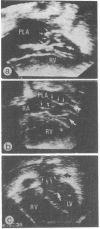
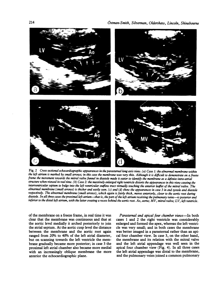
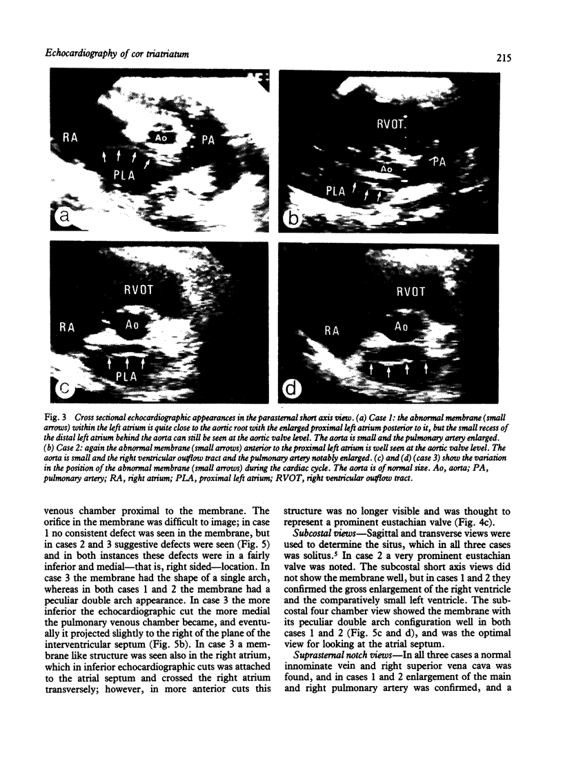
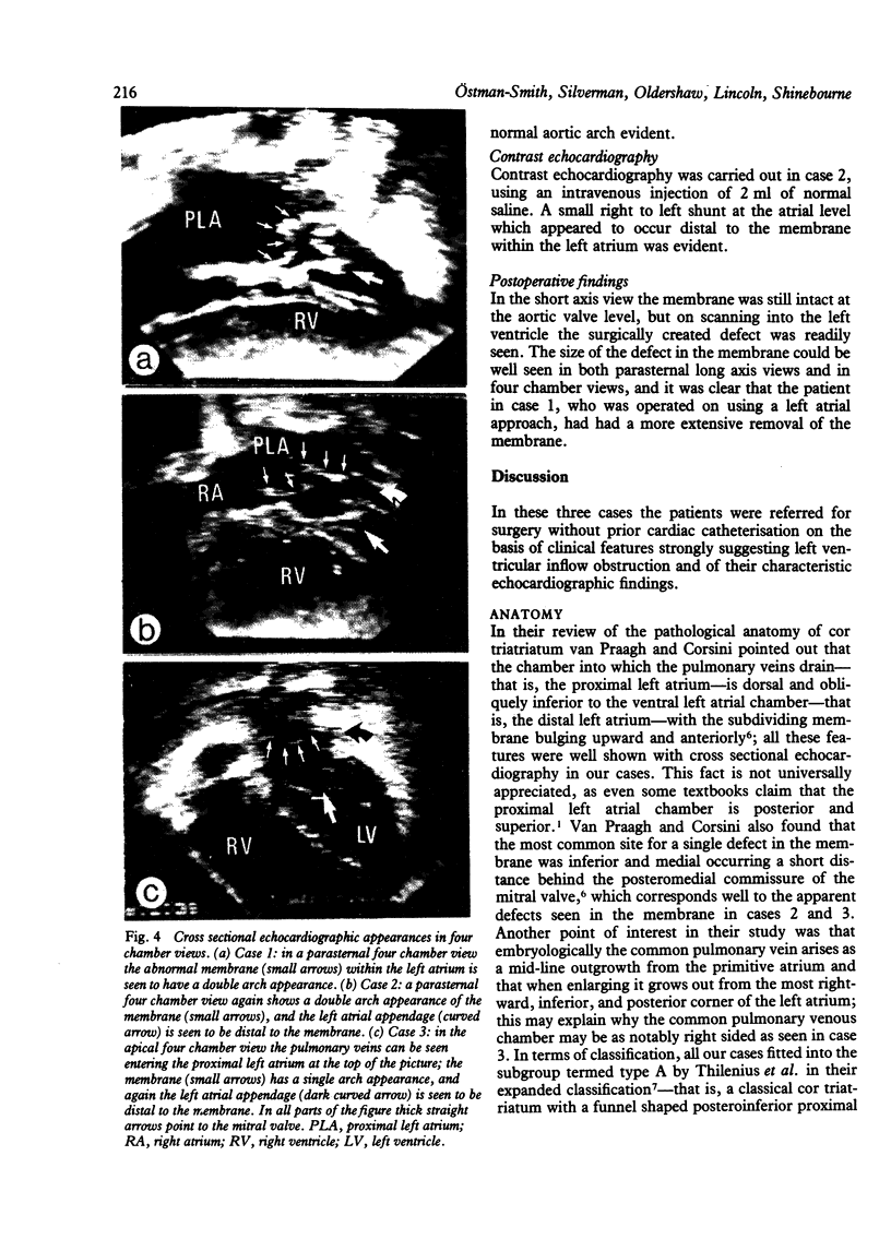
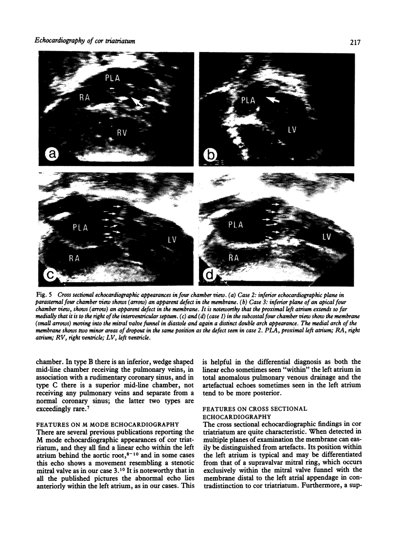
M mode and cross sectional echocardiography was carried out in three cases of cor triatriatum sinistrum (two infants and one adult). In two cases a peculiar double arch appearance, not previously reported, was found. All three cases were referred for surgery without cardiac catheterisation, and the diagnosis proved to be correct. The characteristic echocardiographic feature of cor triatriatum is an intra-atrial membrane detected in multiple planes of examination, curving anteroinferiorly and inserting some distance away from the mitral valve ring, proximal to the left atrial appendage. Superiorly the membrane runs parallel to, and a short distance behind, the aortic root creating a superior recess of the distal left atrial chamber. These features differentiate cor triatriatum from a supravalvar mitral ring. During diastole the membrane moves forward towards the mitral valve funnel. This, together with the arching appearance of the membrane on four chamber views and the more superior position of the membrane, makes it possible to distinguish cor triatriatum from total anomalous pulmonary venous drainage to the coronary sinus. From a review of past experience at the Brompton Hospital of the diagnostic accuracy of cardiac catheterisation in this condition, it is concluded that cross sectional echocardiography is superior to angiography as a technique for diagnosing cor triatriatum.
Full text
PDF
Images in this article
Selected References
These references are in PubMed. This may not be the complete list of references from this article.
- Bett J. H., Graff R. F., Stafford E. G., Dryburgh L. Echocardiographic diagnosis of cor triatriatum. Aust N Z J Med. 1978 Aug;8(4):428–430. doi: 10.1111/j.1445-5994.1978.tb04917.x. [DOI] [PubMed] [Google Scholar]
- Gibson D. G., Honey M., Lennox S. C. Cor triatriatum. Diagnosis by echocardiography. Br Heart J. 1974 Aug;36(8):835–838. doi: 10.1136/hrt.36.8.835. [DOI] [PMC free article] [PubMed] [Google Scholar]
- Huhta J. C., Smallhorn J. F., Macartney F. J. Two dimensional echocardiographic diagnosis of situs. Br Heart J. 1982 Aug;48(2):97–108. doi: 10.1136/hrt.48.2.97. [DOI] [PMC free article] [PubMed] [Google Scholar]
- Jacobstein M. D., Hirschfeld S. S. Concealed left atrial membrane: pitfalls in the diagnosis of cor triatriatum and supravalve mitral ring. Am J Cardiol. 1982 Mar;49(4):780–786. doi: 10.1016/0002-9149(82)91959-2. [DOI] [PubMed] [Google Scholar]
- Jones R. W., Baumer J. H., Joseph M. C., Shinebourne E. A. Arterial oxygen tension and response to oxygen breathing in differential diagnosis of congenital heart disease in infancy. Arch Dis Child. 1976 Sep;51(9):667–673. doi: 10.1136/adc.51.9.667. [DOI] [PMC free article] [PubMed] [Google Scholar]
- Lundström N. R. Ultrasoundcardiographic studies of the mitral valve region in young infants with mitral atresia, mitral stenosis, hypoplasia of the left ventricle, and cor triatriatum. Circulation. 1972 Feb;45(2):324–334. doi: 10.1161/01.cir.45.2.324. [DOI] [PubMed] [Google Scholar]
- Silverman N. H., Schiller N. B. Apex echocardiography. A two-dimensional technique for evaluating congenital heart disease. Circulation. 1978 Mar;57(3):503–511. doi: 10.1161/01.cir.57.3.503. [DOI] [PubMed] [Google Scholar]
- Snider A. R., Roge C. L., Schiller N. B., Silverman N. H. Congenital left ventricular inflow obstruction evaluated by two-dimensional echocardiography. Circulation. 1980 Apr;61(4):848–855. doi: 10.1161/01.cir.61.4.848. [DOI] [PubMed] [Google Scholar]
- Tajik A. J., Seward J. B., Hagler D. J., Mair D. D., Lie J. T. Two-dimensional real-time ultrasonic imaging of the heart and great vessels. Technique, image orientation, structure identification, and validation. Mayo Clin Proc. 1978 May;53(5):271–303. [PubMed] [Google Scholar]
- Thilenius O. G., Bharati S., Lev M. Subdivided left atrium: an expanded concept of cor triatriatum sinistrum. Am J Cardiol. 1976 Apr;37(5):743–752. doi: 10.1016/0002-9149(76)90369-6. [DOI] [PubMed] [Google Scholar]
- Thilenius O. G., Vitullo D., Bharati S., Luken J., Lamberti J. J., Tatooles C., Lev M., Carr I., Arcilla R. A. Endocardial cushion defect associated with cor triatriatum sinistrum or supravalve mitral ring. Am J Cardiol. 1979 Dec;44(7):1339–1343. doi: 10.1016/0002-9149(79)90450-8. [DOI] [PubMed] [Google Scholar]
- Van Praagh R., Corsini I. Cor triatriatum: pathologic anatomy and a consideration of morphogenesis based on 13 postmortem cases and a study of normal development of the pulmonary vein and atrial septum in 83 human embryos. Am Heart J. 1969 Sep;78(3):379–405. doi: 10.1016/0002-8703(69)90046-5. [DOI] [PubMed] [Google Scholar]


