Abstract
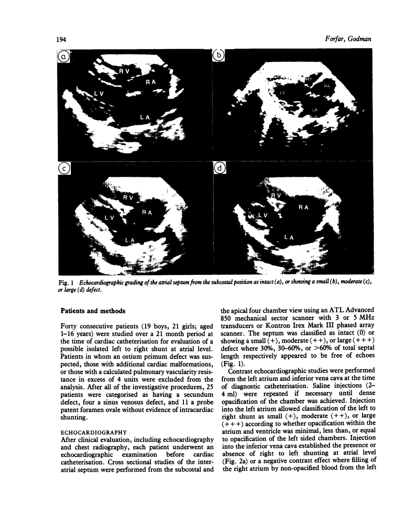
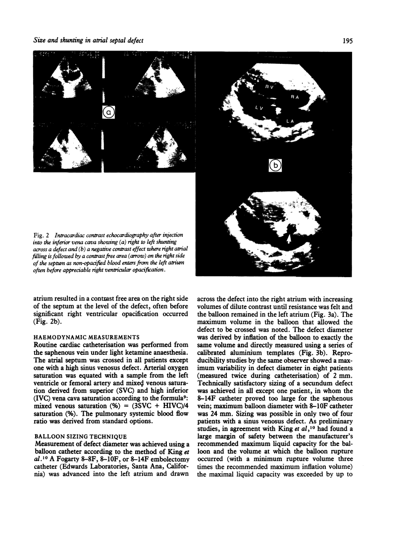
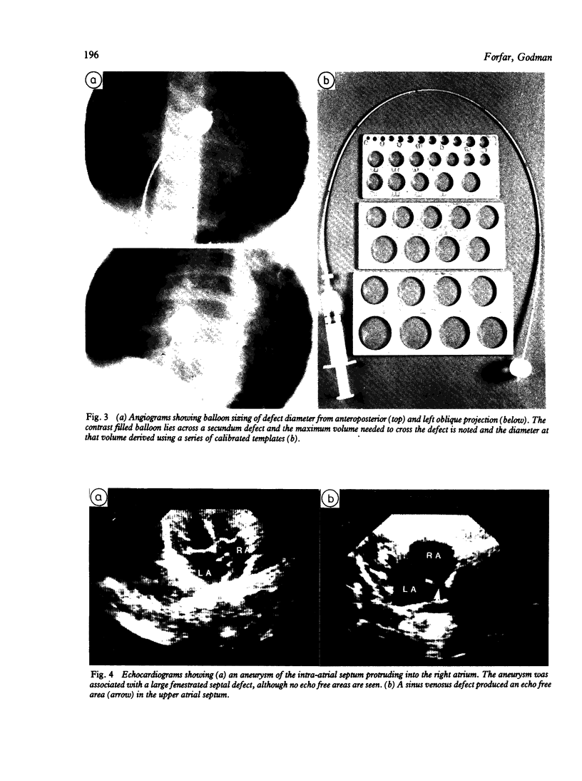
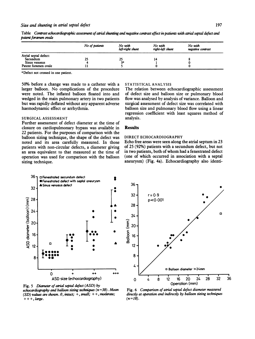
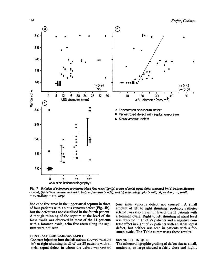
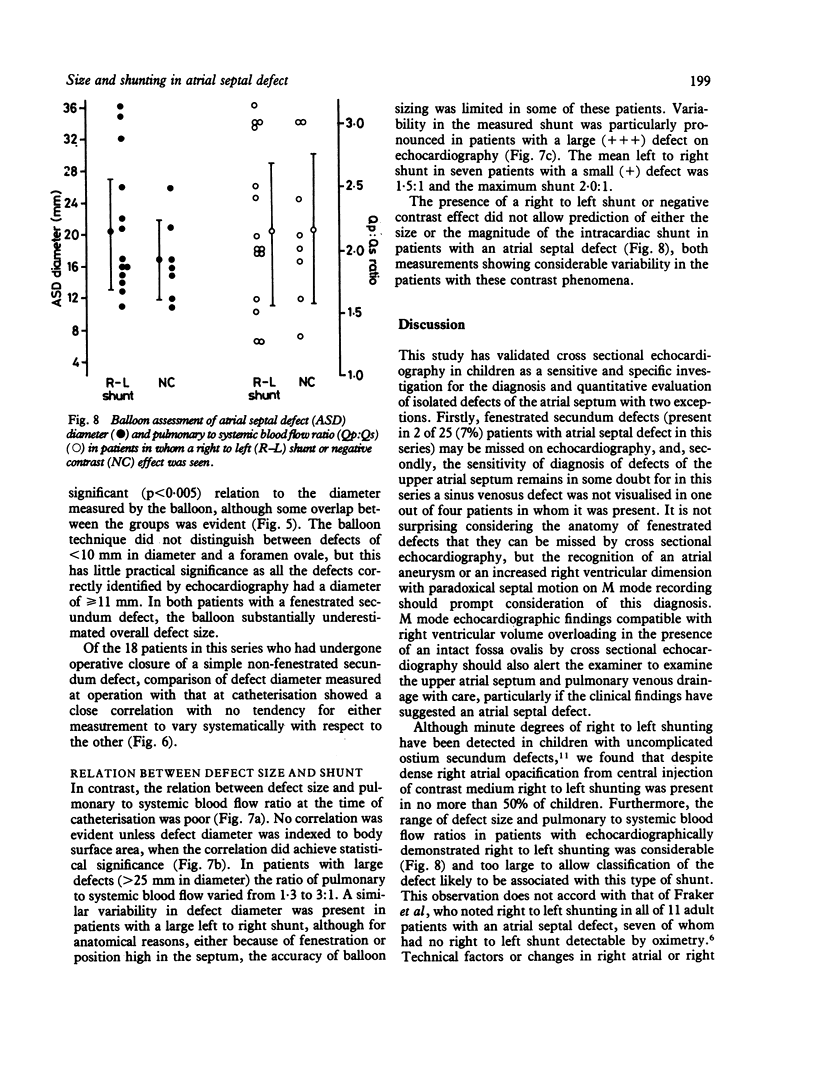
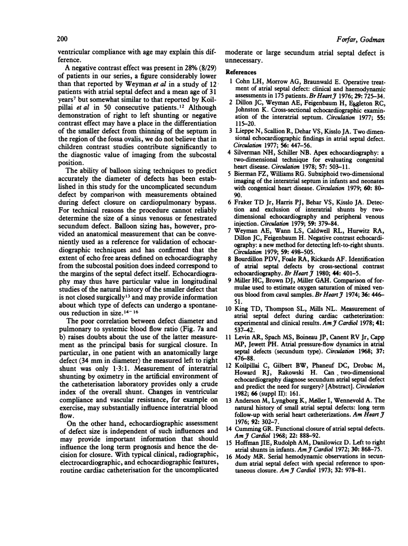
The results of cross sectional echocardiography, intracardiac contrast echocardiography, and balloon sizing techniques and conventional haemodynamic assessment were correlated in 40 consecutive patients evaluated for an isolated left to right shunt at atrial level. Echo free areas along the septum were identified in 23 of 25 patients with a secundum defect, but not in two with a fenestrated defect, and in the upper atrial septum in three of four patients with a sinus venosus defect. No false positive results occurred in 11 patients with a probe patent foramen ovale. Saline contrast injection into the left atrium showed significant left to right shunting in all patients with atrial septal defect; inferior vena caval injection produced right to left shunting in 15 of 29 patients and a negative contrast effect in eight of 29 patients with an atrial septal defect, although neither correlated quantitatively with defect diameter or magnitude of the left to right shunt. Echocardiographic assessment of defect size as small, moderate, or large showed a highly significant correlation with balloon measurement of defect diameter, although some overlap between the groups was evident. In contrast, the correlation between defect diameter and pulmonary to systemic blood flow ratio was poor, mainly because of highly variable shunting in patients with an anatomically large defect. Cross sectional echocardiography has high sensitivity and specificity in the diagnosis of the non-fenestrated atrial septal defect and provides quantitative information about defect diameter. Contrast studies do not add to the diagnostic value of imaging from the subcostal position. The poor correlation between defect size and the measured shunt suggests that the latter may not be the best criterion for surgical management and that size could be an important factor likely to influence both the long term prognosis and the decision for closure.
Full text
PDF
Images in this article
Selected References
These references are in PubMed. This may not be the complete list of references from this article.
- Andersen M., Moller I., Lyngborg K., Wennevold A. The natural history of small atrial septal defects; long-term follow-up with serial heart catheterizations. Am Heart J. 1976 Sep;92(3):302–307. doi: 10.1016/s0002-8703(76)80111-1. [DOI] [PubMed] [Google Scholar]
- Bierman F. Z., Williams R. G. Subxiphoid two-dimensional imaging of the interatrial septum in infants and neonates with congenital heart disease. Circulation. 1979 Jul;60(1):80–90. doi: 10.1161/01.cir.60.1.80. [DOI] [PubMed] [Google Scholar]
- Bourdillon P. D., Foale R. A., Rickards A. F. Identification of atrial septal defects by cross-sectional contrast echocardiography. Br Heart J. 1980 Oct;44(4):401–405. doi: 10.1136/hrt.44.4.401. [DOI] [PMC free article] [PubMed] [Google Scholar]
- Cohn L. H., Morrow A. G., Braunwald E. Operative treatment of atrial septal defect: clinical and haemodynamic assessments in 175 patients. Br Heart J. 1967 Sep;29(5):725–734. doi: 10.1136/hrt.29.5.725. [DOI] [PMC free article] [PubMed] [Google Scholar]
- Cumming G. R. Functional closure of atrial septal defects. Am J Cardiol. 1968 Dec;22(6):888–892. doi: 10.1016/0002-9149(68)90189-6. [DOI] [PubMed] [Google Scholar]
- Dillon J. C., Weyman A. E., Feigenbaum H., Eggleton R. C., Johnston K. Cross-sectional echocardiographic examination of the interatrial septum. Circulation. 1977 Jan;55(1):115–120. doi: 10.1161/01.cir.55.1.115. [DOI] [PubMed] [Google Scholar]
- Fraker T. D., Jr, Harris P. J., Behar V. S., Kisslo J. A. Detection and exclusion of interatrial shunts by two-dimensional echocardiography and peripheral venous injection. Circulation. 1979 Feb;59(2):379–384. doi: 10.1161/01.cir.59.2.379. [DOI] [PubMed] [Google Scholar]
- Hoffman J. I., Rudolph A. M., Danilowicz D. Left to right atrial shunts in infants. Am J Cardiol. 1972 Dec;30(8):868–875. doi: 10.1016/0002-9149(72)90012-4. [DOI] [PubMed] [Google Scholar]
- King T. D., Thompson S. L., Mills N. L. Measurement of atrial septal defect during cardiac catheterization. Experimental and clinical results. Am J Cardiol. 1978 Mar;41(3):537–542. doi: 10.1016/0002-9149(78)90012-7. [DOI] [PubMed] [Google Scholar]
- Lieppe W., Scallion R., Behar V. S., Kisslo J. A. Two-dimensional echocardiographic findings in atrial septal defect. Circulation. 1977 Sep;56(3):447–456. doi: 10.1161/01.cir.56.3.447. [DOI] [PubMed] [Google Scholar]
- Miller H. C., Brown D. J., Miller G. A. Comparison of formulae used to estimate oxygen saturation of mixed venous blood from caval samples. Br Heart J. 1974 May;36(5):446–451. doi: 10.1136/hrt.36.5.446. [DOI] [PMC free article] [PubMed] [Google Scholar]
- Mody M. R. Serial hemodynamic observations in secundum atrial septal defect with special reference to spontaneous closure. Am J Cardiol. 1973 Dec;32(7):978–981. doi: 10.1016/s0002-9149(73)80167-5. [DOI] [PubMed] [Google Scholar]
- Silverman N. H., Schiller N. B. Apex echocardiography. A two-dimensional technique for evaluating congenital heart disease. Circulation. 1978 Mar;57(3):503–511. doi: 10.1161/01.cir.57.3.503. [DOI] [PubMed] [Google Scholar]
- Weyman A. E., Wann L. S., Caldwell R. L., Hurwitz R. A., Dillon J. C., Feigenbaum H. Negative contrast echocardiography: a new method for detecting left-to-right shunts. Circulation. 1979 Mar;59(3):498–505. doi: 10.1161/01.cir.59.3.498. [DOI] [PubMed] [Google Scholar]


